Article

Morphological, Pathogenic and Molecular Characterization of Sclerotinia sclerotiorum, the Causal Agent of White Rot of Cabbage (Brassica oleracea var. capitata), in Serbia

by
Brankica Pešić
1,
Petar Mitrović
2,
Ana Marjanović Jeromela
2,
Federica Zanetti
3,
Milica Mihajlović
1,
Jovana Hrustić
1,
Mira Vojvodić
4,
Miljan Grkinić
4 and
Aleksandra Bulajić
4,*
1
Institute of Pesticides and Environmental Protection, Banatska 31b, 11080 Belgrade, Serbia
2
National Institute of Field and Vegetable Crops, Maksima Gorkog 30, 21000 Novi Sad, Serbia
3
Department of Agricultural and Food Sciences (DISTAL), Alma Mater Studiorum—University of Bologna, Piazza Goidanich 60, 47521 Cesena, Italy
4
Faculty of Agriculture, University of Belgrade, Nemanjina 6, 11080 Belgrade, Serbia
*
Author to whom correspondence should be addressed.
Plants 2025, 14(16), 2478; https://doi.org/10.3390/plants14162478
Submission received: 16 July 2025 / Revised: 4 August 2025 / Accepted: 8 August 2025 / Published: 10 August 2025
(This article belongs to the Section Plant Protection and Biotic Interactions)

Abstract

Sclerotinia sclerotiorum is a globally distributed necrotrophic pathogen with a broad host range, including many Brassicaceae crops. In 2021, white rot symptoms on cabbage were observed in 12 commercial fields in the northern part of Serbia. Twelve representative isolates of S. sclerotiorum, forming white colonies and black sclerotia, were selected for characterization and comparison with an isolate from sunflower, as the most important host plant in Serbia. The isolates showed significant variation in growth rate and sclerotia production, while ITS sequence analysis revealed the complete nucleotide identity and all isolates grouped within the major phylogenetic clade of S. sclerotiorum. Despite the low diversity of the global population of S. sclerotiorum, forty-four haplotypes were identified, with one predominant haplotype encompassing all Serbian isolates. When six Brassicaceae species (cabbage, cauliflower, broccoli, kale, mustard, and oilseed rape) and sunflower were inoculated, sunflower was found to be the most and cauliflower the least susceptible, while isolates from cabbage were generally more aggressive than those derived from sunflower. This work represents the first detailed characterization of S. sclerotiorum infecting cabbage in Serbia and highlights its genetic uniformity and differential pathogenic potential, which are critical factors for integrated disease management and crop rotation planning in Brassicaceae agroecosystems.

1. Introduction

Cruciferous plants (family Brassicaceae) are an important group of oilseeds and vegetable crops that make an important contribution to the economy in many countries around the world. They represent a large share of oil and vegetable production, as well as participating in a growing biofuel industry. Cabbage (Brassica oleracea var. capitata) is the most important cruciferous vegetable. Cabbage production varies worldwide, with China being the leading producer (34,986,293 tons), followed by India (9,825,000 tons), the Republic of Korea (2,428,893 tons) and Russia (2,298,209 tons). In Serbia, it is grown on 7111 ha (FAO, https://www.fao.org/faostat/en/#data/QCL, accessed on 24 May 2025), mainly in the central and western parts of Serbia (42%), followed by the southern and eastern regions (25%) [1]. Several diseases threaten cabbage production, especially diseases caused by fungi such as leaf spot, downy mildew, damping-off, Sclerotinia rot/white rot, yellow rot or Fusarium wilt, blackleg, wire stem and clubroot. Two related Sclerotinia species, Sclerotinia sclerotiorum [2] and S. minor [3], have been described as causal agents of Sclerotinia rot in cabbage and other Brassicaceae plants. Depending on weather conditions, the yield losses caused by both species vary between 5 and 100% in many crops [4]. The average annual incidence of Sclerotina rot in oilseed rape in China is about 10–20% and can be as high as 80% in severe outbreaks [5]. Significant losses due to S. sclerotiorum in oilseed rape (Brassica napus) and mustard (Brassica juncea) have been recorded in Australia, North America, China and Europe [6]. Sclerotinia species attack all growth stages of the crop and cause commercial yield losses in the millions under field, storage and marketing conditions. The reported rot incidence caused by S. sclerotiorum in cabbage varies between 1 and 30% [7,8,9,10,11].
S. sclerotiorum is one of the most devastating soilborne pathogens affecting many agricultural crops [12,13] and is distributed in more than 92 countries all over the world (CABI distribution map, UK, https://plantwiseplusknowledgebank.org, accessed on 29 May 2025). The pathogen has an extraordinarily broad host range within more than 98 families and is associated with 2048 host species and varieties (USDA database, https://biocollections.ars.usda.gov, accessed on 29 May 2025). Most of the hosts are dicotyledonous herbaceous species, but some also belong to monocotyledonous plants. In Serbia, S. sclerotiorum is still rarely studied. Significant losses have been recorded in sunflower production [14], and the occurrence in fields with green beans (Phaseolus vulgaris) [15], faba beans (Vicia faba) [16] and cabbage [17] has been observed. However, in these studies, the pathogen was not isolated and subsequently characterized, so data on pathogenic, morphological and genetic characteristics of isolates from Serbia are missing. Despite the fact that S. sclerotiorum has been described in cabbage and other Brassicaceae vegetables worldwide [2,3,18], its distribution and importance in cabbage production in Serbia had not been investigated prior to this study.
In 2021, typical Sclerotinia rot-like symptoms were observed on cabbage plants in several commercial fields in the northern part of Serbia, including spreading brown lesions and intense rotting of cabbage heads, often accompanied by abundant snow-white mycelium and black sclerotia (Mitrović, personal communication). The aim of the present work was to (a) identify and characterize the causal agent of the observed symptoms on cabbage based on pathogenic and morphological features; (b) determine phylogenetic relationships among isolates; (c) investigate intraspecific diversity by comparing isolates from cabbage and sunflower; (d) determine the population diversity of S. sclerotiorum worldwide and in Serbia based on haplotype analyses; (e) test the aggressiveness of derived isolates against cabbage and five other Brassicaceae species and compare them with sunflower as the main Sclerotinia host plant in Serbia; and (f) determine the relative differences in susceptibility of the main Brassicaceae crops.

2. Results

2.1. Disease Symptoms and Morphological Characterization

Typical symptoms of the Sclerotinia rot outbreak were observed in 12 production fields at two locations, Begeč and Futog, mostly distributed in patches. The first symptoms of the disease were water-soaked, spreading areas on the lower leaves, which became necrotic and were covered with a fluffy white mycelium (Figure 1A,B). In the late stages of disease progression, black sclerotia 2 to 7 mm in size were detected, either embedded in the white mycelium (Figure 1D) or located in the decaying outer leaves of the cabbage heads (Figure 1C). As the infection progressed, head rot and complete collapse of the plants were observed. The incidence of the disease varied slightly among fields. The highest incidence was observed in the field from which isolate M originated and was estimated at 10%, followed by the field from which isolate M3 originated (3%). In the remaining fields, the disease incidence ranged from 1 to 2%.
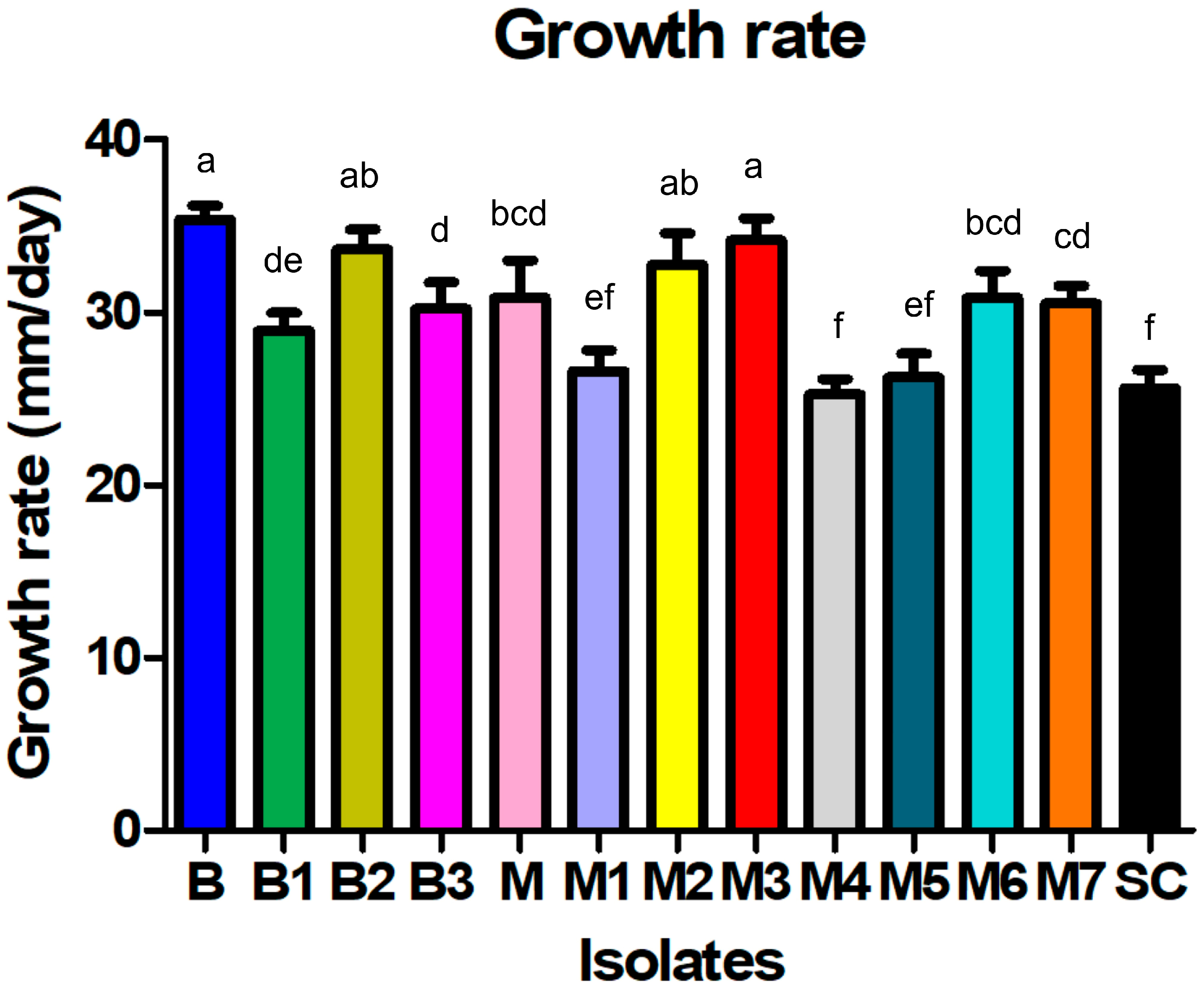
From the collected symptomatic cabbage samples, 12 Sclerotinia-like isolates were obtained, 4 isolates from Begeč (B, B1, B2, and B3) and 8 isolates from Futog (M, M1, M2, M3, M4, M5, M6, and M7), with 1 representative per cabbage production field, all of which showed a similar colony appearance, typical for Sclerotinia spp. Initially, the isolates formed white, fast-growing colonies with uniform margins consisting of sparse, homogeneous mycelium adhering to the medium (Figure 1E,H). After filling the plates, the isolates developed sparse to very dense, floccose, wooly–floccose or wooly aerial mycelium containing tufts and strands, usually arranged in a circular pattern (Figure 1I–L). The growth rate of the isolates varied between 25.3 mm/day (isolate M4) and 35.4 mm/day (isolate B), with an average of 30.1 mm/day. The growth rate of isolate SC from sunflower was 25.6 mm/day. The differences among the isolates in terms of average growth rate were statistically significant (F = 31.1; p < 0.0001) (Figure 2). On the surface of the four-day old colony, all cabbage-derived isolates formed white sclerotial initials that developed to grayish-black, mostly solitary sclerotia after an incubation period of 6–9 days (Figure 1F,D). The majority of isolates from cabbage formed sclerotia after 8 days (Figure 1G). In the culture of the sunflower-derived SC isolate, sclerotia were visible much later, 13 days after inoculation (Table 1). All sclerotia were round or irregularly shaped, had a textured surface and were easily detached from the mycelium. Based on the arrangement of sclerotia in the cultures, the isolates formed two distinct groups—group I, which formed a ring of individual sclerotia (5 isolates) mostly located at the edge of the colony, and group II, which formed two rings of sclerotia (edge and middle ring) (8 isolates). The isolate from the sunflower belonged to group II. The number of sclerotia formed differed significantly among the isolates from cabbage (p < 0.01). After 30 days of incubation, the average number of sclerotia varied between 9.7 in isolate M2 and 33.7 sclerotia/plate in isolate M7. During the period studied, isolate SC from sunflower formed a significantly lower number of sclerotia (2.7 sclerotia/plate) (Table 1).

2.2. Pathogenicity

The first symptoms of necrosis and decay were visible on the inoculated cabbage and sunflower plants 3 days post inoculation, and none of the plants inoculated with sterile agar plugs developed symptoms. All isolates were successfully recovered from infected tissue and thus fulfilled Koch’s postulates.

2.3. Sequence Analysis and Phylogeny

All 13 Serbian S. sclerotiorum isolates (12 from cabbage and 1 from sunflower) had an identical ITS nucleotide sequence (identity of 100% and 0 bp differences). BLAST (BLAST+ 2.17.0) analyses showed that they had 100% nucleotide identity with over 50 sequences of S. sclerotiorum in the GenBank database, and the most similar was isolate S. sclerotiorum from South Korea from Chinese chives (KJ614564) [19]. Phylogenetic analyses of the ITS sequences of 13 Serbian isolates, together with the previously listed sequences of S. sclerotiorum, S. trifloriorum and S. minor, resulted in a stable phylogenetic three with clear separation of all three Sclerotinia spp. (Figure 3). All 13 isolates from Serbia clustered in a well-supported branch comprising all S. sclerotiorum isolates with low diversity and were clearly separated from S. minor or S. trifoliorum, confirming the conventional identification.
Therefore, based on the morphology and growth characteristics of the recovered isolates, reproduced symptoms on inoculated cabbage and sunflower plants, sequence and phylogenetic analysis, the causal agent of the investigated cabbage disease was identified as S. sclerotiorum.

2.4. Haplotype Structure and Genetic Diversity of Sclerotinia sclerotiorum Sequences

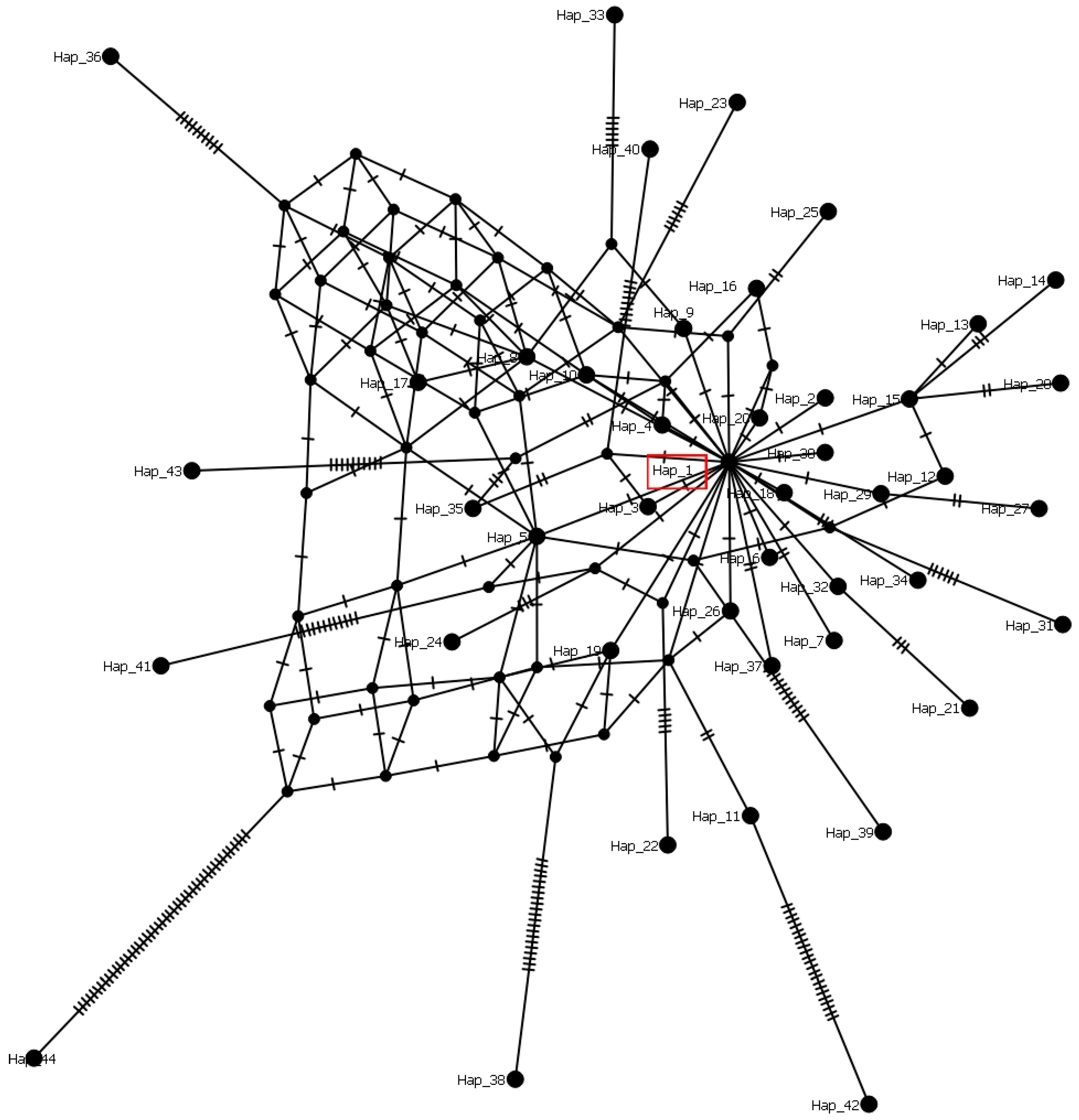
The dataset of 1052 S. sclerotiorum sequences, including Serbian ones, was grouped into 44 haplogroups and characterized by 94 variable positions. The haplotype diversity of 0.102, as well as the nucleotide diversity of 0.00520, indicates a low level of genetic variation among the analyzed sequences (0 to 40 nt difference). The most numerous and dominant haplotype (Hap1) contained 997 sequences from different hosts and countries. The remaining 43 haplogroups (Hap2–44) comprised 55 sequences. The Serbian population of 13 S. sclerotiorum isolates was grouped into Hap1, with 0 nt difference between them. The haplotype network reconstructed using the median joining network algorithm implied star-shaped genealogical relationships with Hap1 in the central position (Figure 4) and numerous single-step mutations leading to peripheral haplotypes in the world population of S. sclerotiorum. The network also exhibited some genetic diversity, as evidenced by multiple mutational steps between specific haplotypes.

2.5. Aggressiveness Towards Different Plant Species

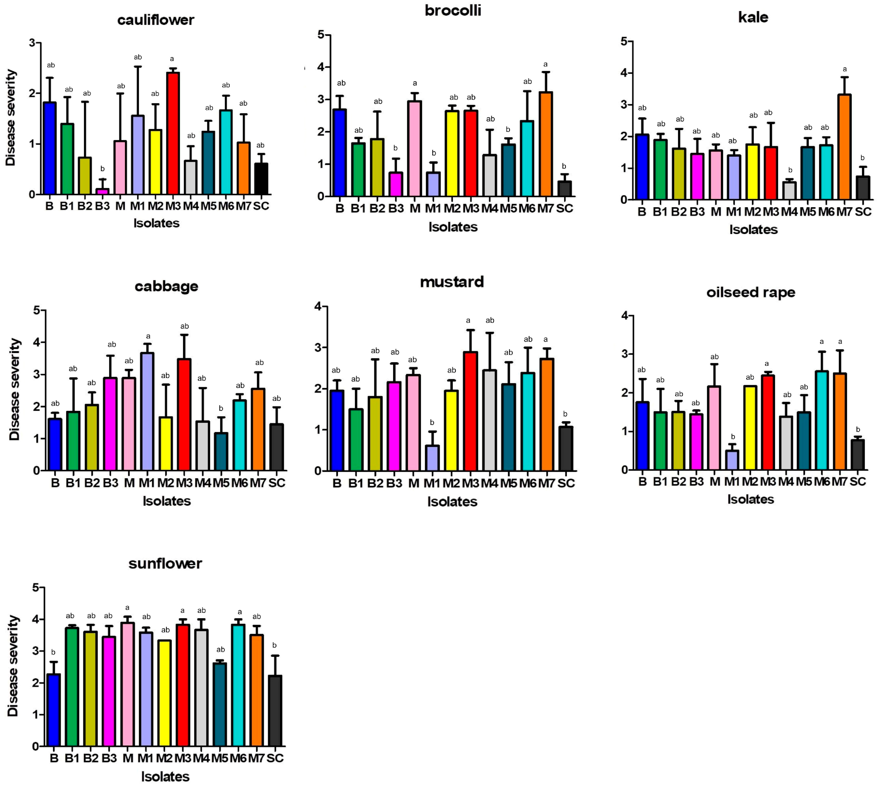
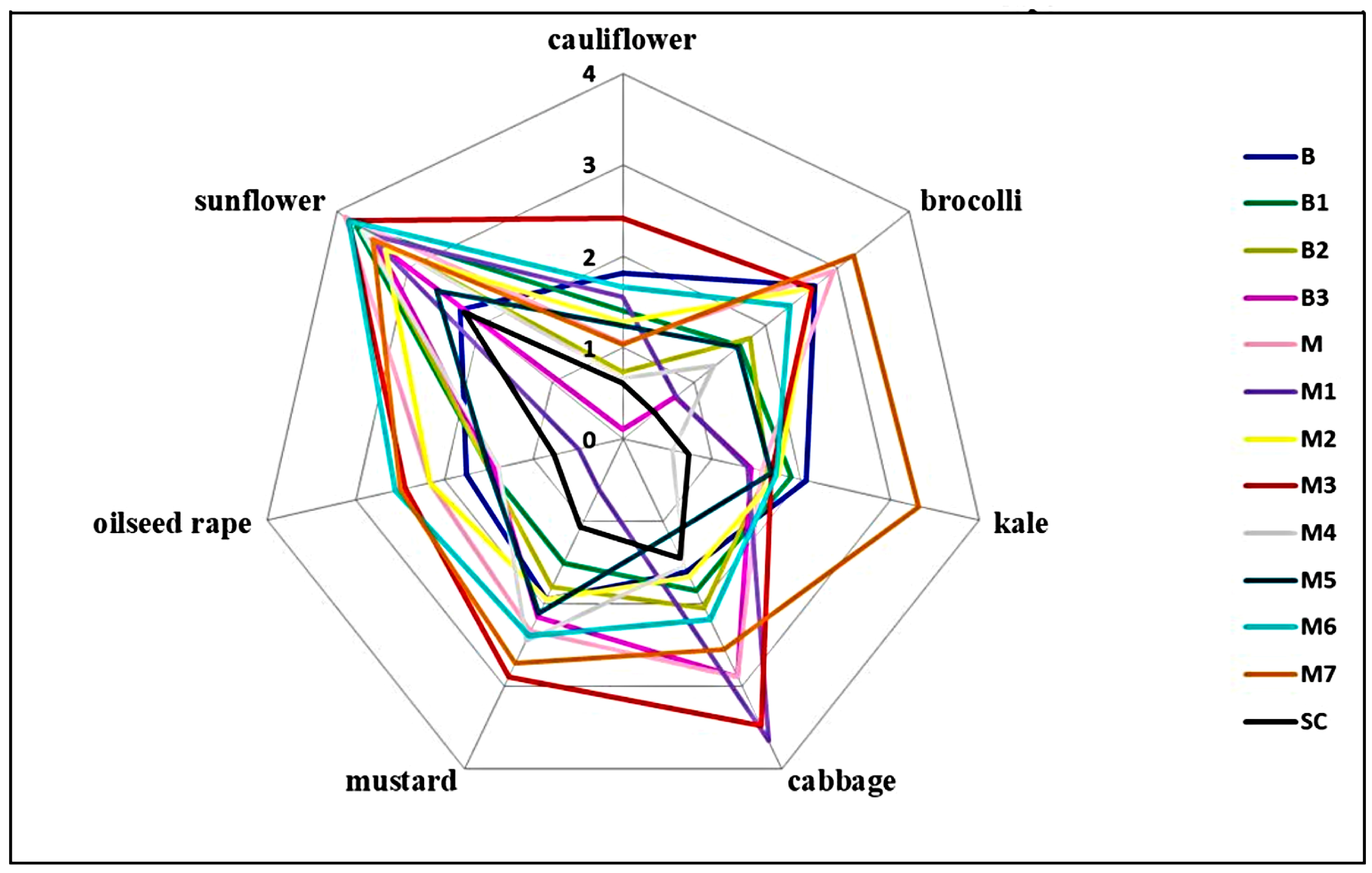
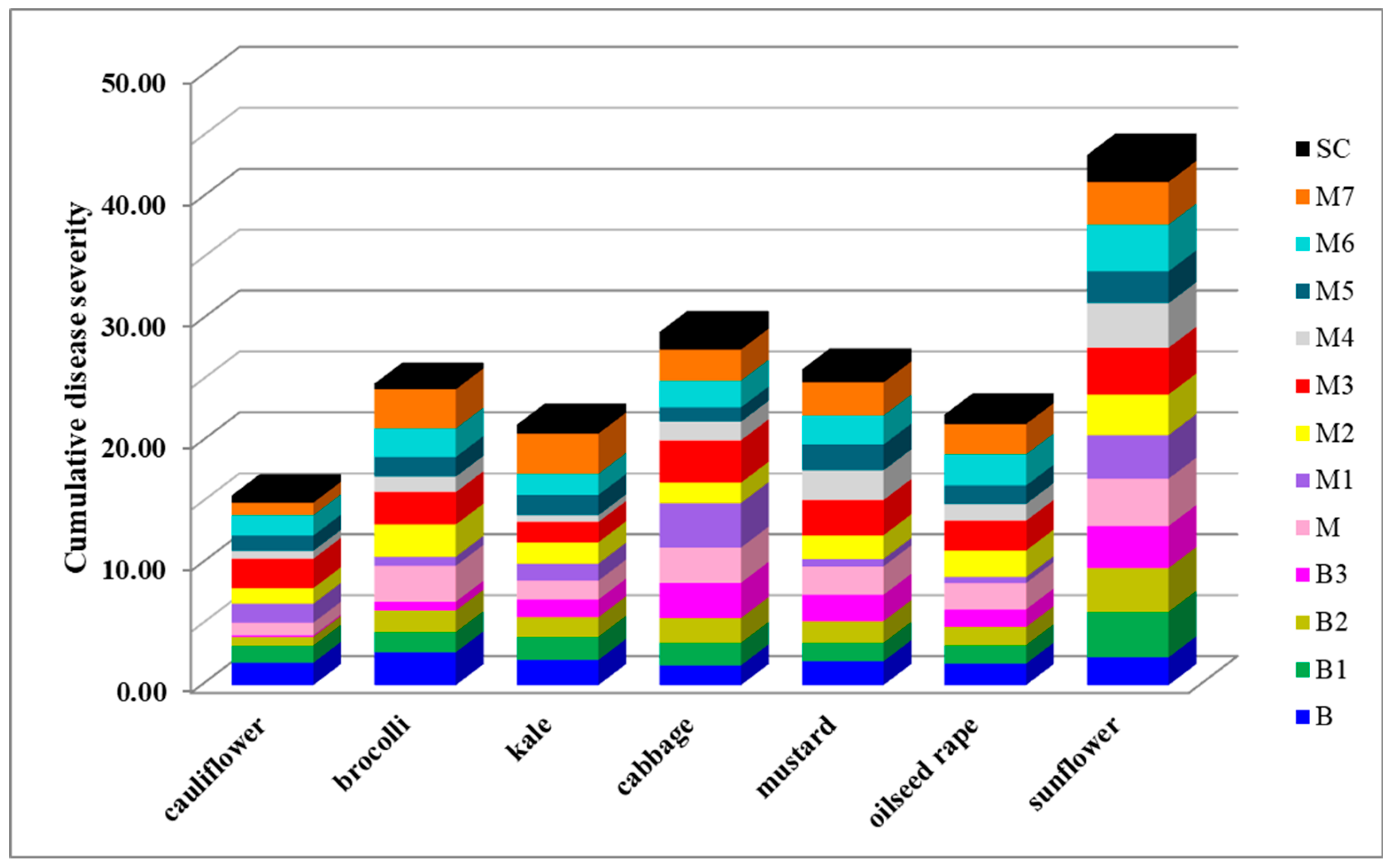
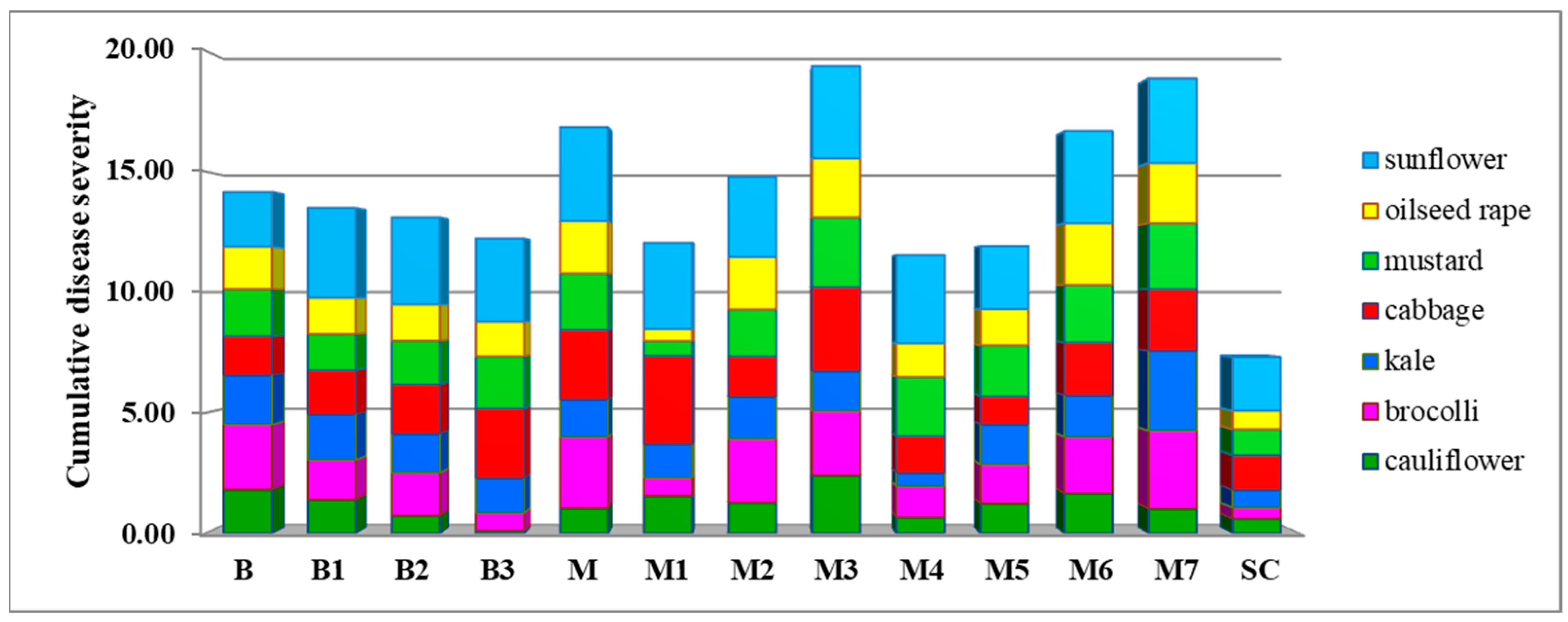
After a three-day incubation period, all inoculated plants of seven tested plant species showed typical symptoms of Sclerotinia rot (Figure 1M,N), while none of the control plants inoculated with sterile agar plugs developed symptoms. Artificial inoculation revealed significant differences between the S. sclerotiorum isolates in terms of their aggressiveness towards a particular plant species, as well as differences in the aggressiveness of a particular isolate towards different plant species (p < 0.05) (Figure 1O, Figure 5, Figure 6, Figure 7 and Figure 8). On average, isolate M3 was the most aggressive for all hosts tested, causing a cumulative disease severity index value of 19.4 (Figure 8). It was the most aggressive to sunflower and least aggressive to kale. The least aggressive isolate overall was isolate SC from sunflower, with a cumulative disease severity index value of 7.3 (individual values ranged from 0.4 to 2.2, depending on the plant species), followed by isolate M4, which was the least aggressive isolate from cabbage (cumulative disease severity index value 11.5, individual values 0.6 to 3.7). A susceptibility profile of the hosts to all isolates tested showed that sunflower was the most susceptible species (disease severity index values 2.2 to 3.9, cumulative 43.5), followed by cabbage (disease severity index between 1.4 and 3.6, cumulative 29.0), while cauliflower had the lowest susceptibility, with disease severity index median values between 0.1 and 2.4 (Figure 5, Figure 6 and Figure 7), depending on the isolate. The cumulative disease severity index for cauliflower was 15.6, confirming its low susceptibility to the isolates tested (Figure 7).

3. Discussion

As far as the authors know, this is the first study reporting a comprehensive characterization of S. sclerotiorum as the causal agent of cabbage rot in Serbia. S. sclerotiorum is a globally recognized causal agent of cabbage white rot, but in Serbia the symptoms were detected for the first time in 1997, published in 2016 [17], and no further research has been conducted to investigate its population. The occurrence of S. sclerotiorum on other host plants in Serbia, especially in the province of Vojvodina, has been documented previously [14]. In this study, we characterized a population of S. sclerotiorum responsible for substantial yield losses in cabbage and evaluated the relative susceptibility of six other crucifers to contribute to the development of a knowledge-based crop rotation strategy with crucifers. Our study is the first comprehensive characterization of the S. sclerotiorum population affecting cabbage in Serbia that offers valuable guidance for cabbage growers, particularly in selecting appropriate Brassicaceae crops for crop rotation.
Following an outbreak of cabbage white rot at 12 locations, the pathogen was identified as S. sclerotiorum using morphological and molecular methods. All isolates formed white colonies accompanied by large black sclerotia typical of S. sclerotiorum, as previously described [20]. The isolates in this study showed a substantial diversity in the appearance of the aerial mycelium, which ranged from sparse to very dense and from wooly to woolly–floccose or floccose in terms of its pattern on the colony surface. The morphological diversity of S. sclerotiorum isolates has been recorded in different crops [21]. Although the variability of colony color, ranging from white to grayish white, has been noted in many studies [21,22,23,24,25,26], our isolates shared the predominant appearance of S. sclerotiorum and formed exclusively white colonies [20,27,28,29,30]. The growth rate of the isolates examined was also within the range of previously reported values [22,23,26,31], although statistically significant differences were observed among the isolates.
All isolates from our study originating from cabbage formed roundish or irregular sclerotia after an incubation period of 6 to 9 days, similar to the isolates originating from B. juncea in India [32]. The previously reported time required for the formation of sclerotia in isolates from soybean ranged from 10.7 to 18 days [25], and 4 to 7 days in isolates from beans [29]. In the present study, the isolate from sunflower formed sclerotia much later (after 13 days). The number of sclerotia formed by isolate SC was also much lower (2.7 sclerotia/plate) compared to isolates from cabbage, which formed 9.7–33.7 sclerotia/plate. No clear correlation was found between the number of sclerotia and the time required for their formation.
The phylogenetic studies based on the ITS sequence allowed for the reliable identification of all isolates from Serbia, which clustered in a well-defined and supported branch that included all S. sclerotiorum isolates and was clearly separated from S. minor or S. trifoliorum. Our data are consistent with the previously published results [33]. A similar study based on phylogenetic analyses of ITS sequences of S. sclerotiorum reported the genetic diversity of 65 S. sclerotiorum isolates derived from B. juncea in India and described the presence of 11 evolutionary lineages [32]. We compared the Serbian isolates and the isolates from that study, and all isolates shared 100% nucleotide similarity (13 Serbian and 64 Indian), with only one isolate (Acc. No. MF408249) showing a 3 nt difference. Thus, we were not able to confirm the presence of diversity and correlate it with the different aggressiveness of Serbian or Indian isolates derived from cabbage or Indian mustard as two Brassicaceae crops. In our study, the susceptibility of mustard as a host plant was slightly lower than that of cabbage, which is the second most susceptible host plant. The Serbian isolates showed differences in aggressiveness, similar to the population of mustard-originating S. sclerotiorum from India [32], but a correlation with molecular diversity could not be established.
The molecular ITS marker is known as a useful barcoding region for the genus identification of fungi, as well as for the species delimitation of several robust genera such as Sclerotinia [33,34] and Monilina [35,36]. Our analyses of the haplotype structure and diversity of S. sclerotiorum revealed considerable genetic uniformity in the population of all sequences available to date. The analyzed set consists of 44 haplotypes, one of which is a major haplotype that accounts for almost 95% of all analyzed sequences and is distributed throughout the world. The central position of the major haplotype in a star-shaped genetic structure indicates its possible role as an ancestor. On the other hand, several haplotypes (e.g., Hap14 and Hap38) were found to be more distantly connected, suggesting possible isolated evolutionary lineages. After analyzing a smaller set of IGS sequences of S. sclerotiorum, a similarly low level of genetic structuring in the population was established [37]. These findings support the hypothesis that S. sclerotiorum isolates represent a genetically uniform population in different regions, with limited intra-population variability. Further studies, using diverse and additional molecular markers, are essential to clarify the extent and biological relevance of this genetic homogeneity.
To assess the potential impact of cross hosts within the crop rotation and the associated agronomic risks, we investigated the variability in aggressiveness of isolates against six Brassicaceae hosts and sunflower, an economically important crop in Serbia and a major host of S. sclerotiorum. To ensure methodological consistency and comparability, lesion length was selected as the primary indicator of susceptibility to S. sclerotiorum, a parameter well-established in the literature, especially in controlled inoculation experiments [5,6,14,21,22], and applicable across plant species with distinct anatomical and morphological features. Artificial inoculation revealed significant differences in the aggressiveness of S. sclerotiorum isolates. All isolates elicited symptoms similar to those observed in the field and described by many authors. In general, they were much more aggressive towards sunflower than all the other crucifers species tested. Among the latter, cauliflower was the least susceptible and cabbage the most susceptible species. A few studies have been published describing a great difference in the susceptibility of different crops to Sclerotinia species, from a study detecting no significant differences between cruciferous crops [38] to study showing some differences between Cucurbitaceae vegetables crops [39]. Based on differences in aggressiveness towards oilseed rape, S. sclerotiorum isolates were divided into three groups: highly aggressive, aggressive and slightly aggressive [40]. A similar categorization was reported by some other studies [21,25,32,41]. In the present study, all isolates were highly aggressive towards sunflower, while the statistically significant differences in aggressiveness towards Brassica species were biologically rather moderate. When analyzing the diversity of S. sclerotiorum isolates derived from B. juncea in India, no correlation between genetic variability and aggressiveness or geographical distribution was observed [32]. In the present study, the aggressiveness of each S. sclerotiorum isolate was largely determined by the specific Brassica host plant. Although isolate M3 was not the most aggressive for all plant species tested, it exhibited the highest cumulative aggressiveness across all plant species, warranting further investigation of the factors involved in S. sclerotiorum pathogenicity. Conversely, the isolate from sunflower showed significantly lower aggressiveness towards all tested host plants compared to the isolates from cabbage. As only one isolate from sunflower was included in this study, further studies with more isolates from sunflower are required to draw a comprehensive conclusion. Nevertheless, our results indicate a remarkable aggressiveness of isolates from cabbage towards sunflower, highlighting potential problems in crop rotation strategies. Pronounced phenotypic variability in S. sclerotiorum isolates, particularly regarding aggressiveness and host specificity, suggests that molecular markers alone may not fully capture the pathogen’s adaptive potential. To unravel the mechanisms underlying phenotypic variability, future research should focus on multi-locus genotyping or whole-genome sequencing, coupled with long-term field surveillance, to detect the emergence of highly aggressive or host-adapted strains. In Serbia, crop rotation is strictly adopted and in all the production fields considered in the present study, cabbage was grown either for the first time or as part of a 3–5 year rotation with soybean, potato and wheat, suggesting possible problems with other included crops. The combined airborne and soilborne nature of S. sclerotiorum and its global economic importance in many crops require detailed characterization of the pathogen in order to develop effective control measures. To the best of our knowledge, this study provided the first detailed description of the morphological, pathogenic and phylogenetic characteristics of S. sclerotiorum isolates from cruciferous plants in Serbia. Variations were observed in the growth rate, relative density and appearance of areal mycelium, as well as in aggressiveness. On average, cauliflower and kale were the least and cabbage the most susceptible Brassica species to the isolates tested. Compared to sunflower, all Brassica species were significantly less susceptible. Although the results of this study provide important insights into the aggressiveness of S. sclerotiorum isolates from cabbage in Serbia, these tests were carried out under controlled conditions which do not fully reflect the complex interactions between the pathogen, host plant, and environmental factors that influence disease development in agroecosystems. Nevertheless, this study provides a valuable foundation for understanding the presence and biological behavior of S. sclerotiorum in cabbage production, as well as guidance for future research aimed at improving sustainable disease management strategies.
The present study clearly revealed that even genetically homogeneous populations of S. sclerotiorum can display considerable phenotypic plasticity, likely driven by environmental conditions and host-specific selective pressures. The elucidation of pathogenic variability within this clonal population highlights the need for incorporating both molecular and phenotypic characterization in the development of integrated disease management strategies in Brassicaceae cropping systems.

4. Materials and Methods

4.1. Sampling and Pathogen Isolation

In September 2021, cabbage plants with head necrosis were observed in 8 fields near the locality of Futog (seven fields with cv. Futoški and one with cv. Bravo F1 hybrid) and 4 fields near Begeč (two fields with cv. Bravo F1 hybrid, one with cv. Bucharest and one with cv. Futoški), all in the province of Vojvodina, Serbia. The observed disease outbreak did not appear to be directly influenced by the weather conditions, since they were typical for that region and the time of year. At each location, disease incidence was estimated by walking the field in a zigzag course and randomly assessing 100 plants in three replicates. A total of 10 symptomatic samples were collected from each location and transported to the laboratory for isolation and identification. For each sample, small fragments of tissue were taken from the border between necrotic and healthy cabbage tissue, surface-sterilized with 2% sodium hypochlorite for 1 min, rinsed with sterile distilled H2O, plated on potato dextrose agar (PDA; 200 g potato, 20 g dextrose, 17 g agar, and 1 L distilled H2O) [42], and incubated at 24 °C for 5 days. Uniform Sclerotinia-like colonies grew from diseased cabbage tissue, and representative isolates from each field were purified by transferring the hyphal tips to PDA and selected for morphological identification and characterization. Isolates were stored on sealed PDA slants at 4 °C in the fungal collection of the Department of Phytopathology, Faculty of Agriculture, University of Belgrade. In addition, a reference isolate SC from sunflower from Rimski Šančevi, Province of Vojvodina, Serbia, was included in the study (fungal collection of the Institute of Vegetable and Field Crops, National Institute of the Republic of Serbia).

4.2. Morphological Identification

Morphological identification of 12 selected Sclerotinia-like isolates from cabbage and a reference isolate from sunflower was based on the colony appearance assessed 15 days after inoculation on PDA at 24 °C in darkness. The growth rate was determined by measuring two perpendicular colony diameters in five replicates per isolate and calculating an average value for each isolate. The presence of sclerotia and the time of first formation were assessed in the first week after subculturing, the distribution of sclerotia was evaluated after 15 days, and the number of sclerotia per colony and the size of sclerotia were determined 30 days after subculturing [43]. Three replicates were used for each isolate and the entire experiment was performed twice.

4.3. Amplification and Sequencing of Isolates’ DNA

Total genomic DNA was extracted from 100 mg dry mycelium of 7-day-old cultures of Sclerotinia isolates grown on potato dextrose broth (PDB; 200 g potato, 20 g dextrose and 1 L distilled H2O) using the DNeasy Plant Mini Kit (Qiagen, Hilden, Germany), according to the manufacturer’s instructions. PCR amplification of ITS (ITS1, 5.8S rDNA and ITS2) with the primers ITS1f/ITS4 [44,45] was performed in a total reaction volume of 25 μL, consisting of 12.5 μL 2 X PCR Master mix (Fermentas, Lithuania), 6.5 μL RNase-free water, 2.5 μL forward and reverse primers (working solution with a final concentration of 100 pmol/μL, Metabion International, Gräfelfing, Germany) and 1 μL template DNA. The amplification conditions were as follows: initial denaturation at 95 °C for 3 min, followed by 35 cycles of denaturation at 95 °C for 30 s, annealing at 52 °C for 1 min, elongation at 72 °C for 1 min and final elongation for 10 min at 72 °C. The amplicons obtained were stained with ethidium bromide, analyzed by 1% agarose gel electrophoresis and visualized using a UV transilluminator. The PCR products were sequenced directly in both directions using an automatic sequencer (Automatic Sequencer Macrogen Inc., Amsterdam, The Netherlands) using the same primers as for amplification. The consensus sequences were calculated with ClustalW 2.1 [46], integrated into the software MEGA 7.0 [47] and deposited in GenBank (http://www.ncbi.nlm.nih.gov, accessed on 7 August 2025). All generated sequences were compared with each other by calculating nucleotide (nt) similarities and with previously deposited isolates of Sclerotinia spp. in GenBank using the BLAST similarity search tool.

4.4. Phylogenetic Analyses

Newly generated Serbian ITS sequences of Sclerotinia isolates from cabbage were analyzed with 24 previously listed type species of the genus Sclerotinia [33,48,49] and an outgroup taxa Trichoderma lixii [50]. Of the 25 sequences retrieved in GenBank, 15 were S. sclerotiorum isolated from different host plants and geographical distributions. The analyses included five representative sequences of S. trifloriorum and four of S. minor (Table 2). A phylogenetic tree was constructed using the Maximum Likelihood Method implemented in MEGA 7.0 software [47]. The Gamma-distributed Kimura’s two-parameter model [51] obtained using the model test implemented in MEGA 7.0 was used as the best fitting model for nucleotide substitution, and all sites with gaps were omitted. The reliability of the obtained trees was evaluated with 1000 bootstrap replicates.

4.5. Haplotype Analysis of Sclerotinia sclerotiourum Sequences

All 1100 available sequences of the ITS region of S. sclerotiorum (accessed 25 May 2025) were retrieved from the NCBI database, and the sequences shorter than 400 nt or containing degenerative positions were manually eliminated, resulting in a final dataset of 1052 sequences, including 13 Serbian sequences from this study, all from different host plants and different geographic regions worldwide. Genetic diversity was analyzed using DnaSP version 6.0 [52], which provided the number of haplotypes (h), haplotype diversity (Hd), number of variable sites (S) and nucleotide diversity (π) for the ITS region. Further analyses for haplotype composition and frequency were performed using PopART 1.7 software [53]. Nucleotide identities between sequences were calculated with MEGA 7.0 software [47], which allowed a comparison of sequence similarity and divergence. A haplotype network of all selected sequences and the Serbian cabbage and sunflower isolates was generated using the median joining network algorithm implemented in PopART [54].

4.6. Pathogenicity and Aggressiveness Testing

To prepare the inoculum for pathogenicity and aggressiveness studies, all studied isolates were subcultured on PDA and incubated for 4 days at 24 °C in the dark. Mycelial plugs with a diameter of 10 mm, cut 10 cm from the edge of the colony, served as the inoculum source in both experiments.
Pathogenicity was tested under laboratory conditions through the inoculation of cabbage and sunflower plants at the growth stage of 4 true leaves. The mycelial plugs were placed on wounded stem tissue, covered with a moist cotton swab and sealed with aluminum foil. For each isolate, 5 plants were inoculated. The control plants were inoculated using the same method, but with sterile pieces of PDA. The inoculated plants were incubated in a greenhouse with a photoperiod of 12 h (light and dark) and a temperature of 24 °C.
To determine the aggressiveness of 13 S. sclerotiorum isolates (12 from cabbage and 1 from sunflower), 6 plant species from the Brassicaceae family and sunflower were used in a greenhouse experiment (Table 3). The seedlings of all experimental plants were grown under greenhouse conditions and inoculated at the growth stage of 6–8 leaves. The second internodes of the plants were superficially wounded using a laboratory needle, and the mycelial plugs of the respective isolates were placed on the injured tissue. The inoculation site was immediately covered with a moist cotton swab and sealed with aluminum foil (Figure 1M). The plants of the control group were inoculated with sterile PDA fragments. The plants were incubated in a greenhouse under natural light with a 12 h photoperiod and temperatures of 20 ± 2 °C. The experiment was a completely randomized block consisting of 6 plants/plant species/isolate and was replicated three times. Disease severity was assessed three days after inoculation. Symptoms were rated using the scale established for this experiment: 0—no reaction; 1—necrosis length up to 0.5 cm; 2—necrosis length 0.5–1 cm; 3—necrosis length 1–2 cm; 4—necrosis length > 2 cm. An average disease severity index was calculated. For each isolate, a cumulative disease severity index was calculated as the sum of the average values for all hosts combined, and for each host, the cumulative disease severity index was calculated as the sum of the average values for all isolates combined.

4.7. Statistical Analyses

The growth rate data (colony diameter of the studied isolates) were verified for normality using the CKolmogorov–-Smirnov and Liliefors tests and processed by one-way ANOVA using Graph Pad Software 5.0 (USA). Mean values were compared using Tukey’s test at the p < 0.05 level of significance. Results are presented as mean daily growth rate in mm/day ± standard deviation (SD).
The recorded number of sclerotia per plate that failed the normality test was subjected to Kruskal–Wallis non-parametric statistical analysis, followed by Dunn’s multiple comparison test at the significance level p < 0.05, using Graph Pad Software 5.0 (San Diego, CA, USA). Data were expressed as means ± SD.
The ordinal data of the greenhouse inoculation experiment were pooled together and subjected to the Kruskal–Wallis non-parametric statistical test separately for each host plant. The medians of the disease severity index were compared using Dunn’s multiple comparison test at the significance level p < 0.05. Data were expressed as means ± SD.

Author Contributions

Conceptualization, A.B., P.M. and B.P.; methodology, B.P., J.H., M.M., P.M., A.B., M.V. and M.G.; software, B.P. and M.V.; validation, B.P., J.H., M.M., A.B. and P.M.; investigation, P.M., M.G., J.H., M.M. and M.V.; resources, A.B., A.M.J. and F.Z.; writing—original draft preparation, B.P., J.H., M.M., A.B. and M.V.; writing—review and editing, A.B., B.P., F.Z. and A.M.J.; visualization, M.V., J.H. and M.M.; funding acquisition, B.P., A.B., A.M.J. and F.Z. All authors have read and agreed to the published version of the manuscript.

Funding

This project was supported by the Ministry of Education, Science and Technological Development of the Republic of Serbia, grants 451-03-136/2025-03/200214, 451-03-137/2025-03/200116 and 451-03-136/2025-03/200032; the Provincial Secretariat for Higher Education and Scientific Research of the Autonomous Province of Vojvodina, Republic of Serbia, contract Nos. 003073815202409418 and 00300000000104003; and the Centre of Excellence for Innovations and Breeding of Climate-Resilient Crops—Climate Crops of Institute of Field and Vegetable Crops, Novi Sad, Serbia.

Data Availability Statement

Dataset available on request from the authors.

Conflicts of Interest

The authors declare no conflicts of interest.

Abbreviations

The following abbreviations are used in this manuscript:
bpBase pair
DNADeoxyribonucleic acid
DSIDisease severity index
HapHaplotype
ITSInternal transcribed spacer
PCRPolymerase chain reaction
PDAPotato dextrose agar
ntNucleotide
SDStandard deviation

References

  1. Červenski, J.; Medić-Pap, S. Proizvodnja kupusa [Cabbage Production]; Institute of Field and Vegetable Crops: Novi Sad, Serbia, 2018. [Google Scholar]
  2. Boland, G.J.; Hall, R. Index of plant hosts of Sclerotinia sclerotiorum. Can. J. Plant Pathol. 1994, 16, 93–108. [Google Scholar] [CrossRef]
  3. Melzer, M.S.; Smith, E.A.; Boland, G.J. Index of plant hosts of Sclerotinia minor. Can. J. Plant Pathol. 1997, 19, 272–280. [Google Scholar] [CrossRef]
  4. Yang, M.; Zhang, W.; Lv, Z.; Shi, L.; Ge, B. Evaluation of the inhibitory effects of Wuyiencin, a secondary metabolite of Streptomyces albulus CK-15, against Sclerotinia sclerotiorum in vitro. Plant Dis. 2022, 106, 156–164. [Google Scholar] [CrossRef]
  5. Li, C.X.; Li, H.; Sivasithamparam, K.; Fu, T.D.; Li, Y.C.; Liu, S.Y.; Barbetti, M.J. Expression of field resistance under Western Australian conditions to Sclerotinia sclerotiorum in Chinese and Australian Brassica napus and Brassica juncea germplasm and its relation with stem diameter. Aust. J. Agric. Res. 2006, 57, 1131–1135. [Google Scholar] [CrossRef]
  6. Khan, M.A.; Cowling, W.; Banga, S.S.; You, M.P.; Tyagi, V.; Bharti, B.; Barbetti, M.J. Quantitative inheritance of Sclerotinia stem rot resistance in Brassica napus and relationship to cotyledon and leaf resistances. Plant Dis. 2022, 106, 127–139. [Google Scholar] [CrossRef]
  7. Purdy, L.H. Sclerotinia sclerotiorum: History, diseases and symptomatology, host range, geographic distribution, and impact. Phytopathology 1979, 69, 875–880. [Google Scholar] [CrossRef]
  8. Dillard, H.; Hunter, J. Association of common ragweed with Sclerotinia rot of cabbage in New York State. Plant Dis. 1986, 70, 26–28. [Google Scholar] [CrossRef]
  9. Krishnamoorthy, K.K.; Sankaralingam, A.; Nakkeeran, S. Management of head rot of cabbage caused by Sclerotinia sclerotiorum through combined application of fungicides and biocontrol Bacillus amyloliquefaciens. Int. J. Chem. Stud. 2017, 5, 401–404. [Google Scholar]
  10. Elahi, F.E.; Islam, M.M.; Rahman, M.M. Sclerotinia sclerotiorum infects cabbage in Bangladesh. Australas. Plant Dis. Notes 2023, 18, 4. [Google Scholar] [CrossRef]
  11. Terrones Salgado, J.; Ortega Acosta, C.; Sánchez Ruiz, F.J.; Ortega Acosta, S.A.; Palemón Alberto, F.; García Sánchez, G.; Rodríguez Márquez, A.; Zárate Aguilar, A. First report of white mold caused by Sclerotinia sclerotiorum on cabbage in Mexico. Plant Dis. 2024, 108, 523. [Google Scholar] [CrossRef]
  12. Ziqin, L.; Zhang, M.; Wang, Y.; Li, R. Mycelial compatibility group and pathogenicity variation of Sclerotinia sclerotiorum population in sunflower from China, Canada and England. Plant Pathol. J. 2008, 7, 131–139. [Google Scholar] [CrossRef]
  13. Sharma, P.; Meena, P.D.; Verma, P.R.; Saharan, G.S.; Mehta, N.; Singh, D.; Kumar, A. Sclerotinia sclerotiorum (Lib.) de Bary causing Sclerotinia rot in oilseed Brassicas: A review. J. Oilseed Brass. 2015, 6, 1–44. [Google Scholar]
  14. Tančić, S.; Dedić, B.; Jocić, S.; Balalić, I.; Lačok, N.; Miladinović, D.; Miklič, V. Sclerotinia wilt occurrence on sunflower in Vojvodina, Serbia. Field Veg. Crop Res. 2011, 48, 353–358. [Google Scholar] [CrossRef]
  15. Radujkov, D.; Maširević, S.; Vujičić, J.; Tarlanović, J.; Vlajić, S. The appearance of Sclerotinia sclerotiorum on green beans and the examination of antifungal effect of Extrasol®. Res. J. Agric. Sci. 2015, 47, 183–187. [Google Scholar]
  16. Vasić, T.; Živković, S.; Marković, J.; Stanojević, I.; Filipović, S.; Terzić, D. Phytopathogenic fungi causers fungal diseases of the faba bean (Vicia faba L.) in Serbia. Biol. Nyssana 2019, 10, 17–21. [Google Scholar] [CrossRef]
  17. Vlajić, S.; Maširević, S.; Barać, R.; Iličić, R.; Gvozdanović–Varga, J.; Božić, V. Diseases of cabbage during 2016. In Proceedings of the XXII Conference on Biotechnology, Čačak, Serbia, 10–11 March 2017; Faculty of Agronomy Čačak, University of Kragujevac: Čačak, Serbia, 2017; pp. 309–314. (In Serbian). [Google Scholar]
  18. Abawi, G.S.; Grogan, R.G. Epidemiology of diseases caused by Sclerotinia species. Phytopathology 1979, 69, 899–904. [Google Scholar] [CrossRef]
  19. Choi, I.Y.; Kim, J.; Lee, W.H.; Cho, S.E.; Shin, H.D. First report of Sclerotinia stem rot caused by Sclerotinia sclerotiorum on Chinese chives in Korea. Plant Dis. 2017, 101, 1953. [Google Scholar] [CrossRef]
  20. Mourde, E.M.; Holliday, P. Sclerotinia sclerotiorum (Sclerotial state). In CMI Descriptions of Pathogenic Fungi and Bacteria; CMI: London, UK, 1976; No. 513. [Google Scholar]
  21. Borah, T.R.; Dutta, S.; Barman, A.R.; Helim, R.; Sen, K. Variability and host range of Sclerotinia sclerotiorum in Eastern and North Eastern India. J. Plant Pathol. 2021, 103, 809–822. [Google Scholar] [CrossRef]
  22. Garg, H.; Kohn, L.M.; Andrew, M.; Li, H.; Sivasithamparam, K.; Barbetti, M.J. Pathogenicity of morphologically different isolates of Sclerotinia sclerotiorum with Brassica napus and B. juncea genotypes. Eur. J. Plant Pathol. 2010, 126, 305–315. [Google Scholar] [CrossRef]
  23. Abreu, M.J.; Souza, E.A. Investigation of Sclerotinia sclerotiorum strains variability in Brazil. Genet. Mol. Res. 2015, 14, 6879–6896. [Google Scholar] [CrossRef]
  24. Upadhyay, P.; Tiwari, A.K.; Bisht, K.S. Cultural, morphological, pathogenic variability and mycelial compatibility among the isolates of Sclerotinia sclerotiorum (Lib.) de Bary cause of Sclerotinia rot. Bioscan 2015, 10, 1813–1817. [Google Scholar]
  25. Zanatta, T.P.; Kulczynski, S.M.; Guterres, C.W.; Fontana, D.C.; Meira, D.; Ceolin, E.L.; Balem, E.; Trevisan, M.; Paraginski, J.A.; Buffon, P.A. Morphological and pathogenic characterization of Sclerotinia sclerotiorum. J. Agric. Sci. 2019, 11, 302–313. [Google Scholar] [CrossRef]
  26. Faruk, M.I.; Rahman, M.M.E. Collection, isolation and characterization of Sclerotinia sclerotiorum, an emerging fungal pathogen causing white mold disease. J. Plant Sci. Phytopathol. 2022, 6, 43–51. [Google Scholar] [CrossRef]
  27. Willetts, H.J.; Wong, J.A.-L.; Kirst, G.D. The biology of Sclerotinia sclerotiorum, S. trifoliorum, and S. minor with emphasis on specific nomenclature. Bot. Rev. 1980, 46, 101–165. [Google Scholar] [CrossRef]
  28. Mondal, B.; Khatua, D.C.; Hansda, S.; Sharma, R. Addition to the host range of Sclerotinia sclerotiorum in West Bengal. Sch. Acad. J. Biosci. 2015, 3, 361–364. [Google Scholar] [CrossRef]
  29. Rather, R.A.; Ahanger, F.A.; Ahanger, S.A.; Basu, U.; Wani, M.A.; Rashid, Z.; Sofi, P.A.; Singh, V.; Javeed, K.; Baazeem, A.; et al. Morpho-cultural and pathogenic variability of Sclerotinia sclerotiorum causing white mold of common beans in temperate climate. J. Fungi 2022, 8, 755. [Google Scholar] [CrossRef]
  30. Jan, N.; Bhat, M.Y.; Wani, A.H.; Malik, M.A.; Jan, M. Incidence of white mould of bean and characterization of its causal pathogen, Sclerotinia sclerotiorum in Kashmir valley, India. Arch. Phytopathol. Plant Prot. 2023, 56, 636–646. [Google Scholar] [CrossRef]
  31. Kim, W.G.; Cho, W.D. Occurrence of Sclerotinia rot on composite vegetable crops and the causal Sclerotinia spp. Mycobiology 2002, 30, 41–46. [Google Scholar] [CrossRef]
  32. Sharma, P.; Samkumar, A.; Rao, M.; Singh, V.V.; Prasad, L.; Mishra, D.C.; Bhattacharya, R.; Gupta, N.C. Genetic diversity studies based on morphological variability, pathogenicity and molecular phylogeny of the Sclerotinia sclerotiorum population from Indian mustard (Brassica juncea). Front. Microbiol. 2018, 9, 1169. [Google Scholar] [CrossRef]
  33. Prova, A.; Akanda, A.M.; Islam, S.; Hossain, M.M. Characterization of Sclerotinia sclerotiorum, an emerging fungal pathogen causing blight in hyacinth bean (Lablab purpureus). Plant Pathol. J. 2018, 34, 367–380. [Google Scholar] [CrossRef]
  34. Derbyshire, M.C.; Newman, T.E.; Khentry, Y.; Owolabi Taiwo, A. The evolutionary and molecular features of the broad-host-range plant pathogen Sclerotinia sclerotiorum. Mol. Plant Pathol. 2022, 23, 1075–1090. [Google Scholar] [CrossRef]
  35. Hu, M.-J.; Cox, K.D.; Schnabel, G.; Luo, C.-X. Monilinia species causing brown rot of peach in China. PLoS ONE 2011, 6, e24990. [Google Scholar] [CrossRef]
  36. Baltazar, E.; Rodrigues, S.; Ares, A.; Camelo, A.; Brandão, I.; Espirito Santo, C.; Trovão, J.; Garcia, E.; Costa, J. Morphological, Molecular and Genomic Identification and Characterisation of Monilinia fructicola in Prunus persica from Portugal. Agronomy 2023, 13, 1493. [Google Scholar] [CrossRef]
  37. Faraghati, M.; Abrinbana, M.; Ghosta, Y. Genetic structure of Sclerotinia sclerotiorum populations from sunflower and cabbage in West Azarbaijan province of Iran. Sci. Rep. 2022, 12, 9263. [Google Scholar] [CrossRef] [PubMed]
  38. Kim, W.G.; Cho, W.D. Occurrence of Sclerotinia rot in cruciferous crops caused by Sclerotinia spp. Plant Pathol. J. 2003, 19, 69–74. [Google Scholar] [CrossRef]
  39. Kim, W.G.; Cho, W.D.; Jee, H.J. Occurrence of Sclerotinia rot on cucurbitaceous vegetable crops in greenhouses. Korean J. Mycol. 1999, 27, 198–205. [Google Scholar]
  40. Yu, Y.; Cai, J.; Ma, L.; Huang, Z.; Wang, Y.; Fang, A.; Yang, Y.; Qing, L.; Bi, C. Population structure and aggressiveness of Sclerotinia sclerotiorum from rapeseed (Brassica napus) in Chongqing City. Plant Dis. 2020, 104, 1201–1210. [Google Scholar] [CrossRef]
  41. Chaudhary, S.; Lal, M.; Sagar, S.; Tyagi, H.; Kumar, M.; Sharma, S.; Chakrabarti, S.K. Genetic diversity studies based on morpho-pathological and molecular variability of the Sclerotinia sclerotiorum population infecting potato (Solanum tuberosum L). World J. Microbiol. Biotechnol. 2020, 36, 177. [Google Scholar] [CrossRef]
  42. Dhingra, O.; Sinclair, J. Basic Plant Pathology Methods, 2nd ed.; CRC Press: Boca Raton, FL, USA, 1995. [Google Scholar]
  43. Mert-Türk, F.; Ipek, M.; Mermer, D.; Nicholson, P. Microsatellite and morphological markers reveal genetic variation within a population of Sclerotinia sclerotiorum from oilseed rape in the Çanakkale Province of Turkey. J. Phytopathol. 2007, 155, 182–187. [Google Scholar] [CrossRef]
  44. Gardes, M.; Bruns, T.D. ITS primers with enhanced specificity for basidiomycetes-application to the identification of mycorrhizae and rusts. Mol. Ecol. 1993, 2, 113–118. [Google Scholar] [CrossRef]
  45. White, T.J.; Bruns, T.D.; Lee, S.; Taylor, J. Amplification and direct sequencing of fungal ribosomal RNA genes for phylogenetics. In PCR Protocols: A Guide to Methods and Applications; White, T.J., Sninsky, J.J., Gelfand, D.H., Innin, M.A., Eds.; Academic Press: San Diego, CA, USA, 1990. [Google Scholar]
  46. Thompson, J.D.; Higgins, D.G.; Gibson, T.J. CLUSTAL W: Improving the sensitivity of progressive multiple sequence alignment through sequence weighting, position-specific gap penalties and weight matrix choice. Nucleic Acids Res. 1994, 22, 4673–4680. [Google Scholar] [CrossRef]
  47. Kumar, S.; Stecher, G.; Tamura, K. MEGA7: Molecular evolutionary genetics analysis version 7.0 for bigger datasets. Mol. Biol. Evol. 2016, 33, 1870–1874. [Google Scholar] [CrossRef]
  48. Baturo-Ciesniewska, A.; Groves, C.; Albrecht, K.; Smith, D.; Grau, C.; Willis, D. Molecular Identification of Sclerotinia trifoliorum and Sclerotinia sclerotiorum Isolates from the United States and Poland. Plant Dis. 2017, 101, 192–199. [Google Scholar] [CrossRef]
  49. Gargouri, S.; Berraies, S.; Gharbi, M.S.; Paulitz, T.; Murray, T.; Burges, L. Occurrence of Sclerotinia stem rot of fenugreek caused by Sclerotinia trifoliorum and S. sclerotiorum in Tunisia. Eur. J. Plant Pathol. 2017, 149, 587–597. [Google Scholar] [CrossRef]
  50. Pavani, P.; Singh, L.N.; Sinha, B.; Bathula, P. In-vitro sensitivity test of native Trichoderma spp. against growth of Rhizoctonia solani f.sp. sasakii causing banded leaf and sheath blight of maize in Manipur. Int. J. Environ. Clim. Chang. 2022, 12, 998–1002. [Google Scholar] [CrossRef]
  51. Kimura, M. A simple method for estimating evolutionary rate of base substitutions through comparative studies of nucleotide sequences. J. Mol. Evol. 1980, 16, 111–120. [Google Scholar] [CrossRef] [PubMed]
  52. Rozas, J.; Ferrer-Mata, A.; Sánchez-DelBarrio, J.C.; Guirao-Rico, S.; Librado, P.; Ramos-Onsins, S.E.; Sánchez-Gracia, A. DnaSP 6: DNA sequence polymorphism analysis of large data sets. Mol. Biol. Evol. 2017, 34, 3299–3302. [Google Scholar] [CrossRef]
  53. Leigh, J.W.; Bryant, D. PopART: Full-feature software for haplotype network construction. Methods Ecol. Evol. 2015, 6, 1110–1116. [Google Scholar] [CrossRef]
  54. Bandelt, H.; Forster, P.; Röhl, A. Median-joining networks for inferring intraspecific phylogenies. Mol. Biol. Evol. 1999, 16, 37–48. [Google Scholar] [CrossRef]
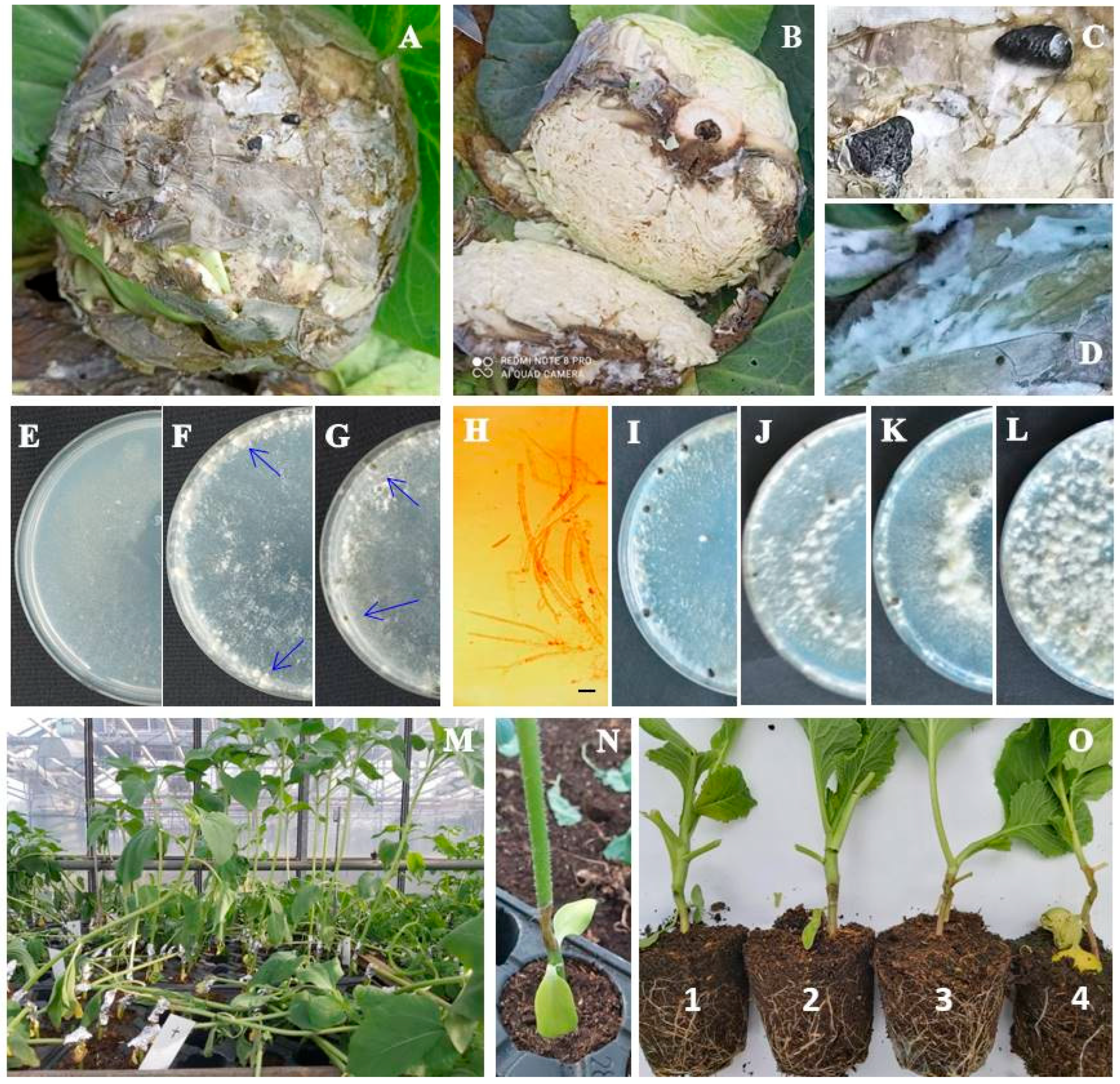
Figure 1. Sclerotinia sclerotiorum: (A,B) cabbage head rot (natural infection); (C,D) sclerotia and mycelium on infected tissue; (E,H) sparse, homogeneous mycelium of two-day old colony on PDA (bar = 20 μm); (F) white sclerotial initials in four-day colony on PDA (blue arrows); (G) fully developed sclerotia in six-day-old colony on PDA (blue arrows). (IL) The 15-day-old colony morphology: (I) sparse floccose, (J) sparse–low dense floccose, (K) sparse–very dense wooly–floccose, (L) very dense, wooly. (M,N) White stem rot of artificially inoculated sunflower plants after three-day incubation. (O) Rating scale for disease incidence evaluation on cabbage plants: 1—necrosis length up to 0.5 cm; 2—necrosis length 0.5–1 cm; 3—necrosis length 1–2 cm; 4—necrosis length > 2 cm.
Plants 14 02478 g001
Figure 2. Growth rate of Sclerotinia sclerotiorum isolates on PDA at 24 °C in the dark. B—Isolates from the Begeč location; M—isolates from the Futog location; SC—isolate from sunflower. Bars represent mean values of 5 replicates. Error bars indicate standard deviation. Values labeled with the same letter do not differ significantly.
Plants 14 02478 g002
Figure 3. Maximum likelihood phylogenetic tree of the internal transcribed spacer rDNA sequences of 13 Serbian and 24 reference isolates of Sclerotinia spp., and the outgroup taxa Trichoderma lixii. The tree was generated in Mega 7.0 using Kimura’s two-parameter model. Bootstrap analyses were performed with 1000 replicates, and bootstrap values (>50%) are shown next to the corresponding branches. Serbian isolates are in bold.
Plants 14 02478 g003
Figure 4. Median-joining network showing the phylogenetic relationships between haplotypes of Sclerotinia sclerotiorum. Black nodes represent median vectors required to connect existing haplotypes. Haplotype to which Serbian isolates belong is marked with a rectangle. The number of hatch marks on branches indicates mutational steps between haplotypes.
Plants 14 02478 g004
Figure 5. Disease severity on seven host plants caused by Sclerotinia sclerotiorum, rated using the following scale: 0—no reaction; 1—necrosis length up to 0.5 cm; 2—necrosis length 0.5–1 cm; 3—necrosis length 1–2 cm; 4—necrosis length > 2 cm. Error bars represent standard deviation. Values marked with the same letter do not differ significantly.
Plants 14 02478 g005
Figure 6. Comparative analysis of susceptibility levels in various host plants to Sclerotinia sclerotiorum isolates (B, B1-3, M, M1-7, SC) based on disease severity index calculated as an average symptom appearance rated using the following scale: 0—no reaction; 1—necrosis length up to 0.5 cm; 2—necrosis length 0.5–1 cm; 3—necrosis length 1–2 cm; and 4—necrosis length > 2 cm.
Plants 14 02478 g006
Figure 7. Sensitivity profiles of the host plants to Sclerotinia sclerotiorum isolates expressed as cumulative disease severity, calculated as the sum of the average disease severity index values (calculated as an average symptom appearance rated using the following scale: 0—no reaction; 1—necrosis length up to 0.5 cm; 2—necrosis length 0.5–1 cm; 3—necrosis length 1–2 cm; and 4—necrosis length > 2 cm) for all isolates combined.
Plants 14 02478 g007
Figure 8. Aggressiveness profiles of the isolates of Sclerotinia sclerotiorum on different host plants expressed as a cumulative disease severity calculated as the sum of the average disease severity index values (calculated as an average symptoms appearance rated using the following scale: 0—no reaction; 1—necrosis length up to 0.5 cm; 2—necrosis length 0.5–1 cm; 3—necrosis length 1–2 cm; and 4—necrosis length > 2 cm) for all hosts combined.
Plants 14 02478 g008
Table 1. Variability in colony appearance and sclerotia formation on PDA in Sclerotinia sclerotiorum isolates.
IsolateColony AppearanceSclerotia
ColorRelative Density and Appearance of Areal MyceliumTiming of Formation (day)Average No. ± SD/PlateAverage Size (mm)Arrangement
BWhiteSparse–very dense,
Wooly–floccose
815.7 ± 1.5 cd *5 × 3 (2–7 × 2–4)Edge ring
B1WhiteSparse–low dense.
Wooly
810.7 ± 0.6 de4 × 3 (2–5 × 2–4)Edge ring
B2WhiteSparse–low dense,
Floccose
810.3 ± 0.6 de6 × 3 (1–10 × 1–5) Edge + middle rings
B3WhiteSparse–very dense,
Wooly–floccose
615.7 ± 2.5 c3 × 2 (1–5 × 1–3) Edge ring
MWhiteDense,
Wooly
810.7 ± 1.5 cde5 × 3 (1–8 × 1–5)Edge + middle rings
M1WhiteSparse,
Floccose
810.7 ± 0.6 cde4 × 3 (1–7 × 1–4)Edge + middle rings
M2WhiteSparse,
Floccose
99.7 ± 0.6 e10 × 4 (2–7 × 2–6) Middle ring
M3WhiteModerately dense,
Floccose
612.7 ± 0.6 cde5 × 4 (2–7 × 2–5)Edge ring
M4WhiteSparse–dense,
Floccose
816.7 ± 0.6 bc6 × 3 (1–11 × 1–4)Edge + middle rings
M5WhiteSparse,
Floccose
623.7 ± 7.0 b3 × 3 (1–5 × 1–4)Edge ring
M6WhiteSparse–very dense,
Wooly–floccose
816.7 ± 1.5 bc5 × 4 (3–6 × 3–4)Edge + middle rings
M7WhiteSparse–dense,
Wooly–floccose
733.7 ± 1.5 a6 × 3 (2–9 × 2–3)Edge + middle rings
SCWhiteVery dense,
Wooly
132.7 ± 0.6 f2 × 2 (1–2 × 1–2)Edge + middle rings
* Values marked with the same letter do not differ significantly.
Table 2. Isolates of Sclerotinia sclerotiorum recovered in this study and species from GenBank included in phylogenic analyses.
SpeciesIsolateAcc No.HostCountryLiterature
Sclerotinia sclerotiorum2KX184720Cabbage (Brassica oleracea var. capitata)Sri Lanka[33]
SSC2JHUMG249967Cotton (Gossypium hirsutum)USA[33]
MAFF 306676AB233346Blueberry (Vaccinium corymbosum)Japan [33]
16-042KY073613Shepherd’s purse (Capsella bursa-pastoris)Korea[33]
SS-BO-SCKP340898Cabbage (Brassica oleracea var. capitata)New Mexico[33]
-No dataJN013184Fan Columbine (Aquilegia flabellata) Italy[33]
DAOM: 241671KF859932No dataCanada[33]
B23 DQ329537No dataAlaska[33]
ms85HQ833450Mulberry (Morus alba)China[33]
SQC-000KY750530Chinese celery (Oenanthe javanica)China[33]
16-119KY073614Cucumber (Cucumis sativus)Korea[33]
ATCC MYA-4521FJ810516No dataUSA[33]
Ss1212HAKT224645Caucasian clover (Trifolium ambiguum)Poland[33]
JBARES2014AKJ614564Chinese chives (Allium tuberosum)Korea[33]
MuRa-103AB937095Napa cabbage (Brassica napa)Japan[33]
BPP179050Cabbage (Brassica oleracea var. capitata)SerbiaThis study
B1PP179051Cabbage (Brassica oleracea var. capitata)SerbiaThis study
B2PP179052Cabbage (Brassica oleracea var. capitata)SerbiaThis study
B3PP179053Cabbage (Brassica oleracea var. capitata)SerbiaThis study
MPP179054Cabbage (Brassica oleracea var. capitata)SerbiaThis study
M1PP179057Cabbage (Brassica oleracea var. capitata)SerbiaThis study
M2PP179055Cabbage (Brassica oleracea var. capitata)SerbiaThis study
M3PP179056Cabbage (Brassica oleracea var. capitata)SerbiaThis study
M4PP179058Cabbage (Brassica oleracea var. capitata)SerbiaThis study
M5PP179059Cabbage (Brassica oleracea var. capitata)SerbiaThis study
M6PP179060Cabbage (Brassica oleracea var. capitata)SerbiaThis study
M7PP179061Cabbage (Brassica oleracea var. capitata)SerbiaThis study
SCPP177498Cabbage (Brassica oleracea var. capitata)SerbiaThis study
Sclerotinia trifoliorumSt03TPKT224652Caucasian clover (Trifolium ambiguum)Poland[33]
CBS122377KT970794Caucasian clover (Trifolium ambiguum)Poland[33]
TN Sc10101KT819299Fenugreek (Trigonella foenum-graecum)Tunisia[49]
St2413TAKT986230Caucasian clover (Trifolium ambiguum)Poland[48]
St1412TAKT224642Caucasian clover (Trifolium ambiguum)Poland[48]
Sclerotinia minorMAFF 238173AB516661No dataJapan [33]
45903AJF279879No dataAustralia[33]
45802AJF279877No dataAustralia[33]
62907JF279880No dataAustralia[33]
Trichoderma lixiNCIPM-78KU904458Rhizosphere soilIndia[50]
Table 3. The experimental plant species used to study the aggressiveness of isolates of Sclerotinia sclerotiorum from cabbage in Serbia.
Common NameLatin NameCultivarCultivar’s Origin
CabbageBrassica oleracea var. capitataFutoškiFutog, Novi Sad, Serbia
CauliflowerBrassica oleracea var. botrytisIncline Sakata Seed Southern Africa (Pty) Ltd., Kempton Park, South Africa
BroccoliBrassica oleracea var. silvestrisMerathonSakata Seed Southern Africa (Pty) Ltd., Kempton Park, South Africa
Kale Brassica oleraceaEstorilSakata Seed Southern Africa (Pty) Ltd., Kempton Park, South Africa
Mustard Sinapis alba NS Bela Institute of field and vegetable crops, Novi Sad, Serbia
Oilseed rape Brassica napus NS SvetlanaInstitute of field and vegetable crops, Novi Sad, Serbia
Sunflower Helianthus annuus LabudInstitute of field and vegetable crops, Novi Sad, Serbia
Disclaimer/Publisher’s Note: The statements, opinions and data contained in all publications are solely those of the individual author(s) and contributor(s) and not of MDPI and/or the editor(s). MDPI and/or the editor(s) disclaim responsibility for any injury to people or property resulting from any ideas, methods, instructions or products referred to in the content.