Communication

An Improved Agrobacterium-Mediated Transformation Method for an Important Fresh Fruit: Kiwifruit (Actinidia deliciosa)

1
Key Laboratory of Quality and Safety Control for Subtropical Fruit and Vegetable, Ministry of Agriculture and Rural Affairs, College of Horticulture Science, Zhejiang A&F University, Hangzhou 311300, China
2
State Key Laboratory for Managing Biotic and Chemical Threats to the Quality and Safety of Agro-Products, Key Laboratory of Traceability for Agricultural Genetically Modified Organisms, Ministry of Agriculture and Rural Affairs, Institute of Virology and Biotechnology, Zhejiang Academy of Agricultural Sciences, Hangzhou 310021, China
*
Authors to whom correspondence should be addressed.
These authors contributed equally to this work.
Plants 2025, 14(15), 2353; https://doi.org/10.3390/plants14152353
Submission received: 17 June 2025 / Revised: 26 July 2025 / Accepted: 28 July 2025 / Published: 31 July 2025
(This article belongs to the Special Issue Plant Transformation and Genome Editing)

Abstract

Genetic transformation is an essential tool for investigating gene function and editing genomes. Kiwifruit, recognized as a significant global fresh fruit crop, holds considerable economic and nutritional importance. However, current genetic transformation techniques for kiwifruit are impeded by low efficiency, lengthy culture durations (a minimum of six months), and substantial labor requirements. In this research, we established an efficient system for shoot regeneration and the stable genetic transformation of the ‘Hayward’ cultivar, utilizing leaf explants in conjunction with two strains of Agrobacterium that harbor the expression vector pBI121-35S::GFP, which contains the green fluorescent protein (GFP) gene as a visible marker within the T-DNA region. Our results show that 93.3% of leaf explants responded positively to the regeneration medium, producing multiple independent adventitious shoots around the explants within a six-week period. Furthermore, over 71% of kanamycin-resistant plantlets exhibited robust GFP expression, and the entire transformation process was completed within four months of culture. Southern blot analysis confirmed the stable integration of GFP into the genome, while RT-PCR and fluorescence microscopy validated the sustained expression of GFP in mature plants. This efficient protocol for regeneration and transformation provides a solid foundation for micropropagation and the enhancement of desirable traits in kiwifruit through overexpression and gene silencing techniques.

1. Introduction

Kiwifruit (Actinidia deliciosa) is a globally important fresh fruit crop for economic and human health [1,2,3,4]. It contains many bioactive compounds such as rich vitamins, soluble sucrose, and flavonoids, and it has antioxidant activity [5,6,7,8,9,10]. The ability of classical breeding to improve the agronomic traits of kiwifruit such as maturity, disease resistance, and nutritional composition has been challenged, and progress has been slow due to the lack of useful genetic resources and longer breeding periods [11,12,13,14]. In contrast with traditional breeding, genetic transformation is a powerful research tool for altering secondary metabolic pathways to improve important traits in fruit through short-term application in gene discovery [15]. Interestingly, recently achieved results in the genomics, transcriptomics, and metabolomics of kiwifruit offer novel insights into the fundamental study of gene function and the regulatory network of important traits, further unraveling pathways for secondary biosynthesis and providing new ideas for new improved kiwifruit cultivars [13,16,17,18].
Genetic transformation is a fundamental tool not only for the development of new cultivars but also for the modulation of gene expression by overexpressing or knocking down genes such as through RNA interference and the CRISPR–Cas9 system [19,20,21,22]. Plant transformation has been accomplished using several techniques including particle gun and Agrobacterium-mediated methods [23,24,25,26]. Among these, Agrobacterium-mediated genetic transformation is the most widely used method for crop improvement and gene function analysis in plants [20,23,27]. Since the Agrobacterium-mediated transformation of kiwifruit from hypocotyls and stems was first reported by Uematsu et al. (1991) [23], several studies have reported Agrobacterium-mediated transformation methods from stems or leaves [28,29,30], but these methods present certain limitations such as low transformation efficiency, with successful transformants obtained through lengthy tissue culture periods and labor-intensive processes. Recently, it was also reported that Agrobacterium rhizogenes mediated transformation [31]. However, Agrobacterium rhizogenes-transformed shoots often exhibit altered morphology, including dwarfism, a decrease in apical dominance, an increase in the number of flowers, and abnormal root production [32].
In this study, we describe a protocol for stable and efficient plant regeneration and the Agrobacterium-mediated genetic transformation of kiwifruit using leaf explants. This stable and efficient plant regeneration and genetic transformation protocol provides useful tools for gene function analysis, micropropagation, and genome editing approaches to the genetic improvement of kiwifruit.

2. Results

2.1. Shoot Regeneration of Leaf Explants

To establish an efficient and renewable shoot regeneration system for kiwifruit cultivar ‘Hayward’, thirty leaf explants as a set were cultured on 3/4 MS basal medium supplemented with various concentrations and combinations of growth hormones (Table S2). After 10 days of culture, small swellings appeared at the leaf margins, and small adventitious shoots emerged in media containing high concentrations of BA alone or in combination with 1 mg/L Zt after four weeks of culture. Among these, more than 93% of the leaf explants responded to 3/4 MS medium supplemented with 5 mg/L BAP + 1 mg/L Zt + 0.15 mg/L IBA (SIM) and developed normal adventitious shoots (Figure 1A; Table S2). After 6 weeks of culture on SIM (Table S1), normally elongated shoots were excised and transferred to RIM (Table S1)-induced roots. More than 90% of shoots grew normally and successfully formed roots within 6 weeks of culture on rooting medium (Figure 1B). Therefore, SIM was chosen as the shoot induction medium for further transformation studies.

2.2. Transformation of Kiwifruit

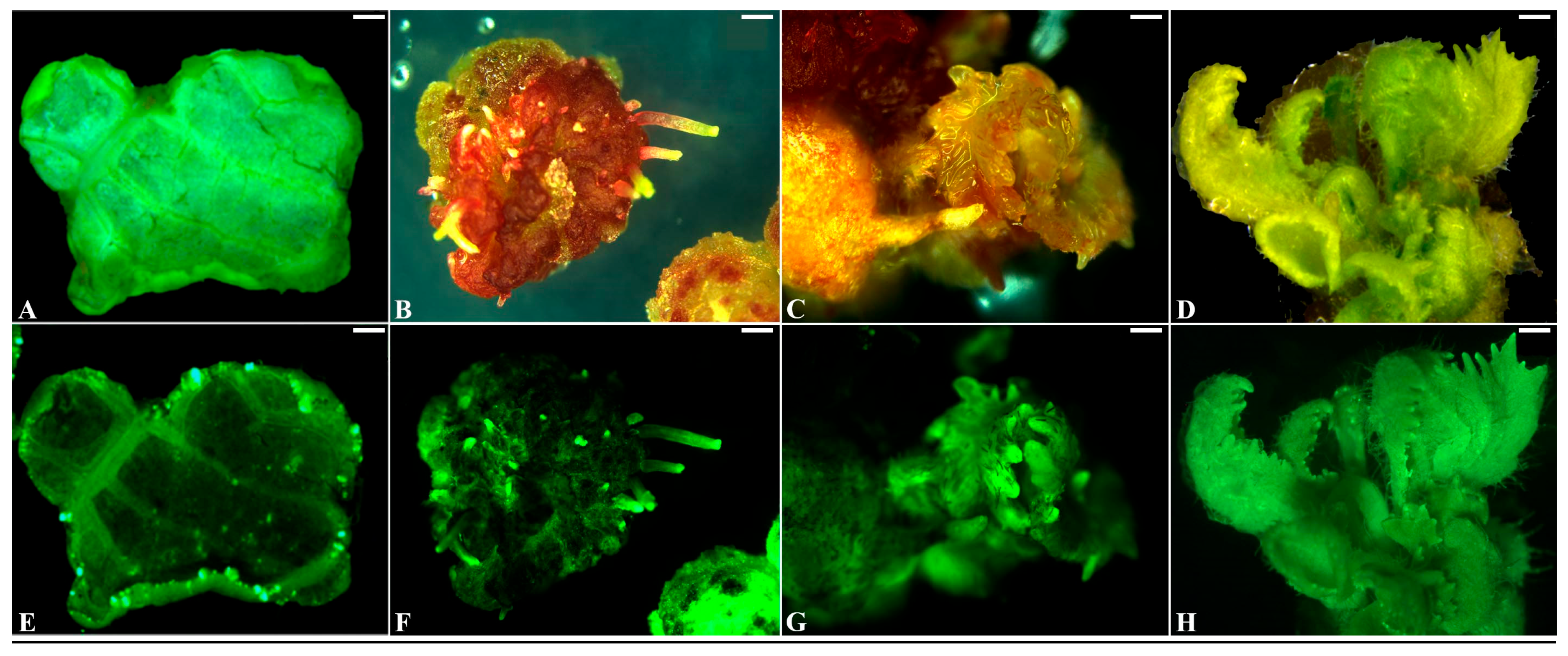
To establish an efficient Agrobacterium-mediated genetic transformation system for kiwifruit, we evaluated two common Agrobacterium strains, EHA105/pBI-35S::GFP and GV3101/pBI-35S::GFP, for their ability to transform leaf explants. The leaf explants were inoculated with diluted Agrobacterium for 15 min, followed by cocultivation for 3 days on CM (Table S2) at 25 °C in the dark and then being transferred to SM (Table S2). The transformation was repeated twice (Table 1). After 10 days of culture on SM, many small swells exhibiting strong GFP expression formed surrounding the leaf explants (Figure 2A,E). After 3 weeks of culture, protuberances with strong GFP expression appeared on many swellings (Figure 2B,F), and adventitious shoots developed from swellings after 5 weeks of culture on SM (Figure 2C,G). After 8 weeks of culture on SM, some normally growing adventitious shoots (Figure 2D,H) were excised and transferred to solid RIM (Table S1) supplemented with 50 mg/L kanamycin to induced roots. Over 71% of the adventitious shoots grew normally, formed roots, and presented strong GFP expression within 7 weeks of culture on RIM (Figure 3A; Table 1).

2.3. PCR Detection of Transgenic Kiwifruit

GFP expression was detected in the leaves, stems, and roots of small, regenerated kiwifruit plants after 6 weeks of culture on RIM (Table S1) supplemented with 50 mg/L kanamycin and 300 mg/L cefotaxime (Figure 3A). To confirm the presence of GFP and the kanamycin resistance gene NPT II in the kiwifruit genome, we used genomic DNA extracted from putatively transformed plants that survived kanamycin treatment and observed GFP expression with PCR amplification analysis. The results showed that the 780 bp band of the GFP gene and the 450 bp band of the NPT II gene were amplified in all investigated transformed kiwifruits (Figure 3B and Figure S1). However, no band was detected in the non-transformed wild-type (WT) kiwifruit plants (Figure 3B and Figure S1).

2.4. Southern Blot Analysis of Transgenic Kiwifruit

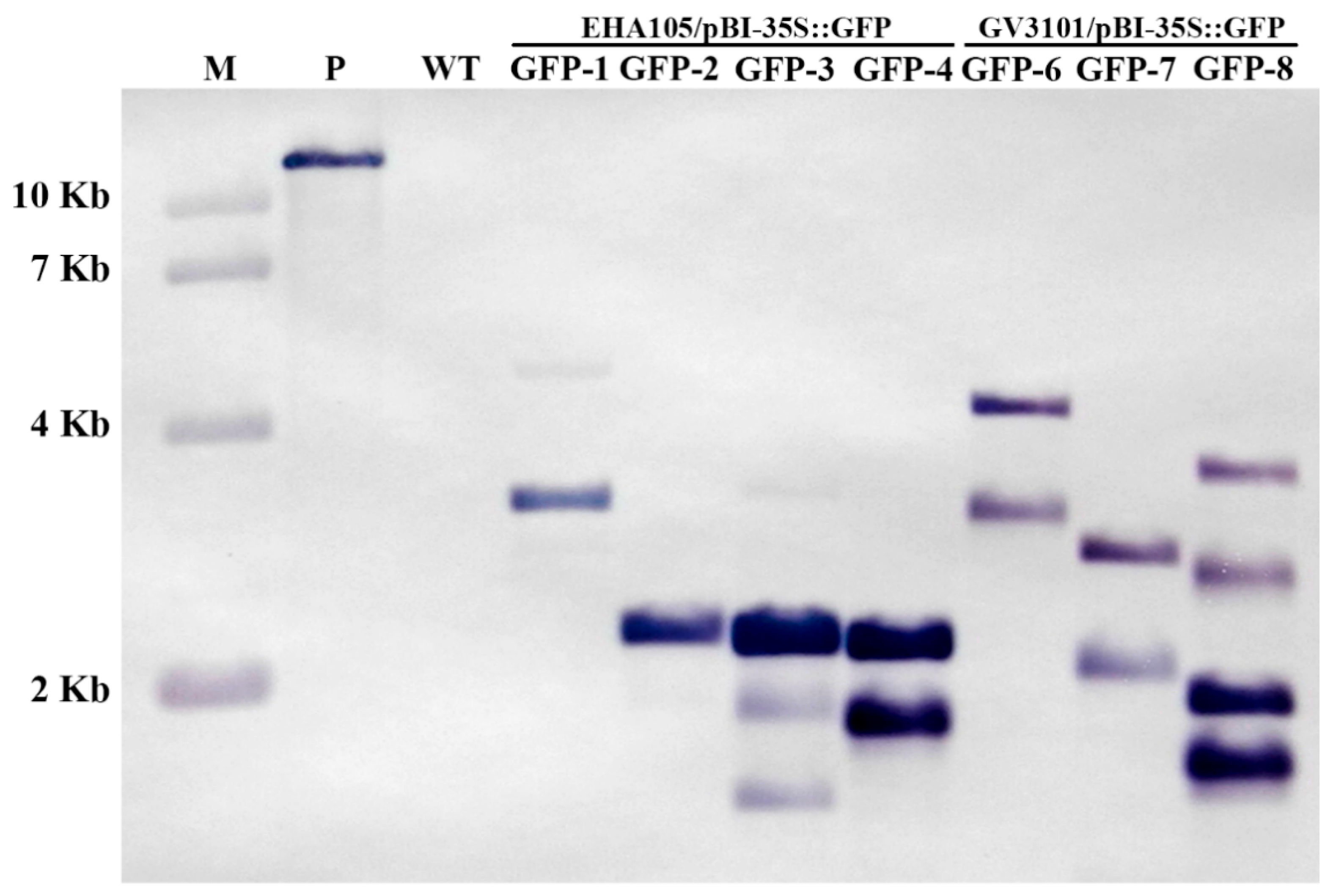
To confirm the integration of GFP in the kiwifruit genome, seven greenhouse-grown transgenic plants were analyzed using Southern hybridization with a probe containing the GFP fragment amplified with PCR from pBI-35S::GFP. The DNA from a wild-type (WT) plant grown under in vitro conditions was used as a negative control. Although all seven plants presented strongly different patterns of hybridization signals, hybridization signals were not detected in the WT plants (Figure 4), providing strong evidence that integrated copies of GFP sequences were present in the genomes of the transgenic plants (Figure 4).

2.5. Observation of GFP Expression in Potted Kiwifruit

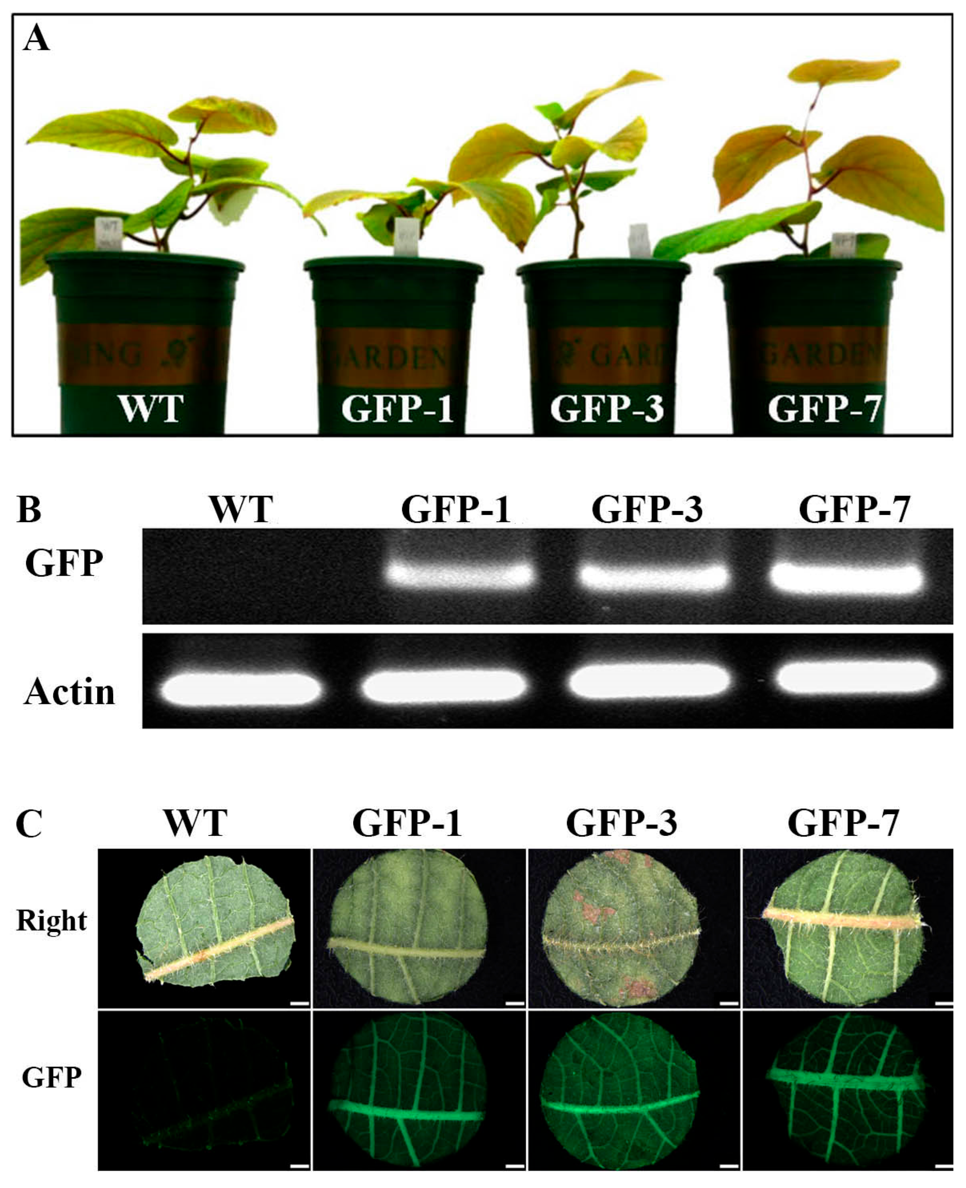
To investigate how the multi-insertion of the GFP gene affects GFP expression in mature kiwifruit plants, DNA extracted from the mature leaves of two 2-copy insertion lines, GFP-1 and GFP-7; a 4-copy insertion line, GFP-3 (Figure 4); and a WT kiwifruit grown for 3 months in the greenhouse (Figure 5A) was analyzed for GFP expression with RT-PCR and fluorescence microscopy. The clear putative GFP band was amplified using RT-PCR analysis from three transgenic kiwifruit plants, while the band was not detected in the WT kiwifruit (Figure 5B). Moreover, strong GFP expression was detected in the leaves of the three transgenic lines, while GFP expression was not detected in the WT (Figure 5C), implying that the stable expression of GFP may not be affected by the number of GFP insertions in the transformed kiwifruit.

2.6. Procedure for Genetic Transformation of Kiwifruit

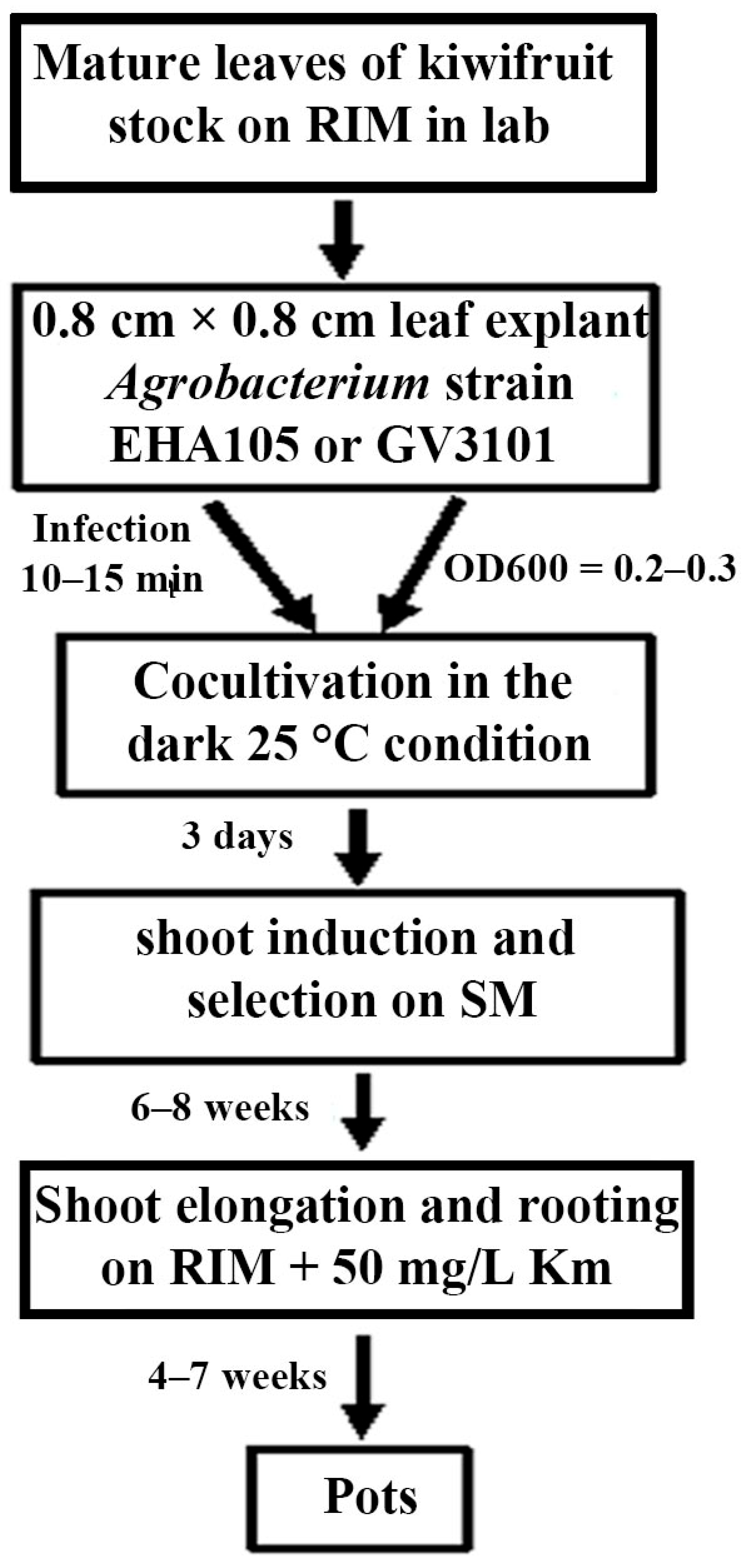
The procedure of plant regeneration and the Agrobacterium-mediated genetic transformation of kiwifruit is summarized in Figure 6. In the first step, leaf explants (0.8 cm × 0.8 cm) were infected with Agrobacterium strain EHA105 or GV3101 for 10–15 min. After three days of cocultivation on CM at 25 °C in the dark, the leaf explants were transferred to shoot induction and selection medium. After six to eight weeks of culture, kanamycin-resistant small adventitious shoots were transferred to shoot elongation and rooting medium. After four to seven weeks of culture, rooted plantlets exhibiting both kanamycin resistance and strong GFP expression were cultivated in pots or in soil. This genetic transformation procedure for kiwifruit required approximately 4 months.

3. Discussion

The tissue culture technique is an essential tool for the micropropagation and genetic transformation of plants [22,33,34]. The success of tissue culture is dependent on the genotype and explant types of the material, the composition of the culture medium, and the combination of plant hormones [35,36,37]. In kiwifruit, several studies have reported successful shoot regeneration and genetic transformation from hypocotyls, stems, and leaves [23,28,30]. For example, a combination of MS medium + 1.0 mg/l 4-PU (N-(2-chloro-4-pyrldyl)-N-phenylurea) or B5 medium + 3.0 mg/l zeatin from the hypocotyl and stems was used, and MS medium + 5 mg/L Zt + 0.1 mg/L NAA regenerated shoots from leaf explants via the callus phase [23,28]. Moreover, Zhang et al. (2018) [30] reported shoot regeneration from leaf explants using a combination of MS medium + Nitsch & Nitsch vitamins + 4 mg/L BA + 1 mg/L NAA. However, these methods have several limitations such as the induction and cultivation of calluses, low-efficiency shoot regeneration, and eventually successful genetic transformation. This requires a lengthy tissue culture period and labor-intensive processes. In the present study, modified 3/4 MS components were used as a basal medium, and the mature leaves of in vitro-grown kiwifruit as a source of explants were investigated for shoot regeneration frequency by treatment with several combinations of the cytokine BA with 1 mg/L Zt, a highly effective regulator for the induction of shoot morphogenesis [38,39]. Interestingly, we found that more than 93.3% of the leaf explants responded to 3/4 MS + 5 mg/L BA + 1 mg/L Zt + 0.15 mg/L IBA medium, and multi-adventitious shoot formation was stimulated from each leaf explant after 6 weeks of culture (Figure 1A), clearly greater compared to any other treatment combination (Table S2). This result implies that Zt is an important plant regulator and that 5 mg/L BA + 1 mg/L Zt is a suitable combination for shoot regeneration from kiwifruit leaf explants (Figure 1; Table S2). Furthermore, on this medium we successfully achieved stable genetic transformation and obtained several kanamycin-resistant and GFP-expressing shoots after 4 months of culture (Figure 2 and Figure 6). This transformation method requires shorter culture periods than other previously reported transformation methods [23,28,30]. Therefore, we suggest that leaves represent a better explant and that combination with modified 3/4 MS-based SIM is an efficient and suitable platform for micropropagation and the genetic transformation of kiwifruit cultivar ‘Hayward’.
The Agrobacterium strain is another important factor for successful genetic transformation in plants [37,40]. In this study, two common, strong Agrobacterium strains, EHA105/pBI-35S::GFP and GV3101/pBI-35S::GFP, were chosen as the T-DNA delivery system, and the transformation efficiency of these strains in kiwifruit was compared. We found that approximately 71.1% kanamycin-resistant plantlets of EHA105/pBI-35S::GFP and 72.5% kanamycin-resistant plantlets of GV3101/pBI-35S::GFP presented strong GFP expression 15 weeks after inoculation (Table 1; Figure 3A) and were stably expressed in three-month-old plants grown in the greenhouse (Figure 5). Moreover, GFP integration in the genome of kiwifruit was detected with Southern blot and PCR analysis, and expression was detected using RT-PCR (Figure 4 and Figure 5B). These findings imply that any strain of Agrobacterium EHA105 and GV3101 could be used to transform kiwifruit.
The Agrobacterium-mediated transformation system is an important deliverer for CRISPR-Cas9 genome editing in plants, with its efficiency significantly influencing genetic modification outcomes [22]). In this study, we employed this system to target the PDS gene in kiwifruit. An analysis of six hygromycin-resistant shoots confirmed the successful integration of the CRISPR-Cas9 editing cassette (Figure S2A,B), and a regenerated shoot displayed an albino phenotype (Figure S2C). Although sequencing validation was not performed, these results indicate that the improved transformation method could be valuable for functional gene studies and genome editing in kiwifruit.
In the present study, we described a reliable and efficient Agrobacterium-mediated kiwifruit transformation protocol using a combination of mature leaf explants and two common Agrobacterium strains, EHA105 and GV3101. The method requires a tissue culture period of approximately 4 months, after which over 71% of kanamycin-resistant transformed plantlets presented strong GFP expression. Moreover, a strong expression of GFP was detected in the mature leaves of three-month-old potted plants. Therefore, this protocol provides a reliable tool for the fundamental study of gene function and can be applied to produce improved new kiwifruit cultivars through biotechnique approaches, such as RNAi and genome editing

4. Materials and Methods

4.1. Plant Materials and Growth Conditions

This study used in vitro-grown ‘Hayward’ kiwifruit (Actinidia deliciosa) plants maintained at Zhejiang A&F University. The plants were cultured on solid RIM (Table S1) at 25 °C under a 16/8 h light/dark photoperiod with cool white fluorescent light (100 µmol m−2 s−1). Shoot tips (2–3 cm) were subcultured on fresh RIM every two months. Fully expanded mature leaves were used for transformation experiments and served as the wild-type (WT) control in molecular and GFP expression analysis.

4.2. Tissue Culture of Leaf Explants

To determine the optimal shoot regeneration conditions from leaves, leaf explants (0.8 cm × 0.8 cm) were excised from plantlets and cultured on ¾-strength MS basal medium [41] supplemented with varying concentrations and combinations of cytokinins (BA and Zt) along with 0.15 mg/L NAA (Table S2). Thirty explants were placed per treatment. After three weeks of cultivation, enlarged explants were subdivided and transferred to three identical fresh media for further culture. Cultures were maintained at 25 °C under a 16/8 h light/dark photoperiod with cool white fluorescent light (100 µmol m−2 s−1). Shoot regeneration frequency was evaluated after five weeks of cultivation.

4.3. Infection of Kiwifruit Leaf Explants

The phytohormones and antibiotics were filter-sterilized (0.22 μm) and aseptically added to the autoclaved culture medium (121 °C, 20 min) after cooling to approximately 70 °C before experimental use.
The kiwifruit transformation was conducted using two A. tumefaciens strains, EHA105/pBI-35S::GFP and GV3101/pBI-35S::GFP, both harboring the binary vector pBI-35S::GFP. This vector contained the neomycin phosphotransferase II (NPT II) gene as a kanamycin selectable marker and the green fluorescent protein (GFP) gene as a visible marker [42]. The Agrobacterium strains were cultured in 4 mL of liquid YEP medium (Table S1) supplemented with 50 mg/L kanamycin and 25 mg/L rifampicin, at 28 °C shaking at 200 rpm for 24 h. The bacterial suspension was then diluted 1:10 (OD600 = 0.2–0.3) with liquid IM medium (Table S1). Fresh kiwifruit leaves were cut into 0.8 cm × 0.8 cm segments, and 30 leaf explants per set were inoculated in the diluted Agrobacterium suspension for 15 min. After inoculation, the explants were cocultured on CM medium (Table S1) at 25 °C in the dark for 3 days. The experiment was performed twice. Following cocultivation, the leaf explants were transferred to SM (Table S1) for kanamycin-resistant shoot induction, with fresh SM replenished every two weeks. After two to three rounds of selection, shoots displaying GFP expression and normal growth were excised and rooted on RIM (Table S1) supplemented with 50 mg/L kanamycin and 300 mg/L cefotaxime. After 6 weeks of culture, rooted plantlets were individually transferred to 400 mL square plastic bottles containing RIM (Table S1) supplemented with 50 mg/L km and 300 mg/L cefotaxime to maintain them and for further molecular analysis and validations of GFP expression. Furthermore, small transformed plants identified by Southern blot analysis were propagated. Selected plants were then transferred to soil and assessed for GFP expression and morphological characteristics.

4.4. Observation of Visible GFP Expression

GFP expression was visualized using a fluorescence microscope (Leica 165 FC, Leica Microsystems, Mannheim, Germany) equipped with a GFP filter. Observations were performed at distinct developmental stages: in the emerging solid callus stage after 10 days post-infection, beginning at the adventitious shoot emergence stage after 3 weeks post-infection, at the elongation of shoot stage after 5 weeks post-infection, at the rooted small plant stage, and at the stage when mature leaves appear in 3-month-old greenhouse-grown plants. Images were captured using a Leica DFC310 FX CCD camera coupled to the microscope system.

4.5. PCR Detection of Transformed Kiwifruit Plants

Genomic DNA was extracted from the mature leaves of putatively transformed kiwifruit plants using a cetyltrimethylammonium bromide (CTAB)-based method [43]. The primer sets (Table S3) were used to amplify the GFP and NPT II genes. PCR amplification reactions were performed on a total volume of 20 µL containing 50 ng of DNA, a 200 µM dNTP mixture, 0.2 µM of each primer, 1 × FastPfu DNA polymerase reaction buffer, and 1 U FastPfu DNA polymerase (TransGen, Beijing, China) under denaturation at 96 °C for 3 min followed by 35 cycles of 96 °C for 30 s, 62 °C for 30 s, and 72 °C for 50 s, with a final extension at 72 °C for 10 min. The amplified product was analyzed with electrophoresis on 1% agarose gels at 100 V for 30 min.

4.6. Southern Blot Analysis

Genomic DNA was extracted from 1 g of fresh mature leaves collected from the pot-grown wild-type (WT) and seven transformed kiwifruit plants using a CTAB-based method [43]. Each 20 µg DNA sample was digested with BamHI, electrophoresed on 0.8% agarose gels at 200 V for 10 h, and then transferred onto Hybond-N+ (Amersham) nylon membranes. A digoxin-labeled GFP probe was synthesized with PCR using the PCR Dig Probe Synthesis Kit (Roche, Basel, Switzerland). The hybridization and signal detection procedures were performed according to the manufacturer’s instructions (Roche).

4.7. Semiquantitative RT–PCR Analysis

Total RNA was isolated from the mature leaves of a wild-type plant and GFP-positive plants growing in the greenhouse using an EasyPure® Plant RNA Kit (TransGen, Beijing, China). cDNA was synthesized with TransScript® First-Strand cDNA Synthesis Super Mix (TransGen, Beijing, China). RT–PCR was carried out using the GFP-specific primer set described above, and the SnActin-specific primer set was used as a positive control (Table S3). The reaction was performed with 1 μL of the first cDNA sample as a template and started with denaturation at 98 °C for 2 min, followed by 30 cycles of 96 °C for 30 s, 62 °C for 30 s, and 72 °C for 50 min, with a final extension at 72 °C for 10 min. The amplified DNA fragments were separated with electrophoresis on 1.2% agarose gels at 100 V for 30 min, stained with ethidium bromide, and observed under UV illumination.

4.8. Construction of pHEE401E-AcPDS Vector

Two target sequences AcPDS-gRNA1 and AcPDS-gRNA2 (Table S3) were selected within the coding region of the Actinidia phytoene desaturase gene (AcPDS, XM_057618388.1) using the website http://www.crisprscan.org. Based on the pHEE401E backbone [44], the pHEE401E-AcPDS editing vector was constructed. The vector was introduced into A. tumefaciens strain GV3101 via electroporation for subsequent transformation assays.

Supplementary Materials

The following supporting information can be downloaded at https://www.mdpi.com/article/10.3390/plants14152353/s1, Supplementary Table S1: Composition of medium used in this study. Supplementary Table S2: Effect of various concentrations and combinations of BA, Zt, and 0.15 mg/L IBA (in 3/4MS basal medium) on shoot regeneration from leaf explants of kiwifruit. Results were scored 5 weeks after culture. Supplementary Table S3: The primers used for the detection of transformed plants, RT-PCR, and the genome editing analysis in this study. Supplementary Figure S1: Detection of kanamycin-resistant NPT II gene in transformed kiwifruits. Supplementary Figure S2: An insertion analysis of the editing cassette and PCR detection of the hygromycin-B gene.

Author Contributions

C.-L.P., M.D. and Y.G.: Data curation, Formal analysis, and Investigation; T.S.: Investigation; Y.Z. and M.-L.C.: Conceptualization, Writing—original draft, and Writing—review and editing. All authors have read and agreed to the published version of the manuscript.

Funding

This work was supported by the Scientific Research Funds of Zhejiang A&F University (2018FR004) given to M.-L.C. and Biological Breeding Project of ZAAS program for Transdisciplinary Research to Y.Z.

Data Availability Statement

All data generated or analyzed during this study are included in the article and its Supplementary Materials.

Conflicts of Interest

The authors declare no conflicts of interest.

References

  1. McGhie, T.K. Secondary metabolite components of kiwifruit. Adv. Food Nutr. Res. 2013, 68, 101–124. [Google Scholar]
  2. Vanneste, J.L. The scientific, economic, and social impacts of the New Zealand outbreak of bacterial canker of kiwifruit (Pseudomonas syringae pv. actinidiae). Annu. Rev. Phytopathol. 2017, 55, 377–399. [Google Scholar] [CrossRef]
  3. Richardson, D.P.; Ansell, J.; Drummond, L.N. The nutritional and health attributes of kiwifruit: A review. Eur. J. Nutr. 2018, 57, 2659–2676. [Google Scholar] [CrossRef] [PubMed]
  4. Kim, Y.M.; Abas, F.; Park, Y.S.; Park, Y.K.; Ham, K.S.; Kang, S.G.; Lubinska-Szczygeł, M.; Ezra, A.; Gorinstein, S. Bioactivities of phenolic compounds from kiwifruit and persimmon. Molecules 2021, 26, 4405. [Google Scholar] [CrossRef]
  5. Tavarini, S.; Degl’iNnocenti, E.; Remorini, D.; Massai, R.; Guidi, L. Antioxidant capacity, ascorbic acid, total phenols and carotenoids changes during harvest and after storage of Hayward kiwifruit. Food Chem. 2008, 107, 282–288. [Google Scholar] [CrossRef]
  6. Latocha, P.; Krupa, T.; Wołosiak, R.; Worobiej, E.; Wilczak, J. Antioxidant activity and chemical difference in fruit of different Actinidia sp. Int. J. Food Sci. Nutr. 2010, 61, 381–394. [Google Scholar] [CrossRef]
  7. Hunter, D.C.; Greenwood, J.; Zhang, J.; Skinner, M.A. Antioxidant and ‘natural protective’ properties of kiwifruit. Curr. Top Med. Chem. 2011, 11, 1811–1820. [Google Scholar] [CrossRef] [PubMed]
  8. Montefiori, M.; Espley, R.V.; Stevenso, D.; Cooney, J.; Datson, P.M.; Saiz, A.; Atkinson, R.G.; Hellens, R.P.; Allan, A.C. Identification and characterization of F3GT1 and F3GGT1, two glycosyltransferases responsible for anthocyanin biosynthesis in red-fleshed kiwifruit (Actinidia chinensis). Plant J. 2011, 65, 106–118. [Google Scholar] [CrossRef]
  9. Pérez-Burillo, S.; Oliveras, M.J.; Quesada, J.; Rufián-Henares, J.A.; Pastoriza, S. Relationship between composition and bioactivity of persimmon and kiwifruit. Food Res. Int. 2018, 105, 461–472. [Google Scholar] [CrossRef] [PubMed]
  10. Liu, X.; Xie, X.; Zhong, C.; Li, D. Comparative Transcriptome Analysis Revealed the Key Genes Regulating Ascorbic Acid Synthesis in Actinidia. Int. J. Mol. Sci. 2021, 22, 12894. [Google Scholar] [CrossRef]
  11. Ferguson, A.R.; Huang, H. Genetic resources of kiwifruit: Domestication and breeding. Hortic. Rev. 2007, 33, 1–121. [Google Scholar]
  12. Ampomah-Dwamena, C.; McGhie, T.; Reginald, W.; Montefiori, M.; Hellens, R.P.; Allan, A.C. The kiwifruit lycopene beta-cyclase plays a significant role in carotenoid accumulation in fruit. J. Exp. Bot. 2009, 60, 3765–3779. [Google Scholar] [CrossRef] [PubMed]
  13. Nardozza, S.; Boldingh, H.L.; Osorio, S.; Höhne, M.; Wohlers, M.; Gleave, A.P.; MacRae, E.A.; Richardson, A.C.; Atkinson, R.G.; Sulpice, R.; et al. Metabolic analysis of kiwifruit (Actinidia deliciosa) berries from extreme genotypes reveals hallmarks for fruit starch metabolism. J. Exp. Bot. 2013, 64, 5049–5063. [Google Scholar] [CrossRef] [PubMed]
  14. Kim, S.H.; Kim, D.R.; Kwak, Y.S. Variations in kiwifruit microbiota across cultivars and tissues during developmental stage. Plant Pathol. J. 2023, 39, 245–254. [Google Scholar] [CrossRef]
  15. Noda, N.; Yoshioka, S.; Kishimoto, S.; Nakayama, M.; Douzono, M.; Tanaka, Y.; Aida, R. Generation of blue chrysanthemums by anthocyanin B-ring hydroxylation and glucosylation and its coloration mechanism. Sci. Adv. 2017, 3, e1602785. [Google Scholar] [CrossRef]
  16. Wu, L.; Lan, J.; Xiang, X.; Xiang, H.; Jin, Z.; Khan, S.; Liu, Y.; Han, Z. Transcriptome sequencing and endogenous phytohormone analysis reveal new insights in CPPU controlling fruit development in kiwifruit (Actinidia chinensis). PLoS ONE 2020, 15, e0240355. [Google Scholar] [CrossRef]
  17. Han, X.; Zhang, Y.; Zhang, Q.; Ma, N.; Liu, X.; Tao, W.; Lou, Z.; Zhong, C.; Deng, X.W.; Li, D.; et al. Two haplotype-resolved, gap-free genome assemblies for Actinidia latifolia and Actinidia chinensis shed light on the regulatory mechanisms of vitamin C and sucrose metabolism in kiwifruit. Mol. Plant 2023, 16, 452–470. [Google Scholar] [CrossRef]
  18. Nazir, M.F.; Lou, J.; Wang, Y.; Zou, S.; Huang, H. Kiwifruit in the Omics Age: Advances in Genomics, Breeding, and Beyond. Plants 2024, 13, 2156. [Google Scholar] [CrossRef]
  19. Kusaba, M. RNA interference in crop plants. Curr. Opin. Biotechnol. 2004, 15, 139–143. [Google Scholar] [CrossRef]
  20. Cui, M.L.; Copsey, L.; Green, A.; Bangham, A.; Coen, E. Quantitative control of organ shape by combinatorial gene activity. PLoS Biol. 2010, 8, e1000538. [Google Scholar] [CrossRef]
  21. Rössner, C.; Lotz, D.; Becker, A. VIGS goes viral: How VIGS transforms our understanding of plant science. Annu. Rev. Plant Biol. 2022, 73, 703–728. [Google Scholar] [CrossRef]
  22. Ding, M.; Piao, C.L.; Zhang, X.; Zhu, Y.; Cui, M.L. Establishment of a high-efficiency transformation and genome editing method for an essential vegetable and medicine Solanum nigrum. Physiol. Plantarum. 2023, 175, e14028. [Google Scholar] [CrossRef]
  23. Uematsu, C.; Murase, M.; Ichikawa, H.; Imamura, J. Agrobacterium-mediated transformation and regeneration of kiwi fruit. Plant Cell Rep. 1991, 10, 286–290. [Google Scholar] [CrossRef] [PubMed]
  24. Kohli, A.; Gahakwa, D.; Vain, P.; Laurie, D.A.; Christou, P. Transgene expression in rice engineered through particle bombardment: Molecular factors controlling stable expression and transgene silencing. Planta 1999, 208, 88–97. [Google Scholar] [CrossRef]
  25. Sailaja, M.; Tarakeswari, M.; Sujatha, M. Stable genetic transformation of castor (Ricinus communis L.) via particle gun-mediated gene transfer using embryo axes from mature seeds. Plant Cell Rep. 2008, 27, 1509–1519. [Google Scholar] [CrossRef]
  26. Li, Y.; Gao, Z.; Piao, C.; Lu, K.; Wang, Z.; Cui, M.L. A stable and efficient Agrobacterium tumefaciens–mediated genetic transformation of the medicinal plant D. purpurea L. Appl. Biochem. Biotechnol. 2014, 172, 1807–1817. [Google Scholar] [CrossRef] [PubMed]
  27. Legendre, M.; Demirer, G.S. Improving crop genetic transformation to feed the world. Trends Biotechnol. 2023, 41, 264–266. [Google Scholar] [CrossRef]
  28. Janssen, B.J.; Gardner, R. The use of transient GUS expression to develop an Agrobacterium-mediated gene transfer system for kiwifruit. Plant Cell Rep. 1993, 13, 28–31. [Google Scholar] [CrossRef] [PubMed]
  29. Wang, T.; Ran, Y.; Atkinson, R.G.; Gleave, A.P.; Cohen, D. Transformation of Actinidia eriantha: A potential species for functional genomics studies in Actinidia. Plant Cell Rep. 2006, 25, 425–431. [Google Scholar] [CrossRef]
  30. Zhang, A.-D.; Wang, W.-Q.; Tong, Y.; Li, M.-J.; Grierson, D.; Ferguson, I.; Chen, K.S.; Yin, X.R. Transcriptome Analysis Identifies a Zinc Finger Protein Regulating Starch Degradation in Kiwifruit. Plant Physiol. 2018, 178, 850–863. [Google Scholar] [CrossRef]
  31. Li, P.; Zhang, Y.; Liang, J.; Hu, X.; He, Y.; Miao, T.; Ouyang, Z.; Yang, Z.; Amin, A.K.; Ling, C.; et al. Agrobacterium rhizogenes-mediated marker-free transformation and gene editing system revealed that AeCBL3 mediates the formation of calcium oxalate crystal in kiwifruit. Mol. Hortic. 2024, 4, 1. [Google Scholar] [CrossRef] [PubMed]
  32. Handa, T. Genetic transformation of Antirrhinum majus L. and inheritance of altered phenotype induced by Ri T-DNA. Plant Sci. 1992, 81, 199–206. [Google Scholar] [CrossRef]
  33. Loyola-Vargas, V.M.; Ochoa-Alejo, N. An introduction to plant tissue culture: Advances and perspectives. Methods Mol. Biol. 2018, 1815, 3–13. [Google Scholar]
  34. Anjanappa, R.B.; Gruissem, W. Current progress and challenges in crop genetic transformation. J. Plant Physiol. 2021, 261, 153411. [Google Scholar] [CrossRef]
  35. Rugini, E. In vitro propagation of some olive (Olea europaea sativa L.) cultivars with different root ability, and medium development using analytical data from developing shoots and embryos. Sci. Hortic. 1984, 24, 123–134. [Google Scholar] [CrossRef]
  36. Abe, T.; Futsuhara, Y. Genotypic variability for callus formation and plant regeneration in rice (Oryza sativa L.). Theor. Appl. Genet. 1986, 72, 3–10. [Google Scholar] [CrossRef]
  37. Akama, K.; Shiraishi, S.; Nakamura, K.; Okada, K.; Shimura, Y. Efficient transformation of Arabidopsis thaliana: Comparison of the efficiencies with various organs, plant ecotypes and Agrobacterium strains. Plant Cell Rep. 1992, 12, 7–11. [Google Scholar] [CrossRef]
  38. Cui, M.L.; Handa, T.; Ezura, H. An improved protocol for Agrobacterium-mediated transformation of Antirrhinum majus L. Mol. Genet. Genomics 2003, 270, 296–302. [Google Scholar] [CrossRef]
  39. Yaroshko, O.; Pasternak, T.; Larriba, E.; Pérez-Pérez, J.M. Optimization of callus induction and shoot regeneration from tomato cotyledon explants. Plants 2023, 12, 2942. [Google Scholar] [CrossRef] [PubMed]
  40. Dehghani, J.; Movafeghi, A.; Barzegari, A.; Barar, J. Efficient and stable transformation of Dunaliella pseudosalina by 3 strains of Agrobacterium tumefaciens. Bioimpacts 2017, 7, 247–254. [Google Scholar] [CrossRef]
  41. Murashige, T.; Skoog, F. A revised medium for rapid growth and bioassays with tobacco tissue culture. Physiol. Plant. 1962, 15, 81–84. [Google Scholar] [CrossRef]
  42. Cui, M.L.; Liu, C.; Piao, C.L.; Liu, C.L. A stable Agrobacterium rhizogenes-mediated transformation of cotton (Gossypium hirsutum) and plant regeneration from transformed hairy root via embryogenesis. Front. Plant Sci. 2020, 11, 604255. [Google Scholar] [CrossRef] [PubMed]
  43. Allen, G.C.; Flores-Vergara, M.A.; Krasynanski, S.; Kumar, S.; Thompson, W.F. A modified protocol for rapid DNA isolation from plant tissues using cetyltrimethylammonium bromide. Nat. Protoc. 2006, 1, 2320–2325. [Google Scholar] [CrossRef] [PubMed]
  44. Xing, H.L.; Dong, L.; Wang, Z.P.; Zhang, H.Y.; Han, C.Y.; Liu, B.; Wang, X.C.; Chen, Q.J. A CRISPR/Cas9 toolkit for multiplex genome editing in plants. BMC Plant Biol. 2014, 14, 327. [Google Scholar] [CrossRef] [PubMed]
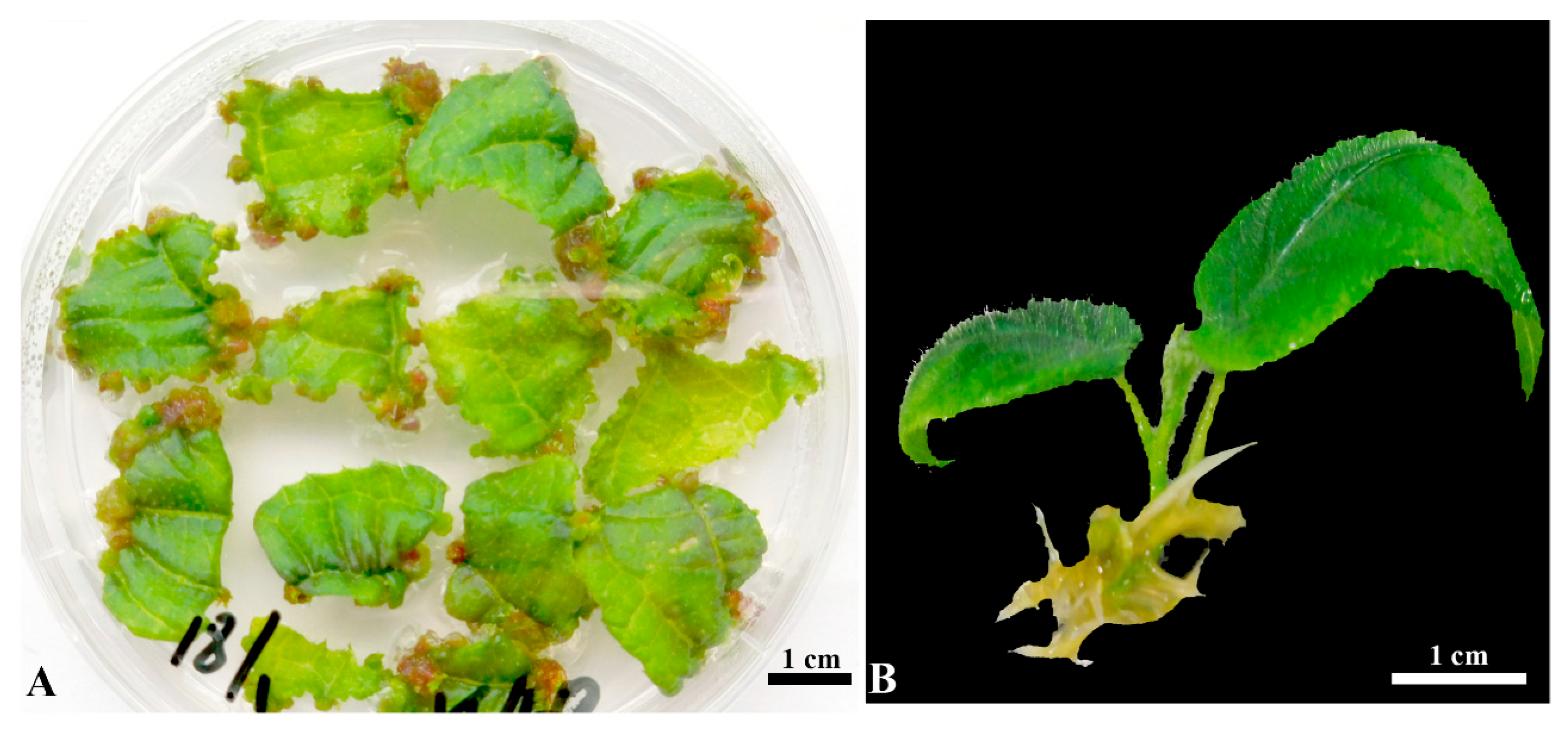
Figure 1. Plant regeneration from leaf explants of kiwifruit. (A) Shoot induction from leaf explants on 3/4 MS+ 5 mg/L BA + 1 mg/L Zt + 0.15 mg/L IBA medium (SIM) after 4 weeks of culture. (B) Root induction from regenerated shoots on 1/2 MS+ 0.25 mg/L IBA medium (RIM) after 6 weeks of culture.
Plants 14 02353 g001
Figure 2. Observation of GFP expression at different steps in Agrobacterium-mediated transformation of kiwifruit. (A) Kanamycin-resistant small swellings emerged on wounded leaf segments on solid SM after 10 days of culture. (B) Protuberance formation on kanamycin-resistant swellings on solid SM after 3 weeks of culture. (C) Kanamycin-resistant small shoots emerged on solid SM after 5 weeks of culture. (D) Normally grown kanamycin-resistant shoots on solid SM after 8 weeks of culture. (E) GFP observation in kanamycin-resistant small swellings (A). (F) GFP observation in kanamycin-resistant protuberances (B). (G) GFP observation in kanamycin-resistant small shoots (C). (H) GFP observation in kanamycin-resistant plantlets (D). Bar = 1 mm.
Plants 14 02353 g002
Figure 3. PCR analysis of transformed kiwifruits. Genomic DNA was extracted from fresh leaves of pot-grown wild-type (WT) plants and ten putative GFP-positive transgenic kiwifruit lines. (A) Rooted transgenic kiwifruit plant exhibiting GFP fluorescence after 6 weeks of culture on RIM supplemented with 50 mg/L kanamycin. (B) PCR detection of GFP transgene in ten independent transgenic lines using gene-specific primers (Table S3). WT: Non-transformed wild-type kiwifruit. GFP1–5: Five independent GFP-positive kiwifruits transformed with EHA105/pBI-35S::GFP. GFP6–10: Five independent GFP-positive kiwifruits transformed with GV3101/pBI-35S::GFP. M: DL1000 DNA marker (Takara, Japan). R: root; L1: First true leaf; AM: Apical meristem.
Plants 14 02353 g003
Figure 4. A Southern blot analysis of GFP integration in transformed kiwifruits. DNA from a non-transformed plant and seven GFP-positive transgenic kiwifruits was digested with BamHI, fractionated using electrophoresis, transferred to a nylon membrane, and hybridized with the full-length GFP probe. WT: a non-transformed wild-type kiwifruit. GFP1–4: four independent transgenic kiwifruits transformed with EHA105/pBI-35S::GFP. GFP6–8: three independent transgenic kiwifruits transformed with GV3101/pBI-35S::GFP. M: DL 10,000 DNA marker (Takara, Japan). P: plasmid pBI-35S::GFP.
Plants 14 02353 g004
Figure 5. GFP observation from WT and three transgenic kiwifruits grown in greenhouse. (A) Three-month-old WT and three transgenic kiwifruits grown in greenhouse. (B) Detection of GFP expression from WT and three transgenic kiwifruits (A) by RT–PCR. (C) Observation of GFP expression from mature leaves of WT and three transgenic kiwifruits (A) using fluorescence microscopy. Bar = 1 mm.
Plants 14 02353 g005
Figure 6. Summary of procedure for Agrobacterium-mediated genetic transformation of kiwifruit. Timetable is displayed on panel.
Plants 14 02353 g006
Table 1. Comparison of transformation efficiency of two common Agrobacterium strains EHA105/pBI-35S::GFP and GV3101/pBI-35S::GFP.
A. tumefaciens Strains (Plasmid)ReplicateNo. of Leaf Explants aKm+ Shoot Numbers bGFP-Positive ShootTransformation Frequency (%) d
NumbersFrequency (%) c
EHA105
(pBI-35S::GFP)
130241770.880
230211571.470
Total60453271.175
GV3101
(pBI-35S::GFP)
130211571.470
230191473.763.3
Total60402972.566.7
a Kiwifruit plant was maintained on RIM in a tissue culture room, and approximately 0.8 cm × 0.8 cm leaf explants were infected with Agrobacterium strains. b A single normally developed kanamycin-resistant (Km+) shoot was excised from a leaf explant after 8 weeks of cultivation on selection medium (SM) and transferred to RIM supplemented with 50 mg/L kanamycin and 300 mg/L cefotaxime. c (No. of GFP-positive shoots / no. of kanamycin-resistant shoots) × 100%. d (No. of kanamycin-resistant shoots / no. of leaf explants) × 100%.
Disclaimer/Publisher’s Note: The statements, opinions and data contained in all publications are solely those of the individual author(s) and contributor(s) and not of MDPI and/or the editor(s). MDPI and/or the editor(s) disclaim responsibility for any injury to people or property resulting from any ideas, methods, instructions or products referred to in the content.