Article

Genome-Wide Analysis of the 12-Oxo-Phytodienoic Acid Reductase Gene Family in Peanut and Functional Characterization of AhOPR6 in Salt Stress

1
Shandong Peanut Research Institute, Qingdao 266100, China
2
Institute of Crop Germplasm Resource, Xinjiang Academy of Agricultural Sciences, Urumqi 830000, China
*
Author to whom correspondence should be addressed.
Plants 2025, 14(10), 1408; https://doi.org/10.3390/plants14101408
Submission received: 10 April 2025 / Revised: 1 May 2025 / Accepted: 6 May 2025 / Published: 8 May 2025
(This article belongs to the Special Issue Physiological and Genetic Responses of Crops to Abiotic Stress)

Abstract

:
12-oxo-phytodienoic acid reductases (OPRs) have been substantiated as pivotal in plant growth and response to biotic and abiotic stresses. However, the functional characterization of OPR genes in the peanut genome remains limited. In this study, we identified a total of 20 OPR genes in a tetraploid cultivar and two diploid peanut species, categorizing them into two subfamilies, OPRI and OPRII. The gene structure and conserved protein motifs within each subfamily were elucidated. Additionally, our findings indicate an uneven chromosomal distribution of peanut OPR genes. Gene duplication events were identified as pivotal in the expansion of the OPR gene family. An analysis of cis-acting elements within OPR gene promoters revealed the presence of numerous phytohormone- and stress-related cis-elements. Furthermore, peanut OPR genes exhibited tissue-specific and stress-inducible expression patterns, underscoring their crucial role in peanut growth and stress response. Additionally, plants overexpressing AhOPR6 exhibited significantly enhanced resistance to salt stress, and the AhOPR6-OE lines demonstrated a higher ability to scavenge reactive oxygen species (ROS). Collectively, these findings offer deeper insights into the roles of peanut OPR genes in stress responses, suggesting that AhOPR6 could serve as a potential candidate gene for improving peanut salt tolerance through genetic transformation.

1. Introduction

The cultivated peanut, one of the most important economic oil crops worldwide, provides healthy oil and high-quality protein to the human diet, and it is used as animal feed [1]. Approximately 53% of the world’s peanut production is used as high-quality edible oil, 32% is used for snack peanuts and candies, and about 15% is used as animal feed and in seed production [2]. Peanut plantation is widely carried out all over the world, from semi-arid tropical to subtropical regions [2,3]. The annual worldwide losses in peanut production as a result of various kinds of biotic and abiotic stresses equal approximately USD 3.2 billion [2]. Peanut is a moderately salt-tolerant crop [4]; planting peanut in saline–alkali areas could solve the urgent problem of the need to improve the utilization efficiency of saline–alkali land, which accounts for about 76% of the world’s cultivated land area [5,6].
JA and related derivatives, namely cis-(+)-12-oxo-phytohormone jasmonic acid (cis-OPDA), methyl jasmonate (MeJA), and jasmonoyl-L-isolucine (JA-lle), participate in signal pathways in response to biotic and abiotic stresses, such as low temperature, hot stress, salinity tolerance, wounding, Hessian flies, aphids, and pathogens [7,8]. Besides contributing to the defense response, JA also acts in many plant growth and development processes, including tendril coiling, fruit ripening, pollen maturation, root growth, and seed germination [9,10]. JA biosynthesis mainly occurs in two organelles, chloroplasts and peroxisomes. JA biosynthesis starts from α-linolenic acid (LA) in chloroplast, which is then mediated by lipoxygenase (LOX), allene oxide synthase (AOS), and allene oxide cyclase (AOC) genes to generate 12-oxophytodienoic acid (OPDA) [11]. Afterward, the OPDA is transported into the peroxisome, where it is catalyzed by OPDA reductase (OPR) and reduced to 12-oxophytoenoic acid (OPC-8:0). Finally, the OPC-8:0 is converted to JA through three cycles of β-oxidation reaction [7].
12-oxo-phytodienoic acid reductases (OPRs) are the key precursors in the biosynthesis of jasmonic acid (JA), and they belong to the old yellow enzyme family (OYE), which contains flavin mononucleotide (FMN) as a non-covalently bound cofactor [12]. This family has several subfamilies in plants [13]. On the basis of the phylogenetic analysis of six green plant groups (green algae, mosses, lycophytes, gymnosperms, monocots, and dicots), OPRs can be divided into seven subfamilies. The OPRs in monocots are classified into five subfamilies (subs. I-V), while those in dicots are classified into two subfamilies (subs. I-II) [14,15]. In Arabidopsis thaliana, which belongs to the dicots, OPR can be divided into two groups (OPRI and OPRII) based on substrate specificity [16]. AtOPR1 and AtOPR2 (OPRI) enzymes preferentially catalyze (9R,13R)-12-oxophytodienoic acid (9R,13R-OPDA), and AtOPR3 (OPRII) can convert 9S,13S-OPDA to oxo-2(2′(Z)-pentenyl)-cyclopentane-1-octanoic acid (OPC-8:0) [14,17]. The classification of subfamilies III, IV, and V in rice (a monocot) was thoroughly explained by Li et al. (2011) [14]. The variable regions MVR i and MVR ii affect the three-dimensional (3D) structure of rice OPR proteins, which in turn influences the substrate specificity and catalytic activity of different subfamily proteins [14]. In rice, sub. I, sub. II, and sub. V have similar 3D structures, except in the MVR ii region. Sub. I and sub. II exhibit strong or moderate catalytic activity with substrates, while sub. V show weak catalytic activity. The distinction between sub. III and sub. IV, which have similar 3D structures, could be seen in MVR i [14].
To date, the identification of OPR genes has been performed in some dicots and monocots, such as Arabidopsis [17,18], rice [14], maize [19], tomato [20,21], and wheat [15]. The function of OPRs, which belong to subfamilies I and II in monocots (like rice, maize, and wheat) and dicots (Arabidopsis, tomato, and cotton), has been well documented; the OPRs participate in biotic and abiotic stress in diverse pathways [20,22,23,24,25]. In Arabidopsis, AtOPR1 and AtOPR2 were highly expressed in the roots and leaves [18], while AtOPR3 was detected in various tissues, especially in the flowers and anthers [26,27]. It has been reported that OPRI genes participate in some stress responses, such as stress caused by salinity, wounding, low temperatures, and pathogens. In wheat, overexpressing TaOPR1 (OPRI) could enhance salinity tolerance, and its heterologous expression in Arabidopsis promoted ABA synthesis and the ABA-dependent stress-responsive pathway [22]. TaOPR2 (OPRII), which is involved in the biosynthesis of JA in wheat, and the expression of TaOPR2 could be induced by wounding, drought, and methyl jasmonate (MeJA) [28]. Additionally, the expression of TaOPR2 increases after infection with Puccinia striiformis f. sp. tritici and Puccinia recondite f. sp. Tritici [28]. In cotton, the transcripts of AOS, AOC, and OPR, along with the increased accumulation of α-linoleic acid, contribute to alleviating salt stress [29]. In rice, the OsOPR7 gene is crucial for JA synthesis, and osOPR7 mutants generated through CRISPR/Cas9 show male sterility with lower JA levels, which can be recovered by exogenous MeJA [30]. In maize, the double mutant OPR7OPR8 exhibited strong developmental defects, delayed leaf senescence, and increased susceptibility to insects and pathogens [23]. Significantly, in plants, only OPR II genes are related to the biosynthesis of JA. Due to the delayed anther filaments and inviable pollen grains, OPR3 (OPR II subfamily) mutant plants are male sterile in Arabidopsis, and this phenotype can be restored by exogenous JA [26]. Silenced OPR3 in tomato is more susceptible to the necrotrophic pathogen Botrytis cinerea and produces fewer seeds than the wild type (WT) [20]. Nevertheless, existing studies focus on the OPRI and OPRII subfamilies primarily in monocot plants and few dicots, and the potential functions of OPR genes in peanut remain unclear. Whether they possess a similar pathway in stress response still requires further validation in peanut.
In this study, all of the OPR genes in peanut (tetraploid cultivar peanut Arachis hypogaea “Tifrunner”, diploid A. duranensis, and A. ipaensis) were identified using the genome-wide analysis method. Then, the putative 20 OPR genes of peanut, 3 genes of Arabidopsis, and 10 genes of rice were used to construct a phylogenetic tree. The gene structure, protein conserved motifs, chromosomal locations, cis-acting elements in the promoter, gene duplication, gene ontology, and interactive networks were systematically investigated. Furthermore, the subcellular location of two OPR genes, AhOPR3 and AhOPR6, was confirmed. One OPR gene (subfamily I) was cloned from the peanut cultivar Huayu71 and named AhOPR6. The overexpression of AhOPR6 in Arabidopsis could enhance salt tolerance. The relative expression levels of twelve genes which related to JA and ABA signaling pathways were detected using qRT-PCR to understand the OPRs’ potential functions in the ABA- or JA-dependent salt-responsive pathway. The findings of this study highlight the roles of peanut OPR genes in peanut growth and development. More importantly, we provide enough evidence that AhOPR6 enhances salt stress tolerance and present a hypothetical model of AhOPR6’s action in improving salt tolerance.

2. Results

2.1. Identification and Analysis of Peanut OPR Genes

Based on the genomic databases of cultivated and wild-type peanut, 23 OPR genes were identified by the HMM profile of OPR (accession: PF00724). After confirming the OPR domain, it was found that 20 OPR genes have the whole domain, and they were used for the following investigation. The coding sequence length of peanut OPRs ranged from 339 (AhOPR5) to 573 (AiOPR2) amino acids. The molecular weights (MWs) of the OPR proteins ranged from 37.6 (AhOPR5) to 64.4 (AiOPR2) kDa, and the theoretical isoelectric points (pI) ranged from 5.52 (AhOPR5) to 7.15 (AhOPR1). The subcellular localization of most OPR proteins (15 out of 20) was predicted to be localized in chloroplast; AdOPR4 and AhOPR9 were localized in mitochondria. In addition, two OPRs (AhOPR3 and AiOPR3) were predicated to be localized in plasma membrane. The predicted subcellular localizations of OPRs need further experimental validation. Detailed information of all of the above OPR genes, including the gene ID, protein length, molecular weight, theoretical pI, genomic location, and prediction of protein subcellular localization, is shown in Table S1.

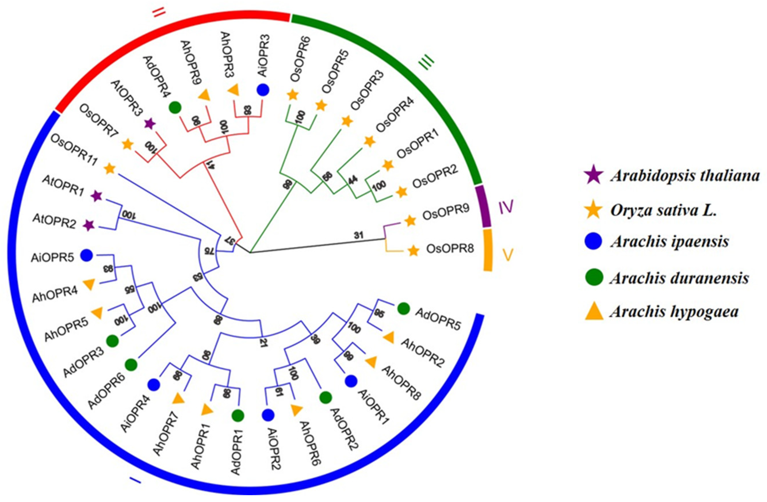
2.2. Phylogenetic Analysis and Classification of OPR Gene Family

To investigate the evolutionary relationships of OPRs, a maximum likelihood phylogenetic tree (with 1000 bootstraps) was constructed using Arabidopsis thaliana, Oryza sativa L., and peanut (Arachis ipaensis, Arachis duranensis, and Arachis hypogaea) species. All OPRs could be classified into five subfamilies: OPR I, II, III, IV, and V (Figure 1). Twenty OPRs from three different peanut species could be divided into two subfamilies, OPR I and OPR II (Figure 1). The classification of peanut OPRs is similar to that of dicot plant Arabidopsis. Among them, the OPR I subfamily contained the highest number of OPR proteins (16 out of 20; 80%), while OPR II contained four OPRs, including AiOPR3, AdOPR4, AhOPR3, and AhOPR9.

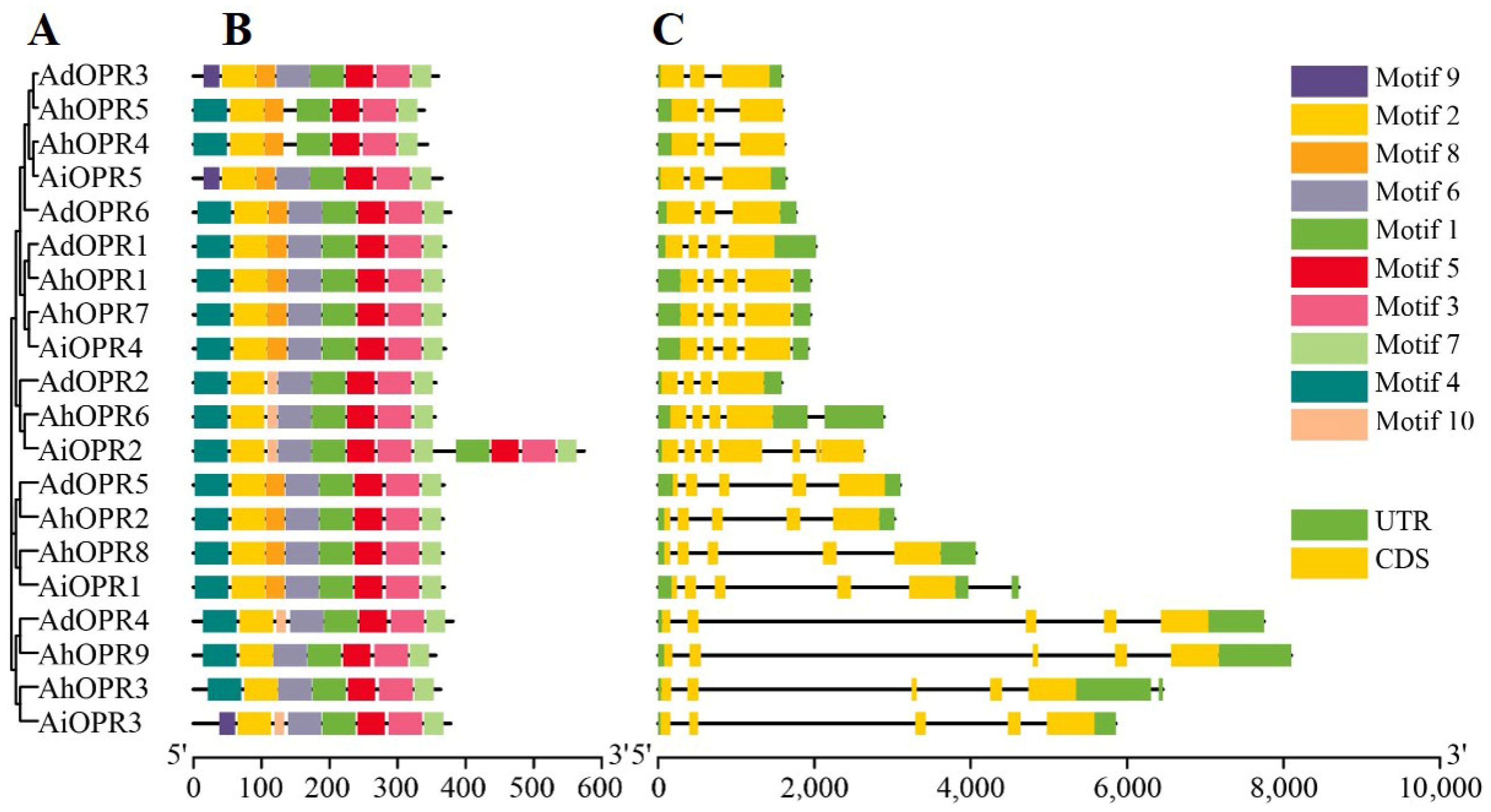
2.3. Conserved Motifs and Gene Structure Analysis of Peanut OPR Genes

A maximum-likelihood phylogenetic tree (with 1000 bootstraps) was constructed using the multiple sequence alignment (ClustalW) of 20 conserved OPR proteins from peanut (Figure 2A). The MEME tool was used to identify the conserved motifs in peanut OPRs. At most, ten conserved motifs, with lengths between 6 and 50 amino acids, were found, showing diversity in each peanut OPR (Figure 2B, Table S2). In the OPR I and OPR II subfamilies, motifs 1–3, motif 5, and motif 7 were present in all OPRs, and they were the most conserved domains (Figure 2B). In AiOPR3, AiOPR5, and AdOPR3, the position of motif 9 was replaced by motif 4. Four OPRs in OPR II lacked motif 8, which was totally associated with the β6 barrel of the protein secondary structures and further affected the 3D structure of AhOPR3 and AhOPR9 (Figure S1, Table S2). In the OPR I subfamily, AhOPR4 and AhOPR5 lacked motif 6, which reveals that they may have different functions (Figure 2B). Similar motifs in the same subfamily may have similar functions.
To further analyze the gene structure of peanut OPR genes, the gene structure features were exhibited by TBtoolsV1.098 (Figure 2C). As shown in Figure 2C, most OPRs of peanut contain 3–5 exons. AiOPR2 contained the highest number of exon 7. In addition, we found that the intron numbers and lengths were diverse in different subfamilies (Figure 2C). All the genes in OPR II contained 4 introns, and the second intron was significantly longer than others compared with genes in the OPR I subfamily (Figure 2C). In OPR I, most OPRs (approximately 70%) had 2–3 introns, and the others contained more than 4 introns (Figure 2C).

2.4. Chromosomal Locations and Synteny Analysis of Peanut OPRs

A total of 20 peanut OPR genes were unevenly distributed on 40 chromosomes (Figure 3). No genes were found on 29 chromosomes, including chromosomes A1–4, A7, B1–4, B8, 1–4, 7, 9–14, and 18. The maximum number of OPR genes appeared in chromosome A5 (4, 20%), and the second largest number of OPR genes were found on chromosome B5 (3, 15%) and 15 (3, 15%). There was one gene on chromosomes A6, A8, B6, B7, 8, and 17 (Figure 3). Two OPR genes were mapped on chromosomes 5 and 16, respectively (Figure 3). To further study the gene duplication events of peanut OPR genes, tandem and segmental duplicated gene pairs were analyzed (Figure 4 and Table S3). There were two groups of tandem duplicated genes, including AdOPR1 and AdOPR2 and AdOPR2 and AdOPR6, which were linked with red lines. In addition, 14 segmental duplication gene pairs are visualized in Figure 4. The number of segmental duplicated gene pairs was dramatically higher than the tandem duplicated gene pairs; this might be the main reason for the expansion of the OPR gene family.

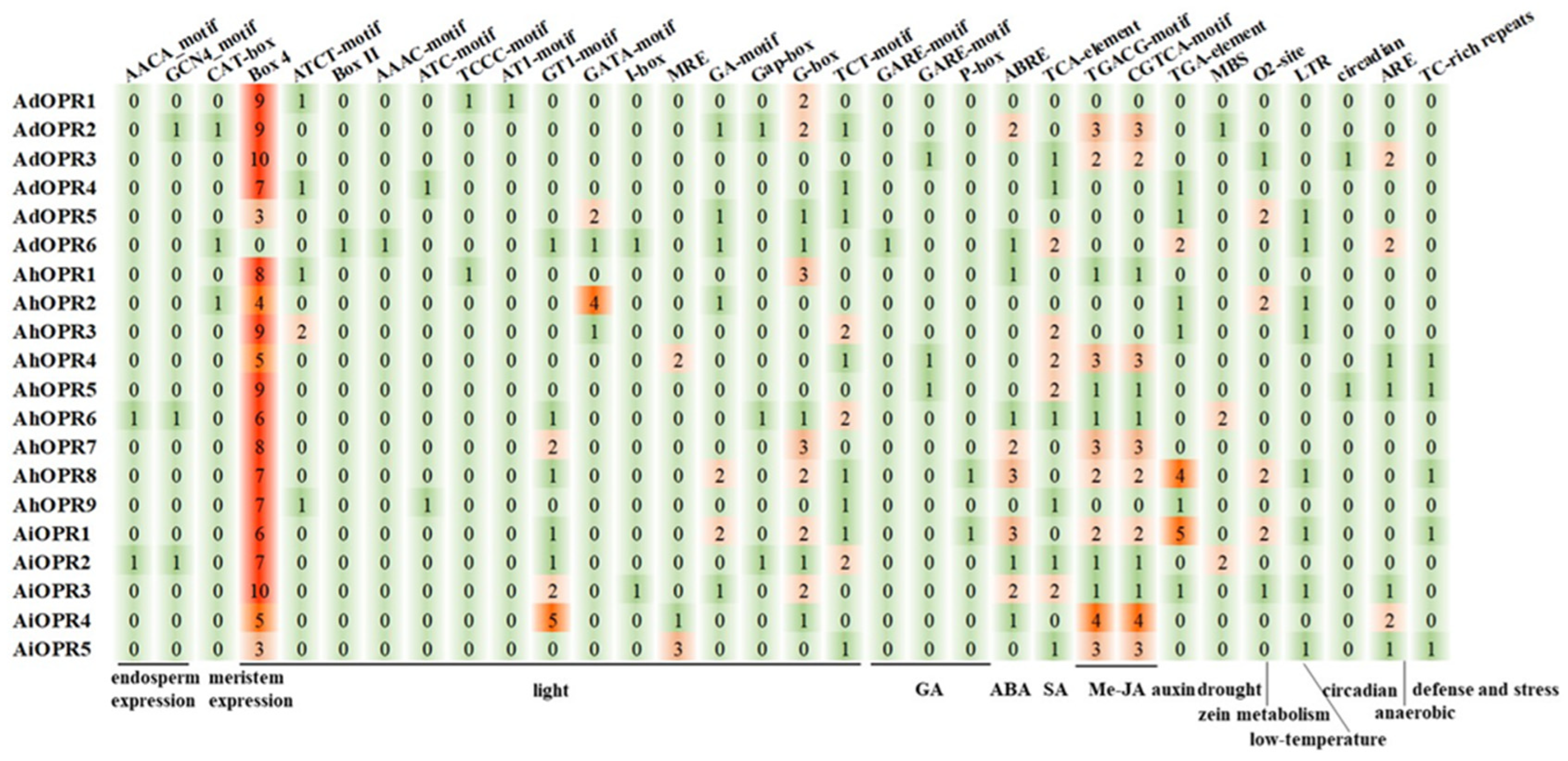
2.5. Prediction and Analysis of Cis-Acting Elements in Promoter Sequences of OPR Genes

To further explore the evolution and functional characterization of peanut OPR genes, the 1.5 kb areas upstream from the translational start site of each gene were analyzed. Various cis-acting elements were found in OPR promoters (Table S4). As shown in Figure 5, peanut OPR promoters contained elements associated with endosperm development, meristem development, light, GA, ABA, SA, MeJA, auxin, drought, zein metabolism, low-temperature, circadian, anaerobic, and defense stresses. The cis-acting elements (Box 4, ATCT-motif, Box II, AAAC-motif, ATC-motif, GT1-motif, GATA-motif, I-box, GA-motif, G-Box, and TCT-motif) related to light were all found in the OPR gene promoter. The majority of the OPRs included elements responsive to ABA (ABRE), SA (TCA-element), and MeJA (TGACG-motif and CGTCA-motif) (Figure 5). A small number of gene promoters were identified to be involved in responses to drought (MBS), GA (GARE-motif and P-box), auxin (TGA-element), low-temperature (LTR), anaerobic (ARE), and defense stresses (TC-rich repeats). These findings indicated that peanut OPR genes might participate in plant growth and development and the response to hormones and diverse stresses.

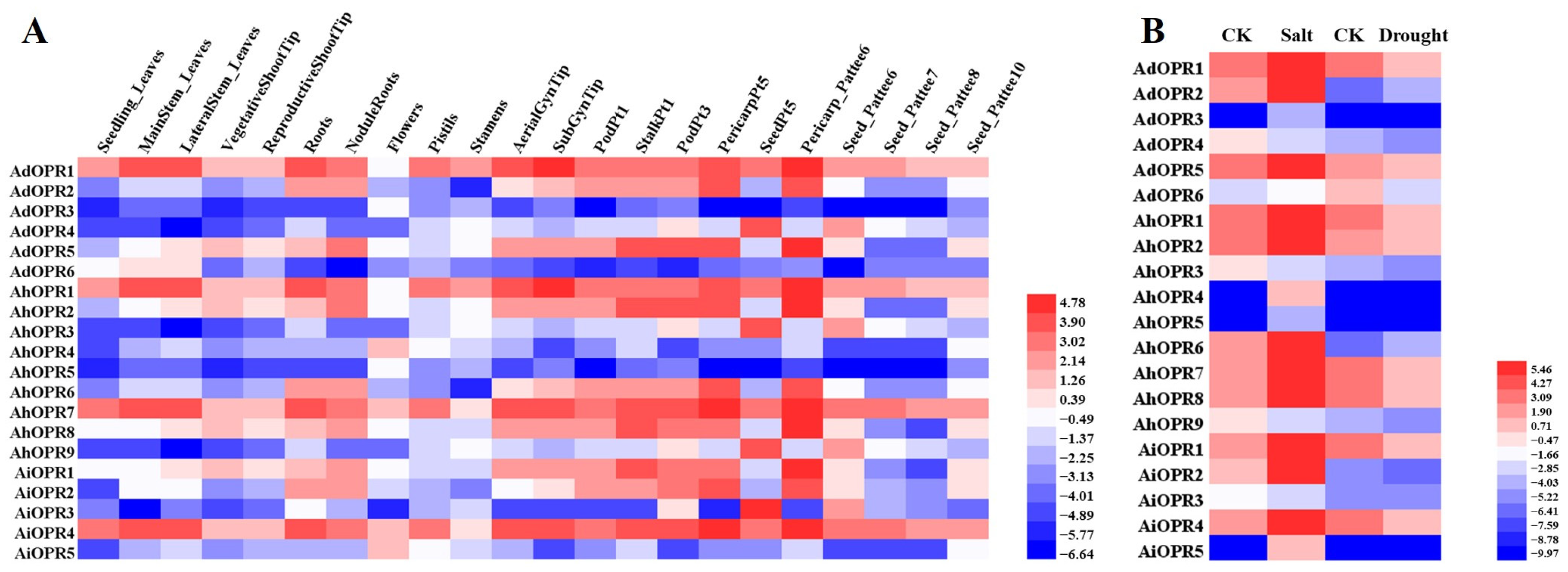
2.6. Expression Patterns of OPR Genes in Different Tissues and in Response to Various Stresses

To gain more insights into the temporal and spatial expression patterns of OPR genes, the RNA sequences of different treatments under abiotic stress (Table S5) and 22 tissues (Table S6) were analyzed. Most genes exhibited different expression levels across 22 tissues (Figure 6A). The results show that four genes had relatively high expression levels in all 22 tissues, while three genes had relatively low expression levels in all 22 tissues (Figure 6A). Some peanut OPR genes exhibited tissue-specific expression patterns, such as AhOPR4 and AiOPR5, which were prominently expressed in flowers. Additionally, AdOPR4, AhOPR3, AhOPR9, and AiOPR3 displayed specific expression in the seeds.
The expression levels of the genes under drought and salt stresses were investigated using our published transcriptome sequencing results. We found that the expression pattern of peanut OPRs was notably different under drought and salt stresses. In Figure 6B, the expression of most peanut OPRs (16 of 20) was upregulated by more than 2-fold under salt stress, and the majority of OPRs were increased by up to 10-fold. Unlike salt treatment, only three OPRs (AdOPR2, AhOPR6, and AiOPR3) were upregulated under drought stress, and the majority OPR genes were downregulated. It is worth noting that the expression of four OPR genes (AdOPR3, AhOPR4, AhOPR5, and AiOPR5) was unchanged in response to drought stress. Only two genes (AdOPR2 and AhOPR6) were upregulated both under salt and drought stresses, and the AhOPR6 gene was selected for further functional study.

2.7. Subcellular Location of Peanut OPR Proteins

AhOPR proteins were predicted to be localized in the chloroplast, plasma membrane, or mitochondria using the online web tool WoLF PSORT (Table S1). In order to confirm the subcellular localization of the peanut proteins, AhOPR3 and AhOPR6 proteins were selected to perform the experiment (Figure 7). The 35S::AhOPR3-GFP and 35S::AhOPR6-GFP fusion proteins were constructed, together with 35S::GFP (the empty vector), and they were transiently expressed in N. benthamiana. Confocal microscopy observations revealed that the green fluorescence of the empty vector was found in the cytoplasm and on the plasma membranes, while the 35S::AhOPR3-GFP fusion protein was distributed on the plasma membranes, and the 35S::AhOPR6-GFP fusion proteins was localized in the chloroplast (Figure 7).

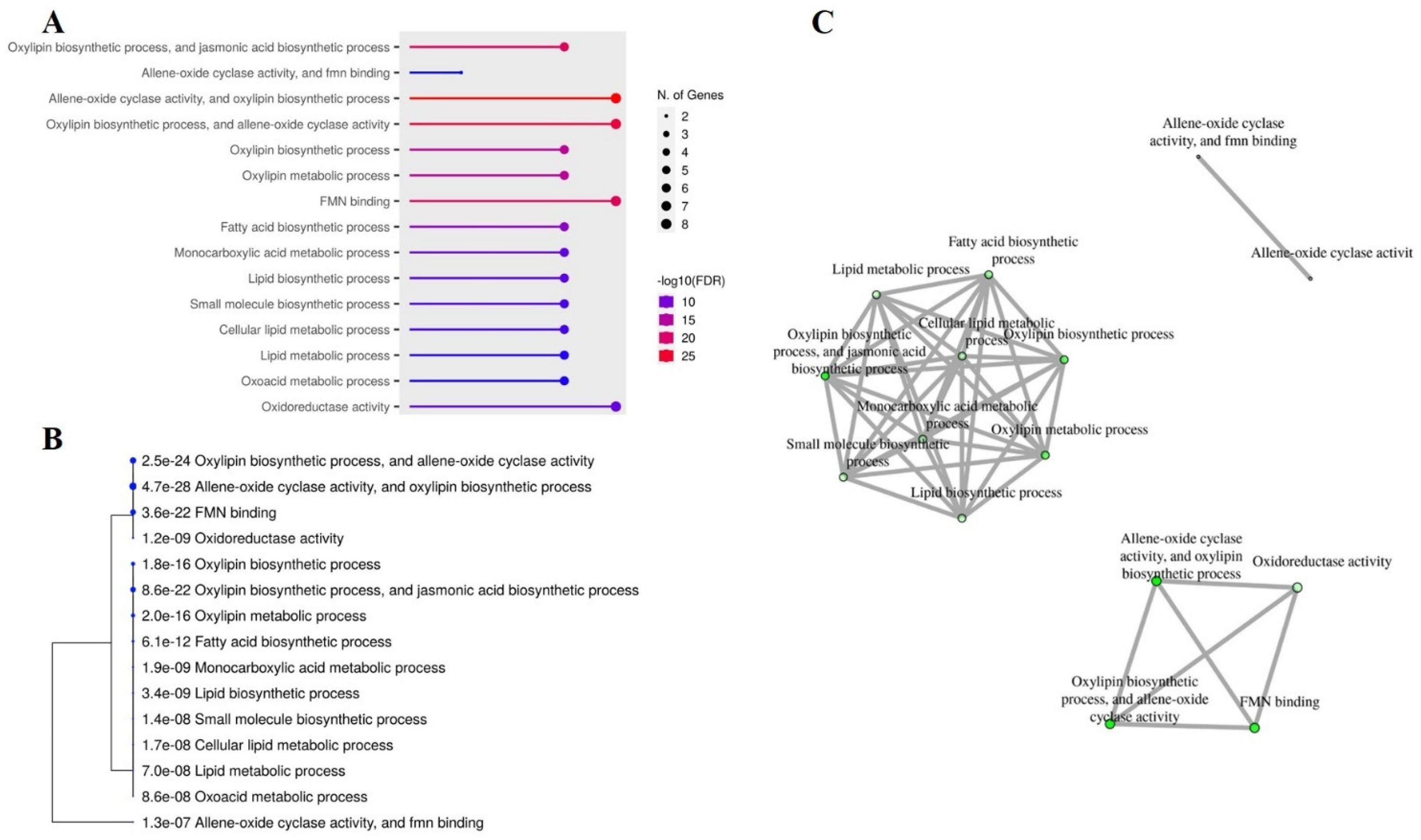
2.8. Gene Ontology (GO) and Interactive Networks of Peanut OPR Genes

GO analysis offers valuable information for investigating potential associations based on their enrichment in molecular function (MF), cellular components (CCs), and biological process (BP). This analytical tool is commonly used in genetics to validate the functions of candidate genes. As shown in Figure 8A, all peanut OPRs were significantly enriched to allene oxide cyclase activity (MF), FMN binding (MF), the oxylipin biosynthetic process (BP), the jasmonic acid (JA) biosynthetic process (BP), and the oxylipin metabolic process (BP). All OPR genes were found to be associated with allene oxide cyclase activity, the oxylipin biosynthetic process, and FMN binding (Figure 8B). A further annotation analysis of AhOPRs using data from ShingGO confirmed a tree of three clusters according to a correlation analysis (Figure 8C).

2.9. Overexpression of AhOPR6 Enhanced Salt Tolerance in Seedlings

To further explore the function of the OPR genes in salt response, overexpressing AhOPR6 transgenic plants were investigated. Overall, twenty T0 transgenic lines were obtained by PCR using Arabidopsis genomic DNA. After cultivation and screening, the homozygous T3 transgenic lines of AhOPR6 were used to analyze the functional roles in salt stress. After being treated with 300 mM of NaCl for one week, most leaves of the wild-type plants were wilted, while only a small number of OE1 and OE2 leaves became wilted (Figure 9A). Furthermore, the activities of SOD (Figure 9B), POD (Figure 9C), and CAT (Figure 9D) enzymes associated with ROS scavenging were increased after salt stress, and gene-overexpressed plants were obviously increased in SOD, POD, and CAT activity compared with wild-type Arabidopsis. Before the salt treatment, the wild-type and transgenic Arabidopsis lines had nearly equal MDA contents (Figure 9E). Transgenic Arabidopsis lines have significantly lower MDA contents after salt treatment (Figure 9E). These results reveal that the overexpression of the peanut AhOPR6 gene enhanced salt tolerance in Arabidopsis.

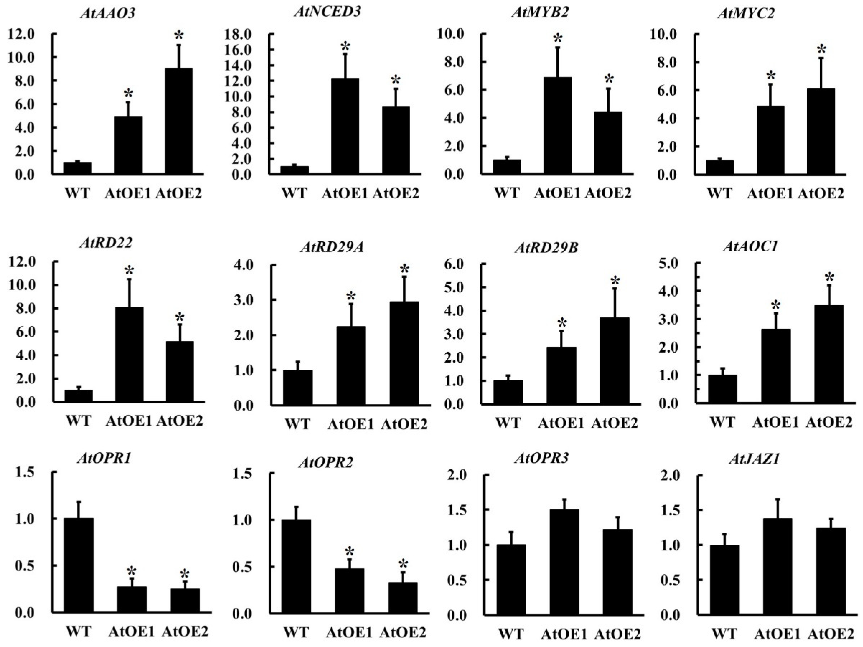
2.10. The Overexpression of AhOPR6 in Arabidopsis Affects the Expression of JA/ABA Pathway-Related Genes

To further validate the role of ABA- and JA-related genes in AhOPR6-mediated salinity stress enhancement, the transcript abundance of 12 genes was monitored (Figure 10, Table S7). Some genes, like AtAAO, AtNCED, AtRD, AtMYB, and AtMYC, were key factors in the ABA-dependent stress-responsive signaling pathway; AtAOC, AtOPR, and AtJAZ were all key components of the JA biosynthesis pathway in Arabidopsis. The transcript abundance of AtAAO3 and AtNCED3 was increased in the AtOE-AhOPR6 lines; AtRD22, AtRD29A, and AtRD29B were also upregulated in the AhOPR6-overexpressing AtOE lines (Figure 10). The expression levels of two upstream transcription factors, AtMYB2 and AtMYC2, were significantly upregulated as expected. In the JA synthesis pathway, the expression levels of AtAOC1, AtOPR1, AtOPR2, and AtJAZ1 were significantly upregulated in transgenetic Arabidopsis lines, while the expression of AtOPR3 was not influenced by the overexpression of peanut AhOPR6.

3. Discussion

Peanut is an important economic and oil crop worldwide. However, annual worldwide losses caused by various biotic and abiotic stresses are destructive. The objective of this study was to explore all the OPRs in peanut and the potential role of OPRs in peanuts’ resistance. OPRs are key enzymes that participate in the last step of JA synthesis. JA and its derivates regulate the development and response to biotic and abiotic stresses in plants [7, 8, 30]. So far, the identification and functional analysis of the OPR gene family have been characterized in many plants, such as Arabidopsis (3), rice (12), tomato (4), maize (8), and wheat (48). However, the OPR gene family in cultivated peanut and its functional characterization have not been investigated. In this study, we constructed a rooted maximum likelihood (ML) phylogenetic tree with 20 peanut OPR genes in accordance with the phylogenetic relationships of two species, Arabidopsis and rice. Accordingly, the OPR gene family in peanut can be divided into two subfamilies (I and II). The number of AhOPR genes (9) is substantially higher compared to most diploid plants but lower than that of hexaploid wheat (48), which might be related to the fact that the allotetraploid genome of cultivated peanut has undergone whole-genome duplication and other gene duplication events [31,32]. There were six AdOPR genes in diploid peanut A. duranensis and five AiOPR genes in diploid peanut A. ipaensis, while the number of allotetraploid AhOPR genes was less than the sum of two diploid OPR gene numbers. This phenomenon could be explained by reports that some homologous genes might have been lost or that some functionally redundant genes disappeared during the polyploidization of the genome [33,34].
Structure diversity within gene families was a mechanism for the evolution of many gene families. Intron gain and loss play a significant role in structural diversity and complexity. We found that most OPRs in peanut contain 3–5 extrons; all the genes in OPRII contain 4 introns, while most OPRs (approximately 70%) in OPR I have 2–3 introns, and the others contain more than 4 introns. In wheat, the OPR genes in subfamilies I, II, IV, and V contain 3–5 introns; 29 of 32 OPR genes in subfamily III contain 1–2 introns. In addition, subfamily III was the youngest subfamily in wheat which possessed the fewest introns [15]. The result was consistent with a previous study showing that the intron loss events occurred during the structural evolution of the OPR gene family [14]. Analogously, four of six maize OPR genes in subgroup I (ZmOPR1-6) contain 0–1 introns; the other two genes in subgroup II (ZmOPR7 and ZmOPR8) have 3 or 4 introns [19]. The above results indicate that intron loss events could be found in different plants, whether dicots or monocots. All peanut OPRs in subfamilies I and II had motif 1, motif 2, motif 3, motif 5, and motif 7. These five motifs were related to the most conserved domains of OPRs and were associated with the alpha or beta barrel of protein secondary structures. Interestingly, the position of motif 9 of three genes (AiOPR3, AiOPR5, and AdOPR3) was replaced with motif 4. Four OPRs in OPRII lacked motif 8, which would impact the β6 barrel of the protein secondary structures. Structural integrity is essential to ensuring gene function, and slight alterations in the DNA structure or protein motifs usually affect gene functions [35,36].
Cis-acting elements are found in the promoter regions upstream of protein-coding genes, and they are transcription factor (TF) binding sites. TFs and cis-acting elements interact to control the precise initiation of gene transcription, resulting in tissue-specific and signal-specific gene expression [37]. Many cis-acting elements related to growth and development (such as endosperm and meristem development), hormones (including MeJA, ABA, SA, GA, and auxin), and stresses (such as drought, zein metabolism, low-temperature, circadian, anaerobic, and defense responses) have been identified in peanut OPR promoters. Among hormone-responsive elements, MeJA responsiveness cis-acting elements (TGACG-motif and CGTCA-motif) were the most abundant, followed by ABA-responsive element ABRE, SA-responsive TCA element, and the auxin-responsive TGA element. Moreover, MeJA responsiveness has been demonstrated to be linked to plant defense mechanisms [38]. Therefore, we can infer the function of peanut OPR genes through the cis-acting elements in the gene promoter, which offered an efficient method for screening potential candidate genes.
The stationary nature of plants makes them susceptible to a variety of stresses, and peanut is no exception. Various abiotic stresses and biotic stresses, like stress caused by salinity, drought, cold, heat, and herbivores, adversely impact staple food crops’ survival, biomass production, and yield by up to 70% [39,40,41]. According to recent research, drought affects approximately 45% of all agricultural land in the world, while salt impacts about 19.5% of it [42]. In accordance with the FAO’s estimates, salinity affects over 6% of the world’s land [41]. According to recent studies, OPRs were positively regulated in Arabidopsis, maize, and wheat when exposed to salt stress [22,43]. In maize, the overexpression of ZmOPR1 affected RD22, RD29B, and ABF2, which are genes associated with osmotic and NaCl stresses [43]. RD22 and RD29B are ABA-responsive genes; ABF2 belongs to the ABF subfamily of bZIP proteins that interact with ABA-responsive elements. They are involved in ABA/stress responses [44,45,46]. Likewise, wheat OPR1 could be induced by ABA and salinity, and the inhibition of ABA synthesis largely counteracts salinity-induced TaOPR1 accumulation. As a result, there is a convincing argument that TaOPR1 expression is induced by ABA synthesis. AtRD22, AtRD29A, and AtRD29B are all upregulated via an ABA-induced signaling cascade [47]. As a result of TaOPR1 expression, AtNCED3 and AtAAO3, which encode enzymes essential for ABA synthesis, were also significantly increased in transcript abundance. AtMYB2, an R2R3-MYB transcription factor gene, was induced by ABA, dehydration, and salt stresses [48,49], indicating that AtMYB2 responds to environmental stresses through ABA-dependent and ABA-independent pathways [50,51]. Aside from AtMYB2, AtMYC2 also activates genes in the ABA signaling pathway [52,53]. It has been documented that AtMYB2 and AtMYC2 can bind to MYB and MYC recognition sites in the promoter sequence of the rd22BP1 gene, respectively, to regulate the expression of ABA-inducible genes (like rd22), thus enhancing transgenic plant stress resistance [53,54]. The expression of AhOPR6 markedly increased the transcript abundance of AtAAO3, AtNCED3, AtRD22, AtRD29A, AtRD29B, AtMYB2, and AtMYC2 (Figure 10). The above investigations provide firm evidence that AhOPR6 plays a positive role in salt stress through ABA-dependent signaling pathways.
There are several factors that lead to salt stress on plants, including ion toxicity [55], osmotic stress [56], and reactive oxygen species (ROS) [57]. Salt stress leads to the accumulation of ROS such as superoxide, H2O2, and hydroxyl radicals; oxidative damage and ROS accumulation are both increased with prolonged salt stress exposure [58]. Similarly, other studies supported our results, indicating that significantly higher antioxidant enzyme activity reduced ROS production and led to improved osmotic adaptation to salt stress [59]. POD plays an important role in ROS signaling and redox reactions, and a high level of POD can increase plants’ salt tolerance [60]. In addition, the activities of POD and CAT were increased to reduce ROS accumulation under salt stress [61], and this result is consistent with our research. MDA, a product of membrane peroxidation, is often used as a prime indicator of oxidative stress in salt-stressed tissues [62,63]. Salt-sensitive plants exhibit a greater amount of MDA than salt-tolerant ones [64]. In our study, the decrease in MDA contents in AtOE lines implied that the overexpression of AhOPR6 increased the plants’ salt tolerance mainly by reducing ROS damage.
JA and its intermediates are critical elements of perception and signaling, and JA-mediated gene expression and signaling pathways can be triggered by several corroborative receptors, activators, transcription factors, repressors, and co-repressors [8,10]. In the first half of JA biosynthesis, LOX, AOS, and AOC sequentially catalyze to generate the intermediate product cis-(+)-OPDA [65]. The expression levels of AtOPR3 and AtJAZ1 (receptor genes of JA) were unchanged (Figure 10); these results indicate that JA synthesis in OE lines does not depend on AhOPR6. Through AtMYC2, the JA-activated MKK3-MPK6 signal negatively regulates the JA pathway and affects JA-dependent gene expression [66]. It is worth noting that the bHLH transcription factor AtMYC2, activated by JA, significantly interacts with the signaling pathways of ABA, JA, GA, and ethylene [67]. We found that the expression level of AtMYC2 was increased, but AtJAZ1 was unchanged in AtOE-AhOPR6 lines. These results could be attributed to the promotion of ABA but not the JA signaling pathway, and it is highly possible that AhOPR6 was not regulating JA synthesis (Figure 11). The research conducted by Dong et al. also verified our hypothesis [22] that the OPRI (TaOPR1) gene exerts no regulatory activity on JA synthesis and JA signaling pathways.

4. Materials and Methods

4.1. Identification of Peanut OPR Gene Family Members

The protein sequence, gene annotation information file (gff3), and coding sequence (CDS) of the newly published peanut genome were obtained from the PeanutBase database (https://www.peanutbase.org/, accessed on 21 January 2022). The Hidden Markov model of the OPR gene family (PF00724) was downloaded from the Pfam database (https://pfam.xfam.org/, accessed on 5 May 2025) [68], and then this profile was used to search the peanut protein database with E-values < 1.2 × 10⁻²⁸ by HMMER 3.0. The protein sequence of OPRs was extracted via Perl language and submitted to the web tools SMART (http://smart.embl.de/smart/batch.pl, accessed on 5 May 2025), Pfam (https://pfam.xfam.org/search, accessed on 5 May 2025), and NCBI-CDD (https://www.ncbi.nlm.nih.gov/cdd/, accessed on 5 May 2025) to verify the structural integrity of OPR. Finally, reliable OPR candidate genes were determined. The molecular weight (MW), amino acid sequence length (aa), and isoelectric point (pI) of the OPR were predicted using the online server ExPASY (https://web.expasy.org/cgi-bin/protparam/protparam, accessed on 5 May 2025) [69]. Subcellular localization of OPRs was predicted using the online web tool WoLF PSORT (https://wolfpsort.hgc.jp/, accessed on 5 May 2025).

4.2. Phylogenetic Analysis

To further understand the evolutionary relationship of the OPR gene family, 3 Arabidopsis OPRs and 11 rice OPRs amino acid sequences were downloaded from the NCBI database, and 20 peanut OPRs were downloaded from PeanutBase (https://www.peanutbase.org/, accessed on 5 May 2025). The multiple sequence alignment of amino acid sequences was conducted via the ClustalW function of MEGA X software, and the phylogenetic relationships of the three species were constructed using the neighbor-joining method; the bootstrap value was set to 1000. The classifications of OPRs in peanut were based on the topological structure of the phylogenetic tree and sequence similarity. The established phylogenetic trees were beautified using Evolview (http://www.evolgenius.info/evolview/, accessed on 5 May 2025) [70].

4.3. Analysis of Gene Structure and Protein Conserved Domains of OPRs

Based on peanut genome annotation information, location information of the exon, intron, and UTR of OPR genes on chromosomes was obtained, which was used to draw gene structure. Conserved motifs with lengths of 6–100 aa in 20 OPR proteins were analyzed using the MEME https://meme-suite.org/meme/tools/meme, accessed on 5 May 2025) program [71]. The maximum number of motifs was set at 10. The gene structure and conserved motifs of OPRs were visualized using TBtoolsV1.098 software [72].

4.4. Cis-Acting Regulatory Elements and Gene Ontology (GO) Annotation

The 2 kb promoter sequence of OPRs was extracted from peanut genomic DNA sequences of the PeanutBase database and uploaded to PlantCARE (http://bioinformatics.psb.ugent.be/webtools/plantcare/html/, accessed on 5 May 2025) [73] to predict cis-acting regulatory elements in the promoter region. The GO analysis was conducted in the ShinyGO 0.80 database (http://bioinformatics.sdstate.edu/go/, accessed on 5 May 2025) by inputting all OPR family genes to ascertain the molecular functions, biological processes, and cell components related to peanut OPRs [74].

4.5. Chromosome Localization and Synteny Analysis

To determine the distribution of OPRs on chromosomes, we obtained the starting and ending positions of OPR genes from peanut genome annotation files (gff3) and submitted their location information to the online tool MapGene2Chrom web v2 (http://mg2c.iask.in/mg2c_v2.0/, accessed on 5 May 2025) to map their chromosome positions. The segmental and tandem repeats of OPR family members in peanut were analyzed via MCScanX (http://chibba.pgml.uga.edu/mcscan2/, accessed on 5 May 2025) software [75]. Two adjacent genes located on the same chromosome with a relative position less than 100 kb are defined as tandem repeat genes [76]. The chromosome distribution of 20 OPR genes and the collinearity of related genes were plotted by using Circos (version 0.69) software [77].

4.6. RNA-Seq Data Analysis of OPRs

The expression patterns of OPR genes in different tissues and different stress treatments were investigated in peanut. In this study, RNA-seq data of 22 tissues were derived from the PeanutBase database [78], and the FPKM values of drought [79] and salt [80] stresses were obtained from our previous work. The PFKM values of OPR family genes were standardized by log2 PFKM, and the heatmap was drawn using Heml_1.0 software.

4.7. Subcellular Localization of AhOPR6 Protein

The coding sequences of AhOPR3 and AhOPR6 were amplified from peanut cultivar “huayu71” and used to construct the 35S::AhOPR3-GFP and 35S::AhOPR6-GFP fusion vectors. The empty vector and the two fusion vectors were transiently expressed in Nicotiana benthamiana leaves. After three days, a confocal microscope (TCS-SP8, Leica, Wetzlar, Germany) was used to observe the fluorescence signals of the fusion proteins in the cells.

4.8. Construction of AhOPR6 Vectors and Generation of Transgenic Arabidopsis Plants

Full-length coding sequence of AhOPR6 was amplified from the cDNA of peanut cultivar “huayu71” by PCR. To construct the overexpression vector, the AhOPR6 gene was placed between the 35S promoter and the Nos terminator in the pCambia2300EC vector. The recombinant plasmid was transformed into Agrobacterium strain GV3101, which was then applied in floral dipping to transform wild-type Arabidopsis. Positive seedlings were derived from the selective media after being grown on MS media with 50 mg/l kanamycin, and T3 homozygous seedlings were employed for subsequent analysis.

4.9. The Treatment and Determination of Salt-Related Physiological Parameters in Transgenic Arabidopsis

Both WT and transgenic Arabidopsis seedlings were grown on 1/2 MS media for 7 days and then transferred to soil for 2 weeks. For salt stress, Arabidopsis seedlings were treated with 300 mM NaCl for one week. The malondialdehyde (MDA) content, superoxide dismutase (SOD), peroxidase (POD), and catalase (CAT) enzyme activities were detected using assay kits (Comin, Suzhou, China) for both overexpressing lines and WT Arabidopsis. The four physiological parameters of the Arabidopsis plants were measured in triplicate.

4.10. RNA Isolation and Real-Time Quantitative PCR (qRT-PCR) Validation of OPR

Trizol reagent (TaKaRa, Dalian, China) was used to extract total RNA from leaves of Arabidopsis under salt stress treatments. The integrity and quality of RNA were detected using 1% agarose gel electrophoresis, and the concentrations of RNA, namely OD260/280 and OD260/230, were detected using the Nanodrop one micro-ultraviolet spectrophotometer. Total RNA was reverse-transcribed into cDNA using the Transcriptor First Strand cDNA Synthesis Kit (TaKaRa, Dalian) and stored at −20 °C. qRT-PCR was performed on ABI7500 (Applied Biosystems, Foster City, CA, USA). The reaction system was as follows: 10 μL of 2×UltraSYBR Mixture, 0.4 μL of forward primer (5.0 μmol/L), 0.4 μL of reverse primer (5.0 μmol/L), 1 μL of cDNA (200 ng), and 7.4 μL of RNase-free water. The amplification procedure was as follows: 95 °C for 5 min, 40 cycles of 95 °C for 10 s, and 60 °C for 1 min; 95 °C for 15 s, 60 °C for 1 min, 95 °C for 15 s, and 60 °C for 15 s. Three biological repeats and three technical repeats were set up in each treatment. The relative expression of genes was calculated using 2−ΔΔCt [81]. The fluorescent quantitative primers were designed using Primer-BLAST (https://www.ncbi.nlm.nih.gov/tools/primer-blast/index.cgi, accessed on 5 May 2025).

5. Conclusions

In this study, a total of 20 OPR genes were identified in the genome of a tetraploid cultivar and two diploid peanut species. They were divided into two subfamilies with Arabidopsis and rice homologous genes. The investigation of the evolutionary relationship, gene structure, protein conserved motifs, chromosome locations, gene duplication, tissue expression patterns, and expression patterns under salt and drought stresses indicated their diversity in different OPR genes. The subcellular location of peanut AhOPR3 and AhOPR6 revealed that they were distributed on the plasma membranes and localized in the chloroplast, respectively. Furthermore, the overexpression of AhOPR6 in Arabidopsis increased tolerance to salt stress. Notably, the detection of JA and ABA signaling pathway-related genes by qRT-PCR indicated that AhOPR6 might enhance salt stress tolerance through an ABA-dependent pathway. Overall, we constructed a hypothetical model of AhOPR6’s action in improving salt tolerance and provided candidate OPR genes for further functional studies in growth and development, as well as peanut stress resistance breeding.

Supplementary Materials

The following supporting information can be downloaded at https://www.mdpi.com/article/10.3390/plants14101408/s1. Table S1: Characteristics of putative peanut OPR genes; Table S2: Detailed information about conserved motifs of AhOPRs; Table S3: Tandem and segmentally duplicated OPR gene pairs in peanut; Table S4: Analysis of cis-acting regulatory elements of peanut OPR promotors; Table S5: FPKM values of peanut OPR genes under two abiotic stresses (drought and salt) in this study; Table S6: FPKM values of peanut OPR genes in 22 tissues in this study; Table S7: Primer sequences used in this study; Figure S1: Three-dimensional structure prediction of peanut OPRs.

Author Contributions

Y.M. conceived and designed the study. Y.M. performed the experiments and wrote the manuscript. Q.S., H.M., J.W. and Q.W. (Qi Wang) analyzed the bioinformatics data. Q.W. (Qianqian Wang), C.Y. (Caixia Yan), C.Y. (Cuiling Yuan) and X.Z. assisted in the stress treatment and data analysis. S.S. and C.L. revised the paper. All authors have read and agreed to the published version of the manuscript.

Funding

This research was funded by the Qingdao Natural Science Foundation (grant number: 23-2-1-44-zyyd-jch), the National Key Research, Development Program of China (grant number: 2022YFD1200403), the Agro-industry Technology Research System of Shandong Province (grant number: SDAIT-04-01), the Key research and development project of Shandong province (grant number: 2024LZGC031), and the Innovation Project of SAAS (grant number: CXGC2024D19), Tianchi talent program.

Data Availability Statement

The original contributions presented in this study are included in the article/supplementary material. Further inquiries can be directed to the corresponding author(s).

Conflicts of Interest

The authors declare no conflicts of interest.

References

  1. Zhang, H.; Tang, Y.; Yue, Y.; Chen, Y. Advances in the evolution research and genetic breeding of peanut. Gene 2024, 916, 148425. [Google Scholar] [CrossRef]
  2. Dwivedi, S.L.; Crouch, J.H.; Nigam, S.N.; Ferguson, M.E.; Paterson, A.H. Molecular breeding of groundnut for enhanced productivity and food security in the semi- arid tropics: Opportunities and challenges. Adv. Agron. 2003, 80, 153–221. [Google Scholar]
  3. Sarkar, T.; Thankappan, R.; Kumar, A.; Mishra, G.P.; Dobaria, J.R.; Zhang, J.S. Heterologous Expression of the AtDREB1A Gene in Transgenic Peanut-Conferred Tolerance to Drought and Salinity Stresses. PLoS ONE 2014, 9, e110507. [Google Scholar] [CrossRef] [PubMed]
  4. Abrol, I.P.; Yadav, J.S.P.; Massoud, F.I. Salt-Affected Soils and Their Management; Food and Agriculture Organization of the United Nations: Rome, Italy, 1988. [Google Scholar]
  5. Yang, S.; Hao, X.; Xu, Y.; Yang, J.; Su, D. Meta-Analysis of the Effect of Saline-Alkali Land Improvement and Utilization on Soil Organic Carbon. Life 2022, 12, 1870. [Google Scholar] [CrossRef] [PubMed]
  6. Liu, Y.; Zhu, J.; Sun, S.; Cui, F.; Han, Y.; Peng, Z.; Zhang, X.; Wan, S.; Li, G. Defining the function of SUMO system in pod development and abiotic stresses in Peanut. BMC Plant Biol. 2019, 19, 593. [Google Scholar] [CrossRef] [PubMed]
  7. Turner, J.G.; Ellis, C.; Devoto, A. The jasmonate signal pathway. Plant Cell 2002, 14, S153–S164. [Google Scholar] [CrossRef]
  8. Wasternack, C.; Hause, B. Jasmonates: Biosynthesis, perception, signal transduction and action in plant stress response, growth and development. An update to the 2007 review in Annals of Botany. Ann. Bot. 2013, 111, 1021–1058. [Google Scholar] [CrossRef]
  9. Wasternack, C. Jasmonates: An update on biosynthesis, signal transduction and action in plant stress response, growth and development. Ann. Bot. 2007, 100, 681–697. [Google Scholar] [CrossRef]
  10. Wasternack, C.; Strnad, M. Jasmonate signaling in plant stress responses and development—Active and inactive compounds. New Biotechnol. 2016, 33, 604–613. [Google Scholar] [CrossRef]
  11. Huang, H.; Liu, B.; Liu, L.; Song, S. Jasmonate action in plant growth and development. J. Exp. Bot. 2017, 68, 1349–1359. [Google Scholar] [CrossRef]
  12. Li, W.; Liu, B.; Yu, L.; Feng, D.; Wang, H.; Wang, J. Phylogenetic analysis, structural evolution and functional divergence of the 12-oxo-phytodienoate acid reductase gene family in plants. Bmc Evol. Biol. 2009, 9, 90. [Google Scholar] [CrossRef]
  13. Breithaupt, C.; Kurzbauer, R.; Schaller, F.; Stintzi, A.; Schaller, A.; Huber, R.; Macheroux, P.; Clausen, T. Structural Basis of Substrate Specificity of Plant 12-Oxophytodienoate Reductases. J. Mol. Biol. 2009, 392, 1266–1277. [Google Scholar] [CrossRef]
  14. Li, W.; Zhou, F.; Liu, B.; Feng, D.; He, Y.; Qi, K.; Wang, H.; Wang, J. Comparative characterization, expression pattern and function analysis of the 12-oxo-phytodienoic acid reductase gene family in rice. Plant Cell Rep. 2011, 30, 981–995. [Google Scholar] [CrossRef] [PubMed]
  15. Mou, Y.; Liu, Y.; Tian, S.; Guo, Q.; Wen, S. Genome-Wide Identification and Characterization of the OPR Gene Family in Wheat (Triticum aestivum L.). Int. J. Mol. Sci. 2019, 20, 1914. [Google Scholar] [CrossRef] [PubMed]
  16. Schaller, F.; Weiler, E.W. Molecular cloning and characterization of 12-oxophytodienoate reductase, an enzyme of the octadecanoid signaling pathway from Arabidopsis thaliana—Structural and functional relationship to yeast old yellow enzyme. J. Biol. Chem. 1997, 272, 28066–28072. [Google Scholar] [CrossRef]
  17. Schaller, F.; Biesgen, C.; Mussig, C.; Altmann, T.; Weiler, E.W. 12-oxophytodienoate reductase 3 (OPR3) is the isoenzyme involved in jasmonate biosynthesis. Planta 2000, 210, 979–984. [Google Scholar] [CrossRef]
  18. Biesgen, C.; Weiler, E.W. Structure and regulation of OPR1 and OPR2, two closely related genes encoding 12-oxophytodienoic acid-10,11-reductases from Arabidopsis thaliana. Planta 1999, 208, 155–165. [Google Scholar] [CrossRef] [PubMed]
  19. Zhang, J.; Simmons, C.; Yalpani, N.; Crane, V.; Wilkinson, H.; Kolomiets, M. Genomic Analysis of the 12-oxo-phytodienoic Acid Reductase Gene Family of Zea mays. Plant Mol. Biol. 2005, 59, 323–343. [Google Scholar] [CrossRef]
  20. Scalschi, L.; Sanmartin, M.; Camanes, G.; Troncho, P.; Sanchez-Serrano, J.J.; Garcia-Agustin, P.; Vicedo, B. Silencing of OPR3 in tomato reveals the role of OPDA in callose deposition during the activation of defense responses against Botrytis cinerea. Plant J. 2015, 81, 304–315. [Google Scholar] [CrossRef]
  21. Strassner, J.; Fürholz, A.; Macheroux, P.; Amrhein, N.; Schaller, A. A homolog of old yellow enzyme in tomato. Spectral properties and substrate specificity of the recombinant protein. J. Biol. Chem. 1999, 274, 35067–35073. [Google Scholar] [CrossRef]
  22. Dong, W.; Wang, M.; Xu, F.; Quan, T.; Peng, K.; Xiao, L.; Xia, G. Wheat Oxophytodienoate Reductase Gene TaOPR1 Confers Salinity Tolerance via Enhancement of Abscisic Acid Signaling and Reactive Oxygen Species Scavenging. Plant Physiol. 2013, 161, 1217–1228. [Google Scholar] [CrossRef]
  23. Yan, Y.; Christensen, S.; Isakeit, T.; Engelberth, J.; Meeley, R.; Hayward, A.; Emery, R.J.N.; Kolomiets, M.V. Disruption of OPR7 and OPR8 Reveals the Versatile Functions of Jasmonic Acid in Maize Development and Defense. Plant Cell 2012, 24, 1420–1436. [Google Scholar] [CrossRef] [PubMed]
  24. Guo, H.-M.; Li, H.-C.; Zhou, S.-R.; Xue, H.-W.; Miao, X.-X. Cis-12-Oxo-Phytodienoic Acid Stimulates Rice Defense Response to a Piercing-Sucking Insect. Mol. Plant 2014, 7, 1683–1692. [Google Scholar] [CrossRef] [PubMed]
  25. Bosch, M.; Wright, L.P.; Gershenzon, J.; Wasternack, C.; Hause, B.; Schaller, A.; Stintzi, A. Jasmonic Acid and Its Precursor 12-Oxophytodienoic Acid Control Different Aspects of Constitutive and Induced Herbivore Defenses in Tomato. Plant Physiol. 2014, 166, 396–410. [Google Scholar] [CrossRef] [PubMed]
  26. Stintzi, A.; Browse, J. The Arabidopsis male-sterile mutant, OPR3, lacks the 12-oxophytodienoic acid reductase required for jasmonate synthesis. Proc. Natl. Acad. Sci. USA 2000, 97, 10625–10630. [Google Scholar] [CrossRef]
  27. Li, S.; Ma, J.; Liu, P. OPR3 is expressed in phloem cells and is vital for lateral root development in Arabidopsis. Can. J. Plant Sci. 2013, 93, 165–170. [Google Scholar] [CrossRef]
  28. Wang, Y.; Yuan, G.; Yuan, S.; Duan, W.; Wang, P.; Bai, J.; Zhang, F.; Gao, S.; Zhang, L.; Zhao, C. TaOPR2 encodes a 12-oxo-phytodienoic acid reductase involved in the biosynthesis of jasmonic acid in wheat (Triticum aestivum L.). Biochem. Biophys. Res. Commun. 2016, 470, 233–238. [Google Scholar] [CrossRef]
  29. Ren, W.; Chen, L.; Xie, Z.M.; Peng, X. Combined transcriptome and metabolome analysis revealed pathways involved in improved salt tolerance of Gossypium hirsutum L. seedlings in response to exogenous melatonin application. BMC Plant Biol. 2022, 22, 552. [Google Scholar] [CrossRef]
  30. Tani, T.; Sobajima, H.; Okada, K.; Chujo, T.; Arimura, S.I.; Tsutsumi, N.; Nishimura, M.; Seto, H.; Nojiri, H.; Yamane, H. Identification of the OsOPR7 gene encoding 12-oxophytodienoate reductase involved in the biosynthesis of jasmonic acid in rice. Planta 2008, 227, 517–526. [Google Scholar] [CrossRef]
  31. Bertioli, D.J.; Cannon, S.B.; Froenicke, L.; Huang, G.; Farmer, A.D.; Cannon, E.K.; Liu, X.; Gao, D.; Clevenger, J.; Dash, S.; et al. The genome sequences of Arachis duranensis and Arachis ipaensis, the diploid ancestors of cultivated peanut. Nat. Genet. 2016, 48, 438–446. [Google Scholar] [CrossRef]
  32. Bertioli, D.J.; Seijo, G.; Freitas, F.O.; Valls, J.; Leal-Bertioli, S.; Moretzsohn, M.C. An overview of peanut and its wild relatives. Plant Genet. Resour. 2011, 9, 134–149. [Google Scholar] [CrossRef]
  33. Lynch, M.; Force, A. The probability of duplicate gene preservation by subfunctionalization. Genetics 2000, 154, 459. [Google Scholar] [CrossRef] [PubMed]
  34. Ma, X.; Ai, X.; Li, C.; Wang, S.; Zhang, N.; Ren, J.; Wang, J.; Zhong, C.; Zhao, X.; Zhang, H.; et al. A Genome-Wide Analysis of the Jasmonic Acid Biosynthesis Gene Families in Peanut Reveals Their Crucial Roles in Growth and Abiotic Stresses. Int. J. Mol. Sci. 2024, 25, 7054. [Google Scholar] [CrossRef]
  35. Yan, L.; Zhang, J.; Chen, H.; Luo, H. Genome-wide analysis of ATP-binding cassette transporter provides insight to genes related to bioactive metabolite transportation in Salvia miltiorrhiza. BMC Genom. 2021, 22, 315. [Google Scholar] [CrossRef] [PubMed]
  36. Nyland, J.; Silbergeld, E. A nanobiological approach to nanotoxicology. Hum. Exp. Toxicol. 2009, 28, 393. [Google Scholar] [CrossRef]
  37. Carlberg, C.; Molnár, F. Mechanisms of Gene Regulation: How Science Works; Springer: Berlin/Heidelberg, Germany, 2020. [Google Scholar]
  38. Motallebi, P.; Niknam, V.; Ebrahimzadeh, H.; Enferadi, S.T.; Hashemi, M. The effect of methyl jasmonate on enzyme activities in wheat genotypes infected by the crown and root rot pathogen Fusarium culmorum. Acta Physiol. Plant. 2015, 37, 237. [Google Scholar] [CrossRef]
  39. Mantri, N.; Patade, V.; Penna, S.; Ford, R.; Pang, E. Abiotic stress responses in plants: Present and future. In Abiotic Stress Responses in Plants; Springer: New York, NY, USA, 2012. [Google Scholar]
  40. Ahmad, P. Salt-induced changes in photosynthetic activity and oxidative defense system of three cultivars of mustard (Brassica juncea L.). Afr. J. Biotechnol. 2012, 11, 2694–2703. [Google Scholar]
  41. Parihar, P.; Singh, S.; Singh, R.; Singh, V.P.; Prasad, S.M. Effect of salinity stress on plants and its tolerance strategies: A review. Environ. Sci. Pollut. Res. 2015, 22, 4056–4075. [Google Scholar] [CrossRef]
  42. Hossain, A.; Khrupnick, T.; Timsina, J.; Mahboob, M.G.; Hasanuzzaman, M. Agricultural Land Degradation: Processes and Problems Undermining Future Food Security. In Environment, Climate, Plant and Vegetation Growth; Springer: Cham, Switzerland, 2020. [Google Scholar]
  43. Gu, D.; Liu, X.; Wang, M.; Zheng, J.; Hou, W.; Wang, G.; Wang, J. Overexpression of ZmOPR1 in Arabidopsis enhanced the tolerance to osmotic and salt stress during seed germination. Plant Sci. 2008, 174, 124–130. [Google Scholar] [CrossRef]
  44. Kim, S.; Kang, J.y.; Cho, D.I.; Park, J.H.; Kim, S.Y. ABF2, an ABRE-binding bZIP factor, is an essential component of glucose signaling and its overexpression affects multiple stress tolerance. Plant J. 2004, 40, 75–87. [Google Scholar] [CrossRef]
  45. Yu, D.; Li, X.; Zhao, X.; Du, C.; Chen, J.; Li, C.; Sun, M.; Wang, L.; Lin, J.; Tang, D. RPN1a negatively regulates ABA signaling in Arabidopsis. Plant Physiol. Biochem. PPB 2016, 108, 279–285. [Google Scholar] [CrossRef]
  46. Gao, F.; Yao, H.; Zhao, H.; Zhou, J.; Luo, X.; Huang, Y.; Li, C.; Chen, H.; Wu, Q. Tartary buckwheat FtMYB10 encodes an R2R3-MYB transcription factor that acts as a novel negative regulator of salt and drought response in transgenic Arabidopsis. Plant Physiol. Biochem. 2016, 109, 387–396. [Google Scholar] [CrossRef] [PubMed]
  47. Fujita, Y.; Fujita, M.; Shinozaki, K.; Yamaguchi-Shinozaki, K. ABA-mediated transcriptional regulation in response to osmotic stress in plants. J. Plant Res. 2011, 124, 509–525. [Google Scholar] [CrossRef] [PubMed]
  48. Abe, H. Role of Arabidopsis MYC and MYB Homologs in Drought and Abscisic Acid-Regulated Gene Expression. Plant Cell 1997, 9, 1859–1868. [Google Scholar]
  49. Zhu, S.; Shi, W.; Jie, Y.; Zhou, Q.; Song, C. A MYB transcription factor, BnMYB2, cloned from ramie (Boehmeria nivea) is involved in cadmium tolerance and accumulation. PLoS ONE 2020, 15, e0233375. [Google Scholar] [CrossRef] [PubMed]
  50. Urao, T. An Arabidopsis myb homolog is induced by dehydration stress and its gene product binds to the conserved MYB recognition sequence. Plant Cell 1993, 5, 1529–1539. [Google Scholar]
  51. Zhao, P.; Hou, S.; Guo, X.; Jia, J.; Yang, W.; Liu, Z.; Chen, S.; Li, X.; Qi, D.; Liu, G. A MYB-related transcription factor from sheepgrass, LcMYB2, promotes seed germination and root growth under drought stress. BMC Plant Biol. 2019, 19, 564. [Google Scholar] [CrossRef]
  52. Li, L.; Yu, X.; Thompson, A.; Guo, M.; Yoshida, S.; Asami, T.; Chory, J.; Yin, Y. Arabidopsis MYB30 is a direct target of BES1 and cooperates with BES1 to regulate brassinosteroid-induced gene expression. Plant J. 2010, 58, 275–286. [Google Scholar] [CrossRef]
  53. Hiroshi, A.; Takeshi, U.; Takuya, I.; Motoaki, S.; Kazuo, S. Arabidopsis AtMYC2 (bHLH) and AtMYB2 (MYB) Function as Transcriptional Activators in Abscisic Acid Signaling. Plant Cell 2003, 15, 63–78. [Google Scholar]
  54. Liang, X.; Li, Y.; Yao, A.; Liu, W.; Yang, T.; Zhao, M.; Zhang, B.; Han, D. Overexpression of MxbHLH18 Increased Iron and High Salinity Stress Tolerance in Arabidopsis thaliana. Int. J. Mol. Sci. 2022, 23, 8007. [Google Scholar] [CrossRef]
  55. Flowers, T.J.; Rana, M.; Colmer, T.D. Sodium chloride toxicity and the cellular basis of salt tolerance in halophytes. Ann. Bot. 2015, 115, 419–431. [Google Scholar] [CrossRef] [PubMed]
  56. Begum, N.; Hasanuzzaman, M.; Li, Y.; Akhtar, K.; Zhang, C.; Zhao, T. Seed Germination Behavior, Growth, Physiology and Antioxidant Metabolism of Four Contrasting Cultivars under Combined Drought and Salinity in Soybean. Antioxidants 2022, 11, 498. [Google Scholar] [CrossRef]
  57. Liya, M.; Huan, Z.; Lirong, S.; Yiheng, J.; Guozeng, Z.; Chen, M.; Fushun, H. NADPH oxidase AtrbohD and AtrbohF function in ROS-dependent regulation of Na/Khomeostasis in Arabidopsis under salt stress. J. Exp. Bot. 2012, 63, 305–317. [Google Scholar]
  58. Xie, Q.; Niu, J.; Xu, X.; Xu, L.; Zhang, Y.; Fan, B.; Liang, X. De novo assembly of the Japanese lawngrass (Zoysia japonica Steud.) root transcriptome and identification of candidate unigenes related to early responses under salt stress. Front. Plant Sci. 2015, 6, 610. [Google Scholar]
  59. Chang, B.; Ma, K.; Lu, Z.; Lu, J.; Cui, J.; Wang, L.; Jin, B. Physiological, Transcriptomic, and Metabolic Responses of Ginkgo biloba L. to Drought, Salt, and Heat Stresses. Biomolecules 2020, 10, 1635. [Google Scholar] [CrossRef]
  60. Chen, F.; Fang, P.; Peng, Y.; Zeng, W.; Zhao, X.; Ding, Y.; Zhuang, Z.; Gao, Q.; Ren, B. Comparative Proteomics of Salt-Tolerant and Salt-Sensitive Maize Inbred Lines to Reveal the Molecular Mechanism of Salt Tolerance. Int. J. Mol. Sci. 2019, 20, 4725. [Google Scholar] [CrossRef]
  61. Xu, L.; Song, J.Q.; Wang, Y.L.; Liu, X.H.; Li, X.L.; Zhang, B.; Li, A.J.; Ye, X.F.; Wang, J.; Wang, P. Thymol improves salinity tolerance of tobacco by increasing the sodium ion efflux and enhancing the content of nitric oxide and glutathione. BMC Plant Biol. 2022, 22, 31. [Google Scholar] [CrossRef]
  62. Mir, M.A.; John, R.; Alyemeni, M.N.; Alam, P.; Ahmad, P. Jasmonic acid ameliorates alkaline stress by improving growth performance, ascorbate glutathione cycle and glyoxylase system in maize seedlings. Sci. Rep. 2018, 8, 2831. [Google Scholar] [CrossRef]
  63. El-Beltagi, H.S.; Salama, Z.A.; El Hariri, D.M. Some Biochemical Markers for Evaluation of Flax Cultivars Under Salt Stress Conditions. J. Nat. Fibers 2008, 5, 316–330. [Google Scholar] [CrossRef]
  64. Koca, H.; Bor, M.; Zdemir, F.; Turkan, I. The effect of salt stress on lipid peroxidation, antioxidative enzymes and proline content of sesame cultivars. Environ. Exp. Bot. 2007, 60, 344–351. [Google Scholar] [CrossRef]
  65. Claus, W.; Bettina, H. A Bypass in Jasmonate Biosynthesis—The OPR3-independent Formation. Trends Plant Sci. 2018, 23, 276–279. [Google Scholar]
  66. Takahashi, F.; Yoshida, R.; Ichimura, K.; Mizoguchi, T.; Seo, S.; Yonezawa, M.; Maruyama, K.; Yamaguchi-Shinozaki, K.; Shinozaki, K. The mitogen-activated protein kinase cascade MKK3-MPK6 is an important part of the jasmonate signal transduction pathway in Arabidopsis. Plant Cell 2007, 19, 805–818. [Google Scholar] [CrossRef] [PubMed]
  67. Hong, G.J.; Xue, X.Y.; Mao, Y.B.; Wang, L.J.; Chen, X.Y. Arabidopsis MYC2 Interacts with DELLA Proteins in Regulating Sesquiterpene Synthase Gene Expression. Plant Cell 2012, 24, 2635–2648. [Google Scholar] [CrossRef]
  68. El-Gebali, S.; Mistry, J.; Bateman, A.; Eddy, S.R.; Luciani, A.; Potter, S.C.; Qureshi, M.; Richardson, L.J.; Salazar, G.A.; Smart, A. The Pfam protein families database in 2019. Nucleic Acids Res. 2019, 47, D427–D432. [Google Scholar] [CrossRef]
  69. Wilkins, M.R.; Gasteiger, E.; Bairoch, A.; Sanchez, J.C.; Hochstrasser, D.F. Protein Identification and Analysis Tools in the ExPASy Server. Methods Mol. Biol. 1999, 112, 531–552. [Google Scholar]
  70. Zilong, H.; Huangkai, Z.; Shenghan, G.; Lercher, M.J.; Wei-Hua, C.; Songnian, H. Evolview v2: An online visualization and management tool for customized and annotated phylogenetic trees. Nucleic Acids Res. 2016, 44, W236–W241. [Google Scholar]
  71. Timothy, L.B.; Nadya, W.; Chris, M.; Wilfred, W.L. MEME: Discovering and analyzing DNA and protein sequence motifs. Nucleic Acids Res. 2006, 34, W369. [Google Scholar]
  72. Chen, C.; Chen, H.; Zhang, Y.; Thomas, H.R.; Xia, R. TBtools: An Integrative Toolkit Developed for Interactive Analyses of Big Biological Data. Mol. Plant 2020, 13, 1194–1202. [Google Scholar] [CrossRef]
  73. Lescot, M. PlantCARE, a database of plant cis-acting regulatory elements and a portal to tools for in silico analysis of promoter sequences. Nucleic Acids Res. 2002, 30, 325–327. [Google Scholar] [CrossRef]
  74. Ge, S.X.; Jung, D.; Yao, R. ShinyGO: A graphical gene-set enrichment tool for animals and plants. Oxf. Acad. 2020, 36, 2628–2629. [Google Scholar] [CrossRef]
  75. Wang, Y.; Tang, H.; Debarry, J.D.; Tan, X.; Li, J.; Wang, X.; Lee, T.-H.; Jin, H.; Marler, B.; Guo, H.; et al. MCScanX: A toolkit for detection and evolutionary analysis of gene synteny and collinearity. Nucleic Acids Res. 2012, 40, e49. [Google Scholar] [CrossRef] [PubMed]
  76. Hu, L.; Liu, S.; Somers, D.J. Genome-wide analysis of the MADS-box gene family in cucumber. Genome 2012, 55, 245–256. [Google Scholar] [CrossRef]
  77. Krzywinski, M.; Schein, J.; Birol, I.; Connors, J.; Gascoyne, R.; Horsman, D.; Jones, S.J.; Marra, M.A. Circos: An information aesthetic for comparative genomics. Genome Res. 2009, 19, 1639–1645. [Google Scholar] [CrossRef] [PubMed]
  78. Josh, C.; Ye, C.; Brian, S.; Ozias-Akins, P. A Developmental Transcriptome Map for Allotetraploid Arachis hypogaea. Front. Plant Sci. 2016, 7, 1446. [Google Scholar]
  79. Zhang, H.; Zhao, X.B.; Sun, Q.X.; Yan, C.X.; Wang, J.; Yuan, C.L.; Li, C.J.; Shan, S.H.; Liu, F.Z. Comparative transcriptome analysis reveals molecular defensive mechanism of Arachis hypogaea in response to salt stress. Int. J. Genom. 2020, 10, 6524093. [Google Scholar] [CrossRef]
  80. Zhao, X.B.; Li, C.J.; Wan, S.B.; Zhang, T.T.; Yan, C.X.; Shan, S.H. Transcriptomic analysis and discovery of genes in the response of Arachis hypogaea to drought stress. Mol. Biol. Rep. 2018, 45, 119–131. [Google Scholar] [CrossRef]
  81. Livak, K.J.; Schmittgen, T.D.L. Analysis of relative gene expression data using real-time quantitative PCR and the 2-DDCt method. Methods 2001, 25, 402–408. [Google Scholar] [CrossRef]
Figure 1. Phylogenetic tree of peanut OPR gene family members. Tree was constructed using neighbor-joining (NJ) method with peanut, rice, and Arabidopsis OPR proteins and bootstrap analysis with 1000 replicates.
Plants 14 01408 g001
Figure 2. The phylogenetic relationship, gene structure, and protein conserved motif analysis of OPR genes in peanut. (A) The NJ phylogenetic tree with 1,000 replicates; (B) the conserved motifs of OPR proteins; (C) the gene structures (exon-intron) of OPR genes.
Plants 14 01408 g002
Figure 3. Chromosomal locations of OPR genes. Genes from different peanut species are shown in different colors (Arachis duranensis: blue, Arachis ipaensis: purple, and Arachis hypogaea: green).
Plants 14 01408 g003
Figure 4. Synteny analysis of OPR genes in peanut. Lines highlighted in red indicate duplicated gene pairs of peanut OPR genes.
Plants 14 01408 g004
Figure 5. Cis-acting element analysis in promoter regions of peanut OPR genes.
Plants 14 01408 g005
Figure 6. Expression profiles of OPR genes in 22 different tissues and stress conditions. (A) Heatmap of OPR family genes in 22 peanut tissues; (B) heatmap of OPR family genes under salt and drought treatments. Fragments per kilobase of transcript per million fragments (FPKM) of peanut OPR genes were log2-transformed using Heml_1.0 software.
Plants 14 01408 g006
Figure 7. The subcellular localization of AhOPR3 and AhOPR6 proteins. The AhOPR3-GFP, AhOPR6-GFP fusion protein, and GFP protein were transiently expressed into tobacco epidermal cells. Bar = 50 μm.
Plants 14 01408 g007
Figure 8. Analysis of gene ontology (GO) and interactive enrichment networks in ShinyGO database. (A) GO of AhOPR6 genes; (B) phylogenetic analysis of AhOPR6 gene annotations; (C) interactive enrichment networks of AhOPR6 genes.
Plants 14 01408 g008
Figure 9. Overexpression of AhOPR6 enhanced salt tolerance in Arabidopsis. (A) Phenotype of wild-type (WT) and OE lines with and without salt treatment. Bar = 1 cm. SOD (B), POD (C), and CAT (D) activities of overexpressing lines and WT Arabidopsis after salt stress. (E) MDA contents. Data are shown as means ± SD from three independent replications. ** p < 0.01 (t-tests).
Plants 14 01408 g009
Figure 10. The relative expression level of JA/ABA pathway-related genes in the WT and OE lines of Arabidopsis. The data are shown as the means ± SD from three independent replications. * p < 0.05 (t-tests).
Plants 14 01408 g010
Figure 11. Salt stress signal and regulatory mechanism of AhOPR6.
Plants 14 01408 g011
Disclaimer/Publisher’s Note: The statements, opinions and data contained in all publications are solely those of the individual author(s) and contributor(s) and not of MDPI and/or the editor(s). MDPI and/or the editor(s) disclaim responsibility for any injury to people or property resulting from any ideas, methods, instructions or products referred to in the content.

