Article

ATP Hydrolases Superfamily Protein 1 (ASP1) Maintains Root Stem Cell Niche Identity through Regulating Reactive Oxygen Species Signaling in Arabidopsis

1
School of Life Sciences, Liaocheng University, Liaocheng 252000, China
2
The Key Laboratory of Plant Development and Environmental Adaptation Biology, Ministry of Education, College of Life Sciences, Shandong University, Qingdao 266237, China
*
Author to whom correspondence should be addressed.
Plants 2024, 13(11), 1469; https://doi.org/10.3390/plants13111469
Submission received: 17 April 2024 / Revised: 16 May 2024 / Accepted: 24 May 2024 / Published: 26 May 2024
(This article belongs to the Special Issue Plant Root: Anatomy, Structure and Development)

Abstract

:
The maintenance of the root stem cell niche identity in Arabidopsis relies on the delicate balance of reactive oxygen species (ROS) levels in root tips; however, the intricate molecular mechanisms governing ROS homeostasis within the root stem cell niche remain unclear. In this study, we unveil the role of ATP hydrolase superfamily protein 1 (ASP1) in orchestrating root stem cell niche maintenance through its interaction with the redox regulator cystathionine β-synthase domain-containing protein 3 (CBSX3). ASP1 is exclusively expressed in the quiescent center (QC) cells and governs the integrity of the root stem cell niche. Loss of ASP1 function leads to enhanced QC cell division and distal stem cell differentiation, attributable to reduced ROS levels and diminished expression of SCARECROW and SHORT ROOT in root tips. Our findings illuminate the pivotal role of ASP1 in regulating ROS signaling to maintain root stem cell niche homeostasis, achieved through direct interaction with CBSX3.

1. Introduction

In the whole life of plants, roots play an irreplaceable role that provides a foundation for adapting to various growth environments [1]. The adjustable growth of roots mainly depends on the constantly produced new cells, which are attributed to stem cells dividing around the clock and ultimately differentiating. The root stem cell niche (RSCN) consists of quiescent center (QC) cells and stem cells (SC) around them. QC cells are formed by the lateral division of pituitary cells at the heart-shaped embryo stage. They are totipotent and are the source of stem cells, and cell division is prevented at the G1/S phase, resulting in inactive cell division [2]. Early studies have shown that QC cells can send short-term non-cell-autonomous signals to surrounding stem cells maintaining the undifferentiated state, thereby regulating the maintenance and differentiation of apical stem cells [3,4]. The stem cells around QC can produce diverse tissue cell types through division and differentiation, thereby regulating the growth and development of roots [5]. Current studies suggest that WOX5 (WUSCHEL-RELATED HOMEOBOX5), SCR (SCARECROW), and SHR (SHORTROOT) of the GRAS transcription factor family, and the PLETHORA family such as PLT1 and PLT2 are key factors that regulate the maintenance of RSCN [6,7,8,9,10,11].
Reactive oxygen species (ROS) are key regulators of plant development [12], containing the root stem cell niche maintenance [13]. To preserve the identification of QC and DSC, the homeostasis of ROS in root tips is crucial. Similar to auxin, either increasing or decreasing ROS levels disturbed the RSCN maintenance, causing an abnormally accelerated QC division and DSC differentiation [14]. Several regulatory factors that are involved in ROS homeostasis to maintain root stem cells have been verified in Arabidopsis thaliana. It has been demonstrated that the peptide hormone RGF1 (Root meristem Growth Factor 1) regulates the new transcription factor RITF1, which helps to maintain the root stem cell niche; moreover, the RGF1 dispersed ROS throughout the root developmental zones, which consequently improved the PLT2 protein’s stability [15]. In addition, Kong reported that PHB3, a prohibitin protein, was involved in determining the identity of the RSCN through affecting the expression of ROS-related genes NDA and NDB [16]. Additionally, it has been proposed that AtSYP81 (SYNTAXIN OF PLANTS81), a syntaxin/Qa-SNARE protein, plays a role in controlling SCN maintenance via ROS signaling. Furthermore, PLT1 and PLT2 were proven to be the downstream factors of AtSYP81 [17]. Phytohormones are essential for regulating cell division and differentiation in SCN. The ABA signaling pathway component ABO8 (ABA-Overly Sensitive 8) affects the transcription level of PLT1 and PLT2 by controlling ROS homeostasis in root tips, creating a connection between ABA, ROS, and PLTs in the maintenance of SCN [18]. The binding of BR to the receptor promotes the increase in intracellular ROS levels, which in turn oxidizes the downstream transcription factor BZR1 (Brassinazole-Resistant 1) and governs the preservation of the root stem cells [19,20]. Salicylic acid was demonstrated to induce the division of QC cells by increasing levels of reactive oxygen species (ROS) and suppressing the expression of PLTs and WOX5 [21]. All the results emphasized the significance of ROS homeostasis in RSCN maintenance; however, the process by which ROS homeostasis contributes to the identity of root stem cells remains obscure.
ATPases were found to be associated with the homeostasis of ROS in animal cells. HuInd1, a human mitochondrial ATPase, is a crucial factor in the assembly of mitochondrial respiratory chain complex I, which is one of the main sources of mitochondrial ROS production. It was found that huInd1 can bind to the iron–sulfur cluster of mitochondrial complex I (NADH dehydrogenase) in an unstable form through its conserved CXXC binding domain [22]. In addition, a recently discovered obg-like ATPase named OLA1 acted as an inhibitor of the antioxidant response. The expression of genes encoding antioxidant enzymes did not reveal substantial changes in OLA1-knockdown cells [23]. In plants, APP1, which belongs to the P-loop NTPase family, was found to cause changes in ROS content in root tips. It was also found that the expression levels of ROS scavenging factors peroxidase genes PER11 and PER55 were significantly increased in app1 mutants. Consistently, the function of mitochondrial complex I was impacted. Nevertheless, the direct regulation mechanism of APP1 on the above factors is still unclear [14].
In this study, an asp1 mutant is characterized as showing enhanced QC cell divisions and root DSC differentiation phenotypes, along with a decreased ROS level in the root tip. We reveal that ASP1 maintains the RSCN identity via regulating ROS homeostasis. ASP1 is shown to localize within the mitochondria, where it engages in interaction with CBSX3 (Cystathionine Beta-Synthase X 3), a well-established regulator of reactive oxygen species (ROS) generation in plant mitochondria. Furthermore, we show that SCR and SHR act downstream of ASP1-mediated ROS signaling in the maintenance of RSCN identity.

2. Results

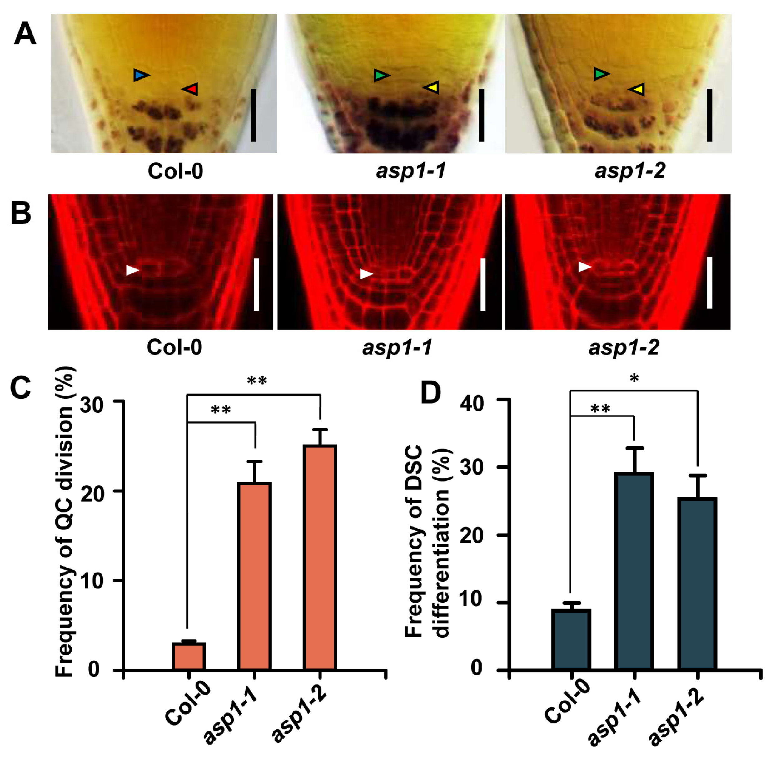
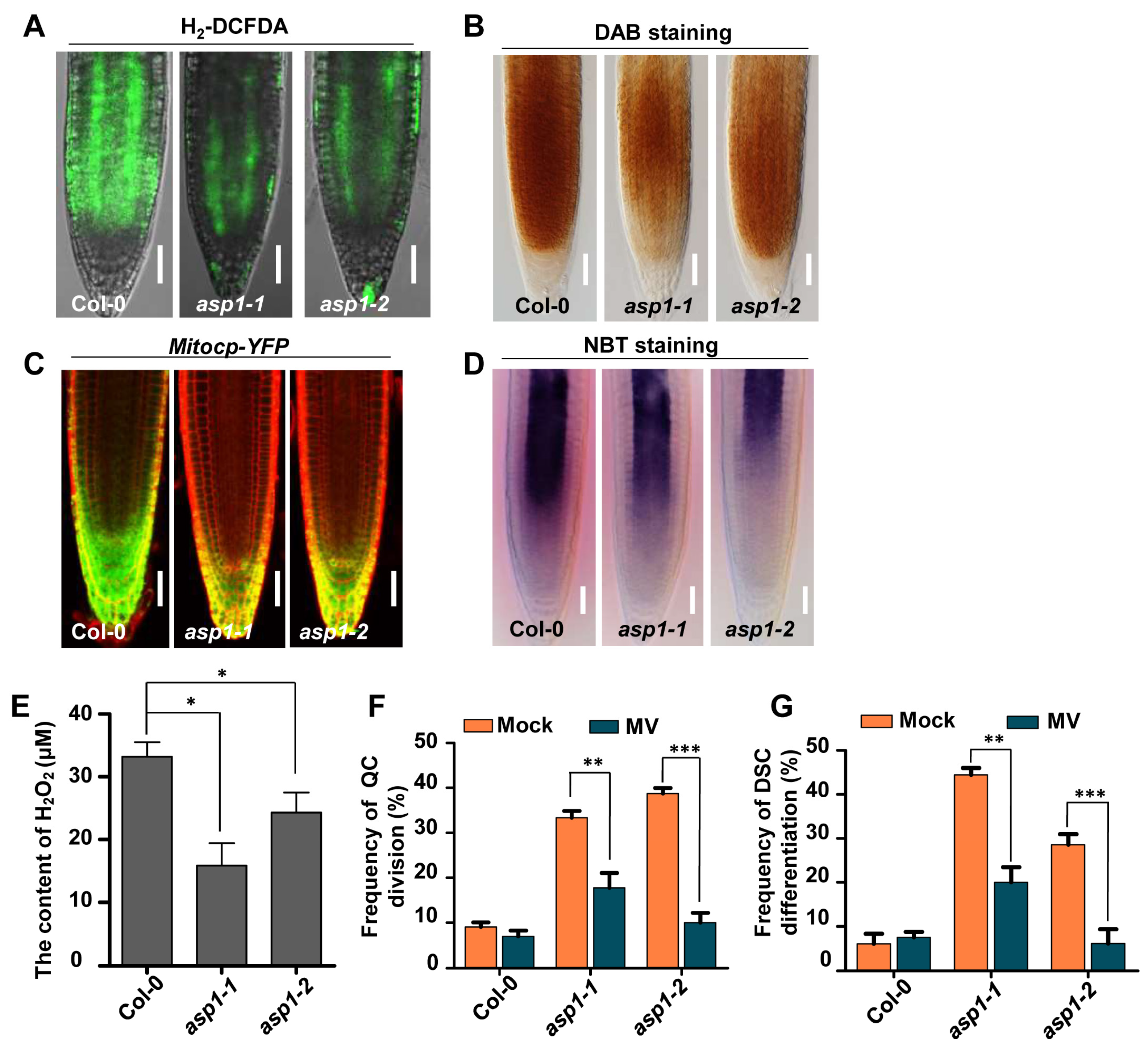
2.1. ASP1 Plays a Role in ROS Maintenance to Control RSCN Identity

A genetic screening was carried out on Arabidopsis mutants to discover new elements associated with the maintenance of root stem cells regulated by ROS. This screening aimed to identify mutants exhibiting impairments in root distal stem cell maintenance and alterations in ROS levels within a mutant pool generated through the insertion of T-DNA [24]. From Lugol’s staining and H2-DCFDA staining, the asp1-1 mutant (Salk_036792) was identified, which displayed increased QC cell divisions and distal stem cell differentiation along with a decreased ROS level in the root tip (Figure 1A and Figure 2A).
Using PI staining (for red fluorescence), the dividing QC cells were observed in the asp1-1 mutant other than the WT (wild type) under a confocal microscope (Figure 1B). In the wild type, less than 5% of QC cells were divided (Figure 1C); however, the frequency of divided QC cells in the asp1-1 mutant was more than 20% (Figure 1C). Furthermore, the asp1-1 mutant showed a higher proportion of DSC differentiation (around 30%), contrary to the low rate in the WT (around 5%) (Figure 1D). Consequently, these results demonstrate that the loss of function of ASP1 results in developmental defects in RSCN.
Tail-PCR analysis showed that the last exon in the asp1-1 (At4g27680) allele is inserted with a T-DNA fragment (Figure S1A). Another mutant asp1 allele (asp1-2, Sail_547_g04) was analyzed to prove this mutation, as the asp1-2 mutant showed the same phenotype as asp1-1 (Figure 1 and Figure S1B). Moreover, the expression of ASP1 was significantly reduced in both mutants (Figure S1B). The defects of root SCN maintenance in the asp1-1 mutant were complemented through crossing with the ASP1p::GFP-ASP1 transgenetic line (Figure S1C). Taken together, these findings provide evidence to show that the observed phenotype in asp1 mutants can be attributed to the mutation in ASP1.
To further confirm the decreased ROS contents in asp1 mutants as indicated by H2-DCFDA, we examined the extent of hydrogen peroxide (H2O2) and superoxide anion (O2−), the two primary kinds of ROS in plants (Figure 2). Compared to WT, the level of H2O2 (indicated by DAB staining) and O2− (indicated by NBT staining) in the asp1 mutants was reduced (Figure 2B,D). When crossed with Mitocp-YFP, a mitochondrial O2− indicator, a weaker signal was observed in the asp1 mutant roots compared to controls (Figure 2C). In addition, the contents of H2O2 were greatly declined in asp1 mutants (Figure 2E). Meanwhile, APX2, APX3, APX4, and APX6—which encode ROS-scavenging enzymes—were up-regulated in the asp1-1 mutant (Figure S2). These findings indicate ASP1 plays a role in regulating the maintenance of ROS levels.
To test the impact of altered ROS levels in asp1 mutants on the defects in root SCN maintenance, Col-0 and asp1 mutants were treated with methyl viologen (MV), a compound known to induce an excess production of superoxide. Different from the WT, the elevated occurrence of division in QC and differentiation in DSC were significantly decreased after treatment with MV in asp1 mutants (Figure 2F,G). These recovered SCN-defective phenotypes in asp1 mutants might be associated with the elevated ROS contents induced by MV. Moreover, the enhanced frequency of abnormal QC and root DSC in ASP1 over-expression lines was attributed to the elevated ROS level (Figure S3). Taken together, we conclude that ASP1 controls RSCN identity through regulating ROS homeostasis.

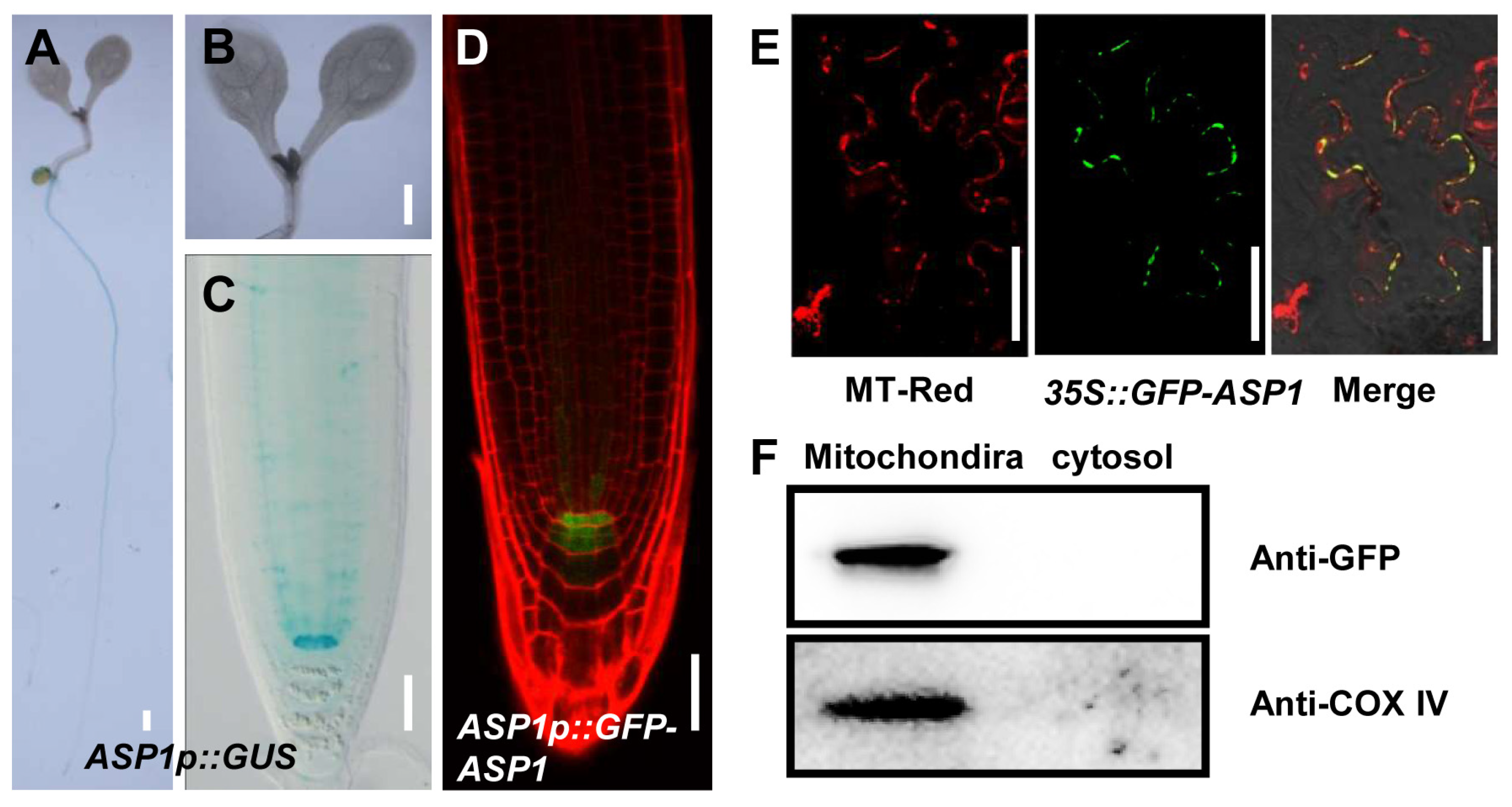
2.2. ASP1 Is Exclusively Expressed in QC Cells and Localized in Mitochondria

To clarify the probable effect of ASP1 in ROS-modulated root SCN maintenance, we constructed ASP1p::GUS and ASP1p::GFP-ASP1 transgenic lines for the purpose of examining the expression profile of ASP1. GUS staining analysis with ASP1p::GUS lines indicates that ASP1 is specifically expressed in QC cells (Figure 3A–C). Consistently, strong GFP signals were detected in the root QC cells through analysis of ASP1p::GFP-ASP1 transgenic lines (Figure 3D).
In addition, we further generated a 35S::GFP-ASP1 construct to study the subcellular localization of ASP1. The results observed under a confocal microscope showed that the green fluorescence signal of ASP1 and the red fluorescence signal of Mito Tracker Red (mitochondrial probe) were largely merged (Figure 3E), indicating ASP1 is localized in mitochondria. To further confirm the mitochondrial localization of ASP1, we isolated mitochondria and cytosol components (without mitochondria) from the 35S::GFP-ASP1 transgenic line. Western blotting analysis detected the ASP1-GFP protein and COX IV; (mitochondria marker protein) from mitochondria proteins other than cytosol components (without mitochondria) (Figure 3F). To explore whether ASP1 is located in other endomembrane systems, a specific antibody marker for endoplasmic reticulum (ER)-associated BIP2 was imported. Immunologic analysis showed minimal co-localization between the signals of GFP-ASP1 and BIP2 (Figure S4).

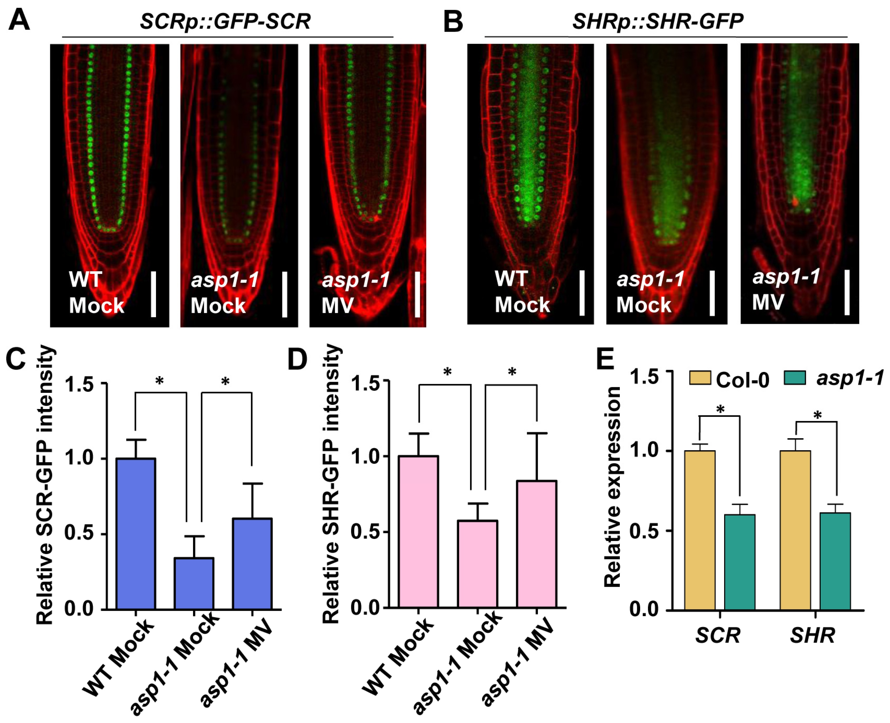
2.3. SCR and SHR Are Implicated in ASP1-Mediated ROS Homeostasis Regulating the SCN Maintenance

To investigate whether the transcription factors important for root SCN maintenance are involved in the ASP1 regulatory pathway, we detected the expression levels of WOX5, PLT1, PLT2, as well as SCR and SHR in the WT and asp1 mutants. The real-time PCR examination indicated that there were no significant differences in the expression levels of WOX5, PLT1, and PLT2 between the asp1 mutants and the wild type (Figure S5A). Similarly, there was no difference in the fluorescence signals of WOX5p::GFP, PLT1p::PLT1-YFP, and PLT2p::PLT2-YFP between Col-0 and asp1 mutants (Figure S5B). However, the levels of expression for SHR and SCR were notably decreased in asp1 mutants, as evidenced by fluorescence examinations using SCRp::GFP-SCR and SHRp::SHR-GFP (Figure 4A–D). The analysis of qRT-PCR revealed a consistent decrease in the expression levels of SHR and SCR in asp1 mutants. This suggests that the down-regulation of SCR and SHR may be involved in the ASP1-mediated regulation of RSCN maintenance (Figure 4E).
Additionally, we explored whether the reduced SCR and SHR expression in asp1 was related to the decreased ROS level. We tested the effect of methyl viologen on SCR and SHR in the SCRp::GFP-SCR and SHRp::SHR-GFP transgenetic lines in the asp1 mutant background. The findings indicated a significant elevation in GFP signals within root tips after methyl viologen treatment (Figure 4A–D), suggesting that ASP1 plays a role in regulating ROS levels to preserve the root stem cell niche via the SCR and SHR pathways.

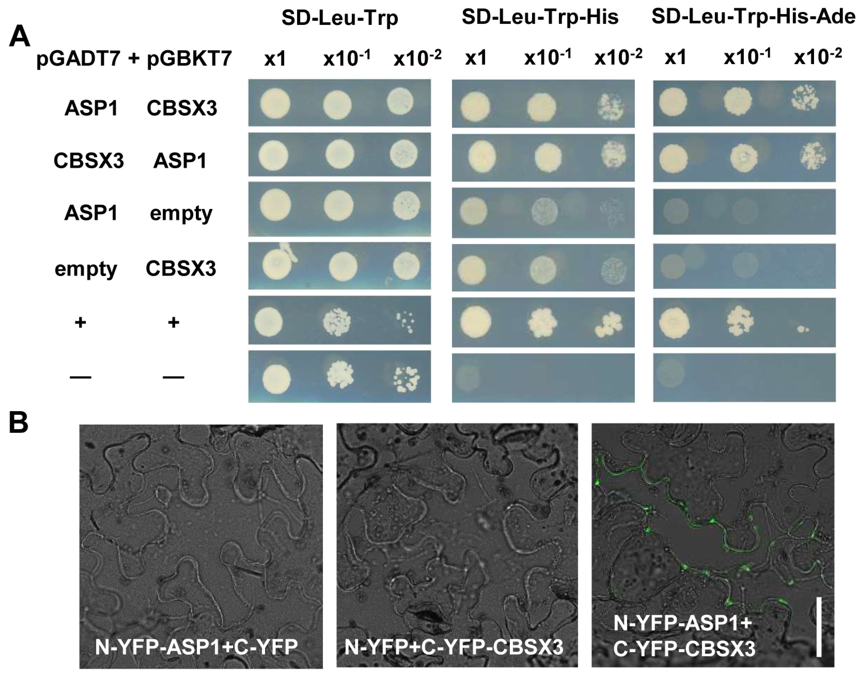
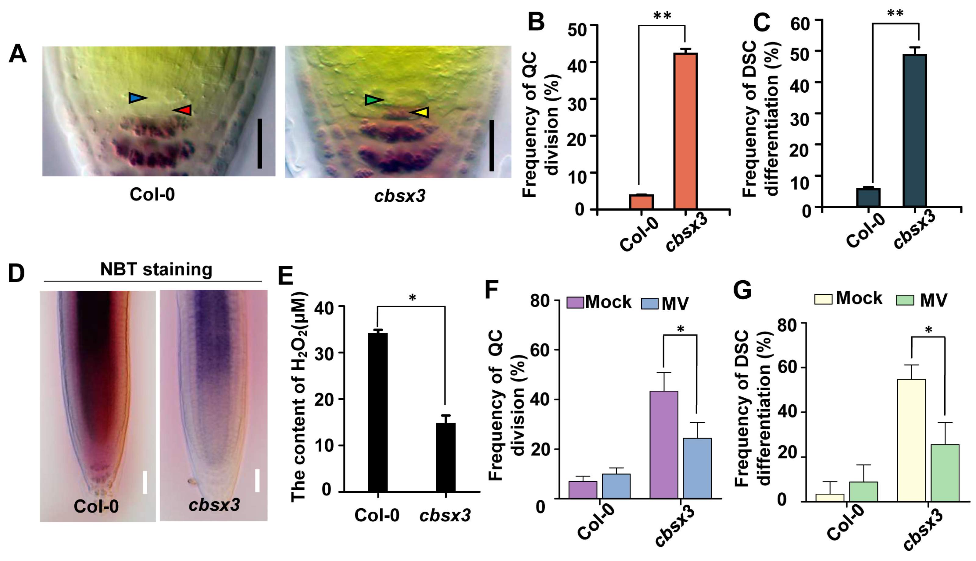
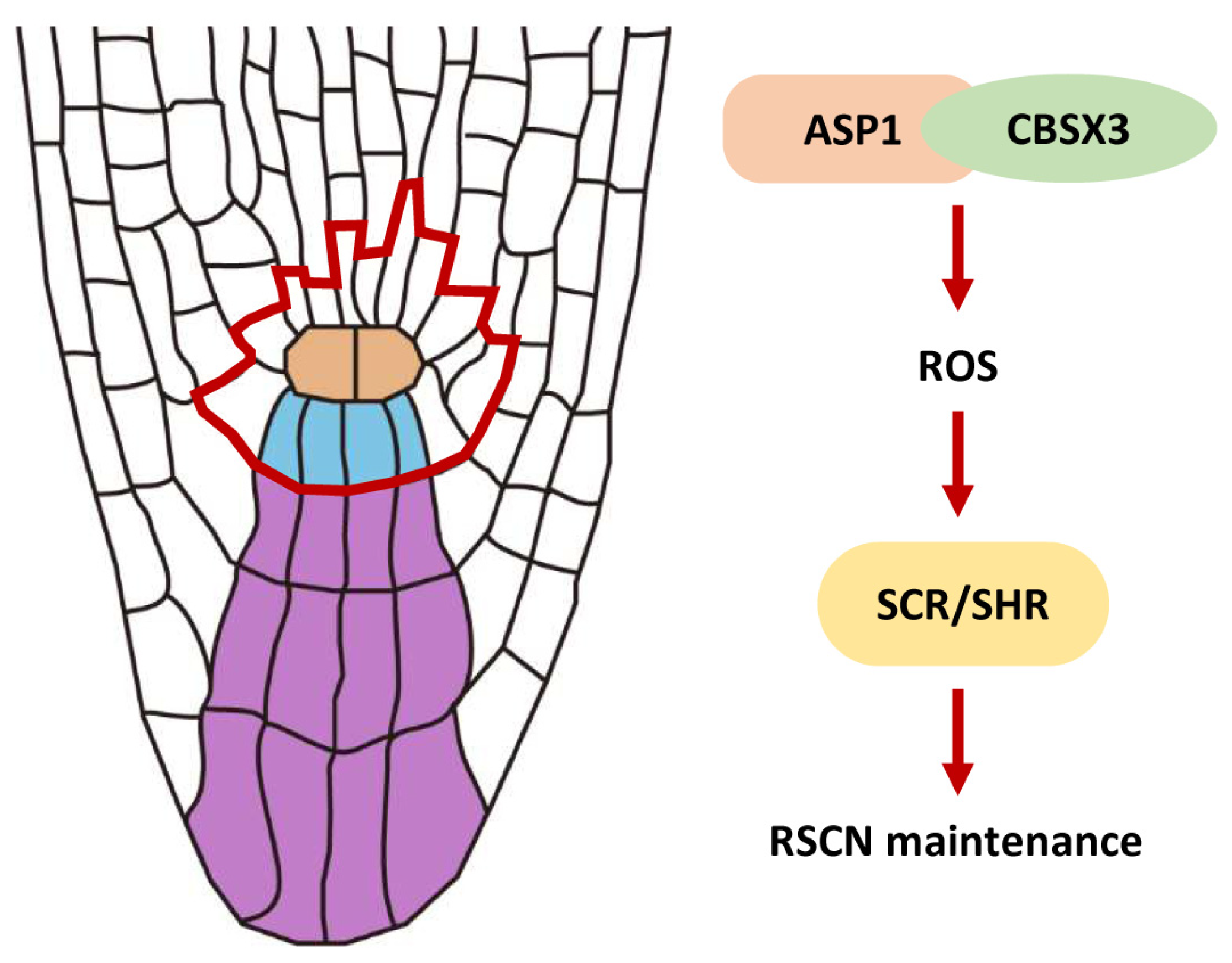
2.4. ASP1 Interacts Directly with CBSX3 to Maintain Root SCN Stability via Manipulating ROS Generation

To uncover how ASP1 regulates ROS homeostasis, a yeast two-hybrid assay was conducted to elucidate the protein interactions of ASP1, leading to the identification of a CBS domain-containing protein, CBSX3 (Figure 5A). The association of ASP1 and CBSX3 was additionally validated through a bimolecular fluorescence complementation (BiFC) assay. In tobacco epidermal cells, a pronounced fluorescence signal was detected when ASP1 was coexpressed with the N-terminal half of YFP (NYFP), and CBSX3 was co-expressed with the C-terminal half of YFP (CYFP). Conversely, no fluorescence signal was detected in the control group where the empty vector was utilized (Figure 5B). All these results indicate that there is an interaction between ASP1 and CBSX3.
CBSX3 has been reported to regulate ROS generation and CBSX3-Ox plants display primary root shortening. Nevertheless, the impact of CBSX3 on the RSCN remains unexplored. Consequently, we investigated the characteristics of the root quiescent center cells and distal stem cells in cbsx3 (Figure 6A). In the cbsx3 mutant, a significant increase in QC cell division rate was observed, exceeding 40% in contrast to the wild-type frequency of 5%. Moreover, approximately half of the mutant roots exhibited a lack of distal stem cells, indicating the potential involvement of CBSX3 in the maintenance of the RSCN (Figure 6B,C). To investigate the role of CBSX3 in maintaining root RSCN integrity through the regulation of ROS homeostasis, we conducted an analysis of ROS levels in the roots of cbsx3. In comparison to the wild type, reduced NBT staining intensity was noted in the cbsx3 root tips (Figure 6D). Furthermore, the concentration of hydrogen peroxide was found to be reduced in cbsx3 in comparison to the wild type (Figure 6E). Therefore, impairment of CBSX3 results in a decrease in ROS generation in the root tips.
To further explore the potential relationship between the increased percentage of divided QC cells, differentiated distal stem cells in cbsx3 mutants, and the reduced levels of ROS, the root stem cell niche was examined after being supplied with methyl viologen. Strikingly, the elevated frequency was strongly decreased after 0.1 μM MV treatments (Figure 6F,G). These results indicate that CBSX3 acts as a controller of reactive oxygen species generation, thereby influencing the root stem cell niche identity.

3. Discussion

The AAA ATPase family is a large and functionally diverse group of enzymes that are involved in numerous cellular processes [25]. All ATPases in this family have a domain of 200–250 amino acids that is responsible for binding ATP [26]. AAA ATPases can be found in all eukaryotic organisms, with the highest diversity in Arabidopsis, with about 140 AAA+ proteins [27]. AAA ATPases play a significant role in controlling various developmental processes in plants, including but not limited to embryonic development, pollen formation, seed development, and the regulation of flowering time [28,29,30,31]; moreover, AAA ATPases play a role in the control of the root meristem. As an illustration, RPT2 maintains the root meristem integrity via proteasome-dependent programmed proteolysis [32]; the function of BRM is to preserve the RSCN by regulating the expression of PIN proteins within the PLT signaling [33]; and MDN1 is essential for the auxin maxima pattern in the embryo and functions in root meristem maintenance [34]. However, the involvement of AAA ATPases in the control of ROS homeostasis for the maintenance of RSCN has not been described. In this study, we provide evidence to show that ASP1, a novel AAA ATPase, maintains the RSCN by manipulating ROS generation in the root tip (Figure 1 and Figure 2). Therefore, our findings broadened our understanding of AAA ATPase function and ROS homeostasis regulation (Figure 7).
Research has demonstrated the significant involvement of ROS homeostasis in the maintenance of the RSCN [13]. The altered ROS levels lead to defects in root SCN maintenance in both asp1 and cbsx3 mutants (Figure 2, Figure 6, and Figure S3). Our findings, together with earlier studies, indicate that maintaining a suitable level of ROS is crucial for preserving the RSCN [14]; however, the molecular mechanism of ROS homeostasis is finely regulated to preserve the identity of RSCN remains unknown. In this research, we present findings that ASP1 interacts with a ROS generation regulator CBSX3 to regulate the homeostasis of ROS (Figure 5 and Figure 6). The CBS (cystathionine β-synthase) domain-containing proteins are a large superfamily of proteins containing a conserved CBS domain [35]. CBSX3 was reported to localize to the mitochondria, and has the same subcellular localization as ASP1 (Figure 3). A previous study showed that CBSX3 regulates ROS generation by interacting with Trxs-o2 in an ADP/ATP ratio-dependent manner [36]. Further studies are needed to explore how ASP1 regulates ROS homeostasis through interacting with CBSX3; therefore, it will also be interesting to study whether ASP1 could affect the interaction between CBSX3 and Trxs-o2 by altering the ADP/ATP ratio.
SCR and SHR, members of GRAS transcription factors, play a crucial role in RSCN maintenance [37]. Both scr and shr mutants display defective QC and DSC maintenance and lead to short-root phenotypes [38,39]. After the synthesis of SHR in the vascular cylinder, it is translocated to the neighboring cell layer to activate SCR [40]. In addition, SCR interacts with PLT and TCP, which is short for teosinte-branched cycloidea PCNA, to determine the specific location of stem cells. These complexes have the ability to stimulate the activation of WOX5 expression, a gene that is uniquely expressed in QC and plays a crucial role in maintaining RSCN integrity, by binding to the WOX5 promoter [9]. A previous study demonstrated that SCR and SHR respond to ROS homeostasis to regulate root DSC maintenance [14]. This report is consistent with our finding that the expression of SCR and SHR was suppressed in asp1 mutants and could be increased by MV (Figure 4 and Figure S5). Based on these observations, we propose that SCR and SHR are involved in ASP1-mediated ROS homeostasis regulating the maintenance of SCN. Further experiments are required to establish the specific correlation between ASP1 and the SCR/SHR pathway.

4. Materials and Methods

4.1. Plant Materials and Growth Conditions

The Arabidopsis mutants and transgenic lines employed in this investigation are derived from the ecotype Columbia (Col-0). The asp1-1 (SALK_036792) and asp1-2 (SAIL_547_G04) were T-DNA insertion mutants acquired from the Arabidopsis Biological Resource Center. The marker lines were previously discussed in other sources: Mito-cpYFP [41], SCRp::GFP-SCR [38], SHRp::SHR-GFP [42], WOX5p::GFP [5], PLT1p::PLT1-YFP [43], PLT2p::PLT2-YFP [43]. The ASP1pro::GUS fusion was generated through the insertion of a 1300-base-pair sequence of the ASP1 promoter region into pKGWFS7.1. The sequences of the ASP1 (AT4G27680) promoter and coding region were integrated into pB7m34GW to acquire the ASP1pro::GFP-ASP1 fusion. The CDS of ASP1 was integrated into pK7WGF2.0 to obtain the 35S::GFP-ASP1 fusion. For MV treatment, 3-day-old seedlings were incubated with 0.1 μM MV in a 1/2 liquid MS medium for 48 h. Seeds were subjected to surface sterilization using chlorine gas and subsequently stored at 4 °C for a period of two days. Following this, the seeds were placed onto solid medium containing half-strength Murashige and Skoog (MS) nutrients and subsequently transferred to a plant growth chamber set at a constant temperature of 20 °C under a 16-h photoperiod.

4.2. Histochemical Assays and Microscopy

To perform Lugol’s staining, the seedlings were steeped in Lugol’s solution for 2 min, then placed in chloral hydrate solution and imaged by a phase-contrast microscope. The H2-DCFDA (Invitrogen, Carlsbad, CA, USA) staining assay was carried out following the previously outlined procedure [44], and then photographed by a laser-scanning confocal microscope (excitation: 488 nm, emission: 525 nm). Five-day-old seedlings were placed in a solution of DAB (Sigma, St. Louis, CA, USA) containing 0.1 mg/mL DAB in a solution of 50 mM Tris-HCl and NBT (Sigma) containing 2 mM NBT in a solution of 0.1 M NaCl and 20 mM potassium phosphate, with a pH of 6.1, for 15 min. Subsequently, they were observed using a light microscope. For measurement of H2O2 content, roots isolated from five-day-old WT and asp1 seedlings were ground with extraction buffer (Beyotime Biotechnology, Shanghai, China) on ice, and measured at 560 nm by a spectrophotometer.

4.3. Western Blotting

The proteins from the mitochondria and cytosol of 35S::GFP-ASP1 roots were separated using the mitochondrial protein extraction kit from Solarbio (Beijing, China). The proteins were separated using SDS-PAGE (Bio-Rad, Hercules, CA, USA) and subsequently moved onto PVDF membranes (Millipore, St. Louis, CA, USA). The membranes were first treated with 5% milk for 2 h, then exposed to GFP and COX IV antibodies overnight at 4 °C, and finally incubated with secondary antibodies conjugated with horseradish peroxidase for 2 h. The SuperSignal West Pico Luminol Solution (ThermoFisher Scientific, Carlsbad, CA, USA) detected the signal.

4.4. Quantitative RT-PCR

The RNA template required was obtained by isolating it using an RNAsimple Total RNA Kit from Tiangen. Next, an initial cDNA strand was created from 2 μg of RNA by utilizing a HiScript III RT SuperMix for qPCR from Vazyme (Nanjing, China), then, DNase I was used to eliminate genomic DNA. QRT-PCRs were conducted on the MyiQTM Real-time PCR Detection System from Bio-Rad, utilizing the AceQ Universal SYBR qPCR Master Mix from Vazyme. Each sample consisted of three biological replicates, with each biological replicate being represented by three technical replicates. The reference gene used was AtACTIN2. The quantitative RT-PCR primers are detailed in the Supplementary Table S1.

4.5. Yeast Two-Hybrid Assay

This assay was conducted following the instructions provided in the manual from Clontech for the Matchmaker GAL4 Two-Hybrid System 3. The coding sequences of ASP1 and CBSX3 were placed into the pGADT7 vector from Takara and separately cloned into the pGBKT7 vector. The modified yeasts were grown on SD–Trp–Leu medium for 2 days before being moved to SD–Trp–Leu–His medium.

4.6. BiFC Assay

The fragment of ASP1 was amplified and inverted into the pSAT6-nYFP, and the CDS of CBSX3 was inserted into the pSAT6-cYFP. The fusions were injected into Nicotiana benthamiana leaves. Signals were observed after culturing at 25 °C for 3 days by using a Zeiss LSM 700 confocal microscope. The blank vector was utilized as a negative control.

Supplementary Materials

The following supporting information can be downloaded at: https://www.mdpi.com/article/10.3390/plants13111469/s1, Figure S1: the validation of asp1 mutants; Figure S2: quantitative real-time PCR was utilized to assess the comparative transcript levels of ROS generation and scavenging genes; Figure S3: the increased ROS concentration observed in ASP1-OE line stimulates the division of QC and differentiation of DSC; Figure S4: the subcellular distribution of ASP1 was examined, revealing minimal overlap between the signal of GFP-ASP1 and the endoplasmic reticulum (ER) marker BIP2; Figure S5: expression of WOX5, PLT1, and PLT2 in asp1 mutants did not show a statistically significant variance compared to those observed in the Col. Table S1: List of primers used in the study.

Author Contributions

Planning and designing of the research, Q.Y. and Z.D.; performing the experiments, Q.Y., H.L. and B.Z.; analyzing the data, Y.S. (Yun Song) and Y.S. (Yueying Sun); writing the paper, Q.Y. All authors have read and agreed to the published version of the manuscript.

Funding

This research was funded by the National Natural Science Foundation of China, grant number 32300292 to Q.Y.; and by the Shandong Province Natural Science Foundation, grant numbers ZR2020QC037 to Q.Y., ZR2021QC200 to Y.S. (Yun Song), and ZR2020QB154 to Y.S. (Yueying Sun).

Data Availability Statement

All data are available in the main text or the Supplementary Materials.

Acknowledgments

We thank Jiri Friml and Zhizhong Gong for sharing published materials. We also thank the editor and the anonymous reviewers for their constructive comments and insightful suggestions, which greatly improved this article.

Conflicts of Interest

The authors declare no conflicts of interest.

References

  1. Ubogoeva, E.V.; Zemlyanskaya, E.V.; Xu, J.; Mironova, V. Mechanisms of stress response in the root stem cell niche. J. Exp. Bot. 2021, 72, 6746–6754. [Google Scholar] [CrossRef] [PubMed]
  2. Jiang, K.; Feldman, L.J. Regulation of root apical meristem development. Annu. Rev. Cell Dev. Biol. 2005, 21, 485–509. [Google Scholar] [CrossRef] [PubMed]
  3. Scheres, B. Stem-cell niches: Nursery rhymes across kingdoms. Nat. Rev. Mol. Cell Biol. 2007, 8, 345–354. [Google Scholar] [CrossRef] [PubMed]
  4. Rahni, R.; Efroni, I.; Birnbaum, K.D. A Case for Distributed Control of Local Stem Cell Behavior in Plants. Dev. Cell 2016, 38, 635–642. [Google Scholar] [CrossRef] [PubMed]
  5. Sarkar, A.K.; Luijten, M.; Miyashima, S.; Lenhard, M.; Hashimoto, T.; Nakajima, K.; Scheres, B.; Heidstra, R.; Laux, T. Conserved factors regulate signalling in Arabidopsis thaliana shoot and root stem cell organizers. Nature 2007, 446, 811–814. [Google Scholar] [CrossRef] [PubMed]
  6. Drisch, R.C.; Stahl, Y. Function and regulation of transcription factors involved in root apical meristem and stem cell maintenance. Front. Plant Sci. 2015, 6, 505. [Google Scholar] [CrossRef] [PubMed]
  7. Aida, M.; Beis, D.; Heidstra, R.; Willemsen, V.; Blilou, I.; Galinha, C.; Nussaume, L.; Noh, Y.S.; Amasino, R.; Scheres, B. The PLETHORA genes mediate patterning of the Arabidopsis root stem cell niche. Cell 2004, 119, 109–120. [Google Scholar] [CrossRef] [PubMed]
  8. Sabatini, S.; Heidstra, R.; Wildwater, M.; Scheres, B. SCARECROW is involved in positioning the stem cell niche in the Arabidopsis root meristem. Genes. Dev. 2003, 17, 354–358. [Google Scholar] [CrossRef] [PubMed]
  9. Shimotohno, A.; Heidstra, R.; Blilou, I.; Scheres, B. Root stem cell niche organizer specification by molecular convergence of PLETHORA and SCARECROW transcription factor modules. Genes Dev. 2018, 32, 1085–1100. [Google Scholar] [CrossRef]
  10. Kong, X.; Lu, S.; Tian, H.; Ding, Z. WOX5 is Shining in the Root Stem Cell Niche. Trends Plant Sci. 2015, 20, 601–603. [Google Scholar] [CrossRef]
  11. Burkart, R.C.; Strotmann, V.I.; Kirschner, G.K.; Akinci, A.; Czempik, L.; Dolata, A.; Maizel, A.; Weidtkamp-Peters, S.; Stahl, Y. PLETHORA-WOX5 interaction and subnuclear localization control Arabidopsis root stem cell maintenance. EMBO Rep. 2022, 23, e54105. [Google Scholar] [CrossRef] [PubMed]
  12. Zhou, X.; Xiang, Y.; Li, C.; Yu, G. Modulatory Role of Reactive Oxygen Species in Root Development in Model Plant of Arabidopsis thaliana. Front. Plant Sci. 2020, 11, 485932. [Google Scholar] [CrossRef] [PubMed]
  13. Yang, S.; Yu, Q.; Zhang, Y.; Jia, Y.; Wan, S.; Kong, X.; Ding, Z. ROS: The Fine-Tuner of Plant Stem Cell Fate. Trends Plant Sci. 2018, 23, 850–853. [Google Scholar] [CrossRef] [PubMed]
  14. Yu, Q.; Tian, H.; Yue, K.; Liu, J.; Zhang, B.; Li, X.; Ding, Z. A P-Loop NTPase Regulates Quiescent Center Cell Division and Distal Stem Cell Identity through the Regulation of ROS Homeostasis in Arabidopsis Root. PLoS Genet. 2016, 12, e1006175. [Google Scholar] [CrossRef] [PubMed]
  15. Yamada, M.; Han, X.; Benfey, P.N. RGF1 controls root meristem size through ROS signalling. Nature 2020, 577, 85–88. [Google Scholar] [CrossRef] [PubMed]
  16. Kong, X.; Tian, H.; Yu, Q.; Zhang, F.; Wang, R.; Gao, S.; Xu, W.; Liu, J.; Shani, E.; Fu, C.; et al. PHB3 Maintains Root Stem Cell Niche Identity through ROS-Responsive AP2/ERF Transcription Factors in Arabidopsis. Cell Rep. 2018, 22, 1350–1363. [Google Scholar] [CrossRef] [PubMed]
  17. Wang, M.; Zhang, H.; Zhao, X.; Zhou, J.; Qin, G.; Liu, Y.; Kou, X.; Zhao, Z.; Wu, T.; Zhu, J.K.; et al. SYNTAXIN OF PLANTS81 regulates root meristem activity and stem cell niche maintenance via ROS signaling. Plant Physiol. 2023, 191, 1365–1382. [Google Scholar] [CrossRef] [PubMed]
  18. Yang, L.; Zhang, J.; He, J.; Qin, Y.; Hua, D.; Duan, Y.; Chen, Z.; Gong, Z. ABA-mediated ROS in mitochondria regulate root meristem activity by controlling PLETHORA expression in Arabidopsis. PLoS Genet. 2014, 10, e1004791. [Google Scholar] [CrossRef]
  19. Lv, B.; Tian, H.; Zhang, F.; Liu, J.; Lu, S.; Bai, M.; Li, C.; Ding, Z. Brassinosteroids regulate root growth by controlling reactive oxygen species homeostasis and dual effect on ethylene synthesis in Arabidopsis. PLoS Genet. 2018, 14, e1007144. [Google Scholar] [CrossRef]
  20. Tian, Y.; Fan, M.; Qin, Z.; Lv, H.; Wang, M.; Zhang, Z.; Zhou, W.; Zhao, N.; Li, X.; Han, C.; et al. Hydrogen peroxide positively regulates brassinosteroid signaling through oxidation of the BRASSINAZOLE-RESISTANT1 transcription factor. Nat. Commun. 2018, 9, 1063. [Google Scholar] [CrossRef]
  21. Wang, Z.; Rong, D.; Chen, D.; Xiao, Y.; Liu, R.; Wu, S.; Yamamuro, C. Salicylic acid promotes quiescent center cell division through ROS accumulation and down-regulation of PLT1, PLT2, and WOX5. J. Integr. Plant Biol. 2021, 63, 583–596. [Google Scholar] [CrossRef]
  22. Sheftel, A.D.; Stehling, O.; Pierik, A.J.; Netz, D.J.; Kerscher, S.; Elsasser, H.P.; Wittig, I.; Balk, J.; Brandt, U.; Lill, R. Human ind1, an iron-sulfur cluster assembly factor for respiratory complex I. Mol. Cell Biol. 2009, 29, 6059–6073. [Google Scholar] [CrossRef] [PubMed]
  23. Zhang, J.; Rubio, V.; Lieberman, M.W.; Shi, Z.Z. OLA1, an Obg-like ATPase, suppresses antioxidant response via nontranscriptional mechanisms. Proc. Natl. Acad. Sci. USA 2009, 106, 15356–15361. [Google Scholar] [CrossRef] [PubMed]
  24. Weigel, D.; Ahn, J.H.; Blazquez, M.A.; Borevitz, J.O.; Christensen, S.K.; Fankhauser, C.; Ferrandiz, C.; Kardailsky, I.; Malancharuvil, E.J.; Neff, M.M.; et al. Activation tagging in Arabidopsis. Plant Physiol. 2000, 122, 1003–1013. [Google Scholar] [CrossRef] [PubMed]
  25. Hanson, P.I.; Whiteheart, S.W. AAA+ proteins: Have engine, will work. Nat. Rev. Mol. Cell Biol. 2005, 6, 519–529. [Google Scholar] [CrossRef] [PubMed]
  26. Iyer, L.M.; Leipe, D.D.; Koonin, E.V.; Aravind, L. Evolutionary history and higher order classification of AAA+ ATPases. J. Struct. Biol. 2004, 146, 11–31. [Google Scholar] [CrossRef] [PubMed]
  27. Frickey, T.; Lupas, A.N. Phylogenetic analysis of AAA proteins. J. Struct. Biol. 2004, 146, 2–10. [Google Scholar] [CrossRef] [PubMed]
  28. Muralla, R.; Lloyd, J.; Meinke, D. Molecular foundations of reproductive lethality in Arabidopsis thaliana. PLoS ONE 2011, 6, e28398. [Google Scholar] [CrossRef]
  29. Girard, C.; Chelysheva, L.; Choinard, S.; Froger, N.; Macaisne, N.; Lemhemdi, A.; Mazel, J.; Crismani, W.; Mercier, R. AAA-ATPase FIDGETIN-LIKE 1 and Helicase FANCM Antagonize Meiotic Crossovers by Distinct Mechanisms. PLoS Genet. 2015, 11, e1005369. [Google Scholar] [CrossRef]
  30. Baek, K.; Seo, P.J.; Park, C.M. Activation of a mitochondrial ATPase gene induces abnormal seed development in Arabidopsis. Mol. Cells 2011, 31, 361–369. [Google Scholar] [CrossRef]
  31. Vinegra de la Torre, N.; Vayssieres, A.; Obeng-Hinneh, E.; Neumann, U.; Zhou, Y.; Lazaro, A.; Roggen, A.; Sun, H.; Stolze, S.C.; Nakagami, H.; et al. FLOWERING REPRESSOR AAA(+) ATPase 1 is a novel regulator of perennial flowering in Arabis alpina. New Phytol. 2022, 236, 729–744. [Google Scholar] [CrossRef]
  32. Ueda, M.; Matsui, K.; Ishiguro, S.; Sano, R.; Wada, T.; Paponov, I.; Palme, K.; Okada, K. The HALTED ROOT gene encoding the 26S proteasome subunit RPT2a is essential for the maintenance of Arabidopsis meristems. Development 2004, 131, 2101–2111. [Google Scholar] [CrossRef]
  33. Yang, S.; Li, C.; Zhao, L.; Gao, S.; Lu, J.; Zhao, M.; Chen, C.Y.; Liu, X.; Luo, M.; Cui, Y.; et al. The Arabidopsis SWI2/SNF2 Chromatin Remodeling ATPase BRAHMA Targets Directly to PINs and Is Required for Root Stem Cell Niche Maintenance. Plant Cell 2015, 27, 1670–1680. [Google Scholar] [CrossRef] [PubMed]
  34. Li, P.C.; Li, K.; Wang, J.; Zhao, C.Z.; Zhao, S.Z.; Hou, L.; Xia, H.; Ma, C.L.; Wang, X.J. The AAA-ATPase MIDASIN 1 Functions in Ribosome Biogenesis and Is Essential for Embryo and Root Development. Plant Physiol. 2019, 180, 289–304. [Google Scholar] [CrossRef]
  35. Yoo, K.S.; Ok, S.H.; Jeong, B.C.; Jung, K.W.; Cui, M.H.; Hyoung, S.; Lee, M.R.; Song, H.K.; Shin, J.S. Single cystathionine beta-synthase domain-containing proteins modulate development by regulating the thioredoxin system in Arabidopsis. Plant Cell 2011, 23, 3577–3594. [Google Scholar] [CrossRef] [PubMed]
  36. Shin, J.S.; So, W.M.; Kim, S.Y.; Noh, M.; Hyoung, S.; Yoo, K.S.; Shin, J.S. CBSX3-Trxo-2 regulates ROS generation of mitochondrial complex II (succinate dehydrogenase) in Arabidopsis. Plant Sci. 2020, 294, 110458. [Google Scholar] [CrossRef]
  37. Dinneny, J.R.; Benfey, P.N. Plant stem cell niches: Standing the test of time. Cell 2008, 132, 553–557. [Google Scholar] [CrossRef]
  38. Di Laurenzio, L.; Wysocka-Diller, J.; Malamy, J.E.; Pysh, L.; Helariutta, Y.; Freshour, G.; Hahn, M.G.; Feldmann, K.A.; Benfey, P.N. The SCARECROW gene regulates an asymmetric cell division that is essential for generating the radial organization of the Arabidopsis root. Cell 1996, 86, 423–433. [Google Scholar] [CrossRef] [PubMed]
  39. Helariutta, Y.; Fukaki, H.; Wysocka-Diller, J.; Nakajima, K.; Jung, J.; Sena, G.; Hauser, M.T.; Benfey, P.N. The SHORT-ROOT gene controls radial patterning of the Arabidopsis root through radial signaling. Cell 2000, 101, 555–567. [Google Scholar] [CrossRef]
  40. Gallagher, K.L.; Paquette, A.J.; Nakajima, K.; Benfey, P.N. Mechanisms regulating SHORT-ROOT intercellular movement. Curr. Biol. 2004, 14, 1847–1851. [Google Scholar] [CrossRef]
  41. Wang, W.; Fang, H.; Groom, L.; Cheng, A.; Zhang, W.; Liu, J.; Wang, X.; Li, K.; Han, P.; Zheng, M.; et al. Superoxide flashes in single mitochondria. Cell 2008, 134, 279–290. [Google Scholar] [CrossRef] [PubMed]
  42. Nakajima, K.; Sena, G.; Nawy, T.; Benfey, P.N. Intercellular movement of the putative transcription factor SHR in root patterning. Nature 2001, 413, 307–311. [Google Scholar] [CrossRef] [PubMed]
  43. Kornet, N.; Scheres, B. Members of the GCN5 histone acetyltransferase complex regulate PLETHORA-mediated root stem cell niche maintenance and transit amplifying cell proliferation in Arabidopsis. Plant Cell 2009, 21, 1070–1079. [Google Scholar] [CrossRef] [PubMed]
  44. Tsukagoshi, H.; Busch, W.; Benfey, P.N. Transcriptional regulation of ROS controls transition from proliferation to differentiation in the root. Cell 2010, 143, 606–616. [Google Scholar] [CrossRef] [PubMed]
Figure 1. The asp1 mutants show defects in root SCN maintenance. (A) Staining with Lugol’s solution was performed on the roots of 5-day-old Col-0 and asp1 mutants. The enhanced rate of divided QC cells and differentiated distal stem cells was revealed by Lugol staining in asp1 mutants. Regular QC cells are represented by blue arrows, standard distal stem cells are represented by red arrows, divided QC cells are represented by green arrows, and abnormal distal stem cells are represented by yellow arrows. The scale bars are 50 μm in length. (B) Mutation of ASP1 resulted in QC cell division indicated by confocal microscope scanning images of PI staining. QC is marked with white arrows. The scale bars are 50 μm in length. (C,D) Measuring the number and analyzing the frequency of divided QC cells and differentiated distal stem cells in WT and asp1. The data presented show the average values with standard error (n = 100), with significance levels indicated as * for p < 0.05 and ** for p < 0.01. The statistical analysis used was Student’s t-test.
Plants 13 01469 g001
Figure 2. The defects of SCN maintenance in asp1 mutants were attributed to the lowered ROS level. (A) Fluorescent examination of H2-DCFDA in the roots of Col-0 and asp1 was conducted. The scale bars represent 50 μm. (B) DAB staining of 5-day-old seedlings. The scale bars represent 50 μm. (C) Detection of the fluorescence signal from Mitocp-YFP in Col-0 and asp1 mutants. The scale bars represent 50 μm. (D) NBT staining of 5-day-old seedlings. The scale bars represent 50 μm. (E) Measurement of endogenous levels of H2O2 in Col-0 and asp1 roots. (F, G) Quantification of divided QC cells and differentiated distal stem cells in Col-0 and asp1 roots treated with or without 0.1 µM MV. Results are presented as averages with standard error (n = 100), with significance levels denoted as * for p < 0.05, ** for p < 0.01, *** for p < 0.001. Statistical analysis was conducted using Student’s t-test.
Plants 13 01469 g002
Figure 3. Patterns of expression and cellular localization of ASP1. (AC) Histochemical staining analysis of ASP1p::GUS transgenic lines. (A) GUS staining of whole seedlings of 5-day-old ASP1p::GUS transgenic line. Scale bar: 10 mm. (B) Magnification of the leaf of ASP1p::GUS transgenic line in (A). Scale bar: 10 mm. (C) Magnification of the leaf and root tips of ASP1p::GUS transgenic line in (A). Scale bar: 50 μm. (D) Image captured using confocal microscopy showing ASP1p::GFP-ASP1 in root tips, with GFP fluorescence in green and PI fluorescence in red. The scale bar represents 50 μm. (E) The signal of GFP-ASP1 is found in the same location as the Mito Tracker Red in tobacco. The scale bar represents 50 μm. (F) Western blotting analysis of mitochondria and cytosol proteins isolated from 35S::GFP-ASP1 seedlings. COX IV was utilized as a reference standard in this examination.
Plants 13 01469 g003
Figure 4. SCR and SHR act downstream of ASP1 and respond to ROS homeostasis to regulate RSCN maintenance. (A) Fluorescence analysis of SCRp::GFP-SCR in Col-0 and asp1 roots treated with or without MV treatment. The scale bar represents 50 μm. (B) The fluorescence signal detection of SHRp::SHR-GFP in Col-0 and asp1 mutants roots with or without MV treatment. The scale bar represents 50 μm. (C,D) Measurement of the intensity of GFP in SCRp::GFP-SCR and SHRp::GFP-SHR seedlings in (A,B) with or without 0.1 µM MV. Results are presented as averages with standard error (n = 30), with statistical significance denoted by * p < 0.05 using Student’s t-test. (E) qRT-PCR results show the relative transcript levels of SCR and SHR in Col-0 and asp1-1 mutant. Results are presented as averages with standard error (n = 3), with statistical significance denoted by * p < 0.05 using Student’s t-test.
Plants 13 01469 g004
Figure 5. ASP1 interacts with CBSX3. (A) The identification of the interaction between ASP1 and CBSX3 by using yeast. Co-transformed cells were plated on SD–Leu–Trp agar to assess their survival and on SD–Leu–Trp–His and SD–Leu–Trp–His–Ade agars to evaluate their interaction. (B) BiFC analysis was conducted to study the interaction between ASP1 and CBSX3. Different combinations of constructs and empty vectors were introduced into tobacco, and then observe the fluorescence signal via confocal microscope. The scale bar on the images was set at 50 μm.
Plants 13 01469 g005
Figure 6. Mutation of CBSX3 affects the root stem cell niche identity via regulating ROS homeostasis. (A) Lugol-staining roots of Col-0 and cbsx3 mutants. Blue arrow indicates regular QC, red arrow indicates the undifferentiated DSC layers, green arrow indicates dividing QC, and yellow arrow indicates the differentiated distal stem cells. Scale bars are 50 μm in length. (B,C) Quantification of divided QC cells and differentiated distal stem cells in Col-0 and cbsx3. (D) Staining with NBT was performed on seedlings of Col-0 and cbsx3 that were 5 days old. Scale bars represent 50 μm. (E) Measurement of H2O2 content in roots of Col-0 and cbsx3. (F,G) The frequency analysis of divided QC cells and differentiated distal stem cells in Col-0 and cbsx3 seedlings with or without MV treatment. Results are presented as averages with standard error (n = 100), with significance levels denoted as * for p < 0.05, ** for p < 0.01. Statistical analysis was conducted using Student’s t-test.
Plants 13 01469 g006
Figure 7. ASP1-mediated ROS homeostasis to regulate RSCN maintenance. ASP1 regulates ROS signaling through interacting with the redox regulator CBSX3. Furthermore, SCR and SHR act downstream of ASP1-mediated ROS signaling in the maintenance of RSCN identity.
Plants 13 01469 g007
Disclaimer/Publisher’s Note: The statements, opinions and data contained in all publications are solely those of the individual author(s) and contributor(s) and not of MDPI and/or the editor(s). MDPI and/or the editor(s) disclaim responsibility for any injury to people or property resulting from any ideas, methods, instructions or products referred to in the content.

