Article

Vernonia britteniana Root Phytochemical Studies, In Vitro Cercaricidal Activity on the Larval Stage of Schistosoma mansoni and Antioxidant Activities

1
Research Institute for Medicines (iMed.ULisboa), Faculty of Pharmacy, Universidade de Lisboa, Av. Professor Gama Pinto, 1649-003 Lisbon, Portugal
2
Instituto de Investigação Veterinária, Bairro Santo António, Huambo 555, Angola
3
Global Health & Tropical Medicine, Medical Parasitology Unit, Universidade Nova de Lisboa, R. da Junqueira 100, 1349-008 Lisbon, Portugal
4
C.I.I.S.A.—Centre for Interdisciplinary Research in Animal Health, Faculty of Veterinary Medicine, University of Lisbon, Av. Universidade Técnica, 1300-477 Lisbon, Portugal
5
Associate Laboratory for Animal and Veterinary Sciences (AL4AnimalS), 1300-477 Lisbon, Portugal
*
Author to whom correspondence should be addressed.
Plants 2023, 12(9), 1788; https://doi.org/10.3390/plants12091788
Submission received: 16 January 2023 / Revised: 24 April 2023 / Accepted: 26 April 2023 / Published: 27 April 2023
(This article belongs to the Special Issue Medicinal Plants and Their Marker Compounds)

Abstract

:
Vernonia britteniana Hiern. (Asteraceae) is a medicinal plant used in traditional Angolan medicine against schistosomiasis. Our study aimed to investigate the phytochemical composition and the cercaricidal and antioxidant activities in vitro of a traditional herbal preparation (Water-Vbr) and a 70% hydroethanolic extract (EtOH70%-Vbr) prepared with this medicinal plant. The activity of the extracts against Schistosoma mansoni cercariae was assessed at different extract concentrations (500, 438, and 125 µg/mL) and at different time intervals, and the phytochemical profiles were obtained by LC-UV-ESI/MS-MS. In addition, the major chemical classes of the identified metabolites were quantified by colorimetry, and the antioxidant potential was assessed using the DPPH and FRAP methods. After 30 min, 100% cercarial mortality was observed at a concentration of 500 μg/mL after exposure, and after 120 min, an LC50 of 438 μg/mL was observed for both extracts. Phenolic acid derivatives (chlorogenic acid, caffeic acid; 3,4-di-O-caffeoylquinic acid; 3,5-di-O-caffeoylquinic acid; and 4,5-di-O-caffeoylquinic acid) and triterpenoids (stigmastane-type steroidal saponins; vernoamyoside D and vernonioside D1; vernoamyoside B; and vernoniamyoside A and C) were identified as the main secondary metabolites. The Water-Vbr extract showed the highest antioxidant activity—DPPH: IC50 = 1.769 ± 0.049 µg/mL; FRAP: mean = 320.80 ± 5.1325 µgAAE/g.

1. Introduction

Over 80% of the African population uses traditional medicine as a primary source of health [1], mainly based on medicinal plants and traditional herbal preparations [2,3,4]. However, according to the latest official Angolan public data, this percentage varies between 30% and 40% in Angola [5]. In this context, medicinal plants are used to treat and prevent various diseases, including parasitic infections such as schistosomiasis [6].
Also known as snail fever, this disease is caused by blood flukes’ parasites of the genus Schistosoma, which are responsible for up to 200,000 deaths a year worldwide, with an estimated 230 million infected in 78 countries in Africa, South America, and Southeast Asia [7,8,9]. In addition, it causes losses of up to 4.5 million disability-adjusted life years annually, mainly in sub-Saharan Africa, where over 90% of morbidity and death are concentrated. Among the six schistosome species that infect humans, Schistosoma haematobium, the causative agent of urogenital schistosomiasis, is the dominant species on the African continent. In contrast, Schistosoma mansoni, one of the five schistosome species responsible for the intestinal form of the disease, is commonly distributed in Africa and South America endemic countries. For example, both types coexist in Angola, resulting in a high disease burden in many rural and suburban areas, with an estimated 5–10% of the population requiring prophylactic chemotherapy [10].
Schistosomiasis transmission to humans occurs when the larval stage of infectious parasites (cercariae) released by freshwater snails penetrates the skin or mucosa after contact with infected water [11]. Development of adult schistosomal worms occurs in the bladder or mesenteric veins, where eggs released from female worms and trapped in tissues induce a wide range of species-specific clinical manifestations in acute and chronic infections [12]. Praziquantel (PZQ) is a synthetic drug used to treat this disease and is the only existing drug for mass administration (MDA) in schistosomiasis control programs. However, because this drug is ineffective against juvenile worms (schistosomula), several schistosomiasis infections go untreated. In addition, the widespread use of PZQ in MDA has raised concerns about the emergence of PZQ-resistant schistosomes, as demonstrated by field isolates from regions involved in extensive MDA programs. Therefore, new therapeutic options are needed to treat this neglected disease [13,14].
Reports on the biological activity of various medicinal plants from the Vernonia Schreb. genus (Asteraceae), describe their anthelminthic, anti-inflammatory, antioxidant, antimicrobial, cytotoxic [15,16,17], antitumor and anticancer [18], renoprotective [19], antimalarial [20], antituberculous [21], antiparasitic [22] and antifungal activities [23]. In addition, various secondary metabolites, such as phenolic acid derivatives, including flavonoids, hydrolyzable and condensed tannins, lactone sesquiterpenes, and steroidal saponins, have been identified in this genus [24,25,26,27].
Vernonia britteniana Hiern. is an endemic species to Angola, and local people traditionally use dried root crops to treat schistosomiasis.
The results of preliminary work on two root extracts of Vernonia britteniana indicate interest in investigating the benefits of this medicinal plant for the treatment of schistosomiasis [28]. The present work aims to evaluate the cercaricidal activity in vitro of a traditional herbal preparation and a 70% hydroethanol extract of this herbal medicine.
For each V. britteniana extract tested, quantification of the major chemical classes of secondary metabolites was performed, and chemical fingerprinting was determined by high-performance liquid chromatography (LC) coupled with an ultraviolet photodiode array (UV) detector and electrospray tandem mass spectrometry (ESI-MS/MS). In addition, the antioxidant activity of these two herbal preparations was determined.

2. Results

2.1. Chemical Studies

2.1.1. Drug-Extract Ratio

The drug-extract ratios were 1:5 for the traditional herbal preparations (Water-Vbr) and 1:7 for the 70% hydroethanolic extract (EtOH70%-Vbr).

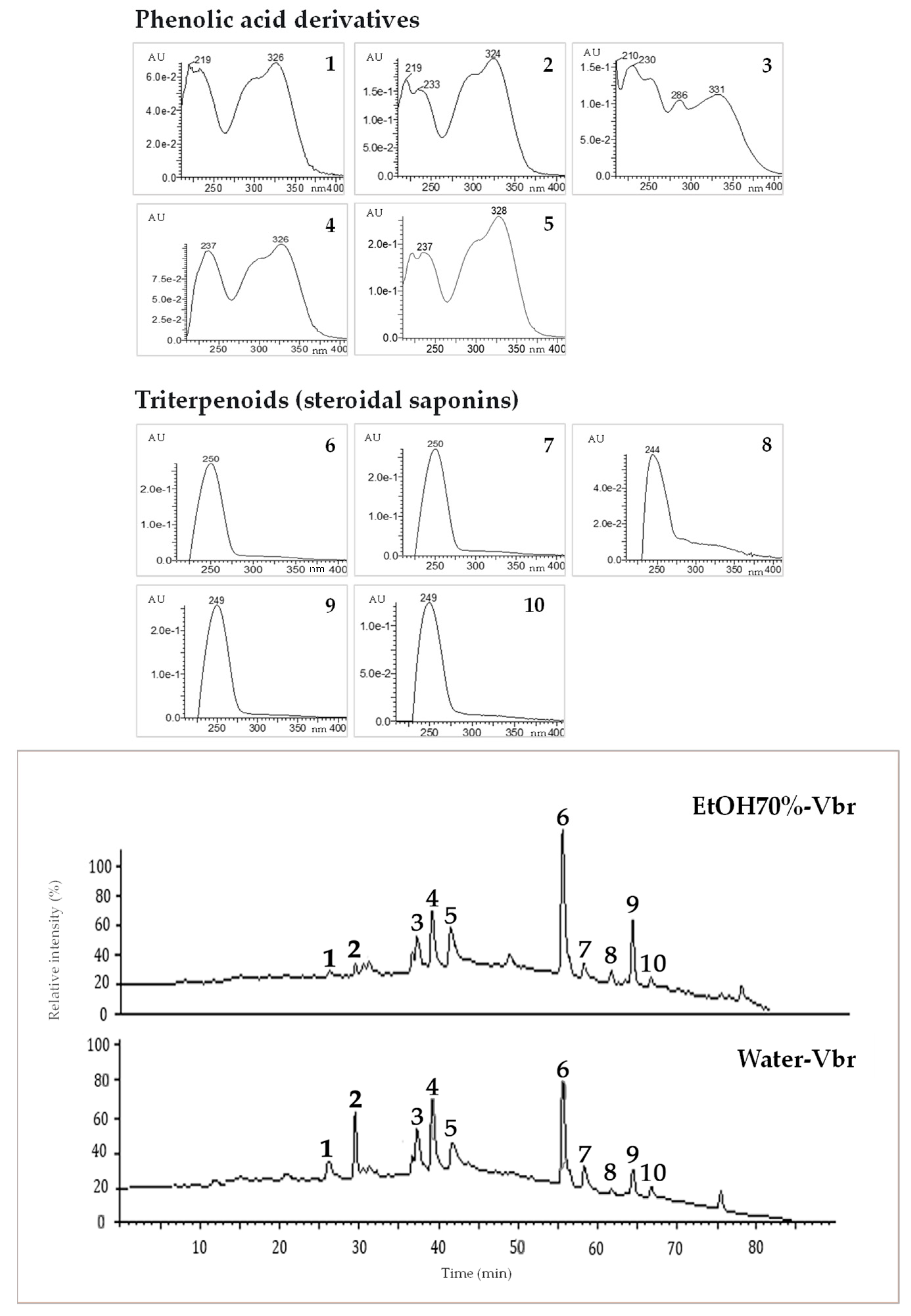
2.1.2. LC-UV-ESI/MS-MS Chemical Profile

The results of V. britteniana root Water-Vbr and EtOH70%-Vbr extract analyzed by LC-UV-ESI/MS-MS are shown in Figure 1 and Table 1. Based on the chromatographic and spectral data, a total of 10 compounds were identified in both extracts, namely one phenolic acid (peak 2), four caffeoylquinic acid derivatives (peaks 1 and peaks 3 to 5), and five triterpenoids (steroidal saponins) (peaks 6 to 10). Obtained data analysis, together with co-chromatography with authentic standards of the commercially available compounds (peaks 1 to 5) and literature data [29,30,31,32,33,34,35], enabled the unequivocal identification of chlorogenic acid (peak 1); caffeic acid (peak 2); 3,4-di-O-caffeoylquinic acid (peak 3); 3,5-di-O-caffeoylquinic acid (peak 4); and 4,5-di-O-caffeoylquinic acid (peak 5) as marker compounds on both extracts. Compared to the reported literature data, peak 6 was tentatively identified as vernoamyoside D [36,37], peak 7 as vernonioside D1 [36], peak 8 as vernoamyoside B [36], peak 9 as vernoniamyoside A [36,37,38], and peak 10 as vernoniamyoside C [36,37,38]. Vernoamyoside D was identified as the principal constituent on both V. britteniana root Water-Vbr and EtOH70%-Vbr extracts.
U—absorbance units; EtOH70%-Vbr—V. britteniana root 70% hydroethanolic extract; nm—nanometers; V. britteniana root Water-Vbr—aqueous extract.

2.1.3. Secondary Metabolites Quantification

The total content of phenols, triterpenoids, and saponins of V. britteniana root Water-Vbr and EtOH-70%-Vbr extracts is presented in Table 2. Both extracts contained saponins as the major classes of secondary metabolites. The Water-Vbr extract showed higher concentrations of all three classes of components relative to the EtOH-70%-Vbr extract. Using the two-tailed unpaired t-test, no statistically significant differences (p > 0.05) were found between these extracts’ total phenols and triterpenoids content. However, statistically significant differences (p = 0.0178) were found between the total saponins content of these extracts. Since saponins are classified as triterpenoids biosynthetically formed from six isoprene units that share the acyclic C30 precursor squalene, and no other type of triterpenoid has been identified in this medicinal plant to date, we conclude that the total triterpenoid content is mainly due to this kind of secondary metabolites [39].

2.2. Biological Activity

2.2.1. Antioxidant Activity

The results of the evaluation of the antioxidant activity of the Water-Vbr and EtOH70%-Vbr extracts from V. britteniana root are presented in Table 3. Both tested extracts showed antioxidant activity, and no statistically significant activity differences were found between these extracts (Water-Vbr p-value > 0.05 vs. EtOH70%-Vbr in both tests). However, in the DPPH assay, the anti-radical activity of both extracts was more than 20-fold higher than that of ascorbic acid. Regarding the results of the FRAP assay, both extracts showed similar ferric iron-reducing capacities, with values approaching those of ascorbic acid.

2.2.2. In Vitro Cercaricidal Activity

The lethal concentration of each V. britteniana root extract required to eliminate 50% of cercariae (LC50) is expressed as the mean and standard deviation. Both extracts, Water-Vbr and EtOH70%-Vbr, showed cercaricidal activity against the larval stage of S. mansoni; the cercariae lacked motility and were deposited at the bottom of the cavity, indicating that they were dead, as shown in Table 4. After 120 min of contact, an LC50 value of 438 µg/mL was obtained for both extracts.
Table 5 shows the results of visible structural changes in cercaria after treatment with root extracts of V. britteniana (Water-Vbr and EtOH70%-Vbr). Both extracts induced structural changes after 1 h of incubation at a 500 µg/mL concentration.
Application of the Student’s t-test showed no significant differences (p = 0.9316) between the mean dead cercariae per time interval by administration of the Water-Vbr and EtOH70%-Vbr extracts. Referring to the mean cercariae killed at a concentration of 438 µg/mL, the unpaired t-test for independent samples was t(df 4) = 0.4472, with a p-value = 0.678.

3. Discussion

Despite efforts to reduce the burden of disease caused by schistosomiasis, it is still the second most important parasitic disease after malaria with significant public health implications in endemic areas of the tropics and subtropics. It is estimated that approximately 785 million people lack access to essential water services, with over 2 billion lacking access to basic sanitation and 3 billion lacking clean water supplies at home [40]. As of 2019, approximately 36% of Angola’s population lived below the poverty line and faced difficulties in accessing basic public services [40]. The World Health Organization Strategic Cooperation Plan (2015–2019) reported that loiasis, lymphocytic filariasis, and schistosomiasis are still of concern in Angola, with an estimated 12 million people at risk of infection and 2.5 million people requiring treatment [41]. As recommended by this entity, praziquantel is the drug also used in Angola for treatment and in MDA control programs. However, due to its use in monotherapies, as well as its ineffectiveness against juvenile worms and the increasing reports of PZQ resistance or loss of sensitivity of parasite isolates [42], there is an urgent need for new alternative treatments [13,43]. Therefore, the use of medicinal plants with pharmacological properties has been considered to be an alternative to treat various diseases such as cancer [18], malaria [20], tuberculosis [21], trypanosomiasis [44], and even schistosomiasis [45].
Several Vernonia species from the African flora are used as an integral part of traditional African medicine. A literature review study conducted by Toyang and Verpoorte (2013) reported that 109 species of Vernonia were used as herbal medicines [46]. Results from in vivo and in vitro biological studies help validate their specific medical utility. Among them, Vernonia amygdalina Del., an African species traditionally used against schistosomiasis, has long been studied for its chemical composition and biological activity, including anti-schistosomiasis activity. Sesquiterpene lactones and steroidal glucosides isolated from this species have been hypothesized to have anti-schistosomal activity [38]. Acheampong et al. (2020) demonstrated the cercaricidal and adulticidal activity of a V. amygdalina whole plant methanol extract against S. mansoni as a function of time and concentration. This extract (3 h IC50 = 35.84 µg/mL) exerted a high cercaricidal activity. The in vivo recovery of the worms after treatment with the plant extract and praziquantel was 48.8% and 59.9%, respectively (p < 0.05), and the mice treated with this extract had lower mean liver and spleen weights compared to those in the untreated groups (p < 0.05) and were considered by the authors to be strongly adulticidal in vivo [45]. In addition, aqueous and ethanolic extracts of this medicinal plant with an LC50 value of 338.8 ppm and an LC90 value of 614.8 ppm proved to be toxic for adult Biomphalaria pfeifferi [45]. The molluscicide whole aqueous and ethanolic extracts of V. amygdalina are also shown against Bulinus globosus with LC50 values of 534 ppm and 208 ppm, respectively [47].
To our knowledge, no studies on the cercaricidal effect of V. britteniana root herbal preparations in the larval stage of S. mansoni have been carried out by others. According to our results, the detected activity (LC50 value of 438 g/mL) seems to be lower than the cercaricidal activity of the V. amygdalina whole plant methanol extract cited above [45]. However, V. britteniana root is explicitly used in traditional Angolan medicine to treat schistosomiasis. Furthermore, no studies on the cercaricidal effect of roots of other Vernonia species were found in the literature.
Consistent with chemotaxonomic data from its botanical genus, we identified phenolic acid derivatives (chlorogenic acid; caffeic acid; 3,4-di-O-caffeoylquinic acid; 3,5-di-O-caffeoylquinic acid; and 4,5-di-O-caffeoylquinic acid) and steroid saponins of the stigmastane type (vernoamyoside D and vernonioside D1; vernoamyoside B; and vernoniamyoside A and C) in V. britteniana root.
Phenolic acids, flavonoids, lactone sesquiterpenes, and steroid saponins) are examples of secondary metabolites identified in V. amygdalina that have anthelmintic properties [48]. The steroid saponins identified in this species, namely vernonioside A1, A2, A3, and B1 showed anti-schistosomal activity [49]. The major marker compounds, vernoamyoside D, vernonioside D1, vernoamyoside B, vernoniamyoside A, and vernonimyoside C, now identified on V. britteniana root might also be involved in the cercaricidal activity of both water and EtOH70% tested extracts.
It has also been reported that dicaffeoylquinic acids are involved in the anti-schistosomal activity of various medicinal plants such as the leaf extracts and branches of Artemisia annua L. and Artemisia afra Jacq. ex Willd [50]. These classes of secondary metabolites have also been recognized as antioxidants and anti-radical agents [30,34].
The antioxidant activity of chlorogenic acid; caffeic acid; 3,4-di-O-caffeoylquinic acid; 3,5-di-O-caffeoylquinic acid; and 4,5-di-O-caffeoylquinic acid isolated from Vernonia anthelmintica seed and from V. amygdalina leaf has been demonstrated [30,51].
The antioxidant activity of the ethanolic extract of Vernonia patula Merril’s whole plant was demonstrated using the DPPH method (IC50 value of 36.59 g/mL) [52]. The aqueous extract of V. amygdalina leaf also showed antioxidant activity, which gave an IC50 value of 1831 ± 0.15 mg/mL by the DDPH method and a mean value of 1.49 ± 0.18 mmol by the FRAP method. The authors justified this activity with the presence of phenolic compounds, such as flavonoids, anthocyanins, and proanthocyanins [53,54,55].
Our results showed that both V. britteniana extracts have a strong antioxidant potential, which can be attributed to the presence of the identified phenolic acid derivatives and steroidal saponins.

4. Materials and Methods

4.1. Chemicals and Reagents

Folin–Ciocalteu reagent; catechin; gallic acid; oleanolic acid; vanillin; 2,2-diphenyl-1-picrylhydrazl (DPPH); chlorogenic acid; caffeic acid; 3,4-di-O-caffeoylquinic acid; 3,5-di-O-caffeoylquinic acid; and 4,5-di-O-caffeoylquinic acid were purchased from Sigma-Aldrich (Bangalore, India). Methanol was purchased from Honeywell (Hamburg, Germany). Sodium acetate trihydrate and 70% perchloric acid were purchased from V.W.R. (Leuven, Belgium). Ferric chloride, sodium carbonate, and acetic acid were purchased from Merck (Darmstadt, Germany). Dimethyl sulfoxide (DMSO) was obtained from Carlo Erba (Val-de-Reuil, France). Formic acid and high-performance liquid chromatography (HPLC) solvents (acetonitrile and methanol) were obtained from Fisher Scientific (Merelbeke, Belgium).

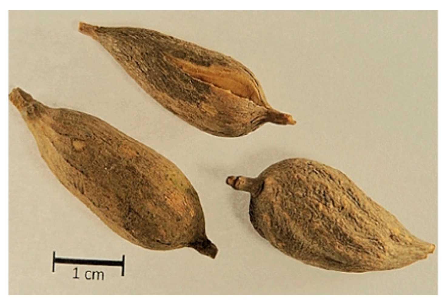
4.2. Collection and Preparation of Plant Material

The roots of V. britteniana (Figure 2) were collected in August 2018 in the Huambo region of Angola in the village of Ndango de Cima (latitude 12°49′17″ S and longitude 15°38′20″ E). After collection, the plant material was dried in the dark and stored in an airy room until further use. Relevant plant parts required for sample identification were also collected, and specimen #LISC13028 was prepared and identified by Maria Cristina Duarte, Scientific Curator of the LISC Herbarium, and from plant collections at the Tropical Botanical Garden and the Lisbon Botanical Garden.

4.3. Extract Preparation

4.3.1. Aqueous Extract

The aqueous extract (Water-Vbr) was prepared to reproduce the recipe provided by traditional medicine practitioners in the Huambo region. Twenty-nine grams of dried V. britteniana root were ground into powder using a grinder (Ika, Multidrive, Staufen, Germany) and extracted by maceration with 1 L of water at room temperature for 90 min. After filtration, the obtained solution was frozen and dried in a lyophilized Heto LyoLab-3000 (Dietikon, Switzerland).

4.3.2. 70% Hydroethanolic Extract

The 70% hydroethanolic extract (EtOH70%-Vbr) was prepared by macerating dried root powder with EtOH70% (ratio 1:10, w/v) at room temperature for 24 h with stirring. After filtration, the obtained extract was concentrated to dryness under reduced pressure (T 40 °C) using a Buchi Rotavapor R-100 (Flawil, Switzerland) [56].

4.4. Phytochemical Studies

4.4.1. LC-UV-ESI/MS-MS Chemical Profile

A Waters Alliance 2695 high-performance liquid chromatography (LC) instrument with autosampler and photodiode array detector (Waters PDA 2996) coupled to a MicroMass Quattromicro API triple quadrupole tandem mass spectrometer (Waters, Drinagh, Ireland). The separation module (Waters, Drinagh, Ireland) comprises a quaternary pump system, degasser, autosampler, and column oven. The chromatograms were recorded at each maximum peak absorbance between the wavelengths of 220 to 410 nm (Maxplot).
The separation was performed on a LiCrospher® 100 RP-18 column 5 µm (250 × 4 mm, Merck); column temperature 35 °C; flow rate 0.3 mL/min; injection volume (20 μL); mobile phase (A) water + 0.1% formic acid/(B) acetonitrile—total time of 90 min under the gradient conditions given in Table 6.
The ionization of the compounds was performed by an electrospray source in positive (ESI+) and negative (ESI) modes at different cone voltages (20 to 40 V). MassLynx software version 4.1 was used for data acquisition and processing.

4.4.2. Total Phenol Content

The phenol content of the Water-Vbr and EtOH70%-Vbr extracts was determined using the Folin–Ciocalteu reagent method [57] with modifications. Briefly, 2 mL of Folin–Ciocalteu reagent (previously diluted 1:10 (v/v) in water) was mixed with 1.6 mL of sodium carbonate (75 g/L) after adding 0.4 mL of sample. After 2 h of incubation at room temperature, protected from light, the absorbance at 765 nm was measured using a Hitachi U-2000 spectrophotometer (Tokyo, Japan). Gallic acid was used as a standard control. Three replicates were performed to obtain the measurements, and the mean and standard deviation values were fitted using the straight-line equation (Y = 00.0016x + 0.0644 and R = 0.9936). Results are presented as mean (±SD) gallic acid equivalents per gram of extract.

4.4.3. Total Triterpenoid Content

The total triterpenoid content of the Water-Vbr and EtOH70%-Vbr extracts was determined using a colorimetric method [58] with modifications. Briefly, 100 μL of the extract was mixed with vanillin/glacial acetic acid (150 μL, 5% (p/v)) and perchloric acid solution (500 μL). The sample solutions were heated at 60 °C for 45 min and then cooled to room temperature. Subsequently, 2.25 mL of glacial acetic acid was added, and the absorbance was measured at 765 nm using a spectrophotometer Hitachi U-2000 (Tokyo, Japan). Oleanolic acid was used as the standard control. Measurements were obtained in triplicate, and the mean and standard deviation values were adjusted using a straight-line equation (Y = 0.0013x + 0.0858 and R2 = 0.9991). The results are presented as the mean (±SD) of oleanolic equivalents per gram of the extract.

4.4.4. Saponins Index (SI)

The saponins content of the Water-Vbr and EtOH70%-Vbr extracts was determined according to the Portuguese Pharmacopeia [59]. Briefly, 1 g of extract was added to 25 mL of alcoholic potassium hydroxide solution (0.5 M) with some glass beads and then heated for 30 min. Then 1 mL of phenolphthalein solution was added, followed by titration with 0.5 M hydrochloric acid (1 mL). A blind test was also carried out under the same conditions. The quantification was based on the formula: SI = 28.05 (n1 − n2)/m.

4.5. DPPH Radical Scavenging Activity

The radical scavenging activity of the Water-Vbr and EtOH70%-Vbr extracts was evaluated using the 2,2-diphenyl-1-picrylhydrazl (DPPH) method [60] with modifications. First, the DPPH solution (3.9 mL, 6 × 10⁻⁵ M) in methanol was mixed with 100 μL of the diluted extract. After incubation at room temperature for 30 min, the absorbance at 517 nm was measured using a Hitachi U-2000 spectrophotometer (Tokyo, Japan). Ascorbic acid was used as a standard control. The scavenging activity of DPPH was calculated using the following formula: % scavenger = (control absorbance, test sample absorbance/control absorbance) × 100. Results are expressed as mean (±SD) and presented as IC50 values.

4.6. Ferric-Reducing Antioxidant Power (FRAP) Activity

The iron (III)-reducing ability of the Water-Vbr and EtOH70%-Vbr extracts was assessed using the Ferric-Reducing Antioxidant Power Assay (FRAP) [61] with modifications. Briefly, 100 μL of the extract was mixed with 3 mL of FRAP reagent (25 mL acetate buffer, 2.5 mL of 2,4,6-tripyridyl-s-triazine, and 2.5 mL FeCl3). 6H2O solution in the portion (1:1:10). After incubation at 37 °C for 4 min, the absorbance at 593 nm was measured using a Hitachi U-2000 spectrophotometer (Tokyo, Japan). Ascorbic acid was used as a standard. Three replicates were performed to obtain the measurements, and the mean and standard deviation values were fitted using a linear equation (Y = 0.001x + 0.0132 and R = 0.9944). Results are presented as mean (±SD) ascorbic acid equivalents per gram of extract.

4.7. Statistical Analysis

Data analysis for total phenol and triterpene content was performed using Microsoft Excel. Results were expressed as the mean (±SD) with units expressed in equivalents of the standards used for each assay. The two-tailed unpaired Student t-test was used to verify the detection of significant differences between the mean content of phenols, triterpenoids, and saponins and the antioxidant activity of Water-Vbr and EtOH70%-Vbr extracts. A p < 0.05 was considered significant Water-Vbr vs. EtOH 70%-Vbr extracts.

4.8. In Vitro Cercaricidal Activity of V. britteniana Root

4.8.1. Sample Preparation

Extemporaneous solutions of each extract (concentration 20 mg/mL) were prepared and used to determine the cercaricidal effect on S. mansoni larvae. First, the aqueous extract was diluted in deionized water, and the hydroethanolic extract was diluted in 1% DMSO. Then the samples were agitated in an ultrasonic bath (BRANSON 3200, USA).

4.8.2. Obtaining Live S. mansoni Cercariae

The assay was performed on live cercariae obtained from the freshwater snail Biomphalaria glabrata (Belo Horizonte, Brazil strain) previously infected with a strain of S. mansoni (Belo Horizonte, Brazil strain), routinely maintained in Mus musculus CD1 male mice at IHMT-UNL.
About eight freshwater snails were placed in a glass beaker containing 10 mL of distilled water and exposed to an artificial light source (70 W lamp) to monitor the release of cercariae by visual observation under a stereomicroscope (4×). All cercaria were placed in a single container. After homogenization, 500 µL of the solution was dispensed onto five glass slides for counting.

4.8.3. Determination of Cercaricidal Activity

The surrounding activity was determined according to the protocol described by Tekwu and co-workers (2017) [62]. In 24-well plates (Polystyrene, Gdynia, Spain), 1 mL of a solution containing approximately 50 cercariae was added to each well. The samples were tested at 125, 438, and 500 g/mL concentrations in 1.5 mL total solution volume. After homogenization with a round stirrer (New Brunswick Scientific, Classic model, Edison Township, NJ, USA), cercariae survival and mortality were observed at intervals of 30 to 120 min incubation using an inverted optical microscope (Olympus CKX41, Japan 4×) and magnification of 10×, 20× and 40×. A positive control (praziquantel 10 µg/mL solution; Thermo Fisher Kandel, Lenzkirch, Germany) and a negative control (distilled water + 1% DMSO) were used in each assay. Mobility, mortality, and structural damage to the cercariae were assessed. Cercariae were considered dead if deposited on the bottom of the well without movement during the time intervals evaluated (30, 60, 90, 120, and 150 min). Altering and destroying the cercaria, such as by severing and crushing their tail, has been considered structural damage. LC50 was determined using the geometric mean of the cercariae count after exposure of each sample to the different concentrations for 2 h and 30 min. All experiments were performed in triplicate.

4.8.4. Statistical Analysis

The data obtained were analyzed using Microsoft Excel software, and the results were expressed as the mean (±SD). An unpaired, two-tailed Student t-test was used to verify the detection of significant differences between the means of the results from the two extracts. A value of p < 0.05 was considered significant concerning Water-Vbr vs. EtOH70%-Vbr extracts.

5. Conclusions

Schistosomiasis is an endemic parasitic disease that is widespread in Africa, particularly in the sub-Saharan region including Angola [10].
The use of traditional medicinal plant preparations is recognized as an added value in combating and eliminating this disease. For the first time, the usefulness of V. britteniana root in schistosomiasis has been evaluated and the major classes of secondary metabolites have been characterized. Phenolic acid derivatives (chlorogenic acid; caffeic acid; 3,4-di-O-caffeoylquinic acid; 3,5-di-O-caffeoylquinic acid; and 4,5-di-O-caffeoylquinic acid) and steroid saponins (vernoniamyoside A and C, vernoamyoside D, vernonioside D1 and vernoamyoside B) were the main components of this medicinal plant. Its possible correlation with the in vitro cercaricidal and antioxidant activities verified with both Water-Vbr and EtOH70%-Vbr extracts will be investigated in the future along with additional studies on the quality, biological activity (against adult worms and eggs), and safety of this medicinal plant.
The present study underlines the importance of ethnopharmacological studies to validate the usefulness of traditional medicines.

Author Contributions

Conceptualization, M.d.A.V. and O.S.; investigation, methodology, and data curation, K.L., M.d.A.V., P.F., S.B. and O.S.; data analysis, M.d.A.V., P.F., I.N. and O.S.; Plant material collection, M.d.A.V., M.P. and P.N.; writing—original draft preparation, M.d.A.V., P.F., R.S., B.S.B., I.B.M.d.S. and O.S.; writing—revision, O.S.; project administration and funding, O.S. All authors have read and agreed to the published version of the manuscript.

Funding

This research was funded by the Foundation for Science and Technology (FCT, Portugal) through national funds FCT/MCTES to iMed.ULisboa (UIDP/04138/2020).

Institutional Review Board Statement

All procedures were conducted in agreement with the animal welfare committee of the Faculty of Pharmacy, Universidade de Lisboa (protocol CEEE 002/16 approved by the Ethics Committee for Animal Experiments (CEEA) in 2016, representing the expert national authority “Direção Geral de Alimentação e Veterinária” (DGAV) according to the EU Directive (2010/63/UE) and Portuguese laws (DR 113/2013, 2880/2015, and 260/2016). Additionally, all experiments were conducted according to ARRIVE Guidelines for Reporting Animal Research.

Informed Consent Statement

Not applicable.

Data Availability Statement

Not applicable.

Acknowledgments

We thank Instituto Nacional de Gestão de Bolsas de Estudo (INAGBE, Angola); Laboratório Central do Instituto de Investigação Veterinária do Huambo (LRIIV, Angola); Centro de Medicina Tradicional Avó Kitoko; and the traditional medicine practitioners from the Province of Huambo for their kind support to our research.

Conflicts of Interest

The authors declare no conflict of interest.

References

  1. World Health Organization. Regional Committee for Africa. Promoting the Role of Traditional Medicine in Health Systems: A Strategy for the African Region. 2000. Available online: https://apps.who.int/iris/handle/10665/95467 (accessed on 15 November 2022).
  2. Gasparetto, J.C.; Martins, C.A.F.; Hayashi, S.S.; Otuky, M.F.; Pontarolo, R. Ethnobotanical and scientific aspects of Malva sylvestris L.: A millennial herbal medicine. J. Pharm. Pharmacol. 2012, 64, 172–189. [Google Scholar] [CrossRef] [PubMed]
  3. Bernardini, S.; Tiezzi, A.; Laghezza Masci, V.; Ovidi, E. Natural products for human health: A historical overview of the drug discovery approaches. Nat. Prod. Res. 2018, 32, 1926–1950. [Google Scholar] [CrossRef]
  4. Newman, D.J.; Cragg, G.M. Natural products as sources of new drugs from 1981 to 2014. J. Nat. Prod. 2016, 79, 629–661. [Google Scholar] [CrossRef]
  5. Ministério da Saúde. Angola—Despesas Públicas no Sector da Saúde 2000–2007; Estudos e Planeamento e Estatística do Ministério da Saúde de Angola: Luanda, Angola, 2007. [Google Scholar]
  6. Simoben, C.V.; Ntie-Kang, F.; Akone, S.H.; Sippl, W. Compounds from African medicinal plants with activities against selected parasitic diseases: Schistosomiasis, trypanosomiasis, and leishmaniasis. Nat. Prod. Bioprospect. 2018, 8, 151–169. [Google Scholar] [CrossRef]
  7. Albuquerque, R.D.D.G.; Mahomoodally, M.F.; Lobine, D.; Suroowan, S.; Rengasamy, K.R.R. Botanical products in the treatment and control of schistosomiasis: Recent studies and distribution of active plant resources according to affected regions. Biology 2020, 9, 223. [Google Scholar] [CrossRef]
  8. Neves, B.J.; Andrade, C.H.; Cravo, P.V.L. Natural products as leads in schistosome drug discovery. Molecules 2015, 20, 1872–1903. [Google Scholar] [CrossRef]
  9. World Health Organization. Fact Sheet Schistosomiasis. 2023. Available online: https://www.who.int/news-room/factsheets/detail/schistosomiasis (accessed on 30 March 2023).
  10. Mendes, E.P.; Okhai, H.; Cristóvão, R.E.; Almeida, M.C.; Katondi, N.; Thompson, R.; Lopes, S. Mapping of schistosomiasis and soil-transmitted helminthiases across 15 provinces of Angola. PLoS. Negl. Trop. Dis. 2022, 16, e0010458. [Google Scholar] [CrossRef] [PubMed]
  11. Gomes, S.E.C.; Mesquita, M.C.S.; Rehn, V.N.C.; Nascimento, W.R.C.; Loyo, R.; Barbosa, C.S. Transmissão urbana da esquistossomose: Novo cenário epidemiológico na zona da mata de Pernambuco. Rev. Bras. Epidemiol. 2016, 19, 822–834. [Google Scholar] [CrossRef] [PubMed]
  12. LoVerde, P.T. Schistosomasis. In Digenetic Trematodes; Toledo, R., Fried, B., Eds.; Springer: Cham, Switzerland, 2019; pp. 45–70. [Google Scholar] [CrossRef]
  13. Oliveira, R.N.; Rehder, V.L.G.; Oliveira, A.S.S.; Jeraldo, V.D.L.S.; Linhares, A.X.; Allegretti, S.M. Anthelmintic activity in vitro and in vivo of Baccharis trimera (Less) DC against immature and adult worms of Schistosoma mansoni. Exp. Parasitol. 2014, 139, 63–72. [Google Scholar] [CrossRef]
  14. Vale, N.; Gouveia, M.J.; Rinaldi, G.; Brindley, P.J.; Gärtner, F.; Correia da Costa, J.M. Praziquantel for schistosomiasis: Single-drug metabolism revisited, mode of action, and resistance. Antimicrob. Agents. Chemother 2017, 61, 5. [Google Scholar] [CrossRef]
  15. Akoto, C.O.; Acheampong, A.; Boakye, Y.D.; Asante, B.; Ohene, S.; Amankwah, F. Anthelminthic, anti-inflammatory, antioxidant, and antimicrobial activities and FTIR analyses of Vernonia camporum stem-bark. J. Chem. 2021, 2021, 3328073. [Google Scholar] [CrossRef]
  16. Habtamu, A.; Melaku, Y. Antibacterial and antioxidant compounds from the flower extracts of Vernonia amygdalina. Adv. Pharmacol. Pharm. Sci. 2018, 2018, 4083736. [Google Scholar] [CrossRef] [PubMed]
  17. Unuofin, J.O.; Oladipo, A.O.; Msagati, T.A.M.; Lebelo, S.L.; Meddows-Taylor, S.; More, G.K. Novel silver-platinum bimetallic nanoalloy synthesized from Vernonia mespilifolia extract: Antioxidant, antimicrobial, and cytotoxic activities. Arab. J. Chem. 2020, 13, 6639–6648. [Google Scholar] [CrossRef]
  18. Lowe, H.I.C.; Daley-Beckford, D.; Toyang, N.J.; Watson, C.; Hartley, S.; Bryant, J. The anticancer activity of Vernonia divaricata Sw against leukaemia, breast and prostate cancers in vitro. West Indian Med. J. 2014, 63, 285. [Google Scholar] [CrossRef]
  19. Dogra, N.K.; Kumar, S. A review on ethnomedicinal uses and pharmacology of Vernonia cinerea Less. Nat. Prod. Res. 2015, 29, 1102–1117. [Google Scholar] [CrossRef]
  20. Bihonegn, T.; Giday, M.; Yimer, G.; Animut, A.; Sisay, M. Antimalarial activity of hydromethanolic extract and its solvent fractions of Vernonia amygdalina leaves in mice infected with Plasmodium berghei. SAGE Open Med. 2019, 7, 2050312119849766. [Google Scholar] [CrossRef] [PubMed]
  21. Kahaliw, W.; Aseffa, A.; Abebe, M.; Teferi, M.; Engidawork, E. Evaluation of the antimycobacterial activity of crude extracts and solvent fractions of selected Ethiopian medicinal plants. BMC Complement. Altern. Med. 2017, 17, 143. [Google Scholar] [CrossRef]
  22. Panda, S.K.; Luyten, W. Antiparasitic activity in Asteraceae with special attention to ethnobotanical use by the tribes of Odisha, India. Parasite 2018, 25, 10. [Google Scholar] [CrossRef]
  23. Rustamova, N.; Gao, Y.; Zhang, Y.; Yili, A. Biological activity of endophytic fungi from the roots of the medicinal plant Vernonia anthelmintica. Microorganisms 2020, 8, 586. [Google Scholar] [CrossRef]
  24. Gahamanyi, N.; Munyaneza, E.; Dukuzimana, E.; Tuyiringire, N.; Pan, C.H.; Komba, E.V.G. Ethnobotany, ethnopharmacology, and phytochemistry of medicinal plants used for treating human diarrheal cases in Rwanda: A review. Antibiotics 2021, 10, 1231. [Google Scholar] [CrossRef]
  25. Boadu, A.; Singh, S.; Karpoormath, R.; Nlooto, M. Review on ethnomedicinal uses, phytochemical constituents and pharmacological evidence on leaf extract of Persea americana and Vernonia amygdalina of the African continent—A Review. Indian Drugs 2019, 56, 7–24. [Google Scholar] [CrossRef]
  26. Alara, O.R.; Abdurahman, N.H.; Ukaegbu, C.I.; Azhari, N.H.; Kabbashi, N.A. Metabolic profiling of flavonoids, saponins, alkaloids, and terpenoids in the extract from Vernonia cinerea leaf using LC-Q-TOF-MS. J. Liq. Chromatog. Relat. Technol. 2018, 41, 722–731. [Google Scholar] [CrossRef]
  27. Dogra, N.K.; Kumar, S.; Kumar, D. Vernonia anthelmintica (L.) Willd.: An Ethnomedicinal, phytochemical, pharmacological, and toxicological review. J. Ethnopharmacol. 2020, 256, 112777. [Google Scholar] [CrossRef]
  28. Valente, M.; Ferreira, P.; Belo, S.; da Silva, I.M.; Nobre, P.; Lima, K.; Neto, I.; Pires, M.; Serrano, R.; Silva, O. In vitro cercaricidal activity and phytochemical profile of Vernonia britteniana root. Planta Med. 2022, 88, 1516. [Google Scholar] [CrossRef]
  29. National Institutes of Health (NIH), National Library of Medicine, PubChem. Available online: https://pubchem.ncbi.nlm.nih.gov/ (accessed on 29 September 2022).
  30. Johnson, C.E.; Lin, L.Z.; Harnly, J.M.; Oladeinde, F.O.; Kinyua, A.M.; Michelin, R.; Bronner, Y. Identification of the phenolic components of Vernonia amygdalina and Russelia equisetiformis. J. Nat. Prod. 2011, 4, 57–64. [Google Scholar]
  31. Sun, J.; Liang, F.; Bin, Y.; Li, P.; Duan, C. Screening non-colored phenolics in red wines using liquid chromatography/ultraviolet and mass spectrometry/mass spectrometry libraries. Molecules 2007, 12, 679–693. [Google Scholar] [CrossRef]
  32. Alara, O.R.; Abdurahman, N.H.; Ukaegbu, C.I. Soxhlet extraction of phenolic compounds from Vernonia cinerea leaves and its antioxidant activity. J. Appl. Res. Med. Aromat. Plants 2018, 11, 12–17. [Google Scholar] [CrossRef]
  33. Willems, J.L.; Khamis, M.M.; Saeid, W.M.; Purves, R.W.; Katselis, G.; Low, N.H.; El-Aneed, A. Analysis of a series of chlorogenic acid isomers using differential ion mobility and tandem mass spectrometry. Anal. Chim. Acta 2016, 933, 164–174. [Google Scholar] [CrossRef]
  34. Zheng, Z.; Wang, X.; Liu, P.; Li, M.; Dong, H.; Qiao, X. Semi-preparative separation of 10 caffeoylquinic acid derivatives using high speed countercurrent chromatogaphy combined with semi-preparative HPLC from the roots of burdock (Arctium lappa L.). Molecules 2018, 23, 429. [Google Scholar] [CrossRef] [PubMed]
  35. Wang, Y.H.; Meng, Y.; Zhai, C.; Wang, M.; Avula, B.; Yuk, J.; Khan, I.A. The Chemical Characterization of Eleutherococcus senticosus and Ci-wu-jia Tea using UHPLC-UV-QTOF/MS. Int. J. Mol. Sci. 2019, 20, 475. [Google Scholar] [CrossRef] [PubMed]
  36. Vasincu, A.; Luca, S.V.; Charalambous, C.; Neophytou, C.M.; Skalicka-Woźniak, K.; Miron, A. LC-HRMS/MS Phytochemical profiling of Vernonia kotschyana Sch. Bip. Ex Walp.: Potential involvement of highly-oxygenated stigmastane-type saponins in cancer cell viability, apoptosis and intracellular ROS production. S. Afr. J. Bot. 2022, 144, 83–91. [Google Scholar] [CrossRef]
  37. Zhao, M.L.; Shan, S.J.; Tao, R.; Cui, L.T.; Li, Q.R.; Luo, J.; Li, Y. Stigmastane-type steroid saponins from the leaves of Vernonia amygdalina Del. Fitoterapia 2021, 150, 104838. [Google Scholar] [CrossRef] [PubMed]
  38. Wang, J.; Song, H.; Wu, X.; Zhang, S.; Gao, X.; Li, F.; Chen, Q. Steroidal saponins from Vernonia amygdalina Del. and their biological activity. Molecules 2018, 23, 579. [Google Scholar] [CrossRef]
  39. Mukherjee, P.K. Quality Control and Evaluation of Herbal Drugs: Evaluating Natural Products and Traditional Medicine; Elsevier: Amsterdam, The Netherlands, 2019; pp. 26–741. [Google Scholar]
  40. World Health Organization. Ending the Neglect to Attain the Sustainable Development Goals: A Global Strategy on Water, Sanitation, and Hygiene to Combat Neglected Tropical Diseases, 2021–2030; Licence: CC BY-NC-SA 3.0 IGO; World Health Organization: Geneva, Switzerland, 2021. [Google Scholar]
  41. Organização Mundial da Saúde. Escritório Regional para a África. Estratégia de cooperação da OMS 2015-2019: Angola. Organização Mundial de Saúde. Escritório Regional Africano. 2016. 58p. Available online: https://apps.who.int/iris/handle/10665/2505162016 (accessed on 26 September 2022).
  42. Wang, W.; Wang, L.; Liang, Y.S. Susceptibility, or resistance of praziquantel in human schistosomiasis: A review. Parasitol. Res. 2012, 111, 1871–1877. [Google Scholar] [CrossRef]
  43. Bergquist, R.; Utzinger, J.; Keiser, J. Controlling schistosomiasis with praziquantel: How much longer without a viable alternative? Infect. Dis. Poverty 2017, 6, 74. [Google Scholar] [CrossRef] [PubMed]
  44. Kimani, N.M.; Matasyoh, J.C.; Kaiser, M.; Brun, R.; Schmidt, T.J. Sesquiterpene lactones from Vernonia cinerascens Sch. Bip. and their in vitro antitrypanosomal activity. Molecules 2018, 23, 248. [Google Scholar] [CrossRef] [PubMed]
  45. Acheampong, D.O.; Owusu-Adzorah, N.; Armah, F.A.; Aninagyei, E.; Asiamah, E.A.; Thomford, A.K.; Anyan, W.K. Ethnopharmacological evaluation of schistosomicidal and cercaricidal activities of some selected medicinal plants from Ghana. Trop. Med. Health 2020, 48, 19. [Google Scholar] [CrossRef] [PubMed]
  46. Toyang, N.J.; Verpoorte, R. A review of the medicinal potentials of plants of the genus Vernonia (Asteraceae). J. Ethnopharmacol. 2013, 146, 681–723. [Google Scholar] [CrossRef] [PubMed]
  47. Oguche, O.; Olofintoye, L.K. Molluscicidal effect of Vernonia amygdalina (Del) and Momordica charantia Linn. on Bulinus (Phy) globosus. Int. J. Multidiscip. Sci. Eng. 2018, 9, 23–28. [Google Scholar]
  48. Oyeyemi, I.T.; Akinlabi, A.A.; Adewumi, A.; Aleshinloye, A.O.; Oyeyemi, O.T. Vernonia amygdalina: A folkloric herb with anthelminthic properties. Beni. Suef. Univ. J. Basic Appl. Sci. 2018, 7, 43–49. [Google Scholar] [CrossRef]
  49. Jisaka, M.; Ohigashi, H.; Takagaki, T.; Nozaki, H.; Tada, T.; Hirota, M.; Koshimizu, K. Bitter steroid glucosides, vernoniosides A1, A2, and A3, and related B1 from a possible medicinal plant, Vernonia amygdalina, used by wild chimpanzees. Tetrahedron 1992, 48, 625–632. [Google Scholar] [CrossRef]
  50. Taljaard, L.; Probst, A.; Tornow, R.; Keiser, J.; Haynes, R.K.; van der Kooy, F. In vitro antischistosomal activity of Artemisia annua and Artemisia afra extracts. Phytomed. Plus 2022, 2, 100279. [Google Scholar] [CrossRef]
  51. Bian, G.L.; Hu, Y.L.; Yan, K.; Cheng, X.J.; Li, D.Q. Characterization of constituents by UPLC-MS and the influence of extraction methods of the seeds of Vernonia anthelmintica Willd.: Extraction, characterization, antioxidant, and enzyme modulatory activities. Heliyon 2022, 8, e10332. [Google Scholar] [CrossRef]
  52. Lyzu, C.; Mitra, S.; Perveen, K.; Khan, Z.; Tareq, A.M.; Bukhari, N.A.; Dashti, M.G. Phytochemical profiling, antioxidant activity, and in silico analyses of Sterculia villosa and Vernonia patula. Evid. Based Complement. Altern. Med. 2022, 2022, 3190496. [Google Scholar] [CrossRef] [PubMed]
  53. Omede, A.; Suleiman, M.S.; Atanu, F.O.; Momoh, S.; Friday, E.T.; Sheneni, V.D.; Jegede, E.R. Evaluation of antioxidant and cytotoxic properties of Vernonia amygdalina. Int. J. Cell Sci. Mol. Biol. 2018, 4, 81–86. [Google Scholar]
  54. Alara, O.R.; Abdurahman, N.H.; Olalere, O.A. Ethanolic extraction of bioactive compounds from Vernonia amygdalina leaf using response surface methodology as an optimization tool. J. Food Meas. Charact. 2018, 12, 1107–1122. [Google Scholar] [CrossRef]
  55. Alara, O.R.; Abdurahman, N.H.; Olalere, O.A. Optimization of microwave-assisted extraction of flavonoids and antioxidants from Vernonia amygdalina leaf using response surface methodology. Food Bioprod. Process. 2018, 107, 36–48. [Google Scholar] [CrossRef]
  56. European Directorate for the Quality of Medicines EDQM. European Pharmacopeia, 1st ed.; Directorate for the Quality of Medicines, Council of Europe: Strasbourg, France, 2019; Available online: https://www.edqm.eu/en/european-pharmacopoeia (accessed on 26 September 2022).
  57. Scalbert, A.; Monties, B.; Janin, G. Tannins in wood: Comparison of different estimation methods. J. Agric. Food Chem. 1989, 37, 1324–1329. [Google Scholar] [CrossRef]
  58. Chang, C.L.; Lin, C.S. Phytochemical composition, antioxidant activity, and neuroprotective effect of Terminalia chebula Retzius extracts. Evid. Based Complement. Altern. Med. 2012, 2012, 125247. [Google Scholar] [CrossRef]
  59. Farmacopeia Portuguesa. Métodos Analíticos-Métodos de Doseamento, 9th ed.; INFARMED: Lisboa, Portugal, 2009. [Google Scholar]
  60. Brand-Williams, W.; Cuvelier, M.E.; Berset, C. Use of a free radical method to evaluate antioxidant activity. LWT Food Sci. Technol. 1995, 28, 25–30. [Google Scholar] [CrossRef]
  61. Benzie, I.F.; Strain, J.J. Ferric reducing/antioxidant power assay: Direct measure of total antioxidant activity of biological fluids and modified version for simultaneous, measurement of total antioxidant power and ascorbic acid concentration. Methods Enzymol. 1999, 299, 15–27. [Google Scholar] [CrossRef] [PubMed]
  62. Tekwu, E.M.; Bosompem, K.M.; Anyan, W.K.; Appiah-Opong, R.; Owusu, K.B.A.; Tettey, M.D.; Kissi, F.A.; Appiah, A.A.; Penla Beng, V.; Nyarko, A.K. In vitro assessment of anthelmintic activities of Rauwolfia vomitoria (Apocynaceae) stem bark and roots against parasitic stages of Schistosoma mansoni and cytotoxicity study. J. Parasitol. Res. 2017, 2017, 2583969. [Google Scholar] [CrossRef] [PubMed]
Figure 1. V. britteniana root extracts’ LC/UV maxplot chromatograms (λ = 220–410 nm) and UV spectral data of main marker secondary metabolites.
Plants 12 01788 g001
Figure 2. Samples of tuberous roots from V. britteniana.
Plants 12 01788 g002
Table 1. LC-UV-ESI/MS-MS identification of main marker V. britteniana root secondary metabolites.
Peak N°tr (min)UVλmax·
(nm)
[M-H]
or [M + H]+
(m/z)
MS-MS
Fragment Ions (m/z)
Assignment
126.31326, 332353 [M-H]191, 179chlorogenic acid
229.64324, 233179 [M-H]135, 89caffeic acid
336.81331, 230515 [M-H]191, 1353,4-di-O-caffeoylquinic acid
439.33326, 237515 [M-H]353, 191, 1793,5-di-O-caffeoylquinic acid
541.96328, 237515 [M-H]353, 191, 1794,5-di-O-caffeoylquinic acid
656.81250649[M + H]+631[M + H-H2O]+
487[M + H-H2O + Glc]+
469[M + H-Glc]+
vernoamyoside D *
758.74250649 [M + H]+631[M + H-H2O]+
469[M + H-Glc]+
451[M + H-H2O-Glc]+
vernonioside D1 *
861.59244649 [M + H]+487[M + H+H2O-Glc]+
469 [M + H-Glc]+
vernoamyoside B *
964.66249647 [M + H]+485[M + H+H2O-Glc]+
467 [M + H-Glc]+
vernoniamyoside A *
1066.80249647[M + H]+485[M + H+H2O-Glc]+
467[M + H-Glc]+
vernoniamyoside C *
* Tentatively assigned based on literature data [34,35,36,37,38]; EtOH70%-Vbr—70% hydroethanolic extract; Glc—glucose; MS-MS—mass spectrometry; m/z—mass-to-charge ratio; [M-H]—negative mass electrospray ionization mode; [M-H]+—positive mass electrospray ionization mode; tr—retention time; UVλmax·—wavelength of maximum absorbance; Vbr—V. britteniana root; Water-Vbr—aqueous extract.
Table 2. Quantification of the main class of secondary metabolites of V. britteniana root.
Class of Secondary MetabolitesExtract
Water-VbrEtOH70%-Vbr
Total phenols (mg GAE/g extract)139.750 ± 3.704 *102.875 ± 1.347
Total triterpenoids (mg OAE/g extract)167.077 ± 2.643 *153.231 ± 16.667
Saponins Index (g extract)370.3 ± 24.450296.0 ± 22.670
EtOH70%-Vbr—70% hydroethanolic extract; GAE—gallic acid equivalents; OAE—oleanolic acid equivalents; SD—standard deviation; Vbr—V. britteniana root; Water-Vbr—aqueous extract; * p-value > 0.05 Water-Vbr vs. EtOH70%-Vbr.
Table 3. Antioxidant activity of V. britteniana root.
ExtractEssay
DPPH
IC50 ± SD
(µg/mL Extract)
FRAP
Mean ± SD
(µg AAE/g Dried Extract)
Water-Vbr1.769 ± 0.049320.800 ± 5.132
EtOH70%-Vbr2.928 ± 0.138286.800 ± 4.780
Ascorbic acid-AAE67.446 ± 0.746256.800 ± 5.706
AAE—ascorbic acid equivalent; EtOH70%-Vbr—70% hydroethanolic extract; IC50—half of the maximum inhibitory concentration; SD—standard deviation; Vbr—V. britteniana root; Water-Vbr—aqueous extract.
Table 4. LC50 of V. britteniana root on S. mansoni cercariae after 120 min in vitro exposure.
ExtractLC50
(µg/mL)
Time
(min)
Mean No. of Dead Cercariae
(µg/mL)
Water-Vbr43812025.7 ± 1.16
EtOH70%-Vbr43812025.3 ± 0.58
PZQ1012050
EtOH70%-Vbr—70% hydroethanolic extract; LC50—lethal concentration that kills 50% of the cercariae; PZQ—Praziquantel, positive control; SD—standard deviation; Vbr—V. britteniana root; Water-Vbr—aqueous extract.
Table 5. Cercariae mortality and structural changes observed after incubation with V. britteniana root.
ExtractConcentration
(µg/mL)
Average Cercaria KilledStructural Changes
Observation Time (min)Observation Time (min)
306090120150306090120150
Water-Vbr125000000----
438017.721.025.70-----
500500000-+---
EtOH70%-Vbr12500000-----
438015.020.725.30-----
5005000000+---
0—no mortality; (-)—no structural changes observed; (+)—structural changes observed; EtOH70%-Vbr—70% hydroethanolic extract; Vbr—V. britteniana root; Water-Vbr—aqueous extract.
Table 6. V. V. britteniana LC-UV-ESI/MS-MS chromatography gradient used.
Time (min)%A%B
0.00955
10.00955
30.008218
44.006436
64.006436
90.001090
Disclaimer/Publisher’s Note: The statements, opinions and data contained in all publications are solely those of the individual author(s) and contributor(s) and not of MDPI and/or the editor(s). MDPI and/or the editor(s) disclaim responsibility for any injury to people or property resulting from any ideas, methods, instructions or products referred to in the content.

