Abstract
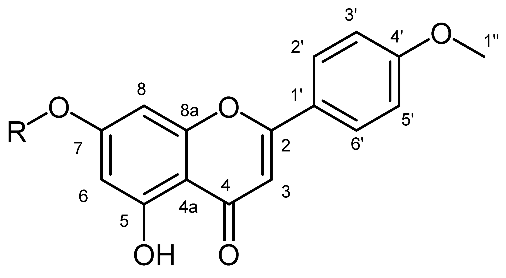
Chemotaxonomy presents various challenges that need to be overcome in order to obtain valid and reliable results. Individual genetic and environmental variations can give a false picture and lead to wrong conclusions. Applying a holistic approach, based on multivariate data analysis, these challenges can be overcome. Thus, a metabolomics approach has to be optimized depending on the subject of research. We used 1H NMR-based metabolomics as a potential chemotaxonomic tool on the selected Euphorbia species growing wild in Serbia. Principal components analysis (PCA), soft independent modeling by class analogy (SIMCA) and Orthogonal Projections to Latent Structures Discriminant Analysis (OPLS-DA) were used to analyze obtained NMR data in order to reveal chemotaxonomic biomarkers. The standard protocol for plant metabolomics was optimized aiming to extract more specific metabolites, which are characteristic for the Euphorbia genus. The obtained models were validated, which revealed that variables unique for each species were associated with certain classes of molecules according to literature data. In E. salicifolia, acacetin-7-O-glycoside (not found before in the species) was detected, and the structure of the aglycone part was solved based on 2D NMR data. In the presented paper, we have shown that metabolomics can be successfully used in Euphorbia chemotaxonomy.
Keywords:
Euphorbia; NMR metabolomics; PCA; OPLS-DA; biomarkers; chemotaxonomy; diterpenes; triterpenes; flavonoids 1. Introduction
Metabolomics analysis has been applied in numerous areas of research related to plant biology and chemistry. Its role could be defined as the extension of traditional phytochemical investigation following the identification of biomarkers that can be statistically correlated to bioactivity changes but also as an attempt to understand complex plant mechanisms as a part of systems biology [1]. In several studies, metabolomics has been helpful in the fingerprinting of species, genotypes or ecotypes for taxonomic or biochemical (gene discovery) purposes [2,3,4,5,6,7,8,9]. For example, the PCA analysis of 1H NMR metabolite fingerprinting was used to discriminate five Verbascum species. Among these species, V. xanthophoeniceum and V. nigrum have higher amounts of the pharmaceutically important harpagoside and verbascoside, forsythoside B and leucosceptoside B [10]. Hierarchical analysis of NMR data was well correlated to the phylogenetic data [11], showing that metabolomics data can be used to make a link between chemotaxonomy and phylogeny. Great care must be taken in such analyses to avoid the influence of environmental and genetic variations of the investigated plants. An unbiased LC-MS-based metabolomics approach applied to Lonicera species flower buds delivered models for classification correlated to taxonomy based on morphological characteristics. Several potential biomarkers were identified using MS-MS analysis and data interpretation [12].
The Euphorbiaceae family, known as the spurge family, includes 322 genera and 8910 species. It is one of the most complex, large and diverse families of angiosperms ranging from large woody trees to simple weeds [13]. Genus Euphorbia is the second largest genus of flowering plants [14] with over 2100 species occurring in all temperate and tropical regions [15]. The taxonomy of Euphorbia is extremely difficult due to the species richness accompanied by a cosmopolitan distribution, the extreme morphological plasticity among certain species and the convergent evolution of certain morphological characters. After recent molecular studies, Euphorbia has been divided into four monophyletic subgenera, including Athymalus (ca. 150 species), Chamaesyce (ca. 600 species), Esula (ca. 500 species) and Euphorbia (ca. 800 species). Many members of the genus Euphorbia contain a poisonous milky-latex sap. In studies performed by Se Jin Park, the metabolomics study of 18 Euphorbia species from the growth chambers was carried out [16]. The latex extraction protocol for LC/MS analysis was optimized. In all tested protocols, polar solvents suitable for LC/MS analysis were used and the numbers of detected peaks were the main elimination criteria. In this study, the author indicated differences in the chemical composition of latex extracts but without further explanation and metabolites identification. In a study performed by S. El-Hawery et al., fifteen Euphorbia species were the subject of metabolomics profiling and searching for species which have the most biologically active compounds against human hepatoma (HepG2) and human breast adenocarcinoma (MCF-7) cell lines [17]. By exploring LC-HRMS and PCA analysis, they identify E. lactea and its two constituents with the molecular formulas C16H18O8 and C20H30O10, which were responsible for cytotoxic activity against MCF-7 and HepG2 cell lines. L. F. Salomé-Abarca et al. used 1H NMR and HPTLC metabolomics in revealing the geographical and inter species variations of two Euphorbia species collected in Serbia [18]. They used methanol extracts of different plant organs (leaves and roots) for their study. Their results showed that the metabolic variation of latexes within species was much more limited than between species and different organs. On the other hand, in order to monitor changes in E. palustris latex after fungal infection, G. Krstić et al. used the 1H NMR spectral data of latex CDCl3 extracts to investigate the effect of plant fungal infections on the chemical compositions of specific metabolites in Euphorbia palustris latex. The infected plants had a greater content of antifungal diterpene metabolites then plants without fungal infection [19]. These results were obtained using multivariate data analysis and in vivo experiments on fungi isolated from infected plants.
In the last three decades, species of the Euphorbia genus have been the subject of extensive phytochemical research [20,21,22,23]. From the results of the aforementioned studies, it is clear that the latex of this genus is rich in specific metabolites of diterpene and triterpene types. These molecules are mostly medium polar or non-polar and the application of the standard protocol for metabolomics analysis of plants is not suitable. Chemotaxonomic differences in the chemical composition of metabolites of Euphorbia species were typically performed by utilizing LC/MS instrumental techniques using an ESI ion source. This ionization technique is suitable for the analysis of polar and some medium polar compounds but not for mostly non-polar, such as diterpenes and triterpenes reported in the latex of the species from the Euphorbia genus. Furthermore, these metabolites usually remain captured in the plant material due to inefficient extraction. MS-based techniques can be useful in the identification of metabolites structure by providing molecular formula or MS/MS fragmentation fingerprints, but the NMR is required for structure identification especially for new compounds [24]. Although the biggest disadvantage of NMR is the low sensitivity compared to MS, realistic molar ratios and the non-selectivity of NMR technique largely overcome this problem, especially in cases where the sample contains compounds of a wide range of polarity as is the case with Euphorbia species.
Consequently, it can be assumed that the application of multivariate analysis would be suitable for a chemotaxonomic study of Euphorbia species, but an optimization and standardization of the extraction protocol is needed. In this paper, we propose an optimized extraction protocol for this purpose and report 1H NMR based metabolomics studies of some Euphorbia species growing wild in Serbia as a potential chemotaxonomic tool.
2. Results and Discussion
2.1. Optimization of the Extraction for the NMR-Based Metabolomic Analysis of Euphorbia Species
In order to obtain as comprehensive a picture as possible of the metabolites present in Euphorbia plants, we tested several solvents for their extraction. For the optimization, we used a standard protocol described by H. K. Kim et al. [25], changing solvents and running 1H NMR spectra with water signals suppression (Figure 1). Solvents for the extraction were selected according to the known literature data. In order to extend the polarity range as much as possible, the following combinations of solvents were tested: 1:1 mixture of deuterated methanol and potassium phosphate buffer in deuterated water (MeOD:KP-D2O); deuterated methanol (MeOD); 1:1 mixture of deuterated methanol and deuterated chloroform (MeOD:CDCl3); and deuterated chloroform (CDCl3). As a model system for these studies, we randomly used finely ground freeze-dried aerial parts (containing stems, leaves and flowers) of E. salicifolia. The criteria for the best solvent for the extraction were the number of the signals in spectra, their intensity and resolution. Additionally, different regions of spectra were integrated, and areas were normalized using the same scale for calibration in comparison to proton signals at oxygenated carbons (from δH 2.9 to 4.8) excluding solvents, in MeOD:KP-D2O extracts (Supplementary Figures S1–S4). In CDCl3 extracts, dominant signals were those of waxes, fatty acids, di- and triterpenes in the region from δH 0.2 to 1.8. More polar metabolites such as sugars and aminoamides were rather sparse, and thus, they were excluded from further investigation. The MeOD:KP-D2O extracts contained a large variety of metabolites, with sugar and amino acid signals dominating (in the region from δH 0.8 to 4.7). They also contained a modest amount and intensity of proton signals from sp2 hybridized carbons as well as protons from sp3 hybridized carbons from di- and tri terpenes skeletons and other non-polar molecules. The “sugar” spectral area contained the most intense signals, whereas those in the aromatic region have a ca. five times smaller area. Comparing the spectra obtained after MeOD and MeOD:CDCl3 extraction showed that more intense and clearly defined signals are observed below δH 0.8 in the latter, and the ratio of areas between the sugar and aromatic areas became much more uniform. According to these data, we had chosen 1:1 mixture of MeOD:CDCl3 as a solvent for this metabolomics study.
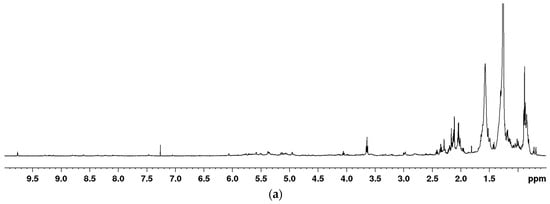
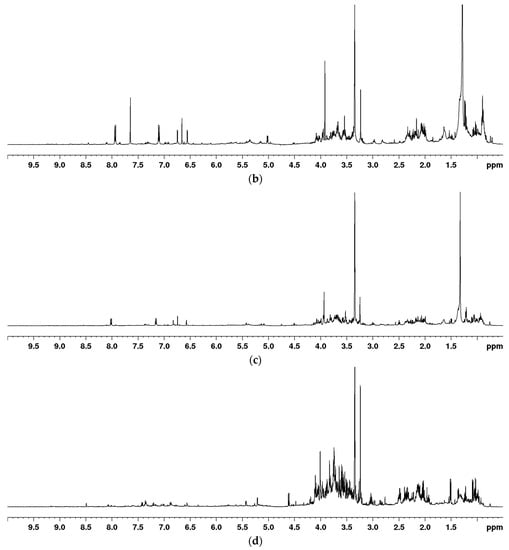
Figure 1.
One-dimensional (1D) NOESY NMR spectra of CDCl3 (a), MEOD/CDCl3 (b), MEOD (c) and MEOD/D2O (d) extracts of E. salicifolia.
2.2. Multivariate Data Analysis
Principal components analysis (PCA) was performed on the NMR metabolomics fingerprints of 60 samples originating from six distinct Euphorbia species. Since PCA is a technique for pattern recognition and unsupervised variable reduction, a smaller number of new variables were formed containing the majority of the original variables’ variation. This analysis yielded a model with eleven principle components that explains 96% of the total variance in the data. E. maculata and E. salicifolia were clearly distinguished from the remaining samples based on the PCA score plots of the first two principal components (Figure 2a). Samples of E. panonica and E. cyparissias were separated along the third component (Figure 2b), whereas E. amygdaloides was separated along the fourth component (Figure 2c). There were no outliers found in any of the score plots.
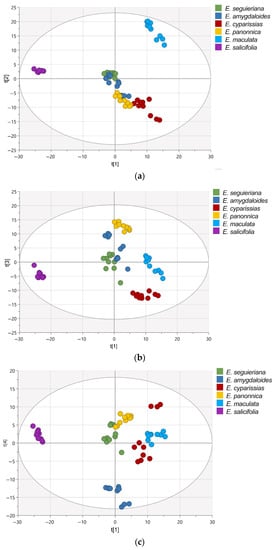
Figure 2.
PCA score plots: PC1/PC2 (a), PC1/PC3 (b), and PC1/PC4 (c). Scores are colored in accordance with Euphorbia species.
The soft independent modeling by class analogy (SIMCA) model was used to confirm the difference between the analyzed Euphorbia species. This is a supervised pattern recognition algorithm based on the PCA of each class individually. The data were separated into training and prediction sets. The logarithmic averaged distances between each class model (DModX) were measured, and class membership was determined by comparing DModX to the critical distance (DCrit). As depicted in Figure 3a–f, all of the samples in the prediction dataset were properly identified, indicating that the model achieved 100% sensitivity and specificity.
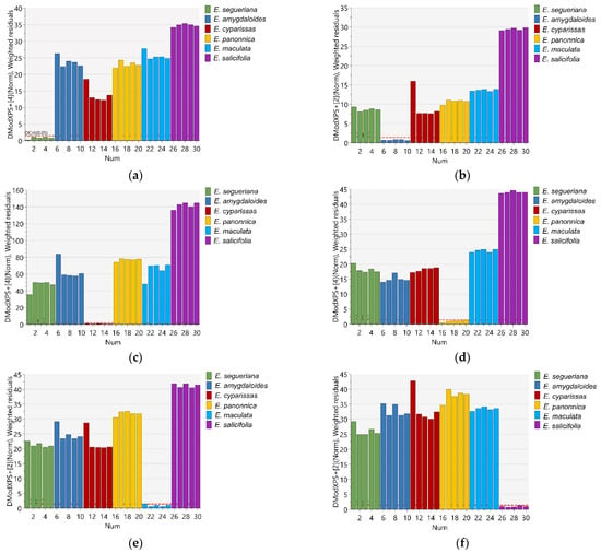
Figure 3.
Logarithmic averaged distance to the models of (a) E. segueriana, (b) E. amygdaloides, (c) E. cyparissias, (d) E. panonnica, (e) E. maculata and (f) E. salicifolia vs. remaining Euphorbia species.
Six OPLS-DA models were used in order to identify metabolites unique to each Euphorbia species investigated. Samples belonging to the species for which distinctive metabolites are to be identified are defined as belonging to one class, while the remaining samples from all other species are defined as belonging to another class. Using this method, novel variables will account for the greatest possible separation between two previously defined classes. Since the systematic variation of variables in the orthogonal model is divided into two components, one of which is linearly related to the class information and the other is orthogonal to it, model interpretation is facilitated [26]. Therefore, OPLS-DA is appropriate for identifying variables with the highest discriminatory power between two preset groups. Cross-validation, permutation testing, and CV-ANOVA were utilized to evaluate the model’s quality (see Supplementary Tables S1–S7 and Figures S5–S10). The most influential variables were chosen based on their impact on the projection scores of the predictive components (VIPpred). Variables having a VIPpred score greater than 1.4 deemed crucial for the separation are shown in Figure 4, Figure 5, Figure 6, Figure 7, Figure 8 and Figure 9.

Figure 4.
Loading plot with red and brown marked variables characteristic for E. seguieriana (Y variables—loading vectors scaled to unit length) in comparison to other species (X variables—chemical shift), obtained from the OPLS-DA model.

Figure 5.
Loading plot with red and brown marked variables characteristic for E. salicifolia (Y variables—loading vectors scaled to unit length) in comparison to other species (X variables—chemical shift), obtained from the OPLS-DA model.

Figure 6.
Loading plot with red and brown marked variables characteristic for E. amygdaloides (Y variables—loading vectors scaled to unit length) in comparison to other species (X variables—chemical shift), obtained from the OPLS-DA model.

Figure 7.
Loading plot with red and brown marked variables characteristic for E. panonnica (Y variables—loading vectors scaled to unit length) in comparison to other species (X variables—chemical shift), obtained from the OPLS-DA model.

Figure 8.
Loading plots with red and brown marked variables characteristic for E. cyparissias (Y variables—loading vectors scaled to unit length) in comparison to other species (X variables—chemical shift), obtained from the OPLS-DA model.

Figure 9.
Loading plot with red and brown marked variables characteristic for E. maculata (Y variables—loading vectors scaled to unit length) in comparison to other species (X variables—chemical shift), obtained from the OPLS-DA model.
The variables that were characteristic and unique for E. seguieriana (Figure 4) in the region from δH 7.3 to 8.8 originate from benzoate substituents of the myrsinol diterpenes type skeleton previously reported by F. Jeske et al. [27]. This assumption is further substantiated with a variable at δH 4.05 from the tetrahydrofuran ring and those in the regions ΔδH = 4.8–4.95 (exomethylene double bonds), ΔδH = 5.85–6.3 (other olefinic protons), acetate esters at δH ca. 2.0 and methylenes at δH 1.8 characteristic for myrsinol diretpenes isolated from E. seguieriana (see Supplementary Figures S11–S14). The same biomarkers were recognized by A. I. Elshamy et al., using agglomerative hierarchical clustering of 32 Euphorbia species based on literature data [28].
According to the literature data, diterpene polyesters and bishomoditerpene lactones are present in E. salicifolia. The three euphosalicins as well as a salicinolide were identified in E. salicifolia by Hohmann et al. [29]. Nevertheless, the highest VIP predictive values exhibited variables at δH 7.94, 7.92, 7.10, 7.08, 6.74, 6.72, 6.65, 6.54, 5.00 and 3. 91 (Figure 5). After further investigation of 2D NMR spectra (see Supplementary Figures S15–S20), it was confirmed that these signals belong to acacetin-7-O-glycoside (Table 1), which can be considered a chemotaxonomic marker for E. salicifolia. Apigenin was previously reported in the roots of E. salicifolia by College et al. [30], but for the first time, apigenin derivative was detected in this species. Other variables from the loading plots were correlated to signals originating from the diterpene skeleton protons, as detected in this species [27].

Table 1.
Structure, 1H and 13C-NMR spectral data of acacetin-7-O-glycoside.
The loading plot of E. amigdaloides contains variables spread across the spectrum (Figure 6) originating from the specific jatrophane diterpenes named amygdaloidins. They contained nicotinate and angeloyl moieties as well as olefinic protons resonating in the region from δH 6.2 to 9.6 (see Supplementary Figures S21–S24). In addition, variables in the area from δH 3.0 to 4.0 are recognized as biomarkers and originate from protons at the oxygenated carbons from the jatrophane skeletons [31,32].
The E. panonnica also known as E. glareosa and E. nicaeensis is rich in diterpenes of the jatrophane and tigliane type [23]. These skeletal types were characterized by a five-membered ring condensed with a twelve-membered ring or seven-membered and six-membered fused rings containing different substituents belonging to jatrophane and tigliane series, respectively. A variable, which corresponds to the chemical shifts the nodal protons of the five-membered ring of tigliane type skeletons (Figure 7), was recognized as a potential biomarker for E. panonnica (see Supplementary Figures S25–S28). These derivatives mostly contain benzoates as aromatic substituents in comparison to jatrophans containing nicotinates. Variables that are not recognized as significant for biomarkers, but are certainly present in E. panonica, are proton signals from the cyclopropane ring which further supports the assumption that benzoate-substituted tiglianes could be used as chemotaxonomic markers for E. panonica.
In contrast to the previously mentioned Euphorbia species, where the highest values of VIP-predictive scores were attributed to signals exhibiting chemical shifts of protons from different diterpene skeletons, in the loading plot of E. cyparissias, the most significant are the variables belonging to the triterpene signals (Figure 8). Previously mentioned protons from the cyclopropane ring (δH 0.41) together with methyl groups of triterpenes (from δH 0.93 to 1.1), methylene skeleton signals at δH 1.72 and 1.92–1.95 as well as a broad singlet signal at δH 4.57 were recognizable in the spectra of acetylated cycloartane [18]. Additionally, two signals in the aromatic region at δH 8.11 and 8.06 are characteristic for E. cyparissias and responsible for the separation (see Supplementary Figures S29–S32).
Accordant to the literature data, E. maculata is rich in the triterpene type of special metabolites [33] and polyphenols [34]. The variables from δH 0.75 to 1.07 and 1.66 characteristic for the E. maculata (Figure 9) match to the methyl groups signals from triterpene skeletons. Variables from δH 3.0 to 4.5 were from proton bonded to oxygenated carbons, and those from δH 5.1 to 7.05 belong to protons on sp2-hybridized carbons with chemical shifts corresponding tannin-type polyphenols (see Supplementary Figures S33–S36) described by I. Agata et al. [34].
3. Materials and Methods
3.1. Chemicals, Samples and Extraction Protocol
The deuterated methanol, deuterated water, KH2PO4 and deuterated chloroform were purchased from Sigma-Aldrich (Saint Louis, MO, USA). Plant material was collected during the May and June 2022. on several locations in Serbia. The samples from E. segueiriana, Neck. Subsp. seguieriana (45°00′00.0″ N 21°01′11.5″ E), E. panonica (44°59′56.1″ N 21°01′09.5″ E and 44°55′48.29″ N 21°11′51.68″ E), and E. cyparissias L. (44°59′07.0″ N 21°01′20.45″ E) were collected at Deliblatska peščara. The samples of E. amygdaloides were collected at Avala (44°41′11.4″ N 20°30′53.2″ E), E. maculata was collected at Zemunski Kej (44°50′41.6″ N 20°24′58.5″ E) and E. salicifolia was collected at Košutnjak (44°45′48.6″ N 20°26′17.3″ E). The identification of plants was carried out by Marijan Niketić (Natural History Museuma, Belgrade, Serbia) and the voucher specimens were deposited at the Herbarium of the Botanical Garden “Jevremovac” University of Belgrade, Belgrade, Serbia (voucher numbers: Euphorbia segueiriana (BEOU17883), Euphorbia panonica (BEOU17884, BEOU17885), Euphorbia cyparissias (BEOU17893), Euphorbia amygdaloides (BEOU17894), Euphorbia maculata (BEOU17881) and Euphorbia salicifolia (BEOU17882)). From each species, ten samples from different locations containing aerial parts of plants were dried and stored on silica gel separately after collection. The samples were ground using a laboratory mill (IKA® A11, IKA®-Werke GmbH & Co. KG, Staufen, Germany) frozen by the addition of liquid nitrogen and stored at −20 °C until analysis. Each sample (50 mg) was measured in 2 mL microtubes and extracted with 0.7 mL of solvent or solvents mixtures on an ultrasonic bath (BANDELIN SONOREX); then, they were centrifuged on 13,400 rpm (MiniSpin Eppendorf) and 500 µL was transferred into 5 mm NMR tubes.
3.2. NMR Measurements and Multivariate Data Analysis
NMR spectra were measured on a Bruker 500 AVANCE III NMR, Fällanden, Switzerland, system equipped with a 5 mm BBI probe head and BVT unit at 298K. All spectra were measured using noesypr1d pulse sequence with 16 scans for optimization and 64 scans for metabolomic studies. For each sample, the transmitter frequency was optimized, and for recorded spectra, the phase and baseline were corrected manually using TopSpin 3.6.pl7 Bruker Biospin, Rheinstetten, Germany software.
The phased 1H-NMR spectra were further processed using the online tool NMRProcFlow v1.4.16, INRA UMR 1332 BFP, Bordeaux Metabolomics Facility, France (https://nmrprocflow.org, accessed on 20 October 2020) for ppm calibration, global baseline correction, and local alignment. To avoid signals of the residual water, MeOH-d4, and CDCl3, respectively, the regions of δH 1.24–1.37, 3.33–3.36, and 7.24–7.26 were removed from the study. The Intelligent Binning approach (resolution factor 0.6, SNR > 10) was used to divide each spectrum into variable size buckets. To build the dataset matrix, the data were normalized to overall spectrum intensity using NMRProcFlow. SIMCA software (version 17, Sartorius Stedim Biotech, Goettingen, Germany) was then utilized for the multivariate data analysis.
4. Conclusions
Presented data confirmed that NMR-based metabolomics involving molecules with a wide range of polarities can be used for the chemotaxonomy of genus Euphorbia. The differences between the analyzed Euphorbia species were confirmed using SIMCA models and distance measurements between each class model. All of the species-specific samples in the prediction dataset were properly identified, and we confirmed model sensitivity and specificity by 100%.
The most important step was the optimization of extraction protocol for plants metabolomics on Euphorbia species. Furthermore, it was demonstrated that the presented optimization has great potential in the more efficient extraction of special metabolites characteristic for this genus and biomarkers for chemotaxonomic classification [29].
As a result of this study, a potentially new metabolite acacetin-7-O-glycoside was detected in E salicifolia, and its presence was confirmed with 2D NMR data. The variables characteristic for myrsinol diterpenes were recognized as biomarkers for E. seguieriana, those characteristic for jatrophanes were recognized as biomarkers for E. amigdaloides, and tiglianes bearing benzoate esters as biomarkers for E. panonnica. The triterpenes with a cyclopropane ring were identified as biomarkers for E. cyparissias, and in E. maculata, these were triterpenes and tannin-type polyphenols.
In conclusion, our experimental data conform to a previously published theoretical study [25] and confirm that our proposed protocol for NMR-based metabolomics could be used in Euphorbia chemotaxonomy.
Supplementary Materials
The following supporting information can be downloaded at: https://www.mdpi.com/article/10.3390/plants12020262/s1, Figure S1: 1H NMR spectrum of E. salicifolia extract of 1:1 mixture of deuterated methanol and potassium phosphate buffer in deuterated water; Figure S2: 1H NMR spectrum of E. salicifolia extract of deuterated methanol; Figure S3: 1H NMR spectrum of E. salicifolia extract of 1:1 mixture of deuterated methanol and deuterated chloroform; Figure S4: 1H NMR spectrum of E. salicifolia extract of deuterated chloroform; Figure S5: Permutation test of OPLS-DA model of E. segueriana vs. remaining E. species; Figure S6: Permutation test of OPLS-DA model of E. salicifolia vs. remaining E. species; Figure S7: Permutation test of OPLS-DA model of E. amygdaloides vs. remaining E. species; Figure S8: Permutation test of OPLS-DA model of E. panonnica vs. remaining E. species; Figure S9: Permutation test of OPLS-DA model of E. cyparissias vs. remaining E. species; Figure S10: Permutation test of OPLS-DA model of E. cyparissias vs. remaining E. species; Figure S11: The 1H NMR spectrum of E. segueriana extract obtained with MeOD:CDCl3 (1:1) with assignment of characteristic resonances; Figure S12: The H,H-J-resoved NMR spectrum of E. segueriana extract obtained with MeOD:CDCl3 (1:1); Figure S13: The COSY NMR spectrum of E. segueriana extract obtained with MeOD:CDCl3 (1:1); Figure S14: The HSQC NMR spectrum of E. segueriana extract obtained with MeOD:CDCl3 (1:1); Figure S15: The 1H NMR spectrum of E. salicifolia extract extract obtained with MeOD:CDCl3 (1:1) with assignment of characteristic resonances; Figure S16: The H,H-J-resoved NMR spectrum of E. salicifolia extract obtained with MeOD:CDCl3 (1:1); Figure S17: The COSY NMR spectrum of E. salicifolia extract obtained with MeOD:CDCl3 (1:1); Figure S18: The NOESY NMR spectrum of E. salicifolia extract obtained with MeOD:CDCl3 (1:1); Figure S19: The HSQC NMR spectrum of E. salicifolia extract obtained with MeOD:CDCl3 (1:1); Figure S20: The HMBC NMR spectrum of E. salicifolia extract obtained with MeOD:CDCl3 (1:1); Figure S21: The 1H NMR spectrum of E. amigdaloides extract extract obtained with MeOD:CDCl3 (1:1) with assignment of characteristic resonances; Figure S22: The H,H-J-resoved NMR spectrum of E. amigdaloides extract obtained with MeOD:CDCl3 (1:1); Figure S23: The COSY NMR spectrum of E. amigdaloides extract obtained with MeOD:CDCl3 (1:1); Figure S24: The HSQC NMR spectrum of E. amigdaloides extract obtained with MeOD:CDCl3 (1:1); Figure S25: The 1H NMR spectrum of E. panonnica extract extract obtained with MeOD:CDCl3 (1:1) with assignment of characteristic resonances; Figure S26: The H,H-J-resoved NMR spectrum of E. panonnica extract obtained with MeOD:CDCl3 (1:1); Figure S27: The COSY NMR spectrum of E. panonnica extract obtained with MeOD:CDCl3 (1:1); Figure S28: The HSQC NMR spectrum of E. panonnica extract obtained with MeOD:CDCl3 (1:1); Figure S29: The 1H NMR spectrum of E. cyparisisas extract extract obtained with MeOD:CDCl3 (1:1) with assignment of characteristic resonances; Figure S30: The H,H-J-resoved NMR spectrum of E. cyparisisas extract obtained with MeOD:CDCl3 (1:1); Figure S31: The COSY NMR spectrum of E. cyparisisas extract obtained with MeOD:CDCl3 (1:1); Figure S32: The HSQC NMR spectrum of E. cyparisisas extract obtained with MeOD:CDCl3 (1:1); Figure S33: The 1H NMR spectrum of E. maculata extract extract obtained with MeOD:CDCl3 (1:1) with assignment of characteristic resonances; Figure S34: The H,H-J-resoved NMR spectrum of E. maculata extract obtained with MeOD:CDCl3 (1:1); Figure S35: The COSY NMR spectrum of E. maculata extract obtained with MeOD:CDCl3 (1:1); Figure S36: The HSQC NMR spectrum of E. maculata extract obtained with MeOD:CDCl3 (1:1); Table S1: Validation paremeters of CV-ANOVA test from OPLS-DA model of E. segueriana vs. remaining E. species.; Table S2. Validation parameters of CV-ANOVA test from OPLS-DA model of E. salicifolia vs. remaining E. species; Table S3. Validation parameters of CV-ANOVA test from OPLS-DA model of E. amygdaloides vs. remaining E. species; Table S4. Validation parameters of CV-ANOVA test from OPLS-DA model of E. panonnica vs. remaining E. species; Table S5. Validation parameters of CV-ANOVA test from OPLS-DA model of E. cyparissias vs. remaining E. species; Table S6. Validation parameters of CV-ANOVA test from OPLS-DA model of E. maculata vs. remaining E. species. Table S7. PCA and OPLS-DA model parameters of tested Euphorbia species.
Author Contributions
Conceptualization and methodology, I.S. and B.A.; software and data curation D.G.; validation, I.S., B.A. and V.T.; formal analysis and investigation, S.I., K.S., J.L. and I.S.; resources, V.T. and S.M.; writing—original draft preparation, B.A. and D.G.; writing—review and editing, S.M.; visualization, S.I.; supervision, V.T.; funding acquisition, S.M. All authors have read and agreed to the published version of the manuscript.
Funding
This research was funded by the Serbian Academy of Sciences and Arts, grant Strategic Project number 01/2022 and financially supported by Serbian Academy of Sciences and Arts projects numbers F188; and Ministry of Education, Science and Technological Development of Republic of Serbia Contract numbers: 451-03-68/2022-14/200168 and 451-03-68/2022-14/200026.
Institutional Review Board Statement
Not applicable.
Informed Consent Statement
Not applicable.
Data Availability Statement
Not applicable.
Acknowledgments
The authors owe a great debt of gratitude to Marijan Niketić from the National History Museum for plants identification and Vlatka Vajs for English language corrections.
Conflicts of Interest
The authors declare no conflict of interest.
References
- Wolfender, J.-L.; Rudaz, S.; Choi, Y.H.; Kim, H.K. Plant metabolomics: From holistic data to relevant biomarkers. Curr. Med. Chem. 2013, 20, 1056–1090. [Google Scholar] [PubMed]
- Böttcher, C.; von Roepenack-Lahaye, E.; Schmidt, J.; Schmotz, C.; Neumann, S.; Scheel, D.; Clemens, S. Metabolome analysis of biosynthetic mutants reveals a diversity of metabolic changes and allows identification of a large number of new compounds in Arabidopsis. Plant Physiol. 2008, 147, 2107–2120. [Google Scholar] [CrossRef] [PubMed]
- Macel, M.; Van Dam, N.M.; Keurentjes, J.J.B. Metabolomics: The chemistry between ecology and genetics. Mol. Ecol. Resour. 2010, 10, 583–593. [Google Scholar] [CrossRef] [PubMed]
- Reynolds, T. The evolution of chemosystematics. Phytochemistry 2007, 68, 2887–2895. [Google Scholar] [CrossRef]
- Duan, L.-X.; Chen, T.-L.; Li, M.; Chen, M.; Zhou, Y.-Q.; Cui, G.-H.; Zhao, A.-H.; Jia, W.; Huang, L.-Q.; Qi, X. Use of the Metabolomics Approach to Characterize Chinese Medicinal Material Huangqi. Mol. Plant 2012, 5, 376–386. [Google Scholar] [CrossRef]
- Cvetković, M.; Anđelković, B.; Stevanović, V.; Jadranin, M.; Đorđević, I.; Tešević, V.; Milosavljević, S.; Gođevac, D. NMR-based metabolomics study of Amphoricarpos species from Montenegro. Phytochem. Lett. 2018, 25, 1–5. [Google Scholar] [CrossRef]
- Alberts, P.S.F.; Meyer, J.J.M. Integrating chemotaxonomic-based metabolomics data with DNA barcoding for plant identification: A case study on south-east African Erythroxylaceae species. S. Afr. J. Bot. 2022, 146, 174–186. [Google Scholar] [CrossRef]
- Shirahata, T.; Ishikawa, H.; Kudo, T.; Takada, Y.; Hoshino, A.; Taga, Y.; Minakuchi, Y.; Hasegawa, T.; Horiguchi, R.; Hirayama, T.; et al. Metabolic fingerprinting for discrimination of DNA-authenticated Atractylodes plants using 1H NMR spectroscopy. J. Nat. Med. 2021, 75, 475–488. [Google Scholar] [CrossRef]
- Lopez, J.M.; Leyva, V.; Maruenda, H. Pure Shift Nuclear Magnetic Resonance: A New Tool for Plant Metabolomics. J. Vis. Exp. 2021, 173, e62719. [Google Scholar] [CrossRef]
- Georgiev, M.I.; Ali, K.; Alipieva, K.; Verpoorte, R.; Choi, Y.H. Metabolic differentiations and classification of Verbascum species by NMR-based metabolomics. Phytochemistry 2011, 72, 2045–2051. [Google Scholar] [CrossRef]
- Kim, H.K.; Saifullah; Khan, S.; Wilson, E.G.; Kricun, S.D.P.; Meissner, A.; Goraler, S.; Deelder, A.M.; Choi, Y.H.; Verpoorte, R. Metabolic classification of South American Ilex species by NMR-based metabolomics. Phytochemistry 2010, 71, 773–784. [Google Scholar] [CrossRef] [PubMed]
- Gao, W.; Yang, H.; Qi, L.-W.; Liu, E.-H.; Ren, M.-T.; Yan, Y.-T.; Chen, J.; Li, P. Unbiased metabolite profiling by liquid chromatography-quadrupole time-of-flight mass spectrometry and multivariate data analysis for herbal authentication: Classification of seven Lonicera species flower buds. J. Chromatogr. A 2012, 1245, 109–116. [Google Scholar] [CrossRef] [PubMed]
- Mwine, J.T.; Damme, P. Van Why do Euphorbiaceae tick as medicinal plants?: A review of Euphorbiaceae family and its medicinal features. J. Med. Plants Res. 2011, 5, 652–662. [Google Scholar]
- Frodin, D.G. History and concepts of big plant genera. Taxon 2004, 53, 753–776. [Google Scholar] [CrossRef]
- Govaerts, R.; Frodin, D.G.; Radcliffe-Smith, A.; Carter, S. World Checklist and Bibliography of Euphorbiaceae (with Pandaceae), 2nd ed.; Royal Botanic Gardens, Kew: Richmond, UK, 2000; pp. 1–4. [Google Scholar]
- Park, S.J. Investigating the Diversity of Latex Metabolites in Species of the Euphorbia. Genus. Thesis, Cornell University, Ithaca, NY, USA, 2012; pp. 1–61. [Google Scholar]
- El-Hawary, S.S.; Mohammed, R.; Tawfike, A.F.; Lithy, N.M.; AbouZid, S.F.; Amin, M.N.; Abdelmohsen, U.R.; Amin, E. Cytotoxic Activity and Metabolic Profiling of Fifteen Euphorbia Species. Metabolites 2021, 11, 15. [Google Scholar] [CrossRef]
- Salomé-Abarca, L.F.; Gođevac, D.; Kim, M.S.; Hwang, G.-S.; Park, S.C.; Jang, Y.P.; Van Den Hondel, C.A.M.J.J.; Verpoorte, R.; Klinkhamer, P.G.L.; Choi, Y.H. Latex Metabolome of Euphorbia Species: Geographical and Inter-Species Variation and its Proposed Role in Plant Defense against Herbivores and Pathogens. J. Chem. Ecol. 2021, 47, 564–576. [Google Scholar] [CrossRef]
- Krstić, G.; Anđelković, B.; Choi, Y.H.; Vajs, V.; Stević, T.; Tešević, V.; Gođevac, D. Metabolic changes in Euphorbia palusrtis latex after fungal infection. Phytochemistry 2016, 131, 17–25. [Google Scholar] [CrossRef]
- Krstić, G.; Kostić, A.; Jadranin, M.; Pešić, M.; Novaković, M.; Aljančić, I.; Vajs, V. Two new jatrophane diterpenes from the roots of Euphorbia nicaeensis: Scientific paper. J. Serbian Chem. Soc. 2021, 86, 1219–1228. [Google Scholar] [CrossRef]
- Jassbi, A.R. Chemistry and biological activity of secondary metabolites in Euphorbia from Iran. Phytochemistry 2006, 67, 1977–1984. [Google Scholar] [CrossRef]
- Kemboi, D.; Peter, X.; Langat, M.; Tembu, J. A Review of the Ethnomedicinal Uses, Biological Activities, and Triterpenoids of Euphorbia Species. Molecules 2020, 25, 4019. [Google Scholar] [CrossRef]
- Krstić, G.; Jadranin, M.; Todorović, N.M.; Pešić, M.; Stanković, T.; Aljančić, I.S.; Tešević, V.V. Jatrophane diterpenoids with multidrug-resistance modulating activity from the latex of Euphorbia nicaeensis. Phytochemistry 2018, 148, 104–112. [Google Scholar] [CrossRef] [PubMed]
- Reynolds, W.F.; Enríquez, R.G. Choosing the Best Pulse Sequences, Acquisition Parameters, Postacquisition Processing Strategies, and Probes for Natural Product Structure Elucidation by NMR Spectroscopy. J. Nat. Prod. 2002, 65, 221–244. [Google Scholar] [CrossRef]
- Kim, H.K.; Choi, Y.H.; Verpoorte, R. NMR-based metabolomic analysis of plants. Nat. Protoc. 2010, 5, 536–549. [Google Scholar] [CrossRef] [PubMed]
- Wiklund, S.; Johansson, E.; Sjöström, L.; Mellerowicz, E.J.; Edlund, U.; Shockcor, J.P.; Gottfries, J.; Moritz, T.; Trygg, J. Visualization of GC/TOF-MS-based metabolomics data for identification of biochemically interesting compounds using OPLS class models. Anal. Chem. 2008, 80, 115–122. [Google Scholar] [CrossRef]
- Jeske, F.; Jakupovic, J.; Berendsohn, W. Diterpenes from Euphorbia seguieriana. Phytochemistry 1995, 40, 1743–1750. [Google Scholar] [CrossRef]
- Elshamy, A.I.; Mohamed, T.A.; Al-Rowaily, S.L.; Abd-ElGawad, A.M.; Dar, B.A.; Shahat, A.A.; Hegazy, M.-E.F. Euphosantianane E-G: Three New Premyrsinane Type Diterpenoids from Euphorbia sanctae-catharinae with Contribution to Chemotaxonomy. Molecules 2019, 24, 2412. [Google Scholar] [CrossRef] [PubMed]
- Hohmann, J.; Evanics, F.; Dombi, G.; Szabó, P. Salicifoline and salicinolide, new diterpene polyesters from Euphorbia salicifolia. Tetrahedron Lett. 2001, 42, 6581–6584. [Google Scholar] [CrossRef]
- Hassana, G.F.; Omera, M.A.; Babadoustb, S.; Naja, D.D. Flavonoids from Euphorbia condylocarpa roots. Int. J. Chem. Biochem. Sci. 2014, 6, 54–60. [Google Scholar]
- Barile, E.; Corea, G.; Lanzotti, V. Diterpenes from Euphorbia as Potential Leads for Drug Design. Nat. Prod. Commun. 2008, 3, 1934578X0800300629. [Google Scholar] [CrossRef]
- Corea, G.; Fattorusso, C.; Fattorusso, E.; Lanzotti, V. Amygdaloidins A–L, twelve new 13 α-OH jatrophane diterpenes from Euphorbia amygdaloides L. Tetrahedron 2005, 61, 4485–4494. [Google Scholar] [CrossRef]
- Sun, Y.; Gao, L.; Tang, M.; Feng, B.; Pei, Y.; Yasukawa, K. Triterpenoids from Euphorbia maculata and Their Anti-Inflammatory Effects. Molecules 2018, 23, 2112. [Google Scholar] [CrossRef] [PubMed]
- Agata, I.; Hatano, T.; Nakaya, Y.; Sugaya, T.; Nishibe, S.; Yoshida, T.; Okuda, T. Tannins and Related Polyphenols of Euphorbiaceous Plants. VIII. Eumaculin A and Eusupinin A, and Accompanying Polyphenols from Euphorbia maculata L. and E. supina RAFIN. Chem. Pharm. Bull. (Tokyo) 1991, 39, 881–883. [Google Scholar] [CrossRef]
Disclaimer/Publisher’s Note: The statements, opinions and data contained in all publications are solely those of the individual author(s) and contributor(s) and not of MDPI and/or the editor(s). MDPI and/or the editor(s) disclaim responsibility for any injury to people or property resulting from any ideas, methods, instructions or products referred to in the content. |
© 2023 by the authors. Licensee MDPI, Basel, Switzerland. This article is an open access article distributed under the terms and conditions of the Creative Commons Attribution (CC BY) license (https://creativecommons.org/licenses/by/4.0/).
