Article

Role of Nodulation-Enhancing Rhizobacteria in the Promotion of Medicago sativa Development in Nutrient-Poor Soils

by
Noris J. Flores-Duarte
1,
Enrique Mateos-Naranjo
2,
Susana Redondo-Gómez
2,
Eloísa Pajuelo
1,
Ignacio D. Rodriguez-Llorente
1 and
Salvadora Navarro-Torre
1,*
1
Departamento de Microbiología y Parasitología, Facultad de Farmacia, Universidad de Sevilla, García González 2, 41012 Sevilla, Spain
2
Departamento de Biología Vegetal y Ecología, Facultad de Biología, Universidad de Sevilla, Apartado 1095, 41012 Sevilla, Spain
*
Author to whom correspondence should be addressed.
Plants 2022, 11(9), 1164; https://doi.org/10.3390/plants11091164
Submission received: 18 March 2022 / Revised: 15 April 2022 / Accepted: 24 April 2022 / Published: 26 April 2022
(This article belongs to the Special Issue Plant–Microbe Interactions for Sustainable Agriculture)

Abstract

:
Legumes are usually used as cover crops to improve soil quality due to the biological nitrogen fixation that occurs due to the interaction of legumes and rhizobia. This symbiosis can be used to recover degraded soils using legumes as pioneer plants. In this work, we screened for bacteria that improve the legume–rhizobia interaction in nutrient-poor soils. Fourteen phosphate solubilizer-strains were isolated, showing at least three out of the five tested plant growth promoting properties. Furthermore, cellulase, protease, pectinase, and chitinase activities were detected in three of the isolated strains. Pseudomonas sp. L1, Chryseobacterium soli L2, and Priestia megaterium L3 were selected to inoculate seeds and plants of Medicago sativa using a nutrient-poor soil as substrate under greenhouse conditions. The effects of the three bacteria individually and in consortium showed more vigorous plants with increased numbers of nodules and a higher nitrogen content than non-inoculated plants. Moreover, bacterial inoculation increased plants’ antioxidant activities and improved their development in nutrient-poor soils, suggesting an important role in the stress mechanisms of plants. In conclusion, the selected strains are nodulation-enhancing rhizobacteria that improve leguminous plants growth and nodulation in nutrient-poor soils and could be used by sustainable agriculture to promote plants’ development in degraded soils.

Graphical Abstract

1. Introduction

Legumes are a family of plants (Fabaceae/Leguminosae) formed by 765 genera and around 19,500 species [1]. This family of plants is characterized by the symbiotic relationship with rhizobia, a group of α- and β-proteobacteria including several genera, among others, such as Bradyrhizobium, Ensifer, Mesorhizobium, Rhizobium, and Sinorhizobium [2]. In this symbiosis, plants offer both a niche and carbon source to the bacteria while the latter provides NH4+ by its ability to reduce atmospheric N2 within the nodules [3]. Thanks to this association, legumes are pioneer plants that can grow and colonize degraded environments, overcoming abiotic stresses (nutrient-poor soils, saline soils, polluted soils, etc.) [4,5] and contributing to enrich degraded soils, with nitrogen improving their quality and fertility [6,7]. For this reason, legumes are used as a transition plant in intercropping to recover soil quality after a crop cultivation [8,9]. Nevertheless, nodulation and the nitrogen fixation effectiveness can be affected by abiotic stress [10,11], which also conditions the rhizobial population present in these kinds of soils [11].
In the rhizosphere, a higher concentration of bacteria exists around roots due to the exudates of plants which bacteria use as a source of nutrients [12,13,14]. Several of these rhizospheric bacteria possess plant growth-promoting (PGP) properties that help plant development and are known as plant-growth-promoting rhizobacteria (PGPR). PGPR assist plants by means of direct and indirect mechanisms such as nutrient (phosphorous, iron, and nitrogen) acquisition, phytohormones production, ethylene level modulation, and the production of lytic enzymes against phytopathogens [5,15,16]. Among these, the solubilization of phosphorous, siderophores production and nitrogen fixation are the major beneficial properties involved in nutrients acquisition. Thus, the presence of these properties in the bacteria helps plants to solubilize insoluble phosphates, improve iron uptake with siderophores, and increases the nitrogen content of the plant through nitrogen fixation [14]. In addition, PGPR can synthesize and produce phytohormones such as auxins (mainly indole-3-acetic acid, IAA), stimulating root elongation and improving nutrient acquisition [14]. The stress level in plants can also be modulated by PGPR’s amino-cyclopropane carboxylic acid (ACC) deaminase activity, an ethylene’s precursor degrading enzyme [14,17].
Among all PGPR, there is a non-rhizobia group which, in addition to its plant promoting properties, improves the symbiotic relationship between legumes and rhizobia enhancing nodulation, known as nodulation-enhancing rhizobacteria (NER) [11]. This kind of bacteria helps the plant to form nodules thanks to PGP properties—IAA production and ACC deaminase activity, the main traits implicated in this nodulation improvement [11].
The aim of this study is to enhance the growth of legumes in soils with nutrient poverty by means of the NER activities in order to recover degraded soils and improve their quality and fertility. For that, we selected soils of the Rio Piedras estuary (Huelva, Spain) as nutrient-poor soil, M. sativa as a model legume, and Ensifer medicae MA11 as the rhizobium to carry out the experiments. The interaction Medicago-Ensifer is well-known [18], and the strain MA11 was isolated from a degraded environment, showing nodulation capabilities under these kinds of conditions [19].
To accomplish the main aim, the following objectives were pursued: (i) isolation of rhizobacteria from the rhizosphere of Medicago spp. from the Rio Piedras estuary; (ii) first screening based on the ability to solubilize phosphate; (iii) identification and characterization of the phosphate solubilizing bacteria displaying additional PGP properties and enzymatic activities; (iv) selection of the best isolated PGPR; (v) determination in vitro of the role of the selected PGPR in the germination and nodulation in M. sativa; (vi) determination of the effect of selected PGPR in the growth and the physiological status of plants of M. sativa grown in soil from the Rio Piedras estuary under greenhouse conditions.

2. Results

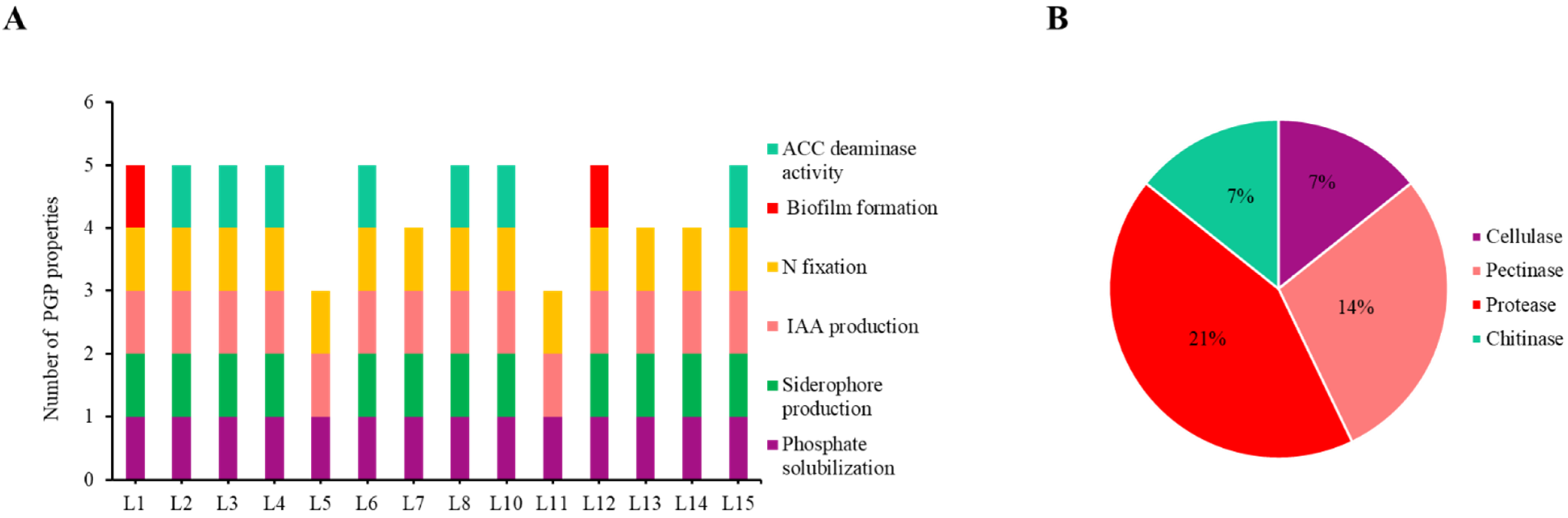
2.1. Isolation and Characterization of Rhizosphere Bacteria

Cultivable bacteria were isolated from rhizosphere soil of Medicago spp. plants from the Rio Piedras estuary. This soil was composed of 92% sand, a very low quantity of organic material (0.98%), and low values of nutrients characterized as a nutrient-poor substrate (Table 1). In addition, pH was alkaline (8.5), and the soil showed a high electric conductivity, corresponding to estuarine sediments.
A total of 71 different bacteria were isolated based on the colonies’ morphology and colour, cell morphology, and Gram staining. Due to the low quantity of P in soils (Table 1), phosphate solubilizing bacteria were searched as a first screening and 14 bacteria were selected (Table S1). Among them, 86% were Gram-negative rods, 7% Gram-positive rods, and 7% sporulated Gram-positive rods (Table S2).
To identify the different isolates, the strains with different colony aspects and different profiles in the Box-PCR (data not shown) were identified by 16S rRNA gene sequencing, and genus Pseudomonas was the most representative (Table 2). The rest of the isolates belonged to genus Chryseobacterium, Priestia, Bacillus, and Butiauxiella.
In relation to the bacterial characterization, PGP properties were checked in the rhizobacteria and all of them showed at least three properties (Figure 1A; Table S1). In addition to phosphate solubilization, all of the isolates were able to produce IAA (Figure 1A). Strains L15, L4, and L8 were the best phosphate solubilizers (18, 14, and 14 mm, respectively), while L15, L10, and L2 produced the highest amount of IAA (11.133, 6.759, and 6.522 mg·L−1, respectively) (Table S1). Eighty-six per cent of the rhizobacteria could fix nitrogen; in addition, siderophores producers had the strain L4 (26 mm of halo; Table S1) as the best one. ACC deaminase activity was present in 50% of the strains, and the highest activity was found in strain L8 with 2.087 μmoles α-ketobutyrate·mg protein−1·h−1, followed by L14 with 1.684 μmoles α-ketobutyrate·mg protein−1·h−1 (Table S1). Finally, biofilm formation was exhibited by L1 and L11, representing 14% of the total bacteria (Table S1).
Enzymatic activities were also studied to characterize the isolates (Table S3). Enzymes such as cellulase, pectinase, protease, and chitinase were detected in the different bacteria. The most abundant enzymatic activity was protease, present in 21% of the isolates, followed by pectinase activity, present in 14% (Figure 1B). On the other hand, cellulase and chitinase activities were found in 7% of the bacteria (Figure 1B). Despite only 21% of isolated bacteria presenting these enzymatic activities, they showed more than one activity, L3 being the strain with more positive enzymatic activities (Table S3).
Based on these results, strains L1, L2, and L3 were selected as inoculants for in planta assays since they had high values in PGP properties and were the only ones that showed the tested enzymatic activities (Tables S1 and S3).

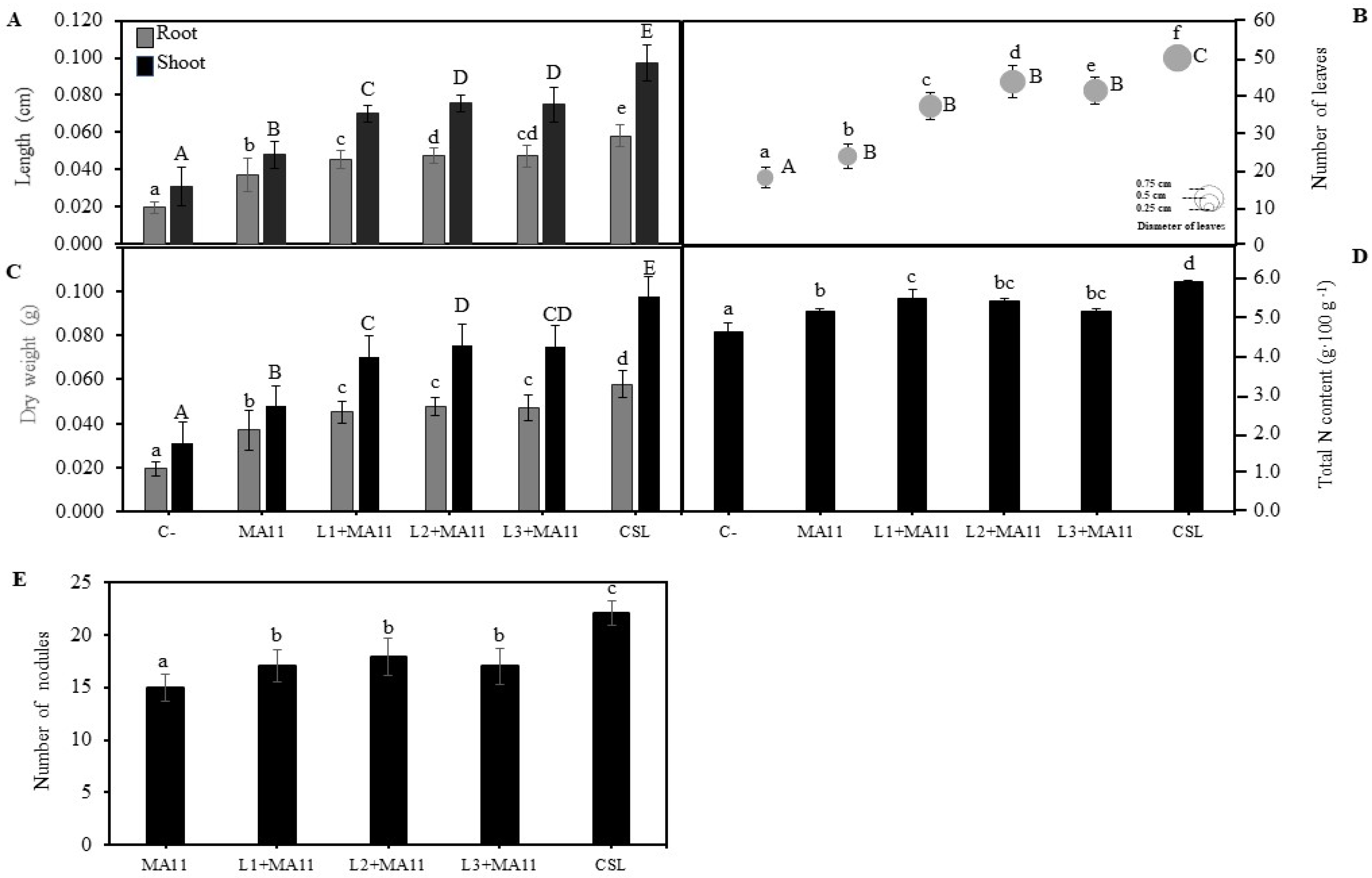
2.2. Effects of Selected PGPR in the Germination, Growth, and Nodulation of M. sativa Plants In Vitro

The selected strains were inoculated to M. sativa seeds to observe their effect on germination. Figure 2A shows that inoculated seeds had a higher percentage of germination than non-inoculated control seeds, particularly those inoculated with the bacterial consortium (CSL treatment), reaching 100% of germinated seeds versus 81% of non-inoculated ones. When tested individually, the three PGPR had a similar germination percentage improvement compared with control seeds, although not as high as the consortium.
In addition to germination, as a preliminary test before greenhouse experiments, the effect of selected PGPR´s inoculation on plant growth and nodulation was studied in vitro. Inoculated plants had more biomass, both shoots and roots, than control plants without inoculation (Figure 2B). Individually, strain L2 inoculation correlated with M. sativa highest dry weight values, particularly in shoots, showing a biomass increase regarding the control of 240% and regarding MA11 inoculation of 54%. However, the more vigorous plants were those inoculated with the consortium, showing an increase on dry weight regarding the controls of 280%. Regarding nodulation, the selected PGPR inoculated individually increased the number of nodules per plant, although the consortium once again showed the highest number, increasing two-fold the nodulation by MA11 (Figure 2C).

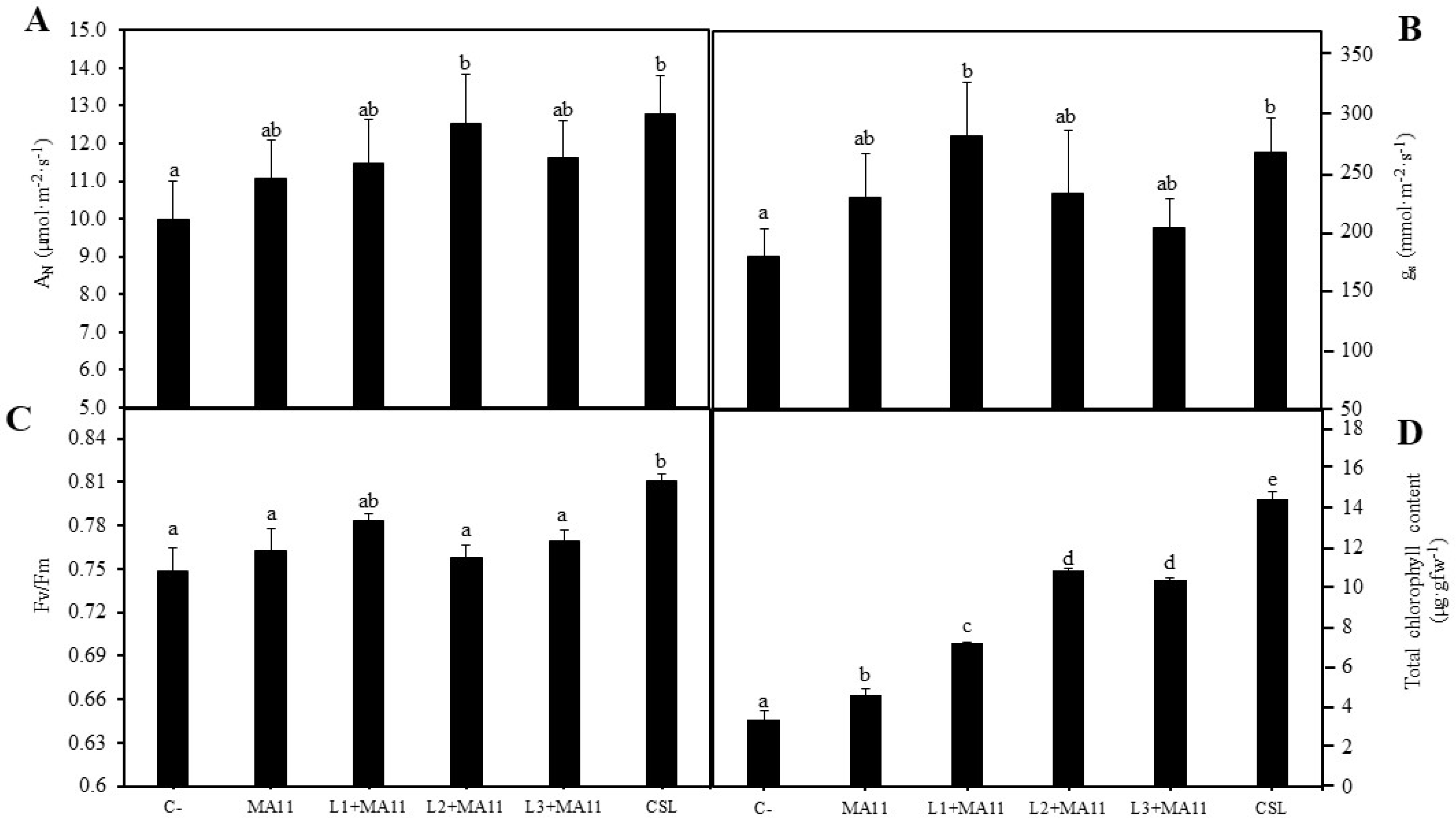
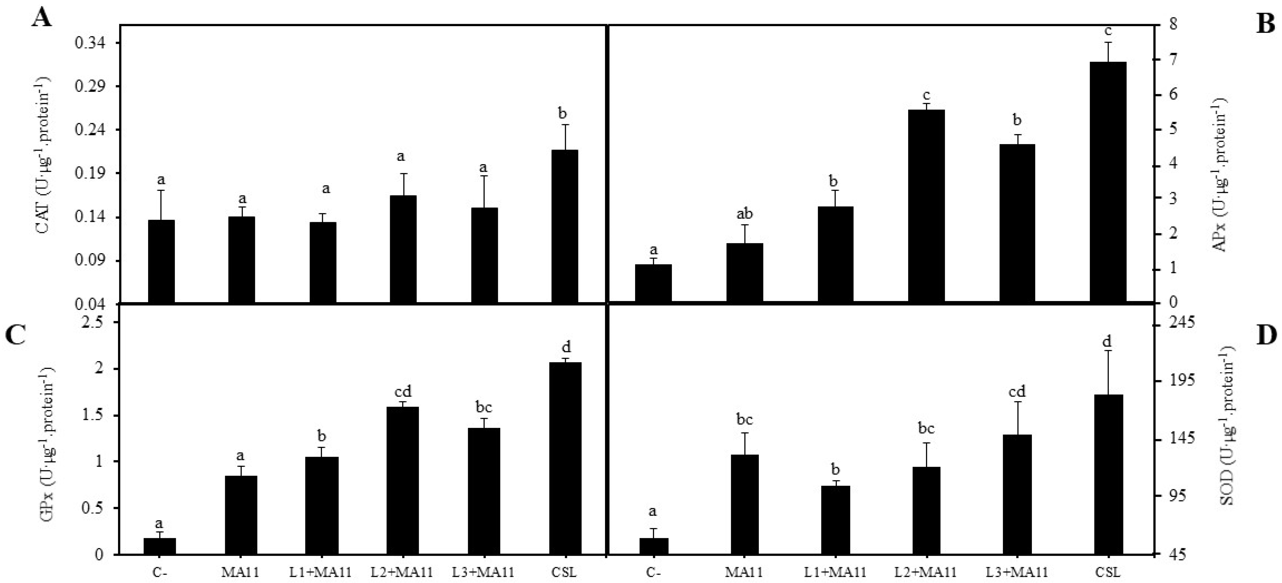
2.3. Effects of Selected PGPR in the Physiological Status and Nodulation of Plants under Nutrient-Poor Soil Stress

M. sativa physiological and nodulation changes induced by in vitro inoculation with selected PGPR were also tested under greenhouse conditions using as substrate a nutrient-poor soil collected from the Rio Piedras estuary. Both the length and size of plants and number of leaves were significantly increased with PGPR’s individual inoculation. The highest values were obtained with the consortium inoculation (Figure 3A,B; Figure S1A,B). Similarly, the control plants’ total biomass was surpassed by the inoculated plants’ biomass, particularly those inoculated with the consortium (Figure 3C). PGPR also improved plant nodulation in nutrient-poor soil. Plants inoculated with the consortium showed a significantly higher number of nodules than those inoculated individually (Figure 3E). Among plants with single inoculation, strain L2 showed the highest values regarding the tested physiological parameters, although no statistically significant difference was observed (Figure 3A–C,E). N content in plants also increased in inoculated plants, particularly with the consortium of PGPR (Figure 3D).
Several parameters were determined in order to establish the physiological state of the plants, namely, the net photosynthetic rate (AN), the stomatal conductance (gs), the intercellular concentration of CO2 (Ci), the efficiency of photosystem II (Fv/Fm), and total chlorophyll content. Regarding gas exchange in plants inoculated with different strains, the net photosynthetic rate was higher in those inoculated with L2 strain and the consortium with similar values (Figure 4A). In the case of the stomatal conductance, plants inoculated with L1 strain showed the highest value, followed by plants inoculated with the consortium without significant differences (Figure 4B). The values of intercellular CO2 concentration in plants were very similar in all of the inoculation conditions, plants inoculated with strain L1 outlined significantly (Figure S2A). In addition, results related to plants’ photochemistry were also statistically very similar among the different inoculation treatments, where the performance of plants inoculated with CSL stood out over the others, showing the highest values in all of the tested parameters (Figure 4C; Figure S2B,C). On the other hand, the total chlorophyll content results showed significant differences among the different inoculations (Figure 4D). Individually, plants inoculated with strains L2 and L3 produced amounts of chlorophyll with significant difference compared with controls; however, the highest increase appeared in plants inoculated with the consortium, with five-fold more chlorophyll than non-inoculated plants and three-fold more than plants inoculated with MA11.
Finally, in order to see the effect of selected bacteria on the stress mechanisms of plants due to the low quantity of nutrients in the substrate, the activities of antioxidant enzymes were measured in leaves. In general, the activities of all tested enzymes increased with bacterial inoculation compared with non-inoculated plants, except for catalase activity (Figure 5A–D). This enzyme activity was statistically similar in all of the treatments apart from CSL inoculation, whose plants showed significantly more activity (Figure 5A). Inoculation with the consortium increased the activities of all of the enzymes, although the inoculation with strain L2 also significantly increased the activity of ascorbate peroxidase and guaiacol peroxidase (Figure 5B,C). Superoxide dismutase was the enzyme with highest activity increase in all of the treatments, while catalase showed the lowest activity change.

3. Discussion

For decades, natural and human activities have caused negative effects in lands, giving rise to degraded soils characterized by poor quality, a decrease in fertility and nutrients content, and an increase of abiotic stresses such as salinity, heavy metals content, or drought. The presence of abiotic stresses in soils reduces plant biodiversity and creates an imbalance of nutrients [20]. Nutrient-poor soils produce a loss of microbial biodiversity and fertility and prevent the growth and development of plants, decreasing the ecological value of estuaries [21,22]. In addition, low fertility is one of the main limitations in crop production [23], so the loss of fertile soils is a worldwide challenge regarding food security [24].
A possible solution for the ecological restoration of agricultural lands is the use of legumes as cover crops. Legumes are beneficial in agricultural practices because their use reduces greenhouse effect gases, reduces the need for nitrogen based artificial fertilizers, avoids soil erosion, and, most importantly, provides organic compounds and nitrogen, increasing the fertility, nutrient content, and quality of soils [25,26,27].
The main goal of this work is the nodulation and development improvement of legumes, particularly M. sativa, in environments with nutrients poverty in order to use them as pioneer crops to recover the quality of degraded soils, promoting a sustainable agriculture. M. sativa was selected, in addition to the benefits that legumes provide to soils, because it is an important forage crop digestible for animals with great yield and high nutrient values [28]. For nodulation to be effective under stress conditions, proper rhizobial strains with high resistance towards stress must be used. In our case, Ensifer medicae MA11 had previously been shown to effectively nodulate Medicago plants under arsenic stress [19]. In addition to using a tolerant rhizobial strain, the isolation, characterization, and selection of PGPR and NER from the rhizosphere of Medicago spp. plants growing in a nutrient-poor estuarine soil were performed, since PGPR can assist plant growth through direct and indirect mechanisms [14,15,16], and NER can improve legumes nodulation [11].
Rhizospheric soil collected from the Rio Piedras estuary was mainly composed of sand, making it a light and drain soil. Furthermore, this soil had less than 1% of organic material and a low quantity of nutrients. These characteristics correspond to the definition of nutrient-poor soil [29,30,31], so the estuary of Rio Piedras could be considered as a degraded environment. The knowledge of the microbial population of this degraded soil is very important to find PGPR and NER able to promote legumes growth under the scarcity of nutrients [32]. In this work, 71 cultivable bacteria were isolated and, due to the low level of phosphorus in the soil, a first screening was performed, selecting those bacteria capable of solubilizing phosphate, reducing the selection of isolates to 14. Phosphorus is one of the most limiting factors for plant development, and phosphate solubilizing bacteria make it available to plants through the secretion of phytases, phosphatases, and organic acids into the soil [33]. The diversity of root-associated bacteria depends on the environment and its stress level [34,35,36,37,38], showing a lower number of microorganism taxa and fewer interactions among taxa in degraded soils with erosion [35,39]. This low diversity was also observed in rhizosphere of Medicago spp. From the Rio Piedras estuary at the genus level, Pseudomonas being the most representative (Table 2). The presence of Pseudomonas as the most represented genus among the cultivable bacteria in this study is not a surprise since it is ubiquitous in soil and has well known genetic, environmental, and physiological adaptability to survive in any environment [40]. In addition, rhizosphere bacteria belonging to genus Pseudomonas had been isolated from other legumes such as peanut, soybean, and broad bean [41,42,43,44]. The rest of the genera isolated in this work, namely, Chryseobacterium, Priestia, Bacillus, and Buttiauxella, have been also described as associated to legumes by other authors [45,46,47,48,49,50].
In addition to phosphate solubilization, isolated bacteria showed individually several PGP properties (Figure 1A). IAA is an auxin, a phytohormone involved in numerous processes in plant development, mainly in root elongation facilitating nutrient absorption by plants [51]. Moreover, IAA is also involved in nodule formation because it intervenes in the relationship between rhizobia and legume, and the functionality of the nodule gets to delay the senescence by the interaction with the bacteroid inside the nodules [11]. This property was observed in all isolates, making all of them good candidates to improve the nodulation of legumes growing in degraded estuaries. Siderophores production is another important PGP property in degraded soil with nutrient deficiency because they have affinity for iron, forming a complex that can be assimilated by plants [52,53]. Furthermore, the production of siderophores is also related to biotic control due to competition for Fe with phytopathogens [12]. A total of 87% of isolated strains in this study were grown in minimal medium without a nitrogen source, indicating that they could fix nitrogen, which would additionally increase the amount of nitrogen fixed within the nodules [54]. ACC deaminase, detected in half of the rhizobacteria (Figure 1A), allows bacteria to modulate the ethylene concentration in plants since this enzyme hydrolyzes the ethylene’s precursor [55,56]. With this modulation, bacteria promote plant growth under stress conditions [57] and even improve the nodulation and the functionality of the nodules because ethylene is also involved in nodule senescence [58,59,60]. Finally, although biofilm formation was the less represented property among isolates, it could help plants to grow in degraded environments since biofilm concentrates bacteria coating roots and facilitates important processes like nitrogen fixation, the mineralization of organic N and P, and nutrients absorption [61].
The secretion of lytic enzymes such as chitinases, lipases, pectinases, or proteases acting against the wall of phytopathogens are also interesting traits to enhance plant development [55]. In this respect, only L1, L2, and L3 showed enzymatic activities for protease, pectinase, and chitinase. Moreover, strain L3 also showed cellulase activity, important to degrading the vegetal cell wall and facilitating the rhizobia entry in roots [62,63]. These three strains, Pseudomonas sp. L1, Chryseobacterium soli L2, and Priestia megaterium L3, which also showed five of the six tested PGP properties with high values individually and all of the PGP properties altogether, were selected to continue with plant experiments.
The inoculation of M. sativa seeds with selected strains showed that all of them increased the number of germinated seeds individually, although inoculation with the three (consortium) showed the highest number of germinated seeds. This positive effect in M. sativa seed germination was also reported in [64], where Bacillus spp. strains improved the number of germinated seeds.
The results concerning plant development and nodulation were similar both in vitro and under greenhouse and nutrient-poor conditions. Inoculated plants showed an increase in both shoots and roots development, the increase in those inoculated with the consortium being the highest one. Supporting these results, several authors verified that the inoculation of M. sativa plants with PGPR improved plant biomass and length in nutrient-poor substrate, both in vitro [65] and in greenhouse conditions [66] and even in the presence of other abiotic stresses such as salinity and heavy metals [28,64,67,68]. In addition to plant development, the nodulation in plants was also higher in inoculated groups, in agreement with the results obtained in other studies where the nodulation in M. sativa was tested under stress conditions [69]. The fact that co-inoculated plants had a greater number of nodules could be related to the nitrogen content in plants since they also showed higher values of nitrogen [28].
The positive effect in plant physiology was also observed in photosynthetic parameters since inoculation with the consortium improved the photosynthetic status in plants under nutrient poverty stress. Similarly, in M. sativa plants growing in a substrate with a low quantity of phosphorous, the inoculation with Priestia megaterium, formerly Bacillus megaterium [70], increased the chlorophyll content [66], and in M. sativa plants under heavy metals stress, inoculation was involved in higher values of physiological parameters such as the ETR, Fv/Fm, ΦPSII, AN, and gs [66].
Finally, the stress level in plants growing in nutrient-poor estuarine soils was determined by the measurement of antioxidant enzymatic activities. The inoculation with the consortium of the selected bacteria showed a significant increase in these enzymatic activities. This increase was also observed in other studies with M. sativa plants under different abiotic stresses [28,64], suggesting that bacterial inoculation could elevate the stress response in plants, improving and recovering the plant status in degraded environments.
According to the results, the inoculation of M. sativa with the consortium of the three selected strains showed more vigorous plants with more nodules than the single inoculations, suggesting that the consortium had the higher effect on the physiological status and nodulation of alfalfa plants under nutrient-poor soil stress. These positive effects could be due to the presence of protease, pectinase, chitinase, and cellulose activities together. As mentioned above, cellulase activity is involved in the nodulation process [62,63] because it facilitates the entry of the rhizobium in the root. In a similar way, pectinase can also degrade the cell wall and could increase the nodulation when plants are inoculated with the consortium [63]. In relation to the improvement in the germination, protease could be involved in the mobilization of the protein reserves in seeds during germination. The presence of all of the PGP properties in the consortium could be the reason for the increase in all of the measured parameters. The biofilm formation and the ACC deaminase activity seem to be the main differences that provide the consortium with the stronger effects, improving the acquisition of nutrients and decreasing the ethylene levels in plants in response to the nutrient poverty in soils [17,61]. More studies should be performed in order to elucidate the role of each PGP property in the plant and nodulation improvement.

4. Materials and Methods

4.1. Collection and Characterization of Soil

Rhizospheric soil of Medicago spp. plants from the Rio Piedras estuary (Huelva, Spain) (37°16′09.1″ N–7°09′36.4″ W) was collected in May 2019. Three rhizosphere samples were picked between 10–20 cm of depth from random plants with gloves and deposited into sterile bottles to be transported to the laboratory. Samples were stored at 4 °C until their utilization.
Soil samples were analyzed chemically as described [71]. The concentration of nutrients in soils was determined by inductively coupled plasma–optical emission spectroscopy (ICP–OES) (ARLFisons3410, Thermo Scientific, Walthman, MA, USA). Conductivity was measured using a Crison-522 (Crison Instrument, S.A., Barcelona, Spain) conductivity meter and redox potential and pH with a Crison pH/mVp-506 (Crison Instrument, S.A., Barcelona, Spain) portable meter [71]. Soil texture (sand, silt, and clay percentage) was determined using the Bouyoucos hydrometer method [72].

4.2. Isolation of Cultivable Rhizosphere Bacteria

Rhizosphere bacteria were isolated from collected soils following the protocol described in [73]. Briefly, soil samples were mixed with sterile 0.9% (w/v) saline solution by agitation for 10 min. Then, after soil decantation, supernatant (100 µL per plate) was deposited into three Petri plates containing tryptic soy agar (TSA) (Intron Biotechnology, Gyeonggi-do, Korea) medium. Inoculated plates were incubated for 72 h at 28 °C. Different bacteria were separated in different TSA plates based on the color and morphology of colonies and on the Gram staining. The pure cultures were stored in sterile 15% (v/v) glycerol at −80 °C.

4.3. Bacterial Screening Based on Phosphate Solubilization

In order to reduce the number of isolated bacteria with potential as PGPR, a first screening was done to select those isolates able to make phosphorus available to plants. For that, bacteria were incubated in NBRIP (National Botanical Research Institute’s phosphate growth medium) plates [74] for 5 days at 28 °C. Phosphate solubilization was observed as a transparent halo around the bacterial colony. The diameter of the halo was measured to semi-quantify the degree of phosphate solubilization.

4.4. Analysis of Diversity and Identification of Isolates

The diversity of P-solubilizers was first analyzed by Box-PCR. Genomic DNA was isolated from bacteria using a G-spin™ Genomic DNA Extraction (for Bacteria) kit (Intron Biotechnology, Gyeonggi-do, Korea) following the instructions supplied by the manufacturer. Box-PCR was performed using 1 µL of DNA and Box A1R primer (5′-CTACGGCAAGGCGACGCTGACG-3′) using the Maxime™ PCR PreMix kit (i-Taq™) (Intron Biotechnology, Gyeonggi-do, Korea) and following the next PCR conditions: initial denaturation at 94 °C for 2 min, 30 cycles of denaturation at 94 °C for 20 s, annealing at 52 °C for 20 s, extension at 72 °C for 1 min, and final extension at 72 °C for 5 min. Electrophoresis was performed in a 1.5% (w/v) agarose gel and a voltage of 70 V for 2 h.
Representative bacteria of each different Box-PCR profile were identified by 16S rRNA gene amplification using 16F27 and 16R1488 primers [73] and the Maxime™ PCR PreMix kit (i-Taq™) (Intron Biotechnology, Gyeonggi-do, Korea) following the next PCR conditions: initial denaturation at 94 °C for 2 min, 30 cycles of denaturation at 94 °C for 20 s, annealing at 58 °C for 10 s, extension at 72 °C for 50 s, and final extension at 72 °C for 5 min. Electrophoresis was performed in a 1% (w/v) agarose gel and a voltage of 120 V for 30 min. PCR products were purified with the enzyme ExoSAP-IT (Affymetrix, Santa Clara, CA, USA), following the manufacturer instructions, and sequenced by the StabVida company (Caparica, Portugal). Then, 16S rRNA gene sequences were compared with those deposited in the EzBioCloud database [75] using the Ez-Taxon-e service (www.ezbiocloud.net/eztaxon; accessed on 7 July 2021). Finally, 16S rRNA sequences were deposited in the NCBI GenBank.

4.5. Bacteria Characterization

4.5.1. Study of Plant Growth Promoting Properties In Vitro

In addition to phosphate solubilization, indole-3-acetic acid (IAA) and siderophores production, nitrogen fixation, biofilm formation, and ACC deaminase activity were tested. IAA production was evaluated by incubating bacteria in tryptic soy broth (TSB) (Intron Biotechnology, Gyeonggi-do, Korea) supplemented with 100 mg·L1 of L-tryptophan at 28 °C for 3 days at 200 rpm. Then, cultures were centrifuged and Salkowski reagent [76] was added to the supernatant. The change of coloration to pink was measured at 535 nm using a spectrophotometer (Lambda25; PerkinElmer, Walthmam, MA, USA), and IAA production was calculated using a pattern curve of known concentrations of IAA.
Siderophores production was tested in a Petri plate with CAS (chrome azurol S) agar medium [77] for 5 days at 28 °C in darkness. An orange halo around the bacterial growth indicated that the bacteria produced siderophores. To semi-quantify the production of siderophores, the diameter of the halo was measured.
Nitrogen fixation was determined in NFB (nitrogen free broth) plates [78]. Massive bacterial growth after 5 days at 28 °C indicated that the bacteria could fix atmospheric nitrogen.
Biofilm formation was determined following the protocol indicated by [79]. Briefly, a 96-well-plate containing TSB was inoculated with isolated bacteria and incubated at 28 °C for 4 days. After that, wells were stained with 0.01% (w/v) violet crystal for 20 min to stain the biofilm, and then biofilm was resuspended in 33% (v/v) acetic acid (in 95% (v/v) ethanol) and absorbance at 570 nm with a microplate reader ASYS UVM-340 (Montreal Biotech, Dorval, QC, Canada).
Finally, ACC deaminase activity was checked as described [80]. Succinctly, bacteria were incubated in DF (Dworkin and Foster) medium supplemented with 3 mM ACC for 24 h at 28 °C, after a previous enrichment in DF medium supplemented with (NH4)2SO4. Then, ACC deaminase activity was measured monitoring the α-ketobutyric acid at 540 nm in a spectrophotometer (Lambda25; PerkinElmer, Walthmam, MA, USA), and the amount of α-ketobutyric acid was calculated using a pattern curve with known concentrations. After that, the total protein concentration of bacteria was determined by the Bradford method [81], using a bovine serum albumin (BSA) standard curve. At the end, the activity of the ACC deaminase was calculated and expressed in μmoles of α-ketobutyrate per mg of protein per hour.

4.5.2. Study of Enzymatic Activities

The presence of amylase, protease, chitinase, cellulase, pectinase, lipase, and DNAse were studied in the isolates. All of the activities were performed in Petri plates and incubated at 28 °C for 5 days.
Amylase activity was assayed in starch agar (Scharlab, Barcelona, Spain) plates. The plates were revealed adding lugol, and a transparent halo appeared around the bacterial biomass when bacteria had this activity. The hydrolysis of casein and lipids by proteases and lipases, respectively, were determined following the protocol described in [82]. Bacteria were incubated in casein or Tween 80 agars plates, and a halo around bacterial biomass indicated that the test was positive in both assays. Chitinase was detected with the appearance of a transparent halo around the bacterial biomass in M9 (minimal medium) plates supplemented with colloidal chitin [83]. Cellulase and pectinase activities were performed according to [84]. For cellulase, bacteria were incubated in carboxymethyl cellulose (CMC) plates and revealed with 0.1% (w/v) Congo Red and 1M NaCl. A halo around the bacterial biomass indicated that the test was positive. For pectinase activity, ammonium mineral agar (AMA) plates were used. The plates were revealed with 2% (w/v) CTAB, and positive bacteria showed a halo around.
Finally, the activity of DNAse enzyme was studied using DNA agar plates (Scharlab, Barcelona, Spain). The appearance of a transparent halo after the addition of 1 M HCl solution indicates that the bacteria are able to hydrolyze DNA.

4.6. Conditions and Inoculum Preparation

Three bacteria were selected based on the PGP properties and enzymatic activities to inoculate Medicago sativa seeds and plants and study the effect over the germination and plant development. Seeds were exposed to five conditions: C- (seeds without bacterial inoculation), L1 (seeds inoculated with Pseudomonas sp. L1), L2 (seeds inoculated with Chryseobacterium soli L2), L3 (seeds inoculated with Priestia megaterium L3), and CSL (seeds inoculated with a consortium of strains L1, L2, and L3).
On the other hand, plants were subjected to six conditions: C- (plants without bacterial inoculation), MA11 (plants inoculated with E. medicae MA11), L1+MA11 (plants inoculated with strains L1 and MA11), L2+MA11 (plants inoculated with strains L2 and MA11), L3+MA11 (plants inoculated with strains L3 and MA11), and CSL (plants inoculated with a consortium of strains L1, L2, L3, and MA11).
The inoculums for each condition were prepared by growing selected strains separately in TSB for 24 h at 28 °C and 200 rpm. Then, cultures containing 108 cells·mL−1 were centrifuged at 8000 rpm for 10 min, washed two times with sterile 0.9 % (w/v) saline solution, and pellets were resuspended in sterile BNM (buffered nodulation medium) [85] or sterile water (depending on the experiment). For the consortium, the four bacteria were mixed after the individual pellet resuspension.
Culture conditions for E. medicae MA11 were described in [19].

4.7. Seed Germination Assay

To study the germination, the surface of seeds of M. sativa were disinfected with 70% (v/v) ethanol as described [86]. Disinfected seeds were bacterized with the corresponding inoculum by immersion into the culture for 1 h under shaking. Control seeds (C-) were immersed into sterile 0.9% (w/v) saline solution. After that, 50 bacterized seeds were plated in 0.9% (w/v) agar plates (10 seeds per plate, 5 replicates per treatment) and incubated at room temperature in darkness for 7 days. Plates were checked every day in order to determinate the kinetic of germination.

4.8. In Vitro Plant Cultivation

Seedlings of M. sativa were transferred to square plates (12 × 12 cm) containing the N-free medium BNM [85] supplemented with 0.9% (w/v) agar (10 seedlings per plate and 5 plates per treatment) and inoculated with 100 µL of the corresponding inoculum. Plates were incubated for 30 days in a plant growth chamber with 16 h light (120–130 μE m−2 s−1) at 22 °C and 8 h dark at 18 °C. After this time, the dry weight of plants and the number of nodules were recorded.

4.9. Greenhouse Experiments

Seedlings of M. sativa were transferred to plastic pots containing sterile Rio Piedras estuary sediments (2 seeds per pot and 8 pots per treatment), and pots for the same treatment were placed in a tray. The soil was sterilized three times by autoclaving 121 °C and 1 atm of overpressure for 20 min.
Plants were maintained for 60 days under greenhouse conditions and irrigated and inoculated with sterile water or the corresponding inoculum (1.25 × 10⁵ cells·g1) every week. At the end of the experiment, the size and dry weight of plants, the number and size of leaves, and the number of nodules were recorded. Moreover, the content of nitrogen in plants was also determined using an InfrAlyzer 300 (Technicon, Tarrytown, NY, USA), as described in [87].

4.9.1. Determination of Photosynthetic Parameters

Gas exchange was measured in random leaves of plants of M. sativa after greenhouse experiments using an infrared gas analyzer (IRGA) LI-6400 (LI-COR Biosciences, Lincoln, NE, USA) equipped with a light leaf chamber Li-6400-02B. Measurements were performed between 10 am to 2 pm hours under a photosynthetic photon flux density of 1500 µmol·m2·s1, a deficit of vapor pressure of 2–3 kPa, a temperature around 25 °C, and a CO2 concentration environment of 400 µmol·mol1 air. The stabilization of the exchange of gases was equilibrated (120 s), and measurements were recorded to determinate the net photosynthetic rate (AN), stomatal conductance (gs), and intercellular CO2 concentration (Ci).
In addition, a fluorometric analysis was performed to study the efficiency of the energy use of photosystem II (PSII). The maximum quantum efficiency of PSII photochemistry (Fv/Fm) and the quantum efficiency of PSII (ΦPSII) were determined using a saturation pulse method [88]. According to [89], selected leaves were dark- and light-adapted for 30 min, and then a saturating actinic light pulse of 10,000 μmol m−2 s−1 was given for 0.8 s at midday (1700 μmol photons m−2 s−1) in them using a modulate fluorimeter FMS-2 (Hansatech Instruments Ltd., Pentney, UK). With the recorded data, the electron transport rate (ETR) was calculated as described [90].

4.9.2. Total Chlorophyll Content

Total chlorophyll content was extracted from 50 mg of random leaves using a mortar containing 100% acetone: 0.9% saline solution (4:1; v/v) [91]. The absorbance of the mixture was measured at 652 nm in a spectrophotometer for duplicate and the total content of chlorophyll was determined by the formula described in [92].

4.9.3. Antioxidant Enzymes Measurement

Enzymatic activity of catalase (CAT), ascorbate peroxidase (APX), superoxide dismutase (SOD), and guaiacol peroxidase (GPX) were measured in the leaves of each treatment for triplicate following the methodology described by [93]. For that, random leaves from different plants of the same treatment were collected in liquid nitrogen at the end of the greenhouse experiment and stored at −80 °C until their homogenization. A total of 500 mg of vegetal material were homogenized in extraction buffer (50 mM sodium phosphate buffer; pH 7.6) and centrifuged at 4 °C and 9000 rpm for 20 min, and the vegetal extract was stored at −80 °C until their use. Total protein concentration was measured in the vegetal extract following the Bradford method [81] and using a pattern curve of BSA. According to [93], catalase activity was determined by measuring the H2O2 disappearance at 240 nm. For the ascorbate peroxidase, the oxidation of L-ascorbate was monitored at 290 nm. Superoxide dismutase was assayed using pyrogallol autooxidation at 325 nm. Finally, for the guaiacol peroxidase activity, the oxidation of guaiacol was measured at 470 nm. To determine the autooxidation of the substrates, control assays were carried out in the absence of enzymatic extract samples. The enzymatic activities were expressed as units per μg of protein.

4.10. Statistical Analyses

Statistical analyses were performed using the Statistica software version 6.0 (Statsoft Inc., Tulsa, OK, USA). The normality of the results was determined by the Kolmogorov-Smirnov test. Results from different treatments were compared using one-way ANOVA, and the Fisher test was carried out to determinate the statistic differences.

5. Conclusions

Rhizosphere of Medicago spp. plants in the Piedras river estuary (southwest Spain) contains PGPR with appropriate properties to be used as biofertilizers. Particularly, the strains Pseudomonas sp. L1, Chryseobacterium soli L2, and Priestia megaterium L3 were able to improve M. sativa development and nodulation in a nutrient-poor soil, acting as NER. Although single bacterial inoculation had a positive effect in M. sativa growth under nutrient poverty stress, the combination of the three PGPR showed the best performance, demonstrating that a consortium of bacteria with complementary traits works better than single inoculation. The next step to confirm the positive obtained results with the inoculation with these bacteria should be to perform a trial in the original soil without sterilization in order to observe the results in a more realistic situation. As a final conclusion, legumes inoculated with Pseudomonas sp. L1, Chryseobacterium soli L2, and Priestia megaterium L3 could be used as biological tools for the ecological restoration of degraded soils and to promote sustainable agriculture.

Supplementary Materials

The following supporting information can be downloaded at: https://www.mdpi.com/article/10.3390/plants11091164/s1, Table S1: PGP properties showed by isolated rhizobacteria; Table S2: Morphological characteristics of the isolates; Table S3: Enzymatic activities detected in isolated bacteria; Figure S1: Comparation of plant size of the different inoculation treatments after 60 days from germination under greenhouse conditions and nutrient-poor soils as substrate; Figure S2: Photosynthetic parameters.

Author Contributions

Conceptualization, E.M.-N., I.D.R.-L., S.R.-G., E.P. and S.N.-T.; Formal analysis, E.M.-N., I.D.R.-L. and S.N.-T.; Funding acquisition, E.M.-N., I.D.R.-L., S.R.-G. and E.P.; Investigation, N.J.F.-D.; Methodology, N.J.F.-D.; Project administration, I.D.R.-L. and S.N.-T.; Supervision, I.D.R.-L. and S.N.-T.; Writing—original draft, S.N.-T.; Writing—review and editing, E.P., S.N.-T. and I.D.R.-L. All authors have read and agreed to the published version of the manuscript.

Funding

This work has been supported by MCIN/AEI/10.13039/501100011033 and UE “NextGenerationEU/PRTR” (PDC2021-120951-I00, Ministry of Science and Innovation of Spain) and the Junta de Andalucía (Programa I+D+i FEDER Andalucía 2014-2020, US-1262036 and Programa I+D+I PAIDI 2020, P20_00682).

Institutional Review Board Statement

Not applicable.

Informed Consent Statement

Not applicable.

Data Availability Statement

Not applicable.

Acknowledgments

N. J. Flores-Duarte thanks the Universidad de Sevilla for personal support by “Ayudas al estudio a refugiados” (Oficina de Cooperación al Desarrollo). We also thank the personnel from the greenhouse service of the Central Services of the Universidad de Sevilla (CITIUS) for their help.

Conflicts of Interest

The authors declare that they have no known competing financial or personal interest that could have appeared to influence the work reported in this paper.

References

  1. Azani, N.; Babineau, M.; Bailey, C.D.; Banks, H.; Barbosa, A.R.; Pinto, R.B.; Boatwright, J.S.; Borges, L.M.; Brown, G.K.; Bruneau, A.; et al. A new subfamily classification of the leguminosae based on a taxonomically comprehensive phylogeny: The Legume Phylogeny Working Group (LPWG). Taxon 2017, 66, 44–77. [Google Scholar] [CrossRef] [Green Version]
  2. Lindström, K.; Mousavi, S.A. Effectiveness of nitrogen fixation in rhizobia. Microb. Biotechnol. 2019, 13, 1314–1335. [Google Scholar] [CrossRef] [PubMed] [Green Version]
  3. Markmann, K.; Parniske, M. Evolution of root endosymbiosis with bacteria: How novel are nodules? Trends Plant Sci. 2009, 14, 77–86. [Google Scholar] [CrossRef] [PubMed]
  4. Chahboune, R.; Barrijal, S.; Moreno, S.; Bedmar, E.J. Characterization of Bradyrhizobium species isolated from root nodules of Cytisus villosus grown in Morocco. Syst. Appl. Microbiol. 2011, 34, 440–445. [Google Scholar] [CrossRef]
  5. Pucciariello, C.; Boscari, A.; Tagliani, A.; Brouquisse, R.; Perata, P. Exploring legume-rhizobia symbiotic models for waterlogging tolerance. Front. Plant Sci. 2019, 10, 578. [Google Scholar] [CrossRef] [PubMed]
  6. Sprent, J.I. Legume Nodulation: A Global Perspective; Wiley-Blackwell: Chichester, UK, 2009; p. 181. [Google Scholar] [CrossRef]
  7. Dai, J.; Qiu, W.; Wang, N.; Wang, T.; Nakanishi, H.; Zuo, Y. From Leguminosae/Gramineae Intercropping Systems to See Benefits of Intercropping on Iron Nutrition. Front. Plant Sci. 2019, 10, 605. [Google Scholar] [CrossRef] [Green Version]
  8. Raza, A.; Asghar, M.A.; Ahmad, B.; Bin, C.; Hussain, M.I.; Li, W.; Iqbal, T.; Yaseen, M.; Shafiq, I.; Yi, Z.; et al. Agro-Techniques for Lodging Stress Management in Maize-Soybean Intercropping System—A Review. Plants 2020, 9, 1592. [Google Scholar] [CrossRef]
  9. Matamoros, M.A.; Becana, M. Molecular responses of legumes to abiotic stress: Post-translational modifications of proteins and redox signaling. J. Exp. Bot. 2021, 72, 5876–5892. [Google Scholar] [CrossRef]
  10. Sharma, M.P.; Grover, M.; Chourasiya, D.; Bharti, A.; Agnihotri, R.; Maheshwari, H.S.; Pareek, A.; Buyer, J.S.; Sharma, S.K.; Schütz, L.; et al. Deciphering the Role of Trehalose in Tripartite Symbiosis Among Rhizobia, Arbuscular Mycorrhizal Fungi, and Legumes for Enhancing Abiotic Stress Tolerance in Crop Plants. Front. Microbiol. 2020, 11, 2219. [Google Scholar] [CrossRef]
  11. Alemneh, A.A.; Zhou, Y.; Ryder, M.H.; Denton, M.D. Mechanisms in plant growth-promoting rhizobacteria that enhance legume-rhizobial symbioses. J. Appl. Microbiol. 2020, 129, 1133–1156. [Google Scholar] [CrossRef]
  12. Babalola, O.O. Beneficial bacteria of agricultural importance. Biotechnol. Lett. 2010, 32, 1559–1570. [Google Scholar] [CrossRef] [PubMed]
  13. Carvalhais, L.C.; Dennis, P.G.; Badri, D.V.; Kidd, B.; Vivanco, J.M.; Schenk, P. Linking Jasmonic Acid Signaling, Root Exudates, and Rhizosphere Microbiomes. Mol. Plant-Microbe Interact. 2015, 28, 1049–1058. [Google Scholar] [CrossRef] [PubMed] [Green Version]
  14. Navarro-Torre, S.; Bessadok, K.; Flores-Duarte, N.J.; Rodríguez-Llorente, I.D.; Caviedes, M.A.; Pajuelo, E. Helping Legumes under Stress Situations: Inoculation with Beneficial Microorganisms. In Legume Crops—Prospects, Production and Uses; Hasanuzzaman, H., Ed.; IntechOpen: London, UK, 2020; pp. 115–135. [Google Scholar] [CrossRef]
  15. Chamkhi, I.; El Omari, N.; Balahbib, A.; El Menyiy, N.; Benali, T.; Ghoulam, C. Is the rhizosphere a source of applicable multi-beneficial microorganisms for plant enhancement? Saudi J. Biol. Sci. 2021, 29, 1246–1259. [Google Scholar] [CrossRef] [PubMed]
  16. Olanrewaju, O.S.; Glick, B.R.; Babalola, O.O. Mechanisms of action of plant growth promoting bacteria. World J. Microbiol. Biotechnol. 2017, 33, 197. [Google Scholar] [CrossRef] [Green Version]
  17. Chandwani, S.; Amaresan, N. Role of ACC deaminase producing bacteria for abiotic stress management and sustainable agriculture production. Environ. Sci. Pollut. Res. 2022, 29, 22843–22859. [Google Scholar] [CrossRef]
  18. Jones, K.; Kobayashi, H.; Davies, B.; Taga, M.E.; Walker, G.C. How rhizobial symbionts invade plants: The Sinorhizobium-Medicago model. Nat. Rev. Microbiol. 2007, 5, 619–633. [Google Scholar] [CrossRef] [Green Version]
  19. Pajuelo, E.; Rodríguez-Llorente, I.D.; Dary, M.; Palomares, A.J. Toxic effects of arsenic on Sinorhizobium-Medicago sativa symbiotic interaction. Environ. Pollut. 2008, 154, 203–211. [Google Scholar] [CrossRef]
  20. Zhu, Q.; Wang, B.; Tan, J.; Liu, T.; Li, L.; Liu, Y.-G. Plant Synthetic Metabolic Engineering for Enhancing Crop Nutritional Quality. Plant Commun. 2020, 1, 100017. [Google Scholar] [CrossRef]
  21. Chodak, M.; Gołębiewski, M.; Morawska-Płoskonka, J.; Kuduk, K.; Niklińska, M. Soil chemical properties affect the reaction of forest soil bacteria to drought and rewetting stress. Ann. Microbiol. 2015, 65, 1627–1637. [Google Scholar] [CrossRef] [Green Version]
  22. Paul, D.; Lade, H. Plant-growth-promoting rhizobacteria to improve crop growth in saline soils: A review. Agron. Sustain. Dev. 2014, 34, 737–752. [Google Scholar] [CrossRef]
  23. Vanlauwe, B.; Descheemaeker, K.; Giller, K.E.; Huising, J.; Merckx, R.; Nziguheba, G.; Wendt, J.; Zingore, S. Integrated soil fertility management in sub-Saharan Africa: Unravelling local adaptation. Soil 2015, 1, 491–508. [Google Scholar] [CrossRef] [Green Version]
  24. Chasek, P.; Akhtar-Schuster, M.; Orr, B.J.; Luise, A.; Ratsimba, H.R.; Safriel, U. Land degradation neutrality: The science-policy interface from the UNCCD to national implementation. Environ. Sci. Policy 2019, 92, 182–190. [Google Scholar] [CrossRef]
  25. Sá, J.C.D.M.; Lal, R.; Cerri, C.C.; Lorenz, K.; Hungria, M.; de Faccio Carvalho, P.C. Low-carbon agriculture in South America to mitigate global climate change and advance food security. Environ. Int. 2017, 98, 102–112. [Google Scholar] [CrossRef] [PubMed] [Green Version]
  26. Vanlauwe, B.; Hungria, M.; Kanampiu, F.; Giller, K. The role of legumes in the sustainable intensification of African smallholder agriculture: Lessons learnt and challenges for the future. Agric. Ecosyst. Environ. 2019, 284, 106583. [Google Scholar] [CrossRef]
  27. Abdalla, M.; Hastings, A.; Cheng, K.; Yue, Q.; Chadwick, D.; Espenberg, M.; Truu, J.; Rees, R.M.; Smith, P. A critical review of the impacts of cover crops on nitrogen leaching, net greenhouse gas balance and crop productivity. Glob. Chang. Biol. 2019, 25, 2530–2543. [Google Scholar] [CrossRef] [Green Version]
  28. Noori, F.; Etesami, H.; Zarini, H.N.; Khoshkholgh-Sima, N.A.; Salekdeh, G.H.; Alishahi, F. Mining alfalfa (Medicago sativa L.) nodules for salinity tolerant non-rhizobial bacteria to improve growth of alfalfa under salinity stress. Ecotoxicol. Environ. Saf. 2018, 162, 129–138. [Google Scholar] [CrossRef]
  29. Garrido Valero, M.S. Interpretación de Análisis de Suelos; Ministerio de Agricultura, Pesca y Alimentación: Madrid, Spain, 1993; p. 40.
  30. Moreira, M.U. Práticas de Solos; Publindústria: Porto, Portugal, 2012; p. 142. [Google Scholar]
  31. Food and Agriculture Organization (FAO). Status of the World’s Soil Resources (SWSR)-Main Report; Food and Agriculture Organization of the United Nations and Intergovernmental Technical Panel on Soils: Rome, Italy, 2015; p. 650. [Google Scholar]
  32. Odelade, K.A.; Babalola, O.O. Bacteria, Fungi and Archaea Domains in Rhizospheric Soil and Their Effects in Enhancing Agricultural Productivity. Int. J. Environ. Res. Public Health 2019, 16, 3873. [Google Scholar] [CrossRef] [Green Version]
  33. Alori, E.T.; Glick, B.R.; Babalola, O.O. Microbial phosphorus solubilization and its potential for use in sustainable agriculture. Front. Microbiol. 2017, 8, 971. [Google Scholar] [CrossRef] [Green Version]
  34. Hartman, K.; Tringe, S.G. Interactions between plants and soil shaping the root microbiome under abiotic stress. Biochem. J. 2019, 476, 2705–2724. [Google Scholar] [CrossRef] [Green Version]
  35. Qiu, L.; Zhang, Q.; Zhu, H.; Reich, P.B.; Banerjee, S.; van der Heijden, M.G.A.; Sadowsky, M.J.; Ishii, S.; Jia, X.; Shao, M.; et al. Erosion reduces soil microbial diversity, network complexity and multifunctionality. ISME J. 2021, 15, 2474–2489. [Google Scholar] [CrossRef]
  36. de Vries, F.T.; Griffiths, R.I.; Bailey, M.; Craig, H.; Girlanda, M.; Gweon, H.S.; Hallin, S.; Kaisermann, A.; Keith, A.M.; Kretzschmar, M.; et al. Soil bacterial networks are less stable under drought than fungal networks. Nat. Commun. 2018, 9, 3033. [Google Scholar] [CrossRef] [PubMed] [Green Version]
  37. Guo, X.; Feng, J.; Shi, Z.; Zhou, X.; Yuan, M.; Tao, X.; Hale, L.; Yuan, T.; Wang, J.; Qin, Y.; et al. Climate warming leads to divergent succession of grassland microbial communities. Nat. Clim. Chang. 2018, 8, 813–818. [Google Scholar] [CrossRef] [Green Version]
  38. Ochoa-Hueso, R.; Collins, S.L.; Delgado-Baquerizo, M.; Hamonts, K.; Pockman, W.T.; Sinsabaugh, R.L.; Smith, M.D.; Knapp, A.K.; Power, S.A. Drought consistently alters the composition of soil fungal and bacterial communities in grasslands from two continents. Glob. Chang. Biol. 2018, 24, 2818–2827. [Google Scholar] [CrossRef] [PubMed]
  39. Araújo, A.S.F.; Borges, C.D.; Tsai, S.M.; Cesarz, S.; Eisenhauer, N. Soil bacterial diversity in degraded and restored lands of Northeast Brazil. Antonie van Leeuwenhoek 2014, 106, 891–899. [Google Scholar] [CrossRef]
  40. Sah, S.; Singh, R. Phylogenetical coherence of Pseudomonas in unexplored soils of Himalayan region. 3 Biotech 2016, 6, 170. [Google Scholar] [CrossRef] [Green Version]
  41. Jia, J.; Wang, X.; Deng, P.; Ma, L.; Baird, S.M.; Li, X.; Lu, S. Pseudomonas glycinae sp. nov. isolated from the soybean rhizosphere. MicrobiologyOpen 2020, 9, e1101. [Google Scholar] [CrossRef]
  42. Zhao, H.; Ma, Y.; Wu, X.; Zhang, L. Pseudomonas viciae sp. nov., isolated from rhizosphere of broad bean. Int. J. Syst. Evol. Microbiol. 2020, 70, 5012–5018. [Google Scholar] [CrossRef]
  43. Wang, M.-Q.; Wang, Z.; Yu, L.-N.; Zhang, C.-S.; Bi, J.; Sun, J. Pseudomonas qingdaonensis sp. nov., an aflatoxin-degrading bacterium, isolated from peanut rhizospheric soil. Arch. Microbiol. 2019, 201, 673–678. [Google Scholar] [CrossRef]
  44. Wang, M.Q.; Zhang, C.S.; Yu, L.N.; Yang, W.Q.; Jiao, K.; Gong, K.J.; Chi, X.Y.; Bi, J.; Song, Y.; Yang, Q.L.; et al. Pseudomonas laoshanensis sp. nov., isolated from peanut field soil. Arch. Microbiol. 2021, 203, 829–834. [Google Scholar] [CrossRef]
  45. Zhang, X.; Guo, X.; Kahaer, M.; Tian, T.; Sun, Y. Chryseobacterium endalhagicum sp. nov., isolated from seed of leguminous plant. Int. J. Syst. Evol. Microbiol. 2021, 71, 005077. [Google Scholar] [CrossRef]
  46. Korir, H.; Mungai, N.W.; Thuita, M.; Hamba, Y.; Masso, C.; Tola, Y.H. Co-inoculation Effect of Rhizobia and Plant Growth Promoting Rhizobacteria on Common Bean Growth in a Low Phosphorus Soil. Front. Plant Sci. 2017, 8, 141. [Google Scholar] [CrossRef] [PubMed] [Green Version]
  47. Zhou, N.; Zhao, S.; Tian, C.-Y. Effect of halotolerant rhizobacteria isolated from halophytes on the growth of sugar beet (Beta vulgaris L.) under salt stress. FEMS Microbiol. Lett. 2017, 364, fnx091. [Google Scholar] [CrossRef] [PubMed]
  48. Benidire, L.; El Khalloufi, F.; Oufdou, K.; Barakat, M.; Tulumello, J.; Ortet, P.; Heulin, T.; Achouak, W. Phytobeneficial bacteria improve saline stress tolerance in Vicia faba and modulate microbial interaction network. Sci. Total Environ. 2020, 729, 139020. [Google Scholar] [CrossRef] [PubMed]
  49. Zhang, H.-L.; Jia, F.; Li, M.; Yu, F.; Zhou, B.; Hao, Q.-H.; Wang, X.-L. Endophytic Bacillus strains isolated from alfalfa (Medicago sativa L.) seeds: Enhancing the lifespan of Caenorhabditis elegans. Lett. Appl. Microbiol. 2018, 68, 226–233. [Google Scholar] [CrossRef] [PubMed]
  50. de Meyer, S.E.; de Beuf, K.; Vekeman, B.; Willems, A. A large diversity of non-rhizobial endophytes found in legume root nodules in Flanders (Belgium). Soil Biol. Biochem. 2015, 83, 1–11. [Google Scholar] [CrossRef] [Green Version]
  51. Turan, M.; Kitir, N.; Alkaya, Ü.; Günes, A.; Tüfenkçi, S.; Yildirim, E.; Nikerel, E. Making Soil More Accessible to Plants: The Case of Plant Growth Promoting Rhizobacteria. In Plant Growth; Rigobelo, E.C., Ed.; IntechOpen: London, UK, 2016. [Google Scholar] [CrossRef] [Green Version]
  52. Alori, E.T.; Babalola, O.O. Microbial Inoculants for Improving Crop Quality and Human Health in Africa. Front. Microbiol. 2018, 9, 2213. [Google Scholar] [CrossRef] [PubMed] [Green Version]
  53. Jha, C.K.; Saraf, M. Plant growth promoting rhizobacteria (PGPR): A review. E3 J. Agric. Res. Dev. 2015, 5, 108–0119. [Google Scholar]
  54. Bashan, Y.; De-Bashan, L.E. Inoculant Preparation and Formulations for Azospirillum spp. In Handbook for Azospirillum: Technical Issues and Protocols; Cassán, F., Okon, Y., Creus, C., Eds.; Springer: Cham, Switzerland; Edinburgh, UK, 2015; pp. 469–485. [Google Scholar] [CrossRef]
  55. Gupta, G.; Parihar, S.S.; Ahirwar, N.K.; Snehi, S.K.; Singh, V. Plant growth promoting rhizobacteria (PGPR): Current and future prospects for development of sustainable agriculture. J. Microb. Biochem. Technol. 2015, 7, 96–102. [Google Scholar] [CrossRef]
  56. Selim, S.M.; Zayed, M.S. Role of Biofertilizers in Sustainable Agriculture Under Abiotic Stresses. In Microorganisms for Green Revolution. Microorganisms for Sustainability; Panpatte, D., Jhala, Y., Vyas, R., Shelat, H., Eds.; Springer: Singapore, 2017; Volume 6, pp. 281–301. [Google Scholar] [CrossRef]
  57. Kang, B.G.; Kim, W.T.; Yun, H.S.; Chang, S.C. Use of plant growth-promoting rhizobacteria to control stress responses of plant roots. Plant Biotechnol. Rep. 2010, 4, 179–183. [Google Scholar] [CrossRef]
  58. Guinel, F.C. Ethylene, a Hormone at the Center-Stage of Nodulation. Front. Plant Sci. 2015, 6, 1121. [Google Scholar] [CrossRef] [Green Version]
  59. Benito, P.; Alonso-Vega, P.; Aguado, C.; Luján, R.; Anzai, Y.; Hirsch, A.M.; Trujillo, M.E. Monitoring the colonization and infection of legume nodules by Micromonospora in co-inoculation experiments with rhizobia. Sci. Rep. 2017, 7, 11051. [Google Scholar] [CrossRef] [PubMed] [Green Version]
  60. Martínez-Hidalgo, P.; Hirsch, A.M. The Nodule Microbiome: N2-Fixing Rhizobia Do Not Live Alone. Phytobiomes J. 2017, 1, 70–82. [Google Scholar] [CrossRef] [Green Version]
  61. Rossi, F. Beneficial biofilms for land rehabilitation and fertilization. FEMS Microbiol. Lett. 2020, 367, fnaa184. [Google Scholar] [CrossRef] [PubMed]
  62. Menéndez, E.; Robledo, M.; Jiménez-Zurdo, J.I.; Velázquez, E.; Rivas, R.; Murray, J.D.; Mateos, P.F. Legumes display common and host-specific responses to the rhizobial cellulase CelC2 during primary symbiotic infection. Sci. Rep. 2019, 9, 13907. [Google Scholar] [CrossRef] [Green Version]
  63. Bhadrecha, P.; Bala, M.; Khasa, Y.P.; Arshi, A.; Singh, J.; Kumar, M. Hippophae rhamnoides L. rhizobacteria exhibit diversified cellulase and pectinase activities. Physiol. Mol. Biol. Plants 2020, 26, 1075–1085. [Google Scholar] [CrossRef]
  64. Zhu, Z.; Zhang, H.; Leng, J.; Niu, H.; Chen, X.; Liu, D.; Chen, Y.; Gao, N.; Ying, H. Isolation and characterization of plant growth-promoting rhizobacteria and their effects on the growth of Medicago sativa L. under salinity conditions. Antonie van Leeuwenhoek 2020, 113, 1263–1278. [Google Scholar] [CrossRef]
  65. Pajuelo, E.; Arjona, S.; Rodríguez-Llorente, I.D.; Mateos-Naranjo, E.; Redondo-Gómez, S.; Merchán, F.; Navarro-Torre, S. Coastal Ecosystems as Sources of Biofertilizers in Agriculture: From Genomics to Application in an Urban Orchard. Front. Mar. Sci. 2021, 8, 685076. [Google Scholar] [CrossRef]
  66. Liu, J.; Liu, X.; Zhang, Q.; Li, S.; Sun, Y.; Lu, W.; Ma, C. Response of alfalfa growth to arbuscular mycorrhizal fungi and phosphate-solubilizing bacteria under different phosphorus application levels. AMB Express 2020, 10, 200. [Google Scholar] [CrossRef]
  67. Yao, X.; Chen, P.; Cheng, T.; Sun, K.; Megharaj, M.; He, W. Inoculation of Bacillus megaterium strain A14 alleviates cadmium accumulation in peanut: Effects and underlying mechanisms. J. Appl. Microbiol. 2021, 131, 819–832. [Google Scholar] [CrossRef]
  68. Raklami, A.; Oufdou, K.; Tahiri, A.-I.; Mateos-Naranjo, E.; Navarro-Torre, S.; Rodríguez-Llorente, I.D.; Meddich, A.; Redondo-Gómez, S.; Pajuelo, E. Safe Cultivation of Medicago sativa in Metal-Polluted Soils from Semi-Arid Regions Assisted by Heat- and Metallo-Resistant PGPR. Microorganisms 2019, 7, 212. [Google Scholar] [CrossRef] [Green Version]
  69. Saidi, S.; Cherif-Silini, H.; Bouket, A.C.; Silini, A.; Eshelli, M.; Luptakova, L.; Alenezi, F.N.; Belbahri, L. Improvement of Medicago sativa Crops Productivity by the Co-inoculation of Sinorhizobium meliloti-Actinobacteria under Salt Stress. Curr. Microbiol. 2021, 78, 1344–1357. [Google Scholar] [CrossRef] [PubMed]
  70. Gupta, R.S.; Patel, S.; Saini, N.; Chen, S. Robust demarcation of 17 distinct Bacillus species clades, proposed as novel Bacillaceae genera, by phylogenomics and comparative genomic analyses: Description of Robertmurraya kyonggiensis sp. nov. and proposal for an emended genus Bacillus limiting it only to the members of the Subtilis and Cereus clades of species. Int. J. Syst. Evol. Microbiol. 2020, 70, 5753–5798. [Google Scholar] [CrossRef] [PubMed]
  71. Mesa-Marín, J.; Naranjo, E.M.; Caviedes, M.; Redondo-Gómez, S.; Pajuelo, E.; Rodríguez-Llorente, I. Scouting contaminated estuaries: Heavy metal resistant and plant growth promoting rhizobacteria in the native metal rhizoaccumulator Spartina maritima. Mar. Pollut. Bull. 2015, 90, 150–159. [Google Scholar] [CrossRef] [PubMed]
  72. Bouyoucos, G.J. Directions for making mechanical analyses of soils by the hydrometer method. Soil Sci. 1936, 42, 225–230. [Google Scholar] [CrossRef]
  73. Navarro-Torre, S.; Naranjo, E.M.; Caviedes, M.; Pajuelo, E.; Rodríguez-Llorente, I. Isolation of plant-growth-promoting and metal-resistant cultivable bacteria from Arthrocnemum macrostachyum in the Odiel marshes with potential use in phytoremediation. Mar. Pollut. Bull. 2016, 110, 133–142. [Google Scholar] [CrossRef] [PubMed]
  74. Nautiyal, C.S. An efficient microbiological growth medium for screening phosphate solubilizing microorganisms. FEMS Microbiol. Lett. 1999, 170, 265–270. [Google Scholar] [CrossRef]
  75. Yoon, S.-H.; Ha, S.-M.; Kwon, S.; Lim, J.; Kim, Y.; Seo, H.; Chun, J. Introducing EzBioCloud: A taxonomically united database of 16S rRNA gene sequences and whole-genome assemblies. Int. J. Syst. Evol. Microbiol. 2017, 67, 1613–1617. [Google Scholar] [CrossRef]
  76. Gordon, S.A.; Weber, R.P. Colorimetric estimation of indoleacetic acid. Plant Physiol. 1951, 26, 192–195. [Google Scholar] [CrossRef] [Green Version]
  77. Schwyn, B.; Neilands, J. Universal chemical assay for the detection and determination of siderophores. Anal. Biochem. 1987, 160, 47–56. [Google Scholar] [CrossRef]
  78. Ji, S.H.; Gururani, M.; Chun, S.-C. Isolation and characterization of plant growth promoting endophytic diazotrophic bacteria from Korean rice cultivars. Microbiol. Res. 2014, 169, 83–98. [Google Scholar] [CrossRef]
  79. del Castillo, I.; Hernández, P.; Lafuente, A.; Rodríguez-Llorente, I.; Caviedes, M.; Pajuelo, E. Self-bioremediation of cork-processing wastewaters by (chloro)phenol-degrading bacteria immobilised onto residual cork particles. Water Res. 2012, 46, 1723–1734. [Google Scholar] [CrossRef] [PubMed]
  80. Penrose, D.M.; Glick, B.R. Methods for isolating and characterizing ACC deaminase-containing plant growth-promoting rhizobacteria. Physiol. Plant. 2003, 118, 10–15. [Google Scholar] [CrossRef] [Green Version]
  81. Bradford, M.M. A rapid and sensitive method for the quantitation of microgram quantities of protein utilizing the principle of protein-dye binding. Anal. Biochem. 1976, 72, 248–254. [Google Scholar] [CrossRef]
  82. Harley, J.P.; Prescott, L.M. Laboratory Exercises in Microbiology, 5th ed.; McGraw-Hill: New York, NY, USA, 2002; p. 320. [Google Scholar]
  83. Mesa-Marín, J.; Naranjo, E.M.; Caviedes, M.A.; Redondo-Gómez, S.; Pajuelo, E.; Rodríguez-Llorente, I.D. Endophytic Cultivable Bacteria of the Metal Bioaccumulator Spartina maritima Improve Plant Growth but Not Metal Uptake in Polluted Marshes Soils. Front. Microbiol. 2015, 6, 1450. [Google Scholar] [CrossRef]
  84. Elbeltagy, A.; Nishioka, K.; Suzuki, H.; Sato, T.; Sato, Y.-I.; Morisaki, H.; Mitsui, H.; Minamisawa, K. Isolation and characterization of endophytic bacteria from wild and traditionally cultivated rice varieties. Soil Sci. Plant Nutr. 2000, 46, 617–629. [Google Scholar] [CrossRef]
  85. Ehrhardt, D.; Atkinson, E.; Long, S.R. Depolarization of alfalfa root hair membrane potential by Rhizobium meliloti Nod factors. Science 1992, 256, 998–1000. [Google Scholar] [CrossRef] [Green Version]
  86. Navarro-Torre, S.; Rodríguez-Llorente, I.D.; Doukkali, B.; Caviedes, M.A.; Pajuelo, E. Competition for alfalfa nodulation under metal stress by the metal-tolerant strain Ochrobactrum cytisi Azn6. Ann. Appl. Biol. 2019, 175, 184–192. [Google Scholar] [CrossRef]
  87. Carrasco, J.A.; Armario, P.; Pajuelo, E.; Burgos, A.; Caviedes, M.A.; Lopez, J.A.C.; Chamber, M.A.; Palomares, A.J. Isolation and characterisation of symbiotically effective Rhizobium resistant to arsenic and heavy metals after the toxic spill at the Aznalcóllar pyrite mine. Soil Biol. Biochem. 2005, 37, 1131–1140. [Google Scholar] [CrossRef]
  88. Genty, B.; Briantais, J.-M.; Baker, N.R. The relationship between the quantum yield of photosynthetic electron transport and quenching of chlorophyll fluorescence. Biochim. Biophys. Acta Gen. Subj. 1989, 990, 87–92. [Google Scholar] [CrossRef]
  89. Schreiber, U.; Schliwa, U.; Bilger, W. Continuous recording of photochemical and non-photochemical chlorophyll fluorescence quenching with a new type of modulation fluorometer. Photosynth. Res. 1986, 10, 51–62. [Google Scholar] [CrossRef]
  90. Krall, J.P.; Edwards, G.E. Relationship between photosystem II activity and CO2 fixation in leaves. Physiol. Plant. 1992, 86, 180–187. [Google Scholar] [CrossRef]
  91. Hiscox, J.D.; Israelstam, G.F. A method for the extraction of chlorophyll from leaf tissue without maceration. Can. J. Bot. 1979, 57, 1332–1334. [Google Scholar] [CrossRef]
  92. Arnon, D.I. Copper enzymes in isolated chloroplasts. Polyphenoloxidase in Beta vulgaris. Plant Physiol. 1949, 24, 1–15. [Google Scholar] [CrossRef] [PubMed] [Green Version]
  93. Duarte, B.; Goessling, J.; Marques, J.C.; Caçador, I. Ecophysiological constraints of Aster tripolium under extreme thermal events impacts: Merging biophysical, biochemical and genetic insights. Plant Physiol. Biochem. 2015, 97, 217–228. [Google Scholar] [CrossRef] [PubMed]
Figure 1. Characterization of isolated bacteria. (A) Number of PGP properties in each strain; X-axis indicated the different isolated strains. (B) Percentage of enzymatic activities in the isolates.
Plants 11 01164 g001
Figure 2. In vitro effects of selected PGPR in M. sativa. (A) Number of germinated seeds in 7 days with different inoculums. Values are mean ± S.D. (n = 50). (B) Dry weight of shoots and roots of M. sativa plants after 30 days in BNM plates. Values are mean ± S.D. (n = 5). Different letters indicate means that are significantly different from each other. Lowercase and uppercase letters are used to qualify different variables and are not comparable among them (one-way ANOVA; LSD test, p < 0.001). (C) Number of nodules in plants of M. sativa after 30 days in BNM plates with different inoculums. Values are mean ± S.D. (n = 5). Different letters indicate means that are significantly different from each other (one-way ANOVA; LSD test, p < 0.001). C-: without inoculation; MA11: inoculation with E. medicae MA11; L1: inoculation with strain Pseudomonas sp. L1; L2: inoculation with Chryseobacterium soli L2; L3: inoculation with Priestia megaterium L3; L1+MA11: inoculation with strains L1 and MA11; L2+MA1: inoculation with strains L2 and MA11; L3+MA11: inoculation with strains L3 and MA11; CSL: inoculation with a consortium of strains L1, L2, L3, and MA11.
Plants 11 01164 g002
Figure 3. Effects of selected PGPR in M. sativa under greenhouse conditions in nutrient-poor soils. (A) Length of shoots and roots of M. sativa plants after 60 days. Values are mean ± S.D. (n = 16). Different letters indicate means that are significantly different from each other. Lowercase and uppercase letters are used to qualify different variables and are not comparable among them (one-way ANOVA; LSD test, p < 0.001). (B) Number and size of leaves in plants of M. sativa after 60 days from germination. Values are mean ± S.D. (n = 16). The size of the circles indicates the mean of the diameter of the leaves. Different letters indicate means that are significantly different from each other. Lowercase letters are used to qualify the number of leaves per plant and uppercase letters are used to qualify the diameter of leaves and are not comparable among them (one-way ANOVA; LSD test, p < 0.0001). (C) Dry weight of shoots and roots of M. sativa plants after 60 days from germination. Values are mean ± S.D. (n = 16). Different letters indicate means that are significantly different from each other. Lowercase and uppercase letters are used to qualify different variables and are not comparable among them (one-way ANOVA; LSD test, p < 0.001). (D) Nitrogen content in plants of M. sativa after 60 days from germination. Values are mean ± S.D. (n = 16). Different letters indicate means that are significantly different from each other. Lowercase and uppercase letters are used to qualify different variables and are not comparable among them (one-way ANOVA; LSD test, p < 0.001). (E) Number of nodules in plants of M. sativa after 60 days from germination. Values are mean ± S.D. (n = 16). Different letters indicate means that are significantly different from each other (one-way ANOVA; LSD test, p < 0.0001). C-: without inoculation; MA11: inoculation with strain MA11; L1+MA11: inoculation with strains L1 and MA11; L2+MA1: inoculation with strains L2 and MA11; L3+MA11: inoculation with strains L3 and MA11; CSL: inoculation with a consortium of strains L1, L2, L3, and MA11.
Plants 11 01164 g003
Figure 4. Photosynthetic parameters. (A) Net photosynthetic rate, (B) stomatal conductance, (C) maximum quantum efficiency of PSII photochemistry, and (D) total chlorophyll content in plants of M. sativa after 60 days from germination under greenhouse conditions with a nutrient-poor soil as substrate and different inoculation treatments. Values are mean ± S.D. (n = 16). Different letters indicate means that are significantly different from each other (one-way ANOVA; LSD test, p < 0.005). C-: without inoculation; MA11: inoculation with strain MA11; L1+MA11: inoculation with strains L1 and MA11; L2+MA1: inoculation with strains L2 and MA11; L3+MA11: inoculation with strains L3 and MA11; CSL: inoculation with a consortium of strains L1, L2, L3, and MA11.
Plants 11 01164 g004
Figure 5. Level of antioxidant activities. (A) Catalase, (B) ascorbate peroxidase, (C) guaiacol peroxidase, (D) superoxide dismutase activities in leaves of M. sativa 60 days after germination under greenhouse conditions with a nutrient-poor soil as substrate and different inoculation treatments. Values are mean ± S.D. (n = 16). Different letters indicate means that are significantly different from each other (one-way ANOVA; LSD test, p < 0.001). C-: without inoculation; MA11: inoculation with strain MA11; L1+MA11: inoculation with strains L1 and MA11; L2+MA1: inoculation with strains L2 and MA11; L3+MA11: inoculation with strains L3 and MA11; CSL: inoculation with a consortium of strains L1, L2, L3, and MA11.
Plants 11 01164 g005
Table 1. Physicochemical properties and nutrient concentrations of soil from the Rio Piedras estuary.
Physicochemical Properties
Texture (%) *Organic Material (%)Conductivity (mS·cm−1)pH
92/4/40.98 ± 0.011.245 ± 0.0098.5 ± 0.014
Nutrient concentration (mg·kg−1)
CaKMgMnNaP
3.162 ± 0.4480.253 ± 0.0210.277 ± 0.029189.822 ± 2.8600.159 ± 0.0060.014 ± 0.002
Values are mean ±S.D. (n = 3). * Texture (sand/slit/clay percentages).
Table 2. Identification of cultivable bacteria isolated from the rhizosphere of Medicago spp. Using the EzBiocloud database. Selected strains appear in bold.
StrainSequence Size (bp)Related Species% IDAccession Number
L1469Pseudomonas kitaguniensis98.01OM397092
L21272Chryseobacterium soli99.76OM397093
L31366Priestia megaterium100OM397094
L61298Pseudomonas canadensis100OM397096
L71270Bacillus paramycoides100OM397095
L101293Pseudomonas baetica99.77OM397097
L121277Pseudomonas moorei99.76OM397098
L131301Pseudomonas moorei99.69OM397099
L14937Pseudomonas moraviensis99.89OM442986
L151317Buttiauxella noackiae99.92OM397100
Publisher’s Note: MDPI stays neutral with regard to jurisdictional claims in published maps and institutional affiliations.

