Article

Evolution of the Anther Gland in Early-Branching Papilionoids (ADA Clade, Papilionoideae, Leguminosae)

by
Viviane Gonçalves Leite
1,2,
Simone Pádua Teixeira
1,*,
Ângela Lúcia Bagnatori Sartori
3 and
Vidal Freitas Mansano
2
1
Departamento de Ciências Farmacêuticas, Faculdade de Ciências Farmacêuticas de Ribeirão Preto, Universidade de São Paulo (USP), Av. do Café, s/n., Ribeirao Preto 14040-903, Brazil
2
Instituto de Pesquisas Jardim Botânico do Rio de Janeiro, Diretoria de Pesquisa Científica, Rua Pacheco Leão, 915, Rio de Janeiro 22460-030, Brazil
3
Centro de Ciências Biológicas e da Saúde, Laboratório de Botânica, Universidade Federal do Mato Grosso do Sul, Caixa Postal 549, Campo Grande 79070-900, Brazil
*
Author to whom correspondence should be addressed.
Plants 2022, 11(7), 835; https://doi.org/10.3390/plants11070835
Submission received: 14 February 2022 / Revised: 7 March 2022 / Accepted: 9 March 2022 / Published: 22 March 2022
(This article belongs to the Special Issue New Progresses in Plant Secretory Structures)

Abstract

:
Papilionoideae is the most diverse subfamily of Leguminosae, especially in terms of floral morphology. The ADA clade shows some exciting floral features among papilionoids, such as anther glands. However, the evolution of the anther glands in such early-branching papilionoids remains unknown. Thus, we compared the occurrence, distribution, morphology, and evolutionary history of the anther glands in species of the ADA clade. Floral buds and/or flowers in 50 species were collected from herbarium specimens and investigated using scanning electron and light microscopy and reconstruction of ancestral character states. The anther apex has a secretory cavity, secretory duct, and phenolic idioblast. The lumen shape of the cavity and duct is closely related to the shape of the anther apex. The oval lumen is located between two thecae, the spherical lumen in the prominent anther apex and the elongated lumen in anthers with a long apex. The occurrence of cavities/ducts in the anther in only two phylogenetically closely related subclades is a unifying character -state. The floral architecture is not correlated with cavity/ducts in the anther but is possibly related to the type of pollinator. Future research needs to combine floral morphology and pollination systems to understand the evolution of floral designs and their diversification.

1. Introduction

Leguminosae is a species-rich family with more than 19,000 species distributed into about 765 genera [1]. The currently accepted classification of the family includes six subfamilies: Cercidoideae LPWG (12 genera/ca. 335 species), Detarioideae Burmeist., Handb. Naturgesch (84 genera/ca. 760 species), Dialioideae LPWG (17 genera/85 species), Duparquetioideae LPWG (1 genera/1 species), Caesalpinioideae DC (12 genera/ca. 335 species) and Papilionoideae DC (503 genera/ca. 14,000 species).
Among the Leguminosae subfamilies, Papilionoideae is considered the most diverse and ecologically successful [2], with a recent history of diversification during the Cenozoic [3,4]. Its diversity is expressed in floral morphology because the presence of papilionaceous flowers characterizes legumes, which also exhibit flowers with other architectural types see [5,6,7,8,9,10].
The early -branching papilionoids comprise plants with exciting flower morphology. They were included in the ADA clade and comprised about 74 species [11,12] (see Figure 1) distributed into three subclades: Amburaneae (eight genera), Angylocalyceae (four genera), and Dipterygeae (four genera) (Figure 1) [1,10,12]. Some members such as Dipteryx alata and Pterodon pubescens exhibit an unusual condition in the family, which is the presence of glandular appendages in the anther containing a secretory cavity. The secretory cavity consists of an isodiametric to elliptical lumen delimited by a uniseriate secretory epithelium and a parenchyma sheath [7,13]. It secretes sticky substances (oleoresins and polysaccharides) with a key role in plant reproduction, aggregating pollen grains and attaching them to the floral visitor’s body [13].
Among the groups within the ADA clade, the Dipterygeae subclade stands out by sharing the presence of secretory cavities and ducts on some parts of the plant body other than anthers, such as the bracteole, sepal, petal [7], pulvine, petiole, rachis [14], leaf [15,16], stem [17], fruit [18], and even early during plant development [19].
For groups closely related to Dipterygeae, there are no records of secretory ducts/cavities in the anther or other floral organs (see [5,8,9]). Interestingly secretory cavities have been found in the leaflets of Cordyla, Myrocarpus, Myroxylon, and Myrospermum [20,21], in genera of the Amburaneae clade, a sister group to the Dipterygeae subclade [1,10,12,22].
The absence of biological information for most species in the ADA clade is due to the sole use of surface analysis (scanning electron microscopy) in the study of flowers without including anatomical sections providing additional information concerning the internal anatomy and intra- and extracellular contents.
In the current study, we present a detailed morphological and evolutionary investigation into the anther glandular appendages of the ADA clade species. We intended to (i) compare the occurrence, distribution and morphology, of the anther glandular appendages in the species of the ADA clade; (ii) to trace the evolutionary history of the secretory structures of the anther based on the recent phylogenetic hypothesis of the ADA clade (Figure 1) [1,10,12]; (iii) and to evaluate whether the presence of this condition is homologous in some groups.
The hypothesis we tested was that the presence of a secretory cavity/duct in the anther is widely distributed in the ADA clade, and thus, it was acquired by these taxa and can be considered as a synapomorphy for the group.

2. Results

Table 1 summarizes the results from our morphological analysis of the ADA clade species and other selected species. Additional information was obtained from the literature (Table 1). Because the presence of glands in the anther of this group is an uncommon condition, we analyzed and compared other characteristics related to the anther (for example, apex shape) and to other floral organs (for example, number per whorl and connation) in order to better understand their function in the flower and their evolutionary history.

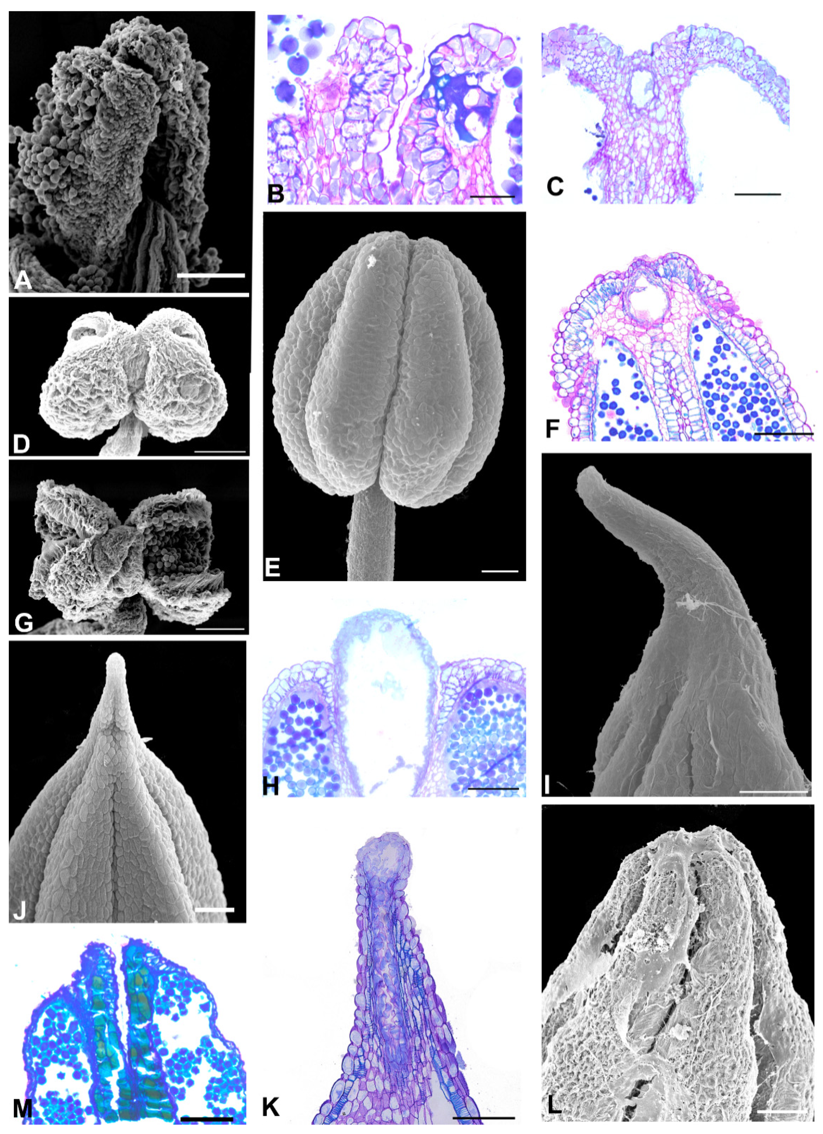
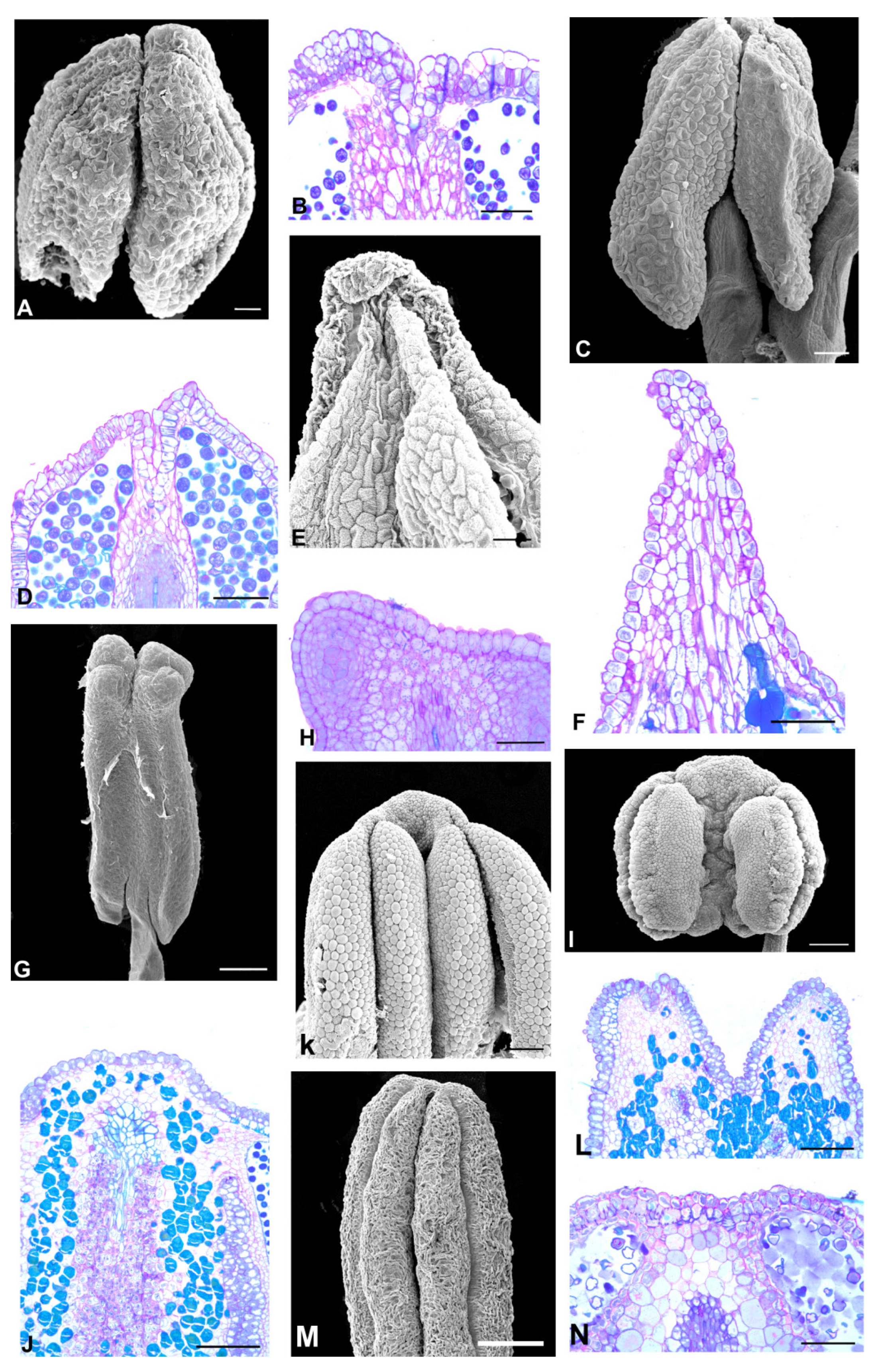
2.1. Flower Morphology

The Amburaneae subclade species (Figure 2 and Figure 3) have flowers with a one-petalled corolla and free stamens (Amburana and Mildbraediodendron), no corolla and free stamens (Cordyla), a papilionaceous corolla and basally united stamens (Dussia and Petaladenium), a non-papilionaceous corolla (five equal petals) and basally united stamens (Myrocarpus), with a papilionaceous corolla and free stamens (Myrospermum), and a non-papilionaceous corolla (widely oval standard) and 10 free homogeneous stamens (Myroxylon). A prominent anther apex occurs in species of Cordyla, Myrospermum, Myroxylon (Figure 2F,G,I, and Figure 3G,I,J), and a non-prominent anther apex occurs in species of Amburana, Dussia, Mildbraediodendron, Myrocarpus, and Petaladenium (Figure 2A,C,D,J–N and Figure 3A,D,E,L).
The Angylocalyceae subclade species (Figure 4 and Figure 5) have flowers with a papilionaceous corolla and basally united stamens (Angylocalyx), a non-papilionaceous corolla that consists of a large vexillum and reduced abaxial petals (Alexa and Castanospermum), a non-papilionaceous corolla that consists of a slightly wider vexillum and four equal abaxial petals and united stamens (Xanthocercis). A prominent anther apex occurs in the two species of Alexa (Alexa grandiflora and A. superba) and Castanospermum australe (Figure 4E,I and Figure 5C,E); a non-prominent anther apex occurs in species of Angylocalyx, in Xanthocercis zambesiaca and six species of Alexa (Figure 4A–C,G,H,L and Figure 5A,G).
The Dipterygeae subclade species (Figure 6 and Figure 7) have flowers with a papilionaceous corolla formed by a vexillum, two wings, and two keels, and a monadelphous androecium (Dipteryx, Pterodon, and Taralea); and a non-papilionaceous corolla that consists of a vexillum, two reduced wings, connate and open keels exposing the free stamens (Monopteryx). A prominent anther apex occurs in seven species of Dipteryx, all species of Pterodon and Monopteryx uaucu (Figure 6A,C,D–F,H,J–M and Figure 7L); a non-prominent anther apex occurs in Dipteryx polyphylla, in all species of Taralea and Monopteryx inpae (Figure 6G and Figure 7A,C,E,G,I–K).
The additional species (Figure 8), Ateleia glazioveana, A. guaraya, Cyathostegia mathewsii, and Uleanthus erythrinoides, exhibit non-prominent anther apices (Figure 8A,C,G,M). In contrast, Candolleodendron brachystachyum and Swartzia langsdorffii have prominent anther apices (Figure 8E,K,I).

2.2. Anther Glands

The anthers of the ADA clade species have three types of glands: secretory cavity, secretory duct, and phenolic idioblast (Table 1).
A secretory cavity occurs embedded in the anther apex of Cordyla, Myrocarpus, Myrospermum species (Amburaneae subclade), of all species of Dipteryx, Pterodon, and of three species of Taralea (T. cordata, T. crassifolia and T. nudipes) (Dipterygeae subclade) (Figure 2H, Figure 3C,F,H, Figure 6B,I,N and Figure 7B,D,F). The lumen shape varies from spherical to oval between Dipteryx and Taralea (compare Figure 6B and Figure 7B).
A secretory duct occurs embedded in the anther apex and extends distally between thecae of the Myroxylon species (Amburaneae subclade) (Figure 3K). The lumen shape is elongated (Figure 3K).
In the remaining species, the anther apex does not show a secretory cavity or a secretory duct (Figure 2B,E,O, Figure 3B,M, Figure 4D,F,J,L, Figure 5B,D,F,H, Figure 7H,M and Figure 8B,D,H,F,J,L,N).
Phenolic idioblasts are found in the anther apex of Petaladenium, six Alexa species, two Monopteryx species, and in Swartzia langsdorffii (Figure 3M, Figure 4D,F,L, Figure 7M and Figure 8J,L). These species do not have cavities or ducts.

2.3. Distribution of Secretory Cavities in the Anther of the Amburaneae and Dipterygeae Subclades

By tracing the evolutionary history of the character “occurrence of secretory cavity/duct in the anther”, it was inferred that the presence of cavity/duct in the anther was acquired in most representatives of the Dipterygeae subclade and some of the Amburaneae subclade (Figure 9).

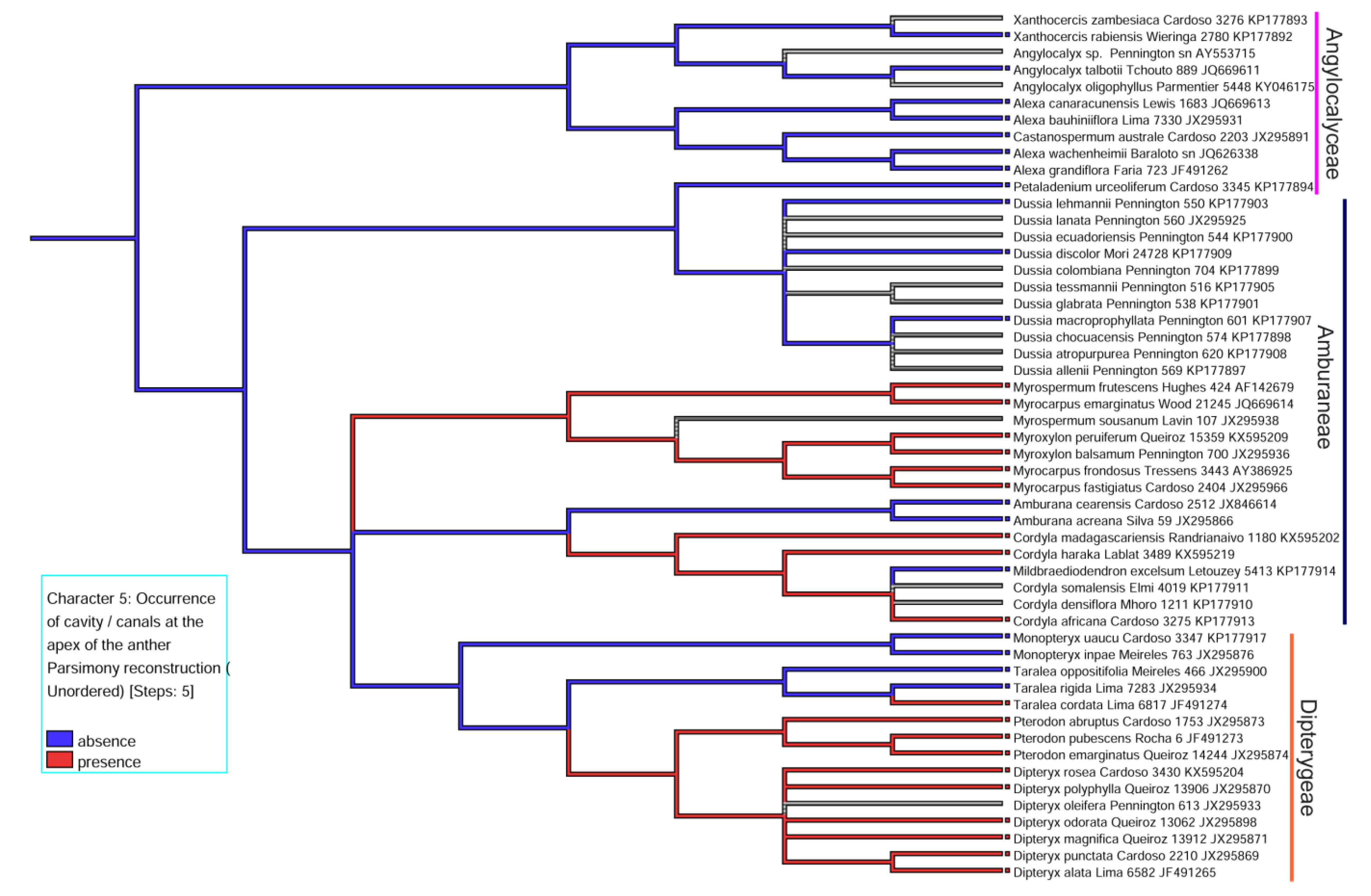
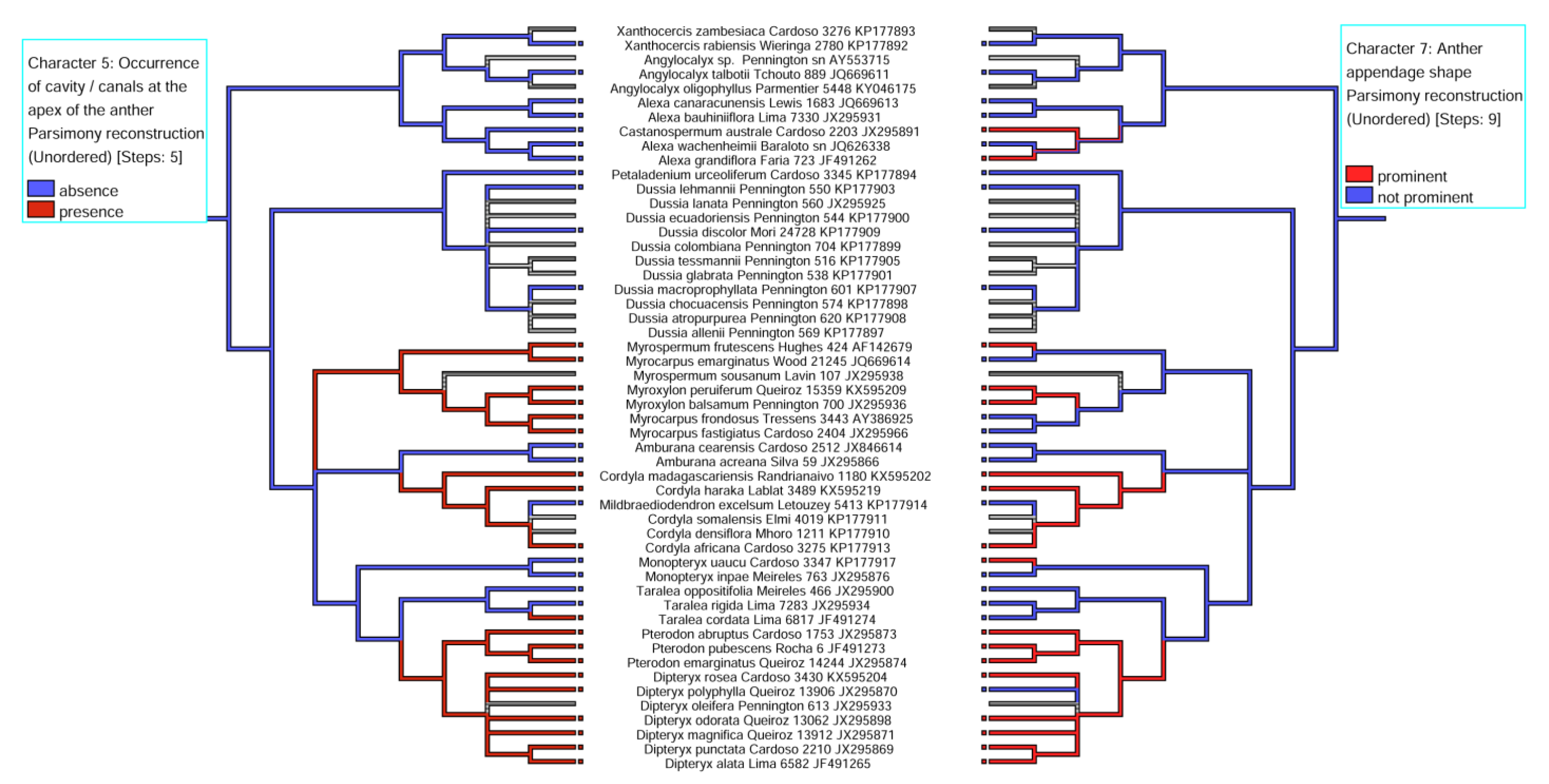
2.4. Correlations between Character -States

The pairings of the character -state reconstructions related to the occurrence of cavity/duct at the anther apex vs. shape of the anther apex indicate a positive correlation between these two characters (Figure 10). However, it is noteworthy that in 5 of the 20 species (Myrocarpus emarginatus, M. fastigiatus, M. frondosus, Dipteryx polyphylla, and Taralea cordata), this correlation was negative that is, even without a prominent anther apex, there was a secretory cavity in the anther.
The pairings of the character-state reconstructions related to the shape of the lumen verified at the anther apex in longitudinal section vs. position of the cavity/duct in the anther indicate a positive correlation between elongated shape and apical and distal position (Myroxylon balsamum and M. peruiferum), spherical shape and apical position (Cordyla africana, C. madasgascariensis, C. haraka, Dipteryx alata, D. magnifica, D. rosea, D. odorata, D. punctata, Pterodon abruptus, P. emarginatus, P. pubescens), and oval shape and apical position (D. polyphylla, Myrocarpus emarginatus, M. fastigiatus, M. frondosus, Myrospermum frutescens, Taralea cordata) (Figure 11).

3. Discussion

Our study highlights how glandular appendages occur in the anther of the ADA clade and provides an opportunity to clarify their enigmatic evolutionary history within early-branching papilionoids. The glandular appendage in the anther has been previously reported in the Dipteryx alata and Pterodon pubescens species of the Dipterygeae subclade [7,13], and also occurring in the species of the Amburaneae subclade.

3.1. Distribution and Location of Secretory Cavities/Ducts

The large ADA clade comprises morphologically eclectic genera with a diverse occurrence and structure of a glandular appendage in the anther, and the shape and location of this gland in the anther. Among 50 species analyzed within the ADA clade, 21 exhibit secretory cavities, two secretory ducts, and nine phenolic idioblasts, for a total of 64% species with a secretory structure in the anther apex.
Anatomical analyses of the anther in a longitudinal section showed that in most species analyzed, the gland is a secretory cavity with lumen shapes ranging from spherical to oval (see Table 1). Myroxylon balsamum and M. peruiferum, two species of the Amburaneae subclade, are the exceptions. The gland in the anther exhibits an elongated lumen so that, the term, secretory duct, becomes more appropriate. Variations in the lumen shape and, consequently, the difficulties generated in the typification of the gland, have been extensively explored in the literature, especially in studies with the leaf [15,20] and the stem [14,15,18,19].
The glandular appendage in the anther is most evident in the species of Pterodon, Dipteryx [7,13], present study, and Cordyla, which have secretory cavities with a spherical lumen, except for D. polyphylla in which the appendix is not very prominent, and the cavities have oval lumens. Similarly, Taralea cordata, T. crassifolia, and T. nudipes exhibit non-prominent anther appendages and contain a secretory cavity with an oval lumen, which is found in the region between the two thecae. In these species, the lumen of the cavity has the same shape as described for the vegetative organs of Taralea oppositifolia [15].
It is interesting to note that the secretory cavity located at the apex of the anther is subepidermal, and the epidermis cells have phenolic compounds see [13]. In Monopteryx and some species of Alexa, the cells of the anther appendix also exhibit phenolic content, although they do not have a secretory cavity. The appendix composed of phenolic cells that the anthers exhibit must be related to the floral structure, which is non-papilionaceous in the species of Alexa and Monopteryx. In these cases, the wing petals are reduced, the keel petals are united and opened, and the free stamens are exposed. The presence of phenolic compounds in the anther apex may be associated with the defense against herbivory or UV radiation since anthers are not protected by the petals as in a papilionaceous flower [34,35,36,37].
An interesting fact is the association between a secretory cavity in the anther and the leaf. Secretory cavities are present at the anther apex of closely related species such as Cordyla africana, C. haraka, C. madagascariensis, Myrocarpus emarginatus, M. fastigiatus, M. frondosus, and Myrospermum frutescens. In contrast, Myroxylon balsamum, and M. peruiferum exhibit a secretory duct at the anther apex. The secretory cavity or duct is absent in the species of Amburana, Dussia, Mildbraediodendron, and Petaladenium (Amburaneae subclade), and in the species of Alexa, Angylocalyx, Castanospermum, and Xanthocercis (Angylocalyceae subclade). The presence of a secretory cavity and ducts in the leaflet is shared by species of Myrocarpus (M. emarginatus, M. fastigiatus, M. frondosus, M. leprosus, M. venezuelensis), Myrospermum (M. frutescens, M. sousanum), Myroxylon (M. peruiferum and M. balsamum) and Cordyla (C. haraka, C. africana and C. madagascariensis [20,21] which demonstrates that these structures also occur in the floral organs of these species.

3.2. Evolutionary History of the Presence of a Secretory Cavity in the Anther of the ADA Clade

Our hypotheses were postulated to explain the occurrence of glands in anthers of the ADA clade species and have two robust explanations: (1) the appearance of the anther glands in the Amburaneae and Dipterygeae subclades or (2) their loss in some species of Amburaneae and Dipterygeae and all species of Angylocaleceae (see Figure 9).
Considering that the secretory cavities are present in other genera of the Amburaneae subclade, it is concluded that they are not a synapomorphy of the Dipterygeae subclade, as suggested by Leite et al. [7].
In the Dipterygeae subclade, our data suggest that secretory cavities may have been acquired in Dipteryx + Pterodon and some species of Taralea. A phenolic glandular appendix may have been acquired in Monopteryx, a sister group of Dipteryx, Pterodon, and Taralea (see distribution in Figure 9). The glandular appendix with a phenolic epidermis in Dipteryx, Pterodon, and Taralea could be a remnant of the Monopteryx phenolic appendage.
The Amburaneae subclade is remarkable because of its high level of floral diversity, production of coumarins (Amburana), red resin from bark and twigs (Dussia), balsams (Myrocarpus, Myrospermum, Myroxylon), and punctate glandular leaves of several genera (Cordyla, Mildbraediodendron, Myrocarpus, Myrospermum, and Myroxylon) [27,38,39]. The presence of glands at the anther apex is also noteworthy. Our data suggest that secretory cavities/ducts may have been acquired in the well-supported clades Myroxylon + Myrocarpus (non-papilionaceous flowers) and Myrospermum (papilionaceous flower). Another interesting fact is the presence of a secretory duct only in Myroxylon, and therefore, an autapomorphy of Myroxylon. Although Cordyla and Mildbraediodendron have a swartzioid-like floral morphology [12,38], the flowers have an entire calyx, no petals, and numerous free stamens, only Cordyla exhibits a secretory cavity in the anther apex. In contrast, the genus Amburana, sister to Cordyla + Mildbraediodendron, does not have a secretory cavity in the anther and has a one-petalled corolla and 10 free stamens. The absence of a secretory cavity in the anther apex of the genera Petaladenium and Dussia reflects their positioning in the phylogenetic tree as sister genera. They also present a papilionaceous corolla and basally united stamens [10]. A peculiar characteristic of Petaladeninum urceoliferum is its wing petals with glands, while in Dussia (its sister genus), the glands are found on the bract and bracteoles [9], although there are no anatomical studies on the composition of these structures.
In the Angylocalyceae subclade, our data suggest that secretory cavities were not acquired in the genera Alexa, Angylocalyx, Castanospermum, and Xanthocercis, defining them as a sister group of the Amburaneae and Dipterygeae subclades. An appendix producing phenolic compounds may have been acquired in some Alexa species, a sister group of Castanospermum, both with similar floral morphology, non-papilionaceous corolla, and 10 free stamens [12]. Angylocalyx and Xanthocercis, sister genera, exhibit a distinct floral morphology [12].
Other relevant data are the occurrence of secretory cavity/duct vs. anther appendage shape (see Figure 10) and lumen shape vs. position of cavity/duct in the anther (see Figure 11). It is likely that the anthers with a prominent apex also exhibit secretory cavities with a spherical lumen, different from those with a non-prominent apex, which exhibit secretory cavities more internalized in the anther, between the thecae, with the lumen being oval. Thus, we suppose that the shape of the anther is related to the lumen shape in species with a secretory cavity in the anther (see Figure 10 and Figure 11). Another interesting result is the lumen shape in the clades Myrocarpus + Myrospermum, and Myroxylon. Myrocarpus+ Myrospermum exhibit secretory cavities more internalized in the anther between the thecae, with an oval lumen and a prominent/non- prominent apex, respectively, different from Myroxylon, which exhibits a secretory duct with a rounded lumen in its apical portion and is elongated in the lower portion between the thecae following the shape of the anther apex.
The secretory cavities of Dipteryx (except D. polyphylla) and Pterodon are anatomically more similar to each other than to those of Taralea cordata, T. crassifolia and T. nudipes, confirming previous data obtained about the flower [7], leaf, stem [15] and leaflets [16] and corroborating the phylogeny data (see LPGW [1]). This fact reflects the topology of a phylogenetic tree in the subclades, which places them as sister groups see [1,12,22].
The inclusion of other species with a non-papilionaceous corolla from the Swartzieae clade (Ateleia glazioveana, A. guaraya, Candolleodendron brachystachyum, Cyathostegia mathewsii, Swartzia langsdorffii) and genistoid clade (Uleanthus erythrinoides) [1] suggests that the presence of secretory cavities/ducts in the anther apex may be restricted to the members of the subclades Amburaneae and Dipterygeae. There are no reports on cavities secretory/duct in the anthers apex of other papilionoid legumes. Hymenaea verrucosa Gaertn. (subfamily Detarioideae) exhibits a secretory cavity in the connective region (ventral region) of the anthers [40]; and in Stryphnodendron adstringens, Tetrapleura tetraptera, Adenanthera pavonine, and Pentaclethra macroloba (Mimosoid clade, subfamily Caesalpinioideae) such anther glandular appendages can comprise secretory emergences [41].

3.3. Evolutionary Significance of the Corolla, Type of Androecium vs. Presence of a Secretory Cavity/Duct in the Anther

Our data plotted in phylogeny have not yet made it possible to correlate the type of corolla and androecium with a secretory cavity/duct in the anther. The presence of a secretory cavity/duct has been reported in species with apetalous, non-papilionaceous flowers with numerous free stamens, including Cordyla africana, C. haraka and C. madagascariensis. Myrocarpus emarginatus, M. fastigiatus, and M. frondosus exhibit five undifferentiated petals with 10 basally united stamens, Myrospermum frutescens exhibits a papilionaceous corolla with 10 free stamens; Myroxylon balsamum and M. peruiferum exhibit a non-papilionaceous corolla with the widely oval banner and 10 free stamens. Therefore, a correlation with pollination seems plausible.
In Dipteryx alata and Pterodon pubescens, species of the Dipterygeae subclade, the anther glands consist of a cavity secreting sticky substances (oleoresins and polysaccharides) that play a key role during the flower’s lifespan by aggregating pollen grains and attaching them to the floral visitor’s body, besides maximizing the pollen release mechanism that is intermediate between the valvular and the explosive [13]. The same mechanism probably occurs in Myrospermum frutescens because these species are pollinated by insects [42]; Myrocarpus frondosus has a non-papilionaceous corolla but is pollinated by insects [43], probably aggregating pollen grains.
In Myroxylon peruiferum, the ducts secrete a substance that aggregates pollen grains; however, when in contact with air, the resinous content secreted together with the pollen grains hardens, probably acting on pollination by sunbirds (personal observation). The same must occur in Cordyla africana, which has petaliferous flowers rich in nectar that are pollinated by sunbirds [44].
Species with phenolic idioblasts at the anther apex, such as those of Alexa and Monopterx, are ornithophilous [27] and entomophilous [45], respectively. There are also reports of sphingophily [46,47] and chiropterophily [48] for Alexa. The lack of information on the pollination biology for these species makes it difficult to understand the function of these glands in early-diverging papilionoids.
However, analyzing the corolla shape and the presence of a secretory structure in the anther concerning the pollinator (insect or bird), probably the corolla type associated with the material exuded by the secretory structure at the anther apex at different proportions found in each species acts by favoring the different pollinators.

3.4. Outlook

We studied the evolution of the anther’s glandular appendage in early-diverging papilionoid genera, reporting this condition in a large number of species.
The diversity of the subfamily Papilionoideae expressed in terms of floral morphology, such as loss of petals, undifferentiated petals, numerous free stamens, entire calyx in bud, and radially symmetric flowers [5,8,49,50,51,52,53,54,55] is particularly common among the early-diverging papilionoid genera [5,6,7,8,9,10]. Additionally, secretory cavities/ducts in the anther in some species of the Dipterygeae and Amburaneae subclades [7,13], present study, is a unifying character-state for these groups. This condition has not been previously reported for representatives of the most recent divergent papilionoids.

4. Materials and Methods

4.1. Sampling Plant Material

We examined at least one species in each genus of the Amburaneae (Amburana, Cordyla, Dussia, Mildbraediodendron, Myrocarpus, Myrospermum, Myroxylon, and Petaladenium), Angylocalyceae (Alexa, Angylocalyx, Castanospermum, and Xanthocercis), and Dipterygeae subclades (Dipteryx, Pterodon, and Monopteryx). In addition, we included six species with non-papilionoid flowers outside the ADA clade to map the character of other distant taxa (Table S1).
Samples of flowers and floral buds (1 or/and 2) were obtained from herbarium specimens (Table S1) and treated with 2% KOH solution for 2 h, washed several times in distilled water [56], and stored in 70% ethanol. The anthers were removed and prepared for observations by scanning electron microscopy (SEM) and light microscopy (LM).

4.2. Scanning Electron Microscopy

For SEM analysis, anthers were critical point dried in a Balzers CPD 030 dryer (Balzers, Liechtenstein), mounted on aluminum stubs with colloidal carbon, coated with gold in a Bal Tec SCD 050 sputter coater, and observed with a Jeol JSM 6610LV scanning electron microscope (Tokyo, Japan).

4.3. Light Microscopy

For LM analysis, anthers were embedded in historesin [57] and longitudinally sectioned (2–3 μm thick) with a rotary microtome (Leica RM 2245, Wetzlar, Germany). Sections were stained with 0.05% toluidine blue in phosphate buffer, pH = 6.8 [58] and photographed with a light microscope (Leica DM5000 B) coupled to a digital camera (Leica DFC295).

4.4. Phylogenetic Analysis

The evolution of the glands in anthers from the ADA clade was investigated based on a recent phylogenetic hypothesis [1]. The characters selected to compose the data matrix were the absence or presence of secretory cavity/ducts, anther appendage shape (prominent, not prominent), anther appendix position (apical, distal, apical/distal), lumen shape (spherical, oval, elongated), and absence or presence of phenolic compound at the apex of the anther (Table 2). We used the Mesquite program [59] to map the selected characters (Table 2) on the RAXML tree obtained by the Legume Phylogeny Working Group (LPWG) [1]. For this purpose, we chose “trace character history”, selecting the option “parsimony ancestral states”.

Supplementary Materials

The following supporting information can be downloaded at: https://www.mdpi.com/article/10.3390/plants11070835/s1, Table S1: Species of Amburaneae, Angylocalyceae, Dipterygeae subclades and outgroup. IAN = Herbário da Embrapa Amazônia Oriental; INPA = Herbário do Instituto Nacional de Pesquisas da Amazônia; KEW = Herbarium Royal Botanic Gardens; MNHN = Muséum national d’Histoire naturelle; MG = Herbário do Museu Paraense Emílio Goeldi; NYBG = The New York Botanical Garden; RB = Herbário do Jardim Botânico do Rio de Janeiro; RBSpirit = Herbário do Jardim Botânico do Rio de Janeiro; SPFR = Herbário do Departamento de Biologia da Faculdade de Filosofia Ciências e Letras de Ribeirão Preto da Universidade de São Paulo; UEC = Herbário da Universidade Estadual de Campinas; US (Smithsonian Institution).

Author Contributions

Conceptualization, V.G.L., S.P.T. and V.F.M.; methodology, V.G.L., S.P.T. and V.F.M.; investigation, V.G.L., S.P.T. and V.F.M.; data analysis, V.G.L., S.P.T., Â.L.B.S. and V.F.M.; writing—original draft preparation, V.G.L., S.P.T. and V.F.M.; writing—review and editing, V.G.L., S.P.T., Â.L.B.S. and V.F.M.; supervision, V.F.M.; project administration, V.F.M.; funding acquisition, V.F.M. and S.P.T. All authors have read and agreed to the published version of the manuscript.

Funding

This research was funded by the National Council for Scientific and Technological Development, CNPq (protocol numbers 155733/2018-4, 154529/2016-8 and 302806/2019-9) and the Coordenação de Aperfeiçoamento de Pessoal de Nível, CAPES (finance code 001).

Institutional Review Board Statement

Not applicable.

Informed Consent Statement

Not applicable.

Data Availability Statement

Data sharing is not applicable to this article.

Acknowledgments

We are grateful to Rodrigo Ferreira Silva (FFCLRPUSP), Maria Dolores Seabra Ferreira, José Augusto Maulin (FMRPUSP) and Edimárcio da Silva Campos (FCFRP/USP) for technical assistance and to Elettra Greene for revision of the English text. We wish to thank the curators, assistant curator, and collections co-managers of the IAN, INPA, KEW, MNHN, MG, NYBG, RB, SPFR, UEC, and US, for their cooperation in herbaria consultation.

Conflicts of Interest

The authors declare no conflict of interest.

References

  1. LPWG (The Legume Phylogeny Working Group). A new subfamily classification of the Leguminosae based on a taxonomically comprehensive phylogeny. Taxon 2017, 66, 44–77. [Google Scholar] [CrossRef] [Green Version]
  2. LPWG (The Legume Phylogeny Working Group). Legume phylogeny and classification in the 21st century: Progress, prospects and lessons for other species-rich clades. Taxon 2013, 62, 217–248. [Google Scholar] [CrossRef]
  3. Lavin, M.; Pennington, R.T.; Klitgaard, B.B.; Sprent, J.I.; Lima, H.C.; Gasson, P.E. The dalbergioid legumes (Fabaceae): Delimitation of a pantropical monophyletic clade. Am. J. Bot. 2001, 88, 503–533. [Google Scholar] [CrossRef]
  4. Schrire, B.D.; Lavin, M.; Lewis, G.P. Global distribution patterns of the Leguminosae: Insights from recent phylogenies. In Plant Diversity and Complexity Patterns: Local, Regional and Global Dimensions; Biologiske Skrifter 55; Friis, I., Balslev, H., Eds.; Kgl. Danske Videnskabernes Selskab: Copenhagen, Denmark, 2005; pp. 375–422. [Google Scholar]
  5. Tucker, S.C. Floral ontogeny in Sophoreae (Leguminosae: Papilionoideae). I. Myroxylon (Myroxylon Group) and Castanospermum (Angylocalyx Group). Am. J. Bot. 1993, 80, 65–75. [Google Scholar] [CrossRef]
  6. Prenner, G. New Aspects In Floral Development of Papilionoideae: Initiated but Suppressed Bracteoles and Variable Initiation of Sepals. Ann. Bot. 2004, 93, 537–545. [Google Scholar] [CrossRef] [PubMed] [Green Version]
  7. Leite, V.G.; Mansano, V.F.; Teixeira, S.P. Floral ontogeny in Dipterygeae (Fabaceae) reveals new insights into one of the earliest branching tribes in papilionoid legumes. Bot. J. Linn. Soc. 2014, 174, 529–550. [Google Scholar] [CrossRef] [Green Version]
  8. Leite, V.G.; Teixeira, S.P.; Mansano, V.F.; Prenner, G. Floral development of the early branching papilionoid legume Amburana cearensis (Leguminosae) reveals rare and novel characters. Int. J. Plant Sci. 2015, 176, 94–106. [Google Scholar] [CrossRef]
  9. Prenner, G.; Cardoso, D.; Zartman, C.E.; Queiroz, L.P. Flowers of the early-branching papilionoid legume Petaladenium urceoliferum display unique morphological and ontogenetic features. Am. J. Bot. 2015, 102, 1780–1793. [Google Scholar] [CrossRef] [Green Version]
  10. Cardoso, D.; São-Mateus, W.M.B.; Cruz, D.T.; Zartman, C.E.; Komura, D.L.; Kite, G.; Prenner, G.; Wieringa, J.J.; Clark, A.; Lewis, G.; et al. Filling in the gaps of the papilionoid legume phylogeny: The enigmatic Amazonian genus Petaladenium is a new branch of the early-diverging Amburaneae clade. Mol. Phylogenetics Evol. 2015, 84, 112–124. [Google Scholar] [CrossRef]
  11. Lewis, G.; Schrire, B.; Mackinder, B.; Lock, M. Legumes of the World; Royal Botanic Gardens, Kew: London, UK, 2005. [Google Scholar]
  12. Cardoso, D.; Pennington, R.T.; Queiroz, L.P.; Boatwright, J.S.; Van Wyk, B.-E.; Wojciechowski, M.F.; Lavin, M. Reconstructing the deep-branching relationships of the papilionoid legumes. S. Afr. J. Bot. 2013, 89, 58–75. [Google Scholar] [CrossRef] [Green Version]
  13. Leite, V.G.; Mansano, V.F.; Pansarin, E.; Teixeira, S.P. Presence of the anther gland is a key feature in pollination of the early-branching papilionoids Dipteryx alata and Pterodon pubescens (Leguminosae). Plant Biol. 2019, 21, 1016–1023. [Google Scholar] [CrossRef]
  14. Rodrigues, T.M.; Machado, S.R. Anatomia comparada do pulvino, pecíolo e raque de Pterodon pubescens Benth (Fabaceae-Faboideae). Acta Bot. Bras. 2004, 18, 381–390. [Google Scholar] [CrossRef] [Green Version]
  15. Palermo, F.; Teixeira, S.P.; Mansano, V.F.; Leite, V.G.; Rodrigues, T.M. Secretory spaces in species of clade Dipterygeae (Leguminosae, Papilionoideae). Acta Bot. Bras. 2017, 31, 374–381. [Google Scholar] [CrossRef] [Green Version]
  16. Silva, N.F.; Arruda, R.C.O.; Alves, F.M.; Sartori, A.L.B. Leaflet anatomy of the Dipterygeae clade (Faboideae: Fabaceae): Evolutionary implications and systematics. Bot. J. Linn. Soc. 2018, 187, 99–117. [Google Scholar] [CrossRef]
  17. Rodrigues, T.M.; Santos, D.C.; Machado, S.R. The role of the parenchyma sheath and PCD during the development of oil cavities in Pterodon pubescens (Leguminosae-Papilionoideae). Comptes Rendus Biol. 2011, 334, 535–543. [Google Scholar] [CrossRef] [PubMed]
  18. Paiva, E.A.S.; Oliveira, D.M.T.; Machado, S.R. Anatomy and ontogeny of the pericarp of Pterodon emarginatus Vogel (Fabaceae, Faboideae), with emphasis on secretory ducts. An. Acad. Bras. Ciênc. 2008, 80, 455–465. [Google Scholar] [CrossRef] [PubMed] [Green Version]
  19. Rodrigues, T.M.; Machado, S.R. Oil glands in Pterodon pubescens Benth. (Leguminosae-Papilionoideae): Distribution, structure and secretion mechanisms. Int. J. Plant Sci. 2012, 173, 984–992. [Google Scholar] [CrossRef] [Green Version]
  20. Sartori, A.L.B.; Tozzi, A.M.G.A. Comparative leaflet anatomy in Myrocarpus Allemão, Myroxylon L. f and Myrospermum Jacq. (Leguminosae–Papilionoideae–Sophoreae) species. Bot. J. Linn. Soc. 2002, 140, 249–259. [Google Scholar] [CrossRef]
  21. Bento, J.P.S.P.; Scremin-Dias, E.; Alves, F.M.; Mansano, V.F.; Sartori, A.L.B. Phylogenetic implications of the anatomical study of the Amburaneae clade (Fabaceae: Faboideae). Bot. J. Linn. Soc. 2020, 192, 69–83. [Google Scholar] [CrossRef]
  22. Cardoso, D.; Queiroz, L.P.; Pennington, R.T.; Lima, H.C.; Fonty, E.; Wojciechowski, M.F.; Lavin, M. Revisiting the phylogeny of Papilionoid Legumes: New insights from comprehensively sampled early-branching lineages. Am. J. Bot. 2012, 99, 1991–2013. [Google Scholar] [CrossRef] [PubMed]
  23. Seleme, E.P.; Lewis, G.P.; Stirton, C.H.; Sartori, A.L.B.; Mansano, V.F. A Taxonomic review and a new species of the south American woody genus Amburana (Leguminosae, Papilionoideae). Phytotaxa 2015, 212, 249–263. [Google Scholar] [CrossRef]
  24. Rudd, V.E. The Genus Dussia (Leguminosae); United States National Museum Contributions from the United States National Herbarium, Smithsonian Institution: Washington, DC, USA, 1963; pp. 247–277. [Google Scholar]
  25. Cowan, R.S. Swartzieae. In Advances in Legume Systematics, Part 1; Polhill, R.M., Raven, P.H., Eds.; Royal Botanic Gardens, Kew: London, UK, 1981; pp. 209–212. [Google Scholar]
  26. WFO. Mildbraediodendron excelsum Harms. Available online: http://www.worldfloraonline.org/taxon/wfo-0000171559 (accessed on 7 February 2022).
  27. Polhill, R.M. Sophoreae. In Advances in Legume Systematics, Part 1; Polhill, R.M., Raven, P.H., Eds.; Royal Botanic Gardens, Kew: London, UK, 1981; pp. 213–230. [Google Scholar]
  28. Sartori, A.L.B.; Tozzi, A.M.G.A. Revisão taxonômica de Myrocarpus Allemão (Leguminosae, Papilionoideae, Sophoreae). Acta Bot. Bras. 2004, 18, 521–535. [Google Scholar] [CrossRef]
  29. Sartori, A.L.B. Revisão Taxonômica e Estudos Morfológicos de Myrocarpus Allemão, Myroxylon L.f. e Myrospermum Jacq. (Leguminosae Papilionoideae Sophoreae). Ph.D. Thesis, Instituto de Biologia, Universidade Estadual de Campinas, Campinas, Brazil, 2000. [Google Scholar]
  30. Ramirez, N. Revision Taxonomica del Genero Alexa Moq. (Fabaceae, Sophoreae). Ann. Mo. Bot. Gard. 1995, 82, 549–569. [Google Scholar] [CrossRef]
  31. Taubert, P. Leguminosae africanae. Engl. Bot. Jahrb. 1896, 23, 172. [Google Scholar]
  32. Louppe, D.; Oteng-Amoako, A.A.; Brink, M. (Eds.) Plant Resources of Tropical Africa 7 (1) Timbers 1; PROTA Foundation: Wageningen, The Netherlands, 2008; p. 701. [Google Scholar]
  33. Rodrigues, W.A. Contribuição para o estudo do gênero Monopteryx Spr. ex Benth. (Leguminosae) da Amazônia. Acta Amaz. 1975, 5, 153–155. [Google Scholar] [CrossRef]
  34. Skubatz, H.; Kunkel, D.D.; Howald, W.N.; Trenkle, R.; Mookherjee, B. The Sauromatum guttatum appendix as an osmophore: Excretory pathways, composition of volatiles and attractiveness to insects. New Phytol. 1996, 134, 631–640. [Google Scholar] [CrossRef] [PubMed]
  35. Jurgens, A.; Dotterl, S.; Meve, A.U. The chemical nature of fetid floral odours in stapeliads (Apocynaceae-Asclepiadoideae-Ceropegieae). New Phytol. 2006, 172, 452–468. [Google Scholar] [CrossRef] [PubMed]
  36. Knudsen, J.T.; Eriksson, R.; Gershenzon, J.; Ståhl, B. Diversity and distribution of floral scent. Bot. Rev. 2006, 72, 1. [Google Scholar] [CrossRef]
  37. Castro, M.M.; Demarco, D. Phenolic compounds produced by secretory structures in plants: A brief review. Nat. Prod. Commun. 2008, 3, 1273–1284. [Google Scholar] [CrossRef] [Green Version]
  38. Du Puy, D.J.; Labat, J.N. Swartzieae. In The Leguminosae of Madagascar; Du Puy, D.J., Labat, J.N., Rabevohitra, R., Villiers, J.F., Bosser, J., Moat, J., Eds.; Royal Botanic Gardens, Kew: London, UK, 2002; pp. 294–297. [Google Scholar]
  39. Sartori, A.L.B.; Lewis, G.P.; Mansano, V.F.; Tozzi, A.M.G.A. A revision of the genus Myroxylon (Leguminosae, Papilionoideae). Kew Bull. 2015, 70, 48. [Google Scholar] [CrossRef]
  40. Kochanovski, F.J.; Paulino, J.V.; Teixeira, S.P.; Tozzi, A.M.G.A.; Mansano, V.F. Floral development of Hymenaea verrucosa: An ontogenetic approach to the unusual flower of Fabaceae subfamily Detarioideae. Bot. J. Linn. Soc. 2018, 1, 46–58. [Google Scholar] [CrossRef]
  41. De Barros, T.C.; Pedersoli, G.D.; Teixeira, S.P. Anther glands in Mimosoideae (Leguminosae) are emergences with a conserved meristematic origin. Flora 2017, 226, 1–9. [Google Scholar] [CrossRef]
  42. Frankie, G.W. Tropical forest phenology and pollinator plant coevolution. In Coevolution of Animals and Plants; Symposium V First International Congress of Systematic and Evolutionary Biology, Boulder, Colorado; Lawrence, E.G., Raven, P.H., Eds.; University of Texas Press: Austin, TX, USA, 1973. [Google Scholar]
  43. Pfeiffer, P.M.M. Plant-Bee Interactions and Pollen Flux in Restored Areas of Atlantic Forest. Ph.D. Thesis, Instituto de Biociências da Universidade de São Paulo, Sao Paulo, Brazil, 2018. [Google Scholar]
  44. Lemmens, R.H.M.J.; Nyunai, N. Plant Resources of Tropical 7(2). Timbers 2; Lemmens, R.H.M.J., Louppe, D., Oteng-Amoako, A.A., Eds.; PROTA (Plant Resources of Tropical Africa) Foundation: Wageningen, The Netherlands, 2012. [Google Scholar]
  45. Rech, A.R. Recursos Polínicos Utilizados por 23 Espécies de Meliponini Lepeletier, 1836 Para Coleta de Pólen ao Longo da Bacia do Rio Negro, Amazonas-Brasil. Master’s Dissertation, Instituto Nacional de Pesquisas da Amazônia, Manaus, Brazil, 2010. [Google Scholar]
  46. Vogel, S. Chiropterophilie in der neotropischen Flora. Neue Mitteilungen II. Flora 1969, 158, 185–222. [Google Scholar]
  47. von Hegnauer, R. Chemotaxonomie der Pflanzen Band XIb-1 Leguminosae Teil 2: Caesalpinoideae und Mimosoideae; Birkhäuser Verlag: Basel, Switzerland; Boston, MA, USA; Berlin, Germany, 1996. [Google Scholar]
  48. Carvalho, C.T. Sobre os hábitos alimentares de Phillostomideos (Mammalia, Chiroptera). Rev. Biol. Trop. 1961, 9, 53–60. [Google Scholar] [CrossRef]
  49. Mansano, V.F.; Tucker, S.C.; Tozzi, A.M.G.A. Floral ontogeny of Lecointea, Zollernia, Exostiles and Harleyodendron (Leguminosae: Papilionoideae: Swartzieae S.L.). Am. J. Bot. 2002, 89, 1553–1569. [Google Scholar] [CrossRef] [PubMed]
  50. McMahon, M.; Hufford, L. Evolution and development in the amorphoid clade (Amorpheae: Papilionoideae: Leguminosae): Petal loss and dedifferentiation. Int. J. Plant Sci. 2005, 166, 383–396. [Google Scholar] [CrossRef] [Green Version]
  51. Paulino, J.V.; Mansano, V.F.; Teixeira, S.P. Elucidating the unusual floral features of Swartzia dipetala (Fabaceae). Bot. J. Linn. Soc. 2013, 173, 303–320. [Google Scholar] [CrossRef] [Green Version]
  52. Pennington, R.T.; Klitgaard, B.B.; Ireland, H.; Lavin, M. New insights into floral evolution of basal papilionoids from molecular phylogenies. In Advances in Legume Systematics, Part 9; Herendeen, P.S., Bruneau, A., Eds.; Royal Botanic Gardens, Kew: London, UK, 2000; pp. 233–248. [Google Scholar]
  53. Tucker, S.C. Loss of floral organs in Ateleia (Leguminosae: Papilionoideae: Sophoreae). Am. J. Bot. 1990, 77, 750–761. [Google Scholar] [CrossRef]
  54. Tucker, S.C. Floral ontogeny in Sophoreae (Leguminosae: Papilionoideae). III. Radial symmetry and random petal aestivation in Cadia purpurea. Am. J. Bot. 2002, 89, 748–757. [Google Scholar] [CrossRef]
  55. Tucker, S.C. Floral Ontogeny in Swartzia (Leguminosae: Papilionoideae: Swartzieae): Distribution and Role of the Ring Meristem. Am. J. Bot. 2003, 90, 1271–1292. [Google Scholar] [CrossRef] [PubMed]
  56. Smith, F.H.; Smith, E.C. Anatomy of the inferior ovary of Darbya. Am. J. Bot. 1942, 29, 464–471. [Google Scholar] [CrossRef]
  57. Gerrits, P.O.; Horobin, R.W. The Application of Glycol Methacrylate in Histotechnology; Some Fundamental Principles; Faculteit der Geneeskunde, Rijksuniversiteit Groningen: Groningen, The Netherlands, 1991. [Google Scholar]
  58. O’ Brien, T.P.; Feder, N.; Mccully, M.E. Polychromatic staining of plant cell walls by Toluidine Blue O. Protoplasma 1964, 59, 368–373. [Google Scholar] [CrossRef]
  59. Maddison, W.P.; Maddison, D.R. Mesquite: A Modular System for Evolutionary Analysis, Version 3.6. 2018. Available online: http://mesquiteproject.org (accessed on 19 December 2019).
Figure 1. Phylogenetic relationships between the three subclades of the ADA clade modified from [1,10,12]. The Angylocalyceae subclade is a sister group to the Amburaneae and Dipterygeae subclades.
Plants 11 00835 g001
Figure 2. Aspects of the anther apex morphology in the species of the Amburaneae subclade. (A,C,D,F,G,IN) = SEM, (B,E,H,O) = light microscopy (LM). (A,B) Amburana acreana. (A) Frontal view of the anther. Note that the anther apex is non-prominent. (B) Longitudinal section showing the anther. Note the absence of a secretory cavity between the two thecae. (C) A. cearensis. Adaxial view of the anther. Note that the anther apex is non-prominent. (D) A. erythrosperma. Frontal view of the anther. Note that the anther apex is non-prominent. (E) Longitudinal section showing the anther. Note the absence of a secretory cavity between the two thecae. (F) Cordyla africana. Adaxial view of the anther. Note that the anther apex is prominent (arrow). (G,H) C. madasgascariensis. (G) Frontal view of the anther. Note that the anther apex is prominent (arrow). (H) Longitudinal section showing the apex with a secretory cavity between the two thecae. (I) C. haraka. Frontal view of the anther. Note that the anther apex is prominent (arrow). (J) Dussia discolor. Abaxial view of the anther. Note that the anther apex is non-prominent. (K) D. lehmanni. Adaxial view of the anther. Note that the anther apex is non-prominent. (L) D. macrophylla. Adaxial view of the anther. Note that the anther apex is non-prominent. (M) D. martinecensis. Adaxial view of the anther. Note that the anther apex is non-prominent. (N,O) D. tesmanni. (N) Lateral view of the anther. (O) Longitudinal section showing the anther. Note the absence of a secretory cavity between the two thecae. Scale bars: (A,B,N), 200 μm; (CE), 400 μm; (FO), 100 μm; (H) 50 μm.
Plants 11 00835 g002
Figure 3. Aspects of the anther apex morphology in the species of the Amburaneae subclade. (A,D,E,G,I,J,L) = SEM, (B,C,F,H,K,M) = light microscopy, ML. (A,B) Mildbraediodendron excelsum. (A) Abaxial view of the anther. Note that the anther apex is non-prominent. (B) Longitudinal section showing the anther. Note the absence of a secretory cavity between the two thecae. (C) Myrocarpus emarginatus. Longitudinal section showing the anther apex with a secretory cavity. (D) M. fastigiatus. Frontal view of the anther. Note that the anther apex is non-prominent. (E,F) M. frondosus. (E) Adaxial view of anther. Note that the anther apex is non-prominent. (F) Longitudinal section showing the anther apex with a secretory cavity. (G,H) Myrospermum frutescens. (G) Frontal view of the anther. (H) Longitudinal section showing the anther apex with a secretory cavity. (I) Myroxylon balsamum. Lateral view of the anther. Note that the anther apex is prominent. (J,K) M. peruiferum. (J) Adaxial view of the anther. Note that the anther apex is prominent. (K) Longitudinal section showing the anther apex with a secretory duct. Note that the lumen shape of the secretory duct follows the shape of the anther apex, rounded in its apical portion (arrowhead) and elongated in the lower portion. (L,M) Petaladenium urceoliferum. (L) Adaxial view of the anther. Note that the anther apex is non-prominent. (M) Longitudinal section of the anther showing the phenolic cells. Scale bars: (A,CL), 100 μm; (B), 50 μm.
Plants 11 00835 g003
Figure 4. Aspects of the anther apex morphology in the species of the Angylocalyceae subclade. (AC,E,GI,K) = light microscopy, (D,F,J,L) = SEM. (A) Alexa bauhiniflora. Adaxial view of the anther. Note that the anther apex is non-prominent. (B) A. canaracunensis. Adaxial view of the anther. Note that the anther apex is non-prominent. (C,D) A. cowanii. (C) Lateral view of the anther. Note that the anther apex is non-prominent. (D) Longitudinal section of the anther showing the phenolic cells. (E,F) A. grandiflora. (E) Lateral view of the anther. Note that the anther apex is a small prominence. (F) Longitudinal section of the anther showing the phenolic cells. (G) A. imperatrizes. Adaxial view of the anther. Note that the anther apex is non-prominent. (H) A. leiopetala. Adaxial view of the anther. Note that the anther apex is non-prominent. (I,J) A. superba. (I) Lateral view of anther. Note that the anther apex is a small prominence. (J) Longitudinal section of the anther. Note the absence of a secretory cavity between the two thecae. (K,L) A. wachenheimii. (K) Lateral view of the anther. Note that the anther apex is non-prominent. (L) Longitudinal section of the anther showing the phenolic cells. Scale bars: (A,C,D), 200 μm; (B,G,L), 50 μm; (E,H), 200 μm; (F,IK) 100 μm.
Plants 11 00835 g004
Figure 5. Aspects of the anther apex morphology in the species of the Angylocalyceae subclade. (A,C,E,F) = SEM, (B,D,F,H) = light microscopy, LM. (A,B) Angylocalyx pynaertii. (A) Adaxial view of the anther. Note that the anther apex is non-prominent. (B) Longitudinal section of the anther. Note the absence of a secretory cavity between the two thecae. (C,D) A. talbotii. (C) Lateral view of the anther. Note that the anther apex is non-prominent. (D) Longitudinal section of the anther. Note the absence of a secretory cavity between the two thecae. (E,F) Castanospermum australe. (E) Adaxial view of the anther. Note that the anther apex is a small prominence. (F) Longitudinal section of the anther. Note the absence of a secretory cavity between the two thecae. (G,H) Xanthocercis zambesiaca. (G) Adaxial view of the anther. Note that the anther apex is non-prominent. (H) Longitudinal section of the anther. Note the absence of a secretory cavity between the two thecae. Scale bars: (A), 200 μm; (BH), 100 μm.
Plants 11 00835 g005
Figure 6. Aspects of the anther apex morphology in the species of the Dipterygeae subclade. (A,CH,JM) = SEM, (B,I,N) = light microscopy, LM. (A,B) Dipteryx alata. (A) Adaxial view of the anther with longitudinal dehiscence and pollen grains adhering to the anther apical region. (B) Longitudinal section showing the anther apex with a secretory cavity. (C) D. lacunifera. Abaxial view of the anther. Note that the anther apex is prominent. (D) D. magnifica. Abaxial view. Note that the anther apex is prominent. (E) D. micrantha. Abaxial view. Note that the anther apex is prominent. (F) D. odorata. Abaxial view of the anther. Note that the anther apex is prominent. (G) D. polyphylla. Abaxial view of the anther. Note that the anther apex is non-prominent. (H,I) D. punctata. H. Lateral view of the anther. Note that the anther apex is prominent. (I) Longitudinal section showing the anther apex with a secretory cavity with a spherical lumen. (J) D. rosea. Lateral view of the anther. Note that the anther apex is prominent. (K) Pterodon abruptus. Adaxial view of the anther. Note that the anther apex is prominent. (L) P. emarginatus. Frontal view of the anther. Note that the anther apex is prominent. (M,N) P. pubescens. (M) Lateral view of the anther. Note that the anther apex is prominent. (N) Longitudinal section showing the anther apex with a secretory cavity. Scale bars (A,D,F,G,J,K,M), 100 μm; (B), 20 μm; (C,E,I,L,H), 50 μm; (N), 200 μm.
Plants 11 00835 g006
Figure 7. Aspects of the anther apex morphology in the species of the Dipterygeae subclade. (A,C,E,G,IL) = SEM, (B,D,F,H,M) = light microscopy, LM. (A,B) Taralea. cordata. (A) Abaxial view of the anther. Note that the anther apex is non-prominent. (B) Longitudinal section showing the anther apex with a secretory cavity with an oval lumen. (C,D) T. crassifolia. (C) Abaxial view of the anther. Note that the anther apex is non-prominent. (D) Longitudinal section showing the anther apex with a secretory cavity with an oval lumen. (E,F) T. nudipes. (E) Adaxial view of the anther. Note that the anther apex is non-prominent. (F) Longitudinal section showing the anther apex with a secretory cavity with an oval lumen. (G,H) T. oppositifolia. (G) Adaxial view of the anther. Note that the anther apex is non-prominent. (H) Longitudinal section showing parenchymal cells (circle) of the anther. (I) T. reticulata. Adaxial view of the anther. Note that the anther apex is non-prominent. (J) T. rigida. Adaxial view of the anther. Note that the anther apex is non-prominent. (K) Monopteryx inpae. Lateral view of the anther. Note that the anther apex is non-prominent (arrowhead). (L,M) M. uaucu. (L) Lateral view of the anther. Note that the anther apex is prominent (arrowhead). (M) Longitudinal section showing the anther apex with phenolic cells. Scale bars: (A,EI,KM), 100 μm; (B,D,J), 200 μm; (C). 50 μm.
Plants 11 00835 g007
Figure 8. Aspects of the anther apex morphology in the species of the Ateleia, Candolleodendron, Cyasthostegia, Swartzia, Uleanthus. (A,C,E,G,K,I,M) = SEM, (B,D,H,F,J,L,N) = light microscopy, LM. (A,B) Ateleia glazioveana. (A) Adaxial view of the anther. Note that the anther apex is non-prominent. (B) Longitudinal section showing parenchymal cells at the anther apex. Note that there is no secretory cavity between the anther’s thecae. (C,D) Ateleia guaraya. (C) Adaxial view of the anther. Note that the anther apex is non-prominent. (D) Longitudinal section showing parenchymal cells at the anther apex. (E,F) Candolleodendron brachystachyum. (E) Adaxial view of the anther. Note that the anther apex is prominent. (F) Longitudinal section showing parenchymal cells at the anther apex. (G,H) Cyathostegia mathewsii. (G) Abaxial view of the anther. Note that the anther apex is non-prominent. (H) Longitudinal section showing parenchymal cells of the anther. (I,J) Swartzia langsdorffii (I) Adaxial view of the anther of the small stamens with a small prominence apex. (J) Longitudinal section showing the anther apex with phenolic cells. (K) Adaxial view of the anther of the large stamens with a small prominent apex. (L) Longitudinal section showing the anther apex with phenolic cells. (M,N) Uleanthus erythrinoides. (M) Adaxial view of the anther. Note that the anther apex is non-prominent. (N) Longitudinal section showing parenchymal cells of the anther. Scale bars: (AG,I,J,N), 100 μm; (H), 50 μm; (L,M), 200 μm.
Plants 11 00835 g008
Figure 9. Representation of the modified LPGW cladogram (2017) for the ADA clade, showing the reconstruction of the character history in the occurrence of glands in anthers of the group. The red color represents the presence of glands in the lineage, the blue color represents the absence of glands, and the gray color represents doubt about the occurrence of the glands. The reconstruction indicates that the presence of glands has two plausible hypotheses: the appearance of the glands in the subclades (Amburaneae and Dipterygeae) or their loss in some representatives of Amburaneae and Dipterygeae and in all representatives of Angylocalyceae.
Plants 11 00835 g009
Figure 10. Modified LPGW cladogram (2017) for the ADA clade showing the mirroring of the character state reconstruction for “secretory cavity/duct occurrence” vs. “shape of the anther apex”.
Plants 11 00835 g010
Figure 11. Modified LPGW cladogram (2017) for the ADA clade showing the mirroring of the character state reconstruction for “lumen shape” vs. “position of the secretory cavity/duct in the anther”.
Plants 11 00835 g011
Table 1. Matrix of morphological data obtained from information in the literature and analysis of herbarium material for the characters selected in the present study. Empty cells mean missing information.
SubcladeSpeciesNumber of SepalsNumber of PetalsNumber of StamensStamen ConnationOccurrence of Cavity/Canal at the AntherPosition of Cavity/Canal at the AntherShape of Lumen of Cavity/CanalShape of the Anther ApexOccurrence of Phenolic Cells at the Anther ApexReferences
AmburaneaeAmburana acreana (Ducke) A.C. Sm.5110freeabsent absence[23], Present study
Amburana cearensis (Allemão) A.C. Sm.5110freeabsent absence[7]
Amburana erythrosperma E. P. Seleme, C. H. Stirt. & V. F. Mansano5110freeabsent absencePresent study
Cordyla africana Lour30numerous cavityapicalsphericalprominent Present study
Cordyla haraka Capuron30numerousfreecavityapicalsphericalprominent Present study
Cordyla madagascariensis R. Vig.30numerousfreecavityapicalsphericalprominent Present study
Dussia discolor (Benth.) Amshoff5510fused at the baseabsent not prominentabsence[6], Present study
Dussia lehmannii Harms5510fused at the baseabsent not prominentabsence[24], Present study
Dussia macroprophyllata (Donn. Sm.) Harms5510fused at the baseabsent not prominentabsence[24], Present study
Dussia martinicensis Krug & Urb. ex Taub5510fused at the baseabsent not prominentabsence[24], Present study
Dussia tessmannii Harms5510 absent not prominentabsencePresent study
Mildbraediodendron excelsum Harms50numerousfreeabsent not prominentabsence[25,26], Present study
Myrocarpus emarginatus A.L.B. Sartori & A.M.G. Azevedo5510fused at the basecavityapicalovalnot prominent [27,28], Present study
Myrocarpus fastigiatus Allemão5510fused at the basecavityapicalovalnot prominent [27,28], Present study
Myrocarpus frondosus Allemão5510fused at the basecavityapicalovalnot prominent [20,27], Present study
Myrospermum frutescens Jacq5510freecavityapicalovalprominent [27,29], Present study
Myroxylon balsamum (L.) Harms5510freeductapical, distalelongateprominent [5,27], Present study
Myroxylon peruiferum L. f.5510freeductapical, distalelongateprominent [27], Present study
Petaladenium urceoliferum Ducke5510(fused at the base = nearly free)absent not prominentabsence[9], Present study
AngylocalyceaeeAlexa bauhiniiflora Ducke558/10freeabsent not prominentpresence[30], Present study
Alexa canaracunensis Pittier5510freeabsent not prominentpresence[30], Present study
Alexa cowanii Yakovlev5510freeabsent not prominentpresence[30], Present study
Alexa grandiflora Ducke5510freeabsent prominentpresence[30], Present study
Alexa imperatricis (R.H. Schomb.) Baill.558/10freeabsent not prominentabsence[30], Present study
Alexa leiopetala Sandwith55 absent not prominentpresence
Alexa superba R.S. Cowan5510/15freeabsent prominentabsence[30], Present study
Alexa wachenheimii Benoist5510freeabsent not prominentpresence
Angylocalyx pynaertii De Wild.5510monadelphousabsent not prominentabsence[31], Present study
Angylocalyx talbotii Hutch. & Dalziel5510monadelphousabsent not prominentabsencePresent study
Castanospermum australe A. Cunn. ex Mudie5510free absent prominentabsence[5], Present study
Xanthocercis zambesiaca (Baker) Dumaz-le-Grand5510fused at the baseabsent not prominentabsence[32], Present study
DipterygeaeDipteryx alata Vogel5510monadelphouscavityapicalsphericalprominent Present study
Dipteryx lacunifera Ducke5510monadelphouscavityapicalsphericalprominent Present study
Dipteryx magnifica (Ducke) Ducke5510monadelphouscavityapicalsphericalprominent Present study
Dipteryx micrantha Harms5510monadelphouscavityapicalspherical prominent Present study
Dipteryx odorata (Aubl.) Willd.5510monadelphouscavityapicalsphericalprominent Present study
Dipteryx polyphylla (Huber) Ducke5510monadelphouscavityapicalovalnot prominent Present study
Dipteryx punctata (Blake) Amshoff5510monadelphouscavityapicalsphericalprominent Present study
Dipteryx rosea Spruce ex Benth5510monadelphouscavityapicalsphericalprominent Present study
Pterodon abruptus (Moric.) Benth.5510monadelphouscavityapicalsphericalprominent Present study
Pterodon emarginatus Vogel5510monadelphouscavityapicalsphericalprominent Present study
Pterodon pubescens (Benth.) Benth.5510monadelphouscavityapicalsphericalprominent Present study
Taralea cordata Ducke5510monadelphouscavityapicalovalnot prominent Present study
Taralea crassifolia (Benth.) Ducke5510monadelphouscavityapicalovalnot prominent Present study
Taralea nudipes (Tul.) Ducke5510monadelphouscavityapicalovalnot prominent Present study
Taralea oppositifolia Aubl.5510monadelphousabsent not prominent Present study
Taralea reticulata (Benth.) Ducke.5510monadelphousabsent not prominent Present study
Taralea rigida Schery5510monadelphousabsent not prominent Present study
Monopteryx inpae W.A.Rodrigues5510freeabsentapical not prominentpresence[10,33], Present study
Monopteryx uaucu Spruce ex Benth.5510freeabsentapical prominentpresence[10,33], Present study
OutgroupAteleia glazioveana Baill.5110freeabsent not prominentabsencePresent study
Ateleia guaraya Herzog5110freeabsent not prominentabsencePresent study
Candolleodendron brachystachyum (DC.) R.S. Cowan51numerousfreeabsent prominentabsencePresent study
Cyathostegia mathewsii (Benth.) Schery51numerousfreeabsent not prominentabsencePresent study
Swartzia langsdorffii Raddi402–3 (smaller stamens), numerous (larger stamens)freeabsent prominentpresencePresent study
Uleanthus erythrinoides Harms5410freeabsent not prominentabsencePresent study
Table 2. Morphological characteristics evaluated in taxa of the Amburaneae, Angylocaleceae, Dipterygeae subclades, Ateleia, Candolleodendron, Cyathostegia, Swartizia and Uleanthus.
Variables
1. Number of sepals on the mature flower(0) five, (1) four, (2) three
2. Number of petals on the mature flower(0) zero, (1) one, (2) four, -(3) five
3. Number of stamens(0) up to 10, (1) 10, -(3) numerous (= over 10)
4. Type of stamen connation(0) free, (1) fused at the base, (2) monadelphous
5. Occurrence of cavity /canals at the apex of the anther(0) absence, (1) presence
6. Position of cavity /canals in the anther(0) apical, (1) distal, (2) apical and distal, (3) absence of position
7. Anther appendage shape(0) prominent, (1) not prominent
8. Phenolic compound tissue at the apex of the anther(0) absence, (1) presence
9. Lumen shape (apex of anther in longitudinal section)(0) spherical, (1) oval, (2) elongate, (3) not applicable
Publisher’s Note: MDPI stays neutral with regard to jurisdictional claims in published maps and institutional affiliations.

