Article

Cytosolic Sodium Influx in Mesophyll Protoplasts of Arabidopsis thaliana, wt, sos1:1 and nhx1 Differs and Induces Different Calcium Changes

1
Plant Botany Department, Faculty of Agriculture, Cairo University, Cairo 12613, Egypt
2
Department of Agronomy, Bangladesh Agricultural University, Mymensingh 2202, Bangladesh
3
Department of Ecology, Environment and Plant Sciences, Stockholm University, SE-106 91 Stockholm, Sweden
*
Author to whom correspondence should be addressed.
Plants 2022, 11(24), 3439; https://doi.org/10.3390/plants11243439
Submission received: 28 October 2022 / Revised: 28 November 2022 / Accepted: 7 December 2022 / Published: 9 December 2022

Abstract

:
The sodium influx into the cytosol of mesophyll protoplasts from Arabidopsis thaliana cv. Columbia, wild type, was compared with the influx into sos1-1 and nhx1 genotypes, which lack the Na+/H+ antiporter in the plasma membrane and tonoplast, respectively. Changes in cytosolic sodium and calcium concentrations upon a 100 mM NaCl addition were detected by use of epifluorescence microscopy and the sodium-specific fluorescent dye SBFI, AM, and calcium sensitive Fura 2, AM, respectively. There was a smaller and mainly transient influx of Na+ in the cytosol of the wild type compared with the sos1-1 and nhx1 genotypes, in which the influx lasted for a longer time. Sodium chloride addition to the protoplasts’ medium induced a significant increase in cytosolic calcium concentration in the wild type at 1.0 mM external calcium, and to a lesser extent in nhx1, however, it was negligible in the sos1-1 genotype. LiCl inhibited the cytosolic calcium elevation in the wild type. The results suggest that the salt-induced calcium elevation in the cytosol of mesophyll cells depends on an influx from both internal and external stores and occurs in the presence of an intact Na+/H+ antiporter at the plasma membrane. The Arabidopsis SOS1 more effectively regulates sodium homeostasis than NHX1.

1. Introduction

1.1. Sodium Uptake and Transport

Salinity stress is one of the main factors that restricts crop productivity and has a great impact on economies worldwide [1]. Soil salinity inhibits plant growth; uptake of water, K+, and Ca2+; and causes ionic imbalance, toxicity, and changed metabolism [2]. Different transporters mediate the uptake of Na+ into plant cells, such as nonselective cation channels; NSCCs, which are of two types: cyclic nucleotide-gated channels and CNGCs; and glutamate receptors, GLRs, high-affinity potassium channels, HKTs, and HAKs, which all have been identified in Arabidopsis [3,4].
Plants have developed several mechanisms to overcome stress and keep ionic homeostasis. Early reports stated that most important for ionic homeostasis are the Na+/H+ antiporters that transport Na+ out of the cell by the Salt-Overly Sensitive pathway, SOS, at the plasma membrane, and NHX antiporters at the tonoplast and other endomembranes that compartmentalize Na+ into the vacuole and subcellular organelles [5]. In halophytes with bladder cells, SOS is also involved in salt secretion from the leaves [6].
Aside from its role in Na+ transport from the cytosol, NHX has a function in K+ homeostasis, endosomal pH control, and vesicle trafficking [7].
It was reported that the overexpression of AtNHX1, and of the wheat Na+/H+ antiporter TaNHX, increases both salt and drought stress tolerance [8,9]. The NHX antiporters mostly are energized by both V-ATPase and V-PPase in the tonoplast, but in Arabidopsis, only the V-PPase is involved in Na+ transport into the vacuole [10]. However, it has been questioned if the NHX at the tonoplast really transports Na+. A possibility is that it instead transports K+ for osmotic and stomata regulation [11]. Some reports demonstrate that intracellular vesicle transport and autophagy are important transport mechanisms for Na+ [12,13].
Results showed that a mutation of AtSOS1, Atsos1-1, induced a significant decrease in the transcripts of AtHKT1;1, AtSKOR, and AtAKT1 in the roots compared with Wt, which led to an accumulation of Na+ in the roots and a decrease in K+ uptake and translocation [14]. The AtHKT1;1 mutation changed transcripts of AtSKOR and AtHAK5 and treatment with 25 mM Na+ caused a decrease in the selective transport of K+ over Na+ in the Athkt1;1 roots compared with the Wt roots. In rice, the OsHKT1;5 was involved in the transport and regulation of K+ and Ca2+ homeostasis [15]. These findings show that the transport system under salinity stress is very complex when considering net ion uptake and translocation between root and shoot.

1.2. Calcium Transport and Signalling under Salinity

Calcium has a crucial signalling function in salt stress and tolerance. Changes in the cytosolic calcium concentration, [Ca2+]cyt depend both on the influx and efflux of Ca2+. High salt causes both ionic and osmotic stresses, which induce different Ca2+ signalling “signatures” and the production of reactive oxygen species, ROS. Mutation of the ROS-producing NADPH oxidase, RBOHD, slows down calcium signalling [16]. An increase in [Ca2+]cyt, can occur by calcium transport from the apoplast via plasma membrane channels or from internal calcium stores, such as the ER or vacuoles [17]. In Arabidopsis, under salt stress calcium is transported into the cytosol by different channels, such as cyclic nucleotide-gated channels, AtCNGCs, and glutamate receptor-like receptors, AtGLRs, in the plasma membrane ([18] and references therein). Wang et al., (2019) showed that the At GLR3-7 interacted with 14-3-3 proteins in the regulation of Ca2+cyt under salt stress [14]. Transport of Ca2+ from the vacuole into cytosol can be involved in calcium signalling by the two-pore channel, TPC1 in the tonoplast [19]. In Arabidopsis, roots ROS are built in the endosomes and then are transported to the central vacuole where they activate Ca-permeable channels [20].
In the cytosol of roots, Ca2+ can bind to the SOS3/CBL4 protein and the complex in turn activates the serine/threonine kinase, SOS2/CIPK24. This protein is transported to the plasma membrane where it phosphorylates SOS1 with subsequent activation of this protein [21,22]. The phosphorylation relieves SOS1 from its autoinhibitory domain in the C-terminal [23]. The former authors stated that the sos1-1 mutant has restricted growth compared with the wild type of Arabidopsis, which can remain green after treatment with 100 mM NaCl. In the sos1-1 mutant, the young leaves turned chlorotic and the old leaves became dark.
In root cells, the putative sensor for calcium, SOS3, is a calcineurin-binding like 10 protein. In Arabidopsis, it is called AtCBL10 and located at the tonoplast. However, in shoots, a related protein is proposed to function in a similar way, the SCABP8, an SOS3-like calcium-binding protein 8 [24,25].
Calcium has a signalling function under stress. Salinity stress in plants mainly induces a transient or prolonged elevation of cytosolic calcium concentration, [Ca2+cyt] [26,27]. The increase in [Ca2+cyt] activates protein kinases via calmodulin, which in turn induces different cellular reactions leading to reduced stress [28].
A report showed that the Arabidopsis sos1 mutant accumulated less total Na+ than the wild type after cultivation in the presence of a low concentration of NaCl (20–25 mM) [29]. In the same report, it was demonstrated that the total Na+ accumulation in Arabidopsis wild type and nhx1 mutant did not differ [30]. Since most investigations on salt uptake and tolerance mechanisms deal with gene expressions and total uptake/transport in roots, we studied the cytosolic uptake of Na+ into mesophyll cells of Arabidopsis when the external concentration of NaCl was high, 100 mM.
In the Atsos1-1 mutant, the accumulated Na+ cannot be transported out of the cell by any SOS1-1 transporter. We hypothesized that the lack of this Na+/H+ antiporter should result in a higher net [Na+]cyt compared to Wt. More Na+ should enter into the cytosol in the presence of 0.1 mM Ca2+ than at 1.0 mM Ca2+, as the latter concentration can block the entrance of Na+ by NSCCs and is supposed to function as the main Na+ -uptake channels at high salinity [4,31,32].
We also hypothesized that the nhx1 protoplasts accumulated a higher net [Na+] in the cytosol than the Wt cells, as they are lacking transport into the vacuoles or endomembrane vesicles by the NHX1.
To get better knowledge about the efficiency of SOS1-1 and NHX1 to exclude Na+ from the cytosol in Arabidopsis mesophyll cells, we measured and compared the kinetic patterns for the net accumulation of Na+ in the cytosol of Arabidopsis wild type, sos1-1, and nhx1 mutants. To investigate calcium signalling in Arabidopsis under salinity we compared the cytosolic [Ca2+] elevation of a 100 mM Na+ addition to mesophyll protoplasts from Arabidopsis wild type, sos1-1, and nhx1 mutants in the presence of different external Ca2+ concentrations. LiCl was used to investigate if an IP3-regulated Ca2+ influx from internal stores took part in the signalling [33].

2. Results

2.1. Sodium Uptake in Mesophyll Protoplasts

When the seedlings were harvested after 7–8 weeks, the mutants looked somewhat smaller than the wild type. In order to measure any influx of sodium into the mesophyll protoplasts of Arabidopsis wild type, sos1-1, and nhx1, protoplasts were loaded with the sodium-specific dye SBFI AM. Only protoplasts correctly loaded with the dye in the cytosol were used for the experiments. The addition of 100 mM NaCl to the Wt protoplasts in the presence of 1 mM Ca2+ caused a transient increase in the fluorescence intensity ratio of 340/380 nm, corresponding to a rather low influx of Na+ when the influx stabilized (Figure 1a).
When 100 mM of NaCl instead was added to the genotypes sos1-1 and nhx1 protoplasts, the cytosolic influx of sodium at the same external concentration of Ca2+ was higher than in the Wt and also remained at a higher level (Figure 1a–c and Figure 2). The maximal influx was obtained in sos1-1, and after stabilization, the highest net uptake of Na+ was seen in the sos1-1 mutant. In that mutant, the influx was rapidly increasing after the addition of Na+. In the nhx1 mutant, the increase of [Na+]cyt was almost transient. Thereafter it increased and was stable at the end of the experiment for more than 200 s.

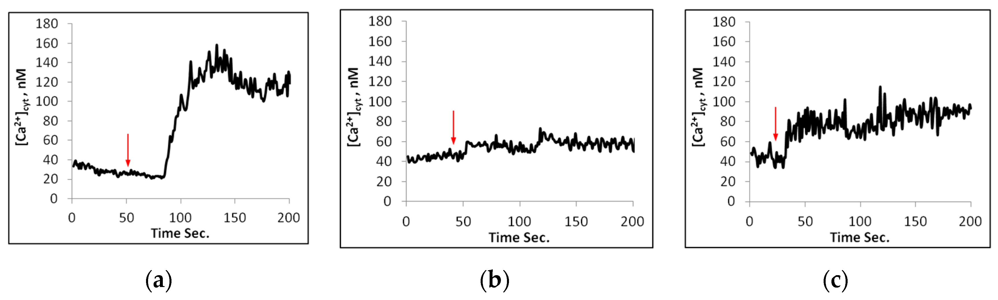
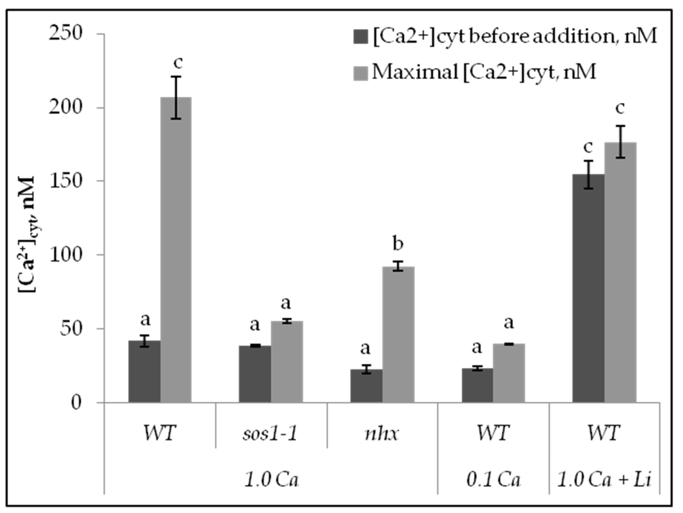
2.2. Calcium Changes in Mesophyll Protoplasts upon NaCl Addition

For cytosolic calcium Ca2+cyt measurements, the protoplasts were loaded with Fura 2, AM, a Ca2+-specific fluorescent dye. Upon addition of 100 mM NaCl to the wild-type protoplasts in the presence of 1 mM Ca, there was a distinct increase in cytosolic calcium concentration, [Ca2]cyt (Figure 3a and Figure 4). Pretreatment of the protoplasts for 15 min in the presence of 5 mM LiCl significantly decreased the calcium elevation. The maximal increase in [Ca2]cyt was obtained in Wt protoplasts in the presence of 1.0 mM external Ca2+. At 0.1 mM external Ca2+, the elevation of Ca2cyt was reduced (Figure 4).
When 100 mM NaCl instead was added to sos1-1 protoplasts in the presence of 0.1 or 1 mM Ca2, there were negligible increases of [Ca2]cyt (Figure 3b and Figure 4). On the other hand, when NaCl was added to the nhx1 protoplasts in the presence of 1 mM Ca2+, there was an elevation of [Ca2]cyt which was less pronounced than in Wt (Figure 3c and Figure 4).

3. Discussion

In the present study, sodium influx into the mesophyll protoplasts was low in the presence of 1.0 mM calcium. Therefore, in Arabidopsis Na+ is probably mainly taken up by nonselective cation channels, NSCCs, which are permeable for both Ca2+ and Na+. Sodium can also be taken up by HKT channels as shown in experiments with rice [31]. The high-affinity transporters HKT2s can mediate Na+-K+ cotransport and Na/K homeostasis under K-starved conditions and salinity [32]. In barley, over-expression of HKT2 caused salt tolerance, however, in Arabidopsis HKT2, expression showed reduced salt tolerance.
The importance of nonselective cation channels for the influx in the presence of a high concentration of sodium has been stressed [31,34].
A higher influx of Na+ was obtained in the sos1-1 and nhx1 mutants than in the wild type (Figure 1a–c and Figure 2). The influx into the sos1-1 protoplasts was faster than in the other genotypes and may depend on the lack of an SOS1 protein, and then decreased due to efflux from the cytosol into the vacuole or intracellular vesicles (Figure 1b). It was reported that SOS1 plays a crucial role in sodium efflux from root cells [21] and the same we found in the A.t. mesophyll cells. Transgene Arabidopsis overexpressing the SOS1 protein caused an enhanced tolerance to NaCl, since this plant accumulated less Na+ in the transpiration stream, and in the shoot, than the wild type [21]. Similar findings were obtained from an sos1 mutant of rice that showed a higher total uptake of Na+ than the wild type [35].
A low and transient uptake into the cytosol is more important for tolerance than a total uptake into cells [36]. Here we show that the net uptake of Na+ into the cytosol of A.t is higher in the sos1-1 mutant than in the wild type. It was demonstrated that the Arabidopsis sos1-1 mutant has reduced growth compared with the wild type, and chlorotic young leaves, which can be expected if higher Na+ is present in the cytosol [23].
The nhx1 mutant, lacking the Na+/H+ antiporter in the tonoplast [8] immediately after NaCl addition, took up less Na+ than the other genotypes, however, the net accumulation of Na+ was higher than the Wt (Figure 1c and Figure 2). Therefore, this genotype can rapidly transport some Na+ out from the cytosol, but not into the vacuole. Thus, our results confirm that AtSOS1-1 is more effective than AtNHX1 in removing Na+ from the cytosol of Arabidopsis mesophyll cells.
In experiments with protoplasts from Arabidopsis, Oryza sativa, and Vicia faba, the rise in [Ca2+]cyt was lower than in intact transgene Arabidopsis subjected to different types of stress [26,27,37,38,39] and might depend on the lack of cell walls. Gao et al. (2004) [38] reported that calcium signalling also could take place in the cell walls. In intact Arabidopsis seedlings expressing the calcium–binding protein aequorin, the Ca2+cyt elevations were inhibited by La, and to a lesser extent by EGTA [37]. However, lantan and EGTA, which block calcium influx from external stores, were only partly inhibiting the influx of Ca2+. Calcium elevation may also result from the activation of PLC, phospholipase C, leading to the hydrolysis of PIP2 to IP3 and subsequent release of Ca2+ from intracellular stores, like the vacuole [40]. Inhibitors to IP3 signaling such as LiCl showed that mannitol caused a calcium concentration increase in the microdomain of the vacuole [37]. Our results with the Arabidopsis protoplasts from the wild type show that LiCl also blocked the Ca2+ elevation caused by NaCl addition. Other findings showed that signalling via phospholipase C was suggested for proline accumulation in Arabidopsis upon ionic, but not nonionic hyperosmotic stress [41]. As reported for Arabidopsis roots, ROS produced by high salt could activate calcium channels in the tonoplasts [20], and the same mechanism is likely to exist in mesophyll cells.
In the present study we show that NaCl addition, in the presence of 1 mM external Ca2+, causes a significant elevation of Ca2+cyt in Arabidopsis mesophyll protoplasts from the wild type, and a smaller elevation in the nhx-1 mutant, however, the elevation is lacking in protoplasts from the sos1-1 genotype (Figure 4). Moreover, in the presence of 0.1 mM Ca2+, the calcium increase is much reduced in Wt. Thus, it is likely that Na+ at first induces openings of the calcium channels in the plasma membrane and then causes a second calcium elevation from internal stores. Similar results were obtained with a salt-sensitive rice cv. BRRI Dhan29, which also showed a more pronounced elevation at 1.0 mM than at 0.1 mM Ca2+ [26].
In Arabidopsis, the long C-terminal tail of the SOS1 protein located in the cytosol was speculated to be a Na+ sensor [5,42,43]. More recent findings suggest that external Na+ can bind to a plasma membrane lipid called glycosyl inositol phosphorylceramide, GIPC [44,45]. The Na-bound protein can enter the cytosol and activate an unknown calcium channel leading to an influx of calcium into the cytosol. These findings are in line with the results from two rice cultivars, the salt-tolerant cv. Pokkali, and the salt-sensitive cv. BRRI Dhan 29, which revealed that Na+ should enter the cytosol before a [Ca2+]cyt elevation could occur [26].

4. Materials and Methods

4.1. Plant Material

Three genotypes of Arabidopsis thaliana, cv. Columbia, wild type, sos1-1, and nhx1, were obtained from Nottingham Arabidopsis Stock Centre (NASC http://arabidopsis, Loughborough, UK). They were cultivated in a climate chamber in a mixture of vermicompost (Blomjord, Hasselfors Garden, Örebro, Sweden), sand, and plant perlite at a ratio of 2:1:0.5, supplemented with a full-strength nutrient solution (Blomstra, Sweden) once a week during 6–8 weeks [46]. The day/night temperature was 18 ± 2 °C at 18/6 h light/dark photoperiod. The light was a mixture of light from fluorescence tubes L30W/77-Fluora and 30W41-827 LUMILUX; OSRAM, Berlin, Germany). Light intensity was 100 ± 20 µmol s−1 m−2, and humidity was about 60%.

4.2. Protoplast Isolation

Leaves (0.5 g) were sliced in 0.5 mm pieces and digested in darkness with 0.5% (w/v) cellulase (lyophilized powder; 6 units mg−1 solid from Trichoderma resei (Sigma, St Louis, MO, USA, EC 3.2.1.4) and 0.2% (w/v) macerase (lyophilized powder; 0.104 units pectinase mg−1), Maceroenzyme R-10, from Rhizopus sp. (Serva, Heidelberg, Germany, EC 3.2.1.4) for 1.5 h at 22 ± 1 °C. The pH of the digestion solution (5 mL) was 5.5 and contained 1.0 mM CaCl2, 0.2% (w/v) BSA (Sigma), 0.05% (w/v) PVP, polyvinyl polypyrrolidone (Sigma) and 20 mM Mes-KOH, [morpholino] ethane sulfonic acid (Sigma).
After digestion, the solution with plant material was filtrated by a tea strainer and washed twice with the same digestion solution, but without enzymes. Thereafter, it was filtrated through a nylon net (100 µm pores) and centrifuged at 100× g for 5 min. The pellet was re-suspended in 1 mL 22% (w/v) sucrose (BHD, England) and centrifuged at 150× g for 5 min. The viable protoplasts floating on the sucrose solution were collected and gently mixed with a washing solution containing 0.5 M mannitol, 1 mM CaCl2, and 20 mM MES-KOH buffer, pH 5.5. The solution was again centrifuged at 100× g and the pellet was suspended in 1 mL mannitol solution.

4.3. Dye Loading and Fluorescence Measurements

The protoplasts were washed twice in the loading medium (medium ‘A’) containing 0.5 M mannitol (Sigma), 1.0 mM CaCl2, 0.05% (w/v) PVP, 0.2 (w/v)% BSA (Sigma) and a buffer (pH 5.5) containing 5 mM Tris (Labassco, Partille, Sweden) and 5 mM MES (Sigma), pH 5.5.
For cytosolic Ca2+ measurement, the protoplasts were loaded with acetoxy methyl ester of calcium-binding benzofuran (Fura 2-AM; Molecular Probes, Leiden, the Netherlands). The Fura 2-AM solution was prepared by mixing 2 µL of Fura 2-AM stock solution (5 mg/mL) in dry (<0.1% v/v water) DMSO, 1.25 µL of pluronic F-127 (Molecular Probes) solution (20% w/v in DMSO), and 6.75 µL of ethanol (99.5% v/v) [47,48,49]. From the Fura 2-AM dye solution, 5 µL was added to 1 mL of protoplast suspension (medium A). Loading was performed at 22 °C for 1.5 h.
For Na+ measurements protoplasts were loaded with SBFI-AM (Molecular Probes, Eugene, OR, USA) The dye was dissolved in dimethyl sulphoxide (DMSO, Merck, Eurolab AB, Stockholm, Sweden, (<0.1% water) to give a 5 mM stock solution. Two µL of the stock solution was diluted with 6.75 µL ethanol (Kemetyl, Stockholm, Sweden) and 1.25 µL pluronic F-127 (Molecular Probes) as described earlier [31,49] and added to 1 mL of protoplast suspension to get a final concentration of 10 µM. Dye loading was performed in medium A for 1.5–2 h at room temperature in darkness. After loading, the samples were centrifuged and pellets were re-suspended into 1 mL of a solution similar to medium A, but with TRIS-MES buffer at pH 7 (medium B). Before measurements, samples were kept in darkness at room temperature for 25 min.

4.4. Fluorescence Measurements

An epi-fluorescence microscope (Axiovert 10; Zeiss, Oberkochem, Germany), supplied with an electromagnetic filter exchanger (Zeiss), Xenon lamp (ZeissXBO 75), photometer (Zeiss 01), microprocessor (MSP 21, Zeiss), and a personal computer was used to determine fluorescence intensity after excitation at 340/380 nm for both
Fura 2- and SBFI measurements. Measurements were taken every 250 ms. Emission wavelengths were 510–550 nm for both SBFI and Fura 2 measurements. All measurements were performed with a Planneofluar x40/0.75 objective (Zeiss) for phase contrast. Adjustments of signals and noise were made automatically. By means of ratio microscopy, the effect of different dye concentrations can be eliminated [50,51]. Micro slides were covered with poly-L-lysine (molecular weight based on viscosity 150,000–300,000; Sigma) 0.2%, to attach protoplasts to their surface.
Since the Arabidopsis protoplasts are very sensitive to osmotic stress, all solutions used had the same osmotic pressure. Thus, when NaCl solutions were added less mannitol was used in the buffer.
In situ calibration for Ca2+ measurement was made using single protoplasts labeled with Fura 2-AM as described earlier [51,52].
In situ calibration was also used for [Na.]cyt measurements and made on single protoplasts labeled with SBFI-AM [39]. Measurements of SBFI-fluorescence at an excitation ratio of 340/380 nm and 510–550 nm emission wavelengths were performed with protoplasts in separate suspension solutions with concentrations of 0, 25, 50, 75, and 100 mM NaCl. KCl was added to the suspension solutions to give a final concentration of 100 mM [Na + K] to approximate physiological ionic strengths. The standard measurements were undertaken 5 to 10 min after the addition of 10 mM gramicidin (Sigma) to equilibrate intracellular and extracellular concentrations of Na. As salt stress also induces cytosolic acidification, 5 mM nigericin was added to avoid a pH effect.

4.5. Statistics

Each plot is a copy of printer plots and shows representative traces of a specific experiment repeated more than, or equal to, five times with protoplasts from independent cultivations. Each value before salt addition is the average of around 25 fluorescent-ratio determinations and after salt addition, 240 fluorescent-ratio determinations. Figure 3 shows data from experiments repeated five or more times, and Figure 4 from experiments repeated 3 times with protoplasts from independent cultivations. Data are presented as means ± SE. All collected data were statistically analyzed using a factorial completely randomized design (CRD) and the means were compared using the least significant difference test (L.S.D.) at a 5% level of probability to indicate treatment differences [53]

5. Conclusions

Our results demonstrate that AtSOS1 under high salinity reduces the Na+ concentration in the cytosol of mesophyll cells more effectively than AtNHX1. Similar results were obtained in rice [54]. In Arabidopsis, high Na+ induces a cytosolic calcium elevation only in cells with an intact SOS1. The calcium elevation is caused by transport from both external and internal stores and is more pronounced in the presence of 1.0 mM external Ca2+ than at 0.1 mM. Therefore, it is likely that calcium first enters the cytosol via calcium channels in the plasma membrane probably activated by a Na+-GIPC complex [44] and thereafter from internal stores. The AtTPC1 channel in the tonoplast is activated by Ca2+cyt and may be involved in calcium transport from the vacuole into the cytosol [48,54].

Author Contributions

S.H.M., M.A.K. and S.L. contributed to the project design. M.A.K. and S.L. performed all the experiments and S.H.M. and S.L. analysed the results and made interpretations. S.L. wrote the text and S.H.M., M.A.K. and S.L. were involved in the final approval and reading. All authors have read and agreed to the published version of the manuscript.

Funding

Guest Scholarships by Swedish Institute, SI 00313, and KSLA L15-0042-CFL, Sweden.

Data Availability Statement

Data are available from the corresponding author.

Acknowledgments

We are grateful to SI and KSLA for project funding. We also thank Docent Maria Greger for allowing us to use her laboratory.

Conflicts of Interest

The authors declare no conflict of interest. The funders had no role in the design of the project, in the material collection, analyses, data interpretation, or writing of the manuscript.

References

  1. Munns, R.; Gilliham, M. Salinity tolerance of crops—What is the costs? New Phytol. 2015, 208, 668–673. [Google Scholar] [CrossRef] [Green Version]
  2. Munns, R.; James, R.A.; Läuchli, A. Approaches to increasing the salt tolerance of wheat and other cereals. J. Exp. Bot. 2006, 57, 1025–1043. [Google Scholar] [CrossRef] [PubMed] [Green Version]
  3. Maathuis, F.J.M. Sodium in plants: Perception, signalling and regulation of sodium fluxes. J. Exp. Bot. 2014, 65, 849–858. [Google Scholar] [CrossRef] [PubMed]
  4. Keisham, M.; Mukherjee, S.; Bhatla, S. Mechanisms of sodium transport in plants—Progresses and challenges. Int. J. Mol. Sci. 2018, 19, 647. [Google Scholar] [CrossRef] [Green Version]
  5. Zhu, J.K. Regulation of ion homeostasis under salt stress. Curr. Opin. Plant Biol. 2003, 6, 441–445. [Google Scholar] [CrossRef]
  6. Guo, Q.; Meng, L.; Han, J.; Mao, P.; Tian, X.; Zheng, M.; Mur, L.A.J. SOS1 is a key systemic regulator of salt secretion and K+/Na+ homeostasis in the recretohalophyte Karelinia caspia. Environ. J. Exp. Bot. 2020, 177, 104098. [Google Scholar] [CrossRef]
  7. Pardo, J.M.; Cubero, B.; Leidi, E.O.; Quintero, F.J. Alkali cation exchangers: Roles on cellular homeostasis and stress tolerance. J. Exp. Bot. 2006, 57, 1181–1199. [Google Scholar] [CrossRef] [Green Version]
  8. Apse, M.P.; Blumwald, E. Na+ transport in plants. FEBS Lett. 2007, 581, 2247–2254. [Google Scholar] [CrossRef] [PubMed] [Green Version]
  9. Brini, F.; Hanin, M.; Mezghani, I.; Gerald, A.; Berkowitz, G.A.; Masmoudi, K. Overexpression of wheat Na+/H+ antiporter TNHX1 and H+-pyrophosphatase TVP1 improve salt- and drought-stress tolerance in Arabidopsis thaliana plants. J. Exp. Bot. 2007, 98, 301–308. [Google Scholar] [CrossRef] [PubMed] [Green Version]
  10. Krebs, M.; Beyhlb, D.; Esther Görlich, E.; Al-Rasheid, K.A.S.; Marten, I.; Stierhofd, Y.-D.; Hedrich, R.; Schumacher, K. Arabidopsis V-ATPase activity at the tonoplast is required for efficient nutrient storage but not for sodium accumulation. Proc. Natl. Acad. Sci. USA 2010, 107, 3251–3256. [Google Scholar] [CrossRef]
  11. Andres, Z.; Perez-Hormaeche, J.; Leidi, E.O.; Schlucking, K.; Steinhorst, L.; McLachlan, D.H.; Schumacher, K.; Hetherington, A.M.; Kudla, J.; Cubero, B.; et al. Control of vacuolar dynamics and regulation of stomatal aperture by tonoplast potassium uptake. Proc. Natl. Acad. Sci. USA 2014, 111, E1806–E1814. [Google Scholar] [CrossRef] [Green Version]
  12. Luo, L.; Zhang, P.; Zhu, R.; Fu, J.; Su, J.; Zheng, J.; Wang, Z.; Wang, D.; Gong, Q. Autophagy is rapidly induced by salt stress and is required for salt tolerance in Arabidopsis. Front. Plant Sci. 2017, 8, 1459. [Google Scholar] [CrossRef] [Green Version]
  13. Köster, P.; Wallrad, L.; Edel, K.H.; Faisal, M.; Alatar, A.A.; Kudla, J. The battles of two ions: Ca2+ signalling against Na+ stress. Plant Biol. 2018, 21, 39–48. [Google Scholar] [CrossRef] [PubMed]
  14. Wang, Q.; Guan, C.; Wang, P.; Ma, Q.; Bao, A.-K.; Zhang, J.-L.; Wan, S.-M. The Effect of AtHKT1;1 or AtSOS1 mutation on the expressions of Na+ or K+ transporter genes and ion homeostasis in Arabidopsis thaliana under salt stress. Int. J. Mol. Sci. 2019, 20, 1085. [Google Scholar] [CrossRef] [PubMed] [Green Version]
  15. Nayef, M.A.; Celymar, S.; Shabala, L.; Ogura, T.; Chen, Z.; Bose, J.; Maathuis, F.J.M.; Venkataraman, G.; Tanoi, K.; Yu, M.; et al. Changes in expression level of OsHKT1; 5 alters activity of membrane transporters involved in K+ and Ca2+ acquisition and homeostasis in salinized rice roots. Int. J. Mol. Sci. 2020, 21, 4882. [Google Scholar] [CrossRef] [PubMed]
  16. Evans, M.J.; Choi, W.-G.; Gilroy, S.; Morris, R.J. A ROS-assisted calcium wave dependent on the AtRBOHD NADPH oxidase and TPCl cation channel propagates the systemic response to salt stress. Plant Physiol. 2016, 171, 1771–1784. [Google Scholar] [CrossRef] [PubMed] [Green Version]
  17. White, P.J.; Broadley, M.R. Calcium in Plants. Ann. Bot. 2003, 92, 487–511. [Google Scholar] [CrossRef]
  18. Park, C.-J.; Shin, R. Calcium channels and transporters: Roles in response to biotic and abiotic stresses. Front. Plant Sci. 2022, 13, 964059. [Google Scholar] [CrossRef]
  19. Ye, F.; Xu, L.; Li, X.; Zen, W.; Gan, N.; Zhao, C.; Yang, W.; Jiang, Y.; Guo, J. Voltage-gating and cytosolic Ca2+ activation mechanisms of Arabidopsis two-pore channel AtTPC1. Proc. Natl. Acad. Sci. USA 2021, 118, e2113946118. [Google Scholar] [CrossRef]
  20. Leshem, Y.; Melamed-Book, N.; Cagnac, O.; Ronen, G.; Nishri, Y.; Solomon, M.; Cohen, G.; Levine, A. Suppression of Arabidopsis vesicle-SNARE expression inhibited fusion of H2O2-containing vesicles with tonoplast and increased salt tolerance. Proc. Natl. Acad. Sci. USA 2006, 103, 18008–18013. [Google Scholar] [CrossRef]
  21. Shi, H.; Quintero, F.J.; Pardo, J.M.; Zhu, J.-K. The Putative plasma membrane Na+/H+ antiporter SOS1 controls long-distance Na+ transports in plants. Plant Cell 2002, 14, 465–477. [Google Scholar] [CrossRef] [Green Version]
  22. Julkowska, M.M.; Testerink, C. Tuning plant signaling and growth to survive salt. Trends Plant Sci. 2015, 20, 586–594. [Google Scholar] [CrossRef] [Green Version]
  23. Quintero, F.J.; Martinez-Atienza, J.; Villalta, I.; Jiang, X.; Kim, W.-Y.; Ali, Z.; Fujii, H.; Mendoza, I.; Yun, D.-J.; Zhu, J.-K.; et al. Activation of the plasma membrane Na/H antiporter Salt-Overly-Sensitive 1 (SOS1) by phosphorylation of an auto-inhibitory C-terminal domain. Proc. Natl. Acad. Sci. USA 2011, 108, 2611–2616. [Google Scholar] [CrossRef] [Green Version]
  24. Quan, R.; Lin, H.; Mendoza, I.; Zhang, Y.; Cao, W.; Yang, Y.; Shang, M.; Chen, S.; Pardo, J.M.; Guo, Y. SCABP8/CBL10, a putative calcium sensor, interacts with the protein kinase SOS2 to protect Arabidopsis shoots from salt stress. Plant Cell 2007, 19, 1415–1431. [Google Scholar] [CrossRef] [PubMed] [Green Version]
  25. Egea, I.; Pined, B.A.; Ortíz-Atienza, A.; Plasencia, F.A.; Drevensek, S.; García-Sogo, B.; Yuste-Lisbona, F.J.; Barrero-Gil, J.; Atarés, A.; Flores, F.B.; et al. The SlCBL10 Calcineurin B-like protein ensures plant growth under salt stress by regulating Na+ and Ca2+ homeostasis. Plant Physiol. 2018, 176, 1676–1693. [Google Scholar] [CrossRef] [PubMed] [Green Version]
  26. Kader, A.; Lindberg, S.; Seidel, T.; Golldack, D.; Yemelyanov, V. Sodium sensing induces different changes in free cytosolic calcium concentration and pH in salt-tolerant and salt-sensitive rice (Oryza sativa L.) cultivars. Physiol. Plant. 2007, 130, 99–111. [Google Scholar] [CrossRef]
  27. Morgan, S.H.; Lindberg, S.; Jha Maity, P.; Geilfus, C.M.; Plieth, C.; Mühling, K.H. Apoplastic and cytosolic Ca2+ and pH dynamics in salt-stressed Vicia faba leaves change in response to calcium. Func. Plant Biol. 2017, 44, 515–524. [Google Scholar] [CrossRef] [Green Version]
  28. Villalobos-Lo’pez, M.A.; Arroyo-Becerra, A.; Quintero-Jiménez, A.; Iturriaga, G. Biotechnological Advances to Improve Abiotic Stress Tolerance in Crops. Int. J. Mol. Sci. 2022, 23, 12053. [Google Scholar] [CrossRef]
  29. Ding, L.; Zhu, J.-K. Reduced Na+ uptake in the NaCI-hypersensitive sos1 mutant of Arabidopsis thaliana. Plant Physiol. 1997, 113, 795–799. [Google Scholar] [CrossRef] [Green Version]
  30. Essah, P.A.; Davenport, R.; Tester, M. Sodium influx and accumulation in Arabidopsis. Plant Physiol. 2003, 133, 307–318. [Google Scholar] [CrossRef]
  31. Kader, M.A.; Lindberg, S. Uptake of sodium in protoplasts of salt-sensitive and salt-tolerant cultivars of rice, Oryza sativa L. determined by the fluorescent dye SBFI. J. Exp. Bot. 2005, 56, 3149–3158. [Google Scholar] [CrossRef] [PubMed] [Green Version]
  32. Tada, Y.; Ohnuma, A. Comparative Functional Analysis of Class II Potassium Transporters, SvHKT2;1, SvHKT2;2, and HvHKT2;1, on Ionic Transport and Salt Tolerance in Transgenic Arabidopsis. Plants 2020, 9, 786. [Google Scholar] [CrossRef]
  33. Yemelyanov, V.V.; Shishova, M.F.; Chirkova, T.V.; Lindberg, S. Anoxia-induced elevation of cytosolic Ca2+ concentration depends on different Ca2+ sources in rice and wheat protoplasts. Planta 2011, 234, 271–280. [Google Scholar] [CrossRef] [PubMed]
  34. Demidchik, V.; Maathuis, F.J.M. Physiological roles of nonselective cation channels in plants: From salt stress to signalling and development. New Phytol. 2007, 175, 387–404. [Google Scholar] [CrossRef] [PubMed]
  35. El Mahi, H.; Péres-Hormaeche, J.; De Luca, A.; Villlalta, I.; Espartero, J.; Gámes-Arjona, F.; Fernández, J.L.; Bundó, M.; Mendoza, I.; Mieulet, D.; et al. A Critical Role of Sodium Flux via the Plasma Membrane Na+/H+ Exchanger SOS1 in the Salt Tolerance of Rice. Plant Physiol. 2019, 180, 1046–1065. [Google Scholar] [CrossRef] [Green Version]
  36. D’Onofrio, C.; Kader, A.; Lindberg, S. Uptake of sodium in quince, wheat and sugar beet protoplasts determined by the fluorescent sodium-binding benzofuran isophthalate dye. J. Plant Physiol. 2005, 162, 421–428. [Google Scholar] [CrossRef]
  37. Knight, H.; Trewavas, A.J.; Knight, M.R. Calcium signal. Plant J. 1997, 12, 1067–1078. [Google Scholar] [CrossRef] [PubMed]
  38. Gao, D.; Knight, M.R.; Trewavas, A.J.; Sattelmacher, B.; Plieth, C. Self-reporting Arabidopsis expressing pH and [Ca2+] indicators unveil ion dynamics in the cytoplasm and in the apoplast under abiotic stress. Plant Physiol. 2004, 134, 898–908. [Google Scholar] [CrossRef] [PubMed] [Green Version]
  39. Morgan, S.H.; Geilfus, C.M.; Lindberg, S.; Mühling, K.H. The leaf ion homeostasis and PM H+-ATPase activity in Vicia faba L. change after extra calcium and potassium supply under salinity. Plant Physiol. Biochem. 2014, 82, 244–253. [Google Scholar] [CrossRef]
  40. Allen, G.J.; Muir, S.R.; Sanders, D. Release of Ca2+ from individual plant vacuoles by both InsP3 and cyclic ADP-ribose. Science 1995, 268, 735–737. [Google Scholar] [CrossRef]
  41. Parre, E.; Ghars, M.A.; Leprince, A.-S.; Thiery, L.; Lefebvre, D.; Bordenave, M.; Richard, L.; Mazars, C.; Abdelly, C.; Savouré, A. Calcium signaling via phospholipase C is essential for proline accumulation upon ionic but not nonionic hyperosmotic stresses in Arabidopsis. Plant Physiol. 2007, 144, 503–512. [Google Scholar] [CrossRef] [Green Version]
  42. Zhang, J.Z.; Creelman, R.A.; Zhu, J.-K. From laboratory to field. Using information from Arabidopsis to engineer salt, cold, and drought tolerance in crops. Plant Physiol. 2004, 135, 615–621. [Google Scholar] [CrossRef] [Green Version]
  43. Shabala, L.; Cuin, T.A.; Newman, I.A.; Shabala, S. Salinity-induced ion flux patterns from the excised roots of Arabidopsis sos mutants. Planta 2005, 222, 1041–1050. [Google Scholar] [CrossRef]
  44. Jiang, Z.; Zhou, X.; Pei, Z.-M. Plant cell-surface GIPC sphingolipids sense salt to trigger Ca2+ influx. Nature 2019, 572, 341–346. [Google Scholar] [CrossRef]
  45. Steinhorst, L.; Kudla, J. How plants perceive salt. Nature 2019, 572, 318–320. [Google Scholar] [CrossRef] [Green Version]
  46. Premkumar, A.; Lindberg, S.; Lager, I.; Rasmussen, U.; Schulz, A. Arabidopsis PLDs with C2-domain function distinctively in hypoxia signaling. Physiol. Plant. 2019, 107, 90–110. [Google Scholar] [CrossRef] [PubMed]
  47. Sebastiani, L.; Lindberg, S.; Vitagliano, C. Cytoplasmic free calcium dynamics in single tomato (Lycopersicon esculentum L) protoplasts subjected to chilling temperatures. Physiol. Plant. 1999, 105, 239–245. [Google Scholar] [CrossRef]
  48. Poenie, M.; Alderton, J.; Steinhardt, R.; Tsien, R. Calcium rises abruptly and briefly throughout the cell cycle and onset of anaphase. Science 1986, 233, 886. [Google Scholar] [CrossRef] [PubMed]
  49. Bright, G.R.; Fisher, G.W.; Rogowska, J.; Taylor, D.L. Fluorescence ratio imaging microscopy: Temporal and spatial measurements of cytoplasmic pH. J. Cell Biol. 1987, 104, 1019–1033. [Google Scholar] [CrossRef] [Green Version]
  50. Tsien, R.Y.; Poenie, M. Fluorescence ratio imaging: A new window into intracellular ionic signalling. Trends Biochem. Sci. 1986, 11, 450–455. [Google Scholar] [CrossRef]
  51. Lindberg, S.; Premkumar, A.; Rasmussen, U.; Schulz, A.; Lager, I. Phospholipases AtPLDζ1 and AtPLDζ2 function differently in hypoxia. Physiol. Plant. 2018, 162, 98–108. [Google Scholar] [CrossRef] [PubMed]
  52. Grynkiewicz, G.; Poenie, M.; Tsien, R.Y. A new generation of Ca2+ indicators with greatly improved fluorescence properties. J. Biol. Chem. 1985, 260, 3440–3450. [Google Scholar] [CrossRef] [PubMed]
  53. Snedecor, G.W.; Cochran, W.G. Statistical Methods, 7th ed.; Iowa State University Press: Ames, IA, USA, 1980. [Google Scholar]
  54. Schulze, C.; Sticht, H.; Meyerhoff, P.; Dietrich, P. Differential contribution of EF-hands to the Ca2+-dependent activation in the plant two-pore channel TPC1. Plant J. 2011, 68, 424–432. [Google Scholar] [CrossRef] [PubMed]
Figure 1. Changes of [Na+cyt] in mM with time upon addition of 100 mM of NaCl to single mesophyll protoplasts of Arabidopsis wild type (a), sos1-1 (b), and nhx1 (c). Measurements at excitation ratio 340/380 nm and emission 510–550 nm. Typical traces.
Plants 11 03439 g001
Figure 2. Changes of [Na+cyt ] in mM of mesophyll protoplasts of Arabidopsis wild type, sos1-1, and nhx1 genotypes upon addition of 100 mM of NaCl, and after stabilization. Means ± SE.
Plants 11 03439 g002
Figure 3. Changes of [Ca2+cyt] in nM with time, upon addition of 100 mM of NaCl to single mesophyll protoplasts of Arabidopsis wild type (a), sos1-1 (b), and nhx1 (c). Measurements at excitation ratio 340/380 nm and emission 510–550 nm. Typical traces.
Plants 11 03439 g003
Figure 4. Changes in [Ca2+cyt] in nM of mesophyll protoplasts of Arabidopsis wild type, sos1-1, and nhx1 genotypes before the addition of 100 mM of NaCl and at maximal changes of [Ca2+cyt]. The external solution was with and without 5 mM LiCl and contained 0.1 or 1.0 mM Ca2+. Means ± SE.
Plants 11 03439 g004
Publisher’s Note: MDPI stays neutral with regard to jurisdictional claims in published maps and institutional affiliations.

