Article

Myrrh and Chamomile Flower Extract Inhibit Mediator Release from IgE-stimulated Mast-Cell-Like RBL-2H3 Cells

1
Institute of Medical Physics and Biophysics, Medical Faculty, Leipzig University, Härtelstr. 16-18, 04107 Leipzig, Germany
2
Repha GmbH Biologische Arzneimittel, Alt-Godshorn 87, 30855 Langenhagen, Germany
*
Authors to whom correspondence should be addressed.
Plants 2022, 11(24), 3422; https://doi.org/10.3390/plants11243422
Submission received: 21 September 2022 / Revised: 28 November 2022 / Accepted: 1 December 2022 / Published: 8 December 2022
(This article belongs to the Special Issue Anti-Inflammatory Bioactivities in Plant Extracts)

Abstract

:
Recent clinical evidence supports the efficacy of a traditional medicinal product (TMP) containing a combination of myrrh (Commiphora myrrha (Nees) Engl.), coffee charcoal (Coffea arabica L.), and chamomile flower dry extract (Matricaria chamomilla L.) in the therapy of diarrhea and inflammatory bowel disease. Mast cells seem to play a key role in the symptom generation of irritable bowel syndrome (IBS). To evaluate the use of the TMP in IBS treatment, the effects of the herbal extracts on the release of mast-cell mediators from stimulated RBL-2H3 cells were investigated. Therefore, degranulation was induced by phorbol-12-myristate-13-acetate (PMA) and calcium ionophore A13187 (CI) or IgE stimulation, and the amounts of released β-hexosaminidase and histamine were quantified. The extracts showed no effect on the mediator release of PMA- and CI-stimulated RBL-2H3 cells. Myrrh and chamomile were able to reduce the β-hexosaminidase release of IgE-stimulated cells, while myrrh showed stronger inhibition of the mediator release than chamomile, which reduced only IgE-stimulated histamine release. Thus, these results indicate a mechanistic basis for the use of the herbal combination of myrrh, coffee charcoal, and chamomile flower extract in the symptom-oriented treatment of IBS patients with diarrheal symptoms.

1. Introduction

Irritable bowel syndrome (IBS) affects about 11% of the global population [1]. Patients suffer a huge variety of symptoms such as pain, discomfort, diarrhea, obstipation, and flatulence, leading to a decrease in their quality of life [2]. While the etiology and pathophysiology of the disease have yet to be fully unraveled, the increased amount and activity of mast cells in IBS patients might play a key role in symptom generation [3,4]. Upon degranulation, mediators such as histamine and proteases are released, activating corresponding receptors on the surface of epithelial cells, which leads to chloride secretion into the gut lumen causing diarrhea [5,6,7,8]. Proteases and protease-activated receptors (PARs) are associated with the impairment of intestinal barrier function [8,9,10]. Furthermore, mast cell mediators such as histamine and tryptase are able to excite visceral nerves [11,12,13], possibly explaining the dysfunction in motility, discomfort, and pain. Therefore, inhibiting the mediator release from mast cells should improve symptoms in IBS patients. This effect could be observed in a randomized, placebo-controlled study with ketotifen, which significantly improved symptoms associated with IBS such as diarrhea, discomfort, and pain, therefore improving the overall quality of life in patients with diarrhea-predominant IBS. It was also shown that ketotifen decreased the spontaneous mediator release from mast cells [14,15].
In severe IBS cases, patients cannot fulfil their daily tasks or participate in social activities, which drastically decreases their quality of life [2,16]. According to the German S3 guideline, IBS treatment is only symptomatic [17]. Due to the lack of satisfying treatment options to fully control symptoms, patients’ interest in complementary medicine (CM) has increased. However, because of scarce evidence, CM has a low acceptance rate by healthcare professionals. Further pharmacological validation of CM might contribute to higher acceptance and rational use of CM and therefore increase the quality of life of IBS patients. Myrrhinil-Intest® is a traditional herbal medicinal product containing a combination of myrrh (Commiphora myrrha (Nees) Engl.), coffee charcoal (Coffea arabica L.), and chamomile flower dry extract (Matricaria chamomilla L.), which is used for the treatment of diarrhea and gut disorders. Clinical efficacy and safety have been shown in diarrhea and IBS patients [18].
Previous pharmacological in vitro studies demonstrated spasmolytic, anti-inflammatory, and intestinal-barrier-stabilizing effects of the plant compounds [19,20,21,22]. To further evaluate the pharmacological profile of the herbal combination product in the treatment of IBS, the aim of this study was to investigate the influence of the herbal components on mast-cell degranulation. Thus, characterized extracts were applied in a mast-cell degranulation model using RBL-2H3 cells, and the mediators released (β-hexosaminidase, histamine) were investigated after chemical as well as IgE stimulation.

2. Results and Discussion

To support the hypothesis that the antidiarrheal effects of the herbal combination might be related to the compounds inhibiting the mediator release from mucosal mast cells, the potential effects were investigated using the cell line RBL-2H3. The cell line can be used as a model for mucosal mast cells, releasing different mediators upon degranulation, induced by a variety of compounds as well as IgE sensitization followed by antigen stimulation [23,24,25]. Degranulation was induced by chemical agents, phorbol-12-myristate-13-acetate (PMA) and calcium ionophore A13187 (CI), or by IgE stimulation. The protease β-hexosaminidase and histamine are preformed mediators stored in RBL-2H3 cell granules and were therefore chosen as degranulation markers.

2.1. Plant Extract Characterization

All extracts used in the following in vitro studies have been previously characterized, mainly via liquid chromatography and subsequent mass spectrometry [20,21,22].
HPLC-DAD analysis of the chamomile extract revealed seven main peaks [20]. UV-spectra for five compounds (Table 1, peaks 3 to 7) showed typical UV spectra of flavonoids. Comparison with reference standards as well as mass spectrometry confirmed apigenin and apigenin derivatives as the main constituents of the extract (Table 1, peaks 3 to 7). Peaks 1 and 2 (Table 1) were identified as derivatives of ferulic acid.
Mass spectrometry alongside 1D and 2D NMR of isolated compounds from the myrrh extract showed the presence of several sesquiterpenes (Table 2, peaks 1 to 6) and one (nor-)triterpenoid (Table 3, peak 7) [21]. Comparison of the NMR and mass spectrometry data with literature allowed for the identification of all seven substances.
Analysis of the coffee charcoal extract via HPLC, reference standards, and literature data led to the identification of caffeine (Table 3 peak 4) and trigonelline (Table 2, peak 1) as well as several caffeic acid derivatives (Table 2, peaks 2, 3, and 5) and two feruloylquinic acid isomers (Table 2, peaks 6 and 7) [22].

2.2. Influence on Cell viability

Before conducting any experiments, the extracts were tested for cytotoxicity using an XTT assay (Figure 1). Only myrrh at a concentration of 100 µg/mL reduced the metabolic activity of the RBL-2H3 cells (59.9% ± 10.2%; p < 0.05). Based on these results, concentrations were chosen for further experiments.

2.3. Influence on Chemical Induced Mediator Release

Upon stimulation with a combination of CI (1 µM) and PMA (40 nM), the plant extracts were unable to exhibit an effect on the release of β-hexosaminidase and histamine from RBL-2H3 cells compared to the stimulated control (Figure 2). Since the degranulation is induced by increasing intracellular Ca2+ concentration through CI, as a pore-forming agent or carrier [25], this method resembles a non-physiological mechanism. Therefore, experiments using a stimulation method closer to physiological conditions were performed to investigate the potential inhibitory effects of the plant extracts.

2.4. Influence on IgE-Induced β-Hexosaminidase Release

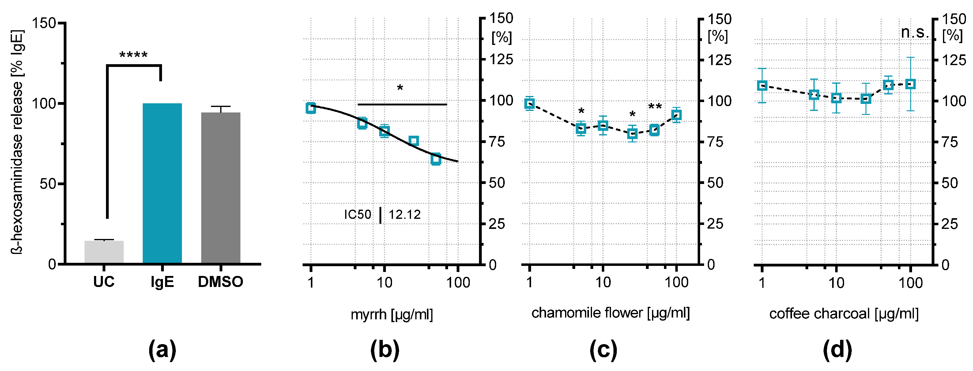
RBL-2H3 cell degranulation can be induced by IgE stimulation. The binding of antigens by surface-bound IgE molecules leads to FcεR-activation, resulting in degranulation of the cells [24], which resembles a more physiological pathway of mast cell degranulation. It is shown that IgE-stimulated β-hexosaminidase release from RBL-2H3 was reduced by myrrh resin extract down to 64.3% (±3.5%, p < 0.05) and chamomile flower extract down to 80.4% (±5.0%, p < 0.05) (Figure 3). The effect of chamomile flower extract aligns with findings showing flavonoids are able to reduce mediator release from RBL-2H3 [26,27]. Apigenin has shown inhibitory effects on β-hexosaminidase release upon IgE stimulation, which is also present in the extract [28,29]. These results indicate a U-shaped response curve. This is commonly observed for phytochemical compounds, where they exhibit positive effects at low concentrations, while the opposite effect can be found at higher concentrations, which is called hormesis [30,31,32].
Myrrh resin extract shows a concentration-dependent inhibitory effect on IgE-induced β-hexosaminidase release, reducing it by about one-third compared to the stimulated control at the highest concentration of 50 µg/mL (64.3% ± 4.9%; p < 0.0001). IC50 is reached at a concentration of 12.12 µg/mL. This result suggests inhibitory properties from mast-cell mediator release upon IgE stimulation, which is underlined by the findings of a study with HMC-1 cells showing Commiphora myrrha (Nees) Engl. extract reducing histamine release and IL-31 production [33]. The coffee charcoal extract had no effect on the mediator release.

2.5. Influence on IgE-Induced Histamine Release

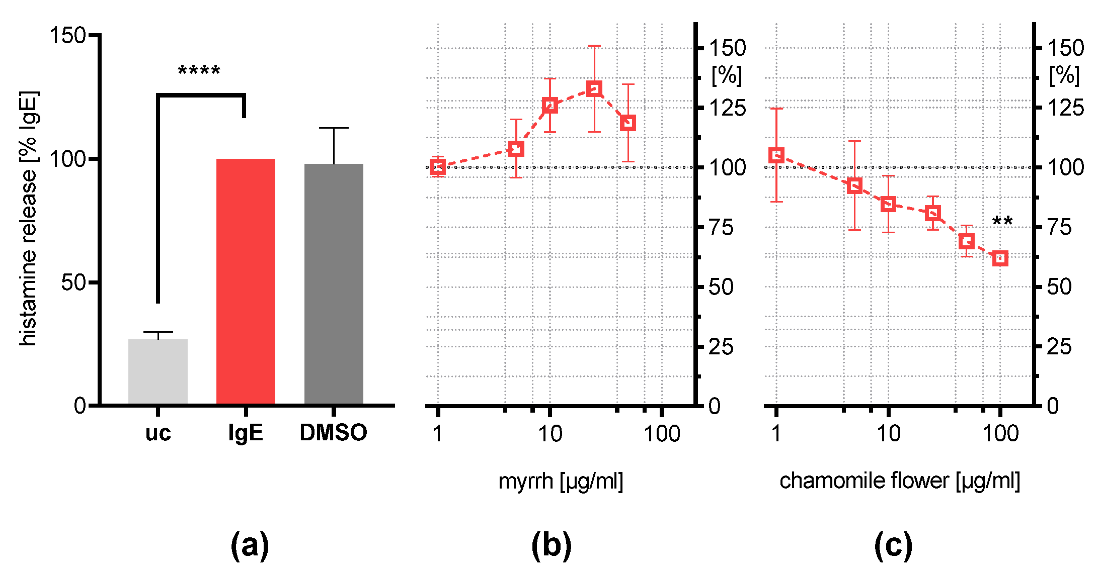
For further investigation of these results, the histamine concentration was determined in the supernatant of IgE/HSA-stimulated cells treated with myrrh and chamomile flower extract (Figure 4). Contrary to the effect observed for β-hexosaminidase release, only the chamomile flower, at a concentration of 100 µg/mL, was able to reduce the histamine release (61.9% ± 6.9%; p < 0.01). The inhibition might be induced by flavonoids such as apigenin and luteolin, as previous studies suggested [26,28,34].
These results indicate that the inhibition of β-hexosaminidase release is independent of the amount of histamine released in IgE-stimulated RBL-2H3 cells. Studies on mice bone marrow mast cells showed that they form various secretory granules, which all store β-hexosaminidase; however, some do not contain histamine [35,36]. Furthermore, mast cells are capable of releasing β-hexosaminidase independently of histamine [36,37]. This might serve as a possible explanation as to why myrrh reduces β-hexosaminidase release without inhibiting histamine secretion. This heterogeneity of mast cell granules could not be observed in human mast cells in vivo [38], which suggests that myrrh resin might be able to inhibit the release of all mediators of mucosal mast cells. This would include PAR-2 agonist tryptase, which could explain the antidiarrheal activity in IBS since PAR-2 activation plays a role in symptom generation [8,9,10,39]. The herbal extracts also exhibit anti-inflammatory and barrier-stabilizing effects on epithelial cells [21,40]. Further studies could investigate the barrier-stabilizing effects of the extracts on the epithelial cells in the presence of PAR-2 agonists, or stimulated mast cells in a co-culture model. Combined with the inhibition of histamine release due to flavonoids in chamomile flower extract, these results indicate multimodal effects, which may underly the symptom reduction observed in IBS patients and further validates the use of a TMP in IBS therapy.

3. Materials and Methods

3.1. Chemicals

Ethanol for extraction was purchased in HSL quality from CSC Jäckelchemie (Rauschwitz, Germany) and purified by evaporation. Dimethyl sulfoxide (DMSO) for solving plant extracts was purchased from VWR (Radnor, PA, USA). Chemicals (p.a. grade) were supplied by Merck (Darmstadt, Germany; glucose, CaCl2·2H2O; MgCl·2H2O; citric acid monohydrate), Carl Roth (Karlsruhe, Germany; HEPES), Fluka (Buchs, Switzerland; KCl, Triton-X), Th. Geyer (Renningen, Germany; NaCl), or Sigma (St. Louis, MO, USA; NaHPO4·2H2O; glycine, bovine serum albumin (BSA); phorbol-12-myristate-13-acetate (PMA); calcium ionophore A13187 (CI); di-nitrophenyl human serum albumin (DNP-HSA); mouse anti-DNP-HSA-IgE; 4-nitrophenyl-n-acetyl-ß-d-glucosaminide (pNAG)). Cell culture supplies MEM, fetal bovine serum (FBS), penicillin-streptomycin (P/S), glutamine stable, trypsin-EDTA and Dulbecco’s phosphate-buffered saline (DPBS) were provided by Biowest (Riverside, MO, USA).

3.2. Cell Culture

RBL-2H3 cells, purchased from DMSZ (Braunschweig, Germany), were cultured with MEM supplemented with 10% FBS, 1% P/S, and 1% glutamine stable. All cell lines were cultivated under standard cell-culture conditions. For chemical stimulation, RBL-2H3 cells were seeded into a sterile 96-well plate (GBO (Frickenhausen, Germany)) at 1 × 10⁵ cells/mL density, grown overnight, washed with DPBS, incubated for 30 min with plant extracts and control solutions, washed again and incubated for 30 min with 100 µL of a freshly prepared solution of 40 nM PMA and 1 µM CI in HEPES-buffer (HEPES 10 mM, NaCl 137 mM, KCl 2.7 mM, Na2HPO4·2H2O 0.4 mM, glucose 5.6 mM, CaCl2·2H2O 1.8 mM, MgCl·2H2O 1.3 mM, BSA 0,4%). For IgE stimulation, 5 × 10⁵ cells/mL were seeded into 96-well plates, before plant extracts and 500 ng/mL anti-DNP-HSA-IgE were added and incubated overnight (18 h) for sensitization. Cells were washed with HEPES buffer and incubated with 100 µL of 1 µg/mL DNP-HSA solution in HEPES for 30 min.

3.3. Plant Material

Ready-to-use chamomile flower dry extract (EtOH 60% m/m; DER: 4–6:1, batch-no. HC0070) as well as powdered myrrh (Myrrhe gum EB/PB pulv., batch-no. NM0160) and powdered coffee charcoal (Carbo Coffea EB 6, batch-no. JB0142) were provided by Lomapharm (Emmerthal, Germany).
Myrrh and coffee charcoal extracts were prepared, lyophilized, and stored as previously described by Weber et al. [21,22].

3.4. Quantification of β-Hexosaminidase Release

The β-hexosaminidase release was analyzed similarly to Kuehn et al. [41]. Briefly, 50 µL of the supernatants or lysed cells (addition of 150 µL of Triton-X 0.1%) was transferred into a 96-well plate (GBO (Frickenhausen, Germany)) and incubated with 100 µL of pNAG-solution (3.5 mg/mL in citrate buffer (citric acid monohydrate 40 nM, Na2HPO4·2H2O 20 mM) pH = 4.5) for 90 min at 37 °C without CO2. The enzymatic reaction was stopped by the addition of 50 µL glycine buffer pH = 10.7 (glycine 400 nM). Extinction was measured at 405 nm (reference 620 nm) with an Infinite® M200 plate reader (TECAN, Salzburg, Austria).

3.5. Quantification of Histamine Release

The histamine concentration in the supernatants was determined using the Histamine ELISA kit (cat no A05890), provided by Bertin (Montigny-le-Bretonneux, France), according to the manufacturer’s instructions.

3.6. Influence on Cell Viability

The measurement of the metabolic activity for the RBL-2H3 cells after overnight incubation in the presence of the plant extract was performed with the XTT-Vitality-Assay Kit provided by Roche (Basel, Switzerland), according to the manufacturer’s instructions.

3.7. Data Analysis

Microsoft Excel (Office 365) was used for data capturing and processing of the extinction measurements. For statistical evaluation, a mixed-model one-way analysis of variance (ANOVA) followed by Dunnett’s multiple comparisons test against the stimulated control was conducted with GraphPad Prism 8.0.2 (* p < 0.05, ** p < 0.01, *** p < 0.001, **** p < 0.0001.

4. Conclusions

The RBL-2H3 cell line was used as a model for mucosal mast cells to investigate the effects of myrrh, coffee charcoal, and chamomile flower extracts on degranulation. In IgE-stimulated cells, myrrh and chamomile flower extracts were able to inhibit the release of the protease β-hexosaminidase, while chamomile flower extract exhibited a moderate effect on histamine release, probably driven by flavonoids.
In this context and with the available clinical evidence suggesting the antidiarrheal properties of the herbal combination of myrrh, coffee charcoal, and chamomile flower, this study indicates a potential target on mucosal mast cells and builds a foundation for further investigation into the mechanism of action, as well as supporting its rational use in IBS. It also underlines the multi-target approach of herbal treatments in complex diseases such as IBS, to improve the quality of life of patients.

Author Contributions

Conceptualization, C.V.; methodology F.A. and L.S.; validation F.A. and L.S., investigation F.A. and L.S.; resources, C.V.; writing—original draft preparation, F.A.; writing—review and editing, C.V. and B.L.; visualization, F.A.; supervision, C.V.; project administration, C.V.; funding acquisition, C.V. All authors have read and agreed to the published version of the manuscript.

Funding

This research was funded by Repha GmbH Biologische Arzneimittel (Grant ID BGAAF-0750). The APC was funded by the Open Access Publishing Fund of Leipzig University, which is supported by the German Research Foundation within the program Open Access Publication Funding.

Data Availability Statement

Not applicable.

Acknowledgments

Rick Raudszus and Kerstin Hill are kindly acknowledged for their input and the RBL-2H3 cells that were used for the initial experiments (Rudolf-Boehm-Institute, Medical Faculty, Leipzig University).

Conflicts of Interest

Authors: Fabian Altenbernd, Bartosz Lipowicz and Cica Vissiennon are employed by Repha GmbH Biologische Arzneimittel. Lena Schwarz has no conflict of interest.

Sample Availability

Samples of the test compounds are available from the authors.

References

  1. Canavan, C.; West, J.; Card, T. The epidemiology of irritable bowel syndrome. Clin. Epidemiol. 2014, 6, 71–80. [Google Scholar] [PubMed] [Green Version]
  2. Simrén, M.; Brazier, J.; Coremans, G.; Dapoigny, M.; Müller-Lissner, S.A.; Pace, F.; Smout, A.J.P.M.; Stockbrügger, R.W.; Vatn, M.H.; Whorwell, P.J. Quality of life and illness costs in irritable bowel syndrome. Digestion 2004, 69, 254–261. [Google Scholar] [CrossRef] [PubMed]
  3. Wouters, M.M.; Vicario, M.; Santos, J. The role of mast cells in functional GI disorders. Gut 2016, 65, 155–168. [Google Scholar] [CrossRef] [PubMed] [Green Version]
  4. Matricon, J.; Meleine, M.; Gelot, A.; Piche, T.; Dapoigny, M.; Muller, E.; Ardid, D. Review article: Associations between immune activation, intestinal permeability and the irritable bowel syndrome. Aliment. Pharmacol. Ther. 2012, 36, 1009–1031. [Google Scholar] [CrossRef] [Green Version]
  5. Santos, J.; Saperas, E.; Nogueiras, C.; Mourelle, M.; Antolín, M.; Cadahia, A.; Malagelada, J.-R. Release of mast cell mediators into the jejunum by cold pain stress in humans. Gastroenterology 1998, 114, 640–648. [Google Scholar] [CrossRef]
  6. Hardcastle, J.; Hardcastle, P.T. The secretory actions of histamine in rat small intestine. J. Physiol. 1987, 388, 521–532. [Google Scholar] [CrossRef]
  7. Russell, D.A. Mast cells in the regulation of intestinal electrolyte transport. Am. J. Physiol. 1986, 251, G253–G262. [Google Scholar] [CrossRef]
  8. Mall, M.; Gonska, T.; Thomas, J.; Hirtz, S.; Schreiber, R.; Kunzelmann, K. Activation of ion secretion via proteinase-activated receptor-2 in human colon. Am. J. Physiol. Gastrointest. Liver Physiol. 2002, 282, G200–G210. [Google Scholar] [CrossRef] [Green Version]
  9. Martínez, C.; Lobo, B.; Pigrau, M.; Ramos, L.; González-Castro, A.M.; Alonso, C.; Guilarte, M.; Guilá, M.; de Torres, I.; Azpiroz, F.; et al. Diarrhoea-predominant irritable bowel syndrome: An organic disorder with structural abnormalities in the jejunal epithelial barrier. Gut 2013, 62, 1160–1168. [Google Scholar] [CrossRef]
  10. Bueno, L. Protease activated receptor 2: A new target for IBS treatment. Eur. Rev. Med. Pharmacol. Sci. 2008, 12 (Suppl. 1), 95–102. [Google Scholar]
  11. Barbara, G.; Wang, B.; Stanghellini, V.; de Giorgio, R.; Cremon, C.; Di Nardo, G.; Trevisani, M.; Campi, B.; Geppetti, P.; Tonini, M.; et al. Mast cell-dependent excitation of visceral-nociceptive sensory neurons in irritable bowel syndrome. Gastroenterology 2007, 132, 26–37. [Google Scholar] [CrossRef] [PubMed]
  12. Buhner, S.; Li, Q.; Vignali, S.; Barbara, G.; de Giorgio, R.; Stanghellini, V.; Cremon, C.; Zeller, F.; Langer, R.; Daniel, H.; et al. Activation of human enteric neurons by supernatants of colonic biopsy specimens from patients with irritable bowel syndrome. Gastroenterology 2009, 137, 1425–1434. [Google Scholar] [CrossRef] [PubMed]
  13. Feng, B.; La, J.H.; Schwartz, E.S.; Gebhart, G.F. Irritable bowel syndrome: Methods, mechanisms, and pathophysiology. Neural and neuro-immune mechanisms of visceral hypersensitivity in irritable bowel syndrome. Am. J. Physiol. Gastrointest. Liver Physiol. 2012, 302, G1085–G1098. [Google Scholar] [CrossRef] [PubMed]
  14. Klooker, T.K.; Braak, B.; Koopman, K.E.; Welting, O.; Wouters, M.M.; van der Heide, S.; Schemann, M.; Bischoff, S.C.; van den Wijngaard, R.M.; Boeckxstaens, G.E. The mast cell stabiliser ketotifen decreases visceral hypersensitivity and improves intestinal symptoms in patients with irritable bowel syndrome. Gut 2010, 59, 1213–1221. [Google Scholar] [CrossRef]
  15. Wang, J.; Wang, Y.; Zhou, H.; Gu, W.; Wang, X.; Yang, J. Clinical efficacy and safety of ketotifen in treating irritable bowel syndrome with diarrhea. Eur. J. Gastroenterol. Hepatol. 2020, 32, 706–712. [Google Scholar] [CrossRef]
  16. Nellesen, D.; Yee, K.; Chawla, A.; Lewis, B.E.; Carson, R.T. A systematic review of the economic and humanistic burden of illness in irritable bowel syndrome and chronic constipation. J. Manag. Care Pharm. JMCP 2013, 19, 755–764. [Google Scholar] [CrossRef]
  17. Layer, P.; Andresen, V.; Pehl, C.H.A.; Bischoff, S.C.; Claßen, M.; Enck, P.; Frieling, T.; Haag, S.; Holtmann, G.; Karaus, M.; et al. S3-Leitlinie Reizdarmsyndrom: Definition, Pathophysiologie, Diagnostik und Therapie. Gemeinsame Leitlinie der Deutschen Gesellschaft für Verdauungs- und Stoffwechselkrankheiten (DGVS) und der Deutschen Gesellschaft für Neurogastroenterologie und Motilität (DGNM) [Irritable bowel syndrome: German consensus guidelines on definition, pathophysiology and management]. Z Gastroenterol. 2011, 49, 237–293. [Google Scholar]
  18. Albrecht, U.; Müller, V.; Schneider, B.; Stange, R. Efficacy and safety of a herbal medicinal product containing myrrh, chamomile and coffee charcoal for the treatment of gastrointestinal disorders: A non-interventional study. BMJ Open Gastroenterol. 2014, 1, e000015. [Google Scholar] [CrossRef] [Green Version]
  19. Vissiennon, C.; Goos, K.-H.; Goos, O.; Nieber, K. Antispasmodic effects of myrrh due to calcium antagonistic effects in inflamed rat small intestinal preparations. Planta Med. 2015, 81, 116–122. [Google Scholar] [CrossRef]
  20. Vissiennon, C.; Hammoud, D.; Rodewald, S.; Fester, K.; Goos, K.H.; Nieber, K.; Arnhold, J. Chamomile Flower, Myrrh, and Coffee Charcoal, Components of a Traditional Herbal Medicinal Product, Diminish Proinflammatory Activation in Human Macrophages. Planta Med. 2017, 83, 846–854. [Google Scholar] [CrossRef] [Green Version]
  21. Weber, L.; Kuck, K.; Jürgenliemk, G.; Heilmann, J.; Lipowicz, B.; Vissiennon, C. Anti-Inflammatory and Barrier-Stabilising Effects of Myrrh, Coffee Charcoal and Chamomile Flower Extract in a Co-Culture Cell Model of the Intestinal Mucosa. Biomolecules 2020, 10, 1033. [Google Scholar] [CrossRef] [PubMed]
  22. Weber, L.; Hammoud Mahdi, D.; Jankuhn, S.; Lipowicz, B.; Vissiennon, C. Bioactive Plant Compounds in Coffee Charcoal (Coffeae carbo) Extract Inhibit Cytokine Release from Activated Human THP-1 Macrophages. Molecules 2019, 24, 4263. [Google Scholar] [CrossRef] [PubMed]
  23. Passante, E.; Frankish, N. The RBL-2H3 cell line: Its provenance and suitability as a model for the mast cell. Inflamm. Res. Off. J. Eur. Histamine Res. Soc. 2009, 58, 737–745. [Google Scholar] [CrossRef] [PubMed]
  24. Choi, O.H.; Kim, J.H.; Kinet, J.P. Calcium mobilization via sphingosine kinase in signalling by the Fc epsilon RI antigen receptor. Nature 1996, 380, 634–636. [Google Scholar] [CrossRef] [PubMed]
  25. Ludowyke, R.I.; Scurr, L.L.; McNally, C.M. Calcium ionophore-induced secretion from mast cells correlates with myosin light chain phosphorylation by protein kinase C. J. Immunol. 1996, 157, 5130–5138. [Google Scholar] [PubMed]
  26. Watanabe, J.; Shinmoto, H.; Tsushida, T. Coumarin and flavone derivatives from estragon and thyme as inhibitors of chemical mediator release from RBL-2H3 Cells. Biosci. Biotechnol. Biochem. 2005, 69, 1–6. [Google Scholar] [CrossRef]
  27. Mastuda, H. Structural requirements of flavonoids for inhibition of antigen-Induced degranulation, TNF-α and IL-4 production from RBL-2H3 cells. Bioorganic Med. Chem. 2002, 10, 3123–3128. [Google Scholar] [CrossRef]
  28. Park, C.-H.; Min, S.-Y.; Yu, H.-W.; Kim, K.; Kim, S.; Lee, H.-J.; Kim, J.-H.; Park, Y.-J. Effects of Apigenin on RBL-2H3, RAW264.7, and HaCaT Cells: Anti-Allergic, Anti-Inflammatory, and Skin-Protective Activities. Int. J. Mol. Sci. 2020, 21, 4620. [Google Scholar] [CrossRef]
  29. McKay, D.L.; Blumberg, J.B. A review of the bioactivity and potential health benefits of chamomile tea (Matricaria recutita L.). Phytother. Res. PTR 2006, 20, 519–530. [Google Scholar] [CrossRef]
  30. Chirumbolo, S. Hormesis, resveratrol and plant-derived polyphenols: Some comments. Hum. Exp. Toxicol. 2011, 30, 2027–2030. [Google Scholar] [CrossRef]
  31. Calabrese, E.J.; Baldwin, L.A. U-Shaped Dose-Responses in Biology, Toxicology, and Public Health. Annu. Rev. Public Health 2001, 22, 15–33. [Google Scholar] [CrossRef] [PubMed]
  32. Calabrese, V.; Cornelius, C.; Trovato, A.; Cavallaro, M.; Mancuso, C.; Di Rienzo, L.; Condorelli, D.; de Lorenzo, A.; Calabrese, E.J. The hormetic role of dietary antioxidants in free radical-related diseases. Curr. Pharm. Des. 2010, 16, 877–883. [Google Scholar] [CrossRef] [PubMed]
  33. Shin, J.Y.; Che, D.N.; Cho, B.O.; Kang, H.J.; Kim, J.; Jang, S.I. Commiphora myrrha inhibits itch-associated histamine and IL-31 production in stimulated mast cells. Exp. Ther. Med. 2019, 18, 1914–1920. [Google Scholar] [CrossRef] [PubMed] [Green Version]
  34. Park, H.-H.; Lee, S.; Son, H.-Y.; Park, S.-B.; Kim, M.-S.; Choi, E.-J.; Singh, T.S.K.; Ha, J.-H.; Lee, M.-G.; Kim, J.-E.; et al. Flavonoids inhibit histamine release and expression of proinflammatory cytokines in mast cells. Arch. Pharmacal Res. 2008, 31, 1303–1311. [Google Scholar] [CrossRef]
  35. Raposo, G.; Tenza, D.; Mecheri, S.; Peronet, R.; Bonnerot, C.; Desaymard, C. Accumulation of major histocompatibility complex class II molecules in mast cell secretory granules and their release upon degranulation. Mol. Biol. Cell 1997, 8, 2631–2645. [Google Scholar] [CrossRef] [Green Version]
  36. Moon, T.C.; Befus, A.D.; Kulka, M. Mast cell mediators: Their differential release and the secretory pathways involved. Front. Immunol. 2014, 5, 569. [Google Scholar] [CrossRef] [Green Version]
  37. Baram, D.; Adachi, R.; Medalia, O.; Tuvim, M.; Dickey, B.F.; Mekori, Y.A.; Sagi-Eisenberg, R. Synaptotagmin II negatively regulates Ca2+-triggered exocytosis of lysosomes in mast cells. J. Exp. Med. 1999, 189, 1649–1658. [Google Scholar] [CrossRef] [Green Version]
  38. Wernersson, S.; Pejler, G. Mast cell secretory granules: Armed for battle. Nat. Rev. Immunol. 2014, 14, 478–494. [Google Scholar] [CrossRef]
  39. Bueno, L.; Fioramonti, J. Protease-activated receptor 2 and gut permeability: A review. Neurogastroenterol. Motil. Off. J. Eur. Gastrointest. Motil. Soc. 2008, 20, 580–587. [Google Scholar] [CrossRef]
  40. Rosenthal, R.; Luettig, J.; Hering, N.A.; Krug, S.M.; Albrecht, U.; Fromm, M.; Schulzke, J.-D. Myrrh exerts barrier-stabilising and -protective effects in HT-29/B6 and Caco-2 intestinal epithelial cells. Int. J. Color. Dis. 2017, 32, 623–634. [Google Scholar] [CrossRef]
  41. Kuehn, H.S.; Radinger, M.; Gilfillan, A.M. Measuring mast cell mediator release. Curr. Protoc. Immunol. 2010, 91, 7–38. [Google Scholar] [CrossRef] [PubMed]
Figure 1. Effects of the plant extracts on the metabolic activity of RBL-2H3 cells. Cells were incubated overnight (18 h) with (a) myrrh resin extract (1–100 µg/mL); (b) chamomile flower extract (1–200 µg/mL); (c) coffee charcoal extract (1–200 µg/mL) and incubated for 2 h with XTT-assay solution. Results are presented as means ± SEM; n = 4 (myrrh); n = 6; * p < 0.05.
Plants 11 03422 g001
Figure 2. Effects of the plant extracts on mediator release of PMA and CI stimulated RBL-2H3 cells. β-hexosaminidase () and histamine () release were determined after 30 min stimulation with 40 nM PMA + 1 µM CI (PMACI = 100%) compared untreated control (UC) and vehicle control (PMACI + 0.2% DMSO) (a,e). (b) Myrrh resin extract (1–50 µg/mL), (c) chamomile flower extract (1–200 µg/mL), or (d) coffee charcoal extract (1–200 µg/mL) were incubated overnight (18 h) before PMACI stimulation. Results are presented as means ± SEM. n = 3; * p < 0.05; ** p < 0.01.
Plants 11 03422 g002
Figure 3. Effects of the plant extracts on β-hexosaminidase release of IgE-stimulated RBL-2H3 cells. β-hexosaminidase release was determined after overnight (18 h) incubation with 500 ng/mL IgE following 30 min stimulation with 1 µg/mL DNP-HSA (IgE = 100%) compared untreated control (UC) and vehicle control (IgE + 0.2% DMSO) (a). (b) Myrrh resin extract (1–50 µg/mL), (c) chamomile flower extract (1–100 µg/mL) or (d) coffee charcoal extract (1–100 µg/mL) were incubated simultaneously to IgE. Results are presented as means ± SEM and non-linear regression for myrrh (continuous line); n = 4–10; * p < 0.05; ** p < 0.01; **** p < 0.0001; n.s. non-significant.
Plants 11 03422 g003
Figure 4. Effects of the plant extracts on the histamine release of IgE-stimulated RBL-2H3 cells. Histamine release was determined after overnight (18 h) incubation with IgE (500 ng/mL) following 30 min stimulation with 1 µg/mL DNP-HSA (IgE = 100%) compared to the untreated control (UC) and vehicle control (IgE + 0.2% DMSO) (a). (b) Myrrh resin extract (1–50 µg/mL), or (c) chamomile flower extract (1–100 µg/mL) were incubated simultaneously to IgE. Results are presented as means ± SEM; n = 5; ** p < 0.01; **** p < 0.0001.
Plants 11 03422 g004
Table 1. Summary of the chromatographic, UV-vis-spectroscopic and mass spectrometric characteristics of the plant compounds identified in chamomile flower extract. [20].
PeakRT (min)Peak Height (mAU)Molecular WeightCompound
142.18273.673.6(Iso)ferulic acid O-glucoside
250.317163.4163.4(Iso)ferulic acid O-glucosyl ester
357.433524.6524.6Apigenin-7-O-Glucoside
460.53365.065.0Apigenin-glucosyl-monoacetate
562.46784.484.4Apigenin-7-O-malonylglucoside
666.317149.9149.9Apigenin-glucosyl-monoacetate
770.48349.749.7Apigenin
Table 2. Summary of the chromatographic, UV-vis-spectroscopic and mass spectrometric characteristics of the plant compounds identified in coffee charcoal extracts [22].
PeakRT (min)λ max (nm)m/zIonCompound
14.1200, 263138.12[M + H]+Trigonelline
232.778244, 325352.96[M − H]Neochlorogenic acid
(3-caffeoylquinic acid)
706.84[2M − H]
339.743244, 325190.92[quinic acid − H]Chlorogenic acid
(5-caffeoylquinic acid)
352.98[M − H]
707.13[2M − H]
440.093218, 272138.12[M + H − OCNCH3]+Caffeine
163.08[M + H − CH3OH]+
195.08[M + H]+
540.646244, 325190.95[quinic acid − H]Cryptochlorogenic acid
(4-caffeoylquinic acid)
352.96[M − H]
707.13[2M − H]
644.807238172.95[quinic acid − H − H2O]Feruloylquinic acid
192.96[ferulic acid − H]
366.99[M − H]
734.89[2M − H]
746.266235172.93[quinic acid – H − H2O]Feruloylquinic acid
190.92[quinic acid − H]
366.98[M − H]
735.15[2M − H]
Table 3. Summary of LC-/GC-MS characteristics of plant compounds identified in myrrh extract. Retention times (RT in min) and mass spectra (m/z) for furanoeudesma-1,3-diene (1), curzerenone (2), 2-methoxy-5-acetoxyfuranogermacr-1(10)-en-6-one (3), 5-αH,8-βH-eudesma-1,3,7(11)-trien-8,12-olide (4), hydroxylindestrenolide (5), hydroxyisogermafurenolide (6), and 3,4-secomansumbinoic acid (7) were determined in the standard solutions and the myrrh extract. Analyses were performed either on LC (a) or GC (b) in positive (c) or negative (d) mode. [21].
StandardRT (Standard)m/z (Standard)RT (Extract)m/z (Extract)Molecular Formula
16.11 a214.1345 c6.11 a214.1347 cC15H18O
26.045 b231.1281 c6.042 b231.1383 cC15H18O2
35.587 b321.1700 c5.581 b321.1699 cC18H24O5
45.473 b231.1379 c5.470 b231.1383 cC15H18O2
54.402 b247.1330 c4.383 b247.1334 cC15H18O3
64.761 b249.1487 c4.749 b249.1486 cC15H20O3
79.024 b329.2491 d9.040 b329.2487 dC22H24O2
Publisher’s Note: MDPI stays neutral with regard to jurisdictional claims in published maps and institutional affiliations.