Article

Neutrophil Immunomodulatory Activity of Nerolidol, a Major Component of Essential Oils from Populus balsamifera Buds and Propolis

1
Department of Microbiology and Cell Biology, Montana State University, Bozeman, MT 59717, USA
2
Department of Pharmacognosy, Faculty of Pharmacy, Anadolu University, Eskisehir 26470, Turkey
3
Kizhner Research Center, Tomsk Polytechnic University, Tomsk 634050, Russia
*
Author to whom correspondence should be addressed.
Plants 2022, 11(23), 3399; https://doi.org/10.3390/plants11233399
Submission received: 6 October 2022 / Revised: 23 November 2022 / Accepted: 1 December 2022 / Published: 6 December 2022
(This article belongs to the Collection Medicinal Chemistry of Aromatic Plants)

Abstract

:
Propolis is a resinous mixture of substances collected and processed from various botanical sources by honeybees. Black poplar (Populus balsamifera L.) buds are one of the primary sources of propolis. Despite their reported therapeutic properties, little is known about the innate immunomodulatory activity of essential oils from P. balsamifera and propolis. In the present studies, essential oils were isolated from the buds of P. balsamifera and propolis collected in Montana. The main components of the essential oil from P. balsamifera were E-nerolidol (64.0%), 1,8-cineole (10.8%), benzyl benzoate (3.7%), α-terpinyl acetate (2.7%), α-pinene (1.8%), o-methyl anisol (1.8%), salicylaldehyde (1.8%), and benzyl salicylate (1.6%). Likewise, the essential oil from propolis was enriched with E-nerolidol (14.4%), cabreuva oxide-VI (7.9%), α-bisabolol (7.1%), benzyl benzoate (6.1%), β-eudesmol (3.6%), T-cadinol (3.1%), 2-methyl-3-buten-2-ol (3.1%), α-eudesmol (3.0%), fokienol (2.2%), nerolidol oxide derivative (1.9%), decanal (1.8%), 3-butenyl benzene (1.5%), 1,4-dihydronaphthalene (1.5%), selina-4,11-diene (1.5%), α-cadinol (1.5%), linalool (1.4%), γ-cadinene (1.4%), 2-phenylethyl-2-methyl butyrate (1.4%), 2-methyl-2-butenol (1.3%), octanal (1.1%), benzylacetone (1.1%), and eremoligenol (1.1%). A comparison between P. balsamifera and propolis essential oils demonstrated that 22 compounds were found in both essential oil samples. Both were enriched in E-nerolidol and its derivatives, including cabreuva oxide VI and nerolidol oxides. P. balsamifera and propolis essential oils and pure nerolidol activated Ca2+ influx in human neutrophils. Since these treatments activated neutrophils, the essential oil samples were also evaluated for their ability to down-regulate the neutrophil responses to subsequent agonist activation. Indeed, treatment with P. balsamifera and propolis essential oils inhibited subsequent activation of these cells by the N-formyl peptide receptor 1 (FPR1) agonist fMLF and the FPR2 agonist WKYMVM. Likewise, nerolidol inhibited human neutrophil activation induced by fMLF (IC50 = 4.0 μM) and WKYMVM (IC50 = 3.7 μM). Pretreatment with the essential oils and nerolidol also inhibited human neutrophil chemotaxis induced by fMLF, again suggesting that these treatments down-regulated human neutrophil responses to inflammatory chemoattractants. Finally, reverse pharmacophore mapping predicted several potential kinase targets for nerolidol. Thus, our studies have identified nerolidol as a potential anti-inflammatory modulator of human neutrophils.

1. Introduction

Propolis is a resinous beehive product that is collected by bees from plant exudates and is used to protect and maintain hive homeostasis [1,2]. Propolis has been used by humans therapeutically to treat inflammation and infectious diseases [3,4,5,6,7,8]. Numerous studies have revealed a range of biological activities of propolis, including antibacterial, antifungal, antiviral, anticancer, anti-inflammatory, immunomodulatory, and antioxidant activities [9,10,11,12,13,14,15]. Although the anti-inflammatory and immunomodulatory activities of propolis are well established (reviewed in [16,17]), it is still unclear how propolis components could contribute to all of these biological properties. To date, over 800 compounds have been reported to be present in propolis, including alcohols, acids and their esters, benzofuranes, benzopyranes, chalcones, flavonoids and their esters, glycosides, glycerol and its esters, lignans, phenylpropanoids, steroids, terpenes, and terpenoids [2,18].
The sources of propolis are mainly plant exudates from bark and buds [19], and the main source of propolis in the northern temperate zone is poplar buds. Indeed, balsam poplar (Populus balsamifera L.) buds are polyphenol-rich, with a chemical composition close to that of propolis [20]. P. balsamifera buds have been used traditionally by American Indians to treat various skin problems, such as psoriasis, eczema, sores, and inflamed wounds [21]. Additionally, the Cherokee people have used a tincture of poplar buds for chronic rheumatism and as a gastrointestinal aid [21]. Extracts from poplar buds were also reported to exhibit anti-inflammatory and immunomodulatory activities [22,23,24].
Poplar buds are a rich source of essential oils [25,26], and essential oils from various plant species are reported to exhibit antimicrobial, anticancer, anti-inflammatory, and immunomodulatory effects (reviewed in [27,28,29]). Recently we found that essential oils from Artemisia kotuchovii Kupr., Ferula akitschkensis B.Fedtsch. ex Koso-Pol., Ferula iliensis Krasn. ex Korovin, Hypericum perforatum L., Grindelia squarrosa, Rhododendron albiflorum Hook., and Juniperus and Artemisia spp. can modulate human neutrophil functions [30,31,32,33,34,35,36]. Moreover, essential oils and some of their compounds (e.g., monoterpenes, sesquiterpenes) are reported to modulate humoral and cellular immune responses [37,38,39,40,41,42]. Thus, we hypothesized that essential oils from P. balsamifera buds and propolis collected nearby might be a novel source of anti-inflammatory compounds.
Neutrophils are the most abundant immune cells, and their activation is critical for innate immunity and initiation of the inflammatory process [43]. These cells rapidly respond to infection and injury to establish the first line of defense through multiple mechanisms, such as reactive oxygen species production, phagocytosis, and microbicidal enzyme secretion [44,45]. On the other hand, unregulated or chronic neutrophil activation through G-protein-coupled receptors (GPCRs) could lead to host tissue damage [46]. Therefore, GPCRs or their downstream molecules are major targets for inhibiting uncontrolled neutrophil activation [47], and numerous natural products, including essential oils, are shown to exhibit neutrophil immunomodulatory activity [30,31,32,33,34,35,36,47,48].
In the present study, essential oils were isolated from the buds of P. balsamifera and propolis collected in Montana and analyzed for their chemical compositions and innate immunomodulatory activities. These essential oils potently inhibited intracellular Ca2+ mobilization ([Ca2+]i) in human neutrophils. Furthermore, nerolidol, which was present at high levels in both essential oil samples, also inhibited human neutrophil functional responses. Thus, nerolidol is likely one of the main active components in these essential oils. Given the critical role of neutrophils in inflammation, these data support the possibility that nerolidol could be considered in the development of new anti-inflammatory agents.

2. Results and Discussion

2.1. Composition of Essential Oil from P. balsamifera Buds and Propolis

Essential oils were obtained from the buds of P. balsamifera (designated by PBO) and propolis (designated by PRO) for subsequent phytochemical and biological characterization. The yields (v/w) of the essential oils from buds of P. balsamifera and propolis were 3.5% and 0.2%, respectively, and the chemical composition of these essential oils was evaluated using simultaneous GC-FID and GC/MS.
A total of 52 compounds were identified in PBO, accounting for 98.4% of the essential oils, with E-nerolidol (64.0%), 1,8-cineole (10.8%), benzyl benzoate (3.7%), α-terpinyl acetate (2.7%), α-pinene (1.8%), o-methyl anisol (1.8%), salicylaldehyde (1.8%), and benzyl salicylate (1.6%) being the major compounds present at >1%. The major classes of compounds in PBO were oxygenated sesquiterpenes (65.4%) and oxygenated monoterpenes (15.6%) (Table 1).
The major compounds identified in PRO were E-nerolidol (14.4%), cabreuva oxide-VI (7.9%), α-bisabolol (7.1%), benzyl benzoate (6.1%), β-eudesmol (3.6%), T-cadinol (3.1%), 2-methyl-3-buten-2-ol (3.1%), α-eudesmol (3.0%), fokienol (2.2%), nerolidol oxide derivative (1.9%), decanal (1.8%), 3-butenyl benzene (1.5%), 1,4-dihydronaphthalene (1.5%), selina-4,11-diene (1.5%), α-cadinol (1.5%), linalool (1.4%), γ-cadinene (1.4%), 2-phenylethyl-2-methyl butyrate (1.4%), 2-methyl-2-butenol (1.3%), octanal (1.1%), benzylacetone (1.1%), and eremoligenol (1.1%). Overall, the major classes of compounds in PRO were oxygenated sesquiterpenes (48.5%) and sesquiterpene hydrocarbons (14.1%) (Table 1).
A comparison of PBO and PRO demonstrated that 22 compounds (from 107 identified compounds) were present in both essential oil samples. The chemical composition of the PBO from Montana was significantly different than essential oils extracted from poplar buds in other locations. The main distinguishing feature of PBO was the extremely high amount of E-nerolidol (64%), whereas essential oils isolated from Canadian P. balsamifera buds were much lower by far (4.0–5.6%) [49]. The Canadian balsam poplar bud’s oils (spring, fall) were characterized by high levels of the monocyclic sesquiterpene alcohol α-bisabolol (18.2–67.7%), as well as γ-cadinene (1.5–4.0%), δ-cadinene (2.2–10.0%), and minor amounts of 1,8-cineole [49]. In contrast, α-bisabolol was absent, γ- and δ-cadinene were present in trace amounts, and large amounts of 1,8-cineole were found (10.8%) in PBO samples. The volatiles extracted from Polonian balsam poplar buds were characterized by high levels of the tertiary bicyclic alcohols guaiol (13.2%) and bulnesol (9.7%), as well as significant amounts of α-, β-, and γ-eudesmol (9.1%) [50]. In contrast, these constituents were not present in PBO.
Previously, nerolidol was identified in essential oils extracted from propolis collected in the Canary Islands (3.2–11.0%) and Brazil (6.6–17.1%) [51,52,53,54], which were a little lower or similar to the levels identified in PRO. Essential oil/volatiles present in propolis from different countries suggest a wide diversity in chemical composition. It is likely that this diversity reflects the phytogeographic characteristics of the bee harvesting site, which makes the comparison of different samples of this natural product difficult [55]. Volatile compounds of propolis consisted mainly of terpenoids and aromatic compounds with low molecular masses. Monoterpenes dominated the essential oils of Greek propolis, including α-pinene (7.9–45.8%) and trans-β-terpineol (2.2–6.6%), with lower levels of sesquiterpenes, such as α-muurolene (1.5–5.0%), δ-cadinene (0.3–8.4%), cedrol (4.3–6.3%), and α-, β-, and γ-eudesmol (12.1%, 4.7%, and 4.0%, respectively) [56]. Bulgarian propolis essential oils were characterized by a high content of sesquiterpenes, β-eudesmol (8.8%), δ-cadinene (5.3%), and γ-muurolene (4.7%) [57]. Brazilian propolis samples contained prenyl and diprenyl acetophenones (3.6–8.2% and 1.7–11.1%, respectively), while the sesquiterpenes were represented by (2Z, 6E)-farnesol (6.1–17.4%), sesquiterpene alcohol [M + =220] (12.9%), δ-cadinene (0.7–3.3%), and ledol (0.1–5.7%) [58]. The major volatile components identified in Dalmatian propolis were terpenes (30%), while benzyl alcohol, benzoic acid, and benzyl benzoate were predominant (49%) in propolis from Slavonia [59]. Recently, propolis essential oil samples from acaricide-treated and untreated beehives in southern Portugal were found to contain thymol (0.1–78.8%), viridiflorol (2.7–37.9%), guaiol (0.1–5.0%), ledol (0.1–4.2%), and β-eudesmol (0.1–3.4%) [55]. These essential oils also contained sesquiterpenoids of the drimane group, such as ambroxide (0.4–5.5%), 8-epi-13-nor-ambreinolide (t-3.8%), 13-nor-ambreinolide (0.1–2.7%), 6-acetoxy-11-nor-drim-7-en-9-one (0.6–0.8%), as well as labdane diterpenoids, labd-7-en-15-ol (0.1–15.1%), labd-8-en-15-ol (0.1–3.3%), 15-nor-labdan-8-ol (1.4–10.8%), lab-8(17)-en-15-ol (0.1–3.2%), and manoyl oxide (0.1–0.4%). In contrast, PRO collected in Montana did not contain diterpenes or drimane group sesquiterpenes. By comparison with the other propolis essential oils analyzed, it is evident that PRO has a unique compositional profile due to the presence of nerolidol derivatives and benzoic acid derivatives.
Nerolidol (3,7,11-trimethyl-1,6,10-dodecatrien-3-ol), an acyclic sesquiterpene, is also known as peruviol and penetrol. It has been reported as a major constituent (up to 90%) in essential oils of many plant species, such as Baccharis dracunculifolia DC (Asteraceae), Melaleuca leucadendra L. (Myrtaceae), Piper calussenianum (Piperaceae) [60,61,62,63,64], and fungal extracts [65]. Because of the geometric and asymmetric center in two carbons, nerolidol has four different isomeric forms: S- and R-enantiomers and geometric E- and Z-isomers [66,67,68]. Although there is not much reported on the enantiomeric status of nerolidol in essential oils, (3S,6E)-nerolidol was identified in cabreuva oil from Myrocarpus frondosus and M. fastigiatus Allem and Peru balsam from Myroxylon pereira Klotzsch [69,70]. Likewise, the seed oils of Aframomum dalzielii Hutchinson, A. letestuianum Gagnepain, and A. pruinosum Gagnepain from Cameroon were characterized by a high content of (3R,6E)-nerolidol (>88.0%) [66].
Table 1. Chemical composition of essential oil (%) isolated from P. balsamifera buds (PBO) and propolis (PRO).
NoRRIRRI @CompoundPBOPRONoRRIRRI @CompoundPBOPRO
110321008–1039 aα-Pinene1.8 5517031629–1704 aSalicylaldehyde1.8
210351012–1039 aα-Thujene0.3 5617041655–1714 aγ-MuuroleneT0.9
310481005–1075 b2-Methyl-3-buten-2-ol 3.15717041682–1704 aγ-Curcumene 0.2
410571026–1088 bToluene 0.75817061659–1724 aα-Terpineol 0.5
510761043–1086 aCamphenet 5917091672–1718 aα-Terpinyl acetate2.7
611181085–1130 aβ-Pinene0.6 6017191702–1708 bZonarene 0.3
711321098–1140 aSabinene0.1 6117261676–1726 aGermacrene D 0.2
811451100–1178 bEthyl benzene 0.26217371713–1748 a(Z,E)-α-Farnesene0.2
911741140–1175 aMyrcene0.30.26317401686–1753 aα-Muurolenet0.8
1011761148–1186 aα-Phellandrenet 6417411698–1748 aβ-Bisabolene 0.7
1111881154–1195 aα-Terpinene0.2 6517421686–1743 aβ-Selinene 0.4
1212031178–1219 aLimonene0.50.66617441696–1748 aα-Selinene 0.7
1312131186–1231 a1,8-Cineole10.80.96717441689–1771 aBenzyl acetate0.10.1
1412551222–1266 aγ-Terpinene0.6 6817551711–1756 aβ-Curcumene 0.4
1512681249–1266 bPrenyl acetate 0.16917581714–1763 a(E,E)-α-Farnesene0.70.2
1612721240–1290 aVinyl benzene 0.87017711726–1773 aγ-Bisabolene 0.3
1712801246–1291 ap-Cymene0.7 7117731722–1774 aδ-Cadinene0.10.2
1812901261–1300 aTerpinolene0.1 7217761735–1782 aγ-Cadinene0.11.4
1912961267–1312 aOctanal 1.17317841763–1786 a(E)-α-Bisabolene 0.2
2013481317–1357 a6-Methyl-5-hepten-2-one0.1 7417861743–1788 aar-Curcumenet0.3
211369-3-Butenyl benzene # 1.57517981727–1809 aMethyl salicylate0.2
2213711308–1328 b2-Methyl-2-butenol 1.3761805-Nerolidol oxide der. *1.01.9
231387-MOMP # 0.37718191815 bα-Cadinene 0.4
2413881398 cDMNT #0.1 7818251823 bCabreuva oxide VI 7.9
2514001390–1432 bo-Methyl anisole1.8 7918381784–1851 a2-Phenylethyl acetate 0.6
2614001370–1414 aNonanal 0.78018531800–1853 acis-Calamenenet0.9
27141613971-Ethenyl-4-methyl benzene d 0.38118591837–1882 bBenzylacetone0.11.1
2814431425–1459 b2,5- Dimethylstyrene 0.18218661842–1866 bMethyl hydrocinnamate 0.4
2914501429–1481 atrans-Linalool oxide0.10.78319001900 eNonadecane 0.3
3014781410–1478 acis-Linalool oxide 0.38419021880–1908 bBenzyl isovalerate0.1
3114931459–1500 aα-Ylangenet0.18519321894–1937 b3-Methylbutyl benzoate0.1
3214971462–1522 aα-Copaenet0.38619411893–1941 aα-Calacorene 0.5
3315061471–1516 aDecanal 1.88719481988 fNerolidol oxide I tr
3415381551 bp-Ethyl anisole * 0.18819501950 bPEMB 1.4
3515411481–1555 aBenzaldehyde0.10.38920252016 fNerolidol oxide II 0.9
3615531507–1564 aLinalool 1.49020501995–2055 aE-Nerolidol64.014.4
3715651532–1570 aLinalyl acetate0.7 9120611986–2065 a4-Ethylguaiacol 0.4
3815941559–1609 btrans-β-Bergamotenet0.29220802019–2090 aCubenol 0.5
3916021582–1604 bMHDO 0.39320882049–2088 bMethyl-o-anisate0.2
4016051600–1642 bepi-Bicyclosesquiphellandrenet 9421562156 gα-Bisabolol oxide B 0.9
4116081542–1628 bβ-Copaenet 9521642170–2187 bFokienol0.42.2
4216111564–1630 aTerpinen-4-ol0.7 9621792164–2210 b4-Ethylphenol 0.4
4316161580–1616 aHotrienol 0.69721872151–2198 bT-Cadinol 3.1
4416301609–1687 aTerpinen-4-yl acetate0.6 9822052204–2205 bEremoligenol 1.1
4516381548–1638 aβ-Cyclocitral 0.39922092151–2209 bT-Muurolol 0.7
4616411583–1656 aMethyl benzoate0.5 1002214-α-Guaiol # 0.8
471641-1,4-Dihydronaphthalene # 1.510122192150–2233 bδ-Cadinol 0.4
4816611624–1668 aAlloaromadendrene 0.410222322178–2234 aα-Bisabolol 7.1
4916681627–1668 a(Z)-β-Farnesene0.10.310322502186–2250 aα-Eudesmol 3.0
5016711607–1699 aAcetophenone 0.610422552180–2255 aα-Cadinol 1.5
5116771672–1692 bepi-Zonarene 0.110522572196–2272 aβ-Eudesmol 3.6
5216851640–1706 aEthyl benzoate0.2 10626552565–2655 aBenzyl benzoate3.76.1
5316881664–1688 bSelina-4,11-diene0.31.510727852760–2810 aBenzyl salicylate1.6
5416941670–1694 bp-Vinylanisole 0.7
Legend: The data are presented as relative % for each component identified. * Correct isomer not identified; # tentative identification from mass spectra similarity. All other compounds were identified by comparison with co-injected standards. RRI, relative retention index, calculated based on retention of n-alkanes; @ RRI, reported in the literature a–g [71,72,73,74,75,76,77]. Trace amounts (t) were present at <0.1%. Nerolidol oxide derivative (7S, 10S,5E)-2,6,10-trimethyl-7,10-epoxy-2,5,11-dodecatriene); DMNT, 4,8-dimethyl-1,3,7-nonatriene; MHDO, 6-methyl-3,5-heptadien-2-one; MOMP, 3-methyl-2-oxo-methyl pentanoate; PEMB, 2-phenylethyl-2-methyl butyrate.

2.2. Effect of PBO, PRO, and Nerolidol on Neutrophil Ca2+ Influx

PBO and PRO were evaluated for their immunomodulatory effects on human neutrophils. Specifically, their effects on intracellular Ca2+ flux [Ca2+]i were evaluated, which is a key component of neutrophil activation and function [78]. Treatment of neutrophils with either PBO or PRO activated human neutrophils, resulting in increased [Ca2+]i (EC50 = 10.5 µg/mL and 18.3 µg/mL, respectively) (Table 2).
Analysis of the direct effect of nerolidol on human neutrophils showed that nerolidol (mixture of E/Z isomers) activated [Ca2+]i with an EC50 = 0.8 µM, and a representative kinetic curve for neutrophil [Ca2+]i induced by nerolidol is shown in Figure 1.
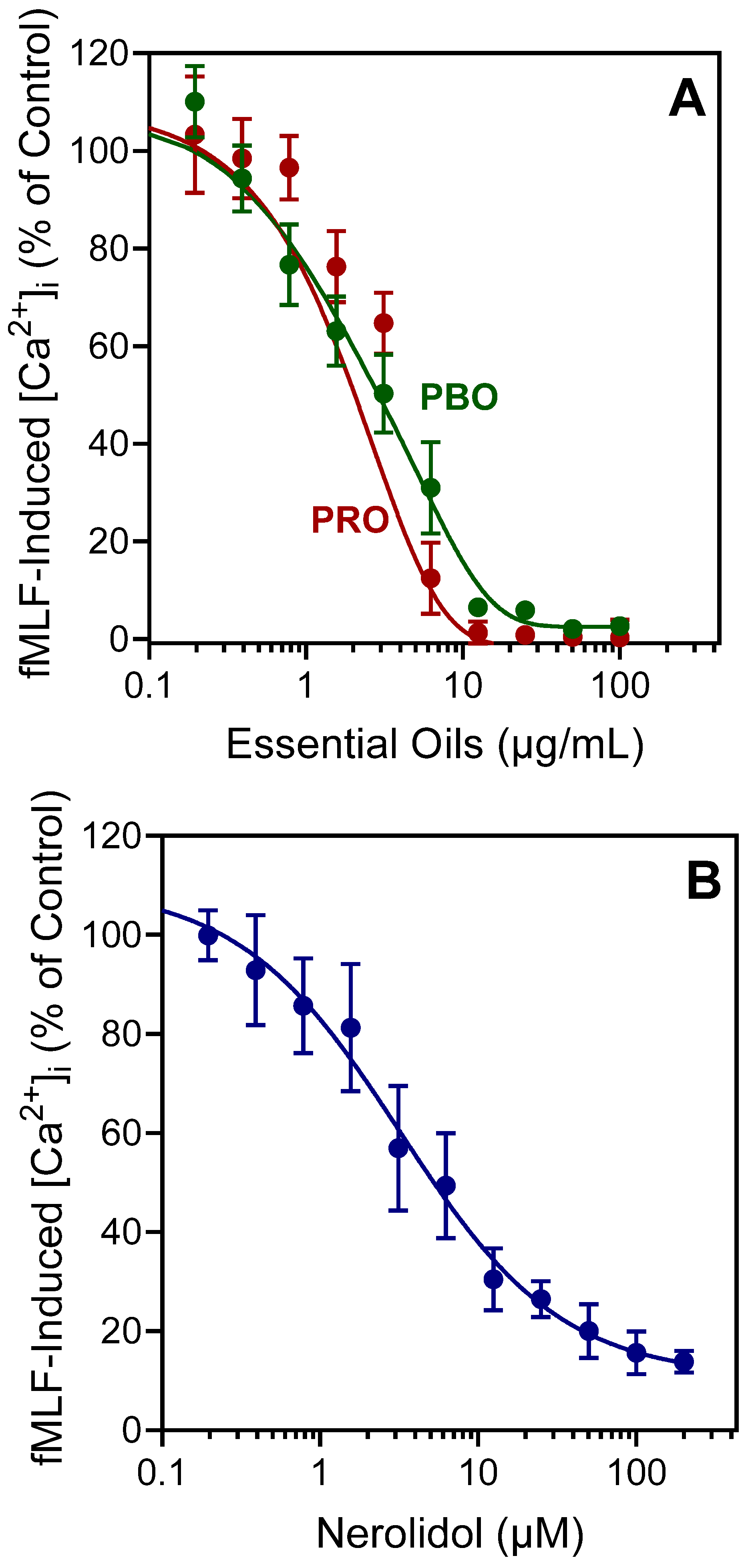
It is well recognized that agonists can down-regulate neutrophil responses to subsequent treatment with heterologous or homologous agonists [79]. Thus, whether PBO, PRO, or nerolidol could alter agonist-induced [Ca2+]i in human neutrophils stimulated with N-formyl chemotactic peptide was evaluated. As shown in Table 2 and Figure 2A, pretreatment with either PBO or PRO inhibited [Ca2+]i in fMLF- and WKYMVM-stimulated neutrophils, with IC50 values in the micromolar range. Likewise, nerolidol pretreatment also potently inhibited fMLF- and WKYMVM-stimulated neutrophil [Ca2+]i (Table 2). A representative, dose-dependent response for the inhibition of fMLF-induced neutrophil [Ca2+]i by nerolidol is shown in Figure 2B.
Previously, several of the compounds that are also present in PBO and PRO (i.e., α-pinene, β-pinene, limonene, 1,8-cineole, myrcene, sabinene, and terpinolene; see Table 1) were shown to have no inhibitory effect on human neutrophil Ca2+ influx [34,35], whereas minor components farnesene (0.2–0.7% in PRO and PBO) and germacrene D (0.2% in PRO) were found previously to inhibit agonist-induced activation of human neutrophils [32,48] and, thus, could contribute to the inhibitory activity observed for PBO and PRO.

2.3. Effect of PBO, PRO, and Nerolidol on Neutrophil Chemotaxis

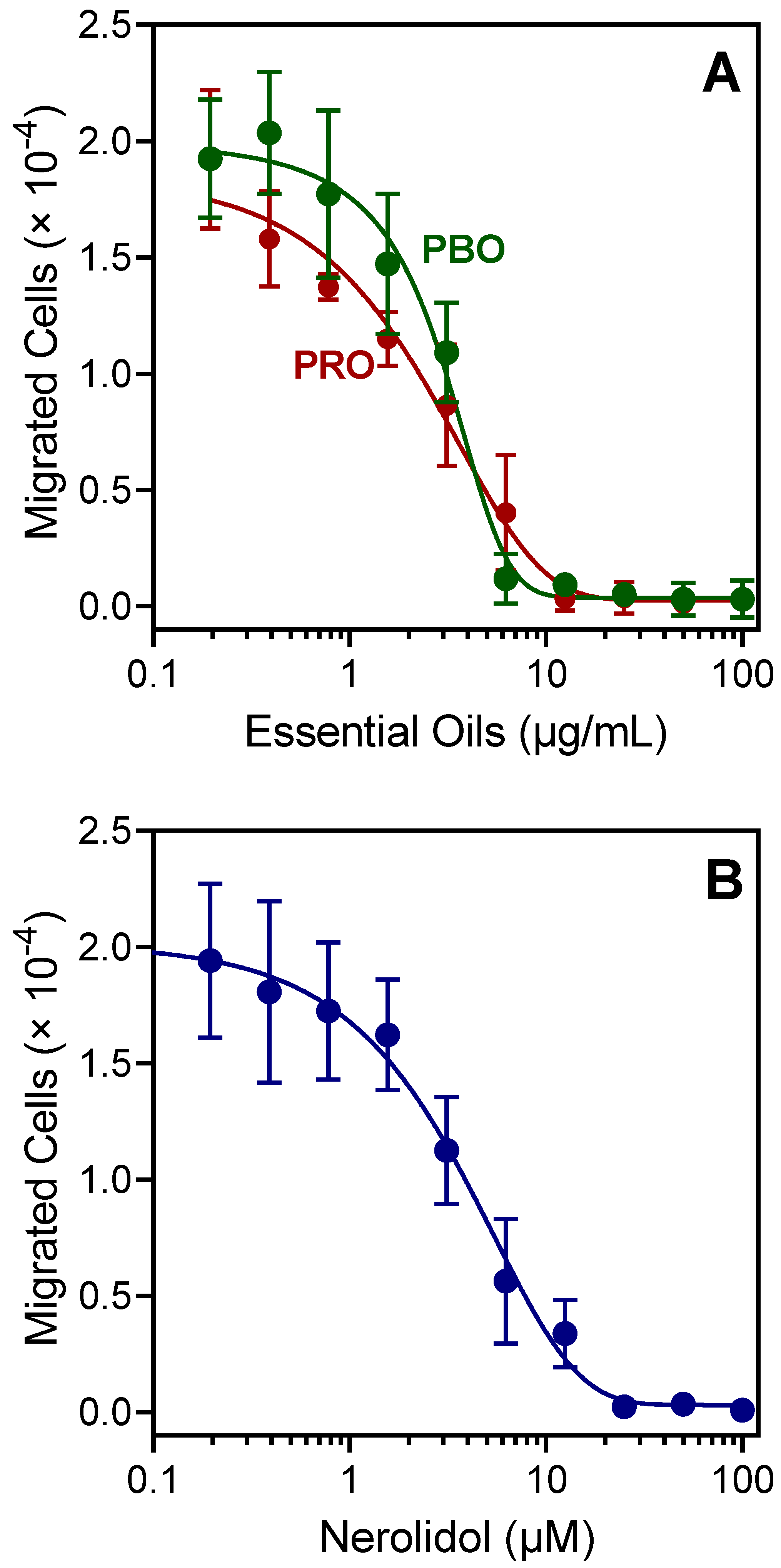
Various essential oils and their components were reported previously to inhibit neutrophil chemotaxis, including farnesene and germacrene D, which are minor components of PBO and PRO (Table 1). In the present study, the effects of PBO and PRO on human neutrophil chemotaxis were evaluated. Pretreatment with these essential oils dose-dependently inhibited fMLF-induced neutrophil chemotaxis (IC50 = 1.5 and 2.9 µg/mL, respectively) (Figure 3A). Likewise, pretreatment with nerolidol also inhibited fMLF-induced human neutrophil chemotaxis (IC50 = 3.9 µM) (Figure 3B).
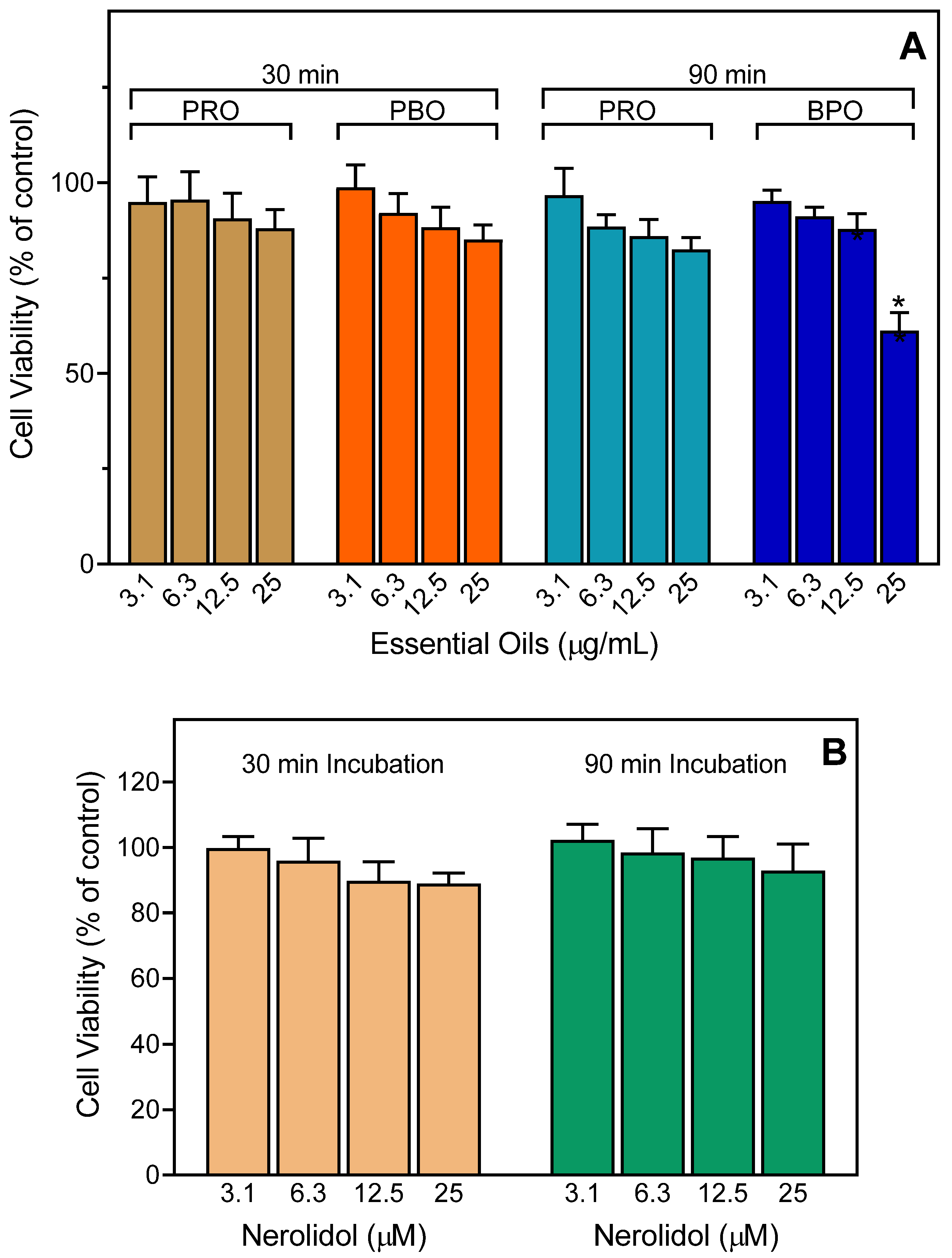
To ensure that the effects of the essential oils and nerolidol were not influenced by possible toxicity, we evaluated the cytotoxicity of the PBO and PRO (up to 25 µg/mL) and pure nerolidol at various concentrations (up to 25 µM) in human neutrophils. We found that the PRO had little to no cytotoxicity at concentrations up to 25 µg/mL during 30- and 90-min incubation periods, which covers the times used to measure Ca2+ influx (up to 30 min) and cell migration (up to 90 min), although PBO had some cytotoxicity at the highest concentration (25 µg/mL) (Figure 4A). Note that the inhibitory effects of PBO on neutrophil functional activity were found at much lower concentrations (1–10 µg/mL) (see Figure 2 and Figure 3). Consistent with these results, nerolidol had no cytotoxicity for neutrophils at all concentrations and times tested (Figure 4B).

2.4. Identification of Potential Protein Targets for Nerolidol

The application of nerolidol is widespread across different industries, and it has been used in cosmetics and non-cosmetic products [80,81]. In fact, the U.S. Food and Drug Administration (FDA) has also permitted the use of nerolidol as a food flavoring agent [82]. The fact that nerolidol is a common ingredient in many products has attracted researchers to explore more medicinal properties of nerolidol that may exert beneficial effects on human health (reviewed in [80]).
In addition to the immunomodulatory activity reported here, nerolidol was reported to exhibit a number of biological activities. For example, nerolidol has demonstrated anticholinesterase, antioxidant, antinociceptive, antibacterial, anti-parasite, anti-inflammatory, and anxiolytic activities, suggesting it may be a promising phytochemical for the development of therapeutic drugs (reviewed in [63,80,83]). Likewise, nerolidol was effective against schistosomiasis [84] and babesiosis [85]. Nerolidol was also reported to have neuroprotective properties, presumably through its anti-inflammatory and antioxidant activities [86,87]. Likewise, nerolidol reduced adjuvant arthritis by down-regulating proinflammatory cytokines and up-regulating anti-inflammatory cytokines [88]. It also attenuated hypertension-induced hypertrophy in spontaneously hypertensive rats through modulation of insulin-like growth factor receptor II (IGF-IIR) signaling [89]. This acyclic sesquiterpene induced apoptosis via phosphatidylinositol-3-kinase (PI3K) and c-Jun N-terminal kinase (JNK) regulation through cell cycle arrest in MG-63 osteosarcoma cells [90]. Finally, changes in tissue myeloperoxidase concentrations, neutrophil and macrophage mRNA expression of monocyte chemoattractant protein-1 (MCP-1), and proinflammatory cytokine content (interleukin (IL)-1β, IL-6, and tumor necrosis factor (TNF)) protein and mRNA levels were significantly reduced by nerolidol [91].
The most important physicochemical parameters of nerolidol were calculated using SwissADME [92]. These parameters were identical for E/Z isomers, with the exception of a slight difference in iLogP values (Table 3). SwissADME also predicted that nerolidol can permeate the blood–brain barrier (BBB). In addition, nanotechnology using nerolidol-loaded nanospheres may also improve passage through the BBB [93].
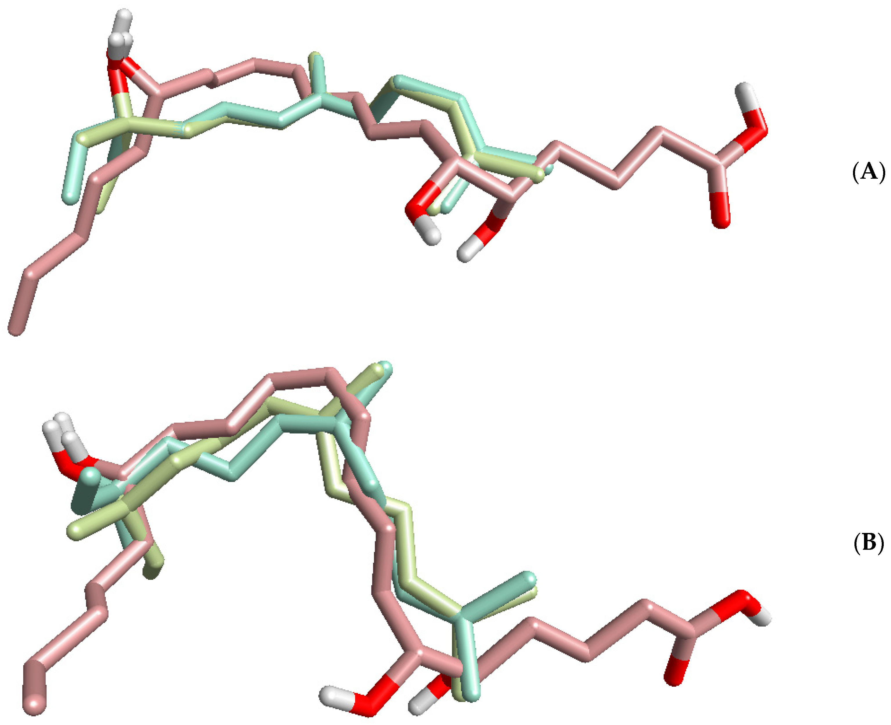
Nerolidol is a lipophilic molecule (Table 3). Thus, the neutrophil signaling inhibitory mechanisms may be based on allosteric interactions of the nerolidol chain with the membrane portion of a receptor, and this issue mechanism is currently being investigated. Indeed, other lipophilic compounds, such as the bile acids deoxycholic acid and chenodeoxycholic acid, were reported to antagonize FPR1 at high concentrations (>100 µM) [94,95,96]. Moreover, lipoxin A4 was also reported as an allosteric modulator of the CB1 cannabinoid receptor and FPR2 [97,98]. To further investigate this issue, lipoxin A4 was aligned with the E- and Z-nerolidol enantiomers using FieldTemplater software (Figure 5). The results demonstrated that the alignments were governed mainly by the hydrophobic hydrocarbon skeletons of the compounds. The combined similarity measure of the superimpositions were relatively high (S = 0.690 and 0.678 for the or Z and E isomers, respectively; each of the superimpositions was obtained with two enantiomers of nerolidol aligned onto lipoxin A4), suggesting that nerolidol might mimic lipoxin A4 and maybe other related specialized pro-resolving mediators, including resolvins, maresins, and protectins [99]. Indeed, many of these molecules were demonstrated to act allosterically on a number of GPCRs (reviewed in [100]). Interestingly, we found previously that other compounds structurally similar to nerolidol (i.e., 6-methyl-3,5-heptadien-2-one (MHDO), geranylacetone, and farnesene) also inhibited agonist-induced neutrophil activity [34,35,48]. Although molecular targets for MHDO and farnesene were not identified, we showed that geranylacetone is a TRPV1 agonist [34].
Reverse-pharmacophore mapping [101] of the molecular structures of E/Z-nerolidol in the R and S enantiomer forms of each geometric isomer was performed to identify potential biological targets. PharmMapper compared a database of pharmacophore patterns with these compounds and generated target information, such as pharmacophoric characteristics and normalized fitness scores. Note, however, that PharmMapper depends on the availability of structures for pharmacophore mapping, and most GPCRs are not represented in the database.
The proper optical isomers of the compounds were submitted to the PharmMapper server, as mapping explicitly accounts for the three-dimensional structure of a molecule. The ten top-ranked potential targets found by PharmMapper are shown in Table 4, and this analysis indicated that kinases could be among the potential targets for nerolidol. Among the top ten ranked kinase targets for the nerolidol were extracellular signal-regulated kinase 2 (ERK2), mitogen-activated protein kinase (MAPK) activated protein kinase 2 (MAPKAPK2), proto-oncogene serine/threonine-protein kinase (Pim-1), c-Jun N-terminal kinase 3 (JNK3), and vascular endothelial growth factor receptor 2 (VEGFR2), a tyrosine kinase (Table 4).
MAPK signaling plays an important role in neutrophil signal transduction cascades [102], and studies have shown that members of the MAPK, JNK, and the ERK families of proteins are activated in response to neutrophil priming/activation (reviewed in [103]). It is also clear from previous studies that one or more of these MAPK pathways is induced by FPR [104,105,106]. Thus, nerolidol may be a general inhibitor of neutrophil activation through GPCRs, and further studies are in progress to evaluate this idea and identify the specific molecular targets.

3. Materials and Methods

3.1. Material

Plant material was collected from wild P. balsamifera trees in May 2022 along Bozeman Creek near Bozeman, MT, USA (45.582238° N, 111.022036° E). Botanical identification of the plant material was performed by botanist Robyn A. Klein from Montana State University (Bozeman). Propolis samples were collected during September 2021 from Apis mellifera hives located approximately 10 miles northwest of Bozeman (45.729971° N, 111.234961° E).

3.2. Materials

Dimethyl sulfoxide (DMSO), N-formyl-Met-Leu-Phe (fMLF), Trp-Lys-Tyr-Val-Met (WKYMVM), nerolidol (as mixture of E- and Z-isomers) and Histopaque 1077 were purchased from Sigma-Aldrich Chemical Co. (St. Louis, MO, USA). n-Hexane was purchased from Merck (Darmstadt, Germany). Fluo-4AM was purchased from Invitrogen (Carlsbad, CA, USA). Roswell Park Memorial Institute (RPMI) 1640 medium was purchased from HyClone Laboratories (Logan, UT, USA). Fetal calf serum and fetal bovine serum were purchased from ATCC (Manassas, VA, USA). Hanks’ balanced salt solution (HBSS; 0.137 M NaCl, 5.4 mM KCl, 0.25 mM Na2HPO4, 0.44 mM KH2PO4, 4.2 mM NaHCO3, 5.56 mM glucose, and 10 mM HEPES, pH 7.4) was purchased from Life Technologies (Grand Island, NY, USA). HBSS without Ca2+ and Mg2+ was designated as HBSS; HBSS containing 1.3 mM CaCl2 and 1.0 mM MgSO4 was designated as HBSS+.

3.3. Essential Oil Extraction

Essential oil was obtained by hydrodistillation of P. balsamifera buds using a Clevenger-type apparatus, as previously described [35]. Crude propolis (100 g) was divided into small pieces and extracted by the same procedure. Conditions accepted by the European Pharmacopoeia (European Directorate for the Quality of Medicines, Council of Europe, Strasbourg, France, 2014) were used to avoid artifacts. Yields of the essential oils were calculated based on the amount of air-dried plant material used. Stock solutions of the essential oils were prepared in DMSO (10 mg/mL) for biological evaluation and in n-hexane (10% w/v) for gas chromatographic analysis.

3.4. Gas Chromatography–Flame Ionization Detector (GC-FID) and Gas Chromatography–Mass Spectrometry (GC-MS) Analysis

GC-MS analysis was performed with an Agilent 5975 GC-MSD system (Agilent Technologies, Santa Clara, CA, USA), as reported previously [107]. An Agilent Innowax FSC column (60 m × 0.25 mm, 0.25 μm film thickness) was used with He as the carrier gas (0.8 mL/min). The GC oven temperature was kept at 60 °C for 10 min, increased to 220 °C at a rate of 4 °C/min, kept constant at 220 °C for 10 min, and then increased to 240 °C at a rate of 1 °C/min. The split ratio was adjusted to 40:1, and the injector temperature was 250 °C. MS spectra were monitored at 70 eV with a mass range of 35 to 450 m/z. GC analysis was performed on an Agilent 6890N GC system. To obtain the same elution order as with GC-MS, the line was split for FID and MS detectors, and a single injection was performed using the same column and appropriate operational conditions. FID temperature was 300 °C. The essential oil components were identified by co-injection with standards (whenever possible), which were purchased commercially or isolated from natural sources. In addition, compound identities were confirmed by comparison of their mass spectra with those in the Wiley GC/MS Library (Wiley, NY, USA), MassFinder software 4.0 (Dr. Hochmuth Scientific Consulting, Hamburg, Germany), Adams Library, and NIST Library. Confirmation was also achieved using the in-house “Başer Library of Essential Oil Constituents” database obtained from chromatographic runs of pure compounds performed with the same equipment and conditions. A C8–C40 n-alkane standard solution (Fluka, Buchs, Switzerland) was used to spike the samples for the determination of relative retention indices (RRI). Relative percentage amounts of the separated compounds were calculated from the FID chromatograms.

3.5. Isolation of Human Neutrophils

For isolation of human neutrophils, blood was collected from healthy donors in accordance with a protocol approved by the Institutional Review Board at Montana State University (protocol #MQ041017). Neutrophils were purified from the blood using dextran sedimentation, followed by Histopaque 1077 gradient separation and hypotonic lysis of red blood cells, as described previously [108]. Isolated neutrophils were washed twice and resuspended in HBSS. Neutrophil preparations were routinely >95% pure, as determined by light microscopy, and >98% viable, as determined by trypan blue exclusion. Neutrophils were obtained from multiple different donors; however, the cells from different donors were never pooled during experiments.

3.6. Ca2+ Mobilization Assay

Changes in intracellular Ca2+ concentrations ([Ca2+]i) were measured with a FlexStation 3 scanning fluorometer (Molecular Devices, Sunnyvale, CA, USA). Briefly, human neutrophils were suspended in HBSS, loaded with Fluo-4AM at a final concentration of 1.25 μg/mL, and incubated for 30 min in the dark at 37 °C. After dye loading, the cells were washed with HBSS, resuspended in HBSS+, separated into aliquots, and loaded into the wells of flat-bottom, half-area-well black microtiter plates (2 × 10⁵ cells/well). To assess the direct effects of test compounds or pure essential oils on Ca2+ influx, the compound/oil was added to the wells (final concentration of DMSO was 1%), and changes in fluorescence were monitored (λex = 485 nm, λem = 538 nm) every 5 s for 240 s at room temperature after addition of the test compound. To evaluate the inhibitory effects of the compounds on N-formyl peptide receptor 1/2 (FPR1/FPR2) dependent Ca2+ influx, the compound or essential oil was added to the wells (final concentration of DMSO was 1%) with human neutrophils. The samples were preincubated for 10 min, followed by the addition of 5 nM fMLF or 5 nM WKYMVM. The maximum change in fluorescence, expressed in arbitrary units over baseline, was used to determine the agonist response. Responses were normalized to the response induced by 5 nM fMLF or 5 nM WKYMVM, which were assigned as 100%. Curve fitting (at least five or six points) and calculation of median effective concentration values (EC50 or IC50) were performed by nonlinear regression analysis of the dose–response curves generated using Prism 9 (GraphPad Software, Inc., San Diego, CA, USA).

3.7. Chemotaxis Assay

Human neutrophils were resuspended in HBSS+ containing 2% (v/v) heat-inactivated fetal bovine serum (2 × 10⁶ cells/mL), and chemotaxis was analyzed in 96-well ChemoTx chemotaxis chambers (Neuroprobe, Gaithersburg, MD, USA). In brief, neutrophils were preincubated with the indicated concentrations of the test sample (essential oil or pure compound) or DMSO (1% final concentration) for 30 min at room temperature and added to the upper wells of the ChemoTx chemotaxis chambers. The lower wells were loaded with 30 µL of HBSS+ containing 2% (v/v) fetal bovine serum and the indicated concentrations of test sample, DMSO (negative control), or 1 nM fMLF as a positive control. Neutrophils were added to the upper wells and allowed to migrate through the 5.0 µm pore polycarbonate membrane filter for 60 min at 37 °C and 5% CO2. The number of migrated cells was determined by measuring ATP in lysates of transmigrated cells using a luminescence-based assay (CellTiter-Glo; Promega, Madison, WI, USA), and chemiluminescence measurements were converted to absolute cell numbers by comparison of the values with standard curves obtained with known numbers of neutrophils. Curve fitting (at least eight to nine points) and calculation of effective concentration values (IC50) were performed by nonlinear regression analysis of the dose–response curves generated using GraphPad Prism 9 (San Diego, CA, USA).

3.8. Cytotoxicity Assay

Cytotoxicity of essential oils and pure nerolidol in human neutrophils was analyzed with a CellTiter-Glo Luminescent Cell Viability Assay Kit (Promega) according to the manufacturer’s protocol. Briefly, human neutrophils were cultured at a density of 104 cells/well with different concentrations of essential oil or nerolidol (final concentration of DMSO was 1%) for 30 and 90 min at 37 °C and 5% CO2. Following treatment, substrate was added to the cells, and the samples were analyzed with a Fluoroscan Ascent FL microplate reader.

3.9. Molecular Modeling

PharmMapper [109] was used to identify potential protein targets for four possible geometric and optical isomers of nerolidol. PharmMapper recognizes potential targets based on “invert” pharmacophore mapping. The protein biotargets are represented by sets of pharmacophore points in reference databases incorporated in the software. PubChem (https://pubchem.ncbi.nlm.nih.gov) and ChemSpider (https://www.chemspider.com/) databases (accessed on 25 August 2022) were used as sources of initial 3D structures of our compounds: (3R,6E)-nerolidol (PubChem CID: 11241545), (3S,6E)-nerolidol (PubChem CID: 5281525), (3S,6Z)-nerolidol (ChemSpider ID: 21427544). The 3D structure of (3R,6Z)-nerolidol was obtained by a mirror reflection of the corresponding S-enantiomer using ChemOffice 2016 software. The 3D structures were saved in SDF format and uploaded into PharmMapper. The system automatically generated up to 300 conformers of each compound based on the software option. Pharmacophore mapping was performed using the “Human Protein Targets Only” database, which contained 2241 targets. The top 250 potential targets were retrieved for each compound evaluated, and the potential targets were sorted by normalized fit score.
The physicochemical properties of nerolidol were computed using SwissADME (http://www.swissadme.ch; accessed on 1 September 2022).
Alignments of lipoxin-A4 and enantiomers of Z- and E-nerolidol were performed using FieldTemplater software (Cresset Group, Cambridgeshire, UK).

3.10. Statistical Analysis

One-way analysis of variance (ANOVA) was performed on the data sets, followed by Tukey’s pair-wise comparisons. Pair-wise comparisons with differences at p < 0.05 were considered statistically significant.

4. Conclusions

Analysis of the composition of essential oils extracted from P. balsamifera buds and propolis collected in Montana showed that they were enriched with nerolidol and its derivatives. Further analysis of the immunomodulatory activity of these essential oils and nerolidol showed that they activated human neutrophils and were able to inhibit FPR1/FPR2 agonist-induced neutrophil activation and chemotaxis, which might contribute to the reported anti-inflammatory activity and other pharmacological properties of these extracts. The biological effects of PBO and PRO might be attributable primarily to nerolidol or to the synergistic effects of nerolidol with other active constituents. However, to verify the key targets responsible for the immunomodulatory effects of nerolidol, further experimental investigation is needed.

Author Contributions

I.A.S. and M.T.Q. conceived and designed the project. I.A.S. and L.N.K. collected the material. I.A.S., G.Ö., T.Ö. and L.N.K. performed the experiments. P.I.K. and A.I.K. conducted the molecular modeling study. I.A.S., G.Ö., T.Ö., L.N.K. and A.I.K. analyzed and interpreted the data. I.A.S., G.Ö., A.I.K. and M.T.Q. drafted and revised the manuscript. All authors have read and agreed to the published version of the manuscript.

Funding

This research was supported in part by National Institutes of Health IDeA Program grant GM103474; USDA National Institute of Food and Agriculture Hatch project 1009546; the Montana State University Agricultural Experiment Station; and the Tomsk Polytechnic University Development Program (Project Priority-2030-NIP/IZ-009-0000-2022).

Institutional Review Board Statement

The study was approved by the Montana State University Institutional Review Board (protocol 2022-168, approved on 23 March 2022).

Informed Consent Statement

Informed consent was obtained from all subjects involved in the study.

Data Availability Statement

Data are contained within the article.

Conflicts of Interest

The authors declare no conflict of interest.

Sample Availability

Samples of essential oils are available from the authors.

References

  1. Saelao, P.; Borba, R.S.; Ricigliano, V.; Spivak, M.; Simone-Finstrom, M. Honeybee microbiome is stabilized in the presence of propolis. Biol. Lett. 2020, 16, 20200003. [Google Scholar] [CrossRef] [PubMed]
  2. Anjum, S.I.; Ullah, A.; Khan, K.A.; Attaullah, M.; Khan, H.; Ali, H.; Bashir, M.A.; Tahir, M.; Ansari, M.J.; Ghramh, H.A.; et al. Composition and functional properties of propolis (bee glue): A review. Saudi J. Biol. Sci. 2019, 26, 1695–1703. [Google Scholar] [CrossRef] [PubMed]
  3. Zulhendri, F.; Perera, C.O.; Tandean, S. Can propolis be a useful adjuvant in brain and neurological disorders and ınjuries? A Systematic Scoping Review of the Latest Experimental Evidence. Biomedicines 2021, 9, 1227. [Google Scholar] [CrossRef] [PubMed]
  4. Zulhendri, F.; Chandrasekaran, K.; Kowacz, M.; Ravalia, M.; Kripal, K.; Fearnley, J.; Perera, C.O. Antiviral, antibacterial, antifungal, and antiparasitic properties of propolis: A review. Foods 2021, 10, 1360. [Google Scholar] [CrossRef] [PubMed]
  5. Zulhendri, F.; Ravalia, M.; Kripal, K.; Chandrasekaran, K.; Fearnley, J.; Perera, C.O. Propolis in metabolic syndrome and ıts associated chronic diseases: A narrative review. Antioxidants 2021, 10, 348. [Google Scholar] [CrossRef] [PubMed]
  6. Zulhendri, F.; Felitti, R.; Fearnley, J.; Ravalia, M. The use of propolis in dentistry, oral health, and medicine: A review. J. Oral. Biosci. 2021, 63, 23–34. [Google Scholar] [CrossRef]
  7. Hermansyah, D.; Zulhendri, F.; Perera, C.O.; Firsty, N.N.; Chandrasekaran, K.; Abdulah, R.; Herman, H.; Lesmana, R. The potential use of propolis as an adjunctive therapy in breast cancers. Integr. Cancer 2022, 21, 15347354221096868. [Google Scholar] [CrossRef]
  8. Zulhendri, F.; Perera, C.O.; Tandean, S.; Abdulah, R.; Herman, H.; Christoper, A.; Chandrasekaran, K.; Putra, A.; Lesmana, R. The potential use of propolis as a primary or an adjunctive therapy in respiratory tract-related diseases and disorders: A systematic scoping review. Biomed. Pharm. 2022, 146, 112595. [Google Scholar] [CrossRef]
  9. Burdock, G.A. Review of the biological properties and toxicity of bee propolis (propolis). Food Chem. Toxicol. 1998, 36, 347–363. [Google Scholar] [CrossRef]
  10. Banskota, A.H.; Tezuka, Y.; Kadota, S. Recent progress in pharmacological research of propolis. Phytother. Res. 2001, 15, 561–571. [Google Scholar] [CrossRef]
  11. Sforcin, J.M.; Bankova, V. Propolis: Is there a potential for the development of new drugs? J. Ethnopharmacol. 2011, 133, 253–260. [Google Scholar] [CrossRef]
  12. Yang, J.; Pi, A.; Yan, L.; Li, J.; Nan, S.; Zhang, J.; Hao, Y. Research progress on therapeutic effect and mechanism of propolis on wound healing. Evid. Based Complement Altern. Med. 2022, 2022, 5798941. [Google Scholar] [CrossRef] [PubMed]
  13. Salatino, A. Perspectives for uses of propolis in therapy against ınfectious diseases. Molecules 2022, 27, 4594. [Google Scholar] [CrossRef] [PubMed]
  14. Elumalai, P.; Muninathan, N.; Megalatha, S.T.; Suresh, A.; Kumar, K.S.; Jhansi, N.; Kalaivani, K.; Krishnamoorthy, G. An ınsight into anticancer effect of propolis and ıts constituents: A review of molecular mechanisms. Evid. Based Complement Altern. Med. 2022, 2022, 5901191. [Google Scholar] [CrossRef] [PubMed]
  15. Mirzoeva, O.K.; Grishanin, R.N.; Calder, P.C. Antimicrobial action of propolis and some of its components: The effects on growth, membrane potential and motility of bacteria. Microbiol. Res. 1997, 152, 239–246. [Google Scholar] [CrossRef]
  16. Magnavacca, A.; Sangiovanni, E.; Racagni, G.; Dell’Agli, M. The antiviral and immunomodulatory activities of propolis: An update and future perspectives for respiratory diseases. Med. Res. Rev. 2022, 42, 897–945. [Google Scholar] [CrossRef]
  17. Dos Santos, F.F.; Morais-Urano, R.P.; Cunha, W.R.; de Almeida, S.G.; Cavallari, P.; Manuquian, H.A.; Pereira, H.A.; Furtado, R.; Santos, M.F.C.; Amdrade, E.S.M.L. A review on the anti-inflammatory activities of Brazilian green, brown and red propolis. J. Food Biochem. 2022, 46, e14350. [Google Scholar] [CrossRef]
  18. Kasote, D.; Bankova, V.; Viljoen, A.M. Propolis: Chemical diversity and challenges in quality control. Phytochem. Rev. 2022, 21, 1887–1911. [Google Scholar] [CrossRef]
  19. Salatino, A.; Salatino, M.L.F.; Negri, G. How diverse is the chemistry and plant origin of Brazilian propolis? Apidologie 2021, 52, 1075–1097. [Google Scholar] [CrossRef]
  20. Stanciauskaite, M.; Marksa, M.; Babickaite, L.; Majiene, D.; Ramanauskiene, K. Comparison of ethanolic and aqueous Populus balsamifera L. bud extracts by different extraction methods: Chemical composition, antioxidant and antibacterial activities. Pharmaceuticals 2021, 14, 1018. [Google Scholar] [CrossRef]
  21. Moerman, D.E. Native American Medicinal Plants: An Ethnobotanical Dictionary; Timber Press: Portland, OR, USA, 2009; p. 799. [Google Scholar]
  22. Wang, K.; Zhang, J.L.; Ping, S.; Ma, Q.X.; Chen, X.; Xuan, H.Z.; Shi, J.H.; Zhang, C.P.; Hu, F.L. Anti-inflammatory effects of ethanol extracts of Chinese propolis and buds from poplar (Populus x canadensis). J. Ethnopharmacol. 2014, 155, 300–311. [Google Scholar] [CrossRef]
  23. Kis, B.; Pavel, I.Z.; Avram, S.; Moaca, E.A.; San Juan, M.H.; Schwiebs, A.; Radeke, H.H.; Muntean, D.; Diaconeasa, Z.; Minda, D.; et al. Antimicrobial activity, in vitro anticancer effect (MCF-7 breast cancer cell line), antiangiogenic and immunomodulatory potentials of Populus nigra L. buds extract. BMC Complement Med. 2022, 22, 74. [Google Scholar] [CrossRef] [PubMed]
  24. Pannucci, E.; D’Eliseo, D.; Ieri, F.; Romani, A.; Santi, L.; Bernini, R.; Sabatti, M.; Velotti, F. Perspectives on Populus spp. (Salicaceae) bud extracts as antioxidant and anti-inflammatory agents. Nat. Prod. Res. 2022, 36, 1648–1652. [Google Scholar] [CrossRef] [PubMed]
  25. Kis, B.; Avram, S.; Pavel, I.Z.; Lombrea, A.; Buda, V.; Dehelean, C.A.; Soica, C.; Yerer, M.B.; Bojin, F.; Folescu, R.; et al. Recent advances regarding the phytochemical and therapeutic uses of Populus nigra L. Buds. Plants 2020, 9, 1464. [Google Scholar] [CrossRef] [PubMed]
  26. Okinczyc, P.; Szumny, A.; Szperlik, J.; Kulma, A.; Franiczek, R.; Zbikowska, B.; Krzyzanowska, B.; Sroka, Z. Profile of polyphenolic and essential oil composition of polish propolis, black poplar and aspens buds. Molecules 2018, 23, 1262. [Google Scholar] [CrossRef] [PubMed] [Green Version]
  27. Sandner, G.; Heckmann, M.; Weghuber, J. Immunomodulatory Activities of Selected Essential Oils. Biomolecules 2020, 10, 1139. [Google Scholar] [CrossRef]
  28. Gandhi, G.R.; Vasconcelos, A.B.S.; Haran, G.H.; Calisto, V.; Jothi, G.; Quintans, J.S.S.; Cuevas, L.E.; Narain, N.; Junior, L.J.Q.; Cipolotti, R.; et al. Essential oils and its bioactive compounds modulating cytokines: A systematic review on anti-asthmatic and immunomodulatory properties. Phytomedicine 2020, 73, 152854. [Google Scholar] [CrossRef] [Green Version]
  29. Valdivieso-Ugarte, M.; Gomez-Llorente, C.; Plaza-Diaz, J.; Gil, A. Antimicrobial, antioxidant, and ımmunomodulatory properties of essential oils: A systematic review. Nutrients 2019, 11, 2786. [Google Scholar] [CrossRef] [Green Version]
  30. Ozek, G.; Schepetkin, I.A.; Yermagambetova, M.; Ozek, T.; Kirpotina, L.N.; Almerekova, S.S.; Abugalieva, S.I.; Khlebnikov, A.I.; Quinn, M.T. Innate ımmunomodulatory activity of cedrol, a component of essential oils ısolated from juniperus species. Molecules 2021, 26, 7644. [Google Scholar] [CrossRef]
  31. Schepetkin, I.A.; Ozek, G.; Ozek, T.; Kirpotina, L.N.; Khlebnikov, A.I.; Quinn, M.T. Chemical composition and ımmunomodulatory activity of essential oils from Rhododendron albiflorum. Molecules 2021, 26, 3652. [Google Scholar] [CrossRef]
  32. Schepetkin, I.A.; Ozek, G.; Ozek, T.; Kirpotina, L.N.; Khlebnikov, A.I.; Quinn, M.T. Chemical composition and ımmunomodulatory activity of Hypericum perforatum essential oils. Biomolecules 2020, 10, 916. [Google Scholar] [CrossRef]
  33. Ozek, G.; Schepetkin, I.A.; Utegenova, G.A.; Kirpotina, L.N.; Andrei, S.R.; Ozek, T.; Baser, K.H.C.; Abidkulova, K.T.; Kushnarenko, S.V.; Khlebnikov, A.I.; et al. Chemical composition and phagocyte immunomodulatory activity of Ferula iliensis essential oils. J. Leukoc. Biol. 2017, 101, 1361–1371. [Google Scholar] [CrossRef] [Green Version]
  34. Schepetkin, I.A.; Kushnarenko, S.V.; Ozek, G.; Kirpotina, L.N.; Sinharoy, P.; Utegenova, G.A.; Abidkulova, K.T.; Ozek, T.; Baser, K.H.; Kovrizhina, A.R.; et al. Modulation of human neutrophil responses by the essential oils from Ferula akitschkensis and their constituents. J. Agric. Food Chem. 2016, 64, 7156–7170. [Google Scholar] [CrossRef] [PubMed] [Green Version]
  35. Schepetkin, I.A.; Kushnarenko, S.V.; Ozek, G.; Kirpotina, L.N.; Utegenova, G.A.; Kotukhov, Y.A.; Danilova, A.N.; Ozek, T.; Baser, K.H.; Quinn, M.T. Inhibition of human neutrophil responses by the essential oil of Artemisia kotuchovii and ıts constituents. J. Agric. Food Chem. 2015, 63, 4999–5007. [Google Scholar] [CrossRef] [PubMed] [Green Version]
  36. Schepetkin, I.A.; Ozek, G.; Ozek, T.; Kirpotina, L.N.; Khlebnikov, A.I.; Quinn, M.T. Neutrophil ımmunomodulatory activity of (-)-borneol, a major component of essential oils extracted from Grindelia squarrosa. Molecules 2022, 27, 4897. [Google Scholar] [CrossRef]
  37. Campra, N.A.; Montironi, I.D.; Reinoso, E.B.; Raviolo, J.; Moreno, F.R.; Maletto, B.; Cariddi, L.N. A natural oil increases specific anti-OVA IgG levels and induces a cellular immune response combined with aluminum hydroxide. J. Leukoc. Biol. 2021, 109, 223–232. [Google Scholar] [CrossRef] [PubMed]
  38. Siveen, K.S.; Kuttan, G. Augmentation of humoral and cell mediated immune responses by Thujone. Int. Immunopharmacol. 2011, 11, 1967–1975. [Google Scholar] [CrossRef] [PubMed]
  39. Khlebnikov, A.I.; Schepetkin, I.A.; Kishkentaeva, A.S.; Shaimerdenova, Z.R.; Atazhanova, G.A.; Adekenov, S.M.; Kirpotina, L.N.; Quinn, M.T. Inhibition of t cell receptor activation by semi-synthetic sesquiterpene lactone derivatives and molecular modeling of their interaction with glutathione and tyrosine kinase ZAP-70. Molecules 2019, 24, 350. [Google Scholar] [CrossRef] [Green Version]
  40. Schepetkin, I.A.; Kirpotina, L.N.; Mitchell, P.T.; Kishkentaeva, A.C.; Shaimerdenova, Z.R.; Atazhanova, G.A.; Adekenov, S.M.; Quinn, M.T. The natural sesquiterpene lactones arglabin, grosheimin, agracin, parthenolide, and estafiatin inhibit T cell receptor (TCR) activation. Phytochemistry 2018, 146, 36–46. [Google Scholar] [CrossRef]
  41. Zonfrillo, M.; Andreola, F.; Krasnowska, E.K.; Sferrazza, G.; Pierimarchi, P.; Serafino, A. Essential Oil from Eucalyptus globulus (Labill.) activates complement receptor-mediated phagocytosis and stimulates podosome formation in human monocyte-derived macrophages. Molecules 2022, 27, 3488. [Google Scholar] [CrossRef]
  42. Sharma, M.; Grewal, K.; Jandrotia, R.; Batish, D.R.; Singh, H.P.; Kohli, R.K. Essential oils as anticancer agents: Potential role in malignancies, drug delivery mechanisms, and immune system enhancement. Biomed. Pharm. 2022, 146, 112514. [Google Scholar] [CrossRef] [PubMed]
  43. Kobayashi, S.D.; DeLeo, F.R.; Quinn, M.T. Microbes and the fate of neutrophils. Immunol. Rev. 2022. Epub ahead of print. [Google Scholar] [CrossRef] [PubMed]
  44. Liew, P.X.; Kubes, P. The neutrophil’s role during health and disease. Physiol. Rev. 2019, 99, 1223–1248. [Google Scholar] [CrossRef] [PubMed]
  45. Malech, H.L.; DeLeo, F.R.; Quinn, M.T. The role of neutrophils in the immune system: An overview. Methods Mol. Biol. 2020, 2087, 3–10. [Google Scholar] [CrossRef] [PubMed]
  46. Tavares, L.P.; Peh, H.Y.; Tan, W.S.D.; Pahima, H.; Maffia, P.; Tiligada, E.; Levi-Schaffer, F. Granulocyte-targeted therapies for airway diseases. Pharm. Res. 2020, 157, 104881. [Google Scholar] [CrossRef]
  47. Liao, H.R.; Kao, Y.Y.; Leu, Y.L.; Liu, F.C.; Tseng, C.P. Larixol inhibits fMLP-induced superoxide anion production and chemotaxis by targeting the βγ subunit of Gi-protein of fMLP receptor in human neutrophils. Biochem. Pharm. 2022, 201, 115091. [Google Scholar] [CrossRef] [PubMed]
  48. Schepetkin, I.A.; Ozek, G.; Ozek, T.; Kirpotina, L.N.; Khlebnikov, A.I.; Klein, R.A.; Quinn, M.T. Neutrophil Immunomodulatory activity of farnesene, a component of Artemisia dracunculus essential oils. Pharmaceuticals 2022, 15, 642. [Google Scholar] [CrossRef]
  49. Piochon-Gauthier, M.; Legault, J.; Sylvestre, M.; Pichette, A. The essential oil of Populus balsamifera buds: Its chemical composition and cytotoxic activity. Nat. Prod. Comm. 2014, 9, 257–260. [Google Scholar] [CrossRef] [Green Version]
  50. Isidorov, V.A.; Vinogorova, V.T. GC-MS analysis of compounds extracted from buds of Populus balsamifera and Populus nigra. Z. Nat. C 2003, 58, 355–360. [Google Scholar] [CrossRef]
  51. Bankova, V.S.; Christov, R.S.; Tejera, A.D. Lignans and other constituents of propolis from the Canary Islands. Phytochemistry 1998, 49, 1411–1415. [Google Scholar] [CrossRef]
  52. de Albuquerque, I.L.; Alves, L.A.; Lemos, T.L.G.; Dorneles, C.A.; de Morais, M.O. Constituents of the essential oil of Brazilian green propolis from Brazil. J. Essent. Oil Res. 2008, 20, 414–415. [Google Scholar] [CrossRef]
  53. Bankova, V.; Popova, M.; Trusheva, B. Propolis volatile compounds: Chemical diversity and biological activity: A review. Chem. Cent. J. 2014, 8, 28. [Google Scholar] [CrossRef] [PubMed] [Green Version]
  54. Marostica, M.R.; Daugsch, A.; Moraes, C.S.; Queiroga, C.L.; Pastore, G.M.; Park, Y.K. Comparison of volatile and polyphenolic compounds in Brazilian green propolis and its botanical origin Baccharis dracunculifolia. Cienc. Tecnol. Alime. 2008, 28, 178–181. [Google Scholar] [CrossRef] [Green Version]
  55. Miguel, M.G.; Nunes, S.; Cruz, C.; Duarte, J.; Antunes, M.D.; Cavaco, A.M.; Mendes, M.D.; Lima, A.S.; Pedro, L.G.; Barroso, J.G.; et al. Propolis volatiles characterisation from acaricide-treated and -untreated beehives maintained at Algarve (Portugal). Nat. Prod. Res. 2013, 27, 743–749. [Google Scholar] [CrossRef]
  56. Melliou, E.; Stratis, E.; Chinou, I. Volatile constituents of propolis from various regions of Greece—Antimicrobial activity. Food Chem. 2007, 103, 375–380. [Google Scholar] [CrossRef]
  57. Bankova, V.; Christov, R.; Popov, S.; Pureb, O.; Bocari, G. Volatile Constituents of Propolis. Z. Nat. C 1994, 49, 6–10. [Google Scholar] [CrossRef]
  58. Bankova, V.; Christov, R.; Kujumgiev, A.; Marcucci, M.C.; Popova, S. Chemical-Composition and Antibacterial Activity of Brazilian Propolis. Z. Nat. C 1995, 50, 167–172. [Google Scholar] [CrossRef]
  59. Borčić, I.; Radonić, A.; Grzunov, K. Comparison of the volatile constituents of propolis gathered in different regions of Croatia. Flavour Frag. J. 1996, 11, 311–313. [Google Scholar] [CrossRef]
  60. Klopell, F.C.; Lemos, M.; Sousa, J.P.B.; Comunello, E.; Maistro, E.L.; Bastos, J.K.; de Andrade, S.F. Nerolidol, an antiulcer constituent from the essential oil of Baccharis dracunculifolia DC (Asteraceae). Z. Nat. C 2007, 62, 537–542. [Google Scholar] [CrossRef] [Green Version]
  61. Padalia, R.C.; Verma, R.S.; Chauhan, A.; Chanotiya, C.S. The essential oil composition of Melaleuca leucadendra L. grown in India: A novel source of (E)-nerolidol. Ind. Crop. Prod. 2015, 69, 224–227. [Google Scholar] [CrossRef]
  62. Marques, A.M.; Barreto, A.L.S.; Curvelo, J.A.D.; Romanos, M.T.V.; Soares, R.M.D.; Kaplan, M.A.C. Antileishmanial activity of nerolidol-rich essential oil from Piper claussenianum. Rev. Bras. Farm. 2011, 21, 908–914. [Google Scholar] [CrossRef]
  63. De Carvalho, R.B.F.; De Almeida, A.A.C.; Campelo, N.B.; Lellis, D.; Nunes, L.C.C. Nerolidol and its pharmacological application in treating neurodegenerative diseases: A review. Recent Pat. Biotechnol. 2018, 12, 158–168. [Google Scholar] [CrossRef] [PubMed]
  64. Limberger, R.P.; Sobral, M.; Henriques, A.T. Intraspecific volatile oil variation in Myrceugenia cucullata (Myrtaceae). Biochem. Syst. Ecol. 2005, 33, 287–293. [Google Scholar] [CrossRef]
  65. Kramer, R.; Abraham, W.R. Volatile sesquiterpenes from fungi: What are they good for? Phytochem. Rev. 2012, 11, 15–37. [Google Scholar] [CrossRef] [Green Version]
  66. Nguikwie, S.K.; Nyegue, M.A.; Belinga, F.N.F.; Ngane, R.A.N.; Romestand, B.; Kouzayha, A.; Casabianca, H.; Zono, P.H.A.; Menut, C. The chemical composition and antibacterial activities of the essential oils from three aframomum species from Cameroon, and their potential as sources of (E)-(R)-nerolidol. Nat. Prod. Comm. 2013, 8, 829–834. [Google Scholar] [CrossRef] [Green Version]
  67. Green, S.A.; Chen, X.Y.; Nieuwenhuizen, N.J.; Matich, A.J.; Wang, M.Y.; Bunn, B.J.; Yauk, Y.K.; Atkinson, R.G. Identification, functional characterization, and regulation of the enzyme responsible for floral (E)-nerolidol biosynthesis in kiwifruit (Actinidia chinensis). J. Exp. Bot. 2012, 63, 1951–1967. [Google Scholar] [CrossRef] [Green Version]
  68. Juchelka, D.; Steil, A.; Witt, K.; Mosandl’, A. Chiral compounds of essential oils. XX. Chirality evaluation and authenticity profiles of neroli and petitgrain oils. J. Essent. Oil Res. 1996, 8, 487–497. [Google Scholar] [CrossRef]
  69. Naves, Y.R. Presenee de nltrolidol dans les huiles essentielles de papilionae. Helv. Chim. Acta 1947, 30, 278–286. [Google Scholar] [CrossRef]
  70. Naves, Y.R. Etudes sur les matières végétales volatiles LXI. Présence de nérolidol dans les huiles essentielles de papilionacées. Helv. Chim. Acta 1948, 31, 408–417. [Google Scholar] [CrossRef]
  71. Babushok, V.I.; Linstrom, P.J.; Zenkevich, I.G. Retention Indices for Frequently Reported Compounds of Plant Essential Oils. J. Phys. Chem. Ref. Data 2011, 40, 43101. [Google Scholar] [CrossRef]
  72. NIST Chemistry WebBook NIST Standard Reference Database Number 69. Last Update to Data: 2022. Available online: Https://webbook.nist.gov/chemistry/ (accessed on 25 August 2022).
  73. Viljoen, A.M.; Moolla, A.; van Vuuren, S.F.; Baser, K.H.C.; Demirci, B.; Ozek, T. A seasonal variation study of the chemical composition and antimicrobial activity of the essential oil of Agathosma ovata (Thunb.) Pillans (Rutaceae). J. Essent. Oil Res. 2006, 18, 30–36. [Google Scholar] [CrossRef]
  74. Boneva, S.; Vassilev, K. Gas chromatographic separation of epoxystyrenes on Carbowax 20 M capillary column. Chromatographia 1996, 43, 208–210. [Google Scholar] [CrossRef]
  75. Weyerstahl, P.; Marschall, H.; Son, P.T.; Giang, P.M. Constituents of the flower essential oil of Aglaia odorata Lour. from Vietnam. Flavour Frag. J. 1999, 14, 219–224. [Google Scholar] [CrossRef]
  76. Şen, A.; Kürkçüoğlu, M.; Bitiş, L.; Doğan, A.; Başer, K.H. Essential oil composition of different parts of Tanacetum cilicicum (Boiss.) Grierson. Nat. Volatiles Essent. Oils 2020, 7, 18–28. [Google Scholar] [CrossRef]
  77. Can, O.D.; Ozkay, U.D.; Kiyan, H.T.; Demirci, B. Psychopharmacological profile of Chamomile (Matricaria recutita L.) essential oil in mice. Phytomedicine 2012, 19, 306–310. [Google Scholar] [CrossRef]
  78. Dixit, N.; Kim, M.H.; Rossaint, J.; Yamayoshi, I.; Zarbock, A.; Simon, S.I. Leukocyte function antigen-1, kindlin-3, and calcium flux orchestrate neutrophil recruitment during inflammation. J. Immunol. 2012, 189, 5954–5964. [Google Scholar] [CrossRef] [Green Version]
  79. Ali, H.; Richardson, R.M.; Haribabu, B.; Snyderman, R. Chemoattractant receptor cross-desensitization. J. Biol. Chem. 1999, 274, 6027–6030. [Google Scholar] [CrossRef] [Green Version]
  80. Chan, W.K.; Tan, L.T.; Chan, K.G.; Lee, L.H.; Goh, B.H. Nerolidol: A Sesquiterpene Alcohol with Multi-Faceted Pharmacological and Biological Activities. Molecules 2016, 21, 529. [Google Scholar] [CrossRef] [Green Version]
  81. Lapczynski, A.; Letizia, C.S.; Api, A.M. Fragrance material review on cis-nerolidol. Food Chem. Toxicol. 2008, 46, S245–S246. [Google Scholar] [CrossRef]
  82. McGinty, D.; Letizia, C.S.; Api, A.M. Addendum to fragrance material review on nerolidol (isomer unspecified). Food Chem. Toxicol. 2010, 48, S43–S45. [Google Scholar] [CrossRef]
  83. Krist, S.; Banovac, D.; Tabanca, N.; Wedge, D.E.; Gochev, V.K.; Wanner, J.; Schmidt, E.; Jirovetz, L. Antimicrobial activity of nerolidol and its derivatives against airborne microbes and further biological activities. Nat. Prod. Comm. 2015, 10, 143–148. [Google Scholar] [CrossRef]
  84. Silva, M.P.N.; Oliveira, G.L.S.; de Carvalho, R.B.F.; de Sousa, D.P.; Freitas, R.M.; Pinto, P.L.S.; de Moraes, J. Antischistosomal Activity of the Terpene Nerolidol. Molecules 2014, 19, 3793–3803. [Google Scholar] [CrossRef] [PubMed] [Green Version]
  85. AbouLaila, M.; Sivakumar, T.; Yokoyama, N.; Igarashi, I. Inhibitory effect of terpene nerolidol on the growth of Babesia parasites. Parasitol. Int. 2010, 59, 278–282. [Google Scholar] [CrossRef] [PubMed]
  86. Javed, H.; Azimullah, S.; Khair, S.B.A.; Ojha, S.; Haque, M.E. Neuroprotective effect of nerolidol against neuroinflammation and oxidative stress induced by rotenone. BMC Neurosci. 2016, 17, 58. [Google Scholar] [CrossRef] [PubMed] [Green Version]
  87. Kaur, A.; Jaiswal, G.; Brar, J.; Kumar, P. Neuroprotective effect of nerolidol in traumatic brain injury associated behavioural comorbidities in rats. Toxicol. Res. 2021, 10, 40–50. [Google Scholar] [CrossRef] [PubMed]
  88. Akhter, S.; Irfan, H.M.; Alamgeer; Jahan, S.; Shahzad, M.; Latif, M.B. Nerolidol: A potential approach in rheumatoid arthritis through reduction of TNF-alpha, IL-1beta, IL-6, NF-kB, COX-2 and antioxidant effect in CFA-induced arthritic model. Inflammopharmacol 2022, 30, 537–548. [Google Scholar] [CrossRef] [PubMed]
  89. Lin, Y.M.; Badrealam, K.F.; Kuo, C.H.; Daddam, J.; Asokan Shibu, M.; Lin, K.H.; Ho, T.J.; Viswanadha, V.P.; Kuo, W.W.; Huang, C.Y. Small molecule compound nerolidol attenuates hypertension induced hypertrophy in spontaneously hypertensive rats through modulation of Mel-18-IGF-IIR signalling. Phytomedicine 2021, 84, 153450. [Google Scholar] [CrossRef]
  90. Yu, Y.; Velu, P.; Ma, Y.; Vijayalakshmi, A. Nerolidol induced apoptosis via PI3K/JNK regulation through cell cycle arrest in MG-63 osteosarcoma cells. Env. Toxicol. 2022, 37, 1750–1758. [Google Scholar] [CrossRef]
  91. Raj, V.; Venkataraman, B.; Almarzooqi, S.; Chandran, S.; Ojha, S.K.; Attoub, S.; Adrian, T.E.; Subramanya, S.B. Nerolidol Mitigates Colonic Inflammation: An Experimental Study Using both In Vivo and In Vitro Models. Nutrients 2020, 12, 2032. [Google Scholar] [CrossRef]
  92. Daina, A.; Michielin, O.; Zoete, V. SwissADME: A free web tool to evaluate pharmacokinetics, drug-likeness and medicinal chemistry friendliness of small molecules. Sci. Rep. 2017, 7, 42717. [Google Scholar] [CrossRef]
  93. Baldissera, M.D.; Souza, C.F.; Boligon, A.A.; Grando, T.H.; MF, D.E.S.; AS, D.A.S.; Stefani, L.M.; Baldisserotto, B.; Monteiro, S.G. Solving the challenge of the blood-brain barrier to treat infections caused by Trypanosoma evansi: Evaluation of nerolidol-loaded nanospheres in mice. Parasitology 2017, 144, 1543–1550. [Google Scholar] [CrossRef] [PubMed]
  94. Ferrari, C.; Macchiarulo, A.; Costantino, G.; Pellicciari, R. Pharmacophore model for bile acids recognition by the FPR receptor. J. Comput. Aided Mol. Des. 2006, 20, 295–303. [Google Scholar] [CrossRef] [PubMed]
  95. Chen, X.; Yang, D.; Shen, W.; Dong, H.F.; Wang, J.M.; Oppenheim, J.J.; Howard, M.Z. Characterization of chenodeoxycholic acid as an endogenous antagonist of the G-coupled formyl peptide receptors. Inflamm. Res. 2000, 49, 744–755. [Google Scholar] [CrossRef] [PubMed]
  96. Chen, X.; Mellon, R.D.; Yang, L.; Dong, H.; Oppenheim, J.J.; Howard, O.M. Regulatory effects of deoxycholic acid, a component of the anti-inflammatory traditional Chinese medicine Niuhuang, on human leukocyte response to chemoattractants. Biochem. Pharm. 2002, 63, 533–541. [Google Scholar] [CrossRef] [PubMed] [Green Version]
  97. Pamplona, F.A.; Ferreira, J.; Menezes de Lima, O., Jr.; Duarte, F.S.; Bento, A.F.; Forner, S.; Villarinho, J.G.; Bellocchio, L.; Wotjak, C.T.; Lerner, R.; et al. Anti-inflammatory lipoxin A4 is an endogenous allosteric enhancer of CB1 cannabinoid receptor. Proc. Natl. Acad. Sci. USA 2012, 109, 21134–21139. [Google Scholar] [CrossRef] [Green Version]
  98. Ge, Y.; Zhang, S.; Wang, J.; Xia, F.; Wan, J.B.; Lu, J.; Ye, R.D. Dual modulation of formyl peptide receptor 2 by aspirin-triggered lipoxin contributes to its anti-inflammatory activity. FASEB J. 2020, 34, 6920–6933. [Google Scholar] [CrossRef] [Green Version]
  99. Das, U.N. Essential Fatty Acids and Their Metabolites in the Pathobiology of Inflammation and Its Resolution. Biomolecules 2021, 11, 1873. [Google Scholar] [CrossRef]
  100. Park, J.; Langmead, C.J.; Riddy, D.M. New advances in targeting the resolution of ınflammation: Implications for specialized pro-resolving mediator GPCR drug discovery. ACS Pharm. Transl. 2020, 3, 88–106. [Google Scholar] [CrossRef]
  101. Huang, H.; Zhang, G.; Zhou, Y.; Lin, C.; Chen, S.; Lin, Y.; Mai, S.; Huang, Z. Reverse Screening Methods to Search for the Protein Targets of Chemopreventive Compounds. Front. Chem. 2018, 6, 138. [Google Scholar] [CrossRef]
  102. Lu, D.J.; Furuya, W.; Grinstein, S. Involvement of multiple kinases in neutrophil activation. Blood Cells 1993, 19, 343–349. [Google Scholar]
  103. Bokoch, G.M. Chemoattractant signaling and leukocyte activation. Blood 1995, 86, 1649–1660. [Google Scholar] [CrossRef] [PubMed]
  104. Schepetkin, I.A.; Khlebnikov, A.I.; Giovannoni, M.P.; Kirpotina, L.N.; Cilibrizzi, A.; Quinn, M.T. Development of small molecule non-peptide formyl peptide receptor (FPR) ligands and molecular modeling of their recognition. Curr. Med. Chem. 2014, 21, 1478–1504. [Google Scholar] [CrossRef] [PubMed] [Green Version]
  105. He, H.Q.; Ye, R.D. The Formyl Peptide Receptors: Diversity of Ligands and Mechanism for Recognition. Molecules 2017, 22, 455. [Google Scholar] [CrossRef] [Green Version]
  106. Knall, C.; Young, S.; Nick, J.A.; Buhl, A.M.; Worthen, G.S.; Johnson, G.L. Interleukin-8 regulation of the Ras/Raf/mitogen-activated protein kinase pathway in human neutrophils. J. Biol. Chem. 1996, 271, 2832–2838. [Google Scholar] [CrossRef] [Green Version]
  107. Ozek, G.; Ishmuratova, M.; Tabanca, N.; Radwan, M.M.; Goger, F.; Ozek, T.; Wedge, D.E.; Becnel, J.J.; Cutler, S.J.; Can Baser, K.H. One-step multiple component isolation from the oil of Crinitaria tatarica (Less.) Sojak by preparative capillary gas chromatography with characterization by spectroscopic and spectrometric techniques and evaluation of biological activity. J. Sep. Sci. 2012, 35, 650–660. [Google Scholar] [CrossRef] [PubMed]
  108. Schepetkin, I.A.; Kirpotina, L.N.; Khlebnikov, A.I.; Quinn, M.T. High-throughput screening for small-molecule activators of neutrophils: Identification of novel N-formyl peptide receptor agonists. Mol. Pharm. 2007, 71, 1061–1074. [Google Scholar] [CrossRef] [PubMed] [Green Version]
  109. Liu, X.; Ouyang, S.; Yu, B.; Liu, Y.; Huang, K.; Gong, J.; Zheng, S.; Li, Z.; Li, H.; Jiang, H. PharmMapper server: A web server for potential drug target identification using pharmacophore mapping approach. Nucleic Acids Res. 2010, 38, W609–W614. [Google Scholar] [CrossRef] [PubMed]
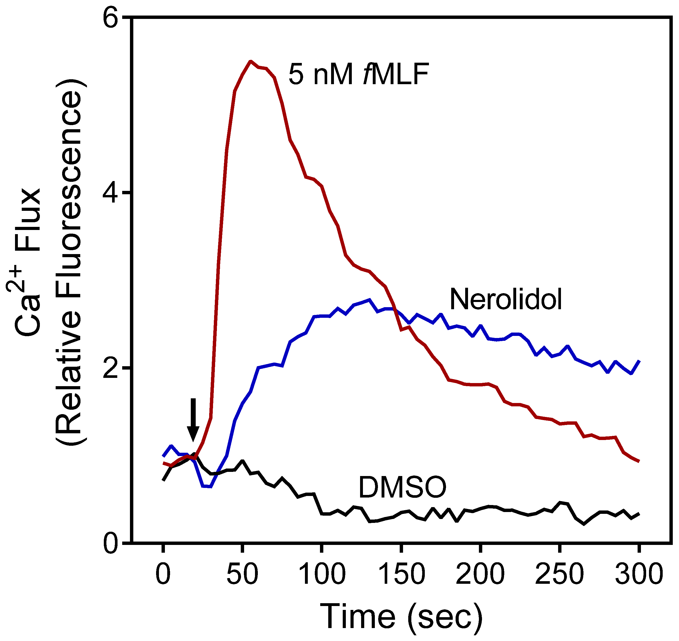
Figure 1. Effect of nerolidol on neutrophil [Ca2+]i. Human neutrophils were treated with 5 µM nerolidol, 5 nM fMLF (positive control), or 1% DMSO (negative control), and [Ca2+]i was monitored for the indicated times (arrow indicates when treatment was added). Data are from one experiment that is representative of three independent experiments.
Plants 11 03399 g001
Figure 2. Effect of PBO, PRO, and nerolidol on fMLF-induced neutrophil [Ca2+]i. Human neutrophils were treated with the indicated concentrations of the essential oils (A), nerolidol (B), or 1% DMSO (negative control) for 10 min. The cells were then activated with 5 nM fMLF, and [Ca2+]i was monitored as described. The data shown are presented as the mean ± SD from one experiment that is representative of three independent experiments with similar results.
Plants 11 03399 g002
Figure 3. Effect of the PBO, PRO, and nerolidol on human neutrophil chemotaxis. Neutrophils were pretreated with the indicated concentrations of the essential oils (A) or nerolidol (B), and neutrophil migration toward 1 nM fMLF was measured, as described. The data are from one experiment that is representative of three independent experiments.
Plants 11 03399 g003
Figure 4. Evaluation of the cytotoxicity of PRO, PBO, and nerolidol. Human neutrophils were preincubated with the indicated concentrations of essential oil (A) or pure nerolidol (B) for 30 min or 90 min, and cell viability was analyzed, as described. Values are the mean ± SD of triplicate samples from one experiment that is representative of three independent experiments with similar results. * p < 0.05 compared to DMSO control.
Plants 11 03399 g004
Figure 5. Alignments of lipoxin A4 with E-nerolidol (A) and Z-nerolidol (B). Alignments were performed using FieldTemplater software. Lipoxin A4 molecule is shown with a pink skeleton; the R and S enantiomers of nerolidol are shown with khaki and green skeletons, respectively.
Plants 11 03399 g005
Table 2. The effect of nerolidol and essential oils from propolis and P. balsamifera buds on human neutrophil [Ca2+]i and chemotaxis.
Source of Essential Oil or Pure CompoundActivation of [Ca2+]iInhibition of [Ca2+]i
fMLF-
Induced
WKYMVM-
Induced
fMLF-Induced Chemotaxis
EC50 (μg/mL)IC50 (μg/mL)
P. balsamifera10.5 ± 1.11.8 ± 0.69.4 ± 1.91.5 ± 0.5
Propolis18.3 ± 3.73.4 ± 0.10.9 ± 0.32.9 ± 1.3
EC50 (μM)IC50 (μM)
Nerolidol0.8 ± 0.14.0 ± 1.73.7 ± 0.43.9 ± 1.3
Legend: EC50 and IC50 values were determined by nonlinear regression analysis of the dose–response curves as described under Materials and Methods. The data are presented as the mean ± SD of three independent experiments.
Table 3. Chemical structures and physicochemical properties of nerolidol isomers according to SwissADME results.
PropertyE-NerolidolZ-Nerolidol
Plants 11 03399 i001Plants 11 03399 i002
FormulaC15H26OC15H26O
M.W.222.37222.37
Heavy atoms1616
Fraction Csp30.60.6
Rotatable bonds77
H-bond acceptors11
H-bond donors11
MR74.074.0
tPSA20.2320.23
iLogP3.643.60
BBB permeationYesYes
Abbreviations: M.W., molecular weight (g/mol); MR, molar refractivity; tPSA, topological polar surface area (Å2); iLogP, lipophilicity; BBB, blood–brain barrier.
Table 4. Potential protein targets for nerolidol isomers identified by PharmMapper.
RankPDB IDTarget NameFit ScoreRankPDB IDTarget NameFit Score
R-(E)-NerolidolS-(E)-Nerolidol
11J96AKR1C22.99911P49Steryl-sulfatase2.987
21E7ESerum albumin2.99723BMPBMP22.983
31L6LApo A-II2.99132JBPMAPKAPK22.981
41P49Steryl-sulfatase2.98943DEJCaspase-32.979
53BMPBMP22.98252Q11β-Secretase 12.977
62PINNR1A22.97862PINNR1A22.977
73BGPPim-12.97071BM6Stromelysin-12.970
81PMEERK22.96681L6LApo A-II2.969
91IIITransthyretin2.96291PMEERK22.965
101TG6CLPP2.960101QKUEstrogen receptor2.961
R-(Z)-NerolidolS-(Z)-Nerolidol
11P49Steryl-sulfatase2.99811P49Steryl-sulfatase3.000
22JBPMAPKAPK22.99123BGPPim-12.993
33CJFVEGFR22.98931F86Transthyretin2.992
43CGFJNK32.98941L6LApo A-II2.991
53BMPBMP22.98352PG2KIF112.987
62PINNR1A22.98063BMPBMP22.982
71SHJCaspase-72.97071IF4CA22.975
81YA8LCE12.96681J96AKR1C22.974
91PMEERK22.96591OJ9MAO-B2.969
102O65Pim-12.964102JBPMAPKAPK22.964
Abbreviations: AKR1C2, aldo-keto reductase family 1 member C2; Apo A-II, apolipoprotein A-II; BMP2, bone morphogenetic protein 2; CA2, carbonic anhydrase 2; CLPP, ATP-dependent Clp protease proteolytic subunit, mitochondrial; ERK2, extracellular signal-regulated kinase 2; JNK3, c-Jun N-terminal kinase 3; KIF11, kinesin family member 11; LCE1, liver carboxylesterase 1; MAO-B, monoamine oxidase B; MAPKAPK2, MAP kinase-activated protein kinase 2; NR1A2, thyroid hormone receptor β; Pim-1, proto-oncogene serine/threonine-protein kinase; VEGFR2, vascular endothelial growth factor receptor 2.
Publisher’s Note: MDPI stays neutral with regard to jurisdictional claims in published maps and institutional affiliations.