Article

Analysis of the RNA-Dependent RNA Polymerase 1 (RDR1) Gene Family in Melon

Department of Plant Pathology and Weed Research, Agricultural Research Organization, The Volcani Center, 68 HaMaccabim Road, P.O. Box 15159, Rishon LeZion 7505101, Israel
*
Author to whom correspondence should be addressed.
Plants 2022, 11(14), 1795; https://doi.org/10.3390/plants11141795
Submission received: 14 May 2022 / Revised: 16 June 2022 / Accepted: 28 June 2022 / Published: 7 July 2022
(This article belongs to the Section Crop Physiology and Crop Production)

Abstract

:
RNA-dependent RNA polymerase 1 (RDR1) plays a crucial defense role against plant viruses by secondary amplification of viral double-stranded RNA in the gene-silencing pathway. In this study, it was found that melon (Cucumis melo) encodes four RDR1 genes (CmRDR1a, b, c1 and c2) similar to the CsRDR1 gene family of cucumber (C. sativus). However, in contrast to cucumber, melon harbors a truncated CmRDR1b gene. In healthy plants, CmRDR1a was expressed, whereas the expression of CmRDR1c1/c2 was not detected. CmRDR1a expression level increased 20-fold upon cucumber mosaic virus (CMV) infection and was not increased in melon plants infected with zucchini yellow mosaic virus (ZYMV), cucumber vein yellowing virus (CVYV) and cucumber green mottle mosaic virus (CGMMV). The expression of CmRDR1c1/c2 genes was induced differentially by infection with viruses from different families: high levels of ~340-, 172- and 115-fold increases were induced by CMV, CVYV and CGMMV, respectively, and relatively low-level increases by potyvirus infection (4- to 6-fold). CMV mutants lacking the viral silencing suppressor 2b protein did not cause increased CmRDR1c/c2 expression; knockout of CmRDR1c1/c2 by CRISPR/Cas9 increased susceptibility to CMV but not to ZYMV. Therefore, it is suggested that the sensitivity of melon to viruses from different families is a result of the loss of function of CmRDR1b.

1. Introduction

Melon is an important vegetable crop and melon world production was 28 million tons on 1.1 million hectares in 2020 [1]. RNA viruses are very serious threats to melon production and early virus infection can lead to total loss [2]. Plant virus infection triggers the plant immune system (post-transcriptional silencing and RNA silencing) by detection of viral replication in the dsRNA form [3]. Endogenous RNA-dependent RNA polymerases (RDRs) generate dsRNA virus fragments as target molecules for gene silencing [4].
Plant RNA silencing is a cellular defense mechanism that regulates transcription or post-transcriptional gene expression in a sequence-specific manner by small interfering RNA (siRNA) molecules [5,6,7]. The silencing mechanism is associated with plant defense against viral infection and RDRs are core factors that initiate the biogenesis of viral-siRNAs [8].
In plants there are six different RDRs; however, RDR1 and RDR6 are the dominant enzymes that amplify single-stranded RNA viruses into aberrant dsRNA, which are digested by the host-encoded Dicer-like (DCL) DCL-4 and DCL-2 proteins into 21–22 nts virus-siRNA duplexes [9,10,11,12]. In addition, during RNA virus infection biogenesis of endogenous small-RNA is activated, dependent on RDR1 activity [13].
RDR1, as a single or duplicated gene occurs in all investigated plant species; however, in cucurbits there is a small RDR1 gene family [14,15,16]. The expression levels of RDR1, induced by DNA and RNA viruses [14,17], phytohormones [18], fungal [19] and insect attack [20], and can be controlled by the alternative oxidase (AOX)-associated defense pathway [21] and plant microRNAs [22]. It has been shown that RDRs play an important role in addition to virus defense: RDR1 is involved with responses to biotic, abiotic stress and defense against insect herbivores via dsRNA production that leads to rapid RNA degradation [23,24,25]. Novel RNA regulation via RDR1/2/6-mediated biosynthesis of antisense RNAs has been described as a response to abiotic stress [23].
The association of virus resistance/susceptibility with low/absent RDR1 expression was first described in Nicotiana bentamiana, a natural RDR1 mutant (with an insertion of 72 nts) that is highly susceptible to a wide range of viruses [26]. Transgenic N. bentamiana transformed with RDR1 genes from a number of plant species exhibited increased resistance to virus infection [26,27]. Correspondingly, virus accumulation increased when RDR1 was silenced in species such as Arabidopsis, tomato, tobacco and potato [17,21,28]. However, overexpression of tobacco NtRDR1 in N. bentamiana increased virus susceptibility [29] and silencing of potato RDR1 did not affect virus susceptibility [15].
RNA virus infection induced salicylic acid accumulation, which led to RDR1 induction [30]. Pretreatment with salicylic acid induced RDR1 expression and enhanced defense against RNA and DNA viruses in several species, whereas decrease of endogenous salicylic acid levels hindered defense against virus infection [31,32,33].
RDR1, known to regulate microRNA levels [24] in rice, plays a role in regulating important endogenous genes via mRNA-mediated DNA methylation, and is associated with the abiotic stress response [34]. In addition, regulation of RDR1 in rice depended on mir144, which was activated by rice stripe virus infection [22]. Notably, RDR1 mutant plants of different species showed no discernible altered phenotypes in growth and development. In contrast, mutants in the RDR6 gene display disturbed leaf development, as RDR6 is essential for tasiRNA biogenesis [13,34,35].
The discovery of the unique cucurbit RDR1 gene family was based on data from the Cucurbit Genomics Database. In cucumber, four functional CsRDR1 genes were identified (CsRDR1a, CsRDR1b, CsRDR1c1 and CsRDR1c2) [14]. In healthy cucumber plants, CsRDR1a and CsRDR1b were expressed, whereas CsRDR1c1 and CsRDR1c2 expression was undetectable. However, virus infection (especially cucumber mosaic virus (CMV) and cucumber vein yellowing virus (CVYV)) dramatically increased CsRDR1c1 and CsRDR1c2 expression, whereas silencing of CsRDR1c1+2 led to increased virus accumulation [14]. In addition, constitutive high levels of CsRDR1b expression were associated with broad virus resistance [14].
Melon (Cucumis melo) and cucumber (C. sativus) are close relatives and belong to the Cucumis genus (Cucurbitaceae). The genomes of melon and cucumber are highly conserved despite genome duplication in melon (ca. 454 Mbp in 12 chromosome pairs), whereas cucumber has 7 chromosome pairs with a genome size of 367 Mbp. Melon is known to be susceptible to infection by a range of viruses of different families such as zucchini yellow mosaic virus (ZYMV), papaya ringspot virus (PRSV-W), water melon mosaic virus (WMV) and cucumber vein yellowing virus (CVYV) (family Potyviridae), CMV (Bromoviridae) and cucumber green mottle mosaic virus (CGMMV; Virgaviridae). Classical breeding for virus resistance in melon has proved insufficient due to limited genetic resistance sources.
In the present study, we characterized the four melon genes of the CmRDR1 family (CmRDR1a, CmRDR1b, CmRDR1c1 and CmRDR1c2) and the differential gene expression responses of plants infected with different viruses. To help in our understanding of the role of CmRDR1c1/c2 genes in the plant response to viral disease, these genes were knocked out using CRISPR/Cas9 technology.

2. Materials and Methods

2.1. Plants, Pathogens and Inoculations

Cucumis melo L. cvs. Arava and Vedrantais were used for CmRDR gene mapping and virus susceptibility analyses. In addition, in 10 melon genotypes (listed in Table S1) the truncated RDR1b genes were mapped. Seeds were planted in a soil mixture and grown in a greenhouse in daylight at 25 °C. The viruses listed in Table S2 were used for virus accumulation analysis and evaluation of RDR gene expression, namely: cucumber mosaic virus CMV—Fny strain, CMV mutant lacking the 2b gene (CMV-Δ2b) [36], CGMMV, CVYV, ZYMV, PRSV-W and WMV. Squash (Cucurbita pepo L. cv. Zuccini) plants were a source of inocula for PRSV-W, WMV and ZYMV. Cucumber (Cucumis sativus L. cv. Bet Alfa) plants were inocula sources for CMV, CMV-Δ2b, CGMMV and CVYV. Melon seedlings at the cotyledon stage with a small true leaf (about 3–5 days post-emergence) were dusted with carborundum (320 grit powder, Fisher Scientific, USA) before mechanical inoculation with virus-bearing sap (ca. 1:10 ratio g tissue/mL H2O) [37].

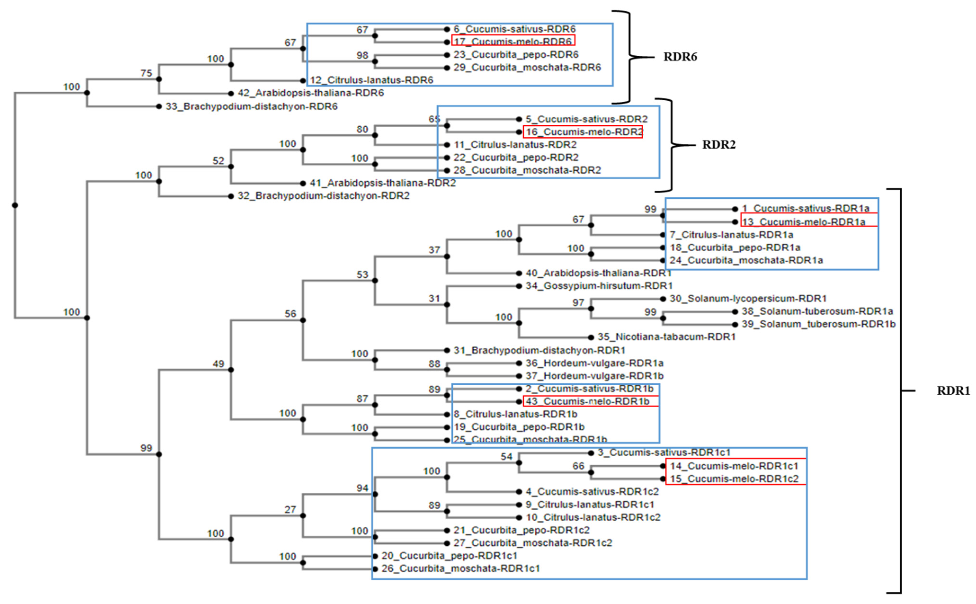
2.2. Phylogenetic Analysis

Protein sequences derived from three RDR clade members (RDR1, RDR2 and RDR6) from eleven plant species (accession numbers in Table S3) were aligned with a modified MAFFT program which allows adjustment of several parameters [38]. CmRDR1c1 and CmRDR1c2 were Sanger sequenced for this study, using primers from the sequences of the cucumber analogues and the Melonomics database (https://melonomics.net (accessed on 28 February 2020)) (Table S3). All other genes were accessed from public databases. Multiple sequence alignment was performed globally for all pairs with a maximum 1000 iterations. The robustness of the multiple sequence alignment was estimated with GUIDANCE2 [39] to be 0.971, which was slightly more robust than the local pair option. The phylogenetic tree was constructed using a likelihood method by running the ‘phyml’ program [40] with the JTT substitution model. The robustness and confidence of the tree nodes was estimated as a percentage of 1000 bootstrap resampling replicates.

2.3. Genome Mapping and Expression Analysis by PCR and QPCR

Total genomic DNA and RNA was extracted from young melon leaves (second or third youngest) using the Dellaporta method [41] and TRI-REAGENT kit (Molecular Research Center, Inc., Cincinnati, OH, USA), respectively. Quantified total RNA was adjusted using a NanoDrop ND1000 spectrophotometer (Thermo Scientific, Wilmington, DE, USA) for equal amounts. First-strand cDNA was synthesized from 2 µg of total RNA using the Verso cDNA Synthesis Kit (Thermo Fisher Scientific, Epsom, UK) including DNase. PCR reactions were performed with the appropriate primers for CmRDR1 (a, b, c1/c2), CmRDR2, CmRDR6, and cyclophilin genes based on the Melonomics database (Table S4). PCR conditions were 2 min at 94 °C, then 30 cycles of 30 s each at 94 °C, 58 °C and 72 °C, and an elongation step of 5 min at 72 °C. Quantitative (Q-PCR) reactions were performed in a volume of 15 µL with 4 µL of diluted cDNA (1/4), 3 pmol of each primer and 7.5 µL of Absolute Q-PCR Sybr Green Mix (Applied Biosystems by Thermo Fisher Scientific, Vilnius, Lithuania). Quantitative analysis was performed using a StepOne (Applied Biosystems) Real-Time PCR system with Q-PCR conditions of 10 min at 95 °C (“hot start”) followed by 40 cycles of 3 s at 94 °C, 15 s at 60 °C and 20 s at 72 °C. The relative expression level of gene accumulation was calculated using the ∆∆Ct method normalized to the housekeeping gene (cyclophilin) using StepOne software version 1.7 (Applied Biosystems) and the samples were compared with one of the samples, which was rated as 1 for comparative purposes. The specificity of the primers was tested prior to the Q-PCR analysis by sequencing of the products and melting curve (Figure S1) and non-specific binding was not found.

2.4. CRISPR/Cas9 Binary Construct Design

The pRCS binary vector containing Cas9-sgRNA was generated based on 35S:Cas9-AtU6:sgRNA-eIF4E [42]. The sgRNA-eIF4E was replaced by a sgRNA-RDR1c sequence using the appropriate primers (Table S4), as per [42].

2.5. Agrobacterium-Mediated Transformation

A Vedrantais melon genotype was used for Agrobacterium-tumefaciens-mediated transformation according to [37] with a number of changes: leaves were surface sterilized for 15 min in sodium hypochlorite (0.5% active chlorine) solution. Kanamycin (150 mg/L) was added to the selection medium. Rooting medium was composed of MS medium supplemented with indole-3-butyric acid (2 mg/L), cefotaxime (500 mg/L) and kanamycin (150 mg/L).

2.6. Genotyping and Mutant Verification

Genomic DNA was isolated from T0 transgenic and non-transgenic plants by the method of [41]. The presence of the Cas9/sgRNA transgene in T0 lines was confirmed by PCR using specific Cas9 primers (Table S4). Transgenic lines were genotyped for indel polymorphisms using primers flanking the CmRDR1c1 and CmRDR1c2 target regions (Table S4) and the PCR products were verified for the genetic changes by sequencing and restriction analysis with BmgBI. Heterozygotic T0 mutant plants were self-pollinated and in the T3 generation homozygotic mutant lines harboring cmrdr1c1 and cmrdr1c2 were selected by PCR analysis for molecular and biological analysis.

3. Results

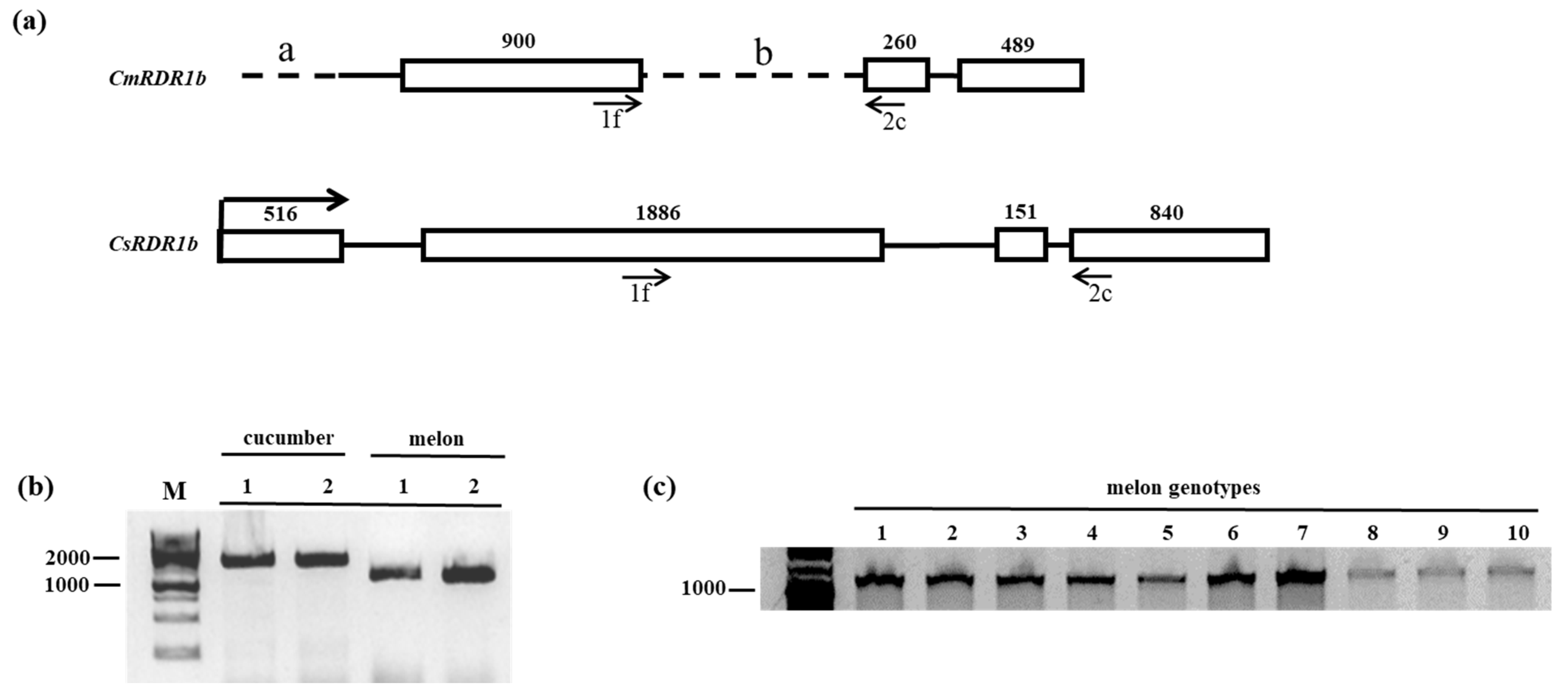
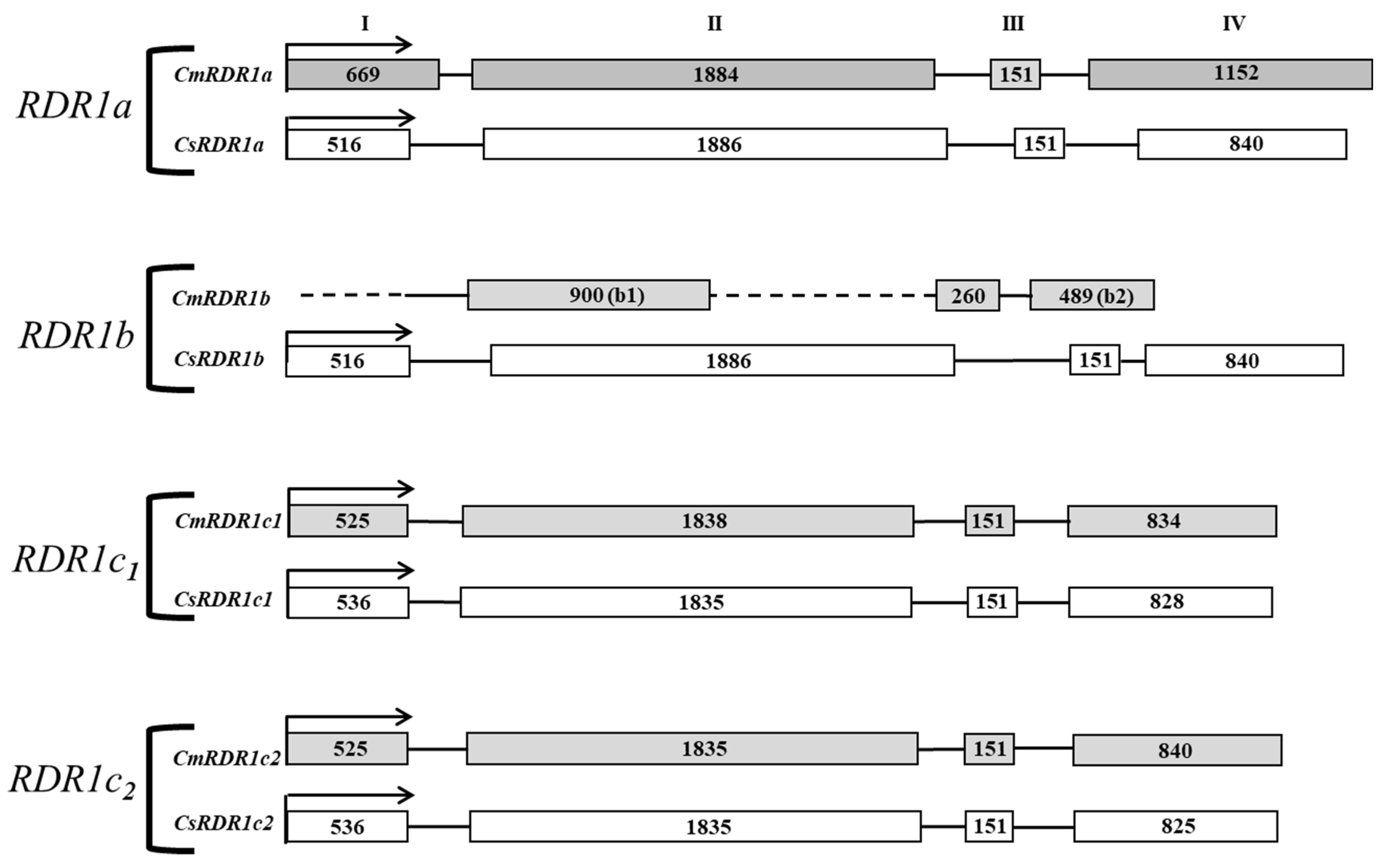
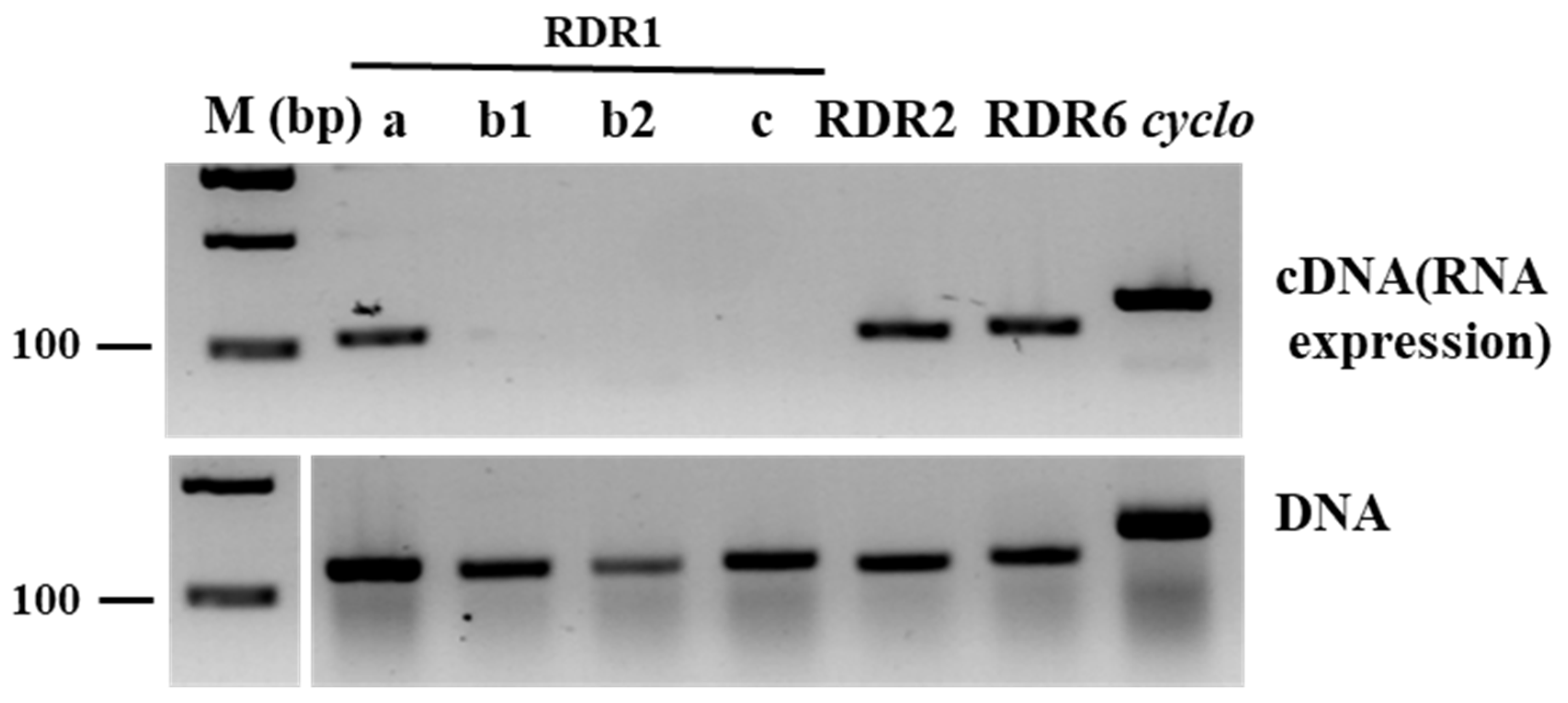
3.1. CmRDR1 Gene Family Mapping in Melon

The CmRDR1 gene family in melon was mapped based on the cucumber CsRDR1 gene (CsRDR1a, b, c1 and c2) sequences [14]. Four putative CmRDR1 genes, CmRDR1a, CmRDR1b and the duplicated CmRDR1c1 and CmRDR1c2, were mapped in melon using the Melonomics Database (Figure 1).
CmRDR1a and CmRDR1b are close to each other (~15 kbp) on chromosome 10 in a positive and negative orientation, respectively, similar to the situation in cucumber [14]. CmRDR1c1 and CmRDR1c2 are duplicated genes located within ~520 kbp of each other on chromosome 9. The exon/intron arrangement of the CmRDR1a, CmRDR1c1 and CmRDR1c2 genes was similar to that of cucumber, with four exons of about the same sizes (Figure 1) [14]. Sequence alignment between CmRDR1c1 and CmRDR1c2 encoding regions exhibited 95% homology and 98% identity on the protein level (Figure S2) and the putative 5′ and 3′ UTR exhibited 78% and 65% homology, respectively. However, the promoter (ca. 3 kb) upstream of the coding regions of CmRDR1c1 and CmRDR1c2 did not exhibit any sequence similarity (data not shown). CmRDR1a and CmRDR1c exhibited 70% amino acid homology, similar to the level of homology in cucumber [14]. Importantly, CmRDR1b, located on Chr 10, was missing two regions at the 5′-end in the first exon (a: 515 bp) and part of exon II (b: 1041 bp) compared with the cucumber gene (Figure 2). The CmRDR1b a and b regions (Figure 2) harbor unknown sequences. Truncated CmRDR1b sequences were mapped in 10 cultivars of different melon varieties by PCR genomic analysis (Figure 2), which indicated that the CmRDR1b enzyme was not generally functional in melon.
A phylogenetic analysis of RDR1, RDR2 and RDR6 genes of cucurbit species was performed (Figure 3). The neighbor-joining tree derived from protein sequences resolved into three clades: RDR1, RDR2 and RDR6. The melon RDR1 gene family mapped to three clusters: CmRDR1a, Cmrdr1b, and CmRDR1c1 and CmRDR1c2 together. Melon RDR1 genes clustered with their cucurbit homologs (Figure 3). All four CmRDR1 genes are closely related to those of cucumber (Figure 3), subsequently related to Citrullus lanatus and lastly to Cucurbita pepo and Cucurbita moschata (Figure 3).

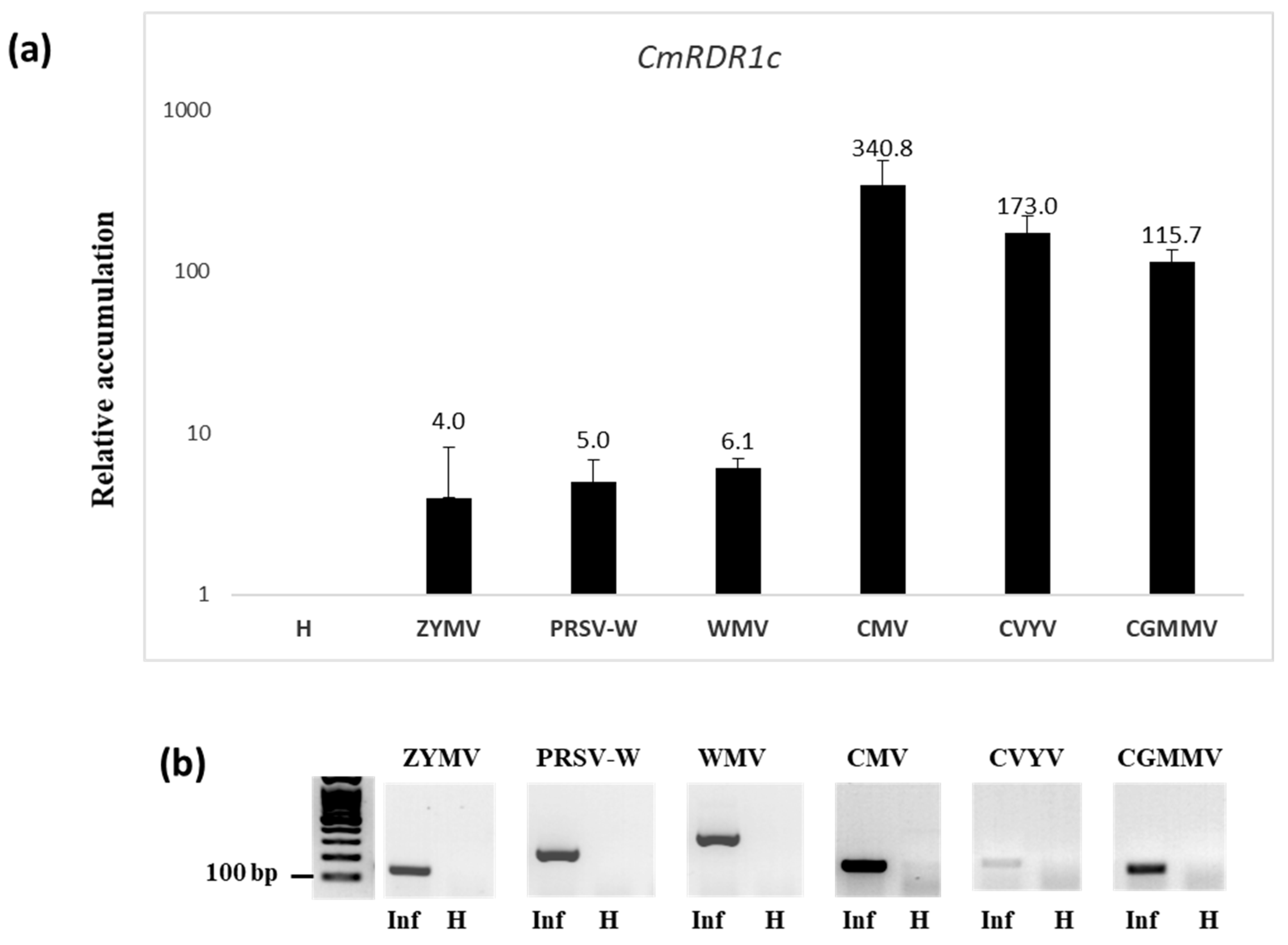
3.2. Expression Level of RDR1 Gene Family in Melon Infected with Different Viruses

The expression level of CmRDR1 genes were determined first in healthy melon by end-point RT-PCR analysis. CmRDR1a was constitutively expressed in leaves similar to the levels of CmRDR2 and CmRDR6 (Figure 4). However, the expression levels of both CmRDR1b and CmRDR1c1/2 were below the detection limit (Figure 4). CmRDR1b is a truncated gene compared with those of cucumber and probably was either not expressed, due to the 5′ gene deletion, or rapidly destroyed as aberrant mRNA (Figure 4) [43].
Melon is susceptible to infection by many viruses; therefore, the relative expression of the CmRDR1 gene family was tested following infection by viruses of different genera and families (Figure 5).
Interestingly, although the CmRDR1c1/2 genes were not expressed in healthy plants, these genes are highly induced by infection with a number of viruses (Figure 5 and Figure 6). The highest expression level of CmRDR1c1/2 was detected in plants upon CMV infection (340-fold increase) (Figure 5a). Following infection by the ipomovirus CVYV or the tobamovirus CGMMV, CmRDR1c1/2 expression level increased by 173-fold or 115-fold, respectively (Figure 5a). A moderate increase of CmRDR1c1/2 gene expression of about 5-fold resulted from potyvirus infection (ZYMV, PRSV and WMV) (Figure 5). The expression level of CmRDR1c1 and CmRDR1c2 were measured together as CmRDR1c, since they have a similar kinetics and response to virus infections (Figure 5 and Figure 6).
We have recently shown that the CMV-2b suppressor protein was associated with induction of CsRDR1c expression [36]. In light of this, we examined the expression level of CmRDR1c1 and CmRDR1c2 separately in melon infected with CMV and the CMV-Δ2b mutant. CmRDR1c1 and CmRDR1c2 were each expressed about 10-fold less in CMV-Δ2b infection compared with CMV at 9 and 16 days post inoculation (dpi) (Figure 6a,b). The lower expression levels of CmRDR1c1 and CmRDR1c2 correlated with the lower level of CMV-Δ2b accumulation and milder symptoms than observed with CMV (Figure 6c). Exceptionally increased expression of CmRDR1c1 and CmRDR1c2 was detected in plant 2b-5 infected with CMV-Δ2b 16 dpi (Figure 6d). Sequencing analysis of CMV-Δ2b in this plant revealed a mutation from ACG to ATG that restored 2b functionality, and as a result, virus accumulation and CmRDR1c1 and CmRDR1c2 expression levels increased in 2b-5 compared with other CMV-Δ2b-infected plants (Figure 6d).

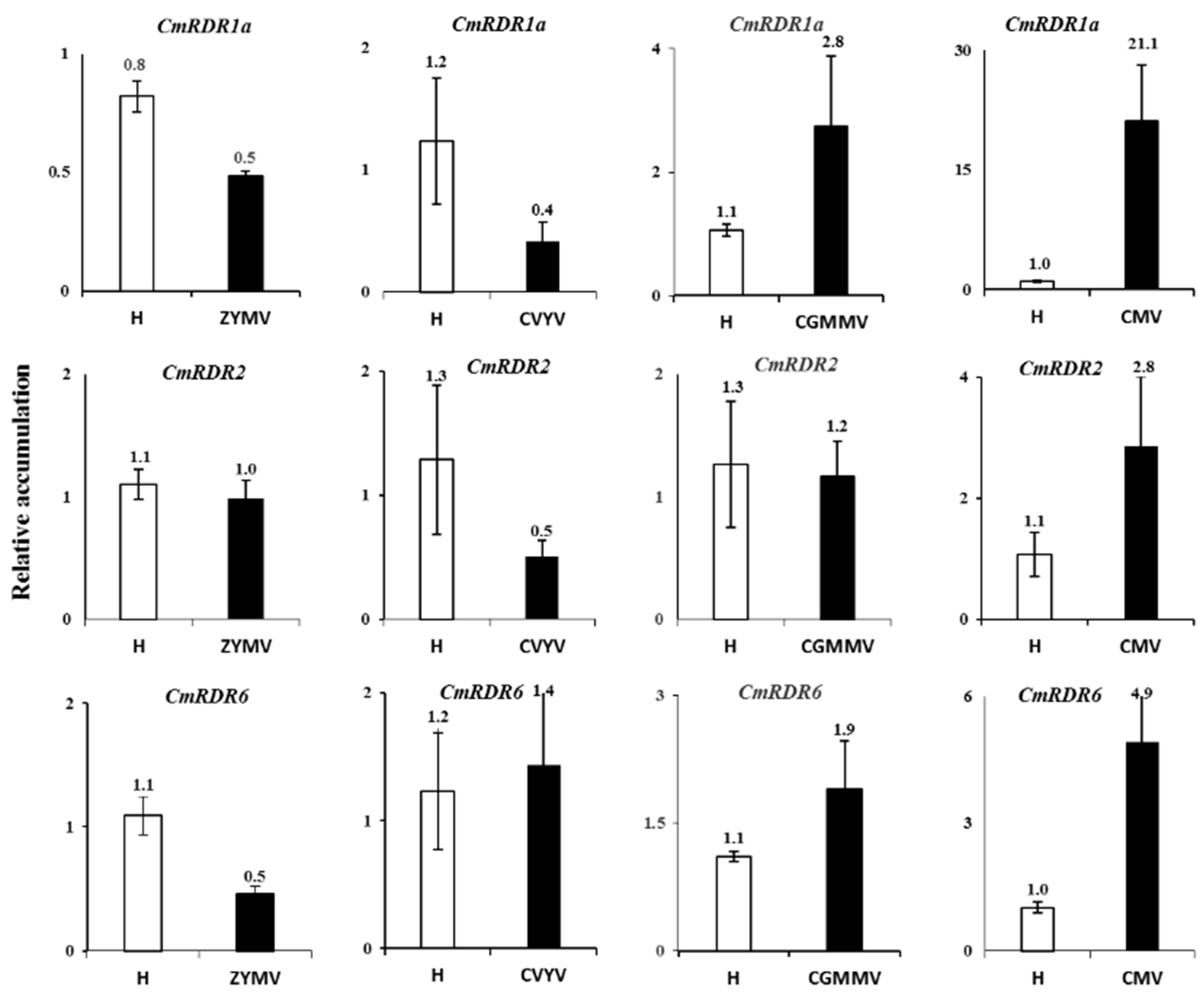
3.3. Expression of CmRDR2, CmRDR6 and CmRDR1a Genes upon Virus Infection

To understand whether other CmRDR genes can complement the loss of CmRDR1b function in melon, we examined the expression levels of CmRDR2, CmRDR6 and CmRDR1a in the susceptible melon cv. Arava in response to different viruses at an early growth stage. The expression levels of CmRDR1a and CmRDR6 decreased ~2-fold and the level of CmRDR2 was unchanged in ZYMV-infected plants at 7 dpi (Figure 7). Infection by CVYV slightly decreased the expression level of CmRDR1a and CmRDR2, but the CmRDR6 level was unchanged. Infection with CGMMV induced CmRDR1a and CmRDR6 expression level ~2-fold, whereas the CmRDR2 expression was unchanged. An exceptional effect was observed on CMV infection, in which the CmRDR1a expression level was induced 21-fold accompanied by moderate increases in CmRDR2 and CmRDR6 of 3- and 5-fold, respectively (Figure 7).

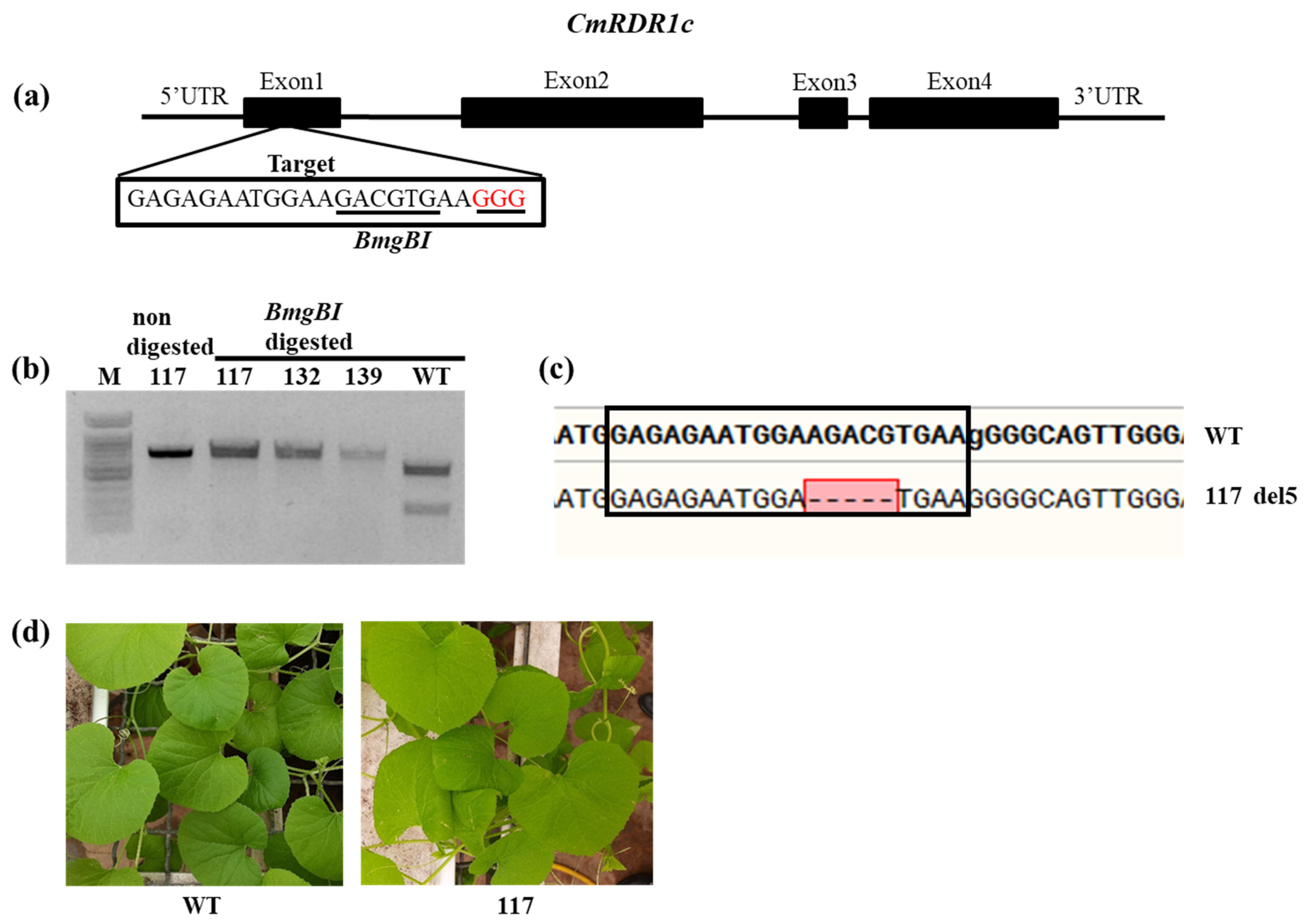
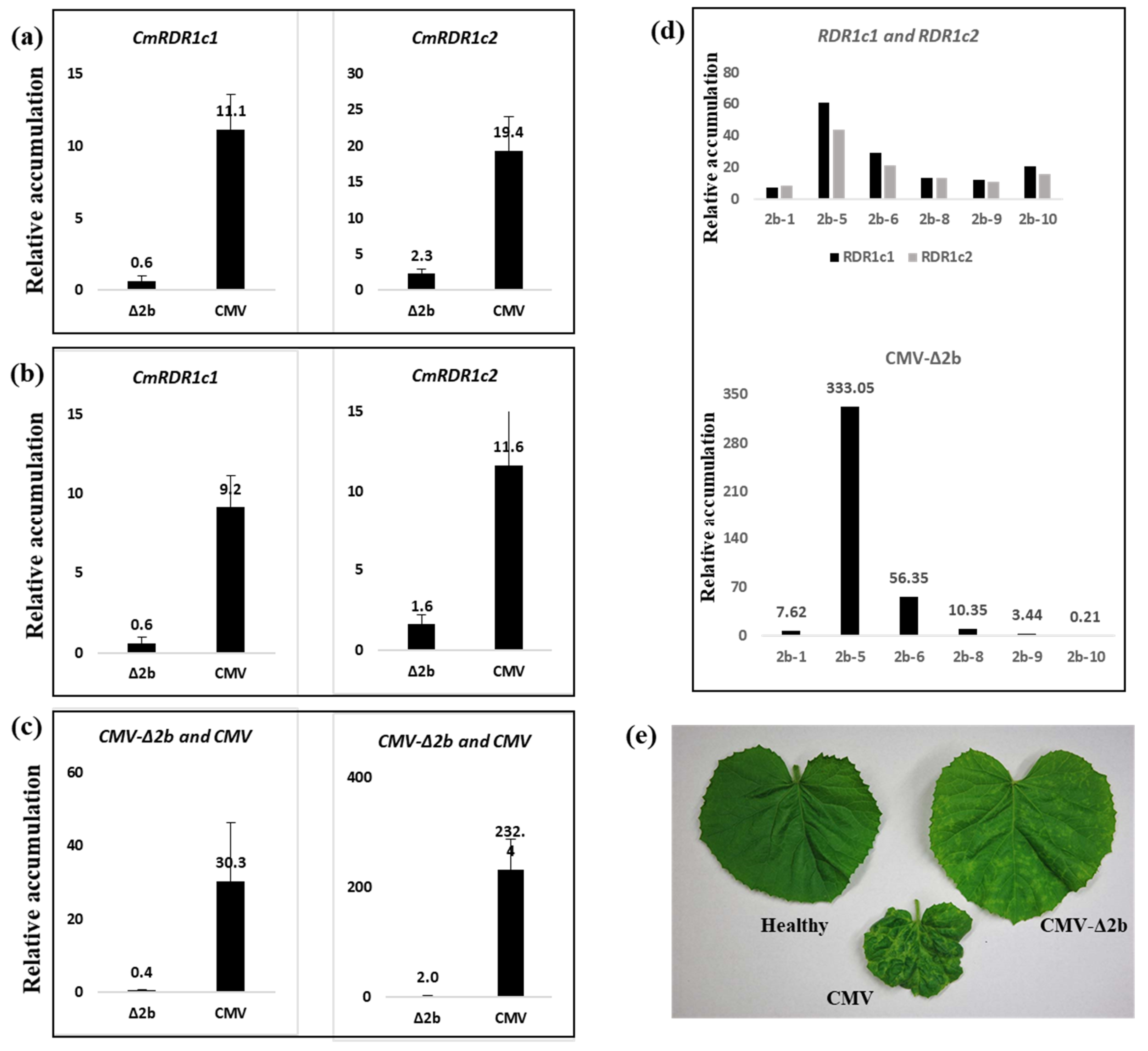
3.4. Evaluation of CmRDR1c (1+2) Contribution to Virus Severity

To study the contribution of the CsRDR1c1/c2 genes to plant virus defense, knockout mutants of these genes were generated by CRISPR/Cas9. Homozygous cmrdr1c1 and cmrdr1c2 T3 mutant plants were generated by self-pollination (Figure 8). The selected cmrdr1c1/c2 mutant line 117 harboring a 5 nts deletion was used in a virus susceptibility assay (Figure 8c). Phenotypically, the mutant cmrdr1c1/2 (Line 117) plants developed similarly to the non-mutant WT control plants (Figure 8d) and no effect was observed on leaf structure and fruit set and plant development.
Cmrdr1c1/c2 mutant plants were inoculated with CMV and ZYMV separately, and symptom expression and virus accumulation were examined. CMV RNA accumulation was similar to WT plants 14 dpi (Figure 9a). Cmrdr1c1/c2 mutant plants (Line 117) infected with CMV exhibited similar severe symptoms as noted in the WT plants (Figure 9b). However, a significant increase of CMV RNA accumulation was measured in mutant plants 24 and 32 dpi. This is despite the fact that the expression level of CmRDR1c1/2 in WT is tens of times higher than in Cmrdr1c1/c2 mutant plants, whereas the expression levels of CmRDR2 and CmRDR6 were slightly increased in cmrdr1c1/2 mutant plants. Mutant cmrdr1c1/c2 plants infected with ZYMV exhibited similar symptoms to the WT plants and ZYMV RNA accumulated to a similar level as in the WT plants at 14 and 24 dpi (Figure 9a,b). Accordingly, the levels of CmRDR1c1/2, CmRDR2 and CmRDR6 did not change between mutant and WT due to ZYMV infection. The low level of CmRDR1c1/2 in mutant plants infected with CMV is probably due to rapid degradation of truncated CmRDR1c1/2 mRNA (Figure 9c), as shown in eukaryotes with mRNAs that prematurely terminate translation [43].

4. Discussion

4.1. CmRDR1 Gene Organization

Here we report genomic characterization of the gene family CmRDR1s in melon, and gene product expression before and after virus infection. Four CmRDR1 genes were identified (RDR1a, RDR1b RDR1c1 and RDR1c2) with similar genome organization (intron/exon) to that of cucumber (Figure 1) [14]. Interestingly, the RDR1 gene family in Cucumis spp. was homologous to those of other genera (Cucurbita and Citrullus) in the Cucurbitaceae (Figure 3), whereas in most plant species only a single or duplicated RDR1 gene has been described [15].
In melon, CmRDR1c1 and CmRDR1c2 genes share high sequence similarity, like the duplicated gene described in cucumber [14]. Likewise, duplication of the RDR1c gene was homologous in the three other cucurbits examined (watermelon, squash and pumpkin) (Figure 3). Duplication of RDR1 genes was characterized in barley (HvRDR1a and HvRDR1b) [16] and potato (StRDR1a and StRDRb) [15].
The close phylogenetic relationship of the RDR1 gene family in the five cucurbit species (Figure 3), suggests that the four RDR1 genes were duplicated before the separation of the ancestral cucurbits into different taxa. Therefore, the unique cucurbit RDR1 gene family may indicate an additional function or interaction between the RDR1 family members in plant defense. In melon, the CmRDR1 gene family mapped to two chromosomes (9 and 10), whereas in cucumber the CmRDR1 gene family was located on chromosome 5 [14], which may reflect a cucumber chromosome fusion from a progenitor species [44]. In plants, the region ~3 kbp upstream to RDR1 has been characterized as a regulatory region [22,45,46]. Therefore, the different sequences (~3 kbp) upstream of the CmRDR1a, b, c1 and c2 genes may imply differential regulation of each RDR1 gene.

4.2. Characterization of Truncated CmRDR1b Gene in Melon Genome

We show here that the CmRDR1b gene from melon accessions worldwide was truncated (cmrdr1b), and the transcript undetectable due to the two missing regions, compared with the CsRDR1b gene in cucumber [14]. We therefore propose that the truncated CmRDR1b in melon was mutated in an ancestor during genome duplication and transposon amplification associated with retrotransposon elements in this region [47].

4.3. Kinetics of RDR1a, b, c1+c2 Gene Expression Response to Virus Infection

Unique CmRDR1a, b, c1+c2 expression levels were determined in melon. CmRDR1a, which shared the highest homology to RDR1 from other species, was continuously expressed in healthy leaves in cucumber and other hosts [14,15]. Expression of the truncated cmrdr1b was not observed in melon (Figure 1 and Figure 3). In cucumber, the high expression level of CsRDR1b in healthy plants was characterized as a marker for broad virus resistance [14] and probably the susceptibility of melon to many viruses may result from malfunction of CmRDR1b. In healthy melon plants the expression level of CmRDR1c1 and CmRDR1c2 were below the detection level (Figure 3) similar to CsRDR1c1 and CsRDR1c2 in cucumber [14], which may indicate that its regulation was due to virus infection. It is important to note that this unique response of RDR1c1 and RDR1c2 was demonstrated only in the genus Cucumis, as in other host families RDR1 is continuously expressed in healthy plants [15,16].
In CMV-infected melon, CmRDR1a gene expression was induced 21-fold, but not by Potyviridae (ZYMV and CVYV) and a tobamovirus (CGMMV) (Figure 5 and Figure 7). Likewise, in cucumber protoplasts, transfection with CMV increased CsRDR1a expression level 29-fold and exogenous application of CsRDR1a reduced CMV accumulation dramatically [36], probably due to 2b induction [36]. However, the involvement of CmRDR1a in defense against potyviruses needs additional study.
The duplicated CmRDR1c1 and CmRDR1c2 genes share high sequence similarity and have similar expression levels following virus infection (Figure 6), although the putative promoters upstream of the genes lack homology. Expression of both genes was induced by virus infection strongly and differentially, depending on virus genus. Whereas potyviruses caused gene induction by 4- to 6-fold, CMV, CVYV and CGMMV caused induction by >100-fold (Figure 5). CMV infection induced CmRDR1c1+2 expression by ~300 fold, similar to CsRDR1c in cucumber cv. Beit-Alfa [14]. However, CMV with a deleted 2b gene induced significantly lower CmRDR1c1+2 transcript accumulation (about 10 to 15-fold lower) (Figure 5), which may indicate that CmRDR1c1+2 gene expression was regulated by CMV level. Alternatively, the 2b protein can enter the nucleus [22] and can elicit PAMP-triggered immunity [36,48]. In addition, in melon infected with the tobamovirus CGMMV, CmRDR1c1+2 expression was induced ~100 fold, probably because of the high level of CGMMV-CP accumulation, an oxidative burst inducer as demonstrated in tobacco by tobacco mosaic virus [49] or via tobacco mosaic virus dsRNA inducer molecules [50]. Interestingly, CVYV (Ipomovirus; Potyviridae) induced CmRDR1c1+2 expression ~20-fold higher than the potyviruses ZYMV, PRSV and WMV during the infection of cucumber [14]. This change may reflect the difference between the suppressors of these genera (P1b of CVYV versus the potyvirus HCPro) [51]. Notably, expression of other RDR genes (CmRDR2 and CmRDR6) was induced in melon by CMV infection, but not by potyviruses and tobamovirus, which suggests that the suppressor 2b of CMV may play a role in PAMP-triggered immunity [36].

4.4. The Sensitivity of cmrdr1c1+2 Mutant Plants to Virus Infection

Expression of CmRDR1c1+2 was rapidly induced to a high level by various viruses (Figure 5), an important role in virus-infected plants. Therefore, cmrdr1c1+2 mutant melon lines were generated to test this role. Mutant lines did not differ in plant growth and fruit development from the WT cultivar. Likewise, ZYMV-infected cmrdr1c1+2 mutant plants showed similar severe symptoms to the WT plants and virus accumulation did not differ between mutant and WT plants. Similarly in potato, reduction of StRDR1 expression level by RNAi did not enhance susceptibility to PVY [15]. In addition, no differences of CmRDR2 and CmRDR6 expression level were observed between mutant and WT plants. These data suggest that in melon the CmRDR1c1+2 genes were not involved with potyvirus defense in our experimental conditions. However, we cannot rule out that under different conditions of abiotic or biotic stresses these genes may be important in protection against potyviruses. In contrast to potyvirus, cmrdr1c1+2 mutant plants infected with CMV showed slightly greater severity compared with WT plants; CMV accumulation was significantly increased 24 and 32 dpi. In addition, CmRDR1c1+2 were highly expressed in WT plants compared with very low levels of expression in mutant plants, suggesting rapid degradation of aberrant RDR1c1 and c2 transcripts of the nonfunctional proteins [43]. These data suggest that CmRDR1c1 and CmRDR1c2 are unique genes associated with defense against CMV infection specifically, and the virus–host interaction in cucumber between the inducer 2b and RDR1c1+c2 probably also occurs in melon [36]. Such virus-specific protection has not been reported.

4.5. Virus Susceptibility in Melon

Melon is a susceptible host to many viruses due to limited genetic resistances. In addition, the virus recovery phenomenon is less common in melon than cucumber. We show that high expression levels of the unique CsRDR1b resistance gene in cucumber is associated with increased and broad virus resistance [14]. We, therefore, suggest that susceptibility of melon to many viruses is a result of a non-functional CmRDR1b and repair of this gene using advanced technologies such as CRISPR/Cas9 may significantly improve melon resistance to a range of pathogenic viruses.

Supplementary Materials

The following supporting information can be downloaded at: https://www.mdpi.com/article/10.3390/plants11141795/s1, Table S1: Genotypes tested for RDR1b mapping. Table S2: List of viruses. Table S3: List of RDR genes. Table S4: Primers used in this study. Figure S1: Melting curves analysis. Figure S2: Amino acid sequence alignment of putative proteins encoded by the CmRDR1c1, CmRDR1c2, CsRDR1c1 and CsRDR1c2 genes of melon cv. Arava and cucumber cv. Beit-Alfa. References [52,53,54,55,56,57,58] are cited in the Supplementary Materials.

Author Contributions

D.L. performed experiments, analyzed data, and wrote the manuscript; E.P. and Y.S., transformed melon; M.S., genotyping of mutant plants; V.G. writing and reviewing the results; A.G.-O. supervision, project administration, funding acquisition and writing. All authors have read and agreed to the published version of the manuscript.

Funding

This research was supported by the Chief Scientist of the Israeli Ministry of Agriculture and Rural Development, Grant No. 20-01-0209 awarded to A.G and grant from the United States–Israel Binational Agricultural Research and Development Fund (BARD) Grant No. IS-5027-17C awarded to A.G. and Shou-wei Ding.

Data Availability Statement

The data presented in this study are available on request from the corresponding author.

Acknowledgments

We thank Ron Ofir for the guidance in the bioinformatics analysis.

Conflicts of Interest

The authors declare that the research was conducted in the absence of any commercial or financial relationships that could be construed as a potential conflict of interest.

References

  1. FAOSTAT. Available online: https://www.fao.org/faostat/en/#home (accessed on 22 June 2022).
  2. Lecoq, H.; Desbiez, C. Viruses of cucurbit crops in the Mediterranean region: An ever-changing picture. Adv. Virus Res. 2012, 84, 67–126. [Google Scholar] [CrossRef] [PubMed]
  3. Calil, I.P.; Fontes, E.P.B. Plant immunity against viruses: Antiviral immune receptors in focus. Ann. Bot. 2017, 119, 711–723. [Google Scholar] [CrossRef] [PubMed] [Green Version]
  4. Wassenegger, M.; Krczal, G. Nomenclature and functions of RNA-directed RNA polymerases. Trends Plant Sci. 2006, 11, 142–151. [Google Scholar] [CrossRef] [PubMed]
  5. Baulcombe, D. RNA silencing in plants. Nature 2004, 431, 356–363. [Google Scholar] [CrossRef] [PubMed]
  6. Ding, S.-W.; Voinnet, O. Antiviral Immunity Directed by Small RNAs. Cell 2007, 130, 413–426. [Google Scholar] [CrossRef] [Green Version]
  7. Meister, G.; Tuschl, T. Mechanisms of gene silencing by double-stranded RNA. Nature 2004, 431, 343–349. [Google Scholar] [CrossRef]
  8. Lopez-Gomollon, S.; Baulcombe, D.C. Roles of RNA silencing in viral and non-viral plant immunity and in the crosstalk between disease resistance systems. Nat. Rev. Mol. Cell Biol. 2022, 1–18. [Google Scholar] [CrossRef]
  9. Donaire, L.; Barajas, D.; Martínez-García, B.; Martínez-Priego, L.; Pagán, I.; Llave, C. Structural and Genetic Requirements for the Biogenesis of Tobacco Rattle Virus-Derived Small Interfering RNAs. J. Virol. 2008, 82, 5167–5177. [Google Scholar] [CrossRef] [Green Version]
  10. Garcia-Ruiz, H.; Takeda, A.; Chapman, E.J.; Sullivan, C.M.; Fahlgren, N.; Brempelis, K.J.; Carrington, J.C. Arabidopsis RNA-dependent RNA polymerases and dicer-like proteins in antiviral defense and small interfering RNA biogenesis during Turnip Mosaic Virus infection. Plant Cell 2010, 22, 481–496. [Google Scholar] [CrossRef] [Green Version]
  11. Qi, X.; Bao, F.S.; Xie, Z. Small RNA Deep Sequencing Reveals Role for Arabidopsis thaliana RNA-Dependent RNA Polymerases in Viral siRNA Biogenesis. PLoS ONE 2009, 4, e4971. [Google Scholar] [CrossRef]
  12. Wang, X.-B.; Wu, Q.; Ito, T.; Cillo, F.; Li, W.-X.; Chen, X.; Yu, J.-L.; Ding, S.-W. RNAi-mediated viral immunity requires amplification of virus-derived siRNAs in Arabidopsis thaliana. Proc. Natl. Acad. Sci. USA 2010, 107, 484–489. [Google Scholar] [CrossRef] [PubMed] [Green Version]
  13. Cao, M.; Du, P.; Wang, X.; Yu, Y.-Q.; Qiu, Y.-H.; Li, W.; Gal-On, A.; Zhou, C.; Li, Y.; Ding, S.-W. Virus infection triggers widespread silencing of host genes by a distinct class of endogenous siRNAs in Arabidopsis. Proc. Natl. Acad. Sci. USA 2014, 111, 14613–14618. [Google Scholar] [CrossRef] [PubMed] [Green Version]
  14. Leibman, D.; Kravchik, M.; Wolf, D.; Haviv, S.; Weissberg, M.; Ophir, R.; Paris, H.S.; Palukaitis, P.; Ding, S.-W.; Gaba, V.; et al. Differential expression of cucumber RNA-dependent RNA polymerase 1 genes during antiviral defence and resistance. Mol. Plant Pathol. 2018, 19, 300–312. [Google Scholar] [CrossRef] [PubMed] [Green Version]
  15. Hunter, L.J.R.; Brockington, S.F.; Murphy, A.M.; Pate, A.E.; Gruden, K.; MacFarlane, S.A.; Palukaitis, P.; Carr, J.P. RNA-dependent RNA polymerase 1 in potato (Solanum tuberosum) and its relationship to other plant RNA-dependent RNA polymerases. Sci. Rep. 2016, 6, 23082. [Google Scholar] [CrossRef] [PubMed] [Green Version]
  16. Madsen, C.T.; Stephens, J.; Hornyik, C.; Shaw, J.; Collinge, D.B.; Lacomme, C.; Albrechtsen, M. Identification and characterization of barley RNA-directed RNA polymerases. Biochim. Biophys. Acta BBA-Gene Regul. Mech. 2009, 1789, 375–385. [Google Scholar] [CrossRef] [PubMed]
  17. Yu, D.; Fan, B.; MacFarlane, S.A.; Chen, Z. Analysis of the Involvement of an Inducible Arabidopsis RNA-Dependent RNA Polymerase in Antiviral Defense. Mol. Plant-Microbe Interact. 2003, 16, 206–216. [Google Scholar] [CrossRef] [Green Version]
  18. He, J.; Dong, Z.; Jia, Z.; Wang, J.; Wang, G. Isolation, expression and functional analysis of a putative RNA-dependent RNA polymerase gene from maize (Zea mays L.). Mol. Biol. Rep. 2010, 37, 865–874. [Google Scholar] [CrossRef]
  19. Gao, Q.; Liu, Y.; Wang, M.; Zhang, J.; Gai, Y.; Zhu, C.; Guo, X. Molecular cloning and characterization of an inducible RNA-dependent RNA polymerase gene, GhRdRP, from cotton (Gossypium hirsutum L.). Mol. Biol. Rep. 2009, 36, 47–56. [Google Scholar] [CrossRef]
  20. Pandey, S.P.; Baldwin, I.T. RNA-directed RNA polymerase 1 (RdR1) mediates the resistance of Nicotiana attenuata to herbivore attack in nature. Plant J. 2007, 50, 40–53. [Google Scholar] [CrossRef]
  21. Liao, Y.-W.-K.; Liu, Y.-R.; Liang, J.-Y.; Wang, W.-P.; Zhou, J.; Xia, X.-J.; Zhou, Y.-H.; Yu, J.-Q.; Shi, K. The relationship between the plant-encoded RNA-dependent RNA polymerase 1 and alternative oxidase in tomato basal defense against Tobacco mosaic virus. Planta 2015, 241, 641–650. [Google Scholar] [CrossRef]
  22. Wang, H.; Jiao, X.; Kong, X.; Hamera, S.; Wu, Y.; Chen, X.; Fang, R.; Yan, Y. A Signaling Cascade from miR444 to RDR1 in Rice Antiviral RNA Silencing Pathway. Plant Physiol. 2016, 170, 2365–2377. [Google Scholar] [CrossRef] [PubMed] [Green Version]
  23. Matsui, A.; Iida, K.; Tanaka, M.; Yamaguchi, K.; Mizuhashi, K.; Kim, J.-M.; Takahashi, S.; Kobayashi, N.; Shigenobu, S.; Shinozaki, K.; et al. Novel Stress-Inducible Antisense RNAs of Protein-Coding Loci Are Synthesized by RNA-Dependent RNA Polymerase. Plant Physiol. 2017, 175, 457–472. [Google Scholar] [CrossRef] [PubMed] [Green Version]
  24. Polydore, S.; Axtell, M.J. Analysis of RDR1/RDR2/RDR6-independent small RNAs in Arabidopsis thaliana improves MIRNA annotations and reveals unexplained types of short interfering RNA loci. Plant J. 2018, 94, 1051–1063. [Google Scholar] [CrossRef] [PubMed] [Green Version]
  25. Pandey, S.P.; Shahi, P.; Gase, K.; Baldwin, I.T. Herbivory-induced changes in the small-RNA transcriptome and phytohormone signaling in Nicotiana attenuata. Proc. Natl. Acad. Sci. USA 2008, 105, 4559–4564. [Google Scholar] [CrossRef] [Green Version]
  26. Yang, S.-J.; Carter, S.A.; Cole, A.B.; Cheng, N.-H.; Nelson, R.S. A natural variant of a host RNA-dependent RNA polymerase is associated with increased susceptibility to viruses by Nicotiana benthamiana. Proc. Natl. Acad. Sci. USA 2004, 101, 6297–6302. [Google Scholar] [CrossRef] [PubMed] [Green Version]
  27. Qin, L.; Mo, N.; Zhang, Y.; Muhammad, T.; Zhao, G.; Zhang, Y.; Liang, Y. CaRDR1, an RNA-Dependent RNA Polymerase Plays a Positive Role in Pepper Resistance against TMV. Front. Plant Sci. 2017, 8, 1068. [Google Scholar] [CrossRef] [PubMed] [Green Version]
  28. Rakhshandehroo, F.; Takeshita, M.; Squires, J.; Palukaitis, P. The influence of RNA-dependent RNA polymerase 1 on potato virus Y infection and on other antiviral response genes. Mol. Plant-Microbe Interact. MPMI 2009, 22, 1312–1318. [Google Scholar] [CrossRef] [Green Version]
  29. Ying, X.-B.; Dong, L.; Zhu, H.; Duan, C.-G.; Du, Q.-S.; Lv, D.-Q.; Fang, Y.-Y.; Garcia, J.A.; Fang, R.-X.; Guo, H.-S. RNA-dependent RNA polymerase 1 from Nicotiana tabacum suppresses RNA silencing and enhances viral infection in Nicotiana benthamiana. Plant Cell 2010, 22, 1358–1372. [Google Scholar] [CrossRef] [Green Version]
  30. Qi, G.; Chen, J.; Chang, M.; Chen, H.; Hall, K.; Korin, J.; Liu, F.; Wang, D.; Fu, Z.Q. Pandemonium Breaks Out: Disruption of Salicylic Acid-Mediated Defense by Plant Pathogens. Mol. Plant 2018, 11, 1427–1439. [Google Scholar] [CrossRef] [Green Version]
  31. Alamillo, J.M.; Saénz, P.; García, J.A. Salicylic acid-mediated and RNA-silencing defense mechanisms cooperate in the restriction of systemic spread of plum pox virus in tobacco. Plant J. Cell Mol. Biol. 2006, 48, 217–227. [Google Scholar] [CrossRef]
  32. Hunter, L.J.R.; Westwood, J.H.; Heath, G.; Macaulay, K.; Smith, A.G.; MacFarlane, S.A.; Palukaitis, P.; Carr, J.P. Regulation of RNA-Dependent RNA Polymerase 1 and Isochorismate Synthase Gene Expression in Arabidopsis. PLoS ONE 2013, 8, e66530. [Google Scholar] [CrossRef] [PubMed]
  33. Lee, W.-S.; Fu, S.-F.; Li, Z.; Murphy, A.M.; Dobson, E.A.; Garland, L.; Chaluvadi, S.R.; Lewsey, M.G.; Nelson, R.S.; Carr, J.P. Salicylic acid treatment and expression of an RNA-dependent RNA polymerase 1 transgene inhibit lethal symptoms and meristem invasion during tobacco mosaic virus infection in Nicotiana benthamiana. BMC Plant Biol. 2016, 16, 15. [Google Scholar] [CrossRef] [PubMed] [Green Version]
  34. Wang, N.; Zhang, D.; Wang, Z.; Xun, H.; Ma, J.; Wang, H.; Huang, W.; Liu, Y.; Lin, X.; Li, N.; et al. Mutation of the RDR1 gene caused genome-wide changes in gene expression, regional variation in small RNA clusters and localized alteration in DNA methylation in rice. BMC Plant Biol. 2014, 14, 177. [Google Scholar] [CrossRef] [PubMed] [Green Version]
  35. Lam, P.; Zhao, L.; McFarlane, H.E.; Aiga, M.; Lam, V.; Hooker, T.S.; Kunst, L. RDR1 and SGS3, Components of RNA-Mediated Gene Silencing, Are Required for the Regulation of Cuticular Wax Biosynthesis in Developing Inflorescence Stems of Arabidopsis. Plant Physiol. 2012, 159, 1385–1395. [Google Scholar] [CrossRef] [PubMed] [Green Version]
  36. Kumari, R.; Kumar, S.; Leibman, D.; Abebie, B.; Shnaider, Y.; Ding, S.-W.; Gal-On, A. Cucumber RDR1s and cucumber mosaic virus suppressor protein 2b association directs host defence in cucumber plants. Mol. Plant Pathol. 2021, 22, 1317–1331. [Google Scholar] [CrossRef]
  37. Leibman, D.; Wolf, D.; Saharan, V.; Zelcer, A.; Arazi, T.; Yoel, S.; Gaba, V.; Gal-On, A. A High Level of Transgenic Viral Small RNA Is Associated with Broad Potyvirus Resistance in Cucurbits. Mol. Plant. Microbe Interact. 2011, 24, 1220–1238. [Google Scholar] [CrossRef] [PubMed] [Green Version]
  38. Katoh, K.; Standley, D.M. MAFFT Multiple Sequence Alignment Software Version 7: Improvements in Performance and Usability. Mol. Biol. Evol. 2013, 30, 772–780. [Google Scholar] [CrossRef] [Green Version]
  39. Sela, I.; Ashkenazy, H.; Katoh, K.; Pupko, T. GUIDANCE2: Accurate detection of unreliable alignment regions accounting for the uncertainty of multiple parameters. Nucleic Acids Res. 2015, 43, W7–W14. [Google Scholar] [CrossRef] [Green Version]
  40. Guindon, S.; Gascuel, O. A simple, fast, and accurate algorithm to estimate large phylogenies by maximum likelihood. Syst. Biol. 2003, 52, 696–704. [Google Scholar] [CrossRef] [Green Version]
  41. Dellaporta, S.L.; Wood, J.; Hicks, J.B. A plant DNA minipreparation: Version II. Plant Mol. Biol. Report. 1983, 1, 19–21. [Google Scholar] [CrossRef]
  42. Chandrasekaran, J.; Brumin, M.; Wolf, D.; Leibman, D.; Klap, C.; Pearlsman, M.; Sherman, A.; Arazi, T.; Gal-On, A. Development of broad virus resistance in non-transgenic cucumber using CRISPR/Cas9 technology. Mol. Plant Pathol. 2016, 17, 1140–1153. [Google Scholar] [CrossRef] [PubMed] [Green Version]
  43. Peccarelli, M.; Kebaara, B.W. Regulation of natural mRNAs by the nonsense-mediated mRNA decay pathway. Eukaryot. Cell 2014, 13, 1126–1135. [Google Scholar] [CrossRef] [PubMed]
  44. Li, D.; Cuevas, H.E.; Yang, L.; Li, Y.; Garcia-Mas, J.; Zalapa, J.; Staub, J.E.; Luan, F.; Reddy, U.; He, X.; et al. Syntenic relationships between cucumber (Cucumis sativus L.) and melon (C. melo L.) chromosomes as revealed by comparative genetic mapping. BMC Genom. 2011, 12, 396. [Google Scholar] [CrossRef] [PubMed] [Green Version]
  45. Prakash, V.; Chakraborty, S. Identification of transcription factor binding sites on promoter of RNA dependent RNA polymerases (RDRs) and interacting partners of RDR proteins through in silico analysis. Physiol. Mol. Biol. Plants Int. J. Funct. Plant Biol. 2019, 25, 1055–1071. [Google Scholar] [CrossRef]
  46. Xu, T.; Zhang, L.; Zhen, J.; Fan, Y.; Zhang, C.; Wang, L. Expressional and regulatory characterization of Arabidopsis RNA-dependent RNA polymerase 1. Planta 2013, 237, 1561–1569. [Google Scholar] [CrossRef] [PubMed]
  47. Garcia-Mas, J.; Benjak, A.; Sanseverino, W.; Bourgeois, M.; Mir, G.; González, V.M.; Hénaff, E.; Câmara, F.; Cozzuto, L.; Lowy, E.; et al. The genome of melon (Cucumis melo L.). Proc. Natl. Acad. Sci. USA 2012, 109, 11872–11877. [Google Scholar] [CrossRef] [Green Version]
  48. Zhou, T.; Murphy, A.M.; Lewsey, M.G.; Westwood, J.H.; Zhang, H.-M.; González, I.; Canto, T.; Carr, J.P.Y. 2014 Domains of the cucumber mosaic virus 2b silencing suppressor protein affecting inhibition of salicylic acid-induced resistance and priming of salicylic acid accumulation during infection. J. Gen. Virol. 2014, 95, 1408–1413. [Google Scholar] [CrossRef] [Green Version]
  49. Allan, A.C.; Lapidot, M.; Culver, J.N.; Fluhr, R. An early tobacco mosaic virus-induced oxidative burst in tobacco indicates extracellular perception of the virus coat protein. Plant Physiol. 2001, 126, 97–108. [Google Scholar] [CrossRef] [Green Version]
  50. Muhammad, T.; Zhang, F.; Zhang, Y.; Liang, Y. RNA Interference: A Natural Immune System of Plants to Counteract Biotic Stressors. Cells 2019, 8, 38. [Google Scholar] [CrossRef] [Green Version]
  51. Valli, A.; Oliveros, J.C.; Molnar, A.; Baulcombe, D.; García, J.A. The specific binding to 21-nt double-stranded RNAs is crucial for the anti-silencing activity of Cucumber vein yellowing virus P1b and perturbs endogenous small RNA populations. RNA 2011, 17, 1148–1158. [Google Scholar] [CrossRef] [Green Version]
  52. Wechter, W.P.; Whitehead, M.P.; Thomas, C.E.; Dean, R.A. Identification of a randomly amplified polymorphic DNA marker linked to the Fom 2 Fusarium wilt resistance gene in muskmelon MR-1. Phytopathology 1995, 85, 1245–1249. [Google Scholar] [CrossRef]
  53. Gal-On, A. A Point Mutation in the FRNK Motif of the Potyvirus Helper Component-Protease Gene Alters Symptom Expression in Cucurbits and Elicits Protection against the Severe Homologous Virus. Phytopathology 2000, 90, 467–473. [Google Scholar] [CrossRef] [PubMed] [Green Version]
  54. Rizzo, T.M.; Palukaitis, P. Construction of full-length cDNA clones of cucumber mosaic virus RNAs 1, 2 and 3: Generation of infectious RNA transcripts. Mol. Gen. Genet. MGG 1990, 222, 249–256. [Google Scholar] [CrossRef] [PubMed]
  55. Reingold, V.; Lachman, O.; Blaosov, E.; Dombrovsky, A. Seed disinfection treatments do not sufficiently eliminate the infectivity of Cucumber green mottle mosaic virus (CGMMV) on cucurbit seeds. Plant Pathol. 2015, 64, 245–255. [Google Scholar] [CrossRef]
  56. Martínez-garcía, B.; Marco, C.F.; Goytia, E.; López-abella, D.; Serra, M.T.; Aranda, M.A.; López-moya, J.J. Development and use of detection methods specific for Cucumber vein yellowing virus (CVYV). Eur. J. Plant Pathol. 2004, 110, 811–821. [Google Scholar] [CrossRef]
  57. Desbiez, C.; Lecoq, H. The nucleotide sequence of Watermelon mosaic virus (WMV, Potyvirus) reveals interspecific recombination between two related potyviruses in the 5’ part of the genome. Arch. Virol. 2004, 149, 1619–1632. [Google Scholar] [CrossRef]
  58. Ali, A. First Complete Genome Sequence of Papaya ringspot virus-W Isolated from a Gourd in the United States. Genome Announc. 2017, 5, 1434–1516. [Google Scholar] [CrossRef] [Green Version]
Figure 1. Genome maps of RDR1a, RDR1b, RDR1c1 and RDR1c2 genes in melon (C. melo—Cm) and cucumber (C. sativus—Cs). Boxes represent exons and lines indicate introns. The dashed lines represent unknown sequences. Genome maps were based on the relevant databases (https://www.melonomics.net/melonomics.html and http://cucurbitgenomics.org (accessed on 28 February 2020)). The numbers indicate exon size (nucleotides) and the start codons (ATG) are represented by an arrow. Exon numbers are marked in Roman numerals (I–IV).
Plants 11 01795 g001
Figure 2. Genome analysis of truncated CmRDR1b in melon genotypes. (a) Schematic representation of truncated CmRDR1b in melon compared with CsRDR1b gene in cucumber. Boxes represent exons and lines indicate introns. Dashed lines indicated non-coding sequences (a and b) and arrows show primer sites used for RDR1b size analysis by PCR amplification. (b) RDR1b size analysis performed by PCR from cucumber 1928 bp (cv. Ilan) and melon 1613 bp (cv. Arava). (c) PCR size analysis of RDR1b genome of 10 different melon varieties (listed in Supplementary Table S1). DNA samples were extracted from the third leaf and amplified PCR fragments were separated on 1% agarose gels and visualized by ethidium bromide staining. Size markers (M) (bp) displayed on the left side of the gels.
Plants 11 01795 g002
Figure 3. Phylogenetic tree of RDR1, RDR2 and RDR6 gene families of some cucurbit and several other plant species. A phylogenetic tree of five cucurbit species and seven other species was constructed using the maximum likelihood method implemented in ‘phyml’. The analysis separated the genes into three distinct clades: RDR1, RDR2 and RDR6. Values left of the internal nodes are the percentage of bootstrap resampling replicates (out of 1000) that support the tree topology. Only bootstrap values of ≥90% and calculated distances are shown. The red and blue boxes represent C. melo and Cucurbitaceae genes, respectively. The numbers beside each species name link to the accession number of each gene (listed in Table S3).
Plants 11 01795 g003
Figure 4. RDR gene expression in healthy melon. Expression of CmRDR1a, CmRDR1b (b1 and b2), CmRDR1c1+2 (c), CmRDR2 and CmRDR6 genes in healthy melon cv. Arava. RNA expression (upper panel) and DNA (lower panel) analysis were performed with specific primers. Amplification of CmRDR1b1 and CmRDR1b2 fragments was performed using primers flanking exon III. RNA and DNA samples were extracted from the third leaf 10 days post germination and amplified RT-PCR and PCR products were separated on 1% agarose gels and visualized by ethidium bromide staining. Size markers (M) (bp) are shown on the left side of gel.
Plants 11 01795 g004
Figure 5. Relative CmRDR1c1/2 gene expression in melon infected with different viruses. (a) Q-PCR analysis of CmRDR1c1/2 genes expression in ZYMV-, PRSV-, WMV-, CMV-, CVYV- and CGMMV-infected leaves 7 days post infection. The relative levels of virus accumulation were calculated using the ∆∆Ct method normalized to the cyclophilin reference gene using StepOne software version 1.7, and the samples were compared with one rated for comparative purposes as 1 (here one of the ZYMV samples). The mean of each sample is shown with standard error from two leaf disks pooled from each plant, from two plants. (b) Virus infection (lower panel) was confirmed by RT-PCR for each virus in healthy (H) and infected (Inf) plants. cDNA samples were separated on 1.5% agarose gels and visualized by ethidium bromide staining. Size markers (bp) are shown on the left side of gel.
Plants 11 01795 g005
Figure 6. CmRDR1c1 and CmRDR1c2 gene expression in melon infected with CMV-Δ2b and CMV. Expression levels of CmRDR1c1 and CmRDR1c2 genes were determined by Q-PCR from leaves infected with CMV-Δ2b and CMV at 9 (a) and 16 (b) dpi. (c) CMV-Δ2b and CMV accumulation in infected plants. (d) CmRDR1c1 and CmRDR1c2 gene expression in melon infected with CMV-Δ2b at 16 dpi. Q-PCR analyses of relative levels of gene accumulation (in (ad)) were calculated using the ∆∆Ct method normalized to the cyclophilin host reference gene using StepOne software version 1.7, and then the samples were compared with one infected with CMV-∆2b, rated for comparative purposes as 1. For each CMV-Δ2b sample, two leaf disks were pooled per plant, and the mean of 5 plants with standard error is reported. For CMV infection, 3 plants were tested separately (two leaf disks pooled per plant), and the mean and standard error reported. (e) Leaf symptoms of healthy melon and those infected with CMV and CMV-Δ2b at 9 dpi.
Plants 11 01795 g006
Figure 7. Expression levels of CmRDR1a, CmRDR2 and CmRDR6 in melon infected with different viruses. Q-PCR analysis of CmRDR1a, CmRDR2 and CmRDR6 expression in melon infected with ZYMV, CVYV, CGMMV and CMV. White bars represent healthy (H) plants and black bars represent virus infected plants at 7 dpi. Each sample is the mean of pools of two leaf disks per plant from each of 4 plants, with standard error. Relative accumulation was calculated using cyclophilin expression level as a host reference gene.
Plants 11 01795 g007
Figure 8. Knockout of CmRDR1c1/2 genes by CRISPR/Cas9. (a) Schematic representation of the CmRDR1c1/2 genomic maps and the sgRNA target site. (b) Restriction analysis for selected mutant lines following digestion of genomic DNA by BmgBI, separated on 1% agarose gel. (c) Sequence analysis of Line 117 at the sgRNA target (deletion of 5 nts marked by red boxed dashed line). (d) Melon phenotype of mutant line 117 compared with WT 14 days post germination.
Plants 11 01795 g008
Figure 9. Relative CMV and ZYMV accumulation and expression of CmRDR genes in cmrdr1c1/c2 mutant plants. (a) Q-PCR analysis of CMV and ZYMV RNA accumulation in infected mutants (117-white bar) and Vedrantais WT control (Ved-black bar) at different days post inoculation (dpi). (b) Disease symptoms in cmrdr1c1/c2 mutants (117) and Vedrantais WT plants at 14 dpi. (c) Q-PCR analysis of CmRDR1c, CmRDR1a, CmRDR2 and CmRDR6 gene expression in mutant (117) and Vedrantais (Ved) plants infected with CMV and ZYMV at 10 dpi. Each result is the mean of pools of two leaf disks per plant from each of 5 plants, with standard error. Cyclophilin was used as an internal host reference for gene expression.
Plants 11 01795 g009
Publisher’s Note: MDPI stays neutral with regard to jurisdictional claims in published maps and institutional affiliations.

