Article

Enhanced Cd-Accumulation in Typha latifolia by Interaction with Pseudomonas rhodesiae GRC140 under Axenic Hydroponic Conditions

by
Gisela Adelina Rolón-Cárdenas
1,2,
Joana Guadalupe Martínez-Martínez
2,
Jackeline Lizzeta Arvizu-Gómez
3,*,
Ruth Elena Soria-Guerra
1,
Ma. Catalina Alfaro-De la Torre
1,
Fulgencio Alatorre-Cobos
4,
Jesús Rubio-Santiago
1,
Regina de Montserrat González-Balderas
1,
Candy Carranza-Álvarez
1,2,
José Roberto Macías-Pérez
2,
Liseth Rubí Aldaba-Muruato
2 and
Alejandro Hernández-Morales
1,2,*
1
Facultad de Ciencias Químicas, Universidad Autónoma de San Luis Potosí, San Luis Potosí 78210, Mexico
2
Facultad de Estudios Profesionales Zona Huasteca, Universidad Autónoma de San Luis Potosí, Ciudad Valles, San Luis Potosí 79060, Mexico
3
Secretaría de Investigación y Posgrado, Centro Nayarita de Innovación y Transferencia de Tecnología (CENITT), Universidad Autónoma de Nayarit, Tepic, Nayarit 63173, Mexico
4
Colegio de Postgraduados Campus Campeche, Campeche 24450, Mexico
*
Authors to whom correspondence should be addressed.
Plants 2022, 11(11), 1447; https://doi.org/10.3390/plants11111447
Submission received: 2 April 2022 / Revised: 11 May 2022 / Accepted: 14 May 2022 / Published: 29 May 2022
(This article belongs to the Special Issue Adaptation of Mutualistic Plant-Microbe Systems to Abiotic Stresses)

Abstract

:
The Typha genus comprises plant species extensively studied for phytoremediation processes. Recently, Pseudomonas rhodesiae GRC140, an IAA-producing bacterium, was isolated from Typha latifolia roots. This bacterium stimulates the emergence of lateral roots of Arabidopsis thaliana in the presence and absence of cadmium. However, the bacterial influence on cadmium accumulation by the plant has not been determined. Moreover, the P. rhodesiae GRC140 effect in Cd phytoextraction by T. latifolia remains poorly understood. In this work, an axenic hydroponic culture of T. latifolia was established. The plants were used to evaluate the effects of cadmium stress in axenic plants and determine the effects of P. rhodesiae GRC140 and exogenous indole acetic acid (IAA) on Cd tolerance and Cd uptake by T. latifolia. Biomass production, total chlorophyll content, root electrolyte leakage, catalase activity, total glutathione, and Cd content were determined. The results showed that Cd reduces shoot biomass and increases total glutathione and Cd content in a dose-dependent manner in root tissues. Furthermore, P. rhodesiae GRC140 increased Cd translocation to the shoots, while IAA increased the Cd accumulation in plant roots, indicating that both treatments increase Cd removal by T. latifolia plants. These results indicate that axenic plants in hydroponic systems are adequate to evaluate the Cd effects in plants and suggest that T. latifolia phytoextraction abilities could be improved by P. rhodesiae GRC140 and exogenous IAA application.

Graphical Abstract

1. Introduction

Environmental pollution by heavy metals has increased due to human activities and industrial processes [1]. Cadmium (Cd) is one of the most toxic heavy metals to humans, even at low concentrations [2]. Therefore, different methods such as encapsulation, electrokinetic extraction, stabilization and soil washing, have been developed to reduce Cd concentrations in the environment and thus avoid its deleterious effects on human health and ecosystems [3]. Phytoremediation is an eco-friendly strategy that uses plants and microorganisms to eliminate heavy metals from the contaminated environment [4].
The Typha genus comprises plant species used in phytoremediation, T. domingensis, T. angustifolia, and T. latifolia being the most utilized species for such purposes [5]. They can grow in polluted sites that contain mining, domestic and industrial wastewater [6]. So, Typha species have been used in tertiary wastewater treatment due to their ability to remove and accumulate Cd, arsenic (As), and lead (Pb), mostly in their root systems [7].
Recently, bacterial-assisted phytoremediation has attracted much attention due to the potential of bacteria to increase plant growth, reduce the adverse effects of environmental stress, and enhance heavy metal phytoaccumulation by modulating endogenous phytohormone production [8,9]. Auxins are important phytohormones that regulate the development and stress response of plants [10]. Exogenous auxin application has been shown to alleviate Cd toxicity and increase Cd accumulation by plants. In A. thaliana plants, the application of exogenous naphthaleneacetic acid (NAA) increased Cd tolerance and Cd retention in the roots [11]. Moreover, auxin-producing bacteria tolerant to heavy metals are an excellent alternative to supply or restore auxin production in plants, thus improving the phytoremediation strategies [9].
Pseudomonas rhodesiae GRC140 is a Cd-tolerant endophytic bacterium isolated from the roots of T. latifolia collected in a Cd contaminated site. This bacterium produces auxins, synthesizes siderophores, solubilizes phosphates, exerts 1-aminocyclopropane-1-carboxylic acid (ACC) deaminase activity, and stimulates lateral root emergence in A. thaliana seedlings exposed and non-exposed to 2.5 mg/L of Cd [12]. However, the effects of P. rhodesiae GRC140 and its main auxin (IAA) on Cd accumulation by T. latifolia remain poorly understood.
Recently, hydroponic systems have been developed to analyze plant response to nutrient availability and plant–microbe interactions [13,14]. Axenic hydroponic systems use liquid nutrient solutions instead of the soil and provide a more precise control of the growth media composition [15]. These systems have been used to dissect the complex interactions between plant, soil, and microorganisms and determine the influence of microorganisms and their metabolites on plant physiology and heavy metal bioremediation [16].
Therefore, an axenic hydroponic culture of T. latifolia was established. These plants were used to (1) evaluate the effects of different Cd concentrations on plant growth; (2) quantify Cd uptake by axenic plants; (3) determine the effect of P. rhodesiae GRC140 and IAA on plant growth and Cd uptake.

2. Results and Discussion

2.1. Plant Authentication

The samples used in this study were obtained from Typha plants previously identified as T. latifolia by Rodríguez-Hernández, et al. [17]. Plant identity was established by amplifying and sequencing the psbA-trnH intergenic spacer, a marker in the plant chloroplast genome used in molecular taxonomy and plant phylogenetic relationship [18]. A sequence of 646 base pairs (bp) yielded by Sanger sequencing (GeneBank access number MZ905385) was searched in the database using the BLAST algorithm. The sequence showed 99.73% similarity with psbA-trnH intergenic sequences of T. latifolia available in the National Center for Biotechnology Information (NCBI) database. These results confirmed the plant identity as T. latifolia.

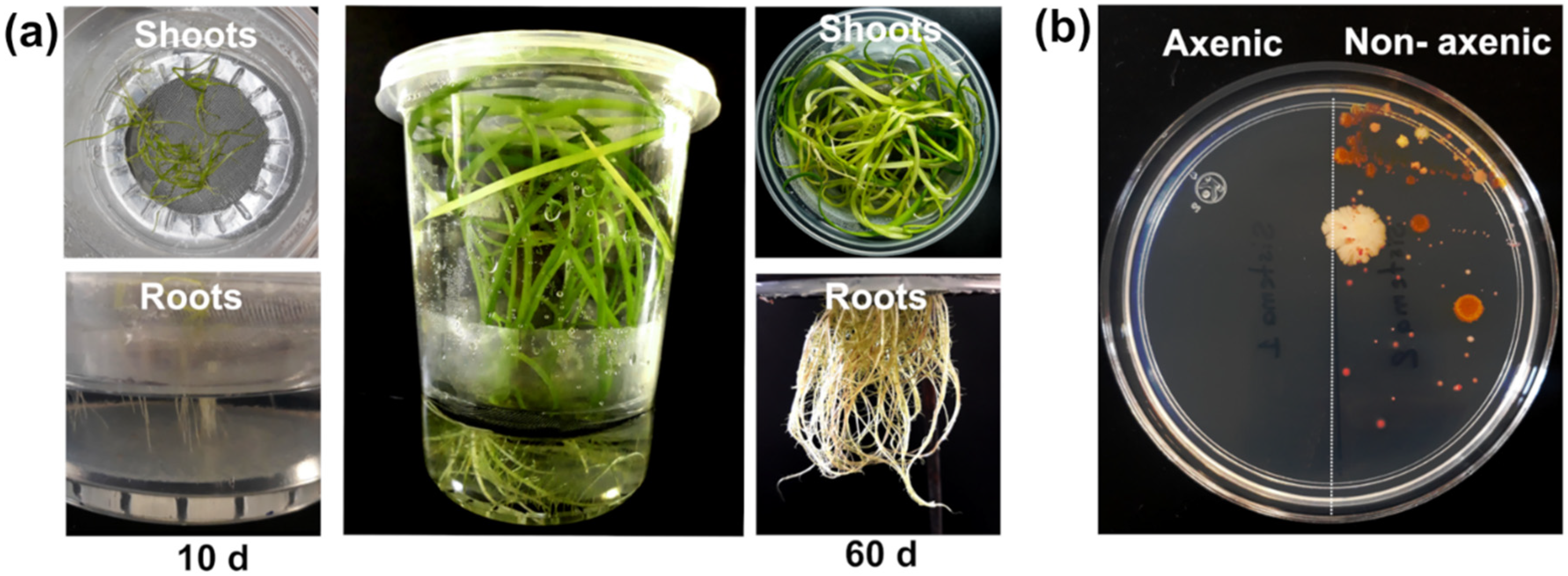
2.2. T. latifolia Culture under Axenic Hydroponic Conditions

Axenic T. latifolia plants have been previously obtained by in vitro micropropagation from mature inflorescences [19,20], leaf sections [20], and germinated seedlings [21,22]. In this work, axenic T. latifolia plants were obtained by seed germination in 0.2× Murashige and Skoog (MS) agar. Under in vitro conditions, the T. latifolia seeds showed 60% germination (Figure S1a), and ten days after sowing showed more vigorous growth of shoots in the 0.2× MS agar (Figure S1b). The T. latifolia seedlings were transferred to a sterile hydroponic system that contained 0.2× MS liquid medium supplemented with glucose as the carbon source. The T. latifolia plants developed shoots and roots in the hydroponic system after 10 days of growth, while after 60 days of growth T. latifolia plants showed good shoot and root development without visual stress signs (Figure 1a). The initial hydroponic solution was maintained during plant development for 60 days. After incubation, no bacterial or fungal contamination was detected in the hydroponic solution, indicating the aseptic conditions of the system (Figure 1b). In contrast with previous studies to obtain axenic T. latifolia plants [20,21,23], our hydroponic system did not use phytohormones for plant differentiation, reducing the time to obtain axenic cultures, and allowing us to carry out research with T. latifolia plants under nutrition-controlled conditions [24].

2.3. Chemical Modeling of Cd Bioavailability in Hydroponics without Glucose

Heavy metal toxicity in plants is closely related to their bioavailability in the hydroponic solution and is influenced by pH, organic matter, ligands, and heavy metal chemical speciation [25]. Complete MS medium has been previously used to evaluate Pb, chromium (Cr), and manganese (Mn) removal by in vitro root culture of T. latifolia [26]. In this work, a diluted MS medium (0.2× strength) without glucose was used to ensure Cd bioavailability in the liquid medium. The prediction analysis by chemical modeling showed that all the CdCl2 was available in different speciation forms, and no Cd precipitation occurred (Table 1). According to our analysis, the ionic form Cd2+ was the predominant species in the 0.2× MS medium at pH 5.7, which was available at 66, 75, and 79% when supplemented with 10, 20, and 40 mg/L of Cd (provided as CdCl2), respectively (Table 1). These levels of available Cd are lower than those observed in the Hoagland solution (pH 6.0) where Cd2+ availability remains between 80–81% when the solution contains 5–100 µM CdCl2 (0.9–18.3 mg/L) [27].

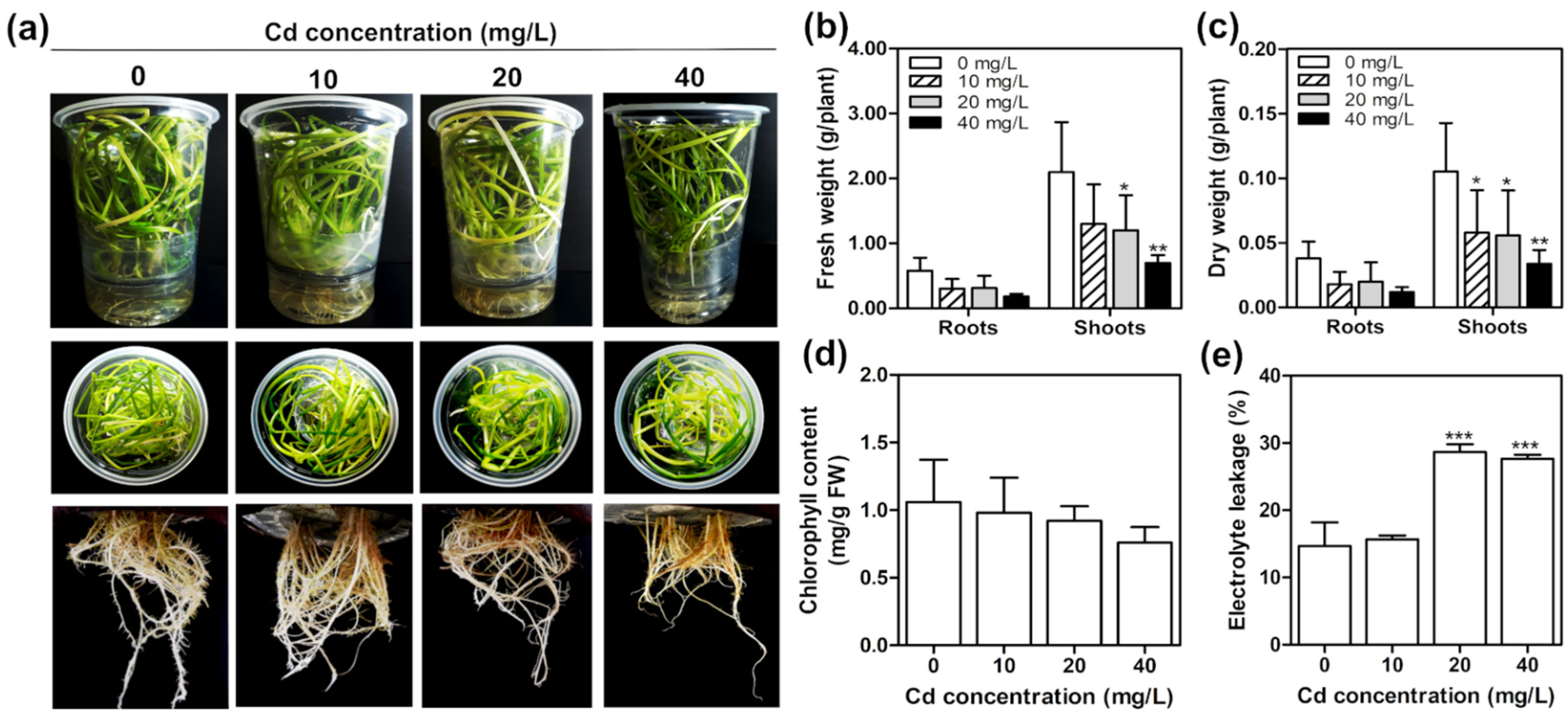
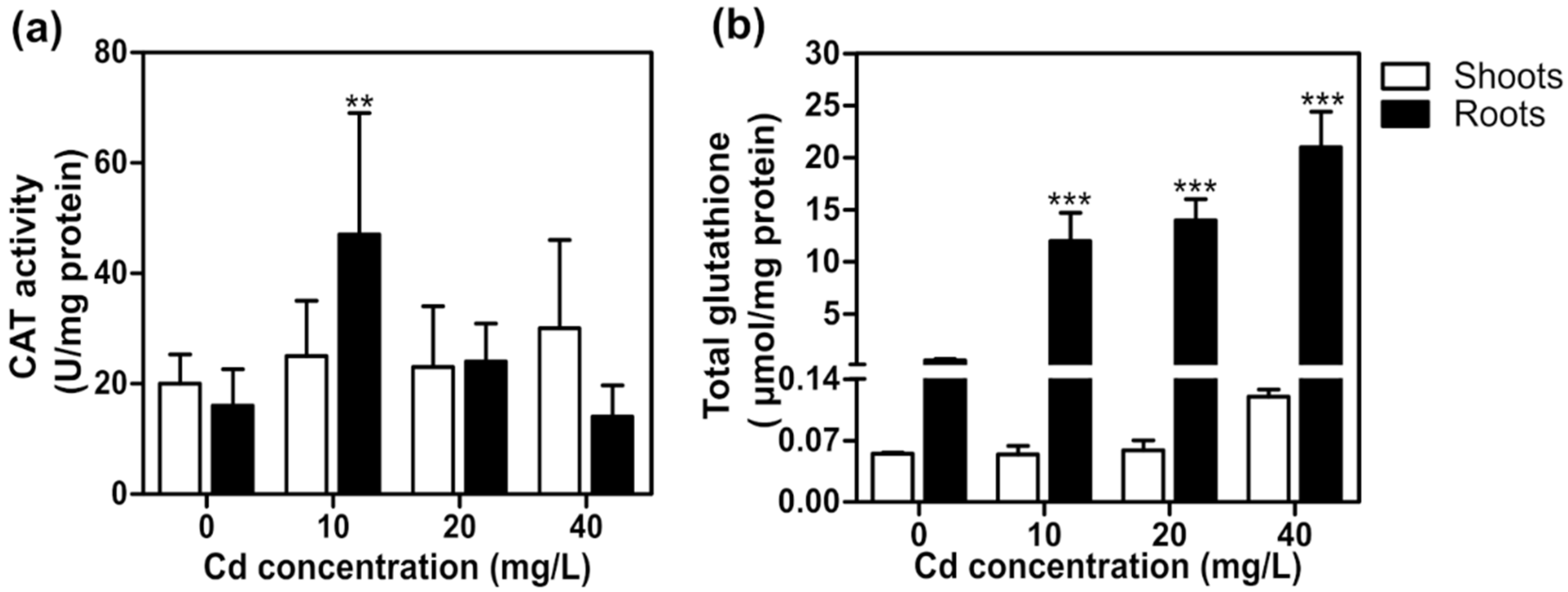
2.4. Cadmium Toxicity Effects on T. latifolia in Hydroponic Culture

Sixty-day-old T. latifolia plants were exposed to 10, 20, and 40 mg/L of Cd under aseptic hydroponic conditions for ten days. Although a reduction in shoot biomass in a Cd dose-dependent manner was observed after Cd exposure (Figure 2b,c), the Cd concentrations tested did not significantly affect either the phenotype or the chlorophyll content of T. latifolia plants (Figure 2a,d). A reduction in dry shoot weight could be due to a significant decrease in the surface area of the leaves caused by the Cd, similar to that observed in Triticum aestivum leaves [28].
Electrolyte leakage is a parameter that measures damage to the cell membrane during plant stress [29]. In the present study, a significant increase in root electrolyte leakage was found in plants exposed to 20 and 40 mg/L Cd (Figure 2e). Previous studies have reported that electrolyte leakage is induced by the increase of reactive oxygen species (ROS) [30]. For instance, in T. angustifolia, Cd exposure increases superoxide (O2) production in a concentration-dependent manner [31]. It is known that ROS activates membrane ion channels that release intracellular potassium (K+) ions, stimulating intracellular hydrolytic enzymes that lead to cell death [30,32]. Although ROS levels were not determined in this study, the results suggest that the root electrolyte leakage in T. latifolia is induced by the ROS generated in the plants exposed at 20 and 40 mg/L of Cd.
Enzymatic and non-enzymatic antioxidant systems are induced in the plant cells to maintain ROS balance [33]. This study evaluated antioxidant defense by quantifying catalase (CAT) activity and total glutathione (GSH) content. Catalase is one of the main antioxidant enzymes that reduce ROS damage by decreasing intracellular hydrogen peroxide (H2O2) levels [33]. We observed that CAT activity increased two-fold in T. latifolia roots exposed to 10 mg/L of Cd, but no significant changes were detected in shoots and roots exposed to 20 or 40 mg/L of Cd (Figure 3a). Similar results were observed in Sassafras tzumu and Triticum aestivum plants exposed to Cd [34,35]. The increase in CAT activity at low Cd levels (10 mg/L) may be attributed to increased ROS production induced by Cd oxidative stress, similar to that observed in T. aestivum [35]. On the other hand, the low CAT activity at high Cd concentrations could result from Cd ions interacting with amino acids of the CAT active center, preventing CAT binding to H2O2 by steric hindrance conditions [36]. These results showed that low Cd levels induce CAT activity, while high Cd levels inhibit CAT activity in T. latifolia plants.
Glutathione (GSH) is an antioxidant compound that regulates H2O2 levels by the ascorbate–glutathione pathway in plants [37]. GSH is also a precursor of phytochelatins, which form complexes with heavy metals and transport them to vacuoles wherein they are sequestered and fixed as nontoxic elements [37]. We observed that Cd stress induces the synthesis of total GSH in T. latifolia only in root tissues. In plants exposed to 10, 20, and 40 mg/L of Cd, total GSH content in roots increased 21-, 24-, and 36-fold compared to non-exposed plants (Figure 3b). These results are in line with those previously found in T. angustifolia, where Cd exposure increased GSH and phytochelatin synthesis to decrease Cd phytotoxicity [1,38]. Our results are consistent with the hypothesis that GSH accumulation could be associated with Cd tolerance in T. latifolia plants.

2.5. Cd Accumulation and Removal by T. latifolia Plants in the Hydroponic System

Cd accumulation in T. latifolia roots and shoots was determined after ten days of Cd exposure under hydroponic conditions. In both organs, Cd levels increased in a dose-dependent manner. The Cd content in roots ranged from 2970 ± 65 to 3520 ± 105 mg/Kg of dry weight (DW), while in the shoots, it was from 406 ± 59 to 739 ± 24 mg/Kg DW (Table 2). The results showed that Cd concentration in T. latifolia roots was from 4.7 to 7.3-fold higher than in shoots, suggesting a low efficiency of Cd translocation. Cd levels in T. latifolia roots were higher than those reported in T. latifolia (1230–2339 mg/Kg) [39], T. angustifolia (1213–2977 mg/Kg) [40], and T. orientalis (300–1100 mg/Kg) [41] where other culture conditions were used.
Bioconcentration (BCF) and translocation (TF) factors are two indicators used to evaluate the plant’s ability to accumulate heavy metals [42]. BCF refers to the metal absorbed by the plant in the roots from the substrate, while TF indicates the ability of the plant to transport the metal from roots to shoots [43]. In this study, T. latifolia BCF ranged from 126 ± 16 to 509 ± 96, while TF ranged from 0.18 ± 0.04 to 0.24 ± 0.01 for Cd (Table 2). These results indicated that T. latifolia plants took up Cd from the hydroponic medium accumulating in their roots and did not efficiently translocate it to the shoots. These results make sense with total chlorophyll and total GSH content detected in shoots that seemed not significantly changed by Cd exposure. Similar values of TF and BCF have been reported in T. angustifolia [31,38,40] and T. orientalis [41]. The low Cd-translocation efficiency in this study could be explained by the findings of Xu, et al. [38] in T. angustifolia where the Casparian band present in plant roots prevents Cd mobilization towards vascular tissues, reducing Cd accumulation in shoots.
Moreover, the level of Cd in the hydroponic solution was determined. Overall, T. latifolia plants removed 37, 43, and 22% of the Cd from the growth media when exposed to 10, 20, and 40 mg/L of Cd, respectively (Table 2). The results showed that the highest Cd removal efficiency occurred at lower concentrations (10 and 20 mg/L of Cd). Similarly, Alonso-Castro, et al. [39] reported that young T. latifolia plants removed 41 and 38% of Cd from aqueous solution when exposed to solutions containing 5.0 and 7.5 mg/L of Cd, respectively.

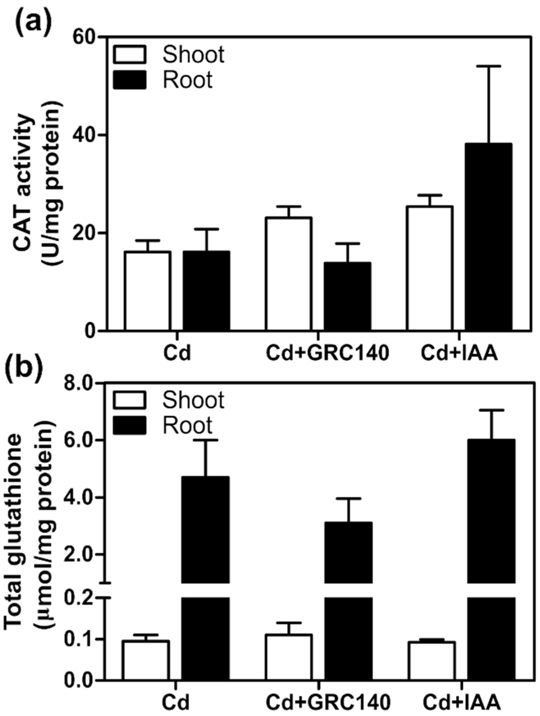
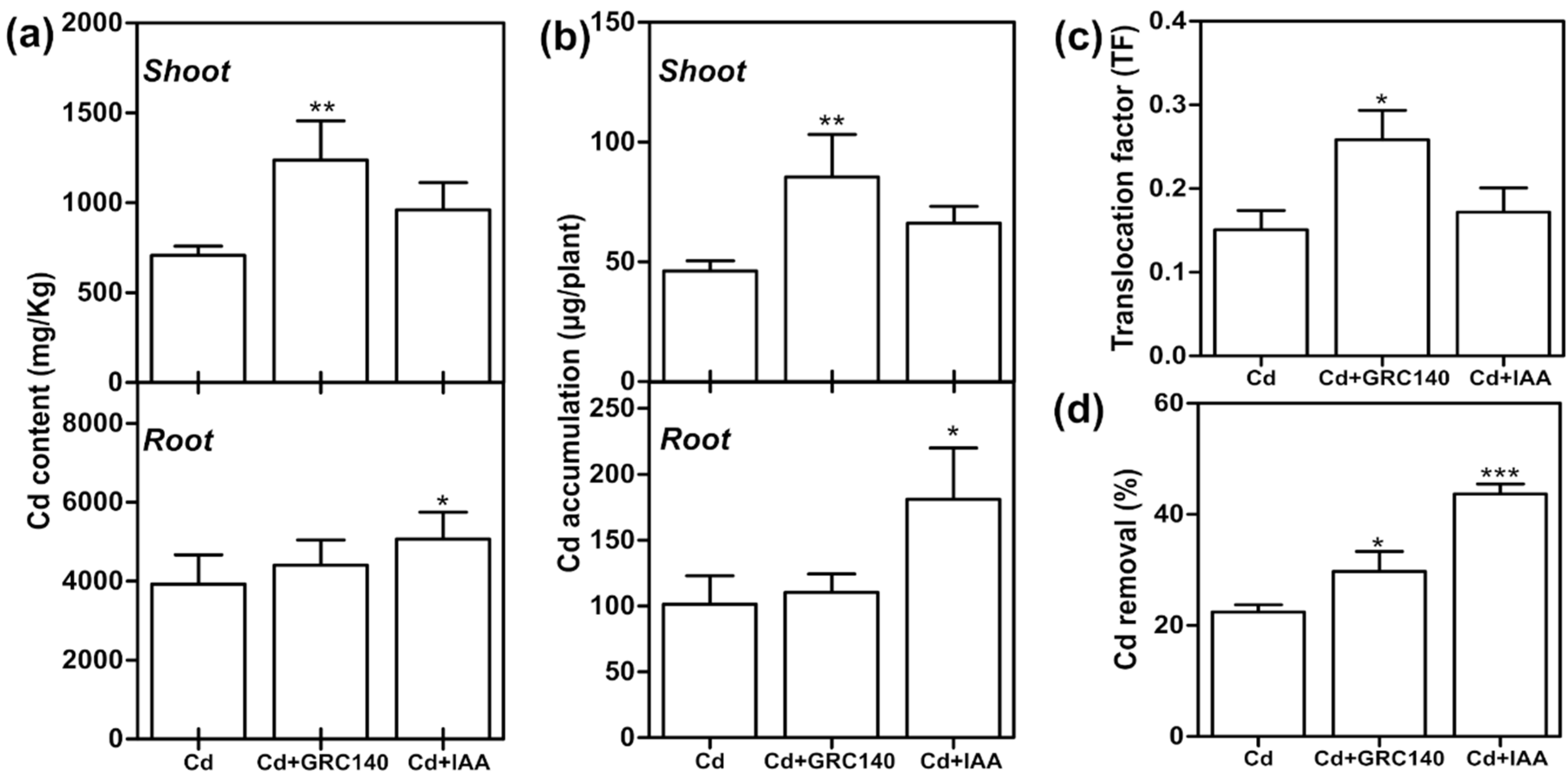
2.6. Effect of P. rhodesiae GRC140 and IAA on T. latifolia Exposed to Cd

Different studies have shown that heavy metal-tolerant plant growth-promoting rhizobacteria (PGPR) improve the phytoremediation processes [44], reduce heavy metal stress in plants, and contribute to plant growth by producing bacterial auxins [9].
Here, we also tested the potential effects of auxin-producing P. rhodesiae GRC40 and exogenous IAA on Cd removal capacity by Typha plants. For this, the bacterial density in the hydroponic medium and bacterial colonization of T. latifolia roots were determined. At 16 days post inoculation (dpi), the number of P. rhodesiae GRC140 in the hydroponic medium was log10 6.6 ± 0.2, while in T. latifolia roots, it was log10 3.9 ± 0.02. No bacterial morphotypes other than P. rhodesiae GRC140 were observed. These results showed that P. rhodesiae GRC140 remains viable in the hydroponic systems and can colonize T. latifolia roots in the presence of 40 mg/L of Cd. Similar results were reported by Wu, et al. [8], who observed that Pseudomonas fluorescens colonizes the roots of Sedum alfredii plants exposed to 25 µM (5.9 mg/L) of Cd(NO3)2.
After Cd exposure, T. latifolia plants inoculated with P. rhodesiae GRC140 (Cd + GRC140 treatment) showed a phenotype similar to plants of Cd + IAA and Cd treatments (Figure 4a), and we did not detect significant changes in fresh weight, dry weight, and electrolyte leakage (Figure 4b,c,e), while exogenous IAA significantly increased only the total chlorophyll content in T. latifolia plants exposed to Cd (Figure 4d). These results agree with Zhou, et al. [45], who observed that exogenous IAA application increased total chlorophyll content in Cinnamomum camphora treated with 30 mg/Kg of Cd.
Auxin-producing bacteria have been reported to protect plants against oxidative stress caused by Cd, increasing the activity of antioxidant enzymes and the synthesis of antioxidant molecules [9]. Therefore, we determined the effect of P. rhodesiae GRC140 and exogenous IAA on CAT activity and GSH content of T. latifolia plants exposed to Cd. Plants inoculated with P. rhodesiae GRC140 did not have differences in the CAT activity and total glutathione content in the roots and shoots of T. latifolia with respect to control plants (Cd) (Figure 5a,b). Likewise, similar results were observed in T. latifolia plants treated with IAA (Figure 5a,b). In our experimental conditions, exogenous IAA and P. rhodesiae GRC140 did not increase the antioxidant defense system in T. latifolia exposed to Cd.

2.7. Effect of P. rhodesiae GRC140 and IAA on Cd Uptake

The Cd content in T. latifolia plants treated with P. rhodesiae GRC140 or exogenous IAA was analyzed by atomic absorption spectrophotometry (AAS). As shown in Figure 6, P. rhodesiae GRC140 increased two-fold the Cd content, and Cd accumulation in shoots compared to the control treatment (Cd) but had no significant effect on the Cd content or Cd accumulation in roots (Figure 6a,b). Furthermore, P. rhodesiae GRC140 inoculation significantly increased the TF value (Figure 6c). These data indicated that P. rhodesiae GRC140 increased Cd translocation in T. latifolia plants. A similar effect has been observed in Miscanthus floridulus Lab and S. alfredii plants inoculated with Lelliottia jeotgali and Sphingomonas, respectively [46,47]. An increase in Cd translocation by P. rhodesiae GRC140 may be attributed to an increase in the gene expression of Cd transporters similar to that observed in S. alfredii [47]. In S. alfredii, Sphingomonas upregulated NRAMP1 and IRT1 genes encoding metal transporters involved in Cd translocation in Oryza sativa [48,49]. However, further research is necessary to understand how P. rhodesiae GRC140 may enhance Cd translocation from roots to shoots in T. latifolia plants.
On the other hand, exogenous IAA application increased the content and the accumulation of Cd in T. latifolia roots but did not affect the Cd translocation factor, suggesting that IAA application increases Cd immobilization in T. latifolia plant roots (Figure 6a–c). This effect may be due to IAA increasing the synthesis of cell wall components upon which Cd fixation occurs [11]. Our results are similar to those observed in A. thaliana [11] and Solanum nigrum [50].
After ten days of incubation, the level of Cd in the hydroponic solution significantly decreased in all treatments (p < 0.05). In T. latifolia plants, the treatments with IAA or P. rhodesiae GRC140 increased by 22 and 8%, the percentage of Cd removal, respectively (Figure 6d). Similar to our results, the inoculation of Cd-resistant microorganisms increased in a 10% removal of Cd2+ by Phragmites australis in constructed wetlands [51]. Overall, Cd removal from axenic hydroponic systems is consistent with Cd accumulation by T. latifolia plants treated with IAA and P. rhodesiae GRC140.

3. Materials and Methods

3.1. Plant Material Sampling and Authentication

Inflorescences and leaves of Typha plants were collected from an artificial wetland at the Faculty of Chemical Sciences, UASLP (San Luis Potosí, SLP, Mexico). Molecular authentication of plants was carried out as follows. DNA was obtained from 100 mg of leaves using the CTAB method previously reported [52]. PCR reaction was performed using SuperMix High Fidelity according to the manufacturer’s instructions (Invitrogen, Waltham, MA, USA). The primers psbA 3F (5′GTTATGCATGAACGTAATGCTC3′) and trnH 3R (5′CGCGCATGGTGGATTCACAATCC3′) were used to amplify the psbA-trnH intergenic spacer region located in the plant chloroplast genome [18]. PCR conditions were 95 °C for 10 min; 35 cycles of 95 °C, 30 s; 50 °C, 30 s; 72 °C for 1 min, and a final extension at 72 °C for 5 min. PCR product was purified and sequenced at Biotechnology Institute, UNAM (Cuernavaca, Morelos, Mexico). The sequence obtained was aligned at GeneBank to determine similarity in the NCBI database.

3.2. Establishment and Cultivation of Axenic T. latifolia Hydroponic Culture

Mature seeds of T. latifolia were surface sterilized with 50% sodium hypochlorite solution and 0.02% Triton X-100 (Sigma-Aldrich, St. Louis, MO, USA) for 15 min, and rinsed six times with sterile distilled water [53]. Then, seeds were germinated on culture flasks with 25 mL MS agar containing Murashige and Skoog (MS; Sigma-Aldrich, St. Louis, MO, USA) salts at 0.2× strength, 3.5 mM 4-Morpholineethanesulfonic acid (MES; pH 5.7, Sigma-Aldrich, St. Louis, MO, USA), 1.0% (w/v) glucose, and 0.8% (w/v) Phytagar (Sigma-Aldrich, St. Louis, MO, USA). Culture flasks were incubated at 28 °C under fluorescent light and a photoperiod of 16/8 h light/darkness. Germinated seeds were aseptically transferred to axenic hydroponic systems (three seeds per system) containing 150 mL of 0.2× MS liquid medium, buffer MES (3.5 mM, pH 5.7), and 1.0% (w/v) glucose [13]. Culture systems were incubated at 28 °C under fluorescent white light with a 16 h light/8 h dark photoperiod for 60 days. Axenic condition of the culture systems was evaluated by plating 20 µL aliquot of the plant-growth medium on LB agar. The LB plates were incubated at 30 °C for five days and then analyzed for microorganism growth.

3.3. Cadmium Plant Treatments and Chemical Modeling of Cd Bioavailability

Sixty-day-old T. latifolia plants, previously grown in hydroponics, were transferred to a hydroponic system with 0.2× MS medium without glucose for six days for acclimatization. After incubation, the systems (n = 3, 12 total systems) were transferred in 0.2× MS liquid medium without glucose at pH 5.7 supplemented with 0, 10, 20, and 40 mg/L of Cd (CdCl2) (Fermont, Monterrey, Mexico). Culture systems were incubated at 28 °C for ten days under fluorescent white light with a 16 h light/8 h dark photoperiod. After Cd exposure, T. latifolia plants were harvested and fresh and dry weight, total chlorophyll content, root electrolyte leakage, catalase activity, and total glutathione were determined. The total Cd content of roots, shoots, and the hydroponic medium was also determined. Cadmium speciation in the 0.2× MS liquid medium was predicted using the software Visual MINTEQ (Version 3.1, KTH, Stockholm, Sweden). Cd speciation was determined at pH 5.7, introducing into the Software data of ionic concentration of each component of MS basal salts (Catalog M5524; Sigma-Aldrich, St. Louis, MO, USA) and MES buffer. The prediction was made at 10, 20, and 40 mg/L of Cd (CdCl2).

3.4. P. rhodesiae GRC140 and IAA Treatments in Plants Exposed to Cd

P. rhodesiae GRC140 was previously isolated from T. latifolia roots by Rolón-Cárdenas, et al. [12]. The bacterial strain was maintained at 4 °C in Luria Bertani (LB) agar plates. For the plant–bacteria interaction, P. rhodesiae GRC140 was grown in LB broth and incubated at 30 °C overnight. After incubation, bacterial cells were harvested by centrifugation at 2450 g for 15 min. The bacterial cells were resuspended in 0.01 M MgSO4 solution and adjusted to OD600 = 1.0 (approximately 109 CFU/mL).
Sixty-day-old T. latifolia plants (n = 3, 9 total systems) were transferred to 0.2× MS medium without glucose. The hydroponic systems were inoculated with 1.5 mL of 0.01 M MgSO4 solution (control), 1.5 mL of P. rhodesiae GRC140 suspension in 0.01 M MgSO4 and supplemented with indole-3-acetic acid (IAA) at a final concentration of 0.1 µM. Six days after incubation, the medium of all systems was replaced with 0.2× MS medium supplemented with a final concentration of 40 mg/L of Cd (CdCl2). The systems were incubated in a plant growth chamber for 10 days in the conditions described above.
After Cd exposure, T. latifolia plants were harvested, and fresh and dry weight, total chlorophyll content, root electrolyte leakage, catalase activity, and total glutathione were determined. The total Cd content of roots, shoots, and hydroponic medium, was also determined as described below.

3.5. Chlorophyll Quantification

Chlorophyll (Chl) content was determined in the leaves of control and treated plants. Chl extraction of T. latifolia plants was carried out according to Lichtenthaler and Buschmann [54]. Fresh leaves (100 mg) were homogenized with 5 mL of acetone, and the extract was kept in darkness for 24 h at 4 °C. The extract absorbance was measured using a UV–Vis spectrophotometer (Evolution 221, Thermo Scientific, Waltham, MA, USA) at 644.8 nm (A644.8) and 661.6 nm (A661.6). Chl a and Chl b were calculated according to Equations (1) and (2), respectively [55].
Chl   a = 11.24   A 661.6 2.040   A 644.8
Chl   b = 20.12   A 644.8 4.19   A 661.6
Total Chl content was defined as the sum of Chl a + Chl b. Data were normalized and expressed as mg/g of fresh weight (FW).

3.6. Electrolyte Leakage

The electrolyte leakage was determined in the roots of controls and treated plants. The roots were washed with 0.1 M ethylenediaminetetraacetic acid (EDTA) for 15 min to remove any the surface-adsorbed metal and rinsed in deionized water to remove surface ions. Root electrolyte leakage was determined using the electrical conductivity method according to Umnajkitikorn, et al. [56]. The root samples (100 mg) were immersed in 10 mL of deionized water at 25 °C for 1 h. The initial electrical conductivity (ECi) of samples was determined using a conductivity meter (PC2700 Oakton, Cole-Parmer, Vernon Hills, IL, USA). Then, samples were incubated at 20 °C for 24 h, and the final electrical conductivity (ECf) was measured. To determine the total electrical conductivity (ECt), the samples were heated at 120 °C in an autoclave for 15 min. Samples were cooled at 25 °C, and electrical conductivity was determined. Root electrolyte leakage was calculated with the formula:
%   EL = ( ECf ECi ECt ECi ) × 100
where ECi: initial electrical conductivity, ECf: final electrical conductivity, ECt: total electrical conductivity.

3.7. Catalase Activity and Total Glutathione Content

Catalase (CAT) activity and glutathione (GSH) content were determined in the shoots and roots of control and treated plants. Fresh plant tissue (250 mg) was powdered in liquid nitrogen and homogenized with ice-cold 500 uL of 100 mM potassium phosphate buffer pH 7.5 containing 5 mM EDTA (KPE buffer). Samples were centrifuged at 16,000× g for 5 min, and the supernatants were collected and used for catalase activity, glutathione total, and protein content.
Total protein was quantified using a standard curve of bovine serum albumin (BSA, Sigma-Aldrich, St. Louis, MO, USA) and Bradford’s reagent (Sigma-Aldrich, St. Louis, MO, USA). Samples were adjusted at 50 µg/mL of total protein, and the catalase activity was determined according to Aebi [57]. A 25 µL aliquot of protein extract was added to the reaction mixture containing 50 mM phosphate buffer (pH 7.0) and 10 mM H2O2. Decomposition of H2O2 was monitored by UV–Vis spectroscopy at 240 nm. Enzymatic activity was calculated using the molar extinction coefficient of H2O2 (39.4 M−1 cm−1). One CAT unit was defined as the amount of enzyme required to decompose 1 µmol of H2O2 per minute [58]. CAT activity was normalized with protein content and expressed as U/mg protein.
The total glutathione content was determined using the enzymatic recycling method reported by Rahman, et al. [59]. The protein extract was mixed with sulfosalicylic acid (10%) and Triton X-100 (0.1%) prepared in a KPE buffer. A 100 µL aliquot of protein extract was added to the reaction mixture of 0.05 mM NADPH, 50 U/mL of glutathione reductase (GR, Sigma-Aldrich, St. Louis, MO, USA), and 0.1 mM DTNB, in 100 mM KPE buffer. The reaction mixture was incubated for 10 min, and the absorbance was quantified at 412 nm using a UV–Vis spectrometer. GSH concentration was determined using a standard curve of GSH (Sigma-Aldrich, St. Louis, MO, USA). GSH content in each sample was normalized with the total protein content and expressed in µmol/mg protein.

3.8. Cadmium Analysis

After ten days of Cd exposure, the hydroponic solution was removed and acidified to pH 2.0 with concentrated HNO3 (Sigma-Aldrich, St. Louis, MO, USA; purity: ≥99.999%). Then, the roots of the control and treated T. latifolia plants were washed with 0.1 M EDTA for 15 min, separated from the shoots, and both samples were dried at 70 °C for 24 h. Dried roots were digested with concentrated HNO3 and 30% H2O2, according to the methodology described by Rodríguez-Hernández, et al. [17]. Dried shoots were digested with HCl (Sigma-Aldrich, St. Louis, MO, USA; purity: ≥99.999%) and HNO3 mixture (3:1 v/v) according to Carranza-Álvarez, et al. [60]. All samples were analyzed by air-acetylene flame AAS (Varian SpectrAA 220FS, Mulgrave, Australia) at a wavelength of 228.8 nm. Cadmium concentration in the samples was estimated using a standard curve of Cd. Cd concentration was normalized with the dry weight in the plant samples and expressed in mg/Kg DW. Cd was also quantified in initial and residual media. All these data obtained were used to calculate the removal percentage (%), the translocation factor (FT), and the bioconcentration factor (BCF). Cd removal percentage was calculated according to the following formula:
%   Removal = ( Ci Cf ) Ci × 100
where Ci is the Cd (mg/L) concentration in the initial medium, Cf is the concentration of residual Cd (mg/L) in the growth medium after treatment.
Translocation factor (TF) was calculated by dividing the concentration of total Cd (mg/Kg DW) in shoots by the concentration of total Cd (mg/Kg DW) in roots [31]. Bioconcentration factor (BCF) was determined by dividing the total Cd in roots (mg/Kg DW) by the total Cd present in the solution (mg/L) after treatment [12].

3.9. Bacteria Growth in the Hydroponic Systems and Root Colonization

The number of viable bacteria (CFU) in the hydroponic solution was estimated by serial dilutions according to Miles, et al. [61]. After Cd exposition, the number of bacteria associated with the roots of inoculated plants was determined according to Balsanelli, et al. [62]. The roots (150 mg) were harvested and homogenized aseptically in 1000 µL saline solution (0.85% NaCl). The extract was serially diluted and 10 µL of each dilution was plated on LB agar. The LB plates were incubated at 30 °C for 48 h.

3.10. Statistical Analysis

All experiments were performed in triplicate, and data were expressed as the mean ± standard deviation (SD). Statistical analyses were performed using the GraphPad Prism program (version 5.01, GraphPad Software Inc., San Diego, CA, USA). Statistical differences were determined by one-way ANOVA and Dunnett post hoc tests, or two-way ANOVA with Bonferroni post hoc tests.

4. Conclusions

In the present study, an axenic hydroponic culture of T. latifolia was established and used to evaluate the effect of P. rhodesiae GRC140 and exogenous IAA on plants exposed to Cd. Initially, we observed similar responses to those reported in T. latifolia exposed to Cd. We observed that Cd reduces shoot biomass in a dose-dependent manner and increases of electrolyte leakage and total GSH content in T. latifolia roots exposed at 20 and 40 mg/L of Cd.
On the other hand, P. rhodesiae GRC140 and exogenous IAA increase the removal and accumulation of Cd by T. latifolia. P. rhodesiae GRC140 increases the Cd content in the shoots, while the IAA increases the Cd content in the roots, similarly to that observed in other plant species inoculated with microorganisms and exposed to Cd. The axenic plants of T. latifolia in a hydroponic system could be used to evaluate the ability of the plant to remove other heavy metals and determine the effect of organic compounds in plant growth, and even establish assays of plant–microorganism interaction.

Supplementary Materials

The following supporting information can be downloaded at: https://www.mdpi.com/article/10.3390/plants11111447/s1, Figure S1: T. latifolia seeds germination.

Author Contributions

G.A.R.-C. wrote the manuscript, A.H.-M. conceived of, and supervised the study, A.H.-M. and M.C.A.-D.l.T. contributed lab Facilities, chemicals and supervised the experiments, G.A.R.-C., J.G.M.-M., R.d.M.G.-B., J.R.-S. performed the experimental work, F.A.-C., J.L.A.-G., R.E.S.-G., C.C.-Á., J.R.M.-P. and L.R.A.-M. analyzed the data and critically reviewed the manuscript. All authors revised the manuscript. All authors have read and agreed to the published version of the manuscript.

Funding

The work reported was funded by grants from CONACYT, Programa Presupuestario F003. Fondo Sectorial de Investigación para la Educación, CB2017-2018 A1-S-40454, to Alejandro Hernández-Morales. Gisela Adelina Rolón-Cárdenas (CVU 712240) thanks CONACYT-Mexico for the financial support given to carry out her Ph.D. studies.

Institutional Review Board Statement

Not applicable.

Informed Consent Statement

Not applicable.

Data Availability Statement

The data that support the findings of this study are available from the corresponding author, AHM, upon reasonable request.

Conflicts of Interest

The authors declare no conflict of interest.

References

  1. Liu, Y.; Chen, J.; Lu, S.; Yang, L.; Qian, J.; Cao, S. Increased lead and cadmium tolerance of Typha angustifolia from Huaihe River is associated with enhanced phytochelatin synthesis and improved antioxidative capacity. Environ. Technol. 2016, 37, 2743–2749. [Google Scholar] [CrossRef] [PubMed]
  2. Rizwan, M.; Ali, S.; Rehman, M.Z.u.; Maqbool, A. A critical review on the effects of zinc at toxic levels of cadmium in plants. Environ. Sci. Pollut. Res. 2019, 26, 6279–6289. [Google Scholar] [CrossRef] [PubMed]
  3. Liu, L.; Li, W.; Song, W.; Guo, M. Remediation techniques for heavy metal-contaminated soils: Principles and applicability. Sci. Total Environ. 2018, 633, 206–219. [Google Scholar] [CrossRef] [PubMed]
  4. Nedjimi, B. Phytoremediation: A sustainable environmental technology for heavy metals decontamination. SN Appl. Sci. 2021, 3, 286. [Google Scholar] [CrossRef]
  5. Zhang, X.H.; Tapia, M.; Webb, J.B.; Huang, Y.H.; Miao, S. Molecular signatures of two cattail species, Typha domingensis and Typha latifolia (Typhaceae), in South Florida. Mol. Phylogenetics Evol. 2008, 49, 368–376. [Google Scholar] [CrossRef]
  6. Bonanno, G.; Cirelli, G.L. Comparative analysis of element concentrations and translocation in three wetland congener plants: Typha domingensis, Typha latifolia and Typha angustifolia. Ecotoxicol. Environ. Saf. 2017, 143, 92–101. [Google Scholar] [CrossRef]
  7. Ali, S.; Abbas, Z.; Rizwan, M.; Zaheer, I.E.; Yavaş, İ.; Ünay, A.; Abdel-DAIM, M.M.; Bin-Jumah, M.; Hasanuzzaman, M.; Kalderis, D. Application of floating aquatic plants in phytoremediation of heavy metals polluted water: A Review. Sustainability 2020, 12, 1927. [Google Scholar] [CrossRef] [Green Version]
  8. Wu, Y.; Ma, L.; Liu, Q.; Vestergård, M.; Topalovic, O.; Wang, Q.; Zhou, Q.; Huang, L.; Yang, X.; Feng, Y. The plant-growth promoting bacteria promote cadmium uptake by inducing a hormonal crosstalk and lateral root formation in a hyperaccumulator plant Sedum alfredii. J. Hazard. Mater. 2020, 395, 122661. [Google Scholar] [CrossRef]
  9. Rolón-Cárdenas, G.A.; Arvizu-Gómez, J.L.; Soria-Guerra, R.E.; Pacheco-Aguilar, J.R.; Alatorre-Cobos, F.; Hernández-Morales, A. The role of auxins and auxin-producing bacteria in the tolerance and accumulation of cadmium by plants. Environ. Geochem. Health 2022. [Google Scholar] [CrossRef]
  10. Li, Z.; Zhu, J.; Wang, Y.; Lin, L.; Liao, M.a.; Wang, J.; Deng, Q.; Tang, Y.; Wang, X.; Wang, J. Effects of exogenous indole acetic acid on growth and cadmium accumulation of Cyphomandra betacea seedlings. Int. J. Environ. Anal. Chem. 2020, 102, 771–779. [Google Scholar] [CrossRef]
  11. Zhu, X.F.; Wang, Z.W.; Dong, F.; Lei, G.J.; Shi, Y.Z.; Li, G.X.; Zheng, S.J. Exogenous auxin alleviates cadmium toxicity in Arabidopsis thaliana by stimulating synthesis of hemicellulose 1 and increasing the cadmium fixation capacity of root cell walls. J. Hazard. Mater. 2013, 263, 398–403. [Google Scholar] [CrossRef] [PubMed]
  12. Rolón-Cárdenas, G.A.; Arvizu-Gómez, J.L.; Pacheco-Aguilar, J.R.; Vázquez-Martínez, J.; Hernández-Morales, A. Cadmium-tolerant endophytic Pseudomonas rhodesiae strains isolated from Typha latifolia modify the root architecture of Arabidopsis thaliana Col-0 in presence and absence of Cd. Braz. J. Microbiol. 2020, 52, 349–361. [Google Scholar] [CrossRef] [PubMed]
  13. Alatorre-Cobos, F.; Calderón-Vázquez, C.; Ibarra-Laclette, E.; Yong-Villalobos, L.; Pérez-Torres, C.-A.; Oropeza-Aburto, A.; Méndez-Bravo, A.; González-Morales, S.-I.; Gutiérrez-Alanís, D.; Chacón-López, A.; et al. An improved, low-cost, hydroponic system for growing Arabidopsis and other plant species under aseptic conditions. BMC Plant Biol. 2014, 14, 69. [Google Scholar] [CrossRef] [PubMed] [Green Version]
  14. Kawasaki, A.; Okada, S.; Zhang, C.; Delhaize, E.; Mathesius, U.; Richardson, A.E.; Watt, M.; Gilliham, M.; Ryan, P.R. A sterile hydroponic system for characterising root exudates from specific root types and whole-root systems of large crop plants. Plant Methods 2018, 14, 114. [Google Scholar] [CrossRef] [Green Version]
  15. Hosseinzadeh, S.; Verheust, Y.; Bonarrigo, G.; Van Hulle, S. Closed hydroponic systems: Operational parameters, root exudates occurrence and related water treatment. Rev. Environ. Sci. Biotechnol. 2017, 16, 59–79. [Google Scholar] [CrossRef]
  16. Golan-Goldhirsh, A.; Barazani, O.; Nepovim, A.; Soudek, P.; Smrcek, S.; Dufkova, L.; Krenkova, S.; Yrjala, K.; Schröder, P.; Vanek, T. Plant response to heavy metals and organic pollutants in cell culture and at whole plant level. J. Soils Sediments 2004, 4, 133–140. [Google Scholar] [CrossRef]
  17. Rodríguez-Hernández, M.C.; Bonifas, I.; Alfaro-De la Torre, M.C.; Flores-Flores, J.L.; Bañuelos-Hernández, B.; Patiño-Rodríguez, O. Increased accumulation of cadmium and lead under Ca and Fe deficiency in Typha latifolia: A study of two pore channel (TPC1) gene responses. Environ. Exp. Bot. 2015, 115, 38–48. [Google Scholar] [CrossRef]
  18. Liu, B.; Yang, Y.; Xie, L.; Zeng, G.; Ma, K. Beilschmiedia turbinata: A newly recognized but dying species of lauraceae from tropical asia based on morphological and molecular data. PLoS ONE 2013, 8, e67636. [Google Scholar] [CrossRef]
  19. Czakó, M.; Feng, X.; He, Y.; Liang, D.; Márton, L. Genetic modification of wetland grasses for phytoremediation. Z. Für Nat. C 2005, 60, 285–291. [Google Scholar] [CrossRef]
  20. Estime, L.; O’Shea, M.; Borst, M. Suspension culture and plant regeneration of Typha latifolia. Hort. Sci. 2002, 37, 406–408. [Google Scholar] [CrossRef] [Green Version]
  21. Rogers, S.D.; Beech, J.; Sarma, K.S. Shoot regeneration and plant acclimatization of the wetland monocot Cattail (Typha latifolia). Plant Cell Rep. 1998, 18, 71–75. [Google Scholar] [CrossRef]
  22. Rogers, S.M.D. Tissue culture and wetland establishment of the freshwater monocots Carex, Juncus, Scirpus, and Typha. In Vitro Cell Dev. Biol. Plant 2003, 39, 1–5. [Google Scholar] [CrossRef]
  23. Liu, H.; Hao, N.; Jia, Y.; Liu, X.; Ni, X.; Wang, M.; Liu, W. The ethylene receptor regulates Typha angustifolia leaf aerenchyma morphogenesis and cell fate. Planta 2019, 250, 381–390. [Google Scholar] [CrossRef] [PubMed]
  24. Doran, P.M. Application of plant tissue cultures in phytoremediation research: Incentives and limitations. Biotechnol. Bioeng. 2009, 103, 60–76. [Google Scholar] [CrossRef] [PubMed]
  25. Shahid, M.; Dumat, C.; Khalid, S.; Niazi, N.K.; Antunes, P.M.C. Cadmium Bioavailability, Uptake, Toxicity and Detoxification in Soil-Plant System. In Reviews of Environmental Contamination and Toxicology Volume 241; de Voogt, P., Gunther, F.A., Eds.; Springer International Publishing: Cham, Swizerland, 2017; pp. 73–137. [Google Scholar]
  26. Santos-Díaz, M.d.S.; Barrón-Cruz, M.d.C. Lead, chromium and manganese removal by in vitro root cultures of two aquatic macrophytes species: Typha Latifolia L. and Scirpus Americanus Pers. Int. J. Phytoremediation 2011, 13, 538–551. [Google Scholar] [CrossRef] [PubMed]
  27. Piotto, F.A.; Carvalho, M.E.A.; Souza, L.A.; Rabêlo, F.H.S.; Franco, M.R.; Batagin-Piotto, K.D.; Azevedo, R.A. Estimating tomato tolerance to heavy metal toxicity: Cadmium as study case. Environ. Sci. Pollut. Res. 2018, 25, 27535–27544. [Google Scholar] [CrossRef] [PubMed]
  28. Agami, R.A.; Mohamed, G.F. Exogenous treatment with indole-3-acetic acid and salicylic acid alleviates cadmium toxicity in wheat seedlings. Ecotoxicol. Environ. Saf. 2013, 94, 164–171. [Google Scholar] [CrossRef]
  29. Takashima, K.; Ahmad Nor, A.S.b.; Ando, S.; Takahashi, H.; Kaneko, T. Evaluation of plant stress due to plasma-generated reactive oxygen and nitrogen species using electrolyte leakage. Jpn. J. Appl. Phys. 2020, 60, 010504. [Google Scholar] [CrossRef]
  30. Demidchik, V. ROS-Activated ion channels in plants: Biophysical characteristics, physiological functions and molecular nature. Int. J. Mol. Sci. 2018, 19, 1263. [Google Scholar] [CrossRef] [Green Version]
  31. Ren, M.; Qin, Z.; Li, X.; Wang, L.; Wang, Y.; Zhang, J.; Huang, Y.; Yang, S. Selenite antagonizes the phytotoxicity of Cd in the cattail Typha angustifolia. Ecotoxicol. Environ. Saf. 2020, 189, 109959. [Google Scholar] [CrossRef]
  32. Demidchik, V.; Straltsova, D.; Medvedev, S.S.; Pozhvanov, G.A.; Sokolik, A.; Yurin, V. Stress-induced electrolyte leakage: The role of K+-permeable channels and involvement in programmed cell death and metabolic adjustment. J. Exp. Bot. 2014, 65, 1259–1270. [Google Scholar] [CrossRef] [PubMed]
  33. Liu, S.; Yang, R. Regulations of reactive oxygen species in plants abiotic stress: An integrated overview. In Plant Life under Changing Environment; Tripathi, D.K., Pratap Singh, V., Chauhan, D.K., Sharma, S., Prasad, S.M., Dubey, N.K., Ramawat, N., Eds.; Academic Press: Cambridge, MA, USA, 2020; pp. 323–353. [Google Scholar]
  34. Zhao, H.; Guan, J.; Liang, Q.; Zhang, X.; Hu, H.; Zhang, J. Effects of cadmium stress on growth and physiological characteristics of sassafras seedlings. Sci. Rep. 2021, 11, 9913. [Google Scholar] [CrossRef] [PubMed]
  35. Zhang, T.; Xiao, J.; Zhao, Y.; Zhang, Y.; Jie, Y.; Shen, D.; Yue, C.; Huang, J.; Hua, Y.; Zhou, T. Comparative physiological and transcriptomic analyses reveal ascorbate and glutathione coregulation of cadmium toxicity resistance in wheat genotypes. BMC Plant Biol. 2021, 21, 459. [Google Scholar] [CrossRef] [PubMed]
  36. Wang, J.; Zhang, H.; Zhang, T.; Zhang, R.; Liu, R.; Chen, Y. Molecular mechanism on cadmium-induced activity changes of catalase and superoxide dismutase. Int. J. Biol. Macromol. 2015, 77, 59–67. [Google Scholar] [CrossRef]
  37. Li, G.-Z.; Chen, S.-J.; Li, N.-Y.; Wang, Y.-Y.; Kang, G.-Z. Exogenous glutathione alleviates cadmium toxicity in wheat by influencing the absorption and translocation of cadmium. Bull. Environ. Contam. Toxicol. 2021, 107, 320–326. [Google Scholar] [CrossRef]
  38. Xu, W.; Shi, W.; Yan, F.; Zhang, B.; Liang, J. Mechanisms of cadmium detoxification in cattail (Typha angustifolia L.). Aquat. Bot. 2011, 94, 37–43. [Google Scholar] [CrossRef]
  39. Alonso-Castro, A.J.; Carranza-Alvarez, C.; Alfaro-De la Torre, M.C.; Chavez-Guerrero, L.; Garcia-De la Cruz, R.F. Removal and accumulation of cadmium and lead by Typha latifolia exposed to single and mixed metal solutions. Arch. Environ. Contam. Toxicol. 2009, 57, 688–696. [Google Scholar] [CrossRef]
  40. Woraharn, S.; Meeinkuirt, W.; Phusantisampan, T.; Chayapan, P. Rhizofiltration of cadmium and zinc in hydroponic systems. Water Air Soil Pollut. 2021, 232, 204. [Google Scholar] [CrossRef]
  41. Wang, Y.; Lv, N.; Mao, X.; Yan, Z.; Wang, J.; Tan, W.; Li, X.; Liu, H.; Wang, L.; Xi, B. Cadmium tolerance and accumulation characteristics of wetland emergent plants under hydroponic conditions. RSC Adv. 2018, 8, 33383–33390. [Google Scholar] [CrossRef] [Green Version]
  42. Sidhu, G.P.S.; Bali, A.S.; Singh, H.P.; Batish, D.R.; Kohli, R.K. Phytoremediation of lead by a wild, non-edible Pb accumulator Coronopus didymus (L.) Brassicaceae. Int. J. Phytoremediation 2018, 20, 483–489. [Google Scholar] [CrossRef]
  43. Mishra, T.; Pandey, V.C. Phytoremediation of Red Mud Deposits Through Natural Succession. In Phytomanagement of Polluted Sites; Pandey, V.C., Bauddh, K., Eds.; Elsevier: Amsterdam, The Netherlands, 2019; pp. 409–424. [Google Scholar]
  44. Sharma, P. Efficiency of bacteria and bacterial assisted phytoremediation of heavy metals: An update. Bioresour. Technol. 2021, 328, 124835. [Google Scholar] [CrossRef] [PubMed]
  45. Zhou, J.; Cheng, K.; Huang, G.; Chen, G.; Zhou, S.; Huang, Y.; Zhang, J.; Duan, H.; Fan, H. Effects of exogenous 3-indoleacetic acid and cadmium stress on the physiological and biochemical characteristics of Cinnamomum camphora. Ecotoxicol. Environ. Saf. 2020, 191, 109998. [Google Scholar] [CrossRef] [PubMed]
  46. Liu, S.; Liu, H.; Chen, R.; Ma, Y.; Yang, B.; Chen, Z.; Liang, Y.; Fang, J.; Xiao, Y. Role of two plant growth-promoting bacteria in remediating cadmium-contaminated soil combined with Miscanthus floridulus (Lab.). Plants 2021, 10, 912. [Google Scholar] [CrossRef]
  47. Pan, F.; Luo, S.; Shen, J.; Wang, Q.; Ye, J.; Meng, Q.; Wu, Y.; Chen, B.; Cao, X.; Yang, X.; et al. The effects of endophytic bacterium SaMR12 on Sedum alfredii Hance metal ion uptake and the expression of three transporter family genes after cadmium exposure. Environ. Sci. Pollut. Res. 2017, 24, 9350–9360. [Google Scholar] [CrossRef] [PubMed]
  48. Lee, S.; An, G. Over-expression of OsIRT1 leads to increased iron and zinc accumulations in rice. Plant Cell Environ. 2009, 32, 408–416. [Google Scholar] [CrossRef] [PubMed]
  49. Takahashi, R.; Ishimaru, Y.; Senoura, T.; Shimo, H.; Ishikawa, S.; Arao, T.; Nakanishi, H.; Nishizawa, N.K. The OsNRAMP1 iron transporter is involved in Cd accumulation in rice. J. Exp. Bot. 2011, 62, 4843–4850. [Google Scholar] [CrossRef] [PubMed] [Green Version]
  50. Zulfiqar, U.; Farooq, M.; Hussain, S.; Maqsood, M.; Hussain, M.; Ishfaq, M.; Ahmad, M.; Anjum, M.Z. Lead toxicity in plants: Impacts and remediation. J. Environ. Manag. 2019, 250, 109557. [Google Scholar] [CrossRef]
  51. Yu, G.; Wang, G.; Li, J.; Chi, T.; Wang, S.; Peng, H.; Chen, H.; Du, C.; Jiang, C.; Liu, Y.; et al. Enhanced Cd2+ and Zn2+ removal from heavy metal wastewater in constructed wetlands with resistant microorganisms. Bioresour. Technol. 2020, 316, 123898. [Google Scholar] [CrossRef]
  52. Porebski, S.; Bailey, L.G.; Baum, B.R. Modification of a CTAB DNA extraction protocol for plants containing high polysaccharide and polyphenol components. Plant Mol. Biol. Rep. 1997, 15, 8–15. [Google Scholar] [CrossRef]
  53. Remy, E.; Duque, P. Assessing tolerance to heavy-metal stress in Arabidopsis thaliana seedlings. In Environmental Responses in Plants: Methods and Protocols; Duque, P., Ed.; Springer: New York, NY, USA, 2016; pp. 197–208. [Google Scholar]
  54. Lichtenthaler, H.K.; Buschmann, C. Extraction of Phtosynthetic Tissues: Chlorophylls and Carotenoids. Curr. Protoc. Food Anal. Chem. 2001, 1, F4.2.1–F4.2.6. [Google Scholar] [CrossRef]
  55. Lichtenthaler, H.K.; Buschmann, C. Chlorophylls and Carotenoids: Measurement and Characterization by UV-VIS Spectroscopy. Curr. Protoc. Food Anal. Chem. 2001, 1, F4.3.1–F4.3.8. [Google Scholar] [CrossRef]
  56. Umnajkitikorn, K.; Fukudome, M.; Uchiumi, T.; Teaumroong, N. Elevated nitrogen priming induced oxinitro-responses and water deficit tolerance in rice. Plants 2021, 10, 381. [Google Scholar] [CrossRef] [PubMed]
  57. Aebi, H. Catalase in vitro. Methods Enzymol. 1984, 105, 121–126. [Google Scholar] [CrossRef] [PubMed]
  58. El-Amier, Y.; Elhindi, K.; El-Hendawy, S.; Al-Rashed, S.; Abd-ElGawad, A. Antioxidant system and biomolecules alteration in Pisum sativum under heavy metal stress and possible alleviation by 5-aminolevulinic acid. Molecules 2019, 24, 4194. [Google Scholar] [CrossRef] [Green Version]
  59. Rahman, I.; Kode, A.; Biswas, S.K. Assay for quantitative determination of glutathione and glutathione disulfide levels using enzymatic recycling method. Nat. Protoc. 2006, 1, 3159–3165. [Google Scholar] [CrossRef]
  60. Carranza-Álvarez, C.; Alonso-Castro, A.J.; Alfaro-De La Torre, M.C.; García-De La Cruz, R.F. Accumulation and distribution of heavy metals in Scirpus americanus and Typha latifolia from an artificial lagoon in San Luis Potosí, México. Water Air Soil Pollut. 2008, 188, 297–309. [Google Scholar] [CrossRef]
  61. Miles, A.A.; Misra, S.S.; Irwin, J.O. The estimation of the bactericidal power of the blood. J. Hyg. 1938, 38, 732–749. [Google Scholar] [CrossRef] [Green Version]
  62. Balsanelli, E.; de Oliveira Pedrosa, F.; de Souza, E.M. Quantification of grass colonization by associative bacteria. Curr. Protoc. Plant Biol. 2017, 2, 108–123. [Google Scholar] [CrossRef]
Figure 1. T. latifolia plants in an axenic hydroponic system (a) Side and top view of shoots and roots of ten and sixty-day-old T. latifolia plants in 0.2× MS medium pH 5.7 supplemented with 1.0% glucose. (b) Evaluation of axenic conditions of the nutrient solution after sixty cultivation days of T. latifolia plants.
Plants 11 01447 g001
Figure 2. Cd effect on T. latifolia growth. Sixty-day-old T. latifolia plants were exposed to 10, 20, and 40 mg/L of Cd for ten days. (a) Shoot and root phenotype appearance, (b) fresh weight, (c) dry weight, (d) leaf chlorophyll content, (e) root electrolyte leakage of Cd exposed plants. The values represent the mean ± SD (n = 3). Asterisks indicate significant differences between control (0 mg/L) and treatment (Dunnett’s test; * p < 0.05, ** p < 0.01 and *** p < 0.001).
Plants 11 01447 g002
Figure 3. T. latifolia biochemical responses to Cd. Sixty-day-old T. latifolia plants were exposed to 10, 20, and 40 mg/L of Cd for ten days. (a) Catalase (CAT) activity and (b) total glutathione levels in shoots and roots. The values represent the mean ± SD (n = 3). Asterisks indicate significant differences between control (0 mg/L) and treatment (Dunnett’s test; ** p < 0.01 and *** p < 0.001).
Plants 11 01447 g003
Figure 4. Effect of P. rhodesiae GRC140 and IAA on T. latifolia exposed to Cd. Sixty-day-old T. latifolia plants were treated with IAA (1.0 μM) or inoculated with P. rhodesiae GRC140 (106 CFU/mL) for 6 days and exposed to 40 mg/L of Cd for ten days. (a) Shoot and root phenotype appearance, (b) fresh weight, (c) dry weight, (d) total chlorophyll content, (e) root electrolyte leakage. The values represent the mean ± SD (n = 3). Asterisks indicate significant differences between control (Cd) and treatment (Dunnett’s test; * p < 0.05).
Plants 11 01447 g004
Figure 5. Effect of P. rhodesiae GRC140 and IAA on total glutathione and CAT activity in T. latifolia plants exposed to 40 mg/L of Cd for ten days. The values represent the mean ± SD (n = 3). Asterisks indicate significant differences between control (Cd) and treatment (Dunnett’s test). (a) CAT activity; (b) total glutathione.
Plants 11 01447 g005
Figure 6. Effect of P. rhodesiae GRC140 and IAA on Cd content and Cd accumulation of T. latifolia. The values represent the mean ± SD (n = 3). Asterisks indicate significant differences between control (Cd) and treatment (Dunnett’s test; * p < 0.05, ** p < 0.01 and *** p < 0.001). (a) Cd content, (b) Cd accumulation, (c) translocation factor, (d) Cd removal.
Plants 11 01447 g006
Table 1. Cd speciation in 0.2× MS medium at pH 5.7 predicted by Visual MINTEQ 3.1.
Species (%)Cd Concentration (mg/L)
102040
Cd2+66.7075.8679.70
CdCl+5.607.119.03
CdCl2 (aq)0.030.040.06
CdSO4 (aq)1.902.172.21
CdNH32+0.030.030.03
CdNO3+1.061.201.25
CdHPO4 (aq)1.421.601.64
CdEDTA2−23.1411.946.06
CdHEDTA0.070.040.02
Dissolved (%)100100100
Precipitated (%)000
aq: aquo complex.
Table 2. Cd content in shoots and roots of T. latifolia plants exposed to Cd for 10 days.
Cd Concentration (mg/L)Cd Content (mg/Kg DW)BCFTFCd Removal (%)
ShootsRoots
0ndndndndnd
10406 ± 59 b2970 ± 65 c509 ± 96 a0.18 ± 0.04 a37 ± 3.8 a
20668 ± 73 a3275 ± 15 b340 ± 38 b0.24 ± 0.01 a43 ± 6.3 a
40739 ± 24 a3520 ± 105 a126 ± 16 c0.23 ± 0.03 a22 ± 5.3 b
BCF: bioconcentration factor; TF: translocation factor; nd: not detected. Values represent the mean ± SD (n = 3). Different letters represent statistically different means (p < 0.05, Tukey’s test).
Publisher’s Note: MDPI stays neutral with regard to jurisdictional claims in published maps and institutional affiliations.

