
A Comprehensive Study of the Antibacterial Activity of Bioactive Juice and Extracts from Pomegranate (Punica granatum L.) Peels and Seeds


Abstract
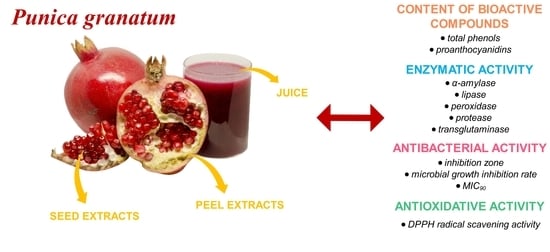
1. Introduction
2. Results
2.1. Enzymatic Activity of P. granatum Fruit
2.2. Content of Other Secondary Metabolites in P. granatum Fruit
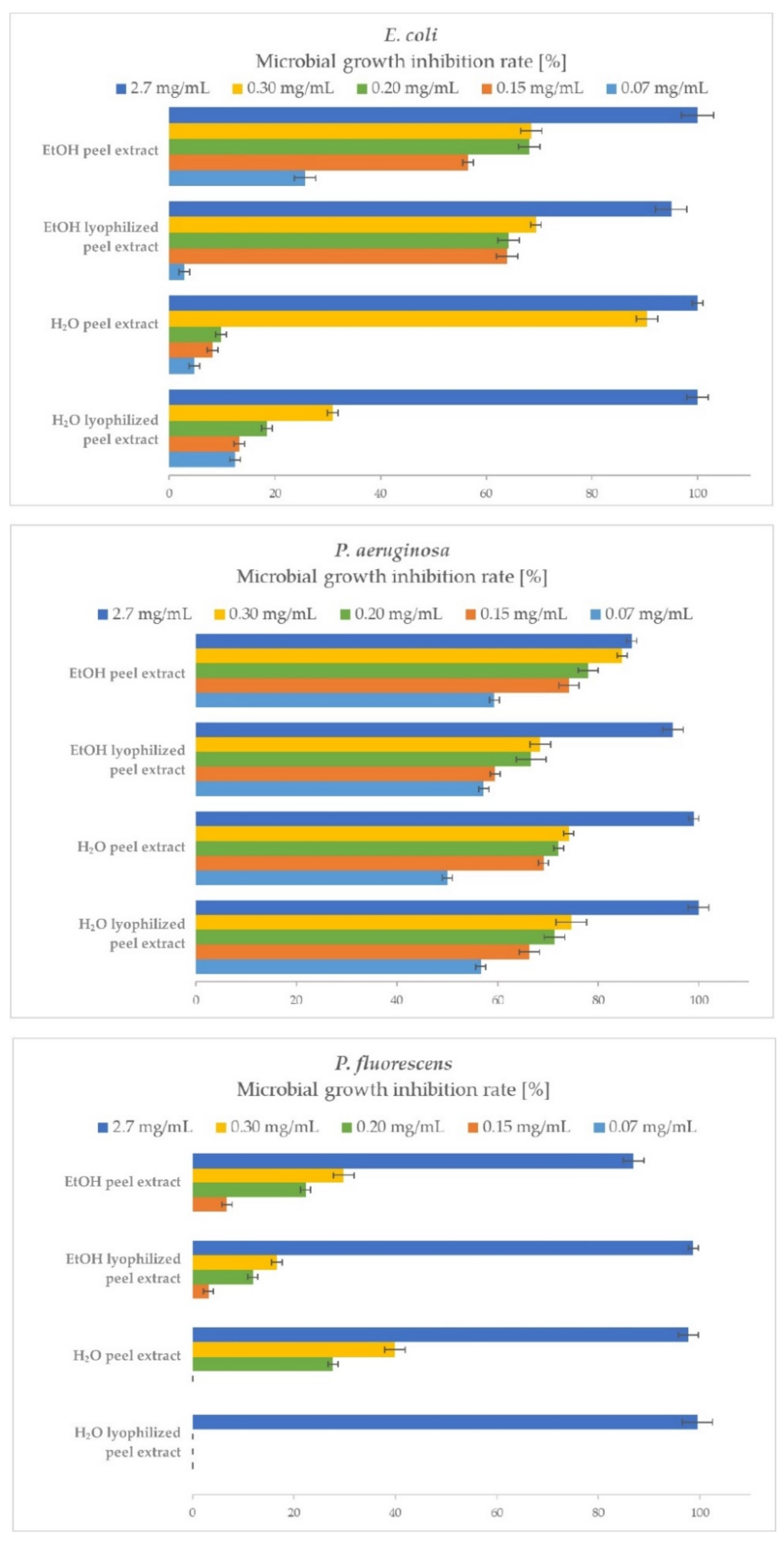
2.3. Antibacterial Activity of P. granatum Extracts
2.3.1. Gram-negative Bacteria
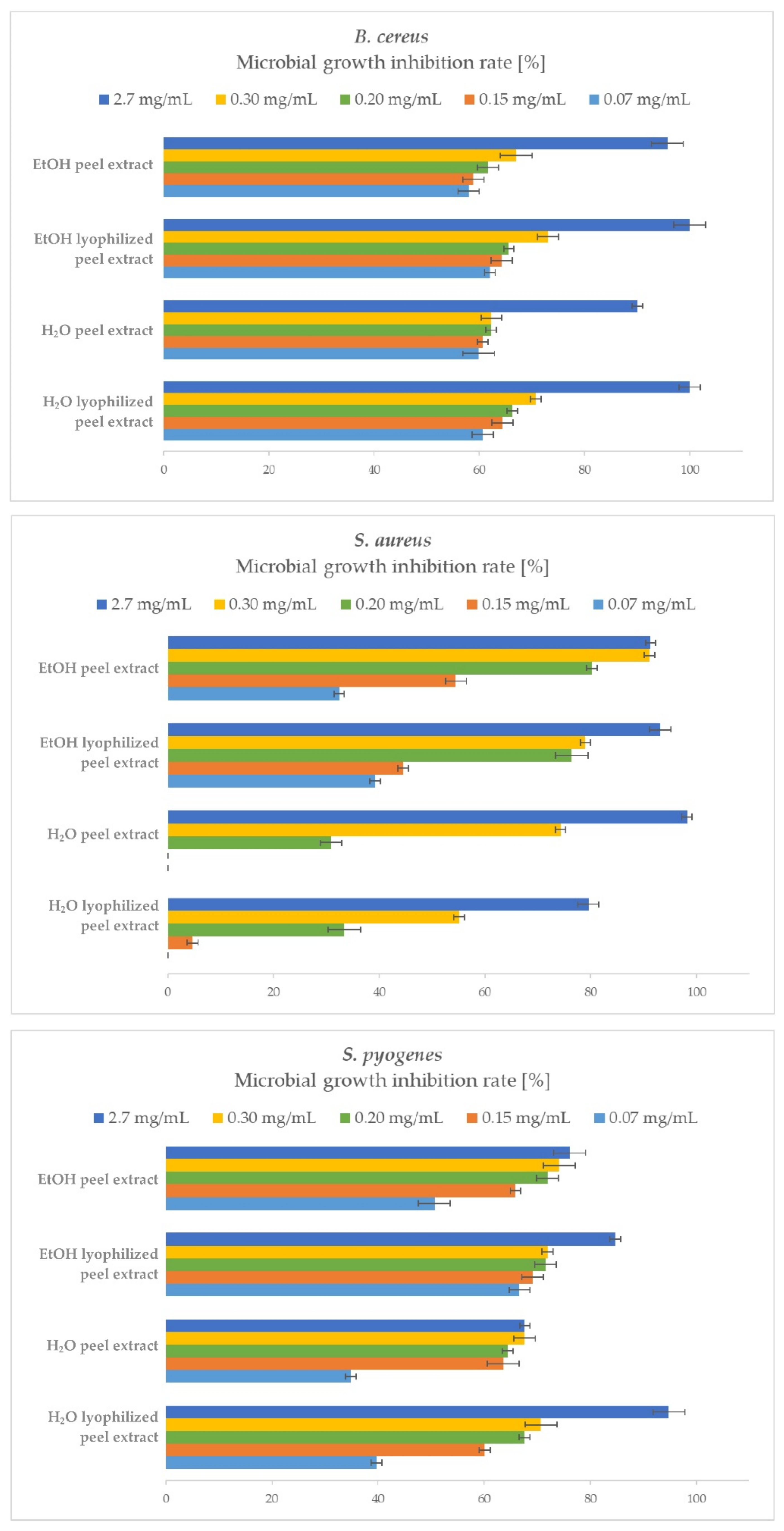
2.3.2. Gram-Positive Bacteria
2.4. The Most Promising P. granatum Samples for Different Applications
3. Discussion
4. Materials and Methods
4.1. Plant Material and Sample Preparation
4.2. Preparation of Extracts
4.2.1. Preparation of Aqueous Extracts
4.2.2. Preparation of Ethanolic Extracts
4.2.3. Applied Concentrations of Samples
4.3. Chemical and Reagents
4.4. Determination of Total Proteins and Enzyme Activities
4.4.1. Total Protein Concentration
4.4.2. Enzyme Activity Assays
4.5. Determination of Phenolics, Proanthocyanidins, and Antioxidant Activity
4.5.1. Total Phenolic and Proanthocyanidin Content
4.5.2. DPPH• Antioxidant Activity Assay
4.6. Antibacterial Analysis
4.6.1. Bacterial Cultures
4.6.2. Qualitative Analysis by Disc Diffusion Method (DDM)
4.6.3. Quantitative Analysis by Broth Microdilution Method (BMD)
4.7. Statistical Analysis
5. Conclusions
Author Contributions
Funding
Data Availability Statement
Acknowledgments
Conflicts of Interest
References
- Theuretzbacher, U. Global antibacterial resistance: The never-ending story. J. Glob. Antimicrob. Resist. 2013, 1, 63–69. [Google Scholar] [CrossRef]
- WHO Antimicrobial Resistance. Available online: https://www.who.int/news-room/fact-sheets/detail/antimicrobial-resistance (accessed on 29 March 2021).
- Cowan, M.M. Plant Products as Antimicrobial Agents. Clin. Microbiol. Rev. 1999, 12, 564–582. [Google Scholar] [CrossRef] [PubMed]
- Lal, M.; Chandraker, S.K.; Shukla, R. Antimicrobial properties of selected plants used in traditional Chinese medicine. In Functional and Preservative Properties of Phytochemicals; Academic Press: Cambridge, MA, USA, 2020; pp. 119–143. [Google Scholar]
- Khan, M.S.; Mustafa, G.; Joyia, F.A. Enzymes: Plant-based Production and their Applications. Protein Pept. Lett. 2018, 25, 136–147. [Google Scholar] [CrossRef]
- Hussain, M.S.; Fareed, S.; Ansari, S.; Rahman, M.A.; Ahmad, I.Z.; Saeed, M. Current approaches toward production of secondary plant metabolites. J. Pharm. Bioallied Sci. 2012, 4, 10–20. [Google Scholar] [CrossRef]
- Gupta, P.D.; Birdi, T.J. Development of botanicals to combat antibiotic resistance. J. Ayurveda Integr. Med. 2017, 8, 266–275. [Google Scholar] [CrossRef] [PubMed]
- Rummun, N.; Somanah, J.; Ramsaha, S.; Bahorun, T.; Neergheen-Bhujun, V.S. Bioactivity of Nonedible Parts of Punica granatum L.: A Potential Source of Functional Ingredients. Int. J. Food Sci. 2013, 2013, 602312. [Google Scholar] [CrossRef]
- Aviram, M.; Dornfeld, L.; Rosenblat, M.; Volkova, N.; Kaplan, M.; Coleman, R.; Hayek, T.; Presser, D.; Fuhrman, B. Pomegranate juice consumption reduces oxidative stress, atherogenic modifications to LDL, and platelet aggregation: Studies in humans and in atherosclerotic apolipoprotein E–deficient mice. Am. J. Clin. Nutr. 2000, 71, 1062–1076. [Google Scholar] [CrossRef] [PubMed]
- Jurenka, J.S. Therapeutic applications of pomegranate (Punica granatum L.): A review. Altern. Med. Rev. 2008, 13, 128–144. [Google Scholar]
- O’Grady, L.; Sigge, G.; Caleb, O.J.; Opara, U.L. Bioactive compounds and quality attributes of pomegranate arils (Punica granatum L.) processed after long-term storage. Food Packag. Shelf Life 2014, 2, 30–37. [Google Scholar] [CrossRef]
- Adiga, S.; Tomar, P.; Rajput, R.R. Effect of Punica Granatum Peel Aqueous Extract on Normal and Dexamethasone Suppressed Wound Healing in Wistar Rats. Int. J. Pharm. Sci. Rev. Res. 2010, 5, 34–37. [Google Scholar]
- Guerrero-Solano, J.A.; Jaramillo-Morales, O.A.; Velázquez, C.; De La O-Arciniega, M.; Castañeda-Ovando, A.; Betanzos-Cabrera, G.; Bautista, M. Pomegranate as a Potential Alternative of Pain Management: A Review. Plants 2020, 9, 419. [Google Scholar] [CrossRef] [PubMed]
- Al-Zoreky, N. Antimicrobial activity of pomegranate (Punica granatum L.) fruit peels. Int. J. Food Microbiol. 2009, 134, 244–248. [Google Scholar] [CrossRef]
- Dahham, S.; Ali, M.N.; Tabassum, H.; Khan, M. Studies on Antibacterial and Antifungal Activity of Pomegranate (Punica granatum L.). Am. Eurasian J. Agric. Environ. Sci. 2010, 9, 273–281. [Google Scholar]
- Nuamsetti, T.; Dechayuenyong, P.; Tantipaibulvut, S. Antibacterial activity of pomegranate fruit peels and arils. Science 2012, 38, 319–322. [Google Scholar] [CrossRef]
- Ferrazzano, G.F.; Scioscia, E.; Sateriale, D.; Pastore, G.; Colicchio, R.; Pagliuca, C.; Cantile, T.; Alcidi, B.; Coda, M.; Ingenito, A.; et al. In Vitro Antibacterial Activity of Pomegranate Juice and Peel Extracts on Cariogenic Bacteria. BioMed Res. Int. 2017, 2017, 2152749. [Google Scholar] [CrossRef]
- Malviya, S.; Jha, A.; Hettiarachchy, N. Antioxidant and antibacterial potential of pomegranate peel extracts. J. Food Sci. Technol. 2014, 51, 4132–4137. [Google Scholar] [CrossRef] [PubMed]
- Elshafie, H.; Caputo, L.; De Martino, L.; Sakr, S.; De Feo, V.; Camele, I. Study of Bio-Pharmaceutical and Antimicrobial Properties of Pomegranate (Punica granatum L.) Leathery Exocarp Extract. Plants 2021, 10, 153. [Google Scholar] [CrossRef] [PubMed]
- Tanveer, A.; Farooq, U.; Akram, K.; Hayat, Z.; Shafi, A.; Nazar, H.; Ahmad, Z. Pomegranate Extracts: A Natural Preventive Measure against Spoilage and Pathogenic Microorganisms. Food Rev. Int. 2014, 31, 29–51. [Google Scholar] [CrossRef]
- Bialonska, D.; Kasimsetty, S.G.; Schrader, K.K.; Ferreira, D. The Effect of Pomegranate (Punica granatum L.) Byproducts and Ellagitannins on the Growth of Human Gut Bacteria. J. Agric. Food Chem. 2009, 57, 8344–8349. [Google Scholar] [CrossRef]
- Fischer, U.A.; Carle, R.; Kammerer, D.R. Identification and quantification of phenolic compounds from pomegranate (Punica granatum L.) peel, mesocarp, aril and differently produced juices by HPLC-DAD–ESI/MSn. Food Chem. 2011, 127, 807–821. [Google Scholar] [CrossRef]
- Gil, M.I.; Tomás-Barberán, F.A.; Hess-Pierce, B.; Holcroft, D.M.; Kader, A.A. Antioxidant Activity of Pomegranate Juice and Its Relationship with Phenolic Composition and Processing. J. Agric. Food Chem. 2000, 48, 4581–4589. [Google Scholar] [CrossRef]
- Reddy, M.K.; Gupta, S.K.; Jacob, M.R.; Khan, S.I.; Ferreira, D. Antioxidant, Antimalarial and Antimicrobial Activities of Tannin-Rich Fractions, Ellagitannins and Phenolic Acids from Punica granatum L. Planta Med. 2007, 73, 461–467. [Google Scholar] [CrossRef]
- Gosset-Erard, C.; Zhao, M.; Lordel-Madeleine, S.; Ennahar, S. Identification of punicalagin as the bioactive compound behind the antimicrobial activity of pomegranate (Punica granatum L.) peels. Food Chem. 2021, 352, 129396. [Google Scholar] [CrossRef] [PubMed]
- Singh, B.; Singh, J.P.; Kaur, A.; Singh, N. Antimicrobial potential of pomegranate peel: A review. Int. J. Food Sci. Technol. 2018, 54, 959–965. [Google Scholar] [CrossRef]
- Robinson, P.K. Enzymes: Principles and biotechnological applications. Essays Biochem. 2015, 59, 1–41. [Google Scholar] [CrossRef]
- Saini, R.; Saini, H.S.; Dahiya, A. Amylases: Characteristics and Industrial Applications. J. Pharmacogn. Phytochem. 2017, 6, 1865–1871. [Google Scholar]
- Murthy, K.N.C.; Jayaprakasha, G.K.; Singh, R.P. Studies on Antioxidant Activity of Pomegranate (Punica granatum) Peel Extract Using in Vivo Models. J. Agric. Food Chem. 2002, 50, 4791–4795. [Google Scholar] [CrossRef]
- Salgado, J.; Ferreira, T.R.B.; Biazotto, F.D.O.; Dias, C. Increased Antioxidant Content in Juice Enriched with Dried Extract of Pomegranate (Punica granatum) Peel. Plant Foods Hum. Nutr. 2012, 67, 39–43. [Google Scholar] [CrossRef]
- Derakhshan, Z.; Ferrante, M.; Tadi, M.; Ansari, F.; Heydari, A.; Hosseini, M.S.; Conti, G.O.; Sadrabad, E.K. Antioxidant activity and total phenolic content of ethanolic extract of pomegranate peels, juice and seeds. Food Chem. Toxicol. 2018, 114, 108–111. [Google Scholar] [CrossRef]
- Li, Y.; Guo, C.; Yang, J.; Wei, J.; Xu, J.; Cheng, S. Evaluation of antioxidant properties of pomegranate peel extract in comparison with pomegranate pulp extract. Food Chem. 2006, 96, 254–260. [Google Scholar] [CrossRef]
- Elfalleh, W.; Hannachi, H.; Tlili, N.; Yahia, Y.; Nasri, N.; Ferchichi, A. Total Phenolic Contents and Antioxidant Activities of Pomegranate Peel, Seed, Leaf and Flower. J. Med. Plants Res. 2012, 6, 4724–4730. [Google Scholar] [CrossRef]
- Zhong, Y.; Shahidi, F. Methods for the assessment of antioxidant activity in foods. In Handbook of Antioxidants for Food Preservation; Woodhead Publishing: Sawston, UK, 2015; pp. 287–333. ISBN 9781782420972. [Google Scholar]
- Olorunmola, F.O.; Kolawole, D.O.; Lamikanra, A. Antibiotic Resistance and Virulence Properties in Escherichia coli Strains from Cases of Urinary Tract Infections. Afr. J. Infect. Dis. 2013, 7, 1–7. [Google Scholar] [CrossRef] [PubMed]
- Panghal, M.; Singh, K.; Kadyan, S.; Chaudary, U.; Yadav, J. The analysis of distribution of multidrug resistant Pseudomonas and Bacillus species from burn patients and burn ward environment. Burns 2015, 41, 812–819. [Google Scholar] [CrossRef] [PubMed]
- Khan, J.A.; Hanee, S. Antibacterial Properties of Punica Granatum Peels. Int. J. Appl. Biol. Pharm. Technol. 2011, 2, 23–27. [Google Scholar]
- Venkata, C.; Indira, P. Bioactive Chemical Constituents from Pomegranate (Punica Granatum) Juice, Seed Peel—A Review. Biology 2011, 1, 1–18. [Google Scholar]
- Negi, P.; Jayaprakasha, G. Antioxidant and Antibacterial Activities of Punica granatum Peel Extracts. J. Food Sci. 2003, 68, 1473–1477. [Google Scholar] [CrossRef]
- Sangeetha, J.; Vijayalakshmi, K. Antimicrobial Activity of Rind Extracts of Punica Granatum Linn. Bioscan 2011, 6, 119–124. [Google Scholar]
- Michalek, I.; John, S.; Dos Santos, F.C. Microbiological contamination of cosmetic products–observations from Europe, 2005–2018. J. Eur. Acad. Dermatol. Venereol. 2019, 33, 2151–2157. [Google Scholar] [CrossRef] [PubMed]
- Chang, H.; Shen, X.; Fu, Z.; Liu, L.; Shen, Y.; Liu, X.; Yu, S.; Yao, K.; Zhao, C.; Yang, Y. Antibiotic resistance and molecular analysis of Streptococcus pyogenes isolated from healthy schoolchildren in China. Scand. J. Infect. Dis. 2009, 42, 84–89. [Google Scholar] [CrossRef]
- Fiedler, G.; Schneider, C.; Igbinosa, E.O.; Kabisch, J.; Brinks, E.; Becker, B.; Stoll, D.A.; Cho, G.-S.; Huch, M.; Franz, C.M.A.P. Antibiotics resistance and toxin profiles of Bacillus cereus-group isolates from fresh vegetables from German retail markets. BMC Microbiol. 2019, 19, 250. [Google Scholar] [CrossRef] [PubMed]
- Guo, Y.; Song, G.; Sun, M.; Wang, J.; Wang, Y. Prevalence and Therapies of Antibiotic-Resistance in Staphylococcus aureus. Front. Cell. Infect. Microbiol. 2020, 10, 107. [Google Scholar] [CrossRef] [PubMed]
- Nozohour, Y.; Golmohammadi, R.; Mirnejad, R.; Fartashvand, M. Antibacterial Activity of Pomegranate (Punica granatum L.) Seed and Peel Alcoholic Extracts on Staphylococcus aureus and Pseudomonas aeruginosa Isolated from Health Centers. J. Appl. Biotechnol. Rep. 2018, 5, 32–36. [Google Scholar] [CrossRef]
- Tayel, A.A.; El-Tras, W.F.; Moussa, S.; El-Sabbagh, S.M. Surface Decontamination and Quality Enhancement in Meat Steaks Using Plant Extracts as Natural Biopreservatives. Foodborne Pathog. Dis. 2012, 9, 755–761. [Google Scholar] [CrossRef]
- Foujdar, R.; Bera, M.B.; Chopra, H.K. Optimization of process variables of probe ultrasonic–assisted extraction of phenolic compounds from the peel of Punica granatum Var. Bhagwa and it’s chemical and bioactivity characterization. J. Food Process. Preserv. 2019, 44, e14317. [Google Scholar] [CrossRef]
- Ranjha, M.M.A.N.; Shafique, B.; Wang, L.; Irfan, S.; Safdar, M.N.; Murtaza, M.A.; Nadeem, M.; Mahmood, S.; Mueen-Ud-Din, G.; Nadeem, H.R. A comprehensive review on phytochemistry, bioactivity and medicinal value of bioactive compounds of pomegranate (Punica granatum). Adv. Tradit. Med. 2021, 1–21. [Google Scholar] [CrossRef]
- Abbasi, H.; Rezaei, K.; Emam-Djomeh, Z.; Mousavi, M. Effect of various extraction conditions on the phenolic contents of pomegranate seed oil. Eur. J. Lipid Sci. Technol. 2008, 110, 435–440. [Google Scholar] [CrossRef]
- Bassiri-Jahromi, S.; Doostkam, A. Comparative evaluation of bioactive compounds of various cultivars of pomegranate (Punica granatum) in different world regions. AIMS Agric. Food 2018, 4, 41–55. [Google Scholar] [CrossRef]
- Pott, D.M.; Vallarino, J.G.; Osorio, S. Metabolite Changes during Postharvest Storage: Effects on Fruit Quality Traits. Metabolits 2020, 10, 187. [Google Scholar] [CrossRef]
- González-Aguilar, G.; Robles-Sánchez, R.; Martínez-Téllez, M.; Olivas, G.; Alvarez-Parrilla, E.; De La Rosa, L. Bioactive compounds in fruits: Health benefits and effect of storage conditions. Stewart Postharvest Rev. 2008, 4, 1–10. [Google Scholar] [CrossRef]
- Kalaycıoğlu, Z.; Erim, F.B. Total phenolic contents, antioxidant activities, and bioactive ingredients of juices from pomegranate cultivars worldwide. Food Chem. 2017, 221, 496–507. [Google Scholar] [CrossRef]
- Akhavan, H.-R.; Barzegar, M.; Weidlich, H.; Zimmermann, B.F. Phenolic Compounds and Antioxidant Activity of Juices from Ten Iranian Pomegranate Cultivars Depend on Extraction. J. Chem. 2015, 2015, 7. [Google Scholar] [CrossRef]
- Barros, M.; Fleuri, L.F.; Macedo, G. Seed lipases: Sources, applications and properties—A review. Braz. J. Chem. Eng. 2010, 27, 15–29. [Google Scholar] [CrossRef]
- Pandey, V.P.; Awasthi, M.; Singh, S.; Tiwari, S.; Dwivedi, U.N. A Comprehensive Review on Function and Application of Plant Peroxidases. Biochem. Anal. Biochem. 2017, 6, 6. [Google Scholar] [CrossRef]
- YaChien, J.; MengJen, T. Research and application of ginger protease in meat tenderization and milk coagulating. Taiwan. J. Agric. Chem. Food Sci. 2017, 55, 225–235. [Google Scholar]
- El-Missiry, M.A. Antioxidant Enzyme; BoD–Books on Demand InTech: London, UK, 2012; ISBN 978-953-51-0789-7. [Google Scholar]
- Mehta, K.; Eckert, R.L. Transglutaminases: Family of Enzymes with Diverse Functions; Karger Medical and Scientific Publishers: Basel, Switzerland, 2005; ISBN 978-3-8055-7901-8. [Google Scholar]
- Álvarez-Martínez, F.J.; Rodríguez, J.C.; Borrás-Rocher, F.; Barrajón-Catalán, E.; Micol, V. The antimicrobial capacity of Cistus salviifolius and Punica granatum plant extracts against clinical pathogens is related to their polyphenolic composition. Sci. Rep. 2021, 11, 588. [Google Scholar] [CrossRef]
- Ahmad, I.; Beg, A.Z. Antimicrobial and phytochemical studies on 45 Indian medicinal plants against multi-drug resistant human pathogens. J. Ethnopharmacol. 2001, 74, 113–123. [Google Scholar] [CrossRef]
- Alzoreky, N.; Nakahara, K. Antibacterial activity of extracts from some edible plants commonly consumed in Asia. Int. J. Food Microbiol. 2003, 80, 223–230. [Google Scholar] [CrossRef]
- Naz, S.; Siddiqi, R.; Ahmad, S.; Rasool, S.; Sayeed, S. Antibacterial Activity Directed Isolation of Compounds from Punica granatum. J. Food Sci. 2007, 72, M341–M345. [Google Scholar] [CrossRef]
- Ge, S.; Duo, L.; Wang, J.; Zhula, G.; Yang, J.; Li, Z.; Tu, Y. A unique understanding of traditional medicine of pomegranate, Punica granatum L. and its current research status. J. Ethnopharmacol. 2021, 271, 113877. [Google Scholar] [CrossRef] [PubMed]
- Kupnik, K.; Primožič, M.; Kokol, V.; Leitgeb, M. Nanocellulose in Drug Delivery and Antimicrobially Active Materials. Polymers 2020, 12, 2825. [Google Scholar] [CrossRef] [PubMed]
- Directorate-General for Environment (European Commission). European Commission Communication from the Commission to the European Parliament, the Council, the Economic and Social Committee and the Committee of the Regions—A New Circular Economy Action Plan for a Cleaner and More Competitive Europe; (COM(2020) 98 Final); European Commission: Brussels, Belgium, 2020. [Google Scholar]
- Pereira, G.A.; Molina, G.; Arruda, H.S.; Pastore, G.M. Optimizing the Homogenizer-Assisted Extraction (HAE) of Total Phenolic Compounds from Banana Peel. J. Food Process. Eng. 2016, 40, e12438. [Google Scholar] [CrossRef]
- Redfern, J.; Kinninmonth, M.; Burdass, D.; Verran, J. Using Soxhlet Ethanol Extraction to Produce and Test Plant Material (Essential Oils) for Their Antimicrobial Properties. J. Microbiol. Biol. Educ. 2014, 15, 45–46. [Google Scholar] [CrossRef]
- Bradford, M.M. A Rapid and Sensitive Method for the Quantitation of Microgram Quantities of Protein Utilizing the Principle of Protein-Dye Binding. Anal. Biochem. 1976, 72, 248–254. [Google Scholar] [CrossRef]
- Leitgeb, M.; Čolnik, M.; Primožič, M.; Zalar, P.; Cimerman, N.G.; Knez, Ž. Activity of cellulase and α-amylase from Hortaea werneckii after cell treatment with supercritical carbon dioxide. J. Supercrit. Fluids 2013, 78, 143–148. [Google Scholar] [CrossRef]
- Sigma Aldrich Lipoprotein Lipase Enzyme Assay. Available online: https://www.sigmaaldrich.com/catalog/product/sigma/l2254?lang=en®ion=SI (accessed on 7 April 2021).
- GoldBio Horseradish Peroxidase Assay. Available online: https://www.goldbio.com/documents/1359/Horseradish%20Peroxidase%20Assay.pdf (accessed on 7 April 2021).
- Primožič, M.; Čolnik, M.; Knez, Ž.; Leitgeb, M. Advantages and disadvantages of using SC CO2 for enzyme release from halophilic fungi. J. Supercrit. Fluids 2019, 143, 286–293. [Google Scholar] [CrossRef]
- Gajšek, M.; Jančič, U.; Vasić, K.; Knez, Ž.; Leitgeb, M. Enhanced activity of immobilized transglutaminase for cleaner production technologies. J. Clean. Prod. 2019, 240, 118218. [Google Scholar] [CrossRef]
- Škerget, M.; Kotnik, P.; Hadolin, M.; Hraš, A.R.; Simonič, M.; Knez, Ž. Phenols, proanthocyanidins, flavones and flavonols in some plant materials and their antioxidant activities. Food Chem. 2005, 89, 191–198. [Google Scholar] [CrossRef]
- Borjan, D.; Leitgeb, M.; Knez, Ž.; Hrnčič, M.K. Microbiological and Antioxidant Activity of Phenolic Compounds in Olive Leaf Extract. Molecules 2020, 25, 5946. [Google Scholar] [CrossRef] [PubMed]
- Hassanbaglou, B.; Abdul Hamid, A.; Mohd Saleh, N.; Abdulamir, A.; Khatib, A.; Sabu, M.C. Antioxidant Activity of Different Extracts from Leaves of Pereskia Bleo (Cactaceae). J. Med. Plants Res. 2012, 6, 2932–2937. [Google Scholar] [CrossRef]
- Kupnik, K.; Primožič, M.; Knez, Ž.; Leitgeb, M. Antimicrobial Efficiency of Aloe arborescens and Aloe barbadensis Natural and Commercial Products. Plants 2021, 10, 92. [Google Scholar] [CrossRef] [PubMed]
- Modarresi-Chahardehi, A.; Ibrahim, D.; Fariza-Sulaiman, S.; Mousavi, L. Screening antimicrobial activity of various extracts of Urtica dioica. Rev. Biol. Trop. 2012, 60, 1567–1576. [Google Scholar] [CrossRef] [PubMed]


| Sample | Total Proteins | α-Amilase | Lipase | Peroxidase | Protease | Transglutaminase |
|---|---|---|---|---|---|---|
| (mg/mL) | (U/mL) | |||||
| EtOH peel extract | 0.0206 ± 0.0008 | n.d. 1 | 0.4682 ± 0.0069 | n.d. | n.d. | 0.1080 ± 0.0543 |
| EtOH lyophilized peel extract | 0.1264 ± 0.0031 | 0.0676 ± 0.0025 | 0.0492 ± 0.0015 | 0.0020 ± 0.0007 | n.d. | n.d. |
| H2O peel extract | 0.1005 ± 0.0084 | n.d. | 0.1570 ± 0.0244 | n.d. | n.d. | n.d. |
| H2O lyophilized peel extract | 0.1221 ± 0.0233 | n.d. | 0.0670 ± 0.0073 | n.d. | n.d. | n.d. |
| EtOH seed extract | 0.0230 ± 0.0003 | 0.0119 ± 0.0057 | n.d. | 0.1122 ± 0.0317 | n.d. | n.d. |
| EtOH lyophilized seed extract | 0.1177 ± 0.0458 | 0.0002 ± 0.0001 | n.d. | n.d. | 0.0326 ± 0.0101 | n.d. |
| H2O seed extract | 0.1000 ± 0.0195 | 0.0025 ± 0.0009 | n.d. | n.d. | 0.0136 ± 0.0084 | 0.0166 ± 0.0075 |
| H2O lyophilized seed extract | 0.1014 ± 0.0043 | 0.0014 ± 0.0001 | n.d. | n.d. | 0.0121 ± 0.0053 | 0.0209 ± 0.0049 |
| Fresh juice | 0.4409 ± 0.0317 | n.d. | 0.0328 ± 0.0076 | 0.1131 ± 0.0098 | n.d. | 0.3371 ± 0.0181 |
| Lyophilized juice | 0.1229 ± 0.0238 | 0.0009 ± 0.0004 | n.d. | n.d. | n.d. | 0.0048 ± 0.0007 |
| Sample | Total Phenols 1,2 (mg/g) | Proanthocyanidins 3 (mg/g) | Antioxidant Activity 4 (% Inhibition) |
|---|---|---|---|
| EtOH peel extract | 24.0599 ± 2.5381 | 3.0549 ± 0.5145 | 90.4518 ± 3.7013 |
| EtOH lyophilized peel extract | 16.7613 ± 3.1294 | 1.3687 ± 0.1807 | 90.0512 ± 2.4588 |
| H2O peel extract | 13.5547 ± 1.0058 | n.d. 5 | 14.2666 ± 1.0647 |
| H2O lyophilized peel extract | 23.3928 ± 1.9437 | 1.7882 ± 0.1234 | 90.3405 ± 3.2261 |
| EtOH seed extract | 15.8798 ± 2.4136 | 4.6710 ± 0.7951 | 15.8256 ± 1.2304 |
| EtOH lyophilized seed extract | 20.1662 ± 2.2164 | 1.5924 ± 0.0243 | 18.2951 ± 2.5168 |
| H2O seed extract | n.d. | 0.0035 ± 0.0011 | 10.1714 ± 3.4629 |
| H2O lyophilized seed extract | n.d. | 0.0068 ± 0.0029 | 14.0128 ± 1.6531 |
| Fresh juice | 3.6387 ± 0.1469 | 0.0755 ± 0.0076 | 5.3416 ± 0.9846 |
| Lyophilized juice | 7.5588 ± 0.5627 | 0.8406 ± 0.0143 | 7.6564 ± 1.3154 |
| Sample | Diameter of the Inhibition Zone (mm) | |||||
|---|---|---|---|---|---|---|
| E. coli | P. aeruginosa | P. fluorescens | ||||
| 106 CFU/mL | 107 CFU/mL | 106 CFU/mL | 107 CFU/mL | 106 CFU/mL | 107 CFU/mL | |
| EtOH peel extract | 18 ± 2 | 16 ± 0 | 15 ± 1 | 14 ± 2 | 21 ± 1 | 20 ± 1 |
| EtOH lyophilized peel extract | 17 ± 1 | 16 ± 1 | 15 ± 1 | 13 ± 1 | 23 ± 2 | 20 ± 1 |
| H2O peel extract | 17 ± 1 | 13 ± 2 | 16 ± 2 | 14 ± 0 | 20 ± 0 | 19 ± 1 |
| H2O lyophilized peel extract | 18 ± 1 | 17 ± 1 | 16 ± 1 | 12 ± 0 | 21 ± 2 | 20 ± 1 |
| EtOH seed extract | 11 ± 0 | / | / | / | 13 ± 1 | / |
| EtOH lyophilized seed extract | 17 ± 1 | 13 ± 0 | / | / | / | / |
| H2O seed extract | 12 ± 2 | / | / | / | 24 ± 2 | 22 ± 1 |
| H2O lyophilized seed extract | / | / | / | / | 12 ± 1 | / |
| Fresh juice | 10 ± 1 | / | / | / | 12 ± 1 | 11 ± 0 |
| Lyophilized juice | 14 ± 1 | / | 16 ± 0 | / | 15 ± 1 | 11 ± 2 |
| Sample | Diameter of the Inhibition Zone (mm) | |||||
|---|---|---|---|---|---|---|
| B. cereus | S. aureus | S. pyogenes | ||||
| 106 CFU/mL | 107 CFU/mL | 106 CFU/mL | 107 CFU/mL | 106 CFU/mL | 107 CFU/mL | |
| EtOH peel extract | 23 ± 2 | 20 ± 2 | 13 ± 2 | / | 13 ± 1 | / |
| EtOH lyophilized peel extract | 23 ± 2 | 21 ± 1 | 19 ± 1 | 14 ± 1 | 11 ± 1 | / |
| H2O peel extract | / | / | 13 ± 1 | 11 ± 1 | 12 ± 0 | / |
| H2O lyophilized peel extract | 23 ± 1 | 21 ± 0 | 13 ± 2 | 8 ± 2 | 12 ± 1 | / |
| EtOH seed extract | 12 ± 0 | 11 ± 0 | / | / | / | / |
| EtOH lyophilized seed extract | 13 ± 2 | 12 ± 0 | 12 ± 2 | / | 11 ± 0 | / |
| H2O seed extract | 14 ± 1 | 10 ± 0 | / | / | / | / |
| H2O lyophilized seed extract | 15 ± 2 | 11 ± 1 | / | / | / | / |
| Fresh juice | 16 ± 1 | 14 ± 1 | / | / | / | / |
| Lyophilized juice | 12 ± 2 | 11 ± 0 | 12 ± 0 | / | 11 ± 0 | / |
| Sample | MIC90 (mg/mL) | |||||
|---|---|---|---|---|---|---|
| E. coli | P. aeruginosa | P. fluorescens | B. cereus | S. aureus | S. pyogenes | |
| EtOH peel extract | 2.7 | > 2.7 | > 2.7 | 2.7 | 0.3 | > 2.7 |
| EtOH lyophilized peel extract | 2.7 | 2.7 | 2.7 | 2.7 | 2.7 | > 2.7 |
| H2O peel extract | 0.3 | 2.7 | 2.7 | 2.7 | 2.7 | > 2.7 |
| H2O lyophilized peel extract | 2.7 | 2.7 | 2.7 | 2.7 | > 2.7 | 2.7 |
| EtOH Peel Extract | EtOH Lyophilized Seed Extract | Lyophilized Juice | ||
|---|---|---|---|---|
| Total phenols (mg/g) 1 | 24.0599 ± 2.5381 | 20.1662 ± 2.2164 | 7.5588 ± 0.5627 | |
| Proanthocyanidins (mg/g) 2 | 3.0549 ± 0.5145 | 1.5924 ± 0.0243 | 0.8406 ± 0.0143 | |
| Antioxidant activity (% inhibition) 3 | 90.4518 ± 3.7013 | 18.2951 ± 2.5168 | 7.6564 ± 1.3154 | |
| Enzymes present | lipase, transglutaminase | α-amylase, protease | α -amylase, transglutaminase | |
| Proven inhibition of bacteria | Gram-negative | E. coli, P. aeruginosa, P. fluorescens | E. coli | E. coli, P. aeruginosa, P. fluorescens |
| Gram-positive | B. cereus, S. aureus, S. pyogenes | B. cereus, S. aureus, S. pyogenes | B. cereus, S. aureus, S. pyogenes | |
Publisher’s Note: MDPI stays neutral with regard to jurisdictional claims in published maps and institutional affiliations. |
© 2021 by the authors. Licensee MDPI, Basel, Switzerland. This article is an open access article distributed under the terms and conditions of the Creative Commons Attribution (CC BY) license (https://creativecommons.org/licenses/by/4.0/).
Share and Cite
Kupnik, K.; Primožič, M.; Vasić, K.; Knez, Ž.; Leitgeb, M. A Comprehensive Study of the Antibacterial Activity of Bioactive Juice and Extracts from Pomegranate (Punica granatum L.) Peels and Seeds. Plants 2021, 10, 1554. https://doi.org/10.3390/plants10081554
Kupnik K, Primožič M, Vasić K, Knez Ž, Leitgeb M. A Comprehensive Study of the Antibacterial Activity of Bioactive Juice and Extracts from Pomegranate (Punica granatum L.) Peels and Seeds. Plants. 2021; 10(8):1554. https://doi.org/10.3390/plants10081554
Chicago/Turabian StyleKupnik, Kaja, Mateja Primožič, Katja Vasić, Željko Knez, and Maja Leitgeb. 2021. "A Comprehensive Study of the Antibacterial Activity of Bioactive Juice and Extracts from Pomegranate (Punica granatum L.) Peels and Seeds" Plants 10, no. 8: 1554. https://doi.org/10.3390/plants10081554
APA StyleKupnik, K., Primožič, M., Vasić, K., Knez, Ž., & Leitgeb, M. (2021). A Comprehensive Study of the Antibacterial Activity of Bioactive Juice and Extracts from Pomegranate (Punica granatum L.) Peels and Seeds. Plants, 10(8), 1554. https://doi.org/10.3390/plants10081554