Article

The Coordinated Upregulated Expression of Genes Involved in MEP, Chlorophyll, Carotenoid and Tocopherol Pathways, Mirrored the Corresponding Metabolite Contents in Rice Leaves during De-Etiolation

1
Gansu Provincial Key Laboratory of Aridland Crop Science, Gansu Agricultural University, Lanzhou 730070, China
2
College of Life Science and Technology, Gansu Agricultural University, Lanzhou 730070, China
3
Department of Plant Production and Forestry Science, University of Lleida-Agrotecnio CERCA Center, Av. Alcalde Rovira Roure, 191, 25198 Lleida, Spain
4
Biochemistry, School of Life Sciences and Environment, Royal Holloway University of London, Egham Hill, Egham, Surrey TW20 0EX, UK
5
School of Life Sciences, Changchun Normal University, Changchun 130032, China
6
School of Life Sciences, Northeast Normal University, Changchun 130024, China
7
ICREA, Catalan Institute for Research and Advanced Studies, Passeig Lluís Companys 23, 08010 Barcelona, Spain
*
Author to whom correspondence should be addressed.
These authors contributed equally to this work.
Plants 2021, 10(7), 1456; https://doi.org/10.3390/plants10071456
Submission received: 14 June 2021 / Revised: 2 July 2021 / Accepted: 8 July 2021 / Published: 16 July 2021
(This article belongs to the Section Plant Molecular Biology)

Abstract

:
Light is an essential regulator of many developmental processes in higher plants. We investigated the effect of 4-hydroxy-3-methylbut-2-enyl diphosphate reductase 1/2 genes (OsHDR1/2) and isopentenyl diphosphate isomerase 1/2 genes (OsIPPI1/2) on the biosynthesis of chlorophylls, carotenoids, and phytosterols in 14-day-old etiolated rice (Oyza sativa L.) leaves during de-etiolation. However, little is known about the effect of isoprenoid biosynthesis genes on the corresponding metabolites during the de-etiolation of etiolated rice leaves. The results showed that the levels of α-tocopherol were significantly increased in de-etiolated rice leaves. Similar to 1-deoxy-D-xylulose-5-phosphate synthase 3 gene (OsDXS3), both OsDXS1 and OsDXS2 genes encode functional 1-deoxy-D-xylulose-5-phosphate synthase (DXS) activities. Their expression patterns and the synthesis of chlorophyll, carotenoid, and tocopherol metabolites suggested that OsDXS1 is responsible for the biosynthesis of plastidial isoprenoids in de-etiolated rice leaves. The expression analysis of isoprenoid biosynthesis genes revealed that the coordinated expression of the MEP (2-C-methyl-D-erythritol 4-phosphate) pathway, chlorophyll, carotenoid, and tocopherol pathway genes mirrored the changes in the levels of the corresponding metabolites during de-etiolation. The underpinning mechanistic basis of coordinated light-upregulated gene expression was elucidated during the de-etiolation process, specifically the role of light-responsive cis-regulatory motifs in the promoter region of these genes. In silico promoter analysis showed that the light-responsive cis-regulatory elements presented in all the promoter regions of each light-upregulated gene, providing an important link between observed phenotype during de-etiolation and the molecular machinery controlling expression of these genes.

1. Introduction

Plant survival depends on the ability to accurately sense and respond to the extracellular environment at the physiological, biochemical, and molecular levels [1,2]. As an energy source, light is an essential regulator of many developmental processes in higher plants. Besides, light is also involved in regulating the growth and developmental processes of the plant including seed germination, seedling photomorphogenesis, phototropism, gravitropism, chloroplast movement, shade avoidance, circadian rhythms, and flower induction [2,3,4]. Many plants have evolved sophisticated photosensory systems to respond to the extracellular environment appropriately. The effects of light on plant growth and development were particularly evident in the morphology during the de-etiolation process [1,2]. Etiolated Arabidopsis (Arabidopsis thaliana) seedlings exhibited a characteristic phenotype, such as a pronounced apical hook, elongated hypocotyls, and undifferentiated chloroplasts [5,6]. When etiolated soybean (Glycine max) seedlings were exposed to light, the cotyledons expanded, the apical hook opened, and the hypocotyls elongated [7]. A similar phenotype was also observed in etiolated monocotyledonous seedlings. In etiolated rice (Oryza sativa), elongated hypocotyls and stem, and rolled and enclosed young leaves were observed [1]. After exposure to light, dramatic morphological changes of etiolated rice seedlings were observed. The leaves expanded and turned green gradually, and apical hooks straightened and opened [1]. During de-etiolation, the morphological changes were accompanied by developmental events, including chloroplast development, pigment synthesis and accumulation, and thylakoid-related photosystem assembly [1]. In Arabidopsis, phytochromes, cytochromes, and phytohormones are crucial to regulate the de-etiolation process, individually or cooperatively [2,7]. Chlorophyll biosynthesis is light-dependent, and etiolated seedlings contain non-photosynthetic etioplasts lacking chlorophylls. Carotenoids and the side chains of chlorophylls and tocochromanols are all derived from the MEP (2-C-methyl-D-erythritol 4-phosphate) pathway [8]. When etiolated Arabidopsis seedlings are exposed to light, protochlorophyllide IX is rapidly converted to chlorophyllide and then to chlorophylls, resulting in the further formation of thylakoid membranes in the developing chloroplasts [9]. Functional photosystems also require various pigments, chlorophyll a/b, and carotenoids in photosynthetic reaction centers. Chlorophyll a/b and carotenoids are crucial to stabilize and assemble newly synthesized photosynthetic complexes [9]. A study by Ghassemian et al. (2006) [9] demonstrated that the amounts of chlorophyll a/b, β-carotene, lutein, and α-tocopherol significantly increased in Arabidopsis seedlings during the de-etiolation process in response to red light. In continuous red light-treated seedlings, mRNA expression and metabolite (e.g., Calvin cycle, biosynthesis of chlorophylls, carotenoids, isoprenoid quinones, thylakoid lipids, sterols, and amino acids) patterns were tightly correlated [9].
The effect of 4-hydroxy-3-methylbut-2-enyl diphosphate (HMBPP) reductase 1/2 genes (OsHDR1/2) and isopentenyl diphosphate isomerase 1/2 gene (OsIPPI1/2) on the biosynthesis of chlorophylls, carotenoids, and phytosterols in 14-day-old etiolated rice leaves during de-etiolation was investigated, which was characterized by a burst of chlorophyll and carotenoid biosynthesis and accumulation, with no obvious changes in phytosterol accumulation [10]. However, little is known about the effect of other isoprenoid biosynthesis genes involved in mevalonate (MVA) and MEP pathways, chlorophyll, carotenoid, and tocopherol biosynthetic pathways on the corresponding metabolites during the de-etiolation of etiolated monocot plant leaves. Therefore, the comparative expression analysis of the selected isoprenoid pathway genes was carried out between 14-day-old etiolated and de-etiolated rice leaves under different illumination. We further investigated the basis of coordinated light-responsive gene expression, specifically the role of light-responsive cis-regulatory motifs in the promoter region of such genes during the de-etiolation process via in silico promoter analysis of all the upregulated genes during de-etiolation.

2. Results

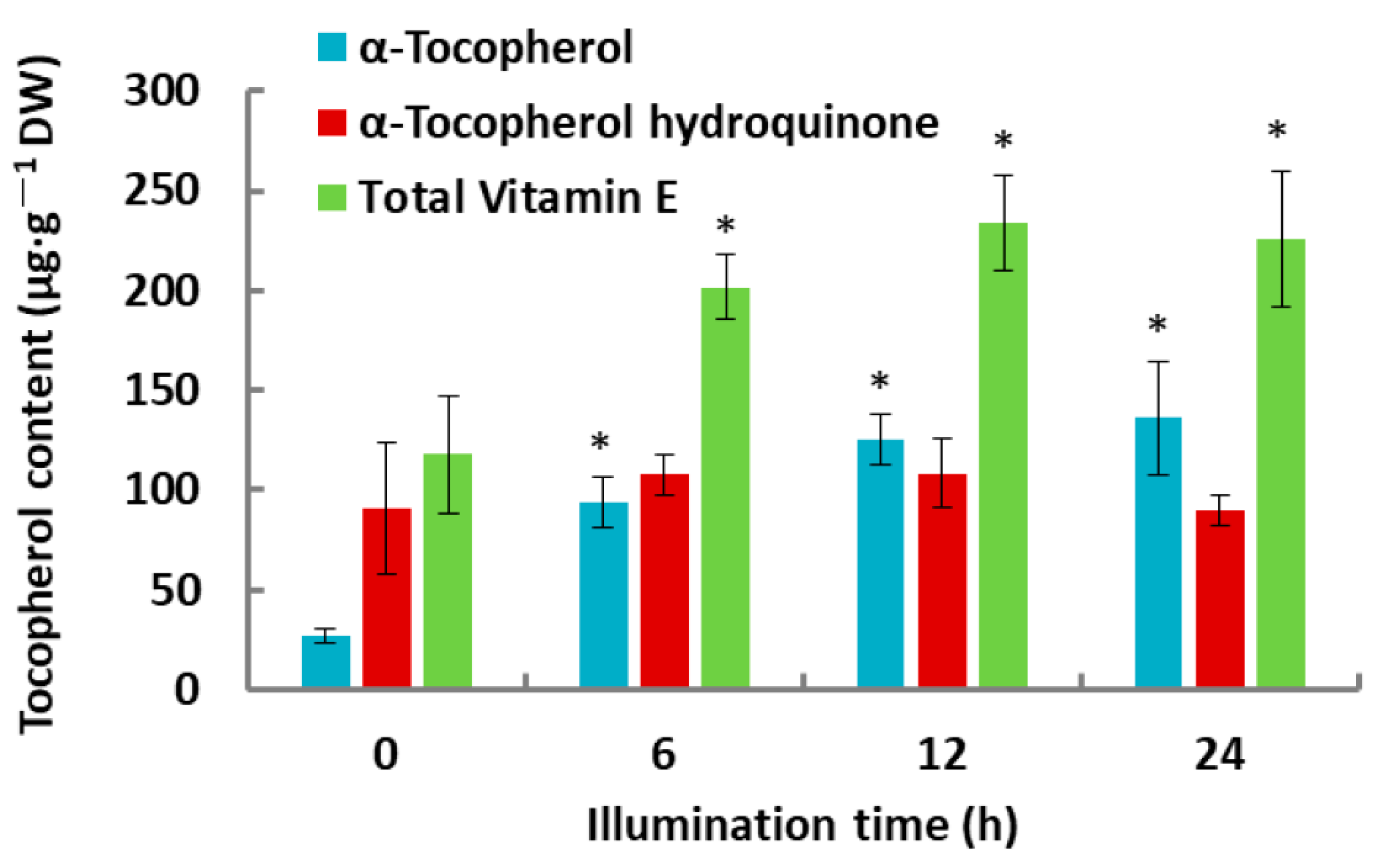
2.1. Tocochromanol Levels in De-Etiolated Rice Leaves

Tocochromanols were identified and profiled by gas chromatography-mass spectrometry (GC-MS) analysis. Only α-tocopherol and α-tocopherol hydroquinone were detectable. The quantity of α-tocopherol significantly increased in response to illumination (Figure 1). In the first 6 h of illumination, α-tocopherol increased 3.4-fold (from 27.30 ± 3.53 μg·g−1 dry weight (DW) to 94. 06 ± 12.42 μg·g−1 DW) and then increased up to 5.0-fold compared with that of the dark control after 24 h of illumination. While α-tocopherol hydroquinone amounts exhibited an increasing trend, and no significant differences were detected during de-etiolation.

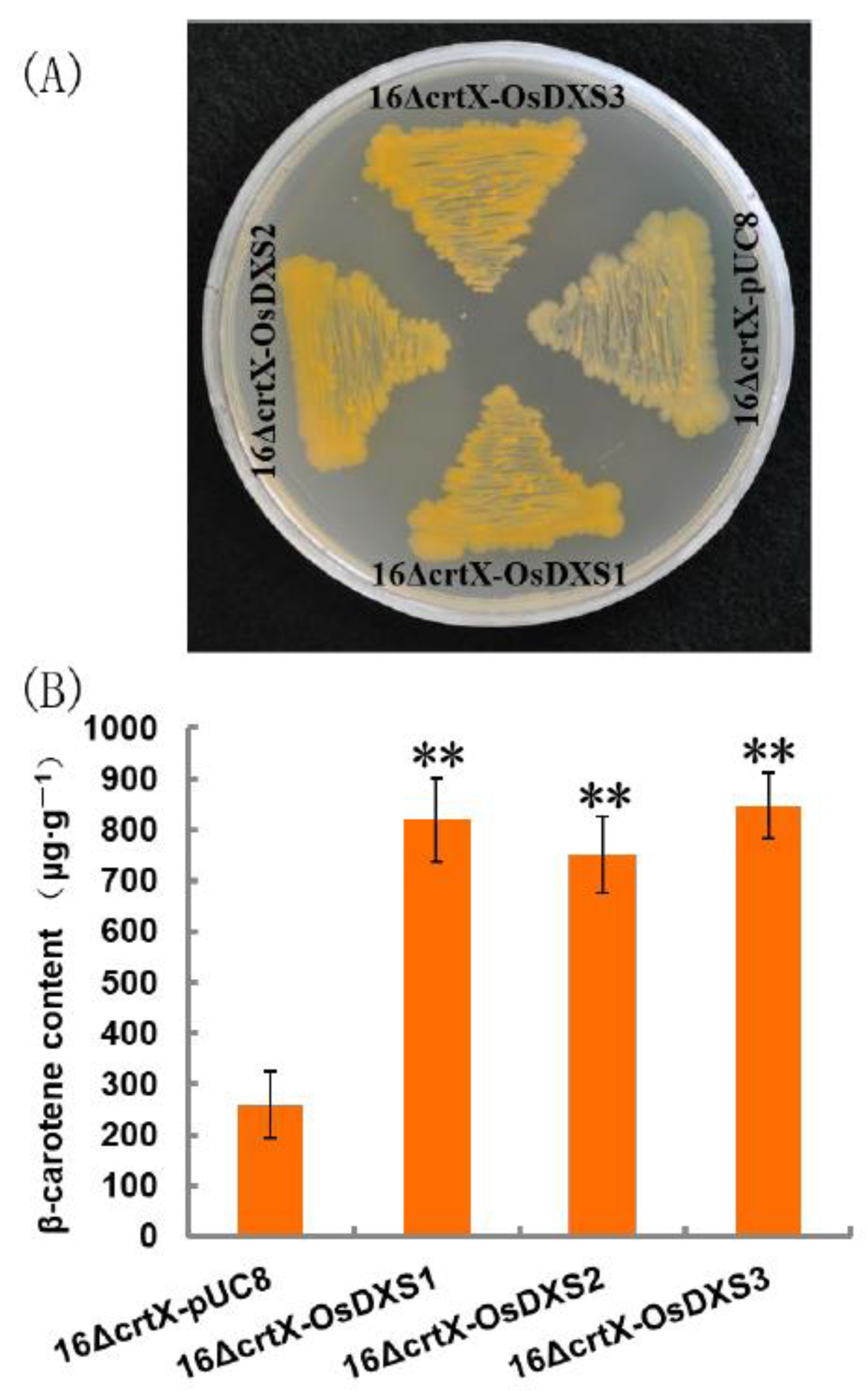
2.2. Functional Analysis of OsDXS Gene Family

In the MEP pathway, pyruvate and glyceraldehyde 3-phosphate are condensed through the action of DXS. DXS is encoded in the rice nuclear genome by three OsDXS gene members [11]. OsDXS3 encoded protein has been demonstrated to have an enzymatic activity that complements the growth of Escherichia coli mutant disrupted in the corresponding DXS gene [12]. A functional E. coli color complementation assay was carried out in a β-carotene accumulating E. coli strain to verify enzymatic activity of OsDXS1 and OsDXS2 encoded proteins. β-Carotene can be synthesized from foreign carotenogenic genes of pACCAR16ΔcrtX which contains all the carotenogenic genes for the generation of β-carotene from farnesyl pyrophosphate (FPP) in non-carotenogenic E. coli as a heterologous host [13]. E. coli cells expressing pACCAR16∆crtX and pUC8 were able to accumulate β-carotene when induced by isopropyl β-D-1-thiogalactopyranoside (IPTG) and showed a light-yellow color (Figure 2A). The color of E. coli strains co-transformed with pACCAR16∆crtX and pUC8-OsDXS1/2/3 were deeper, reflecting the additional flux provided by OsDXS1/2/3 (Figure 2A). Pigments were extracted from liquid cultures and the β-carotene levels were quantified, revealing that co-transformation with pACCAR16∆crtX and the pUC8 empty vector resulted in a β-carotene yield of 258.99 ± 66.03 μg g−1 DW. The addition of pUC8-OsDXS1 resulted in a 3.2-fold increase to 820.47 ± 81.67 μg g−1 DW, whereas the addition of pUC8-OsDXS2 and pUC8-OsDXS3 resulted in a 2.9-fold increase to 750.37 ± 75.36 μg g−1 DW and 3.3-fold increase to 847.37 ± 63.98 μg g−1 DW (Figure 2B). The results indicated that OsDXS1 and OsDXS2 were similar to OsDXS3, encoding functional DXS activities.

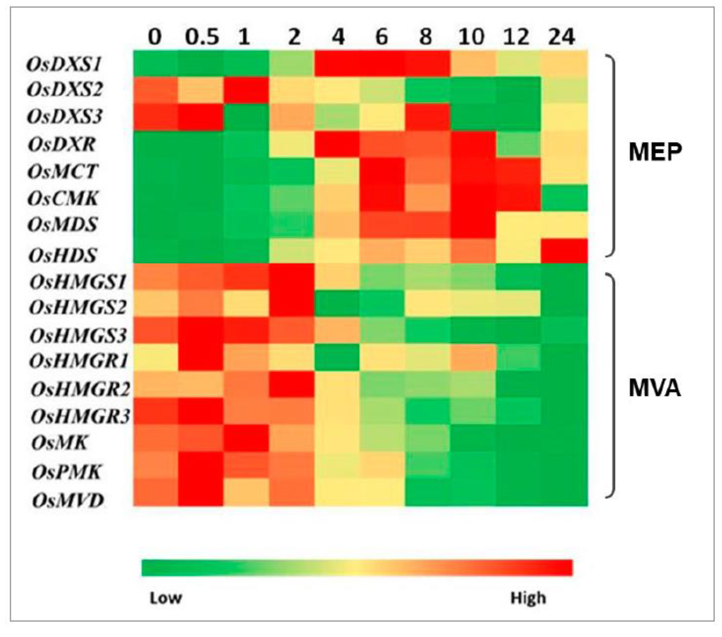
2.3. Expression Analysis of Genes Involved in MEP and MVA Pathways

The function of OsDXS1/2/3, OsDXR (DXP reductoisomerase gene), 4-(cytidine 5′-diphospho)-2-C-methyl-D-erythritol kinase gene (OsCMK), 2-C-methyl-D-erythritol 2,4-cyclodiphosphate synthase gene (OsMDS), and OsIPPI1/2 encoded proteins has been experimentally confirmed (Figure 2) [4,10,12,14]. The expression profiles of MEP and MVA pathway genes were compared to determine whether the light-triggered the isoprenoid biosynthesis gene expression in rice leaves during the de-etiolation process. The changes in expression levels during illumination were investigated by qRT-PCR separately. Figure 3 illustrates the heat map of comparisons of these gene expressions in rice leaves. The genes in MEP and MVA pathways had different responses to light. The majority of the MEP pathway genes were decreased slightly in the first 0.5 h of illumination, then started to increase until peaks, and after that decreased again. There were exceptions such as OsDXS2 and OsDXS3, and the HMBPP synthase gene (OsHDS) among MEP pathway genes. The expression level of OsDXS1 reached the peak after 6 h illumination, which was four-fold greater compared with that in the dark control. Unlike OsDXS1, OsDXS2 and OsDXS3 were downregulated after exposure to light, the highest expression level of OsDXS2 was detected after 1 h (the mRNA level of OsDXS2 was doubled compared with that at 0.5 h), then kept decreasing until 12 h of illumination and slightly increased from 12 to 24 h of illumination. The transcript abundance of OsDXS3 was lower than the other two OsDXS genes in rice leaves and the mRNA level of OsDXS3 after 1 h of illumination dropped to one-tenth of the expression level in dark control. From 1 to 8 h, the expression level of OsDXS3 increased and then decreased sharply (Figure 3).
The expression level of OsDXR (DXP reductoisomerase gene) rose rapidly after 2 h irradiation and reached a peak after 4 h of illumination, which was 7.6-fold higher compared with the dark control. From 4 to 10 h of illumination, the expression level of OsDXR stayed at a high level and then decreased sharply after 12 h irradiation. The highest expression level of OsMCT (2-C-methyl-D-erythritol 4-phosphate cytidylyltransferase gene) was detected after 6 h of irradiation, which was 12-fold higher than that in dark control. After that, the OsMCT expression level decreased slightly and stayed at a high level from 8 to 12 h. After 24 h of irradiation, the expression level of OsMCT declined rapidly, which was 5.8-fold higher than that in the dark control and a half compared with the peak. The expression pattern of OsCMK response to continuous WL (white light) was similar to that of OsMCT. The maximum expression level of OsCMK was detected at 6 h of illumination (10-fold compared with that in dark control). The expression level of OsMDS kept increasing after exposure to WL, and reached the peak after 10 h of illumination, which was 9.9-fold compared to that of the dark control, then decreased to around 6.0-fold after 12 h irradiation. Unlike the rest of the MEP pathway genes, the highest expression level of OsHDS (HMBPP synthase gene) was detected after 24 h of illumination (9.8-fold compared to the dark control). The dynamic changes in mRNA levels of the OsHDR (HMBPP reductase gene) family have been described by Jin et al. (2020) [10].
The mRNA levels of genes in the MVA pathway during the de-etiolation process were opposite to those in the MEP pathway. The expression levels of MVA pathway genes had higher mRNA levels under dark conditions than those after exposure to light. MVA pathway genes had a similar response after exposure to light. The expression levels of the genes at 10 h of illumination were all lower than those in the dark control. The responses of OsHMGS (3-hydroxy-3-methylglutaryl-CoA (HMG-CoA) synthase gene) family genes to WL illumination were not exactly the same. The expression level of OsHMGS1 was increased slightly in the first 2 h of illumination, then decreased sharply from 2 to 6 h, and the expression level of OsHMGS1 was stable at a low level until 10 h (less than half compared with that in dark control). Similar to OsHMGS1, the mRNA level of OsHMGS2 was stable and kept at a low level after 4 h of illumination. The expression level of OsHMGS3 started to decrease after 1 h of illumination, and the lowest expression level was detected after 12 h, which was only 0.08-fold compared with that in the dark control. The OsHMGR (HMG-CoA reductase gene) family also showed distinct reactions to continuous WL illumination. The maximum expression levels of OsHMGR1-3 were detected at 0.5, 2, and 0.5 h of illumination, respectively. The expression level of OsHMGR1 dropped by 50% compared with the dark control after 24 h of illumination. The mRNA level of OsHMGR2 and OsHMGR3 in the dark control was 7.9-fold and 18-fold greater than that after 24 h of illumination, respectively (Figure 3). In the first 1 h, the expression level of OsMK (mevalonate kinase gene) increased slightly, then decreased until 24 h. The expression level of OsMK in the dark control was 3.4-fold higher than that after 24 h illumination. The mRNA level of OsPMK (phospho-MVA kinase gene) and OsMVD (MVA diphosphate decarboxylase gene) both increased slightly in the first 0.5 h of irradiation, and after that kept decreasing. The expression changes in the OsIPPI family, involved in both MVA and MEP pathways, were described by Jin et al. (2020) [10].

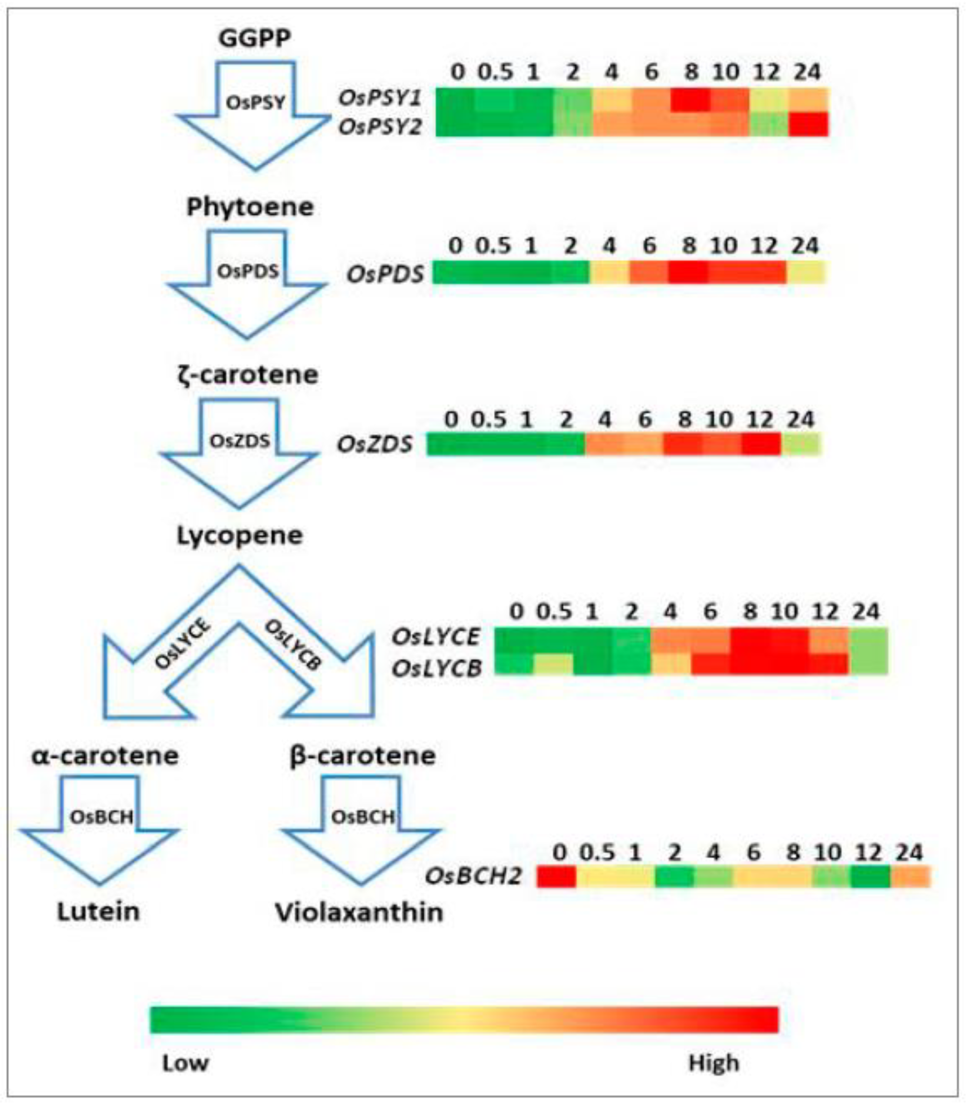
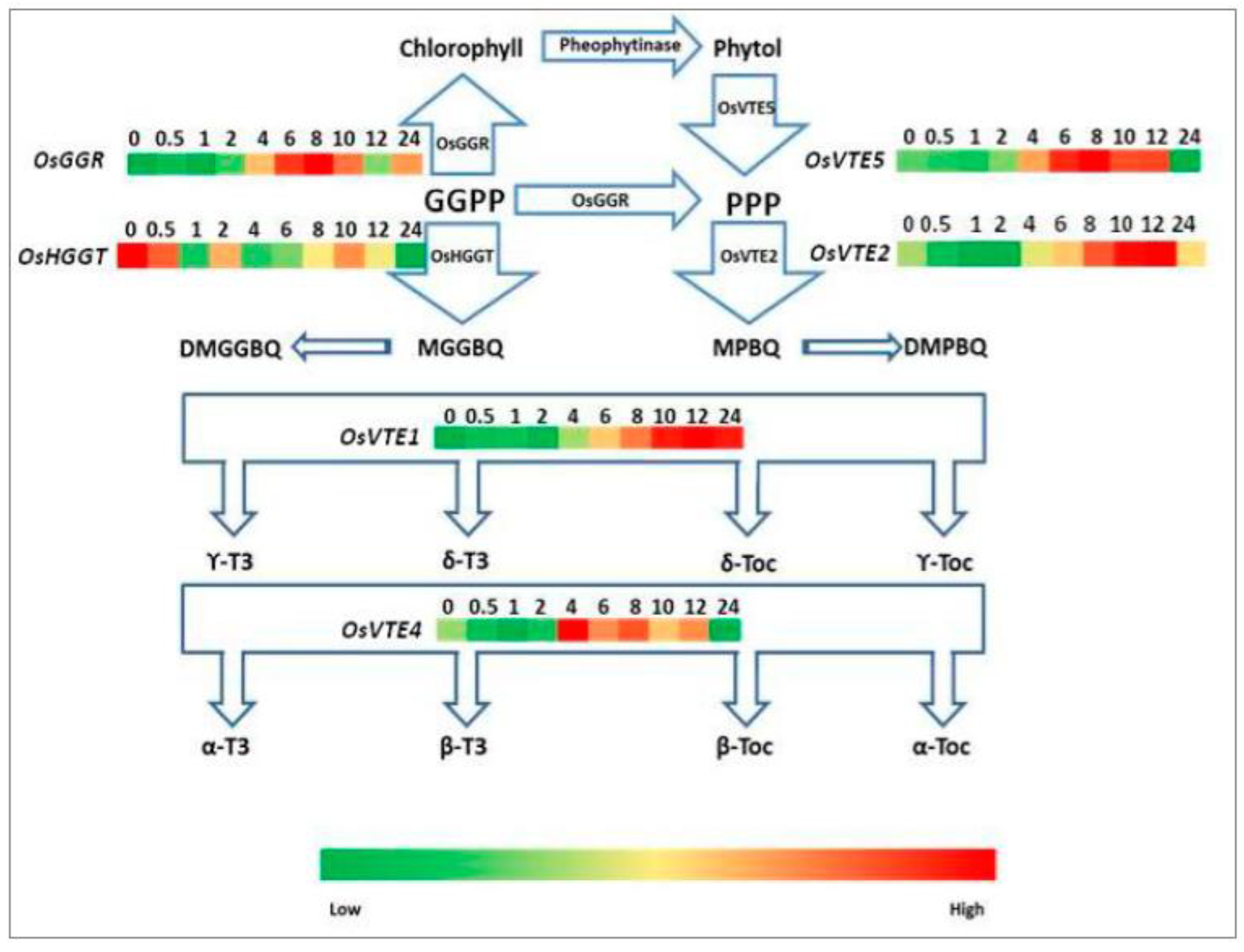
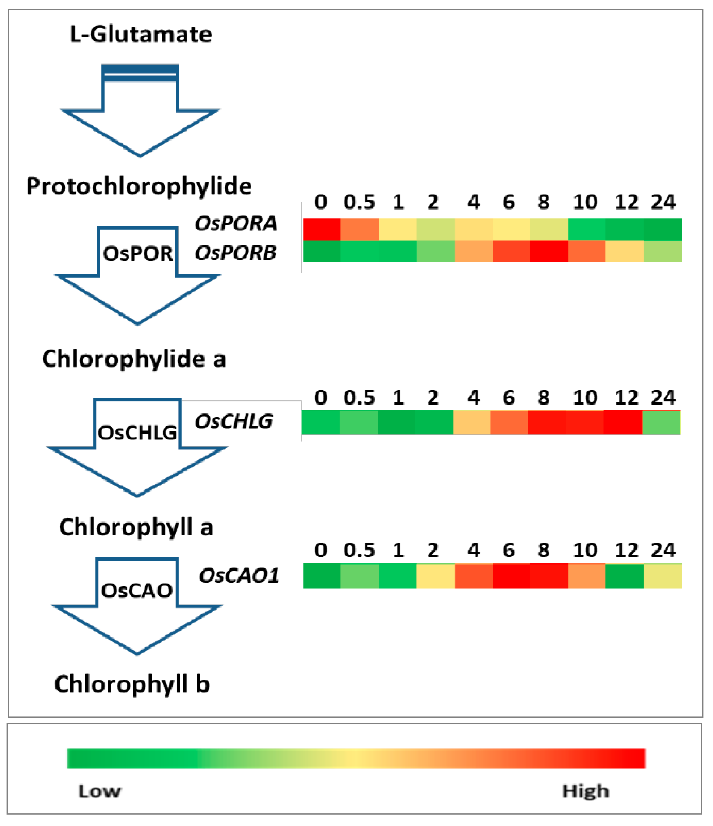
2.4. Expression Analysis of Genes Involved in Carotenoid, Tocopherol, and Chlorophyll Biosynthetic Pathways

The function of rice phytoene synthase 2 (OsPSY2), phytoene desaturase (OsPDS), ζ-carotene desaturase (OsZDS), and lycopene β-cyclase (OsLYCB) has been demonstrated [15,16]. The expression levels of the selected genes related to the carotenoid biosynthetic pathway are shown in Figure 4. The expression level of both OsPSY1 and OsPSY2 increased immediately after exposure to WL illumination. The expression level of both OsPSY1 and OsPSY2 reached high levels after 8 and 10 h of illumination (approximately 5- and 25-fold, respectively, greater than the expression level in the dark control), respectively, but both decreased sharply after 10 h of illumination. The expression of OsPSY1 remained nearly constant from 12 to 24 h of illumination, while the expression of OsPSY2 increased rapidly again after 12 h of illumination and reached its maximum after 24 h. The expression level of OsPDS decreased slightly during the first 1 h of illumination, then increased to the peak after 8 h of illumination (4.1-fold compared with that in the dark control). After the peak, the expression level of OsPDS declined until 24 h, when its expression level was 2.4-fold higher than that of the dark control. The expression level of OsZDS kept increasing in the first 12 h of illumination and then declined at 24 h (8.5- and 4.5-fold higher than the dark control). The mRNA levels of both OsLYCB and OsLYCE (lycopene ε-cyclase gene) reached their peaks after 8 h of illumination (4.7- and 2.3-fold compared to the dark control, respectively). Unlike the other genes, OsBCH2 (β-carotene hydroxylase 2 gene) showed a decrease in expression level after exposure to WL, which decreased to about 50% of that of the dark control after 24 h of illumination (Figure 4).
The key tocochromanol biosynthesis genes in rice have been identified [17], while the function of these genes remained undetermined. The expression levels of the selected tocopherol biosynthetic pathway genes in de-etiolated rice leaves are shown in Figure 5. The expression levels of OsGGR (geranylgeranyl reductase gene), OsVTE5 (phytol kinase gene), OsVTE2 (homogentisate phytyltransferase gene), OsVTE1 (tocopherol cyclase gene), and OsVTE4 (γ-tocopherol methyltransferase gene) were all upregulated by WL. The expression levels of OsGGR increased rapidly during the first 8 h of illumination (10-fold greater than that of dark control) and then plummeted after 12 h of irradiation. The expression level of OsGGR mRNA remained on the rise from 12 to 24 h of illumination. The mRNA of OsVTE1 remained at low levels during the first 2 h of illumination, then started to increase to its maximum after 12 h of illumination (4.9-fold higher than that of dark control), and remained at this high level until 24 h irradiation. There were no significant differences in expression levels among 10-, 12-, and 24-h illuminated samples. Both OsVTE5 and OsVTE2 mRNA levels exhibited a downward trend at the beginning, then started to increase and reached a peak respectively at 8 and 12 h (2.5- and 2.0-fold higher than that in the dark control, respectively). Afterward, the expression levels started to decline, OsVTE5 mRNA level decreased sharply after 24 h of illumination, and the OsVTE2 mRNA level dropped to a level similar to that in the dark control after 24 h irradiation. The expression level of OsVTE4 increased rapidly during the first 4 h of illumination and decreased until 24 h of illumination. However, OsHGGT (homogentisate geranylgeranyltransferase gene) was downregulated by WL. The expression level decreased to three-fold greater than the dark control after 24 h of illumination.
So far, the function of a few chlorophyll biosynthesis genes (rice chlorophyll synthase gene (OsCHLG) and rice chlorophyll a oxygenase 1 gene (OsCAO1)) has been demonstrated [4,18,19]. Changes in the expression levels of the selected chlorophyll biosynthetic pathway genes during rice de-etiolation are shown in Figure 6. The expression levels of OsPORA (protochlorophyllide oxidoreductase A gene) and OsPORB reacted differently to WL illumination. The OsPORB mRNA level increased significantly in the first 8 h (15-fold higher than that in dark control) and decreased rapidly after 24 h of illumination. The expression level at 24 h was approximately three-fold greater than in the dark control. However, the expression of OsPORA started to decline after exposure to WL, indicating that OsPORA was downregulated by light. The expression of OsCHLG increased in the first 0.5 h of illumination, decreased slightly after 2 h of illumination, increased again after 4 h of irradiation, and remained stable from 8 to 12 h. The maximum expression level was 4.5-fold higher than that in the dark control, which was detected after 12 h irradiation. After that, OsCHLG expression decreased immediately to the same level as that in the dark control. The expression level of OsCAO1 reached a peak after 6 h of illumination (8.4-fold), decreased until 12 h, and slightly increased again from 12 to 24 h.

2.5. In Silico Promoter Analysis of Light-Responsive Cis-Regulatory Elements in the Promoter Regions of Light-Upregulated Genes

The full-length CDS (coding sequence) of each rice (O. sativa cultivar: Nipponbare) gene was used to identify its corresponding promoter region from NCBI. The mRNA with the longest 5′-untranslated region and its corresponding promoter sequence (up to 1.0 kb upstream of transcription start site) of each upregulated gene during de-etiolation of etiolated rice leaves were retrieved from NCBI (Table 1). The online tools PlantCare [20] and PLACE [21] were used for the identification of light-responsive cis-acting regulatory elements in the promoter of each gene. The promoters of the upregulated genes (OsDXS1, OsDXR, OsMCT, OsCMK, OsMDS, OsHDS, OsHDR1, OsIPPI1, OsPORB, OsCHLG, OsCAO1, OsPSY1, OsPSY2, OsPDS, OsZDS, OsLYCB, OsLYCE, OsGGR, OsVTE1, OsVTE2, OsVTE4, OsVTE5) during de-etiolation of etiolated rice leaves contained light-upregulated elements, including G-box (CACGTC), IBOXCORE (I-box; GATAA), Box 4 (ATTAAT), GT1-motif (GGTTAA), MRE (AACCTAA), G-box (TAAACGTG), Sp1 (GGGCGG), LTR (CCGAAA), G-box (CCACGTAA), TCCC-motif (TCTCCCT), G-box (GTCGTG), Box II (CCACGTGGC), AE-box (AGAAACAA), G-box (CGCGTC), G-box (GCCACGTGGA), G-box (CACGTT), GATA-motif (GATAGGG), GATA-motif (GATAGGA), and TCT-motif (TCTTAC) (Table 1).

3. Discussion

3.1. The Coordinated Expression Levels of the MEP and MVA Pathway Genes and Carotenoid, Tocopherol and Chlorophyll Biosynthetic Pathway Genes Mirrored the Levels of the Corresponding Metabolite Changes during De-Etiolation

The analysis of Arabidopsis revealed that the genes involved in the MVA and MEP pathways behaved differently in response to light stimulation. The expression levels of MVA pathway genes were inhibited by light, whereas those of MEP pathway genes were upregulated [8]. In rice, Kim et al. (2005) [11] reported that, in the case of light illumination, only the relative expression of OsDXS1 increased relative to the unilluminated control. OsDXS3 has been proposed to be specifically involved in the synthesis of diterpenoid phytoalexins (such as momilactones and phytocassanes) in suspension-cultured rice cells after treatment with a chitin elicitor [12,22].
Carotenoids, tocopherols, and chlorophylls are synthesized primarily from precursors derived from the MEP pathway [8]. The number of carotenoids and chlorophylls increased significantly in response to the illumination of etiolated rice seedlings compared with that of the dark control [10]. The quantity of α-tocopherol significantly increased (Figure 1). The first step in the MEP pathway is catalyzed by DXS. There are three OsDXS genes in the rice genome. The expression of OsDXS1 increased four-fold after 4 h of illumination, whereas the expression levels of OsDXS2 and OsDXS3 decreased after exposure to light (Figure 3). Similar to OsDXS3 [12], both the encoded OsDXS1 and OsDXS2 are shown to be functional enzymes (Figure 2) in engineered E. coli. The appearance time of MEP-derived isoprenoids in the de-etiolated rice leaves coincided with the time of the positive response of OsDXS1 to light (Figure 3; Supplementary Figure S1), suggesting that the biosynthesis of chlorophylls, carotenoids, and tocopherols in the de-etiolated rice leaves is mainly generated by OsDXS1. The rest of the MEP pathway gene (OsDXR, OsMCT, OsCMK, OsMDS, OsHDS, and OsHDR1) expression (Figure 3; Jin et al., 2020 [10] for OsHDR1) had positive correlations with light illumination correlated with the MEP pathway-derived carotenoid, chlorophyll, and tocopherol accumulation. A study by Huang et al. (2018) reported photosynthetic pigments in an OsMDS mutant (IspF), which displayed a yellow-green leaf phenotype, and the chlorophyll and carotenoid levels were significantly lower than those in WT rice. Therefore, OsMDS is essential for the biosynthesis of chlorophylls and carotenoids [14]. The genes encoding enzymes involved in chlorophyll biosynthesis, such as OsCHLG, OsPORB, and OsCAO1, were upregulated by light, except OsPORA (Figure 6), which was associated with the accumulation of chlorophylls.
Phytoene synthase (PSY) is the rate-limiting enzyme of carotenogenesis. In Arabidopsis seedlings, the expression level of PSY increased under the continuous illumination of both FR and red light [23]. In white mustard (Sinapis alba L.), the upregulation of PSY expression was observed after light exposure, while PDS and GGPS (geranylgeranyl pyrophosphate synthase gene) expression levels remained constant [24]. Both rice OsPSY1 and OsPSY2 are subject to phytochrome and involved in carotenoid biosynthesis in photosynthetically active tissues with different expression patterns during chloroplast development [25]. Carotenoid biosynthesis genes exhibit high expression in light-grown rice tissues [26]. In agreement with reports by Welsch et al. (2008) and Chaudhary et al. (2010) [25,26], the expression levels of OsPSY1/2, OsPDS, OsZDS, OsLYCB, and OsLYCE increased after exposure to WL in this study, whereas the OsBCH2 expression level decreased relative to that of the dark control (Figure 4), reflecting the approximately two-fold increases in the abundance of β-carotene after 24 h of illumination compared to that of the dark control [10].
Studies on Arabidopsis found that AtVTE2 showed limited activities in tocopherol synthesis [27]. OsVTE2 was upregulated by light. Both OsVTE1 and OsVTE5 were also upregulated by WL in de-etiolated rice leaves (Figure 5). Meanwhile, OsVTE4 increased rapidly in the early stage of rice de-etiolation. The upregulation of tocopherol biosynthetic genes in de-etiolated rice leaves was reflected by the increase in tocopherol content in rice leaves during de-etiolation.
Phytosterols are derived from the MVA pathway. In a previous study [10], the level of β-sitosterol increased significantly in the first 6 h of light illumination and then decreased after 12 h of illumination to the same level as that of the dark control. After 24 h of light illumination, the levels of campesterol and stigmasterol showed no difference compared with that of the dark control [10]. The expression levels of the selected MVA pathway genes were inhibited with no obvious changes during the de-etiolated rice leaves (Figure 3), consistent with the MVA pathway-derived phytosterol changes.

3.2. The Underpinning Mechanistic Basis of Coordinated Light-Responsive Gene Expression during the De-Etiolation Process

The coordinated expression of light-responsive genes during the de-etiolation can reflect similarities between their promoters. A range of light-responsive elements (LREs) has been documented in different promoters, many of which positively or negatively mediate gene expression in response to light [3]. Although many LREs and their transcription factors have been identified, no single element is found in all light-upregulated gene promoters, suggesting a complex light-regulation network and a lack of a universal switch. It has been suggested that combinations of LREs, rather than individual elements, could confer proper light-responsiveness to a light-insensitive basal promoter [3,28]. To investigate the basis of coordinated light-responsive gene expression during the de-etiolation of rice etiolated leaves, in silico promoter analysis of upregulated genes during de-etiolation was performed. Most promoters contained at least two motifs involved in the response to light, but both OsCMK and OsHDR1 promoters (Table 1) had only one light-responsive motif (IBOXCORE, GATAA), supporting that these genes were upregulated by light. However, surprisingly, except for OsCMT, OsLYCE, and OsVTE2 promoters, other light-upregulated gene promoters all presented a cis-regulatory element with a consensus sequence GATAA (IBOXCORE) (Table 1). IBOXCORE (I-box) is a conserved sequence upstream of light-upregulated genes for both monocots and dicots [21,28]. (http://www.dna.affrc.go.jp/htdocs/PLACE/; 18 March 2020 (See IBOX, S000199)).

4. Materials and Methods

4.1. Plant Material

Wild type rice (Oryza sativa L. cv. EYI105) seeds were sterilized in 3.7% NaClO for 20 min and washed with sterilized water thoroughly, then were aseptically grown on Murashige and Skoog (MS) medium containing 0.3% phytagel in pots (diameter 10 cm, height 20 cm, 9 seeds in each pot) in a controlled growth chamber at 25 °C in complete darkness. Fourteen-day-old etiolated rice seedlings were illuminated with continuous WL (100 μmol m−2 s−1) for 0.5, 1, 2, 4, 6, 8, 10, 12, or 24 h (hour). Leaves of de-etiolated rice seedlings were rapidly frozen in liquid nitrogen after collection and stored at −80 °C. The mixture of all the rice leaves from the same pot was collected and stored as one sample.

4.2. Tocochromanols Analysis

Rice leaf samples were freeze-dried before analysis, and 10 mg of powdered tissue was extracted using a chloroform-methanol mixture, as described [10]. The gas chromatography-mass spectrometry (GC-MS) method was applied to determine tocochromanols as described in detail previously [29]. Compositions of tocochromanols were identified using a mass spectral library built from in-house standards [30]. The samples used for tocochromanols analysis were from the same batch [10] as those used for carotenoids, chlorophylls, phytosterols, and pathway gene expression analysis.

4.3. Rice DXS cDNA Cloning and Functional Complementation Analysis

The OsDXS1, OsDXS2, and OsDXS3 cDNAs were cloned directly from rice leaf mRNA by RT-PCR using two pairs of oligonucleotide primers with terminal EcoRI and BamHI restriction sites for both OsDXS1 and OsDXS3, or EcoRI and HindIII restriction sites for OsDXS2 (Supplementary Table S2). The primers were designed to flank the open reading frame (ORF) of each gene according to sequences in GenBank (accession number XM_015785019 for OsDXS1, XM_015787004 for OsDXS2, and XM_015792458 for OsDXS3). The RT-PCR products were transferred to vector pCRII TOPO (Invitrogen, Carlsbad, CA, USA) to generate pCR-OsDXS1, pCR-OsDXS2, and pCROsDXS3 for sequencing. The OsDXS1, OsDXS2, and OsDXS3 cDNAs were then introduced into pUC8 at the EcoRI/BamHI or EcoRI/HindIII sites to generate vectors pUC8-OsDXS1, pUC8-OsDXS2, and pUC8-OsDXS3 for functional complementation in engineered E. coli cells. The integrity of all intermediate and final constructs was confirmed by sequencing.
Functional color complementation experiments were conducted as previously described [10,13]. E. coli DH5α cells were transformed with pACCAR16ΔcrtX, which contains the Erwinia uredovora carotene biosynthesis genes crtE, crtB, crtI, and crtY encoding geranylgeranyl pyrophosphate (GGPP) synthase, phytoene synthase, phytoene desaturase, and lycopene β-cyclase, respectively, for the generation of β-carotene [31]. β-Carotene levels in engineered E. coli were measured as described previously [10].

4.4. Gene Expression Analysis with Quantitative Real-Time PCR (qRT-PCR)

Total RNA was isolated using the RNeasy Plant Mini Kit (Qiagen, Hilden, Germany) and DNA was removed with DNase I (RNase-Free DNase Set, Qiagen) [10]. First-strand cDNA was synthesized from 1 µg total RNA using Ominiscript Reverse Transcriptase (Qiagen) in a 20 µL total reaction, and quantitative real-time RT-PCR (qRT-PCR) was applied [10,32] using the primers listed in Supplementary Table S1. The amplified DNA fragments for each gene were confirmed by sequencing. The expression levels of the genes in different samples were normalized against rice actin mRNA (OsActin). Three biological replicates each comprising three technical replicates were tested for each sample.

4.5. Retrieval of Promoter Regions and Analysis of Cis-Regulatory Elements

Gene and promoter sequences (1.0 kb upstream of transcription start site) of each rice (O. sativa cultivar: Nipponbare) gene under consideration were retrieved from NCBI (National Center for Biotechnology Information) (https://www.ncbi.nlm.nih.gov/; accessed on 18 March 2020). The tools PlantCare (http://bioinformatics.psb.ugent.be/webtools/plantcare/html/; accessed on 18 March 2020) [20] and PLACE (http://www.dna.affrc.go.jp/htdocs/PLACE/; 18 March 2020) [21] were used to identify cis-regulatory elements in promoters.

4.6. Data and Statistical Analysis

Significant differences in quantitative dependent variables for any two alternative treatments were calculated using Student’s t-test, with a p-value of 0.05 considered significant.

5. Conclusions

Both OsDXS1 and OsDXS2 genes were shown to encode functional DXS enzymes. The appearance of MEP-derived plastidial isoprenoids in the de-etiolated rice leaves correlates well with the observed upregulation of OsDXS1 expression, suggesting that OsDXS1 is responsible for the biosynthesis of chlorophylls, carotenoids, and tocopherols in the de-etiolated rice leaves. The coordinated upregulated expression of genes involved in the MEP pathway, chlorophyll, carotenoid, and tocopherol biosynthetic pathways mirrored the increased levels of chlorophyll, carotenoid, and tocopherol contents in rice leaves during de-etiolation, demonstrating that the synthesis of these plastidial metabolites is under transcriptional control. Moreover, the work was extended to elucidate the underpinning mechanistic basis of coordinated light-responsive gene expression during the de-etiolation process. The light-responsive cis-regulatory elements were presented in all promoter regions (up to 1.0 kb upstream of transcription start site) of each light-upregulated gene, serving as a base for the further characterization of these light-induced genes during the de-etiolation process.

Supplementary Materials

The following are available online at https://www.mdpi.com/article/10.3390/plants10071456/s1, Additional Supporting Information may be found in the online version of this article: Figure S1. Expression of OsDXS1, OsDXS2, and OsDXS3 in etiolated rice (Oryza sativa, EYI105) leaves during de-etiolation at different times after the onset of irradiation with white light, with mRNA levels normalized against OsActin mRNA. Table S1. Primers used for qRT-PCR analysis in this study. Table S2. Primers used for rice DXS cDNA cloning in this study.

Author Contributions

C.Z. conceived and designed the research. X.J., C.B., M.D., Y.S., X.H., W.H., L.S., T.C., P.D.F. and C.Z. conducted the experiments. X.J., C.B., M.D., P.D.F., T.C., P.C. and C.Z. analyzed the data. X.J., C.B., P.D.F., P.C. and C.Z. wrote the manuscript. All authors have read and agreed to the published version of the manuscript.

Funding

This research was funded by Gansu Provincial Key Laboratory of Aridland Crop Science, Gansu Agricultural University (Project No. GSCS-2020-07); The National Natural Science Foundation of China (31870278); the Spanish Ministry of Economy and Competitiveness (MINECO), Spain (RTI2018-097613-B-I00; PGC2018-097655-B-I00); in part by the European Union Framework Program DISCO (613513) “from DISCOvery to products: a next-generation pipeline for the sustainable generation of high-value plant products”, the European Cooperation in Science and Technology project EUROCAROTEN (OC-2015-1-19780), Generalitat de Catalunya Grant 2017 SGR 828 to the Agricultural Biotechnology and Bioeconomy Unit (ABBU), and the International Science and Technology Cooperation Project 20190201013JC (from Jilin Provincial Science and Technology Department, China); GAU-KYQD-2019-06, Gansu Agricultural University.

Institutional Review Board Statement

Not applicable.

Informed Consent Statement

Not applicable.

Data Availability Statement

Not applicable.

Conflicts of Interest

The authors declare that they have no conflict of interest.

References

  1. Yang, P.; Chen, H.; Liang, Y.; Shen, S. Proteomic analysis of de-etiolated rice seedlings upon exposure to light. Proteomics 2007, 7, 2459–2468. [Google Scholar] [CrossRef]
  2. Yang, Y.; Liu, H. Coordinated shoot and root responses to light signaling in Arabidopsis. Plant Commun. 2020, 1, 100026. [Google Scholar] [CrossRef]
  3. Jiao, Y.; Lau, O.S.; Deng, X.W. Light-regulated transcriptional networks in higher plants. Nat. Rev. Genet. 2007, 8, 217–230. [Google Scholar] [CrossRef]
  4. Jung, K.H.; Lee, J.; Dardick, C.; Seo, Y.S.; Cao, P.; Canlas, P.; Phetsom, J.; Xu, X.; Ouyang, S.; An, K.; et al. Identification and functional analysis of light-responsive unique genes and gene family members in rice. PLoS Genet. 2008, 4, e1000164. [Google Scholar] [CrossRef] [PubMed] [Green Version]
  5. Chory, J.; Chatterjee, M.; Cook, R.K.; Elich, T.; Fankhauser, C.; Li, J.; Nagpal, P.; Neff, M.; Pepper, A.; Poole, D.; et al. From seed germination to flowering, light controls plant development via the pigment phytochrome. Proc. Natl. Acad. Sci. USA 1996, 93, 12066–12071. [Google Scholar] [CrossRef] [PubMed] [Green Version]
  6. Clouse, S.D. Integration of light and brassinosteroid signals in etiolated seedling growth. Trends Plant Sci. 2001, 6, 443–445. [Google Scholar] [CrossRef]
  7. Li, Y.; Swaminathan, K.; Hudson, M.E. Rapid, organ-specific transcriptional responses to light regulate photomorphogenic development in dicot seedlings. Plant Physiol. 2011, 156, 2124–2140. [Google Scholar] [CrossRef] [PubMed] [Green Version]
  8. Vranova, E.; Coman, D.; Gruissem, W. Network analysis of the MVA and MEP pathways for isoprenoid synthesis. Annu. Rev. Plant Biol. 2013, 64, 665–700. [Google Scholar] [CrossRef] [PubMed]
  9. Ghassemian, M.; Lutes, J.; Tepperman, J.M.; Chang, H.S.; Zhu, T.; Wang, X.; Quail, P.H.; Lange, B.M. Integrative analysis of transcript and metabolite profiling data sets to evaluate the regulation of biochemical pathways during photomorphogenesis. Arch. Biochem. Biophys. 2006, 448, 45–59. [Google Scholar] [CrossRef]
  10. Jin, X.; Baysal, C.; Gao, L.; Medina, V.; Drapal, M.; Ni, X.; Sheng, Y.; Shi, L.; Capell, T.; Fraser, P.D.; et al. The subcellular localization of two isopentenyl diphosphate isomerases in rice suggests a role for the endoplasmic reticulum in isoprenoid biosynthesis. Plant Cell Rep. 2020, 39, 119–133. [Google Scholar] [CrossRef]
  11. Kim, B.R.; Kim, S.U.; Chang, Y.J. Differential expression of three 1-deoxy-D: -xylulose-5-phosphate synthase genes in rice. Biotechnol. Lett. 2005, 27, 997–1001. [Google Scholar] [CrossRef] [PubMed]
  12. Okada, A.; Shimizu, T.; Okada, K.; Kuzuyama, T.; Koga, J.; Shibuya, N.; Nojiri, H.; Yamane, H. Elicitor induced activation of the methylerythritol phosphate pathway toward phytoalexins biosynthesis in rice. Plant Mol. Biol. 2007, 65, 177–187. [Google Scholar] [CrossRef] [PubMed]
  13. Kajiwara, S.; Fraser, P.D.; Kondo, K.; Misawa, N. Expression of an exogenous isopentenyl diphosphate isomerase gene enhances isoprenoid biosynthesis in Escherichia coli. Biochem. J. 1997, 324, 421–426. [Google Scholar] [CrossRef] [PubMed] [Green Version]
  14. Huang, R.; Wang, Y.; Wang, P.; Li, C.; Xiao, F.; Chen, N.; Li, N.; Li, C.; Sun, C.; Li, L.; et al. A single nucleotide mutation of IspF gene involved in the MEP pathway for isoprenoid biosynthesis causes yellow-green leaf phenotype in rice. Plant Mol. Biol. 2018, 96, 5–16. [Google Scholar] [CrossRef]
  15. Gallagher, C.E.; Matthews, P.D.; Li, F.; Wurtzel, E.T. Gene duplication in the carotenoid biosynthetic pathway preceded evolution of the grasses. Plant Physiol. 2004, 135, 1776–1783. [Google Scholar] [CrossRef] [Green Version]
  16. Fang, J.; Chai, C.; Qian, Q.; Li, C.; Tang, J.; Sun, L.; Huang, Z.; Guo, X.; Sun, C.; Liu, M.; et al. Mutations of genes in synthesis of the carotenoid precursors of ABA lead to pre-harvest sprouting and photo-oxidation in rice. Plant J. 2008, 54, 177–189. [Google Scholar] [CrossRef] [Green Version]
  17. Chaudhary, N.; Khurana, P. Vitamin E biosynthesis genes in rice: Molecular characterization, expression profiling and comparative phylogenetic analysis. Plant Sci. 2009, 177, 479–491. [Google Scholar] [CrossRef]
  18. Lee, S.; Kim, J.H.; Yoo, E.S.; Lee, C.H.; Hirochika, H.; An, G. Differential regulation of chlorophyll a oxygenase genes in rice. Plant Mol. Biol. 2005, 57, 805–818. [Google Scholar] [CrossRef]
  19. Wu, Z.; Zhang, X.; He, B.; Diao, L.; Sheng, S.; Wang, J.; Guo, X.; Su, N.; Wang, L.; Jiang, L.; et al. A chlorophyll-deficient rice mutant with impaired chlorophyllide esterification in chlorophyll biosynthesis. Plant Physiol. 2007, 145, 29–40. [Google Scholar] [CrossRef] [Green Version]
  20. Lescot, M.; Dehais, P.; Thijs, G.; Marchal, K.; Moreau, Y.; Van de Peer, Y.; Rouze, P.; Rombauts, S. PlantCARE a database of plant cis-acting regulatory elements and a portal to tools for in silico analysis of promoter sequences. Nucleic Acids Res. 2002, 30, 325–327. [Google Scholar] [CrossRef]
  21. Higo, K.; Ugawa, Y.; Iwamoto, M.; Korenaga, T. Plant cis-acting regulatory DNA elements (PLACE) database: 1999. Nucleic Acids Res. 1999, 27, 297–300. [Google Scholar] [CrossRef] [Green Version]
  22. Miyamoto, K.; Matsumoto, T.; Okada, A.; Komiyama, K.; Chujo, T.; Yoshikawa, H.; Nojiri, H.; Yamane, H.; Okada, K. Identification of target genes of the bZIP transcription factor OsTGAP1, whose overexpression causes elicitor-induced hyperaccumulation of diterpenoid phytoalexins in rice cells. PLoS ONE 2014, 9, e105823. [Google Scholar] [CrossRef] [Green Version]
  23. Von Lintig, J.; Welsch, R.; Bonk, M.; Giuliano, G.; Batschauer, A.; Kleinig, H. Light-dependent regulation of carotenoid biosynthesis occurs at the level of phytoene synthase expression and is mediated by phytochrome in Sinapis alba and Arabidopsis thaliana seedlings. Plant J. 1997, 12, 625–634. [Google Scholar] [CrossRef] [PubMed]
  24. Welsch, R.; Beyer, P.; Hugueney, P.; Kleinig, H.; von Lintig, J. Regulation and activation of phytoene synthase, a key enzyme in carotenoid biosynthesis, during photomorphogenesis. Planta 2000, 211, 846–854. [Google Scholar] [CrossRef]
  25. Welsch, R.; Wust, F.; Bar, C.; Al-Babili, S.; Beyer, P. A third phytoene synthase is devoted to abiotic stress-induced abscisic acid formation in rice and defines functional diversification of phytoene synthase genes. Plant Physiol. 2008, 147, 367–380. [Google Scholar] [CrossRef] [PubMed] [Green Version]
  26. Chaudhary, N.; Nijhawan, A.; Khurana, J.P.; Khurana, P. Carotenoid biosynthesis genes in rice: Structural analysis, genome-wide expression profiling and phylogenetic analysis. Mol. Genet. Genom. 2010, 283, 13–33. [Google Scholar] [CrossRef] [PubMed]
  27. Collakova, E.; DellaPenna, D. Homogentisate phytyltransferase activity is limiting for tocopherol biosynthesis in Arabidopsis. Plant Physiol. 2003, 131, 632–642. [Google Scholar] [CrossRef] [Green Version]
  28. Terzaghi, W.B.; Cashmore, A.R. Light-regulated transcription. Annu. Rev. Plant Physiol. Plant Mol. Biol. 1995, 46, 445–474. [Google Scholar] [CrossRef]
  29. Enfissi, E.M.; Barneche, F.; Ahmed, I.; Lichtle, C.; Gerrish, C.; McQuinn, R.P.; Giovannoni, J.J.; Lopez-Juez, E.; Bowler, C.; Bramley, P.M.; et al. Integrative transcript and metabolite analysis of nutritionally enhanced DE-ETIOLATED1 downregulated tomato fruit. Plant Cell 2010, 22, 1190–1215. [Google Scholar] [CrossRef] [PubMed] [Green Version]
  30. Almeida, J.; Perez-Fons, L.; Fraser, P.D. A transcriptomic, metabolomic and cellular approach to the physiological adaptation of tomato fruit to high temperature. Plant Cell Environ. 2020. [Google Scholar] [CrossRef]
  31. Misawa, N.; Satomi, Y.; Kondo, K.; Yokoyama, A.; Kajiwara, S.; Saito, T.; Ohtani, T.; Miki, W. Structure and functional analysis of a marine bacterial carotenoid biosynthesis gene cluster and astaxanthin biosynthetic pathway proposed at the gene level. J. Bacteriol. 1995, 177, 6575–6584. [Google Scholar] [CrossRef] [PubMed] [Green Version]
  32. Baysal, C.; He, W.; Drapal, M.; Villorbina, G.; Medina, V.; Capell, T.; Khush, G.S.; Zhu, C.; Fraser, P.D.; Christou, P. Inactivation of rice starch branching enzyme IIb triggers broad and unexpected changes in metabolism by transcriptional reprogramming. Proc. Natl. Acad. Sci. USA 2020, 117, 26503–26512. [Google Scholar] [CrossRef] [PubMed]
Figure 1. Tocopherol composition and content in the etiolated leaves of 14-day-old rice plants during de-etiolation. Values represent the mean of five biological replicates and bars represent standard deviations. Asterisks indicate statistically significant differences compared with the control (0 h illumination) according to the Student’s t-test: * p < 0.05.
Plants 10 01456 g001
Figure 2. Functional analysis of the OsDXS gene family. (A) Color complementation and enhanced β-carotene accumulation due to the expression of OsDXS1, OsDXS2, and OsDXS3, respectively, in E. coli engineered for β-carotene biosynthesis. The plate was divided into four sections, which were inoculated separately with bacteria carrying pACCAR16∆crtX and pUC8-OsDXS1 (16∆crtX-OsDXS1), pACCAR16∆crtX and pUC8-OsDXS2 (16∆crtX-OsDXS2), pACCAR16∆crtX and pUC8-OsDXS3 (16∆crtX-OsDXS3), or pACCAR16∆crtX and pUC8 (empty vector as a control) (16∆crtX-pUC8). (B) Production of β-carotene production by E. coli strains expressing OsDXS1/2/3. Lane 16∆crtX-pUC8, E. coli strain expressing pACCAR16ΔcrtX and pUC8; Lane 16∆crtX-OsDXS1, E. coli strain expressing pACCAR16ΔcrtX and pUC8-OsDXS1; Lane 16∆crtX-OsDXS2, E. coli strain expressing pACCAR16ΔcrtX and pUC8-OsDXS2; Lane 16∆crtX-OsDXS3, E. coli strain expressing pACCAR16ΔcrtX and pUC8-OsDXS3. Values represent the mean of five biological replicates and error bars represent standard deviations. Double asterisks indicate highly significant differences compared with the control (p < 0.01, Student’s t-test).
Plants 10 01456 g002
Figure 3. Relative expression levels of the MEP pathway and the MVA pathway genes in etiolated rice leaves during de-etiolation at various times after the onset of irradiation with white light.
Plants 10 01456 g003
Figure 4. Relative expression levels of carotenoid pathway genes in etiolated rice leaves during de-etiolation at different times after the onset of irradiation with white light.
Plants 10 01456 g004
Figure 5. Relative expression levels of tocopherol pathway genes in etiolated leaves during de-etiolation at different times after the onset of irradiation with white light. Abbreviations: DMGGBQ, 2,3-dimethyl-6-geranylgeranylbenzoquinol; DMPBQ, 2,3-dimethyl-6-phytylbenzoquinol; MGGBQ, 2-methyl-6-geranylgeranylbenzoquinol; MPBQ, 2-methyl-6-phytylbenzoquinol; OsGGR, geranylgeranyl reductase gene; OsHGGT, rice homogentisate geranylgeranyltransferase gene; OsVTE1, tocopherol cyclase gene; OsVTE2, homogentisate phytyltransferase gene; OsVTE4, γ-tocopherol methyltransferase gene; OsVTE5, phytol kinase gene; PPP, phytyl pyrophosphate; α-Toc, α-Tocopherol; β-Toc, β-Tocopherol; γ-Toc, γ-Tocopherol; δ-Toc, δ-Tocopherol; α-T3, α-Tocotrienol; β-T3, β-Tocotrienol; γ-T3, γ-Tocotrienol; δ-T3, δ-Tocotrienol.
Plants 10 01456 g005
Figure 6. Relative expression levels of chlorophyll biosynthesis pathway genes in etiolated rice leaves during de-etiolation at different times after the onset of irradiation with white light.
Plants 10 01456 g006
Table 1. In-silico analysis of light-responsive cis-acting regulatory elements of upregulated genes during de-etiolation of etiolated rice leaves. The positions of the cis-acting regulatory elements are relative to those of cDNA (+1 is the first nucleotide of cDNA).
Gene NameCis-Acting Regulatory Elements Involved in Light ResponsivenessGenBank Accession Numbers
OsDXS1G-box (CACGTC, -102); IBOXCORE (I box; GATAA, -422, -485, -510); Box 4 (ATTAAT, -501, -748); GT1-motif (GGTTAA, -990)XM_015785019 (mRNA);
AP014961 (gDNA, genomic DNA)
OsDXRG-box (CACGTT, -52); IBOXCORE (GATAA, -200, -233); Box 4 (ATTAAT, -664); MRE (AACCTAA, -852)AK099702 (mRNA); AP014957 (gDNA)
OsMCTG-box (TAAACGTG, -35); Box 4 (ATTAAT, -54); Sp1 (GGGCGG, -819, -952) XM_015771410 (mRNA); AP003346 (gDNA)
OsCMKIBOXCORE (GATAA, -552)XM_015766647 (mRNA); AP003221 (gDNA)
OsMDSIBOXCORE (GATAA, -384, -564, -667); GT1-motif (GGTTAA, -857)NC_029257 (mRNA); AP005823 (gDNA)
OsHDSG-box (CACGAC, -46); IBOXCORE (GATAA, -980)XM_015770238 (mRNA); AP004124 (gDNA)
OsHDR1IBOXCORE (GATAA, -17)XM_015777111 (mRNA); AC103550 (gDNA)
OsIPPI1G-box (CACGTG, -143); LTR (CCGAAA, -278); IBOXCORE (GATAA, -898); G-box (CACGAC, -974)XM_015791312 (mRNA); AP014963 (gDNA)
OsPORBBox II (CCACGTGGC, -55); AE-box (AGAAACAA, -143); IBOXCORE (GATAA, -156); G-box (CGCGTC, -514); G-box (GCCACGTGGA, -557); G-box (CACGTT, -566); GATA-motif (GATAGGG, -628)XM_015759459 (mRNA); AC068923 (gDNA)
OsCHLGGATA-motif (GATAGGA, -189); TCT-motif (TCTTAC, -297); IBOXCORE (GATAA, -978) XM_015782164 (mRNA); AC136221 (gDNA)
OsCAO1LTR (CCGAAA, -841, -810); Box 4 (ATTAAT, -749, -697); G-box (CACGTC, -519, -8); Box II (CCACGTGGC, -489); TCT-motif (TCTTAC, -358); IBOXCORE (I-box; GATAA, -70)XM_015758600 (mRNA); AP014966 (gDNA)
OsPSY1TCCC-motif (TCTCCCT, -24); G-box (CACGTC, -30); IBOXCORE (GATAA, -70, -926); Box 4 (ATTAAT, -464, -590, -749); AE-box (AGAAACAA, -877) XM_015787027 (mRNA); AP005750 (gDNA)
OsPSY2G-box (CACGTG, -86, -92); Box 4 (ATTAAT, -183, -464, -630); IBOXCORE (GATAA, -940)AK073290 (mRNA); AL831803 (gDNA)
OsPDSG-box (TACGTG, -173); IBOXCORE (GATAA, -341, -379); G-box (CACGAC, -864)XM_015777615 (mRNA); AC079633 (gDNA)
OsZDSIBOXCORE (GATAA, -66, -538, -569); TCCC-motif (TCTCCCT, -314); Box 4 (ATTAAT, -902)XM_015791038 (mRNA); AP004273 (gDNA)
OsLYCBBox 4 (ATTAAT, -171, -569); IBOXCORE (GATAA, -634); GT1-motif (GGTTAA, -738); G-box (CACGTT, -769)XM_015771748 (mRNA); AP005849 (gDNA)
OsLYCESp1 (GGGCGG, -115, -815); GT1-motif (GGTTAA, -288); G-box (CACGTC, -376)XM_015766712 (mRNA); AP003332 (gDNA)
OsGGRMRE (AACCTAA, -22); IBOXCORE (GATAA, -348, -457); GT1-motif (GGTTAA, -612); GT1-motif (GGTTAAT, -836)XM_015771626 (mRNA); AP004114 (gDNA)
OsVTE1G-box (CCACGTAA, -74); Sp1 (GGGCGG, -156); G-box (CACGTG, -184); IBOXCORE (GATAA, -498, -573)XM_015770843 (mRNA); AP005536 (gDNA)
OsVTE2Box 4 (ATTAAT, -275); MRE (AACCTAA, -916)XM_015789025 (mRNA); AP005760 (gDNA)
OsVTE4Box 4 (ATTAAT, -87); IBOXCORE (GATAA, -746, -809)XM_015767600 (mRNA); AP003994 (gDNA)
OsVTE5AE-box (AGAAACAA, -817); IBOXCORE (GATAA, -846)XM_015769509 (mRNA); AP003252 (gDNA)
Abbreviations: OsDXS1, rice 1-deoxy-D-xylulose-5-phosphate (DXP) synthase 1 gene; OsDXR, rice DXP reductoisomerase gene; OsMCT, rice 2-C-methyl-D-erythritol 4-phosphate cytidylyltransferase gene; OsCMK, rice 4-(cytidine 5′-diphospho)-2-C-methyl-D-erythritol kinase gene; OsMDS, rice 2-C-methyl-D-erythritol 2,4-cyclodiphosphate synthase gene; OsHDS, rice 4-hydroxy-3-methylbut-2-enyldiphosphate (HMBPP) synthase gene; OsHDR1, rice HMBPP reductase 1 gene; OsIPPI1, rice isopentenyl diphosphate isomerase 1 gene; OsPORB, rice protochlorophyllide oxidoreductase B gene; OsCHLG, rice chlorophyll synthase gene; OsCAO1, rice chlorophyll a oxygenase 1 gene; OsPSY1/2, rice phytoene synthase 1/2 genes; OsPDS, rice phytoene desaturase gene; OsZDS, rice zeta-carotene desaturase gene; OsLYCE, rice lycopene epsilon cyclase gene; OsLYCB, rice lycopene beta-cyclase gene; OsGGR, rice geranylgeranyl reductase gene; OsVTE1, rice tocopherol cyclase gene; OsVTE2, rice homogentisate phytyltransferase gene; OsVTE4, rice gamma-tocopherol methyltransferase gene; OsVTE5, rice phytol kinase gene.
Publisher’s Note: MDPI stays neutral with regard to jurisdictional claims in published maps and institutional affiliations.

