Article

Metabolomic Profile and Cytotoxic Activity of Cissus incisa Leaves Extracts

by
Deyani Nocedo-Mena
1,*,
María Yolanda Ríos
2,
M. Ángeles Ramírez-Cisneros
2,
Leticia González-Maya
3,
Jessica N. Sánchez-Carranza
3 and
María del Rayo Camacho-Corona
1,*
1
Facultad de Ciencias Químicas, Universidad Autónoma de Nuevo León, Av. Universidad S/N, Ciudad Universitaria, San Nicolás de los Garza 66451, Nuevo León, Mexico
2
Centro de Investigaciones Químicas-IICBA, Universidad Autónoma del Estado de Morelos, Av. Universidad 1001, Cuernavaca 62209, Morelos, Mexico
3
Facultad de Farmacia, Universidad Autónoma del Estado de Morelos, Av. Universidad 1001, Cuernavaca 62209, Morelos, Mexico
*
Authors to whom correspondence should be addressed.
Plants 2021, 10(7), 1389; https://doi.org/10.3390/plants10071389
Submission received: 31 May 2021 / Revised: 27 June 2021 / Accepted: 30 June 2021 / Published: 7 July 2021
(This article belongs to the Special Issue Bioactive Molecules from Mexican Flora)

Abstract

:
Cissus incisa leaves have been traditionally used in Mexican traditional medicine to treat certain cancerous illness. This study explored the metabolomic profile of this species using untargeted technique. Likewise, it determined the cytotoxic activity and interpreted all data by computational tools. The metabolomic profile was developed through UHPLC-QTOF-MS/MS for dereplication purposes. MetaboAnalyst database was used in metabolic pathway analysis and the network topological analysis. Hexane, chloroform/methanol, and aqueous extracts were evaluated on HepG2, Hep3B, HeLa, PC3, A549, and MCF7 cancer cell lines and IHH immortalized hepatic cells, using Cell Titer proliferation assay kit. Hexane extract was the most active against Hep3B (IC50 = 27 ± 3 μg/mL), while CHCl3/MeOH extract was the most selective (SI = 2.77) on the same cell line. A Principal Component Analysis (PCA) showed similar profiles between the extracts, while a Venn diagram revealed 80 coincident metabolites between the bioactive extracts. The sesquiterpenoid and triterpenoid biosynthesis pathway was the most significant identified. The Network Pharmacology (NP) approach revealed several targets for presqualene diphosphate, phytol, stearic acid, δ-tocopherol, ursolic acid and γ-linolenic acid, involved in cellular processes such as apoptosis. This work highlights the integration of untargeted metabolomic profile and cytotoxic activity to explore plant extracts, and the NP approach to interpreting the experimental results.

Graphical Abstract

1. Introduction

Cancer is the second leading cause of death globally, responsible for an estimated 9.6 million deaths in 2018. Although important medical and technological advances have been made, conventional therapies directed against cancer have severe side effects and complications such as serious toxicities and development of resistance. In this point, the exploration and discovery of anticancer drugs from medicinal plants is playing an important role [1]. From ancient times, several medicinal plants have been consumed by patients in order to prevent and treat cancer, as an alternative therapy. These plants have been used because of their wealth in anticarcinogenic and chemoprotective potentials. Natural extracts from medicinal plants are a key source of antitumor agents with applicability in anticancer modern therapy [2]. It is known that the synergistic effects of plant extracts of a group of metabolites on a biological activity can play a role together, rather than as a single compound.
Recently, untargeted metabolomics have become a useful tool for the simultaneous analysis of many compounds in vegetal extracts. In contrast to targeted analyses, this technique allows the uncovering of as many groups of metabolites as possible without necessarily identifying or quantifying a particular compound [3]. Mass Spectrometry (MS) in combination with high-performance chromatographic separation is considered the most universal approach for metabolome purposes by its sensitivity, specificity, and demonstrated efficiency in the analysis of plant metabolomes. Moreover, it is known that multivariate statistical techniques are frequently used in these studies, and for exploratory data analysis the PCA can be successfully applied [3,4].
On the other hand, a novel paradigm called NP has gained appreciation as method for omics data integration and multitarget drug development, which combines network biology and polypharmacology approaches. NP attempts to understand metabolites actions and interactions with multiple targets. Currently, this approach is getting attention in cancer research from natural products, since these products aim multiple protein targets and thus, are linked to many types of cancers [5].
Mexico stands out for its broad culture into traditional medicine. Despite the rich experience regarding the use of plants to treat diseases, very few have been studied regarding their phytochemical and pharmacological content. One under-explored species is Cissus incisa (Nutt.) Des Moul. Ex S. Watson (syn. C. trifoliata), which belongs to Vitaceae family. This plant is native to southern United States and northern of Mexico. It is fast growing and blooms in the summer. Leaves of this plant are used into traditional Mexican medicine to treat skin infections and tumors [6,7].
Because of our interest in giving scientific authentication and explanation of the traditional use of C. incisa, the antibacterial potential of some phytocompounds and extracts have been previously determined [8]. Further investigations on CHCl3/MeOH extract led to isolation of several compounds such as: ceramides, cerebrosides, β-sitosterol, β-sitosterol-D-glucopyranoside, α-amyrin-3-O-β-D-glucopyranoside, and 2,3-dihydroxypropyl tetracosanoate [9,10]. Another study reported the chemical and biological profile of the stems of this plant [11]. In spite of the above, and to the best of our knowledge, there are no previous investigations about the cytotoxic activities related to the leaves of this plant.
Accordingly, in this work, the untargeted metabolomic technique was used to explore three extracts from C. incisa leaves, by UHPLC-QTOF-MS/MS. Metabolomic fingerprints were obtained by accurate mass measurements, and multivariate analyzes were applied to determine the phytochemical content of the extracts. In addition, the cytotoxic activity of extracts was evaluated on six human cancer cells lines. The integration of the metabolomic study and the cytotoxic activity revealed the cytotoxic metabolites from the bioactive extracts. Finally, a network pharmacology approach was applied to interpret the experimental results.

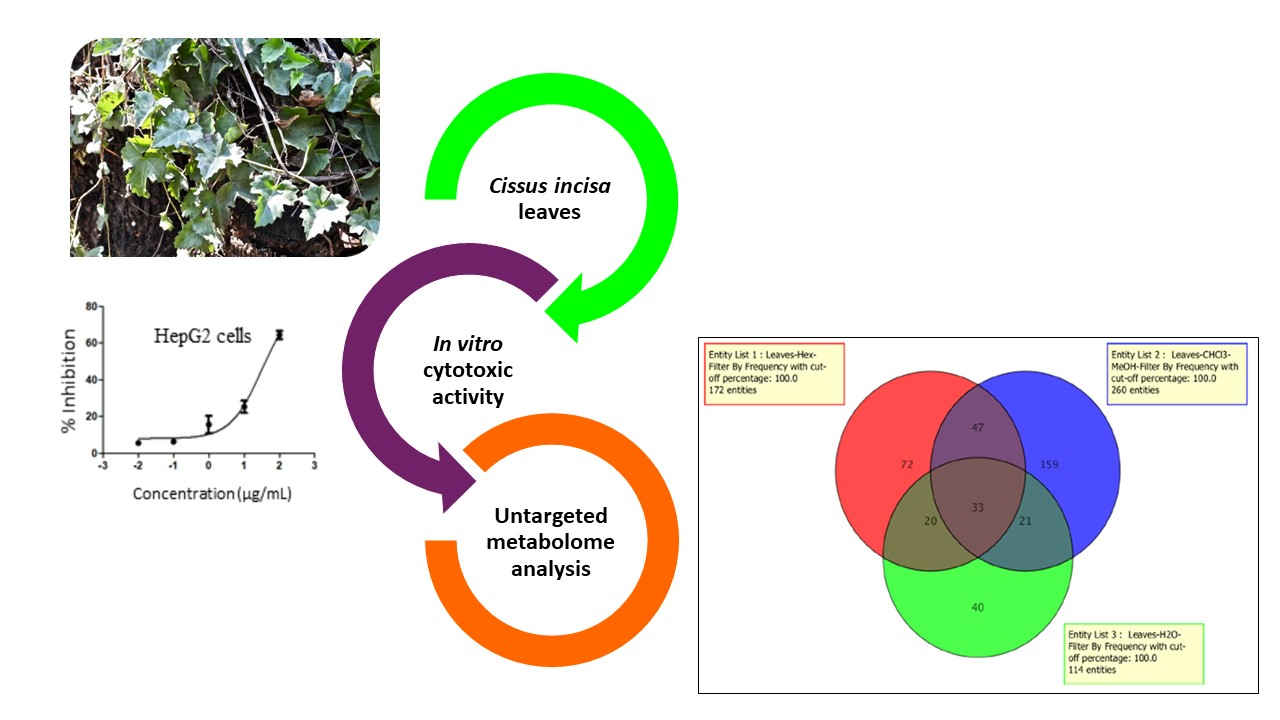
2. Results and Discussion

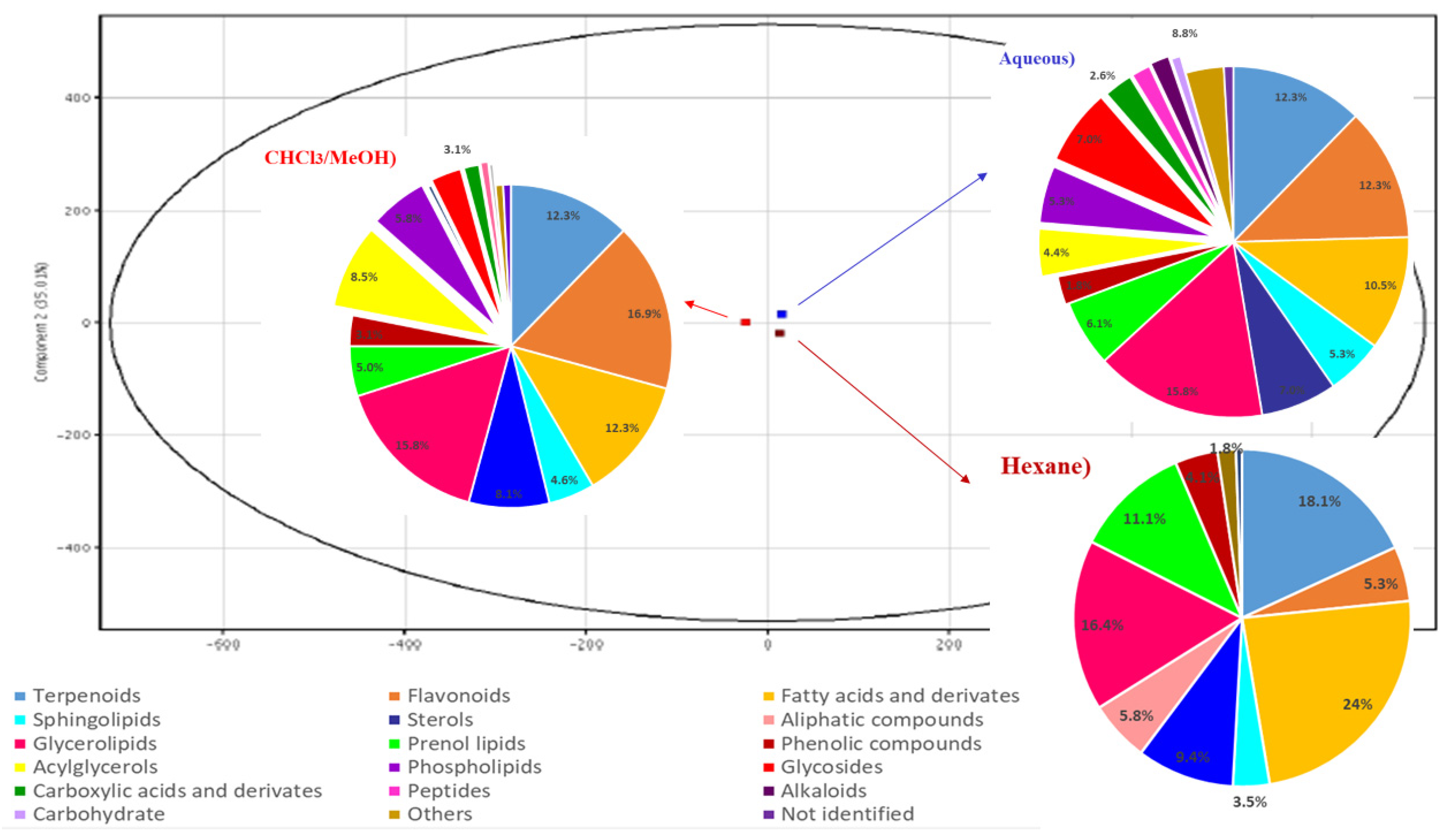
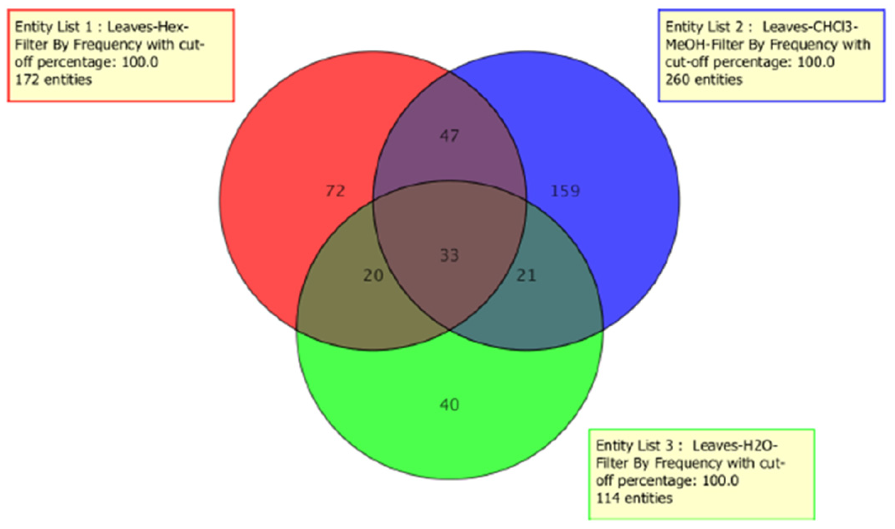
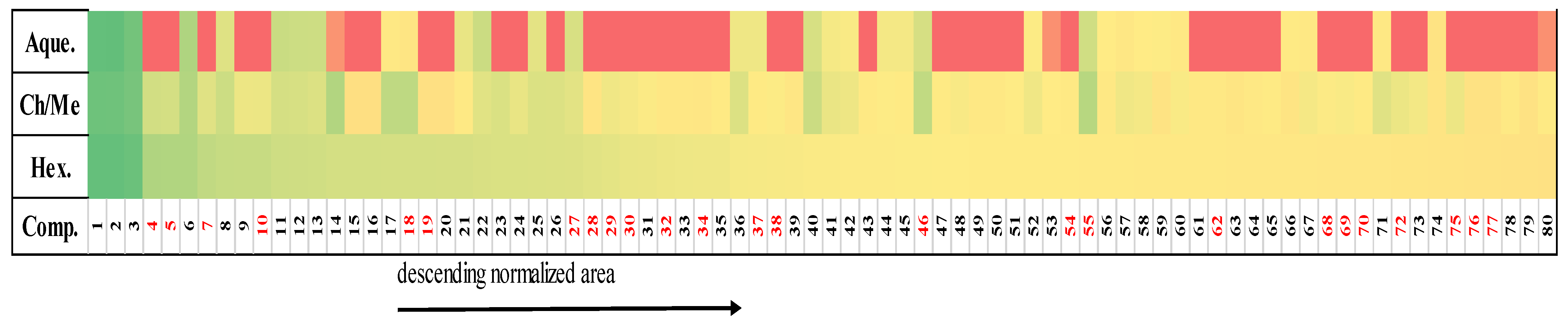
2.1. Metabolomic Profile Analysis of the Extracts

Metabolomic profiling of the extracts from C. incisa leaves by UHPLC-QTOF-MS/MS for dereplication purposes, led to the identification of 171, 260, and 114 metabolites in the hexane, CHCl3/MeOH and aqueous extracts, respectively (Tables S1–S3 in Supplementary Material). Putative identification of compounds detected were made consulting several databases, such as: MEDLINE_Metabolites, Dictionary of Natural Products, KNApSAcK, PubChem, LIPID MAPS, and Human Metabolome Database (HMDB).
Based on the normalized areas data, three common primary metabolites were detected as the most abundant among the three extracts: two glycerophospholipids and a fatty acyl glycoside (Tables S1–S3). The percentages of abundance of each phytocompound in the hexane, CHCl3/MeOH and aqueous extracts were as follows: (0.7491, 0.4864, 1.1213); (0.7480, 0.4856, 1.1222) and (0.7430, 0.4792, 1.0945), respectively. These results are fully comprehensible because glycerophospholipids are the most plentiful phospholipids localized in large amounts in plant cell membranes. In plants, approximately one-third of the organic phosphorus compounds are found in phosphoglycerolipids. In addition, glycerophospholipids participate in cell signaling and as an anchor for proteins in cell membranes [12]. Fatty acyl glucosides, meanwhile, are amphipathic compounds mainly produced by bacteria, yeast, fungi, marine invertebrates, and plants. Recent studies have demonstrated that they play an important role in plant-insect and plant-fungus interactions [13].
Regarding secondary metabolites, α-amyrin acetate and α-tocopherolquinone were the most abundant compounds in the hexane extract. In the CHCl3/MeOH extract, the most abundant secondary metabolites were kazinol A and ursolic acid 3-O-α-L-arabinopyranoside. Meanwhile in the aqueous, armillane and chabrosterol were found to be the most plentiful compounds (Tables S1–S3).
As far as we know, this is the first time that a metabolomic fingerprint of C. incisa leaves is reported, thus contributing to the scientific knowledge of this species. A PCA scores plot was obtained (Figure 1) from a multivariate statistical analysis. The PCA showed close metabolomic profiles for the three analyzed extracts. In Figure 1, a similar composition is observed regarding the presence of fatty acyls, sphingolipids, sterols, glycerolipids, prenol lipids, and terpenoids; although their ratio within the extracts is variable. Thirty-three common compounds between these extracts were found (Figure 2, Table S4). Additionally, 80 common compounds were detected only in the hexane and CHCl3/MeOH, which are included in Table 1.
The findings presented here agree with those reported by Kumar et al. [14] and Chipiti et al. [15] for the leaf extracts of C. quadrangularis and C. cornifolia, respectively.

2.2. Cytotoxic Activity

Cytotoxic activity of C. incisa leaves extracts is also reported here for the first time, which was determined on six human cancer cells. The experimental results are shown in Table 2. According to the National Cancer Institute of the United States of America, an extract is considered active if it achieves an IC50 ≤ 30 μg/mL on tumor cells [16]. In this sense, the hexane extract exhibited cytotoxic activity on Hep3B (IC50 = 27 ± 3 μg/mL) and HepG2 (IC50 = 30 ± 6 μg/mL), being the most active extract. In the case of CHCl3/MeOH extract, it was less active on hepatocellular cancer cells, reaching IC50 = 39 ± 3 μg/mL and 31 ± 2 μg/mL, respectively. Previously, Opoku et al. [17] reported the antiproliferative activity of MeOH extract of C. quadrangularis against the HepG2 cell line with 36.58% of inhibition of proliferation. On the other hand, the hexane extract exhibited certain cytotoxicity on Hela and A549 cancer lines (IC50 = 40 ± 2 and 52 ± 2 μg/mL, respectively), similar to the CHCl3/MeOH extract against MCF7 (IC50 = 50.7 ± 6 μg/mL) and PC3 (57 ± 4 μg/mL).
The Selectivity Index (SI) was determined only for hepatocellular carcinoma cell lines, since they were the most sensitive of all tested (Table 2). It has been reported that SI values less than 2 can indicate toxicity for an extract or a pure compound towards mammal cells [18]. CHCl3/MeOH extract gave a SI = 2.77 on Hep3B, and SI = 2.21 on HepG2, surpassing the values of the control (Paclitaxel) on the same cell lines (2.41 and 1.24, respectively). As consequence, the CHCl3/MeOH extract from C. incisa leaves was the most selective.
The aqueous extract obtained by successive extractions did not show cytotoxic activity in any cancer cell line tested. Different results were obtained by Sáenz et al. [19] evaluating the aqueous extract of C. sicyoides leaves (direct extraction) on HEp-2 cells finding a IC50 = 43.2 ± 2.4 μg/mL. In addition, our aqueous extract did not show cytotoxicity in immortalized cells (IC50 > 100 µg/mL), which is a good first step for further safety studies of the total extract of C. incisa aerial parts.

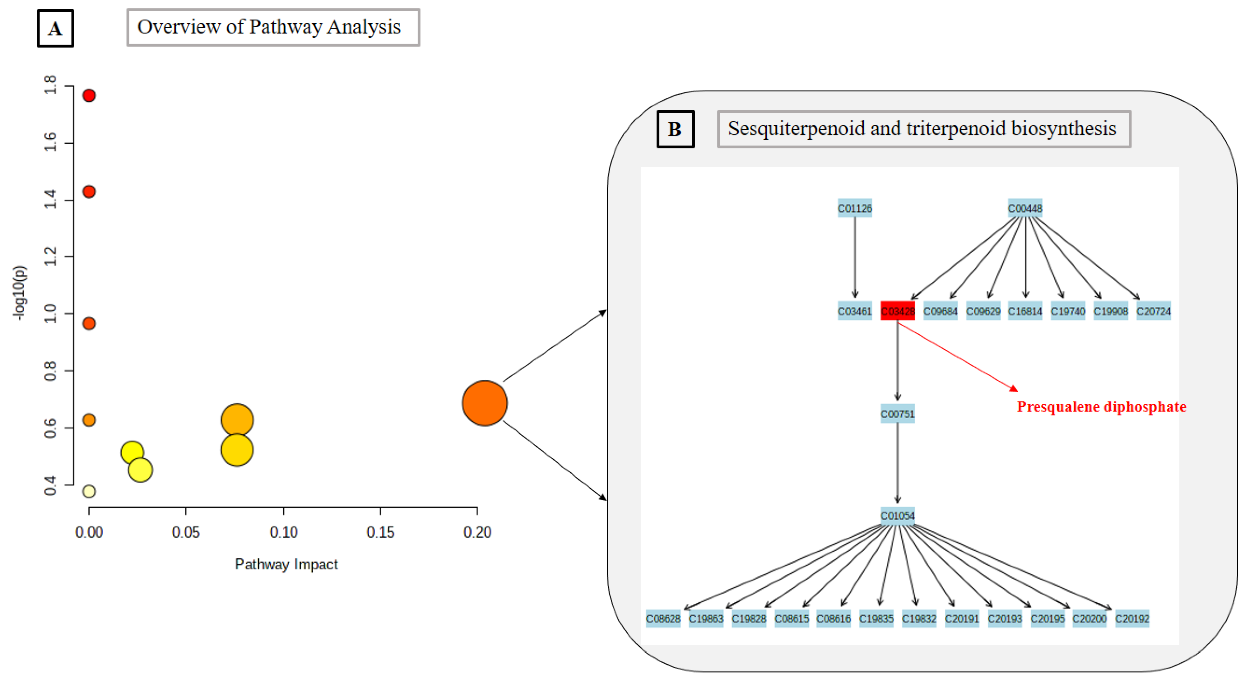
2.3. Metabolomics Pathway Analysis

Based on the biological properties displayed by the hexane and CHCl3/MeOH extracts, we focused on exploring the 80 common metabolites among these extracts, using the Metabolomics Pathway Analysis (MetPA). As a consequence, the most relevant pathways involving these metabolites were identified, in this case, nine networks were revealed (see in Table 1). The threshold of impact was set to 0.10. The pathway is considered to be closely related if its impact value is higher than this value.
The results obtained from MetPA shows four important routes in plants operation, belonging to their primary metabolism: Linoleic acid metabolism, alpha-Linolenic acid metabolism, Glycerophospholipid metabolism, and Fatty acid biosynthesis. However, the most significant pathway identified via MetPA are those related to the biosynthesis of secondary metabolites, specially terpenes and sterols. This is consistent, since terpenoids and sterols from leaves exhibite a multifunctionality role in plants: more specialized chemical interactions and protection in the abiotic and biotic environment [20]. The results from pathway analysis are presented in detail in Table 3, and only the pathway with higher impact is presented graphically (Figure 3).

2.4. Correspondence between Metabolomic Profiling and Cytotoxic Activity

The distribution of the 80 coincident compounds is presented in a heat map (Figure 4), which contains the normalized relative areas of these metabolites, identified in the hexane, CHCl3/MeOH and the aqueous extracts. The heat map also shows the distribution between the three extracts of the cytotoxic metabolites reported against the same cell lines included in this study (or some related ones). It can be seen that most of the cytotoxic compounds are found in a higher proportion within the hexane extract.
As it presented in Table 2, hexane and CHCl3/MeOH extracts had similar cytotoxic results on the hepatocellular cells (even if the hexane extract was more active on Hep3B). These similarities can be explaining by the chemical content, these extracts include 80 common metabolites (Venn diagram Figure 2), showing a correspondence between the metabolomics profiles of the active extracts and the cytotoxic activity on Hep3B and HepG2 cell lines. These cells share common characteristics (for instance Wnt/β-catenin activation [21], providing a unique platform for parallel comparisons, but also HepG2 and Hep3B are from different ethnic origins. Some differential gene expression (for instance; HepG2 cells are known to contain wild-type p53 whereas Hep3B cells are p53 deficient), provide a broad spectrum of mechanisms, particularly for apoptosis induction. Several studies suggested that phytosterols and terpenes disturb the cell cycle and induce apoptosis by activating caspases 3 and 9 in cancer cells. Particularly triterpenes and its derivates glycosides have shown effect against cancer cells and induction of apoptosis mechanism [22]. These phytocompounds are present in both extracts (hexane and CHCl3/MeOH) (Figure 4).
It is necessary to point out that some of these 80 shared metabolites have been previously reported with cytotoxic activity against hepatocellular cancer cells: (5) α-tocopherolquinone, (30) phytol, (29) grandifloric acid, (34) cucurbitacin E, (4) α-amyrin acetate, (37) ursolic acid, (32) δ-linolenic acid, (72) oxyacanthine, (68) stearic acid, and (62) matricin of which, the first six are terpenoids, including three triterpenes. The presence of these cytotoxic metabolites may explain the cytotoxicity of the extracts (numbering is according to the heat map, Figure 4).
α-Tocopherolquinone was dereplicated with molecular formula (C29H50O3) and accurate mass 446.3760. This diterpene has reported good cytotoxic activity on HepG2 cells (IC50 = 6.97 ± 0.5 µg/mL) [23]. Another terpene dereplicated, phytol (C20H40O; accurate mass 296.3079) selectively inhibited the growth of the HepG2 cells with an IC50 value of 78 ± 3.45 μM [24]. Another study showed that phytol exerted antitumor effect in hepatocellular carcinoma cells by activation of caspases 9/3 [25]. The triterpene cucurbitacin E (formula suggested C32H44O8; accurate mass 556.3036) exhibited antiproliferative action on Hep3B cancer cells through inhibition of Wnt/β-catenin activation [26]. Meanwhile, C32H52O2 (468.3967) identified as α-amyrin acetate, and showed moderate activity on HepG2 = 148.9 ± 1.80 µM [27]. Other triterpene, ursolic acid (C30H48O3; accurate mass 456.3603) is distributed among the three extracts; it has been widely studied in relation with anticancer properties. In Hep3B cell lines, ursolic acid has reduced the tumorigenesis in vivo, enhancing apoptosis in tumor tissues [28], and exerting antiangiogenic action [29]. A different work showed that ursolic acid displayed effects on cell viability, DNA fragmentation, mitochondrial membrane potential on human liver cancer HepG2 (IC50 = 4 μM) and Hep3B (IC50 = 8 μM) cells [30]. A study showed in vivo that γ-linolenic acid (C18H30O2; 278.2246) reduced the proliferative and angiogenic effect of carcinoma hepatocellular induced in Wistar rats, by activation of a mitochondrial mediated apoptosis pathway [31]. Likewise, oxyacanthine (C37H40N2O6, 608.28863) attenuated cell proliferation ability and promoted cell apoptosis in mammary, prostatic, liver cancers cells [32], while stearic acid [33] (C18H36O2; 284.2715) and grandifloric acid [34] (terpene; C20H30O3; 318.2194) had the same action on HepG2 cells. A recent study determined the antiproliferative activity of extracts from Australian plants leaves that contained matricin (C17H22O5; 306.1467), a prenol lipid, on HepG2 cells [35].
Other dereplicated compounds with promising anticancer activities reported are: (19) alpinumisoflavone dimethyl ether (C22H20O5; 364.1311) that suppress the proliferation, migration/invasion, tumor angiogenesis and metastasis, and the promotion of apoptosis in various cancers: human oral epidermoid carcinoma KB cells (IC50 = 4.13 μg/mL), and murine leukemia P-388 (IC50 = 4.31 μg/mL) cells [36] and (10) N-(3-hydroxy-dodecanoyl)-homoserine lactone (C16H29NO4; 299.2097) with pro-apoptotic activities [37] (28) Stylisterol A (C28H46O3; 430.3447), (27) stylisterol B (C28H46O4; 446.3396), [38] and (7) gibberellin A12 aldehyde [39] (C20H28O3; 316.2038) have been found to have antiproliferative action in cancer cells, even with apoptosis induction.
In the heat map (Figure 4), it can be observed that the aforementioned metabolites occur in the hexane extract, justifying why this extract is the most active of the three tested. While, only (55) 7-oxo-β-sitosterol [40] (C29H48O2; 428.3654), (46) 5-methoxy-3-(2R-acetoxy-pentadecyl)-1,4-benzoquinone [41] (C24H38O5; 406.2719), (69) 2-hydroxy-6-tridecylbenzoic acid [42] (C20H32O3; 320.2351), (70) stigmastane-3,6-dione [43] (C29H48O2; 428.3654), and (18) 5,7,4′-trimethoxyflavan [44] (C18H20O4; 300.1362) appear preferably in the CHCl3/MeOH extract, and have also displayed anticancer effects. Therefore, all the metabolites presented so far are involved in the cytotoxic activity of the active extracts.
On the other hand, there are other experimental results from cytotoxic assays that are worth discussing. As we presented earlier, two cell lines (HeLa and A549) were more susceptible to hexane extract than CHCl3/MeOH extract (Table 2). In this regard, the fold change analysis detected 30 up-regulated phytocompounds in hexane extract (Table 4). In contrast, PC3 and MCF7 cell lines were more sensitive to the CHCl3/MeOH extract than the hexane one according to Table 2. Thirty-eight up-regulated compounds were identified in the CHCl3/MeOH extract by the fold change analysis. Table 4 also contains these compounds, along with the previous studies against Hela, A549, MCF7 and/or related cell lines.

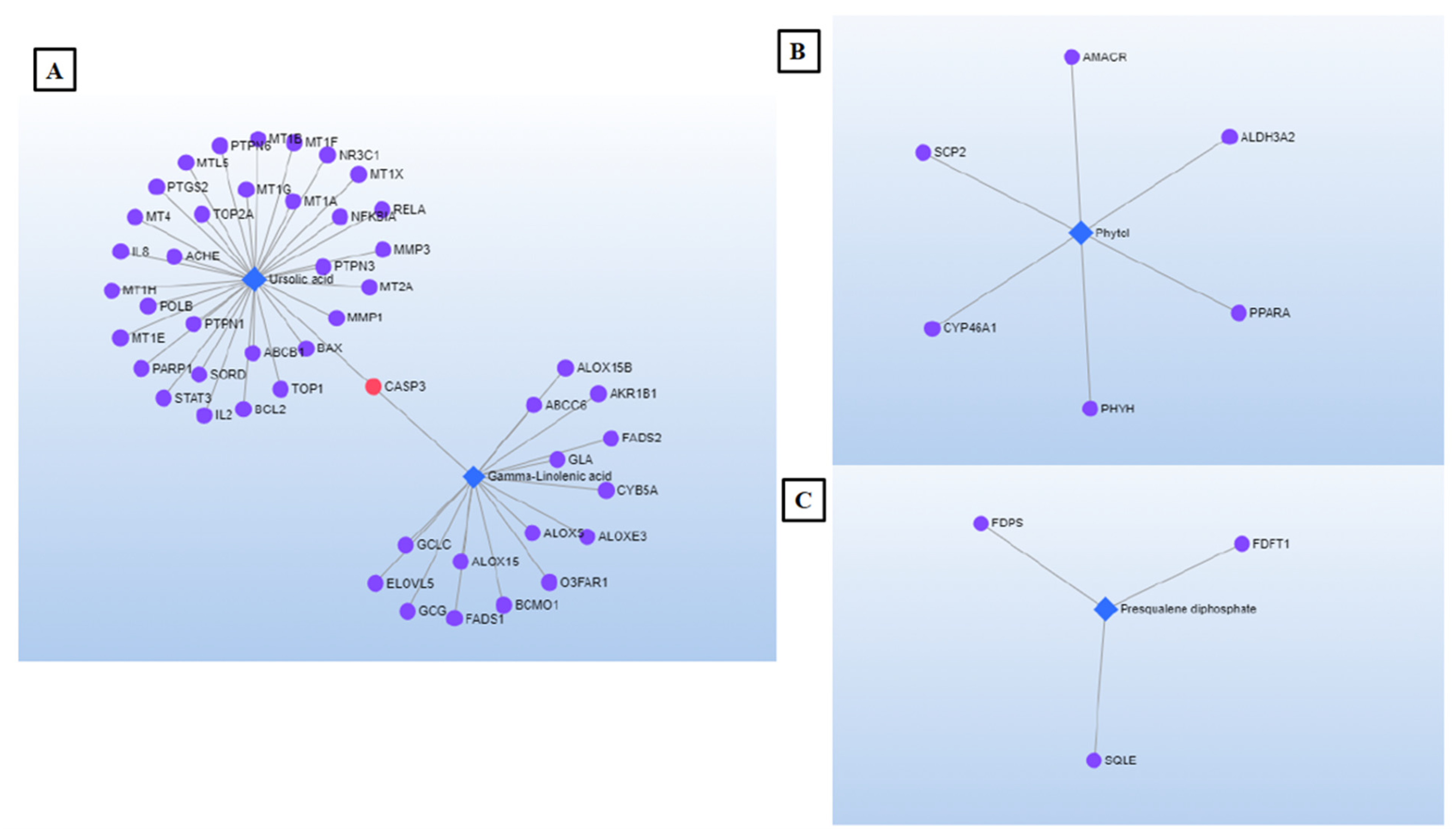
2.5. Network Pharmacology (NP)

NP approach was used to explore metabolite/gen/disease interaction in the cancer context. The results displayed the synergist activity of some metabolites to achieve anticancer effect. Some compounds such as presqualene diphosphate, phytol, stearic acid, δ-tocopherol, ursolic acid and γ-linolenic acid are directly involved in the five sub-networks identified. Figure 5A–C shows three interaction networks selected. Figure 5A is about the most noteworthy network by the largest number of concerned nodes. Some key genes identified in this network are recognized for the National Center for Biotechnology Information (NCBI) for their significant role in drug discovery [53]: CASP3 (caspase 3), the protein encoded by this gene plays a central role in the execution-phase of cell apoptosis. In addition, two nuclear receptors (PARP1; NR3C1) involved in the regulation of several important cellular processes such as differentiation, proliferation, and in the recovery of cell from DNA damage. Likewise, three different genes (BAX, BCL2, STAT3) whose encoded proteins were implicated in cell growth and apoptosis. Two well-known signaling molecules (PTPN6 and PTPN3) were also identified, which regulate a variety of cellular processes including cell growth, differentiation, mitotic cycle, and oncogenic transformation. Last, DNA topoisomerase (TOP2A) was also recognized. The gene encoding this enzyme functions as the target for several anticancer agents and a variety of mutations in this gene have been associated with the development of drug resistance [53].
The analysis of the pharmacological network revealed, as targets, several genes involved in the inflammatory response, which occurs in various pathological conditions, such as cancer (CXCL8, ALOX5 and ALOX15) [53]. Phytol (network Figure 5B), targets PPARα. This gene is implied in cell proliferation, cell differentiation, and immune and inflammation responses. Along with presqualene diphosphate (Figure 5C), phytol targets genes encoding proteins involved in drug metabolism and synthesis of cholesterol, steroids, and other lipids (CYP46A1 and FDFT1, respectively) [53].
Summarizing, the current work presents for the first time the metabolomic fingerprint of C. incisa leaves, and the cytotoxic properties of their extracts. Untargeted metabolomics profiles through UHPLC-QTOF-MS and multivariate analyzes allowed to determine the phytochemical similarities and differences between the three extracts and to understand their cytotoxic effects by the presence of bioactive metabolites. The hexane extract achieved remarkable cytotoxicity on hepatocellular cancer cells, hence, coupling the metabolome data with its biological activity could support a targeted isolation focused on the predicted active metabolites. The NP approach used was successful for the interpretation of the experimental results, because the metabolites that contribute to the cytotoxic activity and the molecular pathways involved were revealed.

3. Materials and Methods

3.1. Vegetal Material and Extracts Preparation

Cissus incisa (Nutt.) Des Moul. Ex S. Watson was collected in Rayones, Nuevo Leon, Mexico (Latitude: 25.0167°, Longitude: −100.05°, Altitude: 900 m) on 10 October 2016. The identification was made by the biologist Ph.D. Mauricio Gonzalez Ferrara (Autonomous University of Nuevo Leon, San Nicolás de los Garza, Mexico). The collected species were deposited in the herbarium of Biological Sciences Faculty of the Autonomous University of Nuevo Leon with Voucher 027499. The plant name has been checked with http://www.theplantlist.org, accessed on 3 February 2020.
Leaves were dried at room temperature for 2 weeks and then milled until obtaining 809 g of dried and grounded plant material. Sequential macerations were made using hexane (10 L), chloroform/methanol (1:1) (7 L), and water (10 L) yielding the organic extracts and the aqueous extract. The extractions were made at room temperature, following the same steps: filtration and vacuum distillation to dryness for the organic extracts. Whereas, a lyophilization was carried out to obtain the dry aqueous extract, yielding 11.6 g of hexane (1.43%), 84 g of CHCl3/MeOH (10.38%), and 19.6 g of aqueous one (2.42%) of dry extracts.

3.2. UHPLC-QTOF-MS/MS Analysis

All solvents LCMS grade Baker (Thermo Fisher Scientific, Waltham, MA, USA) were filtered using membrane filter, NYLON 0.45-micron × 47 mm (DS0215-4045, Thermo Fisher Scientific, Waltham, MA, USA). Three samples per extract were diluted independently (1 mg/mL) in MeOH (50%), sonicated 5 min × 10,000 rpm and filtered using PTFE 0.20 µm Syringe filter (721-1320 Thermo Scientific, Waltham, MA, USA), and transferred to a high-recovery MS Analyzed Type 1 borosilicate amber glass vial (5190-7041/5182-0717, Agilent Technologies, Santa Clara, CA, USA).
Reverse-phase liquid chromatography was performed at 20 °C, using an Agilent 1290 Infinity II Ultra High-Performance Liquid chromatography system (UHPLC Waters, Singapore, Singapore) and the column ZORBAX Eclipse Plus C18 HD 2.1 × 50 mm, 1.8 µm (Agilent Technologies, Santa Clara, CA, USA). The mobile phase was delivered by a binary pump at a flow rate of 0.250 mL/min in a gradient elution using: LCMS grade water + 0.1% v/v formic acid (solvent A) and LCMS grade MeOH + 0.1% v/v formic acid (solvent B) with the following gradient conditions: 0–6 min, from 30 to 100% solvent B; held at 100% B until 10 min; 10–11 min, from 100 to 30% B to return to original conditions. Injection volume was 5 µL. Mass spectrometric analysis was performed using an Agilent 6545 Quadrupole Time of Flight (QTOF) LCMS with an electrospray ionization (ESI) source (Agilent Technologies, Waldbronn, Germany), in positive mode. Detection range of mass-to-charge ratio (m/z) was 100–3000. The nebulizer pressure was set at 35 psi, gas temperature of 320 °C, and a gas flow rate of 8 L/min.

3.3. Data Processing and Metabolic Pathway Analysis

The identification of metabolites was carried out using the METLIN_Metabolites Database on Agilent MassHunter Qualitative Analysis B.08.00 software and the lists for data analysis were generated with compounds present in all the replicates of each extract. Putative assignments for each compound were made based on their accurate mass. Additionally the Dictionary of Natural Products, PubChem, (http://pubchem.ncbi.nlm.nih.gov/, accessed on 11 October 2020), LIPID MAPS (http://www.lipidmaps.org/tools, accessed on 11 October 2020), and Human Metabolome Database (HMDB) (http://www.hmdb.ca, accessed on 11 October 2020) were consulted. Principal component analysis (PCA), Venn diagram, and fold change analysis (cut off 2.0) were carried out for UHPLC-QTOF-MS/MS data on Mass Profiler Professional software. PCA presents the average of replicates by each extract. For all statistical tests performed, ANOVA with cut-off p < 0.05 was taken as significant. The metabolomics pathway analysis and the network topological analysis were performed with MetaboAnalyst (http://www.metaboanalyst.ca/, accessed on 3 February 2021) and STITCH (http://stitch.embl.de/, accessed on 3 February 2021). The metabolite-gene-disease interaction network was selected within the MetPA module, through the integration of network topological analysis, interactive network exploration, and functional enrichment analysis.

3.4. Cytotoxic Activity

3.4.1. Cell Lines

The extracts were evaluated for their cytotoxic activity in human cancer cells: PC3 (prostate ATCC® CRL-1435), Hep3B (hepatocellular ATCC® HB-8064), and HepG2 (hepatocellular ATCC® HB-8065), MCF7 (breast (ATCC® HTB-22), A549 (lung (ATCC® CCL-185), and HeLa (cervical ATCC® CCL-2), all were obtained from ATCC (American Type Culture Collection, Manassas, VA, USA). In addition, a cell line of immortalized human hepatocytes (IHH) was included as control of non-cancerous cells [54]. PC3 cells were cultured in RPMI-1640 medium (Sigma Aldrich, St. Louis, MO, USA), while Hep3B, HepG2, IHH, MCF7, A549 and HeLa in DMEM (Invitrogen, Thermo Fisher Scientific, Inc., Waltham, MA, USA) and supplemented with 10% fetal bovine serum (SFB, Invitrogen, Waltham, MA, USA) and with 2 mM glutamine, all cultures were incubated at 37 °C in a 5% CO2 atmosphere.

3.4.2. IC50 Determination

For the cytotoxic evaluation 4000 cells were cultured per well in 96-well plates. The concentrations used for the extracts and for positive control Paclitaxel were 100, 10, 1, 0.1, 0.01 μg/mL for a dose/response curve.
Prior to the assay, stock solutions of 20 mg/mL (20,000 µg/mL) of each extract were prepared, (1 mg of extract dissolved in 50 µL of DMSO) for organic extracts and sterile water for the aqueous one.
The solutions were prepared from this stock as follows. The concentration 100 µg/mL was prepared from 2.5 µL of a stock solution 20 mg/mL (20,000 µg/mL) in 497.5 µL of culture medium. The concentration 10 µg /mL was prepared from 50 µL of the 100 µg/mL solution in 450 µL of medium. The concentration 1 µg/mL was prepared from 50 µL of the 10 µg/mL solution in 450 µL of medium. The concentration 0.1 µg/mL was prepared from 50 µL of the 1 µg/mL solution in 450 µL of medium. The concentration 0.01 µg/mL was prepared from 50 µL of the 0.1 µg/mL solution in 450 µL of medium.
Subsequently, 100 µL of each solution was added to its corresponding well. Treatment with extracts did not exceed 0.5% of DMSO. In addition, a solvent control was performed at this concentration, not observing cell growth inhibition, which guaranteed that the cytotoxic activity of each extract was associated with the chemical content present in each extract and not with the solvent.
Plates were incubated at 37 °C in 5% CO2 atmosphere for 48 h. The number of viable cells in proliferation was then determined by the Cell Titer 96® aqueous solution cell proliferation assay kit (Promega, Madison, WI, USA) following the supplier’s protocol. Cell viability was determined by absorbance at 450 nm using an automated ELISA reader. The experiments were performed in triplicate in three independent experiments. Data were expressed as means ± SD and were analyzed in the Prism 5.0 statistical program, IC50 values were determined by regression analysis [54].

3.4.3. Selectivity Index

The extracts were tested against IHH normal cell line [54] to determine the selectivity of the cytotoxic activity on hepatocellular lines. The Selectivity Index (SI) was calculated following previous reports [55]: SI = IC50 of extract in a normal cell line/IC50 of the same extract in cancer cell line, where IC50 is the concentration required to kill 50% of the cell population.

Supplementary Materials

The following are available online at https://www.mdpi.com/article/10.3390/plants10071389/s1, Table S1: UHPLC-QTOF-MS/MS results for Hexane extract, Table S2: UHPLC-QTOF-MS/MS results for chloroform/methanol extract, Table S3: UHPLC-QTOF-MS/MS results for aqueous extract, Table S4: Common compounds in the Hexane and CHCl3-MeOH extracts.

Author Contributions

Conceptualization, M.d.R.C.-C.; methodology, M.d.R.C.-C.; software, M.Y.R. and D.N.-M.; validation, J.N.S.-C., and M.Á.R.-C.; formal analysis, D.N.-M. and M.Á.R.-C.; investigation, D.N.-M. and L.G.-M.; resources, L.G.-M. and M.Y.R.; data curation, L.G.-M. and J.N.S.-C.; writing—original draft preparation, D.N.-M.; writing—review and editing, M.d.R.C.-C., M.Y.R., M.Á.R.-C., L.G.-M. and J.N.S.-C.; visualization, D.N.-M.; supervision, M.d.R.C.-C.; project administration, M.d.R.C.-C.; funding acquisition, M.d.R.C.-C. and M.Y.R. All authors have read and agreed to the published version of the manuscript.

Funding

This research was funded by the Universidad Autónoma de Nuevo León (grant number 04-093765-FAR-11/250-FCQ-UANL); and Centro de Investigaciones Químicas-IICBA de la Universidad Autónoma del Estado de Morelos (LANEM Project).

Data Availability Statement

The data presented in this study are available on request from the corresponding authors.

Acknowledgments

D.N.M. thanks CONACYT-MEXICO for the scholarship (605522) to carry out her Ph.D. degree in the Faculty of Chemical Sciences, Autonomous University of Nuevo Leon. The authors thank Tommaso Stefani for his contribution in the preliminary version of this manuscript.

Conflicts of Interest

The authors declare no conflict of interest.

References

  1. Kim, C.; Kim, B. Anti-cancer natural products and their bioactive compounds inducing ER stress-mediated apoptosis: A review. Nutrients 2018, 10, 1021. [Google Scholar] [CrossRef] [PubMed] [Green Version]
  2. Yu, J.; Kinghorn, A.D. Development of Anticancer Agents from Plant-Derived Sesquiterpene Lactones. Curr. Med. Chem. 2016, 23, 2397–2420. [Google Scholar] [CrossRef]
  3. Abdelhafez, O.H.; Othman, E.M.; Fahim, J.R.; Desoukey, S.Y.; Pimentel-Elardo, S.M.; Nodwell, J.R.; Schirmeister, T.; Tawfike, A.; Abdelmohsen, U.R. Metabolomics analysis and biological investigation of three Malvaceae plants. Phytochem. Anal. 2020, 31, 204–214. [Google Scholar] [CrossRef]
  4. Commisso, M.; Strazzer, P.; Toffali, K.; Stocchero, M.; Guzzo, F. Untargeted metabolomics: An emerging approach to determine the composition of herbal pro-ducts. Comput. Struct. Biotechnol. J. 2013, 4, e201301007. [Google Scholar] [CrossRef] [Green Version]
  5. Chandran, U.; Mehendale, N.; Patil, S.; Chaguturu, R.; Patwardhan, B. Network Pharmacology. Innov. Approaches Drug Discov. 2017, 127–164. [Google Scholar] [CrossRef]
  6. PlantDataBase. Cissus Incisa. Available online: https://www.wildflower.org/plants/result.php?id_plant=citr2 (accessed on 12 August 2019).
  7. Alvarado Vázquez, M.A.; Rocha Estrada, A.; Moreno Limón, S. De La Lechuguilla a Las Biopelículas Vegetales: Las Plantas Útiles de Nuevo León; Universidad Autónoma de Nuevo León: Monterrey, Mexico, 2010. [Google Scholar]
  8. Nocedo-Mena, D.; Garza-González, E.; González-Ferrara, M.; Camacho-Corona, M.D.R. Antibacterial Activity of Cissus incisa Extracts against Multidrug- Resistant Bacteria. Curr. Top. Med. Chem. 2020, 20, 318–323. [Google Scholar] [CrossRef] [PubMed]
  9. Nocedo-Mena, D.; Rivas-Galindo, V.M.; Navarro, P.; Garza-González, E.; González-Maya, L.; Ríos, M.Y.; García, A.; Ávalos-Alanís, F.G.; Rodríguez-Rodríguez, J.; Camacho-Corona, M.D.R. Antibacterial and cytotoxic activities of new sphingolipids and other constituents isolated from Cissus incisa leaves. Heliyon 2020, 6, e04671. [Google Scholar] [CrossRef] [PubMed]
  10. Nocedo-Mena, D.; Arrasate, S.; Garza-González, E.; Rivas-Galindo, V.M.; Romo-Mancillas, A.; Munteanu, C.R.; Sotomayor, N.; Lete, E.; Barbolla, I.; Martín, C.A.; et al. Molecular docking, SAR analysis and biophysical approaches in the study of the antibacterial activity of ceramides isolated from Cissus incisa. Bioorganic Chem. 2021, 109, 104745. [Google Scholar] [CrossRef] [PubMed]
  11. Mendez, F.; Garza-González, E.; Ríos, M.Y.; Ramírez-Cisneros, M.Á.; Alvarez, L.; González-Maya, L.; Sánchez-Carranza, J.N.; Camacho-Corona, M.D.R.; González, G. Metabolic profile and evaluation of biological activities of extracts from the stems of cissus trifoliata. Int. J. Mol. Sci. 2020, 21, 930. [Google Scholar] [CrossRef] [Green Version]
  12. Blanco, A.; Blanco, G. Lipids. In Medical Biochemistry; Academic Press: London, UK, 2017; pp. 99–119. [Google Scholar]
  13. Bournonville, C.G.; Filippone, M.P.; Peto, P.D.L.; Ángeles, D.; Trejo, M.F.; Couto, A.S.; de Marchese, A.M.; Ricci, J.C.D.; Welin, B.; Castagnaro, A.P. Strawberry fatty acyl glycosides enhance disease protection, have antibiotic activity and stimulate plant growth. Sci. Rep. 2020, 10, 8196. [Google Scholar] [CrossRef] [PubMed]
  14. Kumar, T.S.; Anandan, A.; Jegadeesan, M. GC-MS Analysis of bioactive components on aerial parts of Cissus quadrangularis LT. Der. Chem. Sin. 2012, 3, 1009–1013. [Google Scholar]
  15. Chipiti, T.; Ibrahim, M.A.; Koorbanally, N.A.; Islam, S. In vitro antioxidant activity and GC-MS analysis of the ethanol and aqueous extracts of Cissus cornifolia (Baker) Splanch (Vitaceae) parts. Acta Pol. Pharm. Drug Res. 2015, 72, 119–127. [Google Scholar]
  16. Suffness, M.; Pezzuto, J. Assays related to cancer drug discovery. In Methods in Plant Biochemistry: Assays for Bioactivity; Academic Press: London, UK, 1990; pp. 71–133. [Google Scholar]
  17. Opoku, A.R.; Geheeb-Keller, M.; Lin, J.; Terblanche, S.E.; Hutchings, A.; Chuturgoon, A.; Pillay, D. Preliminary screening of some traditional Zulu medicinal plants for antineoplastic activities versus the HepG2 cell line. Phytother. Res. 2000, 14, 534–537. [Google Scholar] [CrossRef]
  18. Sahli, R.; Rivière, C.; Dufloer, C.; Beaufay, C.; Neut, C.; Bero, J.; Hennebelle, T.; Roumy, V.; Ksouri, R.; Leclercq, J.; et al. Antiproliferative and Antibacterial Activities of Cirsium scabrum from Tunisia. Evid. Based Complement. Altern. Med. 2017, 2017, 1–9. [Google Scholar] [CrossRef] [Green Version]
  19. Sáenz, M.T.; García, M.D.; Quílez, A.; Ahumada, M.C. Cytotoxic activity of Agave intermixta L. (Agavaceae) and Cissus sicyoides L. (Vitaceae). Phytother. Res. 2000, 14, 552–554. [Google Scholar] [CrossRef]
  20. Toll, D. Biosynthesis and Biological Functions of Terpenoids in Plants. Adv. Biochem. Eng. Biotechnol. 2014, 123, 127–141. [Google Scholar]
  21. Lai, T.; Su, C.; Kuo, W.; Yeh, Y.; Kuo, W.; Tsai, F.; Tsai, C.; Weng, Y. β-catenin plays a key role in metastasis of human hepatocellular carcinoma. Oncol. Rep. 2011, 26, 415–422. [Google Scholar] [PubMed]
  22. Shen, S.; Li, W.; Ouyang, M.-A.; Wang, J. Structure-activity relationship of Triterpenes and derived Glycosides against cancer cells and mechanism of apoptosis induction. Nat. Prod. Res. 2017, 32, 654–661. [Google Scholar] [CrossRef] [PubMed]
  23. Xu, D.-D.; Yan, Y.; Jiang, C.-X.; Liang, J.-J.; Li, H.-F.; Wu, Q.-X.; Zhu, Y. Sesquiterpenes and diterpenes with cytotoxic activities from the aerial parts of Carpesium humile. Fitoterapia 2018, 128, 50–56. [Google Scholar] [CrossRef]
  24. Chen, Y.-C.; Lee, H.-Z.; Chen, H.-C.; Wen, C.-L.; Kuo, Y.-H.; Wang, G.-J. Anti-Inflammatory Components from the Root of Solanum erianthum. Int. J. Mol. Sci. 2013, 14, 12581–12592. [Google Scholar] [CrossRef] [PubMed] [Green Version]
  25. Kim, C.; Lee, H.J.; Jung, J.H.; Kim, Y.H.; Jung, D.B.; Sohn, E.J.; Lee, J.H.; Woo, H.J.; Baek, N.I.; Kim, Y.C.; et al. Activation of Caspase-9/3 and Inhibition of Epithelial Mesenchymal Transition Are Cri-tically Involved in Antitumor Effect of Phytol in Hepatocellular Carcinoma Cells. Phytother. Res. 2015, 29, 1026–1031. [Google Scholar] [CrossRef]
  26. Feng, H.; Zang, L.; Zhao, Z.-X.; Kan, Q.-C. Cucurbitacin-E Inhibits Multiple Cancer Cells Proliferation Through Attenuation of Wnt/β-Catenin Signaling. Cancer Biother. Radiopharm. 2014, 29, 210–214. [Google Scholar] [CrossRef] [PubMed]
  27. Bar, F.M.A.; Abbas, G.M.; Gohar, A.A.; Lahloub, M.-F.I. Antiproliferative activity of stilbene derivatives and other constituents from the stem bark of Morus nigra L. Nat. Prod. Res. 2020, 34, 3506–3513. [Google Scholar] [CrossRef] [PubMed]
  28. Shih, W.-L.; Yu, F.-L.; Chang, C.-D.; Liao, M.-H.; Wu, H.-Y.; Lin, P.-Y. Suppression of AMF/PGI-mediated tumorigenic activities by ursolic acid in cultured hepatoma cells and in a mouse model. Mol. Carcinog. 2012, 52, 800–812. [Google Scholar] [CrossRef] [PubMed]
  29. Lin, C.-C.; Huang, C.-Y.; Mong, M.-C.; Chan, C.-Y.; Yin, M.-C. Antiangiogenic Potential of Three Triterpenic Acids in Human Liver Cancer Cells. J. Agric. Food Chem. 2011, 59, 755–762. [Google Scholar] [CrossRef]
  30. Yan, S.-L.; Huang, C.-Y.; Wu, S.-T.; Yin, M.-C. Oleanolic acid and ursolic acid induce apoptosis in four human liver cancer cell lines. Toxicol. Vitr. 2010, 24, 842–848. [Google Scholar] [CrossRef]
  31. Cui, H.; Han, F.; Zhang, L.; Wang, L.; Kumar, M. Gamma linolenic acid regulates PHD2 mediated hypoxia and mitochondrial apoptosis in DEN induced hepatocellular carcinoma. Drug Des. Devel. Ther. 2018, 12, 4241–4252. [Google Scholar] [CrossRef] [Green Version]
  32. Lv, J.J.; Xu, M.; Wang, D.; Zhu, H.T.; Yang, C.R.; Wang, Y.F.; Li, Y.; Zhang, Y.J. Cytotoxic bisbenzylisoquinoline alkaloids from Stephania epigaea. J. Nat. Prod. 2013, 76, 926–932. [Google Scholar] [CrossRef]
  33. Habib, N.A.; Wood, C.B.; Apostolov, K.; Barker, W.; Hershman, M.J.; Aslam, M.; Heinemann, D.; Fermor, B.; Williamson, R.C.; Jenkins, W.E. Stearic acid and carcinogenesis. Br. J. Cancer 1987, 56, 455–458. [Google Scholar] [CrossRef] [PubMed]
  34. Suo, M.-R.; Tian, Z.; Yang, J.-S.; Lu, Y.; Wu, L.; Li, W. Diterpenes from Helianthus annuus and their cytotoxicity in vitro. Yao Xue Xue Bao Acta Pharm. Sin. 2007, 42, 166–170. [Google Scholar]
  35. Khandanlou, R.; Murthy, V.; Wang, H. Gold nanoparticle-assisted enhancement in bioactive properties of Australian native plant extracts, Tasmannia lanceolata and Backhousia citriodora. Mater. Sci. Eng. C 2020, 112, 110922. [Google Scholar] [CrossRef] [PubMed]
  36. Ateba, S.B.; Mvondo, M.A.; Djiogue, S.; Zingué, S.; Krenn, L.; Njamen, D. A Pharmacological Overview of Alpinumisoflavone, a Natural Prenylated Isoflavonoid. Front. Pharmacol. 2019, 10. [Google Scholar] [CrossRef] [PubMed]
  37. Chen, B.; Cao, X.; Lu, H.; Wen, P.; Qi, X.; Chen, S.; Wu, L.; Li, C.; Xu, A.; Zhao, G. N-(3-oxo-acyl) homoserine lactone induced germ cell apoptosis and suppressed the over-activated RAS/MAPK tumorigenesis via mitochondrial-dependent ROS in C. elegans. Apoptosis 2018, 23, 626–640. [Google Scholar] [CrossRef] [PubMed]
  38. Mitome, H.; Shirato, N.; Hoshino, A.; Miyaoka, H.; Yamada, Y.; Van Soest, R.W. New polyhydroxylated sterols stylisterols A–C and a novel 5,19-cyclosterol hatomasterol from the Okinawan marine sponge Stylissa sp. Steroids 2005, 70, 63–70. [Google Scholar] [CrossRef]
  39. Kim, K.H.; Choi, S.U.; Lee, K.R. Diterpene Glycosides from the Seeds of Pharbitis nil. J. Nat. Prod. 2009, 72, 1121–1127. [Google Scholar] [CrossRef]
  40. Nishiyama, Y.; Noda, Y.; Nakatani, N. Structure of constituents isolated from the bark of Cassipourea malosana and their cy-totoxicity against a human ovarian cell line. J. Nat. Med. 2019, 73, 289–296. [Google Scholar] [CrossRef] [PubMed]
  41. Chang, H.-S.; Lin, Y.-J.; Lee, S.-J.; Yang, C.-W.; Lin, W.-Y.; Tsai, I.-L.; Chen, I.-S. Cytotoxic alkyl benzoquinones and alkyl phenols from Ardisia virens. Phytochemistry 2009, 70, 2064–2071. [Google Scholar] [CrossRef]
  42. Zhou, D.; Jiang, C.; Fu, C.; Chang, P.; Yang, B.; Wu, J.; Zhao, X.; Ma, S. Antiproliferative effect of 2-Hydroxy-6-tridecylbenzoic acid from ginkgo biloba sarcotestas through the aryl hydrocarbon receptor pathway in triple-negative breast cancer cells. Nat. Prod. Res. 2018, 34, 893–897. [Google Scholar] [CrossRef]
  43. Duarte, N.; Ramalhete, C.; Varga, A.; Molnár, J.; Ferreira, M.J.U. Multidrug resistance modulation and apoptosis induction of cancer cells by terpenic compounds isolated from Euphorbia species. Anticancer. Res. 2009, 29, 4467–4472. [Google Scholar]
  44. Zhi-Yong, J.; Xi-Shan, B. Cytotoxic flavanes from Uraria clarkei. J. Asian Nat. Prod. Res. 2013, 15, 979–984. [Google Scholar]
  45. Pejin, B.; Kojic, V.; Bogdanovic, G. An insight into the cytotoxic activity of phytol at in vitro conditions. Nat. Prod. Res. 2014, 28, 2053–2056. [Google Scholar] [CrossRef] [PubMed]
  46. Yang, C.S.; Suh, N. Cancer Prevention by Different Forms of Tocopherols. Top. Curr. Chem. 2012, 329, 21–33. [Google Scholar] [CrossRef]
  47. Gutierrez-Pajares, J.L.; Ben Hassen, C.; Oger, C.; Galano, J.-M.; Durand, T.; Frank, P.G. Oxidized Products of α-Linolenic Acid Negatively Regulate Cellular Survival and Motility of Breast Cancer Cells. Biomolecules 2019, 10, 50. [Google Scholar] [CrossRef] [Green Version]
  48. Ma, G.-L.; Xiong, J.; Osman, E.E.A.; Huang, T.; Yang, G.-X.; Hu, J.-F. LC-MS guided isolation of sinodamines A and B: Chimonanthine-type alkaloids from the endangered ornamental plant Sinocalycanthus chinensis. Phytochemistry 2018, 151, 61–68. [Google Scholar] [CrossRef]
  49. Deyou, T.; Gumula, I.; Pang, F.; Gruhonjic, A.; Mumo, M.; Holleran, J.; Duffy, S.; Fitzpatrick, P.A.; Heydenreich, M.; Landberg, G.; et al. Rotenoids, Flavonoids, and Chalcones from the Root Bark of Millettia usaramensis. J. Nat. Prod. 2015, 78, 2932–2939. [Google Scholar] [CrossRef]
  50. Wang, H.-M.; Zhang, L.; Liu, J.; Yang, Z.-L.; Zhao, H.-Y.; Yang, Y.; Shen, D.; Lu, K.; Fan, Z.-C.; Yao, Q.-W.; et al. Synthesis and anti-cancer activity evaluation of novel prenylated and geranylated chalcone natural products and their analogs. Eur. J. Med. Chem. 2015, 92, 439–448. [Google Scholar] [CrossRef]
  51. Li, Z.; Tran, V.H.; Duke, R.K.; Ng, M.C.; Yang, D.; Duke, C.C. Synthesis and biological activity of hydroxylated derivatives of linoleic acid and conjugated linoleic acids. Chem. Phys. Lipids 2009, 158, 39–45. [Google Scholar] [CrossRef]
  52. Rogovskii, V.; Popov, S.; Sturov, N.; Shimanovskii, N. The Possibility of Preventive and Therapeutic Use of Green Tea Ca-techins in Prostate Cancer. Anticancer Agents Med. Chem. 2019, 19, 1223–1231. [Google Scholar] [CrossRef] [PubMed]
  53. National Center for Biotechnology Information. Available online: https://www.ncbi.nlm.nih.gov/gene (accessed on 20 March 2021).
  54. Basu, A.; Kousuke Saito, M.; Ratna, B.K. Stellate cell apoptosis by a soluble mediator from immortalized human hepatocytes. Apoptosis 2006, 11, 1391–1400. [Google Scholar] [CrossRef] [PubMed]
  55. Badisa, R.B.; Darling-Reed, S.F.; Joseph, P.; Cooperwood, J.S.; Latinwo, L.M.; Goodman, C.B. Selective cytotoxic activities of two novel synthetic drugs on human breast carcinoma MCF-7 cells. Anticancer Res. 2009, 29, 2993–2996. [Google Scholar] [PubMed]
Figure 1. PCA plot of C. incisa leaves extracts. PC1 (64.99%), PC2 (35.01%). Within the PCA graph the hexane extract is represented with brown color, CHCl3/MeOH in red and the aqueous one in blue. Different classes of metabolites identified in each extract are also represented.
Plants 10 01389 g001
Figure 2. Venn diagram showing the common compounds among the three extracts (n = 33), and the common compounds among the most active extracts (hexane and CHCl3/MeOH, n = 80).
Plants 10 01389 g002
Figure 3. Pathway analysis (A) metabolome view, (B) pathway with higher impact.
Plants 10 01389 g003
Figure 4. Heat map with the distribution according to the normalized areas of the 80 shared metabolites, identified in the active extracts. Metabolites with previous reports of cytotoxic activity are highlighted in red. Aque *: Aqueous extract; Ch/Me *: CHCl3/MeOH extract; Hex *: Hexane extract; Comp *: Compounds: 1: 1-(1Z-octadecenyl)-2-(5Z,8Z,11Z,14Z,17Z-eicosapentaenoyl)-glycero-3-phospho-(1′-sn-glycerol) 2: 1,2-dihexadecanoylphosphatidylglycerol phosphate; 3: (3S,5R,6S,7E,9x)-7-megastigmene-3,6,9-triol 9-glucoside; 4: α-amyrin acetate; 5: α-tocopherolquinone; 6: 1-(9Z-hexadecenoyl)-2-(11Z-eicosenoyl)-glycero-3-phosphoserine; 7: gibberellin A12 aldehyde; 8: 16β-16-hydroxy-3-oxo-1,12-oleanadien-28-oic acid; 9: (3E)-4-(2,3-dihydroxy-2,5,5,8a-tetramethyl-decahydronaphthalen-1-yl)but-3-en-2-one; 10: N-(3-hydroxy-dodecanoyl)-homoserine lactone; 11: (all-E)-6′-apo-y-caroten-6′-al; 12: campesteryl p-coumarate; 13: 1-docosanoyl-glycero-3-phospho-(1′-sn-glycerol); 14: 1,2,6a,6b,9,9,12a-heptamethyl-10-[(3,4,5-trihydroxyoxan-2-yl)oxy]-1,2,3,4,4a,5,6,6a,6b,7,8,8a,9,10,11,12,12a,12b,13,14b-icosahydropicene-4a-carboxylate; 15: 7,9,13,17-tetramethyl-7S,14S-dihydroxy-2E,4E,8E,10E,12E,16-octadecahexaenoic acid; 16: calycanthidine; 17: not identified; 18: 5,7,4′-trimethoxyflavan; 19: alpinumisoflavone dimethyl ether; 20: gancaonin R; 21: β-citraurinene; 22: spheroidenone; 23: 6-deoxohomodolichosterone; 24: 3β,18β-3-methoxy-11-oxo-12-oleanen-30-oic acid; 25: (5α,25R)-spirostan-3,6-dione; 26: 5,6-epoxy-5,6-dihydro-10′-apo-β,γ-carotene-3,10′-diol; 27: stylisterol B; 28: stylisterol A; 29: grandifloric acid; 30: phytol; 31: 7′,8′-Dihydro-8′-hydroxycitraniaxanthin; 32:γ-linolenic acid; 33: Amabiline; 34: cucurbitacin E; 35: 2-stearyl citric acid; 36: (1-cyano-2-methylprop-2-en-1-yl) 9Z,12Z-octadecadienoate; 37: ursolic acid; 38: ent-9-L1-phytoP; 39: 17-phenyl heptadecanoic acid; 40: 1-dodecanoyl-glycero-3-phospho-(1′-sn-glycerol); 41: 14-O-(α-L-rhamnopyranosyl)-7S,14R-dihydroxy-7,9,13,17-tetramethyl-2E,4E,8E,10E,12E,16E-octadecahexaenoic acid; 42: 1-octadecanoyl-2-docosanoyl-sn-glycero-3-phosphate; 43: 10-methoxyheptadec-1-en-4,6-diyne-3,9-diol; 44: (12S,15S)-15-O-demethyl-10,29-dideoxy-11,12-dihydro-striatin C; 45: fragarin; 46: 5-methoxy-3-(2R-acetoxy-pentadecyl)-1,4-benzoquinone; 47: Yucalexin B16; 48: Junceic acid; 49: Yucalexin B5; 50: N-tetradecanoyl glutamine; 51: 10,13-Epoxy-11-methyloctadeca-10,12-dienoic acid; 52: 19α-19-hydroxy-3,11-dioxo-12-ursen-28-oic acid; 53: 1-monoacylglycerol; 54: (−)-folicanthine; 55: 7-oxo-β-sitosterol; 56: 1-dodecanoyl-sn-glycero-3-phosphocholine; 57: 1-(9Z,12Z-octadecadienoyl)-rac-glycerol; 58: 1-(11Z,14Z-eicosadienoyl)-glycero-3-phosphate; 59: Flavoxate; 60: (−)-Epicatechin 3′-O-sulfate; 61: 2-Heptadecylfuran; 62: matricin; 63: heneicosan-2-one; 64: austroinulin; 65: heliotrine; 66: doristerol; 67: crispane; 68: stearic acid; 69: 2-hydroxy-6-tridecylbenzoic acid; 70: stigmastane-3,6-dione; 71: 1-pentadecanoyl-2-arachidonoyl-sn-glycero-3-phosphate; 72: oxyacanthine; 73: all-trans-Heptaprenyl diphosphate; 74: cavipetin D; 75: presqualene diphosphate; 76: lucidone A; 77: δ-tocopherol; 78: 1-(4Z,7Z,10Z,13Z,16Z,19Z-docosahexaenoyl)-2-(13Z-docosenoyl)-sn-glycero-3-phosphocholine; 79: (22E,24R)-stigmasta-4,22-diene-3,6-dione; 80: 2-monopalmitoylglycerol. * tentative assignment based on accurate mass.
Plants 10 01389 g004
Figure 5. (AC). Graphs of the selected interaction networks.
Plants 10 01389 g005
Table 1. Common compounds identified in the hexane and CHCl3/MeOH extracts (UHPLC-QTOF-MS/MS)- and metabolomic pathways.
Identified MetabolitesMolecular FormulaAccurate MassMetabolite ClassRelated Pathway
α-Tocopherolquinone *C29H50O3446.3760Diterpenoid
Alpinumisoflavone dimethyl ether *C22H20O5364.1311Flavonoid
7,9,13,17-tetramethyl-7S,14S-dihydroxy-2E,4E,8E,10E,12E,16-octadecahexaenoic acid *C22H32O4360.2301Fatty acid derivative
7-oxo-β-SitosterolC29H48O2428.3654Sterol
Heneicosan-2-one *C21H42O310.3236Fatty Acyl
Gancaonin R *C24H30O4382.2144Stilbene
1-MonoacylglycerolC21H36O4352.2614Acyl glycerol
14-O-(α-L-rhamnopyranosyl)-7S,14R-dihydroxy-7,9,13,17-tetramethyl-2E,4E,8E,10E,12E,16E-octadecahexaenoic acidC28H42O8506.2880Fatty acid glycoside
Calycanthidine *C23H28N4360.2314Alkaloid
1,2,6α,6β,9,9,12α-Heptamethyl-10-[(3,4,5-trihydroxyoxan-2-yl)oxy]-1,2,3,4,4α,5,6,6α,6β,7,8,8α,9,10,11,12,12α,12β,13,14β-icosahydropicene-4α-carboxylateC35H56O7588.4026Terpenoid
2-Heptadecylfuran *C21H38O306.2923Heteroaromatic compound
5-Methoxy-3-(2R-acetoxy-pentadecyl)-1,4-benzoquinoneC24H38O5406.2719Quinone
Phytol *C20H40O296.3079Diterpenoid
Oxyacanthine *C37H40N2O6608.2886Lignan
Yucalexin B16 *C20H28O2300.2089Diterpenoid
Campesteryl p-coumarateC37H54O3546.4072Steroid ester
1-dodecanoyl-glycero-3-phospho-(1′-sn-glycerol)C18H37O9P428.2175Glycerophospholipid
10,13-Epoxy-11-methyloctadeca-10,12-dienoic acid *C19H32O3308.2351Fatty Acyl derivative
SpheroidenoneC41H58O2582.4437Carotene derivative
2-MonopalmitoylglycerolC19H38O4330.2771Monoglyceride
DoristerolC27H46O386.3549 Sterol
(12S,15S)-15-O-demethyl-10,29-dideoxy-11,12-dihydro-striatin CC25H38O6434.2668Terpene
δ-Tocopherol *C27H46O2402.3498Prenol lipidUbiquinone and other terpenoid-quinone biosynthesis
16β-16-Hydroxy-3-oxo-1,12-oleanadien-28-oic acidC30H44O4468.3240Triterpene
(3E)-4-(2,3-dihydroxy-2,5,5,8α-tetramethyl-decahydronaphthalen-1-yl)but-3-en-2-one *C18H30O3294.2195Sesquiterpenoid
3β,18β-3-Methoxy-11-oxo-12-oleanen-30-oic acid *C31H48O5484.7104Triterpenoid
(1-cyano-2-methylprop-2-en-1-yl) 9Z,12Z-octadecadienoateC23H37NO2359.2824Fatty Acyl
FragarinC21H21O10434.1207Flavonoid
FlavoxateC24H25NO4391.1784Flavonoid
β-CitraurineneC30H42O418.3236Triterpenoid
N-(3-hydroxy-dodecanoyl)-homoserine lactone *C16H29NO4299.2097Fatty Acyl
1-(11Z,14Z-eicosadienoyl)-glycero-3-phosphateC23H43O7P462.2746GlycerophospholipidGlycerophospholipid metabolism
Cavipetin DC25H38O5418.2719Diterpenoid
10-Methoxyheptadec-1-en-4,6-diyne-3,9-diol *C18H28O3292.2038Fatty Acyl
1-pentadecanoyl-2-arachidonoyl-sn-glycero-3-phosphateC38H67O8P682.4574Glycerophospholipid
Diisobutyl phthalateC16H22O4278.1516Pollutant
Lucidone A *C24H34O5402.2406Sesquiterpenoid
1-(9Z,12Z-octadecadienoyl)-rac-glycerolC21H38O4354.2770Glycerolipid
(3S,5R,6S,7E,9x)-7-Megastigmene-3,6,9-triol9-glucosideC19H34O8390.2253Fatty acyl glycosides
5,7,4′-TrimethoxyflavanC18H20O4300.1362Flavonoid
all-trans-Heptaprenyl diphosphate *C35H60O7P2654.3814Prenol lipid
1-(1Z-octadecenyl)-2-(5Z,8Z,11Z,14Z,17Z-eicosapentaenoyl)-glycero-3-phospho-(1′-sn-glycerol)C44H77O9P780.5305Glycerophospholipid
Heliotrine *C16H27NO5313.1889Member of pyrrolizines
(all-E)-6′-Apo-y-caroten-6′-alC32H42O442.3272Prenol lipid
1-dodecanoyl-sn-glycero-3-phosphocholineC20H42NO7P439.2699Glycerophospholipid
1-(9Z-hexadecenoyl)-2-(11Z-eicosenoyl)-glycero-3-phosphoserineC42H78NO10P787.5363Glycerophospholipid
Stylisterol BC28H46O4446.3396Sterol Lipid
Stylisterol A *C28H46O3430.3447Sterol Lipid
Cucurbitacin E *C32H44O8556.3036Triterpenoid
(22E,24R)-Stigmasta-4,22-diene-3,6-dioneC29H44O2424.3341Lipid
Junceic acid *C21H30O3330.2195Prenol lipid
2-Hydroxy-6-tridecylbenzoic acid *C20H32O3320.2351phenolic compound
1-docosanoyl-glycero-3-phospho-(1′-sn-glycerol)C28H57O9P568.3740Glycerophospholipid
Gibberellin A12 aldehyde *C20H28O3316.2038Prenol lipidDiterpenoid biosynthesis
Matricin *C17H22O5306.1467Prenol lipid
19-α-19-hydroxy-3,11-dioxo-12-ursen-28-oic acidC30H44O5484.3188Triterpenoid
α-Amyrin acetate *C32H52O2468.3967Triterpenoid
(5α,25R)-Spirostan-3,6-dione C27H40O4428.2926Sterol
Ent-9-L1-phytoP * C18H28O4308.1988Fatty Acyl
γ-Linolenic Acid *C18H30O2278.2246Fatty acid Biosynthesis of unsaturated fatty acids
CrispaneC20H32O3320.2351Terpene
Austroinulin *C20H34O3322.2508Diterpenoid
Presqualene diphosphate *C30H52O7P2586.3188TerpenoidSesquiterpenoid and triterpenoid biosynthesis; Steroid biosynthesis
(−)-Folicanthine *C24H30N4374.2470Indoles derivative
1-Octadecanoyl-2-docosanoyl-sn-glycero-3-phosphateC43H85O8P760.5982Glycerophospholipid
Ursolic acidC30H48O3456.3603Triterpenoid
Amabiline *C15H25NO4283.1784Carboxylic ester
(−)-Epicatechin 3′-O-sulfateC15H14O9S370.0358Flavonoid
Stearic acid *C18H36O2284.2715Fatty acid Biosynthesis of unsaturated fatty acids
Grandifloric acid *C20H30O3318.2194Terpene
1,2-Dihexadecanoylphosphatidylglycerol phosphateC38H76O13P2802.4761Glycerophospholipid
Yucalexin B5 *C20H26O3314.1881Terpene
6-Deoxohomodolichosterone *C29H50O4462.3709Sterol Lipid
7′,8′-Dihydro-8′-hydroxycitraniaxanthin *C33H44O3488.3290Triterpenoid
5,6-Epoxy-5,6-dihydro-10′-apo-β,γ-carotene-3,10′-diol *C27H38O3410.2820Carotenoid
N-tetradecanoyl glutamine *C19H36N2O4356.2675Fatty Acyl
Stigmastane-3,6-dione *C29H48O2428.3654Sterol Lipid
2-Stearyl citric acid *C24H44O7444.3087Tricarboxylic acid
1-(4Z,7Z,10Z,13Z,16Z,19Z-Docosahexaenoyl)-2-(13Z-docosenoyl)-sn-glycero-3-phosphocholine *C52H90NO8P887.6404Glycerophospholipid
17-Phenyl heptadecanoic acid *C23H38O2346.2872Fatty Acyl
* Not detected in aqueous extract.
Table 2. Cytotoxic activity of C. incisa leaves extracts.
Cell LinesHexane ExtractCHCl3/MeOH ExtractAqueous ExtractPaclitaxel
IC50
(µg/mL)
SIIC50
(µg/mL)
SIIC50
(µg/mL)
SIIC50
(µg/mL)
SI
HepG230 ± 61.539 ± 32.21>100ND64 × 10−31.24
Hep3B27 ± 31.6631 ± 22.77>100ND33 × 10−32.41
HeLa40 ± 2ND61 ± 4ND>100ND4.78 × 10−3ND
A54952 ± 2ND77 ± 6ND>100ND5.12 × 10−3ND
PC376 ± 5ND57 ± 4ND>100ND10.2 × 10−3ND
MCF774 ± 6ND50.7 ± 6ND>100ND4.27 × 10−3ND
IHH45 ± 3 86 ± 5 >100 79.4 × 10⁻³
Values expressed are ±SD of three independent experiments (n = 3); ND = not determined.
Table 3. Results from Pathway Analysis with MetaboAnalyst.
No.Pathway NameTotal *ExpectedHits *Raw p *Holm p *FDR p *Impact *
1Biosynthesis of unsaturated fatty acids220.2121.72 × 10−21.001.000.00
2Linoleic acid metabolism40.0413.73 × 10−21.001.000.00
3Sesquiterpenoid and triterpenoid biosynthesis240.2312.05 × 10−11.001.000.20374
4alpha-Linolenic acid metabolism280.2612.35 × 10−11.001.000.00
5Diterpenoid biosynthesis280.2612.35 × 10−11.001.000.07625
6Glycerophospholipid metabolism370.3512.99 × 10−11.001.000.07614
7Ubiquinone and other terpenoid-quinone
biosynthesis
380.3613.06 × 10−11.001.000.02227
8Steroid biosynthesis450.4213.52 × 10−11.001.000.02644
9Fatty acid biosynthesis560.5614.18 × 10−11.001.000.00
* Total is the total number of compounds in the pathway; the Hits is the actually matched number from the user uploaded data; the Raw p is the original p value calculated from the enrichment analysis; the Holm p is the p value adjusted by Holm-Bonferroni method; the FDR p is the p value adjusted using False Discovery Rate; the Impact is the pathway impact value calculated from pathway topology analysis.
Table 4. Fold change analysis results in the hexane and CHCl3/MeOH extracts.
MetabolitesMolecular FormulaAccurate MassUp-Regulation [Hexane Extract]Up-Regulation [CHCl3/MeOH Extract]Biological Activity/
References
HeLa, A549 and/or related cell lines
α-TocopherolquinoneC29H50O3446.3760Yes-[23]
Alpinumisoflavone dimethyl etherC22H20O5364.1311Yes-H2108 (IC50 = 33.5 µM);
H1299 (IC50 = 38.8 µM) [36]
7,9,13,17-tetramethyl-7S,14S-dihydroxy-2E,4E,8E,10E,12E,16-octadecahexaenoic acidC22H32O4360.2301Yes-
Heneicosan-2-oneC21H42O310.3236Yes-
Gancaonin RC24H30O4382.2144Yes-
CalycanthidineC23H28N4360.2314Yes-
2-HeptadecylfuranC21H38O306.2923Yes-
PhytolC20H40O296.3079Yes-Hela (IC50 = 15.51 ± 0.76 µM);
A549 (IC50 = 56.98 ± 2.68 µM) [45]
Yucalexin B16C20H28O2300.2089Yes-
δ-TocopherolC27H46O2402.3498Yes-[46]
(3E)-4-(2,3-dihydroxy-2,5,5,8α-tetramethyl-decahydronaphthalen-1-yl)but-3-en-2-oneC18H30O3294.2195Yes-
3β,18β-3-Methoxy-11-oxo-12-oleanen-30-oic acidC31H48O5484.7104Yes-
N-(3-hydroxy-dodecanoyl)-homoserine lactoneC16H29NO4299.2097Yes-[37]
10-methoxyheptadec-1-en-4,6-diyne-3,9-diolC18H28O3292.2038Yes-
Lucidone AC24H34O5402.2406Yes-
Stylisterol AC28H46O3430.3447Yes-HeLa (IC50 = 14.1 µM) [38]
Cucurbitacin EC32H44O8556.3036Yes-[26]
gibberellin A12 aldehydeC20H28O3316.2038Yes-[39]
α-Amyrin acetateC32H52O2468.3967Yes-[27]
Ent-9-L1-phytoPC18H28O4308.1988Yes-[47]
γ-Linolenic AcidC18H30O2278.2246Yes-[31]
(−)-FolicanthineC24H30N4374.2470Yes-A549 (IC50 = 7.76 µM) [48]
AmabilineC15H25NO4283.1784Yes-
Grandifloric acidC20H30O3318.2194Yes-[34]
Yucalexin B5C20H26O3314.1881Yes-
7′,8′-Dihydro-8′-hydroxycitraniaxanthinC33H44O3488.3290Yes-
5,6-Epoxy-5,6-dihydro-10′-apo-β,γ-carotene-3,10′-diolC27H38O3410.2820Yes-
N-tetradecanoyl glutamineC19H36N2O4356.2675Yes-
2-Stearyl citric acidC24H44O7444.3087Yes-
17-phenyl heptadecanoic acidC23H38O2346.2872Yes-
PC3, MCF7 and/or related cell lines
4′-O-GeranylisoliquiritigeninC25H28O4392.1988-YesMDB-MB-231
(IC50 = 125.5 µM) [49]
1-MonoacylglycerolC20H34NO4352.2619-Yes
Sanguisorbin BC35H56O7588.4026-Yes
OxyacanthineC37H40N2O6608.2886-Yes[32]
3-Methyl-5-pentyl-2-furannonanoic acidC19H32O3308.2351-Yes
2-MonopalmitoylglycerolC19H38O4330.2771-Yes
15-hydroxy-5,9-dimethyl-14-methylidenetetracyclo[11.2.1.01,10.04,9]hexadecane-5-carboxylic acidC20H30O3318.2194-Yes
4-hydroxy-8-cis-sphingenineC18H37NO3315.2773-Yes
N-(5-aminopentyl)-N’-(5-{[4-({5-[butylidene(oxido)-lambda(5)-azanyl]pentyl}amino)-4-oxobutanoyl](hydroxy)amino}pentyl)-N-hydroxybutanediamideC27H52N6O7572.3897-Yes
1,2-di-(9Z-pentadecenoyl)-sn-glycerolC33H60O5536.4441-Yes
4,2′,4′-Trihydroxy-3′,5′-diprenylchalconeC25H28O4392.1988-Yes[50]
5,7,4′-TrimethoxyflavanC18H20O4300.1362-Yes
all-trans-Heptaprenyl diphosphateC35H60O7P2654.3814-Yes
HeliotrineC16H27NO5313.1889-Yes
(22E,24R)-Stigmasta-4,22-diene-3,6-dioneC29H44O2424.3341-Yes
Methyl 9R-hydroxy-10E,12E-octadecadienoateC19H34O3310.2508-Yes[51]
1,2-di-(9Z,12Z-octadecadienoyl)-sn-glycerolC39H68O5616.5067-Yes
N-(1,3-dihydroxypropan-2-yl)hexadecanamideC21H30O3330.2195-Yes
2-Hydroxy-6-tridecylbenzoic acidC20H32O3320.2351-YesMDA-MB-231
(IC50 = 117.25 µM); 4T-1 (IC50 = 102.39 µM) [42]
β-isorenierataneC40H72552.5634-Yes
MatricinC17H22O5306.1467-Yes[35]
9,10,13-trihydroxy-octadecanoic acidC18H36O5332.2563-Yes
AustroinulinC20H34O3322.2508-Yes
Butyl 3-O-β-D-glucopyranosyl-butanoateC14H26O8322.1628-Yes
Presqualene diphosphateC30H52O7P2586.3188-Yes
2-O-(β-D-galactopyranosyl-(1->6)-β-D-galactopyranosyl) 2S,3R-dihydroxynonanoic acidC21H38O14514.2262-Yes
Catechin 3-O-rutinosideC27H34O15598.1898-Yes[52]
Stearic acidC18H36O2284.2715-Yes[33]
1-O-(2R-methoxy-4Z-heneicosenyl)-sn-glycerolC25H50O4414.3709-Yes
AmmothamnidinC25H28O5408.1937-Yes
6-DeoxohomodolichosteroneC29H50O4462.3709-Yes
1-hexadecyl-glycero-3-phospho-(1′-sn-glycerol)C22H47O8P470.3009-Yes
28-Glucopyranosyl-3-methyloleanolic acidC37H60O8632.4285-Yes
Stigmastane-3,6-dioneC29H48O2428.3654-Yes
N-(dodecanoyl)-sphing-4-enineC30H59NO3481.4503-Yes
PhyllospadineC21H21NO6383.1369-Yes
Tricosylic acidC23H46O2354.3498-Yes
1-Docosahexaenoyl-2-erucoyl-sn-glycero-3-phosphocholineC52H90NO8P887.6404-Yes
Publisher’s Note: MDPI stays neutral with regard to jurisdictional claims in published maps and institutional affiliations.

