Review

Feedback Regulation of Signaling Pathways for Precise Pre-Placodal Ectoderm Formation in Vertebrate Embryos

Graduate School of Arts and Sciences, The University of Tokyo, 3-8-1, Komaba, Meguro-ku, Tokyo 153-8902, Japan
*
Author to whom correspondence should be addressed.
J. Dev. Biol. 2022, 10(3), 35; https://doi.org/10.3390/jdb10030035
Submission received: 25 July 2022 / Revised: 19 August 2022 / Accepted: 24 August 2022 / Published: 26 August 2022

Abstract

:
Intracellular signaling pathways are essential to establish embryonic patterning, including embryonic axis formation. Ectodermal patterning is also governed by a series of morphogens. Four ectodermal regions are thought to be controlled by morphogen gradients, but some perturbations are expected to occur during dynamic morphogenetic movement. Therefore, a mechanism to define areas precisely and reproducibly in embryos, including feedback regulation of signaling pathways, is necessary. In this review, we outline ectoderm pattern formation and signaling pathways involved in the establishment of the pre-placodal ectoderm (PPE). We also provide an example of feedback regulation of signaling pathways for robust formation of the PPE, showing the importance of this regulation.

1. Introduction

Embryonic patterning is one of the most crucial steps for constructing a complex body shape from a simple egg. The fundamental concept of embryonic fate determination involves localization of signaling molecules inside an egg and differential activation of pathways in each embryonic area, directing localized expression of specific genes. To establish cell fates precisely, strict regulation of signaling strength in each area is essential [1]. There are two types of pattern formation, self-organization and boundary organization [2]. In the “Turing pattern”, a primary example of self-organization, it is possible to create a periodic pattern such as a fish skin pigmentation pattern, simply by employing at least two molecules that differ in diffusion rates and activities [3,4]. This model is very simple; it is impossible to form the precise pattern reproducibly. The second is the so-called “French-flag model”, by which cells generate a pattern due to the strength of a morphogen gradient. This model allows definition of fixed areas more reproducibly than self-organization. In ectoderm patterning, the principle of boundary formation is adopted.
Ectoderm patterning is established after fertilization in vertebrates. The ectoderm consists of four distinct regions, the neural plate (NP), the neural crest (NC), the pre-placodal ectoderm (PPE, also called the pre-placodal region (PPR)), and the epidermis. Patterning is dependent on positional information provided by several types of signaling molecules secreted from mesendodermal tissues. Major signaling types involved in ectodermal patterning include bone morphogenetic protein (BMP), fibroblast growth factor (FGF), retinoic acid (RA), and Wnt. In addition to the ligands themselves, antagonists of each morphogen also contribute to gradient formation in embryos. For example, in Xenopus gastrula, several proteins such as chordin, noggin, and follistatin allow formation of BMP gradients. Both FGF and Wnt signaling are important for anterior–posterior neural patterning. Wnt antagonists (dkk, cer, frzb etc.), secreted from anterior mesendoderm, induce anterior neural structure, including the brain [5,6]. PPE formation requires cooperative actions of BMP, FGF, Wnt, and RA signaling to determine the position of the PPE in naïve ectoderm [7].
The question is whether only the concentration of these molecules enables establishment of the precise ectoderm pattern, because fluctuations of concentration occur, according to various, unexpected environmental factors, resulting in uncertainty in the region of each tissue. To avoid untenable fluctuations, molecular mechanisms must be able to counter such influences. There are several strategies to establish robustness against noise in embryonic patterning (Figure 1).
One of these is establishment of steep gradients (Figure 1A). The larger the difference in morphogen concentration among cells, the more easily each cell is able to detect differences in signal levels [8]. Another strategy is mutual inhibition by two transcription factors (Figure 1B). At an early stage, both genes are expressed in the same cells, whereas expression of one of these genes is decreased, resulting in boundary formation between two regions that each express one of these genes. The third strategy is “cell sorting” (Figure 1C). Gathering cells that receive similar levels of morphogen enables a region to absorb (or average) the noise of patterning, e.g., a salt and pepper cell array around the boundary. The fourth strategy is “local” regulation of signaling, including feedback regulation of intracellular signaling pathways, especially in two regions. Positive feedback regulation makes the two regions more discrete, whereas negative feedback enables them to maintain stable levels of signaling against local turbulence of signal intensity (Figure 1D,E) [2]. Among ectodermal regions, the PPE and the NC are narrow; therefore, a system to precisely form them is more critical than in the NP and the epidermis. In this review, we will focus mainly on PPE formation and will discuss the importance of feedback regulation for local control of appropriate signaling.

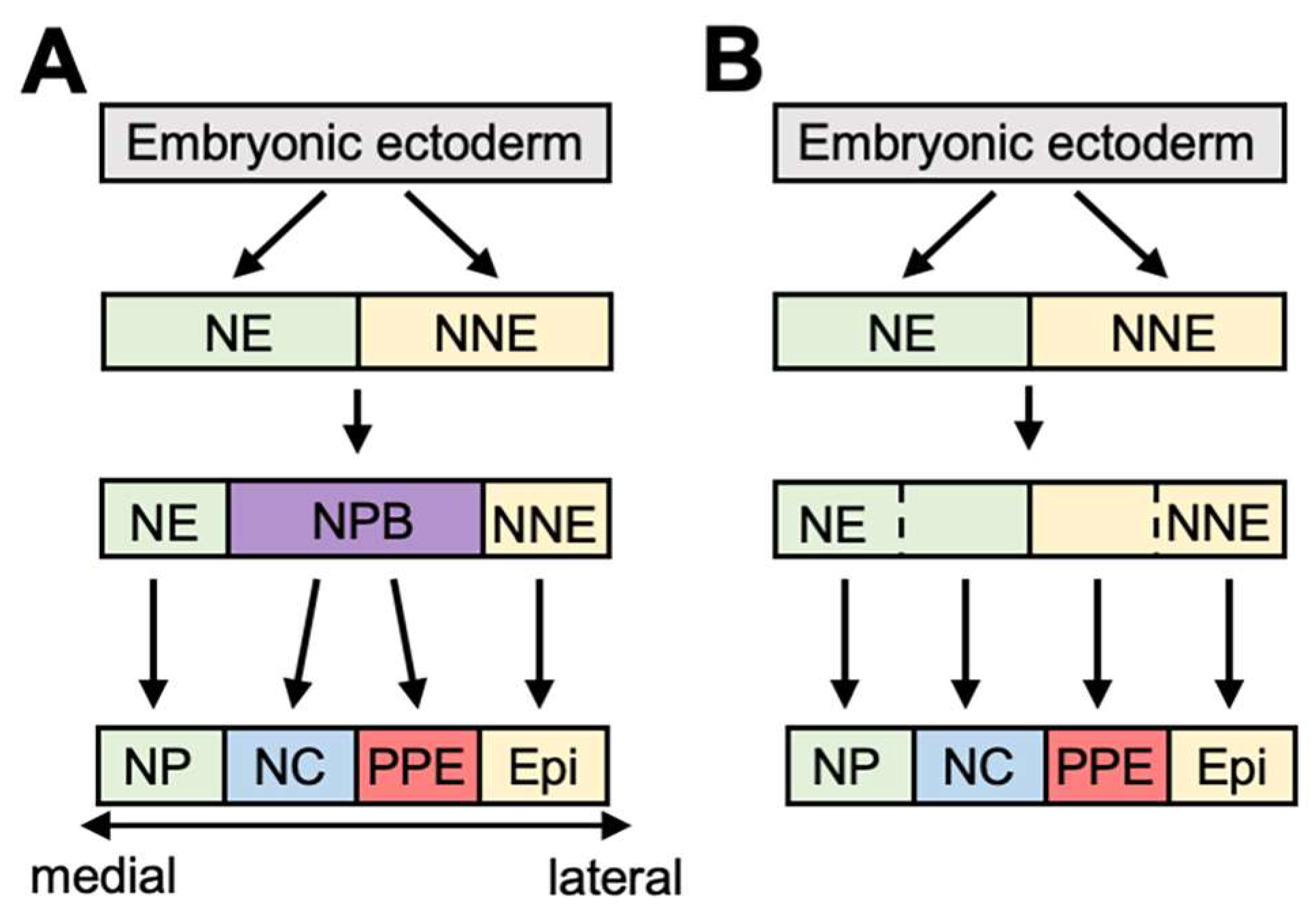
2. An Outline of PPE Formation

The PPE is a narrow, horseshoe-shaped region induced around the boundary between the neuroectoderm (NE) and the non-neural ectoderm (NNE) during gastrulation [9,10,11]. The NC is also derived from a boundary region and forms craniofacial structures [12,13,14]. The model for dividing the PPE and the NC is discussed later.
PPE cells give rise to cranial sensory organs, including lens, olfactory epithelium, inner ear, some of the cranial ganglia, and the anterior pituitary gland [15,16,17,18,19,20]. In contrast to NC cells, a part of PPE cells remain on the surface of the ectoderm, and after neural tube closure, various patterns of cell migration occur, according to the subtypes of placode [21]. Olfactory epithelium, lens, and otic cells are mainly rearranged to form their final shape, whereas trigeminal and epibranchial cells migrate and aggregate. Many genes are involved in PPE specification and construct a gene network [22]. Six1, the homolog of sine oculis (si) in Drosophila, encodes a homeodomain protein and is uniformly expressed in the PPE [23,24]. Eya1 is a cofactor with Six1 and is also expressed in the PPE [25]. These genes are well utilized as pan-placodal markers. The experiment on both upregulation and downregulation has shown that Six1 is required for the gene regulatory network of PPE formation [26,27]. Many other transcription factors including GATA2, Dlx3/5, FoxI1/3 and AP2 are involved with PPE formation (reviewed in [28]). Nonetheless, the molecular mechanism for segregation of the PPE and the NC is controversial in ectoderm patterning, and there are several models to explain PPE/NC formation (Figure 2).
In the “binary competence” model, determination of the neuroectoderm and the non-neural ectoderm occurs during gastrulation, followed by subdivision of the PPE and the epidermis from the non-neural ectoderm, whereas the NC and the NP are derived from the neuroectoderm (Figure 2A). Evidence that supports this model includes the fact that transplantation of NP cells into ventral ectoderm induces Six1, but expression is only seen in the recipient NNE region and not in the donor NP, indicating a difference in competence between the neural and the non-neural ectoderm [6,29]. In addition, Dlx3 plays a role for the formation of differential competence for the PPE [29]. Furthermore, complete inhibition of BMP signaling by dorsomorphin (an antagonist of BMP) at the blastula stage greatly reduced PPE marker expression [30], indicating the importance of at least some BMP signaling at an early stage.
The second model is the “NPB model” (Figure 2B). In this model, the neural plate border (NPB) region is initially induced between the neural and the non-neural ectoderm, followed by subdivision into the NC and the PPE. For NPB formation, several genes are important. Pax3 and Zic1 are typical NPB markers. Knockdown of Zic1 and Pax3 reduced Six1 expression, indicating the necessity of both gene functions for PPE formation [31]. The latter study of conserved enhancers revealed that expression of Pax3 and Zic1 is regulated by BMP, Wnt, and FGF, and the balance of these signals during the late gastrula stage is essential for Zic1/Pax3 expression [32]. FGF signaling is important for Pax3 transcription via specific enhancers (called IR2), whereas Wnt signaling positively regulates zic3 transcription via both E1 and E2 enhancers [32]. Pax3 expression is positively regulated by itself [33]. Immunostaining with several markers indicates that the PPE and the NC, in addition to the NP, overlap before the neurula stage in chick embryos, supporting this model [34]. Very recently, another model was proposed [35]. The “gradient border model” draws upon both of the previous models. In this model, the neural plate border is induced, but in this area, cells that express NC or PPE genes are distinct, suggesting that NPB already possesses two regions before neurulation.
In the following section, we will discuss intracellular signaling involved in ectoderm patterning. In this patterning, several signaling pathways, including BMP, FGF, Wnt, and RA, participate, but in this review, we focus mainly on BMP and FGF signaling. On the subject of feedback regulation, we will also discuss the implications of RA signaling.

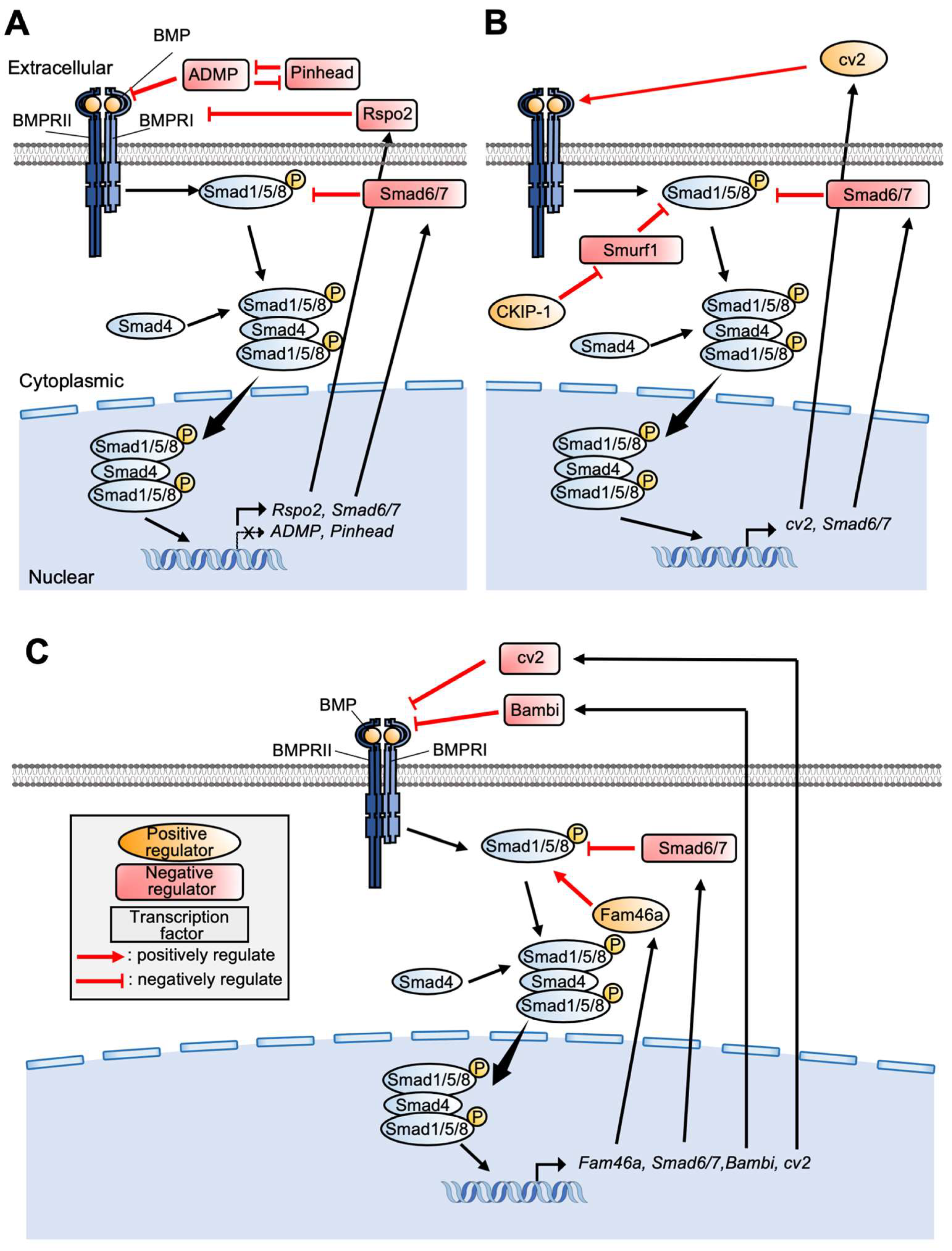
3. Control of BMP Signaling in PPE Formation

BMP serves important functions in various biological events, including many kinds of organ development in both vertebrates and invertebrates. Interaction of BMP ligands with BMP receptors promotes phosphorylation of the C-terminal serine residue of Smad1, directing it to bind Smad4, and regulating target gene expression (Figure 3) [5,36].
A morphogen gradient of BMP signaling is essential to establish embryonic patterning, as in dorsoventral axis formation [37]. Similarly, BMP signaling is crucial for ectoderm patterning. BMP4 and 7 are expressed in NNE, next to the PPE [38,39,40,41,42], whereas BMP antagonists are expressed in mesoderm underlying the PPE or in the PPE itself, contributing to differential control of the BMP level [43,44]. Animal cap experiments indicate that NP gene expression decreases as the dose of BMP increases [45]. Despite the fact that determination of the ectodermal region is crucial for precise body plan formation, the molecular mechanism by which BMP morphogen establishes each region is not still fully understood.
For NC formation, various animal models indicate the importance of BMP signaling, although what level of BMP signal is necessary remains controversial. In Xenopus embryos, signaling from DLMZ during gastrulation is important, whereas the signal from intermediate mesoderm, as well as adjacent ectoderm is important for maintenance of the NC region, indicating the necessity of stage-dependent inhibition of BMP signaling for NC formation (low BMP level in the early stage, whereas high level in the late stage) [46,47]. On the other hand, positive regulation of BMP signaling is necessary to induce the NC from the neural plate in chick embryos [48,49]. Furthermore, a zebrafish study indicated that intermediate levels of BMP specify a cranial neural crest progenitor [50].
For PPE formation, what function does BMP signaling serve? We need to consider the mechanism along with the binary competence and NPB models described above. According to the NPB model, intermediate levels of BMP signaling during gastrulation and neurulation are necessary for PPE formation, and evidence that supports the NPB model from the point of BMP signaling has been presented. An intermediate level of BMP signaling activity directs PPE induction. In chick embryos, the NPB region shows intermediate intensity of phosphorylated Smad1 protein [51]. A Xenopus study using animal cap cells indicated that Six1 expression is highest with an intermediate dose of noggin or chordin [27,52]. Moreover, dlx5 and dlx6 are both expressed in NPB, and the quantitative level of expression was highest in Xenopus embryos injected with an intermediate amount of chordin (chd) mRNA [53]. Another zebrafish study indicated that for PPE formation, a somewhat higher level of BMP signaling is necessary than for the NC [54]. A similar experiment was carried out using zebrafish embryos [55]. In summary, intermediate BMP levels enable induction of NPB/placode gene expression, at least in several experimental systems employing Xenopus, zebrafish, and chick embryos.
In the binary state model, it is likely that positive regulation of BMP signaling before gastrulation is important for inducing the PPE, whereas the chick and Xenopus study indicated that attenuation of BMP signaling is necessary at late gastrula/neurula stages to induce the PPE in naïve ectoderm [6,7]. Similarly, using various doses and variable timing of treatments with dorsomorphin, a zebrafish study showed that BMP inhibition at blastula or early gastrula greatly reduced PPE marker expression (sox3, six4 and pax2), whereas BMP inhibition at a later stage is important [30]. Tfap2A/C, Fox1i and Gata3, which are necessary to acquire PPE formation competence, are induced by BMP, whereas BMP signaling is not necessary to specify PPE fate after gastrulation [29,30,56]. A chick study also indicated that BMP signaling is required for formation of olfactory and lens placodes [57]. From these studies, it is suggested that during gastrulation BMP promotes PPE formation but subsequently inhibits PPE formation in the non-neural ectoderm.

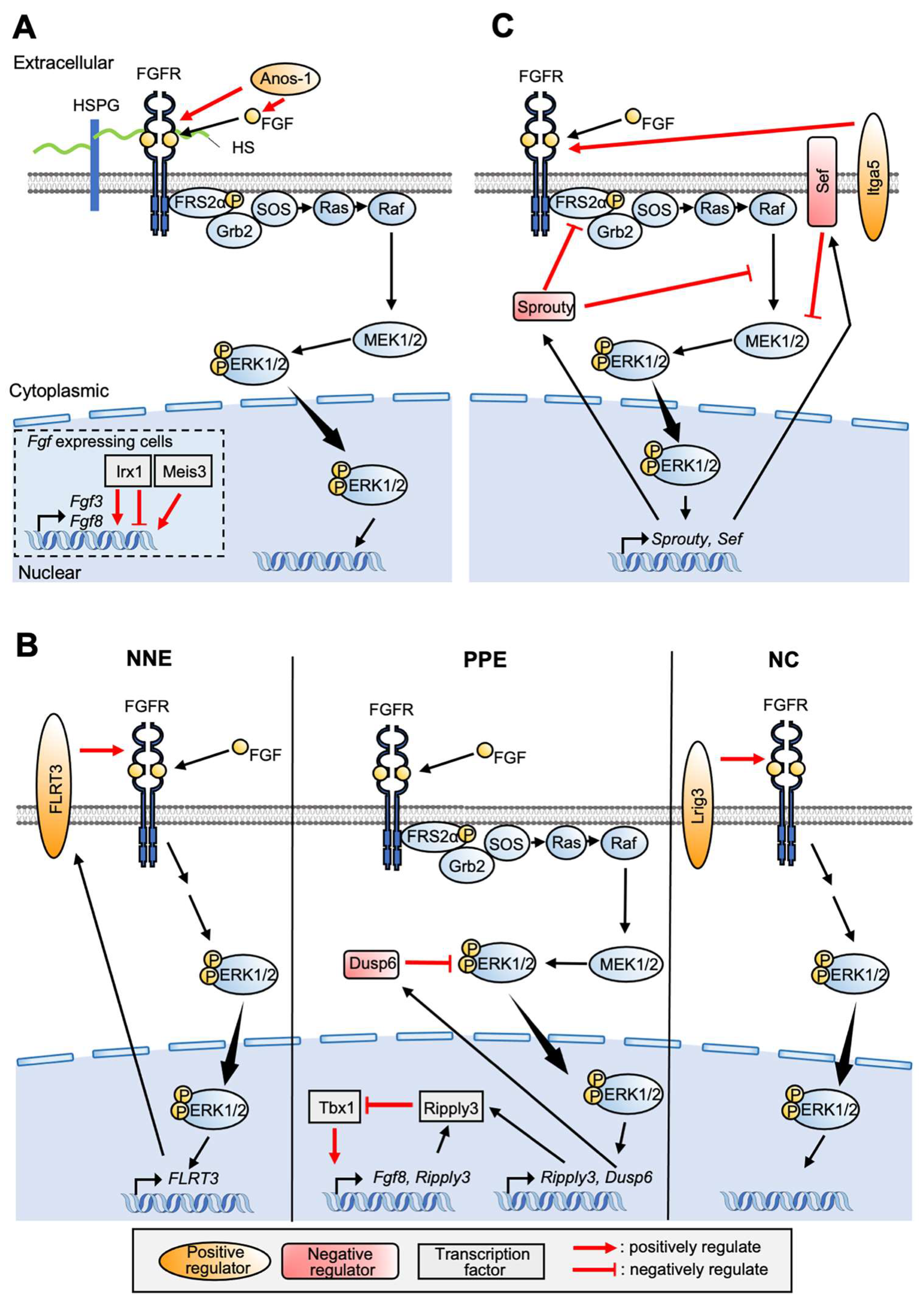
4. Involvement of FGF Signaling for PPE Formation

Many studies have reported that relevant genes are involved in NPB/NC formation (Figure 4). Anosmin-1(Anos1), an ECM-associated, glycosylated protein directly interacts with FGF ligands and facilitates FGF8-FGFR1 interaction in chick embryo (Figure 4A) [58,59,60,61]. Xenopus Anos1 is expressed downstream of Pax3 and Zic1 and contributes to formation of both the NC and the PPE [62]. Meis3 is also expressed downstream of Zic3 and Pax3 and positively regulates Fgf3 and Fgf8 (Figure 4A) [63]. Lrig3, expressed in the NP and the NC, interacts with FGFR1 and modulates FGF signaling in NC induction and specification (Figure 4B) [64]. For establishment of the NC, the balance of ERK and AKT is important [65]. In the NC state of animal cap cells exhibited by Foxd3 and Sox9 expression, the pERK level is high and pAKT is low. Thus, NC formation is inhibited by either ERK inhibition or AKT activation [66].
The importance of FGF signaling for PPE formation has also been shown by a series of studies. In Xenopus embryos, Fgf3, Fgf4, and Fgf8 are expressed in the dorsolateral marginal zone [67]. In chicken blastula, Fgf8 is distributed in almost all parts of the epiblast, and expression accumulates in the primitive streak at early gastrula stage [68]. Additionally, Fgf8 is expressed in the anterior neural ridge, adjacent to the PPE [7,69,70]. In Xenopus embryos, knockdown of Fgf8 by morpholino anti-sense oligo (MO) decreased Six1 expression [6,31], and experiments using SU5402 (FGFR inhibitor) also indicated that an FGF signal is required for placode induction [6]. Although FGF signaling is necessary for PPE specification [6,7,31], overactivation of FGF signaling represses a PPE marker gene, Six1 [27,31]. In addition, slight inhibition of FGF signaling enhances Six1 expression [52], suggesting that an appropriate level of FGF is required for PPE induction.
Irx, which encodes Iroquois homeodomain protein, regulates Fgf8 expression and is involved in NPB specifier-gene expression, including Msx1, Pax3, and Zic1 (Figure 4A) [71]. Irx1 is upregulated by Six1 and Eya1, whereas Irx1 promotes Six1 expression in early PPE formation. Irx1 expression overlaps with that of the NPB gene at first, but expression accumulates only in the PPE region. Interestingly, Irx1 changes to suppress Six1 expression, suggesting a differential stage-dependent role [71].
For specific placode formation from the pan-placodal domain, FGF signaling is necessary [19,72,73]. Mouse KO experiments also indicate essential roles of Fgf3, Fgf8, and Fgf10 in otic placode formation [73,74]. Integrin-α5 (Itga5) is expressed in the PPE, and its knockdown impaired trigeminal, epibranchial, and otic cells (Figure 4C) [75]. In addition, dlx3/dlx4 negatively regulates Fgfr1/2 expression, resulting in malformation of otic placode [44].

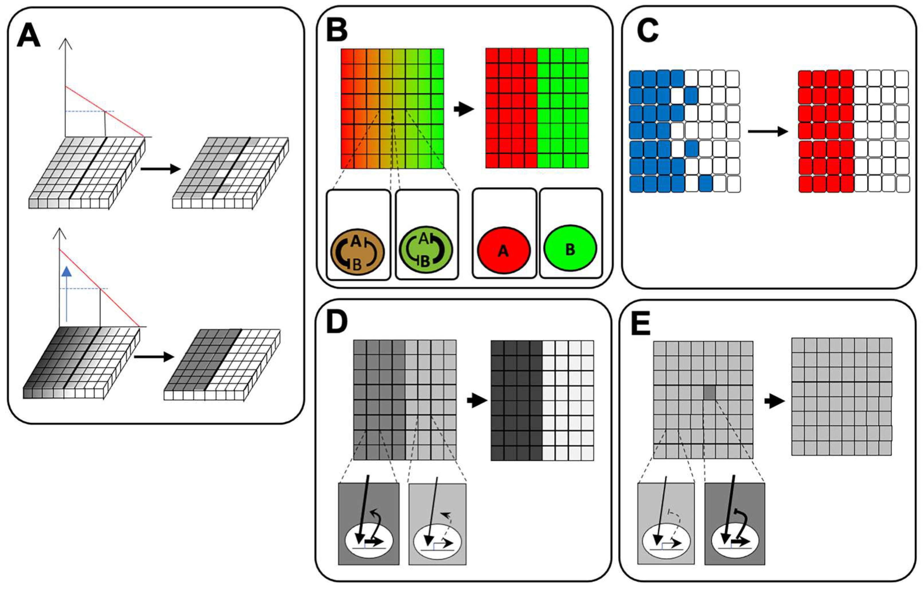
5. Feedback Regulation of Signaling Pathways for Ectodermal Patterning

As shown above, feedback regulation is a useful way not only to establish discrete areas, but also to maintain levels of intracellular signaling against fluctuations. We will show some examples of feedback regulation in BMP and FGF pathways and their contributions to PPE formation.

5.1. BMP Signaling

Several studies have addressed feedback regulation of BMP signaling in the context of embryonic development. In zebrafish embryos, both Pinhead and ADMP encode BMP-like ligands that promote chd degradation, whereas their transcription is repressed by BMP signaling (Figure 3A) [76,77,78]. In Xenopus ectoderm formation, R-spondins (RSPOs) antagonize BMP signaling by associating with the BMP receptor, affecting dorsoventral patterning. Biochemical analysis indicates that BMP promotes Rspo2 transcription, whereas RSPO protein antagonizes BMP signaling extracellularly, suggesting feedback loop formation (Figure 3A) [79]. Bambi is induced by BMP4, whereas Bambi represses the ligand–receptor complex, indicating negative loop formation (Figure 3C) [80]. This negative feedback regulation extends the dynamic range of BMP signaling because this system enables responses to more intense BMP signaling, contributing to attenuation of morphogen fluctuation in embryos [81]. Actually, in Xenopus embryos, the myf5 expression domain induced by intermediate BMP levels is perturbed by Bambi knockdown.
For PPE/NPB formation, there are not many studies that directly demonstrate the contribution of feedback regulation of BMP signaling. In Xenopus embryos, expression of crossveinless2 (cv2), which interacts with both chd and BMP, is seen in high BMP regions, although knockdown of cv2 with cv2 MO increased vent1 and cv2 and decreased Six3 and chd, indicating that cv2 participates in a negative feedback loop of BMP signaling (Figure 3C) [82,83]. On the other hand, the zebrafish study indicates that cv2 forms the positive feedback loop by acting as pro-BMP factor and is required for NC induction by locally enhancing BMP activity and regulating the NPB gene network [84,85]. In the PPE region, dlx3 expression domain is outside the cv2 expression domain in the 5-somite stage of zebrafish embryos. Dlx3b enhances bambi-b in the PPE, suggesting that discrete expression of these genes specifies both the NC and the PPE region [85]. In chick embryos, casein kinase interacting protein 1 (CKIP-1) and Smurf1, which encodes a ubiquitin ligase, are both expressed in NPB and establish an intermediate BMP level with Smurf1 for NC formation. Smurf1 attenuates BMP signaling via degradation of Smad1/5/8 but also degrades itself. At the same time, CKIP-1 directly interacts with Smurf1, promoting Smurf1 degradation. In summary, CKIP-1/Smurf1 double-negative attenuation maintains appropriate BMP signal levels in NPB (Figure 3B) [86].
Our analysis indicates the importance of Fam46a in PPE formation. Knockdown of Fam46a inhibits PPE-specific gene expression, including Six1. Fam46a protein directly interacts with the N-half region of Smad1, including the linker domain, and increases the quantitative level of Smad1. The linker region of Smad1 is phosphorylated by GSK3β, followed by ubiquitination and degradation via the proteosome system; thus, it is suggested that Fam46a upregulates BMP signaling via stabilization of Smad1 protein. Moreover, Fam46a transcription is promoted by BMP signaling, indicating formation of a positive feedback loop in BMP signaling (Figure 3C). Notably, activation of BMP signaling by Fam46a is not intense because Fam46a contributes to stabilization of Smad1 but not to direct activation via promotion of C-terminal phosphorylation of Smad1 [87].

5.2. FGF Signaling

For FGF signaling, feedback controls in either NPB/PPE/specific placode formation have been more widely reported than for BMP signaling. Tbx1 and Ripply3 contribute to regulation of PPE gene expression. In detail, Tbx1 facilitates expression of Fgf8, Six1, Eya1, and Ripply3. Additionally, Fgf8 promotes Ripply3 expression. On the other hand, Ripply3 suppresses expression of Fgf8 and Tbx1 and forms a negative feedback loop with Fgf8, Ripply3, and Tbx1 (Figure 4B). This feedback loop contributes to the postero–lateral boundary during formation of the PPE by restricting the expressing region of fgf [88]. Fibronectin-leucine-rich transmembrane protein 3 (FLRT3) functions as a positive regulator of Ras-MAPK signaling and also promotes ERK phosphorylation. FLRT3 transcription is upregulated by FGF signaling, suggesting positive feedback formation (Figure 4B) [89,90]. Xenopus FLRT3 is co-expressed with Fgf8 in the anterior neural ridge. From the fact that overactivation of FGF signaling inhibits PPE formation, FLRT3 may play a role in boundary formation outside the PPE region [90,91]. Recently, we showed that Dual specificity phosphatase 6 (Dusp6, also known as MKP3) is important to precisely form the PPE region. Dusp6 is a phosphatase that specifically interacts with dual tyrosine and threonine residues of ERK1/2, attenuating Ras/ERK signaling (Figure 4B) [92,93,94,95]. Our study showed that Dusp6 is expressed in the PPE at mid-neurula of Xenopus embryos in an FGF signal-dependent manner and is necessary for both NPB and PPE formation by modulating FGF signaling. An experiment combining FGF bead transplantation with Dusp6 knockdown demonstrated the importance of negative feedback control for PPE formation. In this study, it was suggested that stable spatial pattern formation against perturbation of FGF ligands is accomplished by suppressing intracellular signaling activity [96].
Furthermore, several genes involved in FGF signaling contribute to specific placode formation. Sprouty (Spry) functions as an intracellular negative feedback regulator of FGF signaling in several developmental contexts [97,98,99]. Spry is expressed in an FGF-dependent manner [100,101], and in mouse embryos, conditional knockout of Spry1 causes defective craniofacial and cardiac development, indicating the importance of NC formation [102]. Spry1 and Spry2 are expressed in posterior PPE and participate in otic placode formation by inhibiting FGF signaling [103,104]. Spry1 and Spry2 also contribute to epibranchial placode formation and neuronal differentiation [105]. Malformation of otic placode by Spry1 and Spry2 knockdown was rescued by haploinsufficiency of Fgf8 gene function, suggesting the importance of feedback loop-based fine tuning of FGF signaling (Figure 4C) [105]. Similar expression of fgf (Sef) regulates Ras-MAPK signaling, as well as other types of signaling [61,106]. In both zebrafish and Xenopus embryos, Sef is expressed in an FGF signaling-dependent manner, whereas FGF target gene expression is suppressed by Sef overexpression. Injection with Sef MO expanded the Fgf8 expression region in the midbrain–hindbrain boundary (MHB) [107]. In chick embryos, Sef is expressed in otic placode [108], suggesting negative feedback loop regulation via Sef, at least in otic placode (Figure 4C).
RA signaling functions cooperatively with FGF signaling. RA nuclear receptor, RARa2, reduced the expression of Ripply3, Tbx1 and Six1. As shown above, Ripply3 suppresses FGF signaling, suggesting that RA and FGF signaling form a negative feedback loop via these genes [109]. Pitx2c is induced by RA and promotes transcription of Fgf8, followed by upregulation of Cyp26c1 (an RA metabolizing enzyme) expression adjacent to the PPE. This negative feedback regulation via both FGF and RA signaling suggests a role in PPE specification [110]. In otic vesicle formation, FGF signaling is required for aldh3 (RA synthesizing enzyme) expression, whereas RA treatment itself downregulates fgf8 expression, resulting in feedback loop formation [111].

6. Conclusions

In this review, we discussed the role of signaling pathways in PPE formation. In particular, we focused on BMP and FGF signaling and showed examples of their feedback regulation in PPE patterning. Signal adjustment is obviously important not only to form clear boundaries, but also to pattern narrow areas robustly. In particular, feedback adjustment contributes to noise suppression, which reduces signal fluctuation, and contributes to robust acquisition of patterns.

7. Future Directions

Further analysis is needed to fully elucidate the mechanisms of formation of the PPE region, the NP, the NC, and the epidermis. In addition, other experimental approaches may be important: one of them is to artificially change the feedback cycle by changing the intron length of a target gene and examining the effect on PPE formation [112]. Furthermore, other principles may need to be considered. One of these is mechanical regulation. Recently, a study using human pluripotent stem cells indicated that NPB fate determination is affected by external forces via changes in BMP signaling [113]. For directional migration of NC cells, a gradient of stiffness in surrounding cells, so-called “durotaxis”, is important [114]. Additionally, our studies indicate that there is a difference in cell tension between neural and epidermal ectoderm [115,116]. From these results, it appears that mechanical forces may contribute to form each ectodermal region and to establish their properties. Another point concerns extracellular control of ligand diffusion. Various molecules, including ECM, membrane protein (receptors, etc.), and other cellular processes, including endocytosis, affect ligand diffusion; thus, these mechanisms are also expected to contribute to ectoderm patterning. Other studies report that ECM protein is involved in NC/PPE formation [70]. In addition, anos1, which associates with FGF ligands, also binds to heparan sulfate (HS) [60,117]. By investigating the contributions of these mechanisms to regulation of intracellular signaling, we will better understand the robust and precise system of embryonic pattern formation.

Funding

Preparation of this article was supported in part by JSPS KAKENHI (Grant Number 21K06183 to TM).

Institutional Review Board Statement

Not applicable.

Informed Consent Statement

Not applicable.

Data Availability Statement

Not applicable.

Acknowledgments

We apologize that we could not fully include studies regarding ectoderm patterning. We thank Steven D. Aird for technical editing of the manuscript.

Conflicts of Interest

The authors declare no conflict of interest.

References

  1. Wolpert, L. One Hundred Years of Positional Information. Trends Genet. 1996, 12, 359–364. [Google Scholar] [CrossRef]
  2. Lander, A.D. Pattern, Growth, and Control. Cell 2011, 144, 955–969. [Google Scholar] [CrossRef] [PubMed]
  3. Turing, A.M. The Chemical Basis of Morphogenesis. Philos. Trans. R. Soc. Lond. B Biol. Sci. 1952, 237, 37–72. [Google Scholar] [CrossRef]
  4. Kondo, S.; Asal, R. A Reaction-Diffusion Wave on the Skin of the Marine Angelfish Pomacanthus. Nature 1995, 376, 765–768. [Google Scholar] [CrossRef]
  5. Bier, E.; De Robertis, E.M. BMP Gradients: A Paradigm for Morphogen-Mediated Developmental Patterning. Science 2015, 348, aaa5838. [Google Scholar] [CrossRef] [PubMed]
  6. Ahrens, K.; Schlosser, G. Tissues and Signals Involved in the Induction of Placodal Six1 Expression in Xenopus Laevis. Dev. Biol. 2005, 288, 40–59. [Google Scholar] [CrossRef] [PubMed]
  7. Litsiou, A.; Hanson, S.; Streit, A. A Balance of FGF, BMP and WNT Signalling Positions the Future Placode Territory in the Head. Development 2005, 132, 4051–4062. [Google Scholar] [CrossRef]
  8. Lander, A.D.; Lo, W.-C.; Nie, Q.; Wan, F.Y.M. The Measure of Success: Constraints, Objectives, and Tradeoffs in Morphogen-Mediated Patterning. Cold Spring Harb. Perspect. Biol. 2009, 1, a002022. [Google Scholar] [CrossRef]
  9. Bhattacharyya, S.; Bronner-Fraser, M. Hierarchy of Regulatory Events in Sensory Placode Development. Curr. Opin. Genet. Dev. 2004, 14, 520–526. [Google Scholar] [CrossRef]
  10. Streit, A. The Preplacodal Region: An Ectodermal Domain with Multipotential Progenitors That Contribute to Sense Organs and Cranial Sensory Ganglia. Int. J. Dev. Biol. 2007, 51, 447–461. [Google Scholar] [CrossRef] [Green Version]
  11. Pla, P.; Monsoro-Burq, A.H. The Neural Border: Induction, Specification and Maturation of the Territory That Generates Neural Crest Cells. Dev. Biol. 2018, 444, S36–S46. [Google Scholar] [CrossRef] [PubMed]
  12. Steventon, B.; Carmona-Fontaine, C.; Mayor, R. Genetic Network during Neural Crest Induction: From Cell Specification to Cell Survival. Semin. Cell Dev. Biol. 2005, 16, 647–654. [Google Scholar] [CrossRef] [PubMed]
  13. Mayor, R.; Aybar, M.J. Induction and Development of Neural Crest in Xenopus Laevis. Cell Tissue Res. 2001, 305, 203–209. [Google Scholar] [CrossRef] [PubMed]
  14. Milet, C.; Monsoro-Burq, A.H. Neural Crest Induction at the Neural Plate Border in Vertebrates. Dev. Biol. 2012, 366, 22–33. [Google Scholar] [CrossRef] [PubMed]
  15. Baker, C.V.H.; Bronner-Fraser, M. Vertebrate Cranial Placodes I. Embryonic Induction. Dev. Biol. 2001, 232, 1–61. [Google Scholar] [CrossRef] [PubMed]
  16. Schlosser, G. Induction and Specification of Cranial Placodes. Dev. Biol. 2006, 294, 303–351. [Google Scholar] [CrossRef]
  17. Schlosser, G. Making Senses: Development of Vertebrate Cranial Placodes. In International Review of Cell and Molecular Biology; Elsevier Inc.: Amsterdam, The Netherlands, 2010; Volume 283, pp. 129–234. [Google Scholar]
  18. Grocott, T.; Tambalo, M.; Streit, A. The Peripheral Sensory Nervous System in the Vertebrate Head: A Gene Regulatory Perspective. Dev. Biol. 2012, 370, 3–23. [Google Scholar] [CrossRef]
  19. Saint-Jeannet, J.-P.; Moody, S.A. Establishing the Pre-Placodal Region and Breaking It into Placodes with Distinct Identities. Dev. Biol. 2014, 389, 13–27. [Google Scholar] [CrossRef]
  20. Singh, S.; Groves, A.K. The Molecular Basis of Craniofacial Placode Development. Wiley Interdiscip. Rev. Dev. Biol. 2016, 5, 363–376. [Google Scholar] [CrossRef]
  21. Breau, M.A.; Schneider-Maunoury, S. Cranial Placodes: Models for Exploring the Multi-Facets of Cell Adhesion in Epithelial Rearrangement, Collective Migration and Neuronal Movements. Dev. Biol. 2015, 401, 25–36. [Google Scholar] [CrossRef]
  22. Streit, A. Specification of Sensory Placode Progenitors: Signals and Transcription Factor Networks. Int. J. Dev. Biol. 2018, 62, 195–205. [Google Scholar] [CrossRef] [PubMed]
  23. Pandur, P.D.; Moody, S.A. Xenopus Six1 Gene Is Expressed in Neurogenic Cranial Placodes and Maintained in the Differentiating Lateral Lines. Mech. Dev. 2000, 96, 253–257. [Google Scholar] [CrossRef]
  24. Schlosser, G.; Ahrens, K. Molecular Anatomy of Placode Development in Xenopus Laevis. Dev. Biol. 2004, 271, 439–466. [Google Scholar] [CrossRef] [PubMed]
  25. David, R.; Ahrens, K.; Wedlich, D.; Schlosser, G. Xenopus Eya1 Demarcates All Neurogenic Placodes as Well as Migrating Hypaxial Muscle Precursors. Mech. Dev. 2001, 103, 189–192. [Google Scholar] [CrossRef]
  26. Maharana, S.K.; Schlosser, G. A Gene Regulatory Network Underlying the Formation of Pre-Placodal Ectoderm in Xenopus Laevis. BMC Biol. 2018, 16, 79. [Google Scholar] [CrossRef]
  27. Brugmann, S.A.; Pandur, P.D.; Kenyon, K.L.; Pignoni, F.; Moody, S.A. Six1 Promotes a Placodal Fate within the Lateral Neurogenic Ectoderm by Functioning as Both a Transcriptional Activator and Repressor. Development 2004, 131, 5871–5881. [Google Scholar] [CrossRef]
  28. Schlosser, G. Early Embryonic Specification of Vertebrate Cranial Placodes. Wiley Interdiscip. Rev. Dev. Biol. 2014, 3, 349–363. [Google Scholar] [CrossRef]
  29. Pieper, M.; Ahrens, K.; Rink, E.; Peter, A.; Schlosser, G. Differential Distribution of Competence for Panplacodal and Neural Crest Induction to Non-Neural and Neural Ectoderm. Development 2012, 139, 1175–1187. [Google Scholar] [CrossRef]
  30. Kwon, H.-J.; Bhat, N.; Sweet, E.M.; Cornell, R.A.; Riley, B.B. Identification of Early Requirements for Preplacodal Ectoderm and Sensory Organ Development. PLoS Genet. 2010, 6, e1001133. [Google Scholar] [CrossRef]
  31. Hong, C.-S.; Saint-Jeannet, J.-P. The Activity of Pax3 and Zic1 Regulates Three Distinct Cell Fates at the Neural Plate Border. Mol. Biol. Cell 2007, 18, 2192–2202. [Google Scholar] [CrossRef] [Green Version]
  32. Garnett, A.T.; Square, T.A.; Medeiros, D.M. BMP, Wnt and FGF Signals Are Integrated through Evolutionarily Conserved Enhancers to Achieve Robust Expression of Pax3 and Zic Genes at the Zebrafish Neural Plate Border. Development 2012, 139, 4220–4231. [Google Scholar] [CrossRef] [PubMed]
  33. Plouhinec, J.L.; Roche, D.D.; Pegoraro, C.; Figueiredo, A.L.; Maczkowiak, F.; Brunet, L.J.; Milet, C.; Vert, J.P.; Pollet, N.; Harland, R.M.; et al. Pax3 and Zic1 Trigger the Early Neural Crest Gene Regulatory Network by the Direct Activation of Multiple Key Neural Crest Specifiers. Dev. Biol. 2014, 386, 461–472. [Google Scholar] [CrossRef] [PubMed]
  34. Roellig, D.; Tan-Cabugao, J.; Esaian, S.; Bronner, M.E. Dynamic Transcriptional Signature and Cell Fate Analysis Reveals Plasticity of Individual Neural Plate Border Cells. eLife 2017, 6, e21620. [Google Scholar] [CrossRef] [PubMed]
  35. Thiery, A.; Buzzi, A.L.; Hamrud, E.; Cheshire, C.; Luscombe, N.; Briscoe, J.; Streit, A. A Gradient Border Model for Cell Fate Decisions at the Neural Plate Border. bioRxiv 2022. [Google Scholar] [CrossRef]
  36. Miyazono, K.; Kamiya, Y.; Morikawa, M. Bone Morphogenetic Protein Receptors and Signal Transduction. J. Biochem. 2010, 147, 35–51. [Google Scholar] [CrossRef]
  37. Hill, C.S. Establishment and Interpretation of NODAL and BMP Signaling Gradients in Early Vertebrate Development, 1st ed.; Elsevier Inc.: Amsterdam, The Netherlands, 2022; Volume 149, ISBN 9780128170977. [Google Scholar]
  38. Fainsod, A.; Steinbeisser, H.; De Robertis, E.M. On the Function of BMP-4 in Patterning the Marginal Zone of the Xenopus Embryo. EMBO J. 1994, 13, 5015–5025. [Google Scholar] [CrossRef]
  39. Hemmati-Brivanlou, A.; Thomsen, G.H. Ventral Mesodermal Patterning InXenopus Embryos: Expression Patterns and Activities of BMP-2 and BMP-4. Dev. Genet. 1995, 17, 78–89. [Google Scholar] [CrossRef]
  40. Liem, K.F.; Tremml, G.; Roelink, H.; Jessell, T.M. Dorsal Differentiation of Neural Plate Cells Induced by BMP-Mediated Signals from Epidermal Ectoderm. Cell 1995, 82, 969–979. [Google Scholar] [CrossRef]
  41. Schmidt, J.E.; Suzuki, A.; Ueno, N.; Kimelman, D. Localized BMP-4 Mediates Dorsal/Ventral Patterning in the Early Xenopus Embryo. Dev. Biol. 1995, 169, 37–50. [Google Scholar] [CrossRef]
  42. Streit, A.; Stern, C.D. Establishment and Maintenance of the Border of the Neural Plate in the Chick: Involvement of FGF and BMP Activity. Mech. Dev. 1999, 82, 51–66. [Google Scholar] [CrossRef]
  43. Ogita, J.; Isogai, E.; Sudo, H.; Sakiyama, S.; Nakagawara, A.; Koseki, H. Expression of the Dan Gene during Chicken Embryonic Development. Mech. Dev. 2001, 109, 363–365. [Google Scholar] [CrossRef]
  44. Esterberg, R.; Fritz, A. Dlx3b/4b Are Required for the Formation of the Preplacodal Region and Otic Placode through Local Modulation of BMP Activity. Dev. Biol. 2009, 325, 189–199. [Google Scholar] [CrossRef]
  45. Wilson, P.A.; Lagna, G.; Suzuki, A.; Hemmati-Brivanlou, A. Concentration-Dependent Patterning of the Xenopus Ectoderm by BMP4 and Its Signal Transducer Smad1. Development 1997, 124, 3177–3184. [Google Scholar] [CrossRef] [PubMed]
  46. Marchant, L.; Linker, C.; Ruiz, P.; Guerrero, N.; Mayor, R. The Inductive Properties of Mesoderm Suggest That the Neural Crest Cells Are Specified by a BMP Gradient. Dev. Biol. 1998, 198, 319–329. [Google Scholar] [CrossRef]
  47. Steventon, B.; Araya, C.; Linker, C.; Kuriyama, S.; Mayor, R. Differential Requirements of BMP and Wnt Signalling during Gastrulation and Neurulation Define Two Steps in Neural Crest Induction. Development 2009, 136, 771–779. [Google Scholar] [CrossRef]
  48. Selleck, M.A.J.; García-Castro, M.I.; Artinger, K.B.; Bronner-Fraser, M. Effects of Shh and Noggin on Neural Crest Formation Demonstrate That BMP Is Required in the Neural Tube but Not Ectoderm. Development 1998, 125, 4919–4930. [Google Scholar] [CrossRef] [PubMed]
  49. Endo, Y.; Osumi, N.; Wakamatsu, Y. Bimodal Functions of Notch-Mediated Signaling Are Involved in Neural Crest Formation during Avian Ectoderm Development. Development 2002, 129, 863–873. [Google Scholar] [CrossRef]
  50. Schumacher, J.A.; Hashiguchi, M.; Nguyen, V.H.; Mullins, M.C. An Intermediate Level of Bmp Signaling Directly Specifies Cranial Neural Crest Progenitor Cells in Zebrafish. PLoS ONE 2011, 6, e27403. [Google Scholar] [CrossRef]
  51. Faure, S.; De Santa Barbara, P.; Roberts, D.J.; Whitman, M. Endogenous Patterns of BMP Signaling during Early Chick Development. Dev. Biol. 2002, 244, 44–65. [Google Scholar] [CrossRef]
  52. Watanabe, T.; Kanai, Y.; Matsukawa, S.; Michiue, T. Specific Induction of Cranial Placode Cells from Xenopus Ectoderm by Modulating the Levels of BMP, Wnt, and FGF Signaling. Genesis 2015, 53, 652–659. [Google Scholar] [CrossRef]
  53. Luo, T.; Matsuo-Takasaki, M.; Lim, J.H.; Sargent, T.D. Differential Regulation of Dlx Gene Expression by a BMP Morphogenetic Gradient. Int. J. Dev. Biol. 2001, 45, 681–684. [Google Scholar] [PubMed]
  54. Nguyen, V.H.; Schmid, B.; Trout, J.; Connors, S.A.; Ekker, M.; Mullins, M.C. Ventral and Lateral Regions of the Zebrafish Gastrula, Including the Neural Crest Progenitors, Are Established by Abmp2b/SwirlPathway of Genes. Dev. Biol. 1998, 199, 93–110. [Google Scholar] [CrossRef] [PubMed]
  55. Neave, B.; Holder, N.; Patient, R. A Graded Response to BMP-4 Spatially Coordinates Patterning of the Mesoderm and Ectoderm in the Zebrafish. Mech. Dev. 1997, 62, 183–195. [Google Scholar] [CrossRef]
  56. Bhat, N.; Kwon, H.-J.; Riley, B.B. A Gene Network That Coordinates Preplacodal Competence and Neural Crest Specification in Zebrafish. Dev. Biol. 2013, 373, 107–117. [Google Scholar] [CrossRef] [PubMed]
  57. Sjödal, M.; Edlund, T.; Gunhaga, L. Time of Exposure to BMP Signals Plays a Key Role in the Specification of the Olfactory and Lens Placodes Ex Vivo. Dev. Cell 2007, 13, 141–149. [Google Scholar] [CrossRef] [PubMed]
  58. Hu, Y.; Guimond, S.E.; Travers, P.; Cadman, S.; Hohenester, E.; Tumbull, J.E.; Kim, S.H.; Bouloux, P.M. Novel Mechanisms of Fibroblast Growth Factor Receptor 1 Regulation by Extracellular Matrix Protein Anosmin-1. J. Biol. Chem. 2009, 284, 29905–29920. [Google Scholar] [CrossRef]
  59. Endo, Y.; Ishiwata-Endo, H.; Yamada, K.M. Extracellular Matrix Protein Anosmin Promotes Neural Crest Formation and Regulates FGF, BMP, and WNT Activities. Dev. Cell 2012, 23, 305–316. [Google Scholar] [CrossRef]
  60. Hu, Y.; González-Martínez, D.; Kim, S.H.; Bouloux, P.M.G. Cross-Talk of Anosmin-1, the Protein Implicated in X-Linked Kallmann’s Syndrome, with Heparan Sulphate and Urokinase-Type Plasminogen Activator. Biochem. J. 2004, 384, 495–505. [Google Scholar] [CrossRef]
  61. Korsensky, L.; Ron, D. Regulation of FGF Signaling: Recent Insights from Studying Positive and Negative Modulators. Semin. Cell Dev. Biol. 2016, 53, 101–114. [Google Scholar] [CrossRef]
  62. Bae, C.-J.; Hong, C.-S.; Saint-Jeannet, J.-P. Anosmin-1 Is Essential for Neural Crest and Cranial Placodes Formation in Xenopus. Biochem. Biophys. Res. Commun. 2018, 495, 2257–2263. [Google Scholar] [CrossRef]
  63. Gutkovich, Y.E.; Ofir, R.; Elkouby, Y.M.; Dibner, C.; Gefen, A.; Elias, S.; Frank, D. Xenopus Meis3 Protein Lies at a Nexus Downstream to Zic1 and Pax3 Proteins, Regulating Multiple Cell-Fates during Early Nervous System Development. Dev. Biol. 2010, 338, 50–62. [Google Scholar] [CrossRef] [PubMed]
  64. Zhao, H.; Tanegashima, K.; Ro, H.; Dawid, I.B. Lrig3 Regulates Neural Crest Formation in Xenopus by Modulating Fgf and Wnt Signaling Pathways. Development 2008, 135, 1283–1293. [Google Scholar] [CrossRef] [PubMed]
  65. Dinsmore, C.J.; Soriano, P. MAPK and PI3K Signaling: At the Crossroads of Neural Crest Development. Dev. Biol. 2018, 444, S79–S97. [Google Scholar] [CrossRef] [PubMed]
  66. Geary, L.; LaBonne, C. FGF Mediated MAPK and PI3K/Akt Signals Make Distinct Contributions to Pluripotency and the Establishment of Neural Crest. eLife 2018, 7, e33845. [Google Scholar] [CrossRef]
  67. Monsoro-Burq, A.H.; Fletcher, R.B.; Harland, R.M. Neural Crest Induction by Paraxial Mesoderm in Xenopus Embryos Requires FGF Signals. Development 2003, 130, 3111–3124. [Google Scholar] [CrossRef]
  68. Lawson, A.; Colas, J.F.; Schoenwolf, G.C. Classification Scheme for Genes Expressed during Formation and Progression of the Avian Primitive Streak. Anat. Rec. 2001, 262, 221–226. [Google Scholar] [CrossRef]
  69. Fletcher, R.B.; Baker, J.C.; Harland, R.M. FGF8 Spliceforms Mediate Early Mesoderm and Posterior Neural Tissue Formation in Xenopus. Development 2006, 133, 1703–1714. [Google Scholar] [CrossRef]
  70. Tereshina, M.B.; Ermakova, G.V.; Ivanova, A.S.; Zaraisky, A.G. Ras-Dva1 Small GTPase Regulates Telencephalon Development in Xenopus Laevis Embryos by Controlling Fgf8 and Agr Signaling at the Anterior Border of the Neural Plate. Biol. Open 2014, 3, 192–203. [Google Scholar] [CrossRef]
  71. Sullivan, C.H.; Majumdar, H.D.; Neilson, K.M.; Moody, S.A. Six1 and Irx1 Have Reciprocal Interactions during Cranial Placode and Otic Vesicle Formation. Dev. Biol. 2019, 446, 68–79. [Google Scholar] [CrossRef]
  72. Schimmang, T. Expression and Functions of FGF Ligands during Early Otic Development. Int. J. Dev. Biol. 2007, 51, 473–481. [Google Scholar] [CrossRef] [Green Version]
  73. Wright, T.J.; Mansour, S.L. Fgf3 and Fgf10 Are Required for Mouse Otic Placode Induction. Development 2003, 130, 3379–3390. [Google Scholar] [CrossRef] [PubMed]
  74. Domínguez-Frutos, E.; Vendrell, V.; Alvarez, Y.; Zelarayan, L.C.; López-Hernández, I.; Ros, M.; Schimmang, T. Tissue-Specific Requirements for FGF8 during Early Inner Ear Development. Mech. Dev. 2009, 126, 873–881. [Google Scholar] [CrossRef] [PubMed]
  75. Bhat, N.; Riley, B.B. Integrin-A5 Coordinates Assembly of Posterior Cranial Placodes in Zebrafish and Enhances Fgf-Dependent Regulation of Otic/Epibranchial Cells. PLoS ONE 2011, 6, e27778. [Google Scholar] [CrossRef] [PubMed]
  76. Yan, Y.; Ning, G.; Li, L.; Liu, J.; Yang, S.; Cao, Y.; Wang, Q. The BMP Ligand Pinhead Together with Admp Supports the Robustness of Embryonic Patterning. Sci. Adv. 2019, 5, eaau6455. [Google Scholar] [CrossRef]
  77. Moos, M.; Wang, S.; Krinks, M. Anti-Dorsalizing Morphogenetic Protein Is a Novel TGF-Beta Homolog Expressed in the Spemann Organizer. Development 1995, 121, 4293–4301. [Google Scholar] [CrossRef]
  78. Lele, Z.; Nowak, M.; Hammerschmidt, M. Zebrafish Admp Is Required to Restrict the Size of the Organizer and to Promote Posterior and Ventral Development. Dev. Dyn. 2001, 222, 681–687. [Google Scholar] [CrossRef]
  79. Lee, H.; Seidl, C.; Sun, R.; Glinka, A.; Niehrs, C. R-Spondins Are BMP Receptor Antagonists in Xenopus Early Embryonic Development. Nat. Commun. 2020, 11, 1–16. [Google Scholar] [CrossRef]
  80. Onichtchouk, D.; Chen, Y.G.; Dosch, R.; Gawantka, V.; Delius, H.; Massagué, J.; Niehrs, C. Silencing of TGF-β Signalling by the Pseudoreceptor BAMBI. Nature 1999, 401, 480–485. [Google Scholar] [CrossRef]
  81. Paulsen, M.; Legewie, S.; Eils, R.; Karaulanov, E.; Niehrs, C. Negative Feedback in the Bone Morphogenetic Protein 4 (BMP4) Synexpression Group Governs Its Dynamic Signaling Range and Canalizes Development. Proc. Natl. Acad. Sci. USA 2011, 108, 10202–10207. [Google Scholar] [CrossRef]
  82. Coffinier, C.; Ketpura, N.; Tran, U.; Geissert, D.; De Robertis, E.M. Mouse Crossveinless-2 Is the Vertebrate Homolog of a Drosophila Extracellular Regulator of BMP Signaling. Mech. Dev. 2002, 119, S179–S184. [Google Scholar] [CrossRef] [Green Version]
  83. Ambrosio, A.L.; Taelman, V.F.; Lee, H.X.; Metzinger, C.A.; Coffinier, C.; De Robertis, E.M. Crossveinless-2 Is a BMP Feedback Inhibitor That Binds Chordin/BMP to Regulate Xenopus Embryonic Patterning. Dev. Cell 2008, 15, 248–260. [Google Scholar] [CrossRef] [PubMed]
  84. Rentzsch, F.; Zhang, J.; Kramer, C.; Sebald, W.; Hammerschmidt, M. Crossveinless 2 Is an Essential Positive Feedback Regulator of Bmp Signaling during Zebrafish Gastrulation. Development 2006, 133, 801–811. [Google Scholar] [CrossRef] [PubMed]
  85. Reichert, S.; Randall, R.A.; Hill, C.S. A BMP Regulatory Network Controls Ectodermal Cell Fate Decisions at the Neural Plate Border. Development 2013, 140, 4435–4444. [Google Scholar] [CrossRef] [PubMed]
  86. Piacentino, M.L.; Bronner, M.E. Intracellular Attenuation of BMP Signaling via CKIP-1/Smurf1 Is Essential during Neural Crest Induction. PLoS Biol. 2018, 16, e2004425. [Google Scholar] [CrossRef] [PubMed]
  87. Watanabe, T.; Yamamoto, T.; Tsukano, K.; Hirano, S.; Horikawa, A.; Michiue, T. Fam46a Regulates BMP-Dependent Pre-Placodal Ectoderm Differentiation in Xenopus. Development 2018, 145, dev166710. [Google Scholar] [CrossRef]
  88. Janesick, A.; Shiotsugu, J.; Taketani, M.; Blumberg, B. RIPPLY3 Is a Retinoic Acid-Inducible Repressor Required for Setting the Borders of the Pre-Placodal Ectoderm. Development 2012, 139, 1213–1224. [Google Scholar] [CrossRef]
  89. Böttcher, R.T.; Pollet, N.; Delius, H.; Niehrs, C. The Transmembrane Protein XFLRT3 Forms a Complex with FGF Receptors and Promotes FGF Signalling. Nat. Cell Biol. 2004, 6, 38–44. [Google Scholar] [CrossRef]
  90. Cho, G.S.; Choi, S.C.; Han, J.K. BMP Signal Attenuates FGF Pathway in Anteroposterior Neural Patterning. Biochem. Biophys. Res. Commun. 2013, 434, 509–515. [Google Scholar] [CrossRef]
  91. Cho, G.-S.; Park, D.-S.; Choi, S.-C.; Han, J.-K. Tbx2 Regulates Anterior Neural Specification by Repressing FGF Signaling Pathway. Dev. Biol. 2017, 421, 183–193. [Google Scholar] [CrossRef]
  92. Huang, C.-Y.; Tan, T.-H. DUSPs, to MAP Kinases and Beyond. Cell Biosci. 2012, 2, 24. [Google Scholar] [CrossRef] [Green Version]
  93. Muhammad, K.A.; Nur, A.A.; Nurul, H.S.; Narazah, M.Y.; Siti, R.A.R. Dual-Specificity Phosphatase 6 (DUSP6): A Review of Its Molecular Characteristics and Clinical Relevance in Cancer. Cancer Biol. Med. 2018, 15, 14. [Google Scholar] [CrossRef] [PubMed]
  94. Bermudez, O.; Pagès, G.; Gimond, C. The Dual-Specificity MAP Kinase Phosphatases: Critical Roles in Development and Cancer. Am. J. Physiol. Physiol. 2010, 299, C189–C202. [Google Scholar] [CrossRef] [PubMed]
  95. Gómez, A.R.; López-Varea, A.; Molnar, C.; de la Calle-Mustienes, E.; Ruiz-Gómez, M.; Gómez-Skarmeta, J.L.; de Celis, J.F. Conserved Cross-Interactions in Drosophila and Xenopus between Ras/MAPK Signaling and the Dual-Specificity Phosphatase MKP3. Dev. Dyn. 2005, 232, 695–708. [Google Scholar] [CrossRef] [PubMed]
  96. Tsukano, K.; Yamamoto, T.; Watanabe, T.; Michiue, T. Xenopus Dusp6 Modulates FGF Signaling to Precisely Pattern Pre-Placodal Ectoderm. Dev. Biol. 2022, 488, 81–90. [Google Scholar] [CrossRef] [PubMed]
  97. Mason, J.M.; Morrison, D.J.; Basson, M.A.; Licht, J.D. Sprouty Proteins: Multifaceted Negative-Feedback Regulators of Receptor Tyrosine Kinase Signaling. Trends Cell Biol. 2006, 16, 45–54. [Google Scholar] [CrossRef] [PubMed]
  98. Cabrita, M.A.; Christofori, G. Sprouty Proteins, Masterminds of Receptor Tyrosine Kinase Signaling. Angiogenesis 2008, 11, 53–62. [Google Scholar] [CrossRef]
  99. Kawazoe, T.; Taniguchi, K. The Sprouty/Spred Family as Tumor Suppressors: Coming of Age. Cancer Sci. 2019, 110, 1525–1535. [Google Scholar] [CrossRef]
  100. Sasaki, A.; Taketomi, T.; Wakioka, T.; Kato, R.; Yoshimura, A. Identification of a Dominant Negative Mutant of Sprouty That Potentiates Fibroblast Growth Factor-but Not Epidermal Growth Factor-Induced ERK Activation. J. Biol. Chem. 2001, 276, 36804–36808. [Google Scholar] [CrossRef]
  101. Ozaki, K.; Kadomoto, R.; Asato, K.; Tanimura, S.; Itoh, N.; Kohno, M. Erk Pathway Positively Regulates the Expression of Sprouty Genes. Biochem. Biophys. Res. Commun. 2001, 285, 1084–1088. [Google Scholar] [CrossRef]
  102. Yang, X.; Kilgallen, S.; Andreeva, V.; Spicer, D.B.; Pinz, I.; Friesel, R. Conditional Expression of Spry1 in Neural Crest Causes Craniofacial and Cardiac Defects. BMC Dev. Biol. 2010, 10, 48. [Google Scholar] [CrossRef] [Green Version]
  103. Wright, K.D.; Mahoney Rogers, A.A.; Zhang, J.; Shim, K. Cooperative and Independent Functions of FGF and Wnt Signaling during Early Inner Ear Development Organogenesis. BMC Dev. Biol. 2015, 15, 1–15. [Google Scholar] [CrossRef] [PubMed]
  104. Mahoney Rogers, A.A.; Zhang, J.; Shim, K. Sprouty1 and Sprouty2 Limit Both the Size of the Otic Placode and Hindbrain Wnt8a by Antagonizing FGF Signaling. Dev. Biol. 2011, 353, 94–104. [Google Scholar] [CrossRef] [PubMed]
  105. Simrick, S.; Lickert, H.; Basson, M.A. Sprouty Genes Are Essential for the Normal Development of Epibranchial Ganglia in the Mouse Embryo. Dev. Biol. 2011, 358, 147–155. [Google Scholar] [CrossRef] [PubMed]
  106. Yang, R.B.; Ng, C.K.D.; Wasserman, S.M.; Kömüves, L.G.; Gerritsen, M.E.; Topper, J.N. A Novel Interleukin-17 Receptor-like Protein Identified in Human Umbilical Vein Endothelial Cells Antagonizes Basic Fibroblast Growth Factor-Induced Signaling. J. Biol. Chem. 2003, 278, 33232–33238. [Google Scholar] [CrossRef] [PubMed]
  107. Tsang, M.; Friesel, R.; Kudoh, T.; Dawid, I.B. Identification of Sef, a Novel Modulator of FGF Signalling. Nat. Cell Biol. 2002, 4, 165–169. [Google Scholar] [CrossRef]
  108. Harduf, H.; Halperin, E.; Reshef, R.; Ron, D. Sef Is Synexpressed with FGFs during Chick Embryogenesis and Its Expression Is Differentially Regulated by FGFs in the Developing Limb. Dev. Dyn. 2005, 233, 301–312. [Google Scholar] [CrossRef]
  109. Dubey, A.; Yu, J.; Liu, T.; Kane, M.A.; Saint-Jeannet, J.-P. Retinoic Acid Production, Regulation and Containment through Zic1, Pitx2c and Cyp26c1 Control Cranial Placode Specification. Development 2021, 148, dev193227. [Google Scholar] [CrossRef]
  110. Maier, E.C.; Whitfield, T.T. RA and FGF Signalling Are Required in the Zebrafish Otic Vesicle to Pattern and Maintain Ventral Otic Identities. PLoS Genet. 2014, 10, e1004858. [Google Scholar] [CrossRef]
  111. Jaurena, M.B.; Juraver-Geslin, H.; Devotta, A.; Saint-Jeannet, J.P. Zic1 Controls Placode Progenitor Formation Non-Cell Autonomously by Regulating Retinoic Acid Production and Transport. Nat. Commun. 2015, 6, 7476. [Google Scholar] [CrossRef]
  112. Swinburne, I.A.; Miguez, D.G.; Landgraf, D.; Silver, P.A. Intron Length Increases Oscillatory Periods of Gene Expression in Animal Cells. Genes Dev. 2008, 22, 2342–2346. [Google Scholar] [CrossRef] [Green Version]
  113. Xue, X.; Sun, Y.; Resto-Irizarry, A.M.; Yuan, Y.; Aw Yong, K.M.; Zheng, Y.; Weng, S.; Shao, Y.; Chai, Y.; Studer, L.; et al. Mechanics-Guided Embryonic Patterning of Neuroectoderm Tissue from Human Pluripotent Stem Cells. Nat. Mater. 2018, 17, 633–641. [Google Scholar] [CrossRef] [PubMed]
  114. Shellard, A.; Mayor, R. Collective Durotaxis along a Self-Generated Stiffness Gradient in Vivo. Nature 2021, 600, 690–694. [Google Scholar] [CrossRef] [PubMed]
  115. Yamashita, S.; Tsuboi, T.; Ishinabe, N.; Kitaguchi, T.; Michiue, T. Wide and High Resolution Tension Measurement Using FRET in Embryo. Sci. Rep. 2016, 6, 28535. [Google Scholar] [CrossRef] [PubMed]
  116. Hirano, S.; Yamamoto, T.; Michiue, T. FRET-Based Tension Measurement across Actin-Associated Mechanotransductive Structures Using Lima1. Int. J. Dev. Biol. 2018, 62, 631–636. [Google Scholar] [CrossRef] [PubMed]
  117. Soussi-Yanicostas, N.; Hardelin, J.P.; Arroyo-Jimenez, M.D.M.; Ardouin, O.; Legouis, R.; Levilliers, J.; Traincard, F.; Betton, J.M.; Cabanié, L.; Petit, C. Initial Characterization of Anosmin-1, a Putative Extracellular Matrix Protein Synthesized by Definite Neuronal Cell Populations in the Central Nervous System. J. Cell Sci. 1996, 109, 1749–1757. [Google Scholar] [CrossRef] [PubMed]
Figure 1. The strategy for robust pattern formation: (A) steep gradient formation of a morphogen; (B) mutual inhibition of transcription factors; (C) cell sorting and clear boundary formation of a tissue; (D) positive feedback regulation of morphogen gradients; (E) negative feedback regulation of morphogen gradients.
Jdb 10 00035 g001
Figure 2. A model of PPE and NC formation: (A) the neural plate border (NPB) model; Before division of the NC and the PPE, the NPB region is formed between the neuroectoderm and the non-neural ectoderm; (B) binary competence model; The NC is derived from the neuroectoderm, whereas the PPE is from the non-neural ectoderm.
Jdb 10 00035 g002
Figure 3. An outline of the BMP signaling pathway and a list of related factors described in this review: Factors involved in DV patterning (A), NPB formation (B), and PPE formation (C) are shown.
Jdb 10 00035 g003
Figure 4. An outline of the FGF signaling pathway and a list of related factors described in this review: Factors involved in NPB (A), NC/PPE/NNE (B), and posterior placode (C) formation are shown. Properties of each factor and their actions on targets are shown by color and by arrows. Factors involved in PPE formation are shown in red or orange.
Jdb 10 00035 g004
Publisher’s Note: MDPI stays neutral with regard to jurisdictional claims in published maps and institutional affiliations.

