Article

Isolation and Identification of Duck Intestinal Probiotics and Their Effects on the Production and Immune Performance of Pekin Ducks

1
College of Animal Science and Technology, Northwest A&F University, Yangling 712100, China
2
Livestock and Veterinary Workstation in Zhichuan Town, Hancheng 715400, China
*
Author to whom correspondence should be addressed.
Biomolecules 2025, 15(9), 1217; https://doi.org/10.3390/biom15091217
Submission received: 14 July 2025 / Revised: 8 August 2025 / Accepted: 20 August 2025 / Published: 22 August 2025

Abstract

The purpose of this study was to investigate the effects of duck-derived probiotics added to drinking water on the production and immune performance of Pekin ducks. Two strains with good biological characteristics were isolated from the cecum of Pekin duck and identified as Lactiplantibacillus plantarum (L. plantarum) and Enterococcus faecalis (E. faecalis) by species identification. Then, a total of 90 uniformly sized and healthy 7-day-old Pekin ducks were randomly divided into three groups (six replicates per group, five ducks per replicate). Ducks in the control group were fed the basal diet (control group), and those in the experimental groups were fed the basal diet and supplemented with 1 × 10⁷ CFU/mL L. plantarum (LP group) and E. faecalis (EF group) in drinking water, respectively. The supplementation of L. plantarum and E. faecalis in drinking water could significantly improve the average daily feed intake (ADFI) and average daily gain (ADG) of Pekin ducks, as well as the live weight, eviscerated weight, half-eviscerated weight, breast muscle weight, and leg muscle weight (p < 0.05). Compared with the control group, the duodenal villus height, duodenal V/C (villus height and crypt depth ratio), and ileal villus height were significantly increased in LP and EF groups (p < 0.05). Moreover, supplementing the L. plantarum and E. faecalis significantly improved the immune organ index and serum immunoglobulin A (IgA) content, and reduced the serum immunoglobulin G (IgG) content (p < 0.05). They also significantly decreased the number of pathogenic bacteria in the cecum of Pekin ducks and increased the number of Lactobacillus sp. (p < 0.05). This study indicated that adding duck-derived L. plantarum and E. faecalis can improve the production and immune performance of Pekin ducks, as well as enhance the structure of their gut microbiota and protect intestinal health. These findings deepen our understanding of the functions of duck-derived probiotics and provide a foundation for their use as feed additives.

1. Introduction

The structure and function of the gut microbiota are crucial for the health of poultry, as it has a significant impact on the development of avian intestinal epithelium and the regulation of maintaining intestinal homeostasis (immunity, nutritional digestion, intestinal barrier integrity) [1,2]. Conversely, these functions are essential for optimizing the efficiency of the host in extracting and utilizing energy [3]. Gut microbiota are closely related to poultry productivity, and their interrelationships have been widely studied [4,5]. Microbial dysbiosis can lead to alterations and functional damage at the gene or protein level, ultimately resulting in disruptions in the response of intestinal epithelial cells and the immune system, as well as changes in intestinal metabolites [6,7]. Specifically, when intestinal epithelial cells are damaged, their ability to digest and absorb nutrients declines, ultimately leading to problems such as slow growth, weight loss, and weakened resistance in poultry. Antibiotics mainly exert their effects by inhibiting bacterial cell wall synthesis, enhancing bacterial cell membrane permeability, interfering with bacterial protein synthesis, and inhibiting bacterial nucleic acid replication and transcription. Excessive use of antibiotics can lead to a loss of balance in the gut microbiota, resulting in an increase in the number of yeast (such as Candida albicans) and bacteria (such as Proteobacter sp., Staphylococcus sp., and Clostridium difficile). The number of these bacteria in the intestine is usually very small, and their increase can decline in digestive function or the occurrence of gut-related diseases [8,9,10]. As a result, antibiotics can reduce the diversity and abundance of the gut microbiota, causing a decrease in competitive rejection ability. This indirectly disrupts the community structure, thereby interfering with the interactions between microbial species and the complementary system of nutrient metabolism pathways, leading to widespread fluctuations in the gut environment. Even after stopping the medication for a few months, these changes have not been completely reversed [11,12]. Currently, the use of antibiotics to enhance growth and feed efficiency and reduce mortality has been banned in animal nutrition in China.
The word Probiotics is derived from a Greek word and means “life”. It is defined as “living microorganisms that, when given in sufficient amounts, produce health benefits for the host”. Lactic acid bacteria, including several genera such as Lactobacillus sp., Streptococcus sp., Lactococcus sp., Pediococcus sp., and Enterococcus sp., can tolerate low pH, high salt concentrations, and heat treatment [13]. Lactic acid bacteria can utilize multiple carbon sources for energy metabolism, produce lactic acid through glycolysis, and yield metabolites such as ethanol, acetic acid, and carbon dioxide through the pentose phosphate pathway [14]. Most lactic acid bacteria are probiotic microbes that generate enzymes with antibiotic, anticancer, and immunosuppressive properties [15]. In animal production, lactic acid bacteria are mainly used as feed additives, which have the functions of improving intestinal microbiota balance, promoting growth, and enhancing immunity. Lactic acid bacteria regulate intestinal pH by producing metabolites such as lactic acid to inhibit the proliferation of pathogenic microorganisms and maintain gastrointestinal health [16]. In addition, lactic acid bacteria can also significantly improve the taste of food, strengthen the body’s absorption of nutrients, and reduce blood lipids, cholesterol, and other functions [17]. In order to meet the huge demand for probiotics, it is necessary to conduct new sources of probiotics and extensive routine screening studies, which can screen for new candidate bacteria and conduct efficacy tests in animals for application in the health, feed, meat, or egg production. Ahmed et al. isolated 21 strains of Lactobacillus from the chicken gut. These strains have significant acid resistance, bile salt resistance, and antibacterial activity, and they can be used in poultry feed formulations [18]. Suryadi et al. found that dietary supplementation of a novel probiotic comprising microorganisms from cattle rumen and chicken intestine had significant effects on increasing the protein content and decreasing fat content of broiler meat [19]. The first step in selecting potential probiotics is to isolate and identify microorganisms from the gut, feces, and milk of respective animals. It is then demonstrated whether these identified probiotics can improve animal production performance, reduce the risk of intestinal disease, protect animal health, and thus improve product quality and revenue.
The production of meat and eggs in poultry (chicken, waterfowl) plays a significant role in meeting the growing demand for animal protein. China is not only the world’s largest producer of waterfowl (including ducks and geese) but also the largest consumer of waterfowl products, with the total import and export volume of waterfowl products ranking first in the world. In recent years, the duck industry in China has developed rapidly, with obvious increases in the amount of breeding, slaughtering, and meat production. The production of duck farming is gradually developing toward scale, intensification, and industrialization, and the yield and quality of ducks have been greatly improved. Therefore, ensuring the green, healthy, high-quality, and rapid growth of ducks has attracted more and more attention. In 2024, the number of commercial meat ducks sold in China reached 4.22 billion, with a meat production of approximately 10 million tons. Pekin ducks are a famous meat duck breed with high reproductive performance, fast growth rate, and good meat quality. In order to explore the effects of duck-derived probiotics on duck production and immune performance, Pekin ducks were selected as the research objects to screen and identify the intestinal probiotics and detect their biological characteristics in this study. Afterward, it was determined whether probiotics could promote the growth and slaughter performance of Pekin ducks and improve their immune level through feeding experiments, providing scientific references for the application of probiotics in the duck production.

2. Materials and Methods

2.1. Source of Probiotic Samples

The bacterial isolates used in this study were isolated from the cecum of Pekin ducks. Eight 35-day-old Pekin ducks, raised under the same environmental conditions (net-flat rearing) and free access to feed and water, were selected from Shaanxi Fuqiang Hongtu Animal Husbandry Co., Ltd. (Hancheng, Shanxi, China). The ducks were not fed any antibiotics.

2.2. Isolation and Identification of Probiotics from Duck Intestine

After slaughter, 0.1 g of intestinal contents was immediately collected from the cecum and placed in a centrifuge tube containing 10 mL of sterile physiological saline. After sufficient shaking, the mixture was diluted in a gradient to 10−7 g/mL. Then, 100 μL of diluted solution was inoculated into an MRS agar plate (DeMan, Rogosa, and Sharpe medium) and incubated for 48 h at 37 °C under anaerobic and aerobic conditions. Colonies with obvious calcium-soluble circles were selected, and then isolation and purification of colonies were carried out by subculturing onto MRS agar 3 times. The morphology of isolated colonies was observed, and a Gram stain test was performed.
The selected colonies were inoculated into bacterial micro-biochemical identification tubes according to the instructions (Hope Bio, Qingdao, China), and the identification results were compared with those in the instructions. Biochemical tests included the catalase test, V-P test, MR-VP test, gelatin liquefaction test, hydrogen sulfide test, and complete biochemical identification test of lactic acid bacteria. DNA extraction of the selected bacteria was conducted by using a DNA extraction kit (TIANGEN, Beijing, China), and PCR amplification of 16S rDNA was performed using bacterial universal primers (27 F: 5′-AGAGTTTGATCCTGGCTCAG-3′, 1492R: 5′-ACGGTTACCTTGTTACGACTT-3′, fragment length 1500 bp). The PCR product was checked by 1.5% agarose gel electrophoresis, purified, and sequenced. The nucleotide sequence was used for sequence identity analysis through NCBI (https://www.ncbi.nlm.nih.gov/; accessed on 18 March 2023) to determine the types of probiotics.

2.3. Biological Characteristics of the Isolates

Biological characterization of the potential probiotic isolates was conducted, including growth and acid production capacity, tolerance to low pH and bile salt, and antagonistic pathogenic bacteria, which were the main features of potential probiotics. All tests were performed three times. The potential probiotics were grown separately in 10 mL of MRS broth media for 24 h at 37 °C for further experimentation.

2.3.1. The Growth and Acid Production Capacity

MRS broth was inoculated with 1% probiotics and cultured at 37 °C and 180 rpm for 4 h, 8 h, 12 h, 16 h, 20 h, and 24 h, respectively. The growth capacity of isolates was determined by measuring turbidity of culture media using a spectrophotometer (752N, INESA, Shanghai, China) at 600 nm. Similarly, the acid production capacity of isolates was detected by measuring the pH of the medium using a pH meter (ST20, OHAUS, Parsippany, NJ, USA).

2.3.2. Tolerance to Low pH

MRS broths at pH 2.0, pH 3.0, pH 4.0, pH 5.0, and pH 6.0 were inoculated with 1% isolated strains and incubated at 37 °C for 24 h, respectively. The survival of probiotics at different pH levels was measured using a spectrophotometer at 600 nm, and the pH 6.0 medium was used as the control to calculate the survival rate, as follows:
Survival rate (%) = Nx/N0 × 100%
Nx is the absorbance after 24 h of culture in different pH media. N0 is the absorbance after 24 h of culture in a pH 6.0 medium.

2.3.3. Tolerance to Bile Salt

MRS broths supplemented with 0.1%, 0.15%, 0.2%, 0.25%, and 0.3% (w/v) of bile salt (pig) were inoculated with 1% isolates and incubated at 37 °C for 24 h, respectively. The survival of probiotics at different bile salt levels was measured using a spectrophotometer at 600 nm, and the medium with no bile salt was used as the control to calculate the survival rate, as follows:
Survival rate (%) = Nx/N0 × 100%
Nx is the absorbance after 24 h of culture in different bile salt concentration media. N0 is the absorbance after 24 h of culture in bile salt-free medium.

2.3.4. Antimicrobial Activity

Pathogenic strains, including Staphylococcus aureus, Salmonella gallinarum, and Escherichia coli (E. coli), were used as test organisms. A volume of 100 μL overnight culture of each indicator bacterium was swabbed evenly over the surface of a nutrient agar plate with sterile triangular glass rods. The Oxford cups with upright position were placed on the dried plate, and 200 μL of the isolated bacteria was added to each well and incubated at 37 °C for 24 h. The diameter of the inhibition zone around the well was measured to determine the antibacterial ability of the probiotics.

2.4. Effects of Isolated Probiotics on the Production and Immunity of Pekin Ducks

A total of 90 healthy 7-day-old Pekin ducks were separated into 3 groups randomly (6 replicates in each group and 5 ducks in each replicate). The ducks in the LP group and EF group were fed 1 × 10⁷ CFU/mL of L. plantarum and E. faecalis in drinking water, respectively. The control group ducks were allowed to drink water without probiotics. All ducks had free access to feed and water (Table 1). The routine husbandry management was carried out through the experiments.

2.4.1. Analysis of the Growth and Slaughter Performance

The body weight and feed consumption of each duck were recorded every week, and the average daily feed intake (ADFI), average daily gain (ADG), and feed conversion ratio (FCR) were calculated. Six ducks were randomly selected from each group and weighed individually at 42 d (before slaughter) after 24 h of fasting. After slaughter, the carcass weight, half-eviscerated weight, eviscerated weight, breast muscle weight, and leg muscle weight were measured, and the indicators, including percentage of half-eviscerated yield, percentage of eviscerated yield, percentage of breast, and percentage of leg muscle, were calculated.

2.4.2. Histomorphological Examination and RT-PCR for Small Intestine

The small intestinal tissues of 3 groups, including duodenum, jejunum, and ileum, were collected. One part of the small intestine was used for histomorphological determination, and the other part was used for RT-PCR of immune-related genes.
The duodenum, jejunum, and ileum were washed in ice-cold saline and fixed in 4% polyformaldehyde solution for histomorphology determination. Then, the samples were washed under running water, dehydrated with an ethyl alcohol series, cleared in xylene, and embedded in paraffin wax. The slices of 6 μm thickness were cut and stained with hematoxylin–eosin staining (H&E staining). The villus height and crypt depth of the small intestine were measured using a microscope photography system (Olympus, Tokyo, Japan), and the villus height and crypt depth ratio (V/C) was calculated. Five fields of each slice were randomly selected for statistical analysis, and an average was taken.
The RNA of duodenum, jejunum, and ileum was extracted with Trizol reagent (Invitrogen, Carlsbad, CA, USA) according to the manufacturer’s instructions. The RNA concentration was detected by Nanodrop 2000 (Thermo Fisher Scientific, Waltham, CA, USA), and quality was measured using 1% agarose electrophoresis. Then, the first strand of cDNA was synthesized according to PrimeScriptTM RT Reagent Kit with gDNA Eraser (TaKaRa, Dalian, China), and the amplification was performed with an SYBR Premix Ex Taq II (Takara, Kyoto, Japan) by a CFX Opus 96 Real-Time PCR System (Bio-Rad, Hercules, CA, USA). The primers for duck immune-related genes interferon-gamma (INF-γ), mucin2 (MUC2), interleukin 6 (IL-6), and the housekeeping gene β-actin are shown in Table 2. All reactions contained 3 technical replicates, and gene expression level was analyzed using the 2−∆∆CT method [20].

2.4.3. Content of Serum IgG and IgA

The blood was collected from the duck wing vein (from the ducks sampled above). After standing at room temperature for 2 h, the serum samples were obtained by centrifuging at 4000 rpm for 10 min and stored at −20 °C. The content of serum immunoglobulin A (IgA) and immunoglobulin G (IgG) was measured according to the instructions of the ELISA kits (Jiancheng, Nanjing, China).

2.4.4. Changes in Cecal Microflora

The total bacterial DNA of three duck groups was extracted according to the manufacturer’s instructions (Solarbio, Beijing, China). The ratios of 260 nm/280 nm and 260 nm/230 nm were used as an indicator of both DNA quality and quantity. According to the quantitative primers of the 16S rRNA gene sequence of Escherichia coli, Helicobacter pullorum, Campylobacter sp., Salmonella sp., and Lactobacillus sp. (Table 2), each bacterial fragment was amplified using PCR. The fragment was mixed with a pMD™19-T cloning vector (TaKaRa, Dalian, China), ligated overnight at 16 °C, and then the plasmid was transferred into DH5α competent E. coli.
DH5α was swabbed evenly over the surface of a Luria–Bertani (LB) broth with ampicillin and cultured at 37 °C for 24 h. The colony PCR was performed, and the product was examined by 1.5% agarose gel electrophoresis, purified, and sequenced. Then, the plasmid of each bacterium was extracted according to the instructions (OMEGA, Norcross, GA, USA). According to the molecular weight and mass concentration of the plasmid, the copy number was calculated to make the standard product. The calculation formula is as follows: copies/μL = (6.02 × 10²³ copy number/mol) × (plasmid concentration ng/μL × 10−9)/(plasmid base number × 660/mol). The standard product was diluted according to a 10 times the gradient ratio (diluting 8 gradients), and 2 μL of each gradient was used as a template for RT-PCR amplification. Based on the results, a standard curve was established with the logarithm of plasmid copy number as the x-axis and Ct value as the y-axis. Then, the RT-PCR was performed, and the copy number of each strain in the sample was calculated according to the standard curve and the Ct value of the sample. Each sample was analyzed in triplicate.

2.5. Statistical Analysis

All data were presented as mean ± SD, percentage, and figures. Statistical analyses of the data were conducted in SPSS 18.0 software (version 1.0). Significant differences among treatments were determined by one-way ANOVA followed by Tukey’s test and Duncan’s test with a level of significance at p < 0.05.

3. Results

3.1. Isolation and Identification of Bacterial Strains

In this study, two bacterial species (named YS-1 and YS-2) with probiotic potential were isolated from the cecum of Pekin ducks. The colony morphology showed that YS-1 was white, round, with a neat edge and a calcium-soluble circle; YS-2 was white, round, with a moist and opaque surface, and a neat edge. Both YS-1 and YS-2 showed positive results by Gram staining, among which YS-1 was rod-shaped bacteria and YS-2 was coccus-shaped bacteria (Figure 1). The results of bacterial micro-biochemical identification, including a catalase test, V-P test, MR-VP test, gelatin liquefaction test, hydrogen sulfide test, and complete biochemical identification test of lactic acid bacteria, also indicated that YS-1 and YS-2 were positive in biochemical tests. The 16s rDNA sequencing showed that YS-1 and YS-2 were 100% similar to L. plantarum and E. faecalis, respectively (Table S1).

3.2. Biological Characteristics of the Probiotics

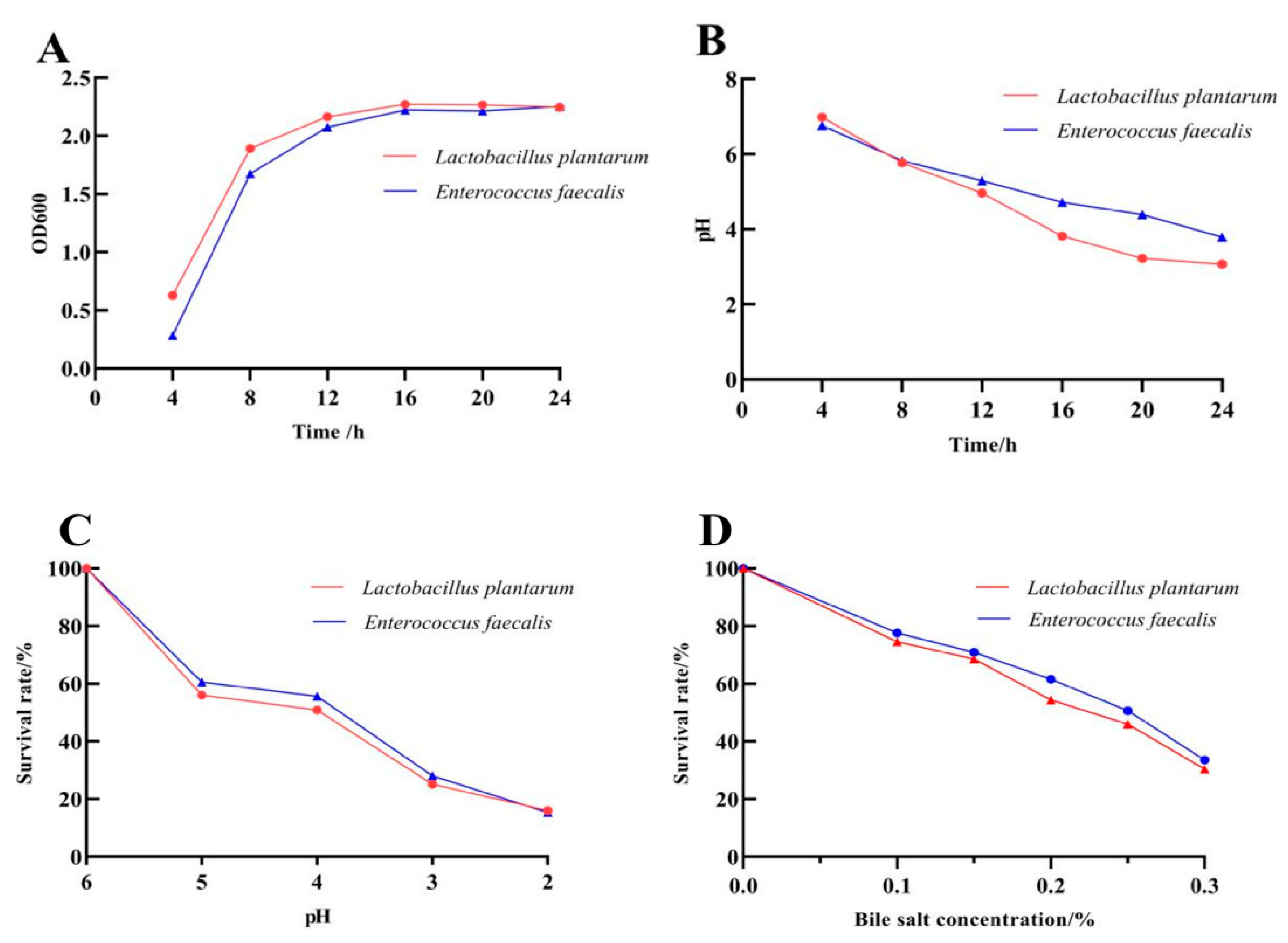
L. plantarum and E. faecalis were in the logarithmic growth stage from 4 to 16 h, with rapid reproduction, indicating good growth characteristics. Moreover, with the passage of time, the two strains produced acid during their growth process, and the pH decreased from about 7.0 to around 3.5, suggesting that they had good acid-producing capacity. After 24 h of exposure at pH 3, L. plantarum and E. faecalis showed high survival rates of (25.10 ± 0.49)% and (28.04 ± 0.46)%, respectively, suggesting the isolates exhibited high tolerance to low pH. The survival rate of L. plantarum and E. faecalis was reduced when the bile salt concentration was increased. Both strains showed a high level of tolerance to a bile salt concentration of 0.3 % after exposure for 24 h, with survival rates of (30.36 ± 0.38)% and (33.51 ± 0.29)%, respectively (Figure 2).
Both L. plantarum and E. faecalis were able to inhibit the growth of pathogenic bacteria such as Staphylococcus aureus, Salmonella gallinarum, and Escherichia coli in this study. The inhibition zones of L. plantarum and E. faecalis against pathogenic bacteria were all greater than 12 mm (Table 3). Therefore, L. plantarum and E. faecalis had good biological characteristics.

3.3. Effects of L. plantarum and E. faecalis on Production and Immunity of Pekin Duck

Compared to the control group, the addition of L. plantarum and E. faecalis significantly increased the ADFI and ADG of Pekin ducks (p < 0.05) and improved the live weight, eviscerated weight, half-eviscerated weight, breast muscle weight, and leg muscle weight (p < 0.05). The FCR was not affected by the supplementation of L. plantarum and E. faecalis in drinking water, and no significant differences were found in the carcass weight between the test groups and the control group either (p > 0.05) (Table 4).
A significant increase was shown in the duodenal villus height, duodenal V/C, and ileal villus height of ducks drinking water with L. plantarum and E. faecalis in comparison with the control group (p < 0.05). No significant differences were found in duodenal crypt depth, ileal crypt depth, or ileal V/C, nor in the villus height, crypt depth, or V/C of jejunum between the experimental groups and control group (p > 0.05) (Table 5).
Compared with the ducks in the control group, the bursa index and spleen index were significantly increased in the ducks drinking the L. plantarum and E. faecalis (p < 0.05). Despite the slight increase in the thymus index, there were no significant differences between the probiotics-treated groups and the control group (p > 0.05). The serum IgG levels in the LP group and EF group were significantly decreased, while the serum IgA levels in the two experimental groups were significantly increased compared with the control (p < 0.05) (Table 5). Moreover, no significant differences were observed in the relative expression levels of INF-γ, IL-6, and MUC2 in the duodenum, jejunum, and ileum between the ducks of the two experiment groups and the control group (p > 0.05) (Figure 3). These results indicated that L. plantarum and E. faecalis can improve the production and immune performance of ducks.

3.4. Effects of L. plantarum and E. faecalis on the Intestinal Flora of Pekin Ducks

The similarity between the PCR product sequences of 5 strains (Escherichia coli, Helicobacter pullorum, Campylobacter sp., Salmonella sp., and Lactobacillus sp.) and the strain sequences provided in NCBI was higher than 95%. The correlation coefficient of the standard curves for each strain was above 0.9883, and the amplification efficiency ranged from 98.8% to 107.5% (Table 6).
RT-PCR for Escherichia coli, Helicobacter pullorum, Campylobacter sp., Salmonella sp., and Lactobacillus sp. was performed, and the results showed that adding L. plantarum and E. faecalis significantly decreased the numbers of Escherichia coli, Campylobacter sp., and Salmonella sp. of ducks in comparison with the control group (p < 0.05). However, despite a slight decrease, there were no significant differences in the number of Helicobacter pullorum between the experiment and control groups (p > 0.05). Moreover, the quantity of Lactobacillus sp. in LP and EF groups was extremely significantly higher than that in the control group (p < 0.01), indicating that Lactobacillus sp. were the dominant bacteria in the two experimental groups (Table 7).

4. Discussion

Probiotics could directly adhere to or degrade harmful substances through themselves and their metabolites or exert indirect probiotic effects by regulating the intestinal flora or metabolic enzyme activity of flora, host immune activity, and part of enzyme activity [30,31,32]. Therefore, supplementing probiotics to regulate the gut microbiota and improve host growth and immune function has become a popular method in the international livestock industry, especially in improving individual gastrointestinal digestion and absorption as well as related diseases. Studies have shown that Lactobacillus salivarius can act as a potential probiotic to improve growth performance, fecal microbiota, and the immune response of chickens [33]. The addition of L. plantarum can also enhance the growth performance and intestinal health of Pekin ducks [34]. In recent years, there have been many studies on probiotics, and the efficacy of probiotics has shown obvious strain specificity and individual differences. Therefore, screening, identifying, and further promoting the development and utilization of probiotics in production were of great significance for improving the production and immune performance of ducks and promoting the efficient development of duck farming.
L. plantarum and E. faecalis were both probiotics and played important roles in maintaining intestinal health. L. plantarum can be used as a fermentation agent for food and was also a gastrointestinal probiotic that can adhere to and colonize on the intestinal mucosa, playing a key role in the competitive elimination of pathogenic bacteria [35,36]. E. faecalis, a facultative anaerobic bacterium belonging to the Enterococcus family in the order Lactobacillales, had strong tolerance to animal gastric fluid, intestinal fluid, and bile salts. It enhanced animal growth performance, maintained gastrointestinal microbiota balance, and strengthened the body’s resistance to diseases by producing extracellular polysaccharides to increase intestinal adhesion and promote probiotic colonization [37,38]. In this study, two isolates were screened from the cecum of Pekin ducks. Through colony morphology observation, biochemical identification, and 16S rDNA sequence analysis, the two strains were identified as L. plantarum and E. faecalis, respectively.
The growth and acid-producing capacity of lactic acid bacteria are important indicators for evaluating the metabolic ability of bacterial strains, and the gastrointestinal digestion tolerance of lactic acid bacteria is also used to evaluate whether they have potential probiotic properties. The growth of lactic acid bacteria will be inhibited after being affected by gastric acid and bile salt, so the survival of the strain in the gastrointestinal tract is the basis of determining its probiotic properties. The pH of the gastric juices of birds has been reported to be an average of 3.5 [39]. In this experiment, L. plantarum and E. faecalis showed good growth ability, acid production, acid resistance, and bile salt resistance, indicating that they had strong probiotic properties and exert probiotic functions in the gastrointestinal tract. Meanwhile, the two strains were able to inhibit the growth of Escherichia coli, Staphylococcus aureus, and Salmonella gallinarum, suggesting that they may be used as feed additives to improve the antibacterial ability of animals and reduce the occurrence of diseases.
Probiotic strains discovered from natural hosts were more desirable than isolates obtained from other sources due to their beneficial effects on host organisms and safety records, and it was the most promising probiotic for poultry nutrition [40]. In animal production, lactic acid bacteria were mainly used as probiotic feed additives, which had the functions of improving intestinal microbiota balance, promoting growth, and enhancing immunity. Studies have shown that L. plantarum GX17 was beneficial for the growth performance and improvement in intestinal barrier/gut microbiota function in yellow-feathered broiler chickens [41]. E. faecalis-1, isolated from healthy chickens, could promote the growth and immune performance of broiler chickens and regulate the cecal microbiota [42]. In this study, the ADFI, ADG, live weight, eviscerated weight, half-eviscerated weight, breast muscle weight, and leg muscle weight in the LP group and EF group were significantly higher than the control group, indicating that the isolated L. plantarum and E. faecalis could improve the growth and slaughter performance of Pekin ducks. Intestinal villus height and crypt depth were the important indicators that affected the digestion and absorption of nutrients, which determined the intestinal absorption capacity of animals. The addition of L. plantarum and E. faecalis in this experiment significantly increased the villus height and V/C of the duodenum in Pekin ducks, as well as the villus height of the ileum, suggesting that L. plantarum and E. faecalis could promote the digestion and absorption of nutrients in the duck intestine. Therefore, the addition of L. plantarum and E. faecalis could improve the production performance of Pekin ducks.
IgA and IgG played a key role in protecting the body from attack by infectious microorganisms [43,44]. Moreover, the genes INF-γ, MUC2, and IL-6 have been shown to play important roles in the body’s immune functions [45,46]. The levels of serum IgA and IgG, as well as the relative expression levels of the intestinal immune-related genes INF-γ, MUC2, and IL-6, were important indicators for detecting the immunomodulatory effects of Lactobacillus on the body. The results of this study showed that the supplementation of L. plantarum and E. faecalis in drinking water could significantly increase the immune organ index and serum IgA content and decrease the serum IgG content of Pekin ducks. At the same time, the relative expression levels of INF-γ and MUC2 in the duodenum, jejunum, and ileum were increased, and the relative expression levels of IL-6 were decreased. These results indicated that L. plantarum and E. faecalis could enhance the immune performance of Pekin ducks.
The process of intestinal digestion was closely related to the gut microbiota, such as nutrient absorption, feed digestibility, energy harvest, and productivity, all of which were influenced by the composition and diversity of the microbiota [47,48]. Intestinal pathogens and their toxins can disrupt the intestinal barrier in animals. Disruption of the dynamic balance of animal gut microbiota can lead to impaired digestion and immune function, as well as increased susceptibility to pathogens, resulting in diarrhea and decreased growth performance [49,50]. Some studies showed that the duck-derived lactic acid bacteria can improve the growth performance and meat quality of Muscovy ducks by regulating intestinal morphology and microbial community [51]. Similarly, supplementation of Lactobacillus acidophilus and Bacillus subtilis could significantly change the growth performance, serum immunity, and cecal microbiota of Cherry Valley ducks during the fattening period [52]. In this study, the addition of L. plantarum and E. faecalis could reduce the number of pathogenic bacteria, such as Escherichia coli, Campylobacter sp., Salmonella sp., and Helicobacter pullorum, in the cecum of Pekin ducks and increase the content of Lactobacillus sp., so as to improve the structure of intestinal microflora and promote intestinal microecological balance.

5. Conclusions

In summary, two strains were isolated from the cecum of Pekin ducks in this experiment and identified as L. plantarum and E. faecalis by colony morphological observation, biochemical identification, and 16S rDNA sequence analysis. Both strains had good biological characteristics. The supplementation of L. plantarum and E. faecalis in drinking water can improve the production and slaughter performance of Pekin ducks, as well as enhance their immune levels. In addition, they can also reduce the number of pathogenic bacteria in the cecum and maintain the stability of intestinal flora. These findings indicated that duck-derived L. plantarum and E. faecalis may be used as probiotic feed additives to promote the intestinal health of Pekin ducks and improve their production and immune performance. Due to the small sample size, there is not yet sufficient evidence to prove the effectiveness of duck-derived probiotics. In the future, we will increase the number of experimental ducks to more accurately determine the application effect of duck-derived probiotics.

Supplementary Materials

The following supporting information can be downloaded at: https://www.mdpi.com/article/10.3390/biom15091217/s1, Table S1: The sequences of YS-1 and YS-2.

Author Contributions

Conceptualization, Z.H. and X.L.; laboratory and feeding experiments, Z.Z., H.Z., and D.X.; bioinformatic analyses and biological interpretation, J.Z. (Jie Zhou) and M.C.; writing—original draft preparation, Z.H. and J.Z. (Jianqin Zhang); Writing—review and editing, Z.H. and Z.Z.; Supervision, X.L. All authors have read and agreed to the published version of the manuscript.

Funding

This study was supported by funds from the China Agriculture Research System of MOF and MARA (CARS-42-2) and the National Key Research and Development Program of China (2023YFD1300303).

Institutional Review Board Statement

All procedures, including handling the duck samples, were approved and implemented by the Institutional Animal Care and Institutional Ethics Committee of Northwest A&F University. Necessary efforts were undertaken to minimize animal suffering (No. ethic code: #1123/203).

Informed Consent Statement

Not applicable.

Data Availability Statement

Data are contained within the article and Supplementary Materials.

Conflicts of Interest

The authors declare that there are no conflicts of interest.

References

  1. Round, J.L.; Mazmanian, S.K. The gut microbiome shapes intestinal immune responses during health and disease. Nat. Rev. Immunol. 2009, 9, 313–323. [Google Scholar] [CrossRef]
  2. Shang, Y.; Kumar, S.; Oakley, B.; Kim, W.K. Chicken gut microbiota: Importance and detection technology. Front. Vet. Sci. 2018, 5, 254. [Google Scholar] [CrossRef]
  3. Kers, J.G.; Velkers, F.C.; Fischer, E.A.J.; Hermes, G.D.A.; Stegeman, J.A.; Smidt, H. Host and environmental factors affecting the intestinal microbiota in chickens. Front. Microbiol. 2018, 9, 235. [Google Scholar] [CrossRef]
  4. Carrasco, J.M.D.; Casanova, N.A.; Miyakawa, M.E.F. Microbiota, gut health and chicken productivity: What is the connection? Microorganisms 2019, 7, 374. [Google Scholar] [CrossRef] [PubMed]
  5. Kers, J.G.; Velkers, F.C.; Fischer, E.A.J.; Hermes, G.D.A.; Lamot, D.M.; Stegeman, J.A.; Smidt, H. Take care of the environment: Housing conditions affect the interplay of nutritional interventions and intestinal microbiota in broiler chickens. Anim. Microbiome 2019, 1, 10. [Google Scholar] [CrossRef] [PubMed]
  6. Clavijo, V.; Flórez, M.J.V. The gastrointestinal microbiome and its association with the control of pathogens in broiler chicken production: A review. Poult. Sci. 2018, 97, 1006–1021. [Google Scholar] [CrossRef] [PubMed]
  7. Chen, V.L.; Kasper, D.L. Interactions between the intestinal microbiota and innate lymphoid cells. Gut Microbes 2014, 5, 129–140. [Google Scholar] [CrossRef]
  8. Clemente, J.C.; Ursell, L.K.; Parfrey, L.W.; Knight, R. The impact of the gut microbiota on human health: An integrative view. Cell 2012, 148, 1258–1270. [Google Scholar] [CrossRef]
  9. Kamada, N.; Seo, S.U.; Chen, G.Y.; Núñez, G. Role of the gut microbiota in immunity and inflammatory disease. Nat. Rev. Immunol. 2013, 13, 321–335. [Google Scholar] [CrossRef]
  10. Potgieter, M.; Bester, J.; Kell, D.B.; Pretorius, E. The dormant blood microbiome in chronic, inflammatory diseases. FEMS Microbiol. Rev. 2015, 39, 567–591. [Google Scholar] [CrossRef]
  11. Modi, S.R.; Collins, J.J.; Relman, D.A. Antibiotics and the gut microbiota. J. Clin. Investig. 2014, 124, 4212–4218. [Google Scholar] [CrossRef]
  12. Yoon, M.Y.; Yoon, S.S. Disruption of the gut ecosystem by antibiotics. Yonsei Med. J. 2018, 59, 4–12. [Google Scholar] [CrossRef]
  13. Echegaray, N.; Yilmaz, B.; Sharma, H.; Kumar, M.; Pateiro, M.; Ozogul, F.; Lorenzo, J.M. A novel approach to Lactiplantibacillus plantarum: From probiotic properties to the omics insights. Microbiol. Res. 2023, 268, 127289. [Google Scholar] [CrossRef]
  14. Mokoena, M.P. Lactic acid bacteria and their bacteriocins: Classification, biosynthesis and applications against uropathogens: A mini-review. Molecules 2017, 22, 1255. [Google Scholar] [CrossRef]
  15. Troche, J.M.R.; Adame, E.C.; Díaz, M.A.V.; Escudero, O.G.; Chávez, M.E.I.; Chávez-Barrera, J.A.; Mondragón, F.Z.; Velasco, J.A.R.V.; Tavares, G.R.A.; Pedrín, M.A.L.; et al. Lactobacillus acidophilus LB: A useful pharmabiotic for the treatment of digestive disorders. Therap. Adv. Gastroenterol. 2020, 13, 1756284820971201. [Google Scholar] [CrossRef]
  16. De Filippis, F.; Pasolli, E.; Ercolini, D. The food-gut axis: Lactic acid bacteria and their link to food, the gut microbiome and human health. FEMS Microbiol. Rev. 2020, 44, 454–489. [Google Scholar] [CrossRef]
  17. Meruvu, H.; Harsa, S.T. Lactic acid bacteria: Isolation-characterization approaches and industrial applications. Crit. Rev. Food Sci. Nutr. 2023, 63, 8337–8356. [Google Scholar] [CrossRef]
  18. Ahmed, Z.; Vohra, M.S.; Khan, M.N.; Ahmed, A.; Khan, T.A. Antimicrobial role of Lactobacillus species as potential probiotics against enteropathogenic bacteria in chickens. J. Infect. Dev. Ctries. 2019, 13, 130–136. [Google Scholar] [CrossRef] [PubMed]
  19. Suryadi, U.; Nugraheni, Y.R.; Prasetyo, A.F.; Awaludin, A. Evaluation of effects of a novel probiotic feed supplement on the quality of broiler meat. Vet. World 2019, 12, 1775–1778. [Google Scholar] [CrossRef] [PubMed]
  20. Livak, K.J.; Schmittgen, T.D. Analysis of relative gene expression data using real-time quantitative PCR and the 2(-Delta Delta C(T)) Method. Methods 2001, 25, 402–408. [Google Scholar] [CrossRef] [PubMed]
  21. Hou, X.L.; Liu, G.; Zhang, H.H.; Hu, X.F.; Zhang, X.Y.; Han, F.; Cui, H.Z.; Luo, J.J.; Guo, R.; Li, R.; et al. High-mobility group box 1 protein (HMGB1) from Cherry Valley duck mediates signaling pathways and antiviral activity. Vet. Res. 2020, 51, 12. [Google Scholar] [CrossRef] [PubMed]
  22. Liu, Y.H.; Liu, C.; An, K.Y.; Gong, X.W.; Xia, Z.F. Effect of dietary Clostridium butyricum supplementation on growth performance, intestinal barrier function, immune function, and microbiota diversity of Pekin ducks. Animals 2021, 11, 2514. [Google Scholar] [CrossRef]
  23. Pechrkong, T.; Incharoen, T.; Hwanhlem, N.; Kaewkong, W.; Subsoontorn, P.; Tartrakoon, W.; Numthuam, S.; Jiménez, G.; Charoensook, R. Effect of Bacillus toyonensis BCT-7112T supplementation on growth performance, intestinal morphology, immune-related gene expression, and gutmicrobiome in Barbary ducks. Poult. Sci. 2023, 102, 102991. [Google Scholar] [CrossRef]
  24. Hu, Z.G.; Cao, J.T.; Zhang, J.Q.; Ge, L.Y.; Zhang, H.L.; Liu, X.L. Skeletal muscle transcriptome analysis of Hanzhong Ma duck at different growth stages using RNA-Seq. Biomolecules 2021, 11, 315. [Google Scholar] [CrossRef] [PubMed]
  25. Wang, Y.P.; Dong, L.; Guo, S.J.; Xu, Q.Q.; Miao, L.Z.; Xie, J.W.; Zhang, Y.; Ma, L.; Shen, Z.Q. Establishment of SYBR Green Ⅰ qPCR detection method for irp2 gene of poultry pathogenic Escherichia coli virulence island. Chin. Vet. Sci. 2017, 47, 871–876. (In Chinese) [Google Scholar]
  26. Pasquali, F.; Rossi, M.; Manfreda, G.; Zanoni, R. Complete nucleotide sequence of the gyrA gene of Helicobacter pullorum and identification of a point mutation leading to ciprofloxacin resistance in poultry isolates. Int. J. Antimicrob. Agents 2007, 30, 222–238. [Google Scholar] [CrossRef] [PubMed]
  27. Li, F.H.; Lou, L.P.; Jiang, T.T.; Liu, M.X.; Deng, Y.G.; Liu, J.C. Establishment of SYBR Green Ⅰ multiplex quantitative PCR method for Escherichia coli, Salmonella, Campylobacter jejuni and Proteus mirabilis in chicken. Mod. J. Anim. Husb. Vet. Med. 2022, 11, 1–6. (In Chinese) [Google Scholar]
  28. Sun, S.D.; Tian, Y.; Gao, Y.W.; Niu, L.Y.; Du, Y.Y.; Wang, W.J.; Zhong, J.H.; Tang, X.Q.; Liu, J.Q.; Zhou, W.P. Construction and application of fluorescent quantitative PCR method for avian Salmonella. Hunan J. Ani. Sci. Vet. Med. 2024, 1, 41–45. (In Chinese) [Google Scholar]
  29. Yang, M.; Yin, Y.; Wang, F.; Bao, X.T.; Long, L.N.; Tan, B.; Yin, Y.L.; Chen, J.S. Effects of dietary rosemary extract supplementation on growth performance, nutrient digestibility, antioxidant capacity, intestinal morphology, and microbiota of weaning pigs. J. Anim. Sci. 2021, 99, 237. [Google Scholar] [CrossRef]
  30. Seddik, H.A.; Bendali, F.; Gancel, F.; Fliss, I.; Spano, G.; Drider, D. Lactobacillus plantarum and its probiotic and food potentialities. Probiotics Antimicrob. Proteins 2017, 9, 111–122. [Google Scholar] [CrossRef]
  31. Quigley, E.M.M. Probiotics in irritable bowel syndrome: An immunomodulatory strategy? J. Am. Coll. Nutr. 2007, 26, 684S–690S. [Google Scholar] [CrossRef]
  32. Power, S.E.; O’Toole, P.W.; Stanton, C.; Ross, R.P.; Fitzgerald, G.F. Intestinal microbiota, diet and health. Br. J. Nutr. 2014, 111, 387–402. [Google Scholar] [CrossRef]
  33. Sureshkumar, S.; Lee, H.C.; Jung, S.K.; Kim, D.J.; Oh, K.B.; Yang, H.; Jo, Y.J.; Lee, H.S.; Lee, S.; Byun, S.J.; et al. Inclusion of Lactobacillus salivarius strain revealed a positive effect on improving growth performance, fecal microbiota and immunological responses in chicken. Arch. Microbiol. 2021, 203, 847–853. [Google Scholar] [CrossRef] [PubMed]
  34. An, K.; Gao, W.; Li, P.; Li, L.; Xia, Z. Dietary Lactobacillus plantarum improves the growth performance and intestinal health of Pekin ducks. Poult. Sci. 2022, 101, 101844. [Google Scholar] [CrossRef] [PubMed]
  35. Fidanza, M.; Panigrahi, P.; Kollmann, T.R. Lactiplantibacillus plantarum-nomad and ideal probiotic. Front. Microbiol. 2021, 12, 712236. [Google Scholar] [CrossRef] [PubMed]
  36. Talib, M.; Mohamad, N.E.; Yeap, S.K.; Hussin, Y.; Aziz, M.N.M.; Masarudin, M.J.; Sharifuddin, S.A.; Hui, Y.W.; Ho, C.L.; Alitheen, N.B. Isolation and characterization of Lactobacillus spp. from kefir samples in Malaysia. Molecules 2019, 24, 2606. [Google Scholar] [CrossRef]
  37. Daca, A.; Jarzembowski, T. From the friend to the foe-Enterococcus faecalis diverse impact on the human immune system. Int. J. Mol. Sci. 2024, 25, 2422. [Google Scholar] [CrossRef]
  38. Ditu, L.M.; Chifiriuc, M.C.; Bezirtzoglou, E.; Voltsi, C.; Bleotu, C.; Pelinescu, D.; Mihaescu, G.; Lazar, V. Modulation of virulence and antibiotic susceptibility of enteropathogenic Escherichia coli strains by Enterococcus faecium probiotic strain culture fractions. Anaerobe 2011, 17, 448–451. [Google Scholar] [CrossRef]
  39. Kassa, G.; Alemayehu, D.; Andualem, B. Isolation, identification, and molecular characterization of probiotic bacteria from locally selected Ethiopian free range chickens gastrointestinal tract. Poult. Sci. 2024, 103, 103311. [Google Scholar] [CrossRef]
  40. Reuben, R.C.; Roy, P.C.; Sarkar, S.L.; Alam, R.U.; Jahid, I.K. Isolation, characterization, and assessment of lactic acid bacteria toward their selection as poultry probiotics. BMC Microbiol. 2019, 19, 253. [Google Scholar] [CrossRef]
  41. Yin, Y.Y.; Liao, Y.Y.; Li, J.; Pei, Z.; Wang, L.P.; Shi, Y.; Peng, H.Y.; Tan, Y.Z.; Li, C.T.; Bai, H.L.; et al. Lactobacillus plantarum GX17 benefits growth performance and improves functions of intestinal barrier/intestinal flora among yellow-feathered broilers. Front. Immunol. 2023, 14, 1195382. [Google Scholar] [CrossRef] [PubMed]
  42. Shehata, A.A.; Tarabees, R.; Basiouni, S.; ElSayed, M.S.; Gaballah, A.; Krueger, M. Effect of a potential probiotic candidate Enterococcus faecalis-1 on growth performance, intestinal microbiota, and immune response of commercial broiler chickens. Probiotics Antimicrob. Proteins 2020, 12, 451–460. [Google Scholar] [CrossRef]
  43. De Sousa-Pereira, P.; Woof, J.M. IgA: Structure, Function, and Developability. Antibodies 2019, 8, 57. [Google Scholar] [CrossRef]
  44. Damelang, T.; Brinkhaus, M.; van Osch, T.L.J.; Schuurman, J.; Labrijn, A.F.; Rispens, T.; Vidarsson, G. Impact of structural modifications of IgG antibodies on effector functions. Front. Immunol. 2024, 14, 1304365. [Google Scholar] [CrossRef]
  45. Quagliato, L.A.; Nardi, A.E. Cytokine alterations in panic disorder: A systematic review. J. Affect. Disord. 2018, 228, 91–96. [Google Scholar] [CrossRef]
  46. Liu, Y.; Yu, Z.; Zhu, L.; Ma, S.; Luo, Y.; Liang, H.; Liu, Q.; Chen, J.; Guli, S.; Chen, X. Orchestration of MUC2-The key regulatory target of gut barrier and homeostasis: A review. Int. J. Biol. Macromol. 2023, 236, 123862. [Google Scholar] [CrossRef]
  47. Mancabelli, L.; Ferrario, C.; Milani, C.; Mangifesta, M.; Turroni, F.; Duranti, S.; Lugli, G.A.; Viappiani, A.; Ossiprandi, M.C.; van Sinderen, D.; et al. Insights into the biodiversity of the gut microbiota of broiler chickens. Environ. Microbiol. 2016, 18, 4727–4738. [Google Scholar] [CrossRef]
  48. Ha, C.W.Y.; Lam, Y.Y.; Holmes, A.J. Mechanistic links between gut microbial community dynamics, microbial functions and metabolic health. World J. Gastroenterol. 2014, 20, 16498–16517. [Google Scholar] [CrossRef]
  49. Sommer, F.; Anderson, J.M.; Bharti, R.; Raes, J.; Rosenstiel, P. The resilience of the intestinal microbiota influences health and disease. Nat. Rev. Microbiol. 2017, 15, 630–638. [Google Scholar] [CrossRef] [PubMed]
  50. Yang, B.G.; Hur, K.Y.; Lee, M.S. Alterations in gut microbiota and immunity by dietary fat. Yonsei Med. J. 2017, 58, 1083–1091. [Google Scholar] [CrossRef] [PubMed]
  51. Li, Z.; Zhou, H.; Liu, W.; Wu, H.; Li, C.; Lin, F.; Yan, L.; Huang, C. Beneficial effects of duck-derived lactic acid bacteria on growth performance and meat quality through modulation of gut histomorphology and intestinal microflora in Muscovy ducks. Poult. Sci. 2024, 103, 104195. [Google Scholar] [CrossRef] [PubMed]
  52. Zhang, M.; Yu, A.; Wu, H.; Xiong, X.; Li, J.; Chen, L. Lactobacillus acidophilus and Bacillus subtilis significantly change the growth performance, serum immunity and cecal microbiota of Cherry Valley ducks during the fattening period. Anim. Sci. J. 2024, 95, e13946. [Google Scholar] [CrossRef] [PubMed]
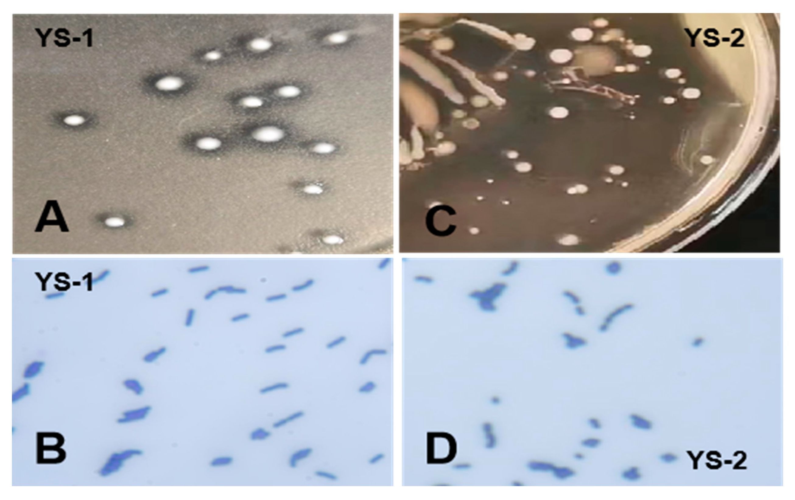
Figure 1. Morphology of bacterial strains. (A) Colony morphology of YS-1. (B) Gram staining of YS-1. (C) Colony morphology of YS-2. (D) Gram staining of YS-2.
Biomolecules 15 01217 g001
Figure 2. Biological characteristics of the probiotics. (A) Growth capacity curve. (B) Acid production capacity curve. (C) Acid tolerance curve. (D) Bile salt tolerance curve.
Biomolecules 15 01217 g002
Figure 3. Relative expression levels of INF-γ, MUC2, and IL-6 in the duodenum, jejunum, and ileum of Pekin ducks.
Biomolecules 15 01217 g003
Table 1. The feed composition of Pekin duck.
IngredientContent (%)NutrientContent (%)
Corn56.00Avian metabolizable energy2875 Mcal·kg−1
Soybean meal23.80Crude protein15.700
Corn gluten meal10.00Calcium0.900
Limestone7.00Total phosphorus0.680
CaHPO41.50Available phosphorus0.450
Premix1.00Salt0.370
NaCl0.30Lysine0.760
Lys·HCl0.30Methionine0.387
DL-Met0.10Methionine + Cystine0.654
Total100.00Isoleucine0.534
Threonine0.579
Tryptophan0.194
Crude fiber4.100
Crude fat3.400
Crude ash5.200
Table 2. Primer sequences for INF-γ, MUC2, IL-6 and bacterial strains.
Name (Target Gene)Primer (5′-3′)LengthReference
INF-γF:GCTGATGGCAATCCTGTTTT247 bp[21]
R:GGATTTTCAAGCCAGTCAGC
MUC2F:AGTTCTTGCCTAATTCCTCAGTCT146 bp[22]
R:TTGCCGTTCATATCCAGGTTCA
IL-6F:TTCGACGAGGAGAAATGCTT150 bp[23]
R:CCTTATCGTCGTTGCCAGAT
β-actinF:CCCTGTATGCCTCTGGTCG194 bp[24]
R:CTCGGCTGTGGTGGTGAAG
Escherichia coli (irp2)F: GCTCTGTGCCCTTTGA261 bp[25]
R: GGCGGGAGGAGTAGTT
Helicobacter pullorum (gyrA)F: CAAGAATCGTGGGTGATG351 bp[26]
R: GTGGAATATTTGTCGCCA
Campylobacter sp. (aicsxii)F: CACATTAAATCTTTATTTTCAACCCGCTGAA73 bp[27]
R: ACAATCCATCTTCTATCATTGCCTTAGC
Salmonella sp. (invA)F: CAATGGCGGCGAATTACGAG100 bp[28]
R: AAGGCTGAGGAAGGTACTGC
Lactobacillus sp.F: CGATGAGTGCTAGGTGTTGGA186 bp[29]
R: CAAGATGTCAAGACCTGGTAAG
Table 3. Antimicrobial activity of L. plantarum and E. faecalis from duck cecum against pathogens.
NameStrain and Diameter of Antibacterial Zone (mm)
Staphylococcus aureusEscherichia coliSalmonella gallinarum
L. plantarum12.20 ± 0.9212.92 ± 0.7413.42 ± 1.40
E. faecalis13.92 ± 2.1212.48 ± 0.2314.07 ± 2.41
Table 4. Effects of L. plantarum and E. faecalis on growth and slaughter performance of Pekin ducks.
PerformanceControl GroupLP GroupEF Group
ADFI (g/d)251.77 ± 3.89 b259.34 ± 4.32 a257.18 ± 4.18 b
ADG (g/d)110.32 ± 2.04 b118.15 ± 1.84 a116.58 ± 1.99 a
FCR2.28 ± 0.222.20 ± 0.232.21 ± 0.26
Live weight (g)3278.74 ± 30.76 b3462.15 ± 47.85 a3432.69 ± 40.32 a
Carcass weight (g)2866.32 ± 27.552902.38 ± 35.692887.65 ± 34.82
Eviscerated weight (g)2401.58 ± 35.83 b2573.46 ± 33.28 a2568.14 ± 34.49 a
Half-eviscerated weight (g)2698.14 ± 40.48 b2832.08 ± 38.77 a2828.78 ± 39.21 a
Breast muscle weight (g)231.23 ± 8.54 b245.55 ± 9.85 a242.14 ± 9.42 a
Leg muscle weight (g)266.12 ± 7.15 b282.27 ± 8.56 a289.32 ± 8.58 a
Note: Same superscript within a row indicates no significant differences (p > 0.05), different superscripts of lowercase letters within a row indicate significant differences (p < 0.01), and different superscripts of capital letters within a row indicate significant differences (p < 0.05). The same below.
Table 5. Effects of L. plantarum and E. faecalis on Immune Organ Index and Serum IgG, IgA Content in Pekin Ducks.
ItemsControl GroupLP GroupEF Group
Villus height in duodenum (μm)1007.35 ± 71.33 b1107.54 ± 50.05 a1101.21 ± 53.26 a
Crypt depth in duodenum (μm)207.78 ± 20.26213.98 ± 25.45208.85 ± 31.23
V/C in duodenum4.85 ± 0.33 b5.18 ± 0.42 a5.27 ± 0.40 a
Villus height in jejunum (μm)637.65 ± 45.01 b656.56 ± 37.75 a650.50 ± 38.44 a
Crypt depth in jejunum (μm)208.42 ± 18.83211.23 ± 21.36210.38 ± 24.45
V/C in jejunum3.06 ± 0.353.11 ± 0.223.09 ± 0.33
Villus height in ileum (μm)664.83 ± 87.35 b679.88 ± 56.58 a677.67 ± 67.43 a
Crypt depth in ileum (μm)155.90 ± 20.63158.54 ± 24.08156.38 ± 23.15
V/C in ileum4.26 ± 0.254.29 ± 0.384.33 ± 0.29
Thymus index (%)1.71 ± 0.211.78 ± 0.161.75 ± 0.16
Bursa index (%)0.62 ± 0.11 b0.70 ± 0.09 a0.72 ± 0.10 a
Spleen index (%)0.57 ± 0.07 b0.64 ± 0.05 a0.63 ± 0.08 a
Serum IgG content (μg/mL)752.79 ± 42.34 a679.08 ± 48.25 b684.23 ± 36.12 b
Serum IgA content (μg/mL)123.28 ± 12.48 b136.78 ± 15.87 a137.74 ± 13.75 a
Table 6. Standard curves for RT-PCR of various bacterial strains.
StrainsStandard CurveCorrelation Coefficient
(R2)
Amplification Efficiency (E)
Escherichia coliy = −2.8283x + 24.1130.9883102.3 %
Helicobacterpullorumy = −2.9817x + 28.3370.9956107.5 %
Campylobacter sp.y = −3.0549x + 28.9610.9913100.1 %
Salmonella sp.y = −3.1228x + 33.1360.994298.8 %
Lactobacillus sp.y = −3.2248x + 31.4560.9938100.9 %
Table 7. Effects of L. plantarum and E. faecalis on cecal microflora of Pekin ducks.
NameControl GroupLP GroupEF Group
Escherichia coli5.28 ± 0.13 a4.72 ± 0.08 b4.49 ± 0.11 b
Helicobacter pullorum3.73 ± 0.113.64 ± 0.083.58 ± 0.09
Campylobacter sp.4.27 ± 0.27 a3.84 ± 0.15 b4.02 ± 0.23 b
Salmonella sp.4.79 ± 0.20 a3.53 ± 0.35 b3.77 ± 0.45 b
Lactobacillus sp.3.66 ± 0.22 B5.38 ± 0.29 A4.86 ± 0.36 A
Disclaimer/Publisher’s Note: The statements, opinions and data contained in all publications are solely those of the individual author(s) and contributor(s) and not of MDPI and/or the editor(s). MDPI and/or the editor(s) disclaim responsibility for any injury to people or property resulting from any ideas, methods, instructions or products referred to in the content.