Article

3-O Sulfated Heparan Sulfate (G2) Peptide Ligand Impairs the Infectivity of Chlamydia muridarum

1
Arizona College of Osteopathic Medicine, Midwestern University, Glendale, AZ 85308, USA
2
Department of Biological Sciences, Sunandan Divatia School of Science, SVKM’s Narsee Monjee Institute of Management Studies (NMIMS) Deemed-to-Be University, Mumbai 400056, Maharashtra, India
3
Department of Microbiology & Immunology, Midwestern University, Downers Grove, IL 60515, USA
*
Authors to whom correspondence should be addressed.
These authors contributed equally to this work.
Biomolecules 2025, 15(7), 999; https://doi.org/10.3390/biom15070999 (registering DOI)
Submission received: 22 April 2025 / Revised: 18 June 2025 / Accepted: 8 July 2025 / Published: 12 July 2025
(This article belongs to the Section Chemical Biology)

Abstract

Background: Heparan sulfate (HS) is widely implicated as a receptor for Chlamydia cell attachment and infectivity. However, the enzymatic modification of HS modified by the 3-O sulfotransferase-3 (3-OST-3) enzyme in chlamydial cell entry remains unknown. Methodology: To rule out the possibility that host cell 3-O sulfated heparan sulfate (3-OS HS) plays a significant role in C. muridarum entry, a Chinese hamster ovary (CHO-K1) cell model lacking endogenous 3-OST-3 was used. In addition, we further tested the efficacy of the phage-display-derived cationic peptides recognizing heparan sulfate (G1 peptide) and the moieties of 3-O sulfated heparan sulfate (G2 peptide) against C. muridarum entry using human cervical adenocarcinoma (HeLa 229) and human vaginal epithelial (VK2/E6E7) cell lines. Furthermore, molecular dynamics simulations were conducted to investigate the interactions of the Chlamydia lipid bilayer membrane with the G1 and G2 peptides, focusing on their binding modes and affinities. Results: The converse effect of 3-OST-3 expression in the CHO-K1 cells had no enhancing effect on C. muridarum entry. The G2 peptide significantly (>80%) affected the cell infectivity of the elementary bodies (EBs) at all the tested concentrations, as evident from the reduced fluorescent staining in the number of inclusion bodies. The observed neutralization effect of G2 peptide on C. muridarum entry suggests the possibility of sulfated-like domains being present on the EBs. In addition, data generated from our in silico computational structural modeling indicated that the G2 peptide ligand had significant affinity towards the C. muridarum lipid bilayer. Conclusions: Taken together, our findings show that the pretreatment of C. muridarum with 3-O sulfated heparan sulfate recognizing G2 peptide significantly prevents the entry of EBs into host cells.

1. Introduction

Chlamydia spp. is a gram-negative, obligate intracellular mucosal pathogen, with a broad host and organ-site infection range, responsible for a wide range of diseases [1,2]. While C. trachomatis serovars A, B, Ba and C are the leading causes of infectious blindness in the non-industrial world (keratoconjunctivitis), C. trachomatis serovars L1–L3 and D–K cause urogenital infections, including lymphogranuloma venereum (LGV), cervicitis, urethritis, including serious sequelae in the form of pelvic inflammatory disease (PID), and salpingitis, resulting in infertility and ectopic pregnancy [3]. Additionally, exposure to C. pneumoniae can lead to the development of acute upper and lower respiratory illnesses, including bronchitis, pharyngitis, sinusitis and pneumonia [4]. The murine-adapted strain, C. muridarum, is commonly used as a rodent model for studying human female genital tract infections caused by Chlamydia, and mirrors observations made concerning the upper genital tract pathology of C. trachomatis in human female genital infections [5].
Regardless of the host, the infectious cycle of Chlamydia begins in the epithelium of the targeted tissues with the attachment and internalization of infectious elementary bodies (EBs) [1]. During the initial attachment process, Chlamydia spp. uses host cell surface heparan sulfate (HS) [6,7,8]. In addition, Chlamydia infection is enhanced in a heparan sulfate proteoglycan (HSPG)-dependent manner via the fibroblast growth factor 2 (FGF2)-mediated signaling pathway [9]. Furthermore, the level and position of sulfation in HSPGs appear to play a role in the binding of C. muridarum and C. trachomatis L2 to epithelial cells [10].
Structurally, HS is composed of a core protein covalently linked to the chains of unbranched sulfated anionic polysaccharides [11]. Modification of the HS chain is vital in mediating critical interactions with multiple ligands [12]. One such modification is the generation of unique sulfate moieties, such as N-/2-O/6-O- and 3-O-sulfated HS, which play a role in maintaining cellular homeostasis [13]. The final step in this HS modification is carried out by 3-O sulfotransferase (3-OST-3) at the C3 position of the glucosamine residue [14]. These modifications are essential in multiple biological processes as modified HS provides specific binding sites for a variety of viral and bacterial proteins, including adhesion molecules [15,16]. For example, Chlamydia hijacks host HS receptors during cell entry [6,7,8]. Thus, HS and their enzymatically sulfated variants provide a molecular basis for potentially targeting anti-chlamydial therapy [17,18,19].
Previous studies have shown that the initial interaction between EBs and the target cell can be significantly blocked by the addition of excess soluble HS, recombinant cysteine-rich outer membrane complex protein (OmcB), or anti-OmcB antibody [20,21]. Furthermore, the removal of 6-O sulfation by 6-O-endosulfatases significantly decreases C. muridarum EB attachment and vacuole formation [22]. However, the role of 3-O sulfation in heparan sulfate in C. muridarum infectivity has not been determined. The goal of the current study was to determine whether 3-O sulfation in heparan sulfate also plays a role in C. muridarum attachment and entry. In this direction, we first tested whether the cell surface expression of 3-O sulfated heparan sulfate (3-OS HS) would have any impact on the infectivity of C. muridarum. Our data clearly showed that the presence of the 3-OS HS receptor did not enhance the infectivity of C. muridarum. Since HS contains many negatively charged sulfate groups, it was logical to test whether HS- and 3-OS HS-recognizing G1/G2 peptides would influence the entry of Chlamydia. The G1 and G2 peptides were originally isolated to generate glycomimetic reagents that would bind to cellular heparan sulfate (G1 peptide) and its 3-O-sulfated heparan sulfate (G2 peptide) site [23]. One additional reason to test these peptides against Chlamydia was the fact that the above two cationic peptides were previously shown to inhibit the entry of herpes simplex virus type 1 (HSV-1) and Cytomegalovirus (CMV) [23]. The above pathogens, along with Chlamydia, are recognized as important agents in venereal and neonatal diseases [24,25,26]. It is also worth noting that the G1/G2 peptide provides a unique tool for studying multiple other pathologies, such as Alzheimer’s disease, hemorrhagic shock, and prostate, breast and hepatocellular cancer, in which enhanced expression of 3-O sulfation has been reported [27,28,29,30,31].
Structurally, the G1 peptide (LRSRTKIIRIRH) and the G2 peptide (MPRRRRIRRRQK) used in this study were distinct. For example, G1 peptide possesses alternating positively charged residues while G2 peptide has strings of cationic residues [23]. Interestingly, our previous findings have shown that G2 peptide preferentially recognizes sulfated domains on the HS chain [32]. In this study, we took two independent experimental approaches. In the first approach, EBs of C. muridarum were pre-exposed before being challenged with human-derived cervical and/or vaginal epithelial cell lines. In the second approach, the target cells were first exposed to G1 and/or G2 peptides before they were challenged with C. muridarum. Our results found that the application of the pre-treatment with G2 peptide to Chlamydia’s EBs showed a robust inhibition in the entry of C. muridarum as compared to G1 peptide in both the tested cell lines. On the other hand, the pre-treatment of target cells with G1/G2 peptides was not as effective in terms of cell infectivity compared to pre-treatment of G1 or G2 peptide to Chlamydia’s EBs. The above data were further supported by molecular dynamics simulation studies, which showed that the G2 peptide interacted better with the lipid bilayer membrane of C. muridarum than the G1 peptide.

2. Materials and Methods

2.1. Cell Cultures

The three cell lines used in this study were purchased from ATCC (American Type Culture Collection). Two were human female genital tissue-derived cell lines, i.e., HeLa 229 (human epithelial cervix adenocarcinoma, ATCC CCL-2.1TM) and VK2/E6E7 (human vaginal epithelial; ATCC CRL-2616™) cell lines. The third cell line, i.e., Chinese hamster ovary (CHO-K1; ATCC CCL-61™), served as the control to investigate the role of the 3-O-sulfated heparan sulfate (3-OS HS), as it lacks the 3-OST-3 enzyme responsible for generating 3-OS HS receptor [33]. HeLa 229 cells were grown at 37 °C with 5% CO2 in complete Dulbecco’s Modified Eagle Medium (DMEM; Corning Cell-gro) with 4.5 g/L glucose, 10% fetal bovine serum (VWR International), 110 mg/L L-glutamine and 1% gentamicin (HyClone). VK2/E6E7 cells were grown in Keratinocyte serum-free media (Life Technologies) supplemented with 0.1 ng/mL human recombinant epidermal growth factor (EGF), 0.05 mg/mL bovine pituitary extract, 0.4 mM calcium chloride and penicillin/streptomycin (Gibco/BRL).

2.2. Expression of 3-O Sulfotransferase-3 (3-OST-3)

To determine whether host cell surface 3-O-sulfated heparan sulfate (3-OS HS) plays a role in C. muridarum entry, a CHO-K1 cell model lacking endogenous 3-O-sulfotransferase-3 (3-OST-3) was used. Wild-type CHO-K1 cells were grown in Ham’s F-12 medium (Gibco/BRL, Carlsbad, CA, USA) supplemented with 10% fetal bovine serum (FBS), penicillin and streptomycin (Gibco/BRL). CHO-K1 cells were transfected with human encoded 3-OST-3 mammalian expression plasmid (OriGene) at 1.5 µg/µL using lipofectamine 2000 (Invitrogen), according to the manufacturer’s recommendations. CHO-K1 cells transfected in parallel with an empty vector (pcDNA3.1) plasmid were used as a negative control. After 72 h, post-transfected cells were challenged with C. muridarum to compare the cell infectivity in the presence and absence of 3-OST-3 enzyme.

2.3. C. muridarum Infectivity Assay

For infectivity assays, C. muridarum Nigg strain from a frozen −80 °C stock at multiplicity of infection (MOI) of 1.0 was used. C. muridarum Nigg strain was originally obtained from Dr Roger Rank of the University of Arkansas, who obtained it from American Type Culture Collection (Rockville, MD, USA). Following 24 h incubation, cells were fixed with methanol and stained with mouse anti-C. muridarum primary antibody and goat anti-mouse IgG secondary antibody (Invitrogen). Images of C. muridarum inclusions were captured at 10× using an EVOS FL Auto Imaging fluorescent microscope (Life Technologies). The level of infectivity was determined by counting the number of inclusion-forming units (IFU) in a minimum of 10 high-power fields (Zeiss Axiovert 25 fluorescent microscope) for each replicate with a minimum of three replicates per treatment to calculate the IFU/mL for each sample. The IFU/mL was calculated as follows: total number of inclusions per sample X sample dilution factor X field factor. Field factors vary depending on: (i) the number of fields counted; (ii) the well area in each plate; (iii) the microscope magnification. For these studies, the field factor is 6.15 for a 96-well plate (Genesee Scientific Corporation, El Cajon, CA, USA) with 10 counted fields at a magnification of 40×.

2.4. Chemical Structure of G1 and G2 Peptide Ligands

The 12-amino acid residue (12-mer) G1 and G2 peptide ligands were generated against plain-type HS and 3-O sulfated HS and synthesized according to a previously reported scheme [23]. The peptide sequence and characterization/purification of G1 and G2 peptide ligands are shown in Figure 1.

2.5. Pretreatment of C. muridarum with G1 and or G2 Peptide Ligands

The cell lines tested were grown in 96-well plates for 24 h to a confluency of 100%. The elementary bodies of C. muridarum (Nigg strain) at a MOI of 1.0 were pre-treated with HS (G1) peptide ligand or 3-OS HS (G2) peptide ligand in a two-fold-dependent manner, ranging from 0.00036 µM to 0.0030 µM, followed by rocking for 1 h (Scigene hybridization incubator, 4 RPM at 35 °C). The supernatant was removed by aspiration, and DMEM containing either G1 or G2 peptide ligand-treated C. muridarum was added (50 µL) to the target cells. Untreated C. muridarum (MOI 1.0, 50 µL) was added to cells as a positive control. Plates containing cells with G1- or G2-treated C. muridarum and controls were centrifuged for 1 h (1100× g, 35 °C). After centrifugation, the supernatant was removed by aspiration. Fresh, complete DMEM (100 µL) with 0.5 µg/mL cycloheximide was then added, and the plates were incubated for 24 h before fixing and staining the cells for fluorescent imaging of inclusion bodies. To understand the affinity of G1 and G2 peptides with the lipid bilayer of C. muridarum, molecular modeling and molecular dynamics simulations were employed.

2.6. Target Cell Pretreatment with G1 and or G2 Peptide Ligands

Target HeLa and VK2 cells were cultured in 96-well plates for 24 h to a confluency of 100%. The cells were independently pre-treated with HS (G1) peptide ligand and/or 3-OS HS (G2) peptide ligand in a two-fold-dependent manner ranging from 0.00036 to 0.0030 μM, followed by rocking for 1 h. Supernatant was removed by aspiration. The cells were then challenged with C. muridarum (Nigg strain) at a MOI of 1.0 for 24 h before fixing and staining the cells for fluorescent imaging of inclusion bodies, as described above. Untreated cells infected with C. muridarum at MOI of 1.0 were considered as a positive controls.

2.7. Exploring the Binding Mode and Affinity of G1 and G2 Peptides with the Lipid Bilayer Membrane Complex of C. muridarum Using Molecular Dynamics Simulation

2.7.1. G1 and G2 Peptide Modeling

The three-dimensional structures of the HS (G1-LRSRTKIIRIRH) and 3-O-sulfated HS (G2-MPRRRRIRRRQK) peptide ligands were predicted using the I-TASSER server, which employs a homology-based multiple-threading strategy to generate atomic-level 3D models of peptides [34]. The confidence score, TM-score and RMSD were used to obtain the best-fit model for both the G1 and G2 peptides. Further, molecular dynamics (MD) simulations were conducted to examine the dynamic behavior and stability of the peptides in interaction with the C. muridarum membrane.

2.7.2. C. muridarum Membrane Generation and MD Simulation

To elucidate the interaction and stability of G1 and G2 peptides with the C. muridarum membrane, MD simulations were performed using Gromacs 2021.5 [35]. The CHARMM-GUI interface [36] was used to construct these MD simulation systems, including the G1-membrane and G2-membrane complexes. Both peptides, G1 and G2, were initially positioned 20 Å above the C. muridarum membrane surface using translation and flip operations along the Z-axis. These MD simulation systems were solvated in a rectangular box filled with TIP3P water molecules, with K+ and Cl- ions added at a concentration of 0.15 M to maintain charge neutrality and physiological conditions. To model the C. muridarum outer membrane, we utilized lipid composition data obtained from the Orientations of Proteins in Membranes (OPM) database [37]. This Gram-negative bacterial pathogen possesses a unique trilaminar outer membrane. The inner leaflet of our model incorporated a mixture of phosphatidylethanolamine (PE), phosphatidylglycerol (PG) and cardiolipin (CL) lipid headgroups in a 75:20:5 ratio. Given that Chlamydia species feature a truncated LPS antigen (LPSA) in their outer membrane, characterized by a group-specific epitope composed of a KDO trisaccharide (K-KDO-(2C8)-K-KDO-(2C4)-K-KDO) [38], we included this LPSA component in both our membrane systems. Calcium ions were added for neutralizing the LPSA, and these assembled peptide–membrane complexes were further subjected to the CHARMM36m force field [39]. Further, molecular dynamics simulations were performed using GROMACS 2021.5 [35]. Initial steric clashes in the systems were removed through energy minimization using the steepest descent method for 5000 steps, followed by 1 ns of NVT (constant number of particles, volume and temperature) and NPT (constant number of particles, pressure and temperature) equilibration. The velocity-rescaling thermostat [40] and Berendsen barostat [41] were employed for temperature and pressure control during the NVT and NPT equilibration phases, respectively. Lastly, the production MD simulations were conducted for a duration of 300 ns, and the subsequent analysis of the resulting trajectories was performed using Gromacs utilities. The MD simulation trajectory was visualized using Visual Molecular Dynamics (VMD) software (1.9.4) [42].

2.7.3. Binding Energy Calculations

To estimate the binding affinity of G1 and G2 peptides with the lipid membrane, we employed the MM-GBSA method as implemented in the gmx_MMPBSA tool [43]. The binding energy calculations were performed on the last 100 ns (from 200–300 ns), and 1001 frames were extracted from the stable trajectory. The total binding energy for both systems will eventually help us understand the effectiveness of the peptides on the C. muridarum membrane. The entropy contribution was omitted during calculation due to computational cost, as in an earlier study [44].

2.8. Statistical Analysis

The data presented in this study involved three independent experiments (n = 3), which were repeated three independent times. Statistical analysis was performed using GraphPad Prism 10 software. A one-way analysis of variance (ANOVA) was used to determine the significance between the control and the experimental groups. In all figures, columns represent the mean of the data collected, and error bars represent SEM. Asterisks signify p-values: * < 0.05, ** < 0.005, *** < 0.0005 and **** < 0.0001.

3. Results

3.1. Expression of 3-O Sulfotransferase-3 (3-OST-3) and C. muridarum Entry

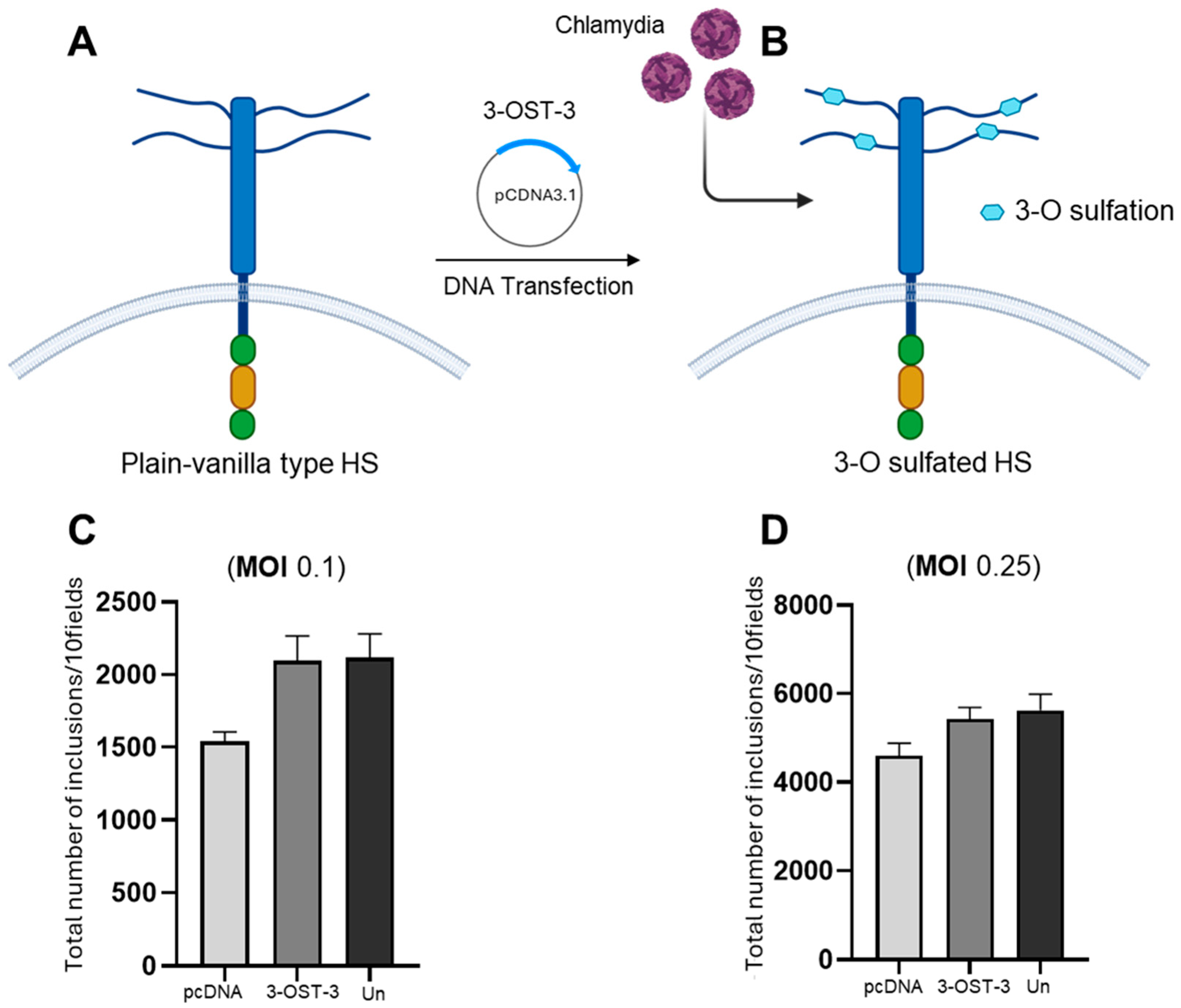
While the variable chains of heparan sulfate (HS)-containing glycosaminoglycans (GAGs) on the surfaces of mammalian cells play an important role in microbial invasion, the direct role of 3-O sulfated heparan sulfate (3-OS HS) as an independent determinant in Chlamydia cell infectivity remains unknown. Therefore, we tested the hypothesis that the presence of 3-OS HS is essential for chlamydial infection using a widely accepted and experimentally simple assessment in the CHO-K1 cell model. We used the wild-type CHO-K1 cells because they lack endogenous 3-OST-3 enzymes, which modify heparan sulfate (HS) to generate 3-OS HS receptors [33]. The expression of 3-OST-3 in CHO-K1 cells was confirmed by running the HSV-1 entry assay in parallel, since HSV-1 independently uses 3-OS HS to enter the CHO-K1 cell). Similarly, the role of the 3-OST-3 enzyme has been well-documented in mediating the entry of medically relevant viruses, including HSV-1 and CMV, into the host cell and virus cell-to-cell spread [23,45]. In this experiment, the CHO-K1 cells were transfected with the 3-OST-3 mammalian expression plasmid, while in parallel, CHO-K1 cells were transfected with an equal amount of an empty vector pCDNA3.1 (Figure 2A,B). Untransfected or mock transfected CHO-K1 cells were used as an internal control. The group of CHO-K1 cells expressing pCDNA3.1, 3-OST-3 and/or mock transfected control were then independently infected with C. muridarum at two different MOIs (0.1 and 0.25) for cell entry. As indicated in the Methods section, fluorescent-based staining followed by imaging to count the number of inclusion bodies was used as a parameter to define the cell infectivity of C. muridarum. There was no statistical difference in C. muridarum infectivity at the tested MOIs in the cells expressing the empty vector pCDNA3.1 control and or 3-OST-3 enzyme (Figure 2C,D). Taken together, our results indicate that the cell surface expression of 3-OS HS receptor does not serve an essential, integral function in promoting C. muridarum infection.

3.2. Preincubation of G1 and G2 Peptide Effects on Elementary Body (EB) Infectivity

Interestingly, previous findings suggest that heparin-like sulfated moieties are present on Chlamydia outer membrane protein (OMP) [46,47,48], while the cysteine-rich outer membrane polypeptide, OmcB, is a surface-exposed protein that functions as a chlamydial adhesin [49]. Published studies in the entry field suggest that the level and position of sulfation within HSPG play a role in binding, entry and inclusion formation [23,50,51]. Therefore, we next aimed to target a chlamydial infectious EB particle by directly exposing it to phage-display-derived G1 and G2 peptides recognizing heparan sulfate and 3-O sulfated HS ligand, respectively, before infecting the target HeLa (Figure 3) and VK2 cells (Figure 4). The details regarding the origin of the G1/G2 peptides are shown in supplementary Figure S1.
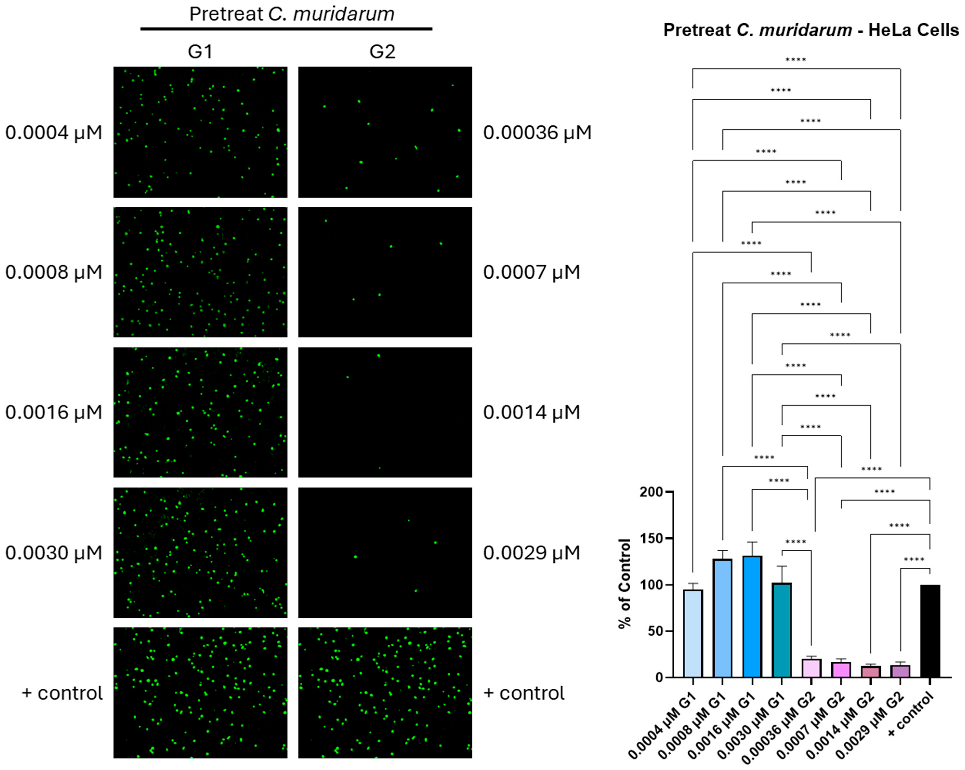
As previously mentioned, the formation of a membrane-bound compartment in the form of inclusion bodies was used as an indicator for Chlamydia’s cell infectivity in the presence and absence of the peptides. The pre-treatment of EBs with the G2 peptide ligand in a dosage-dependent manner showed a significant (p < 0.001) inhibition of C. muridarum infectivity of HeLa cells, as evident from the significant reduction in the green punctate, which corresponded to the number of inclusion bodies (Figure 3). The extent of inhibition over the range of G2 concentrations (0.00036–0.0029-μM) was similar, ranging from 80.2% to 88.5% inhibition, as compared to control, respectively. In contrast, preincubation of EBs with G1 peptide resulted in an enhancement in the number of inclusion bodies. Specifically, the EBs treated with 0.0016 μM of G1 peptide resulted in a 32% increase in infectivity into HeLa cells, while those with cell infection at the remaining concentrations (0.0007, 0.00036 and 0.0029 μM) were similar to that measured for the untreated EB control (Supplementary Figure S2).
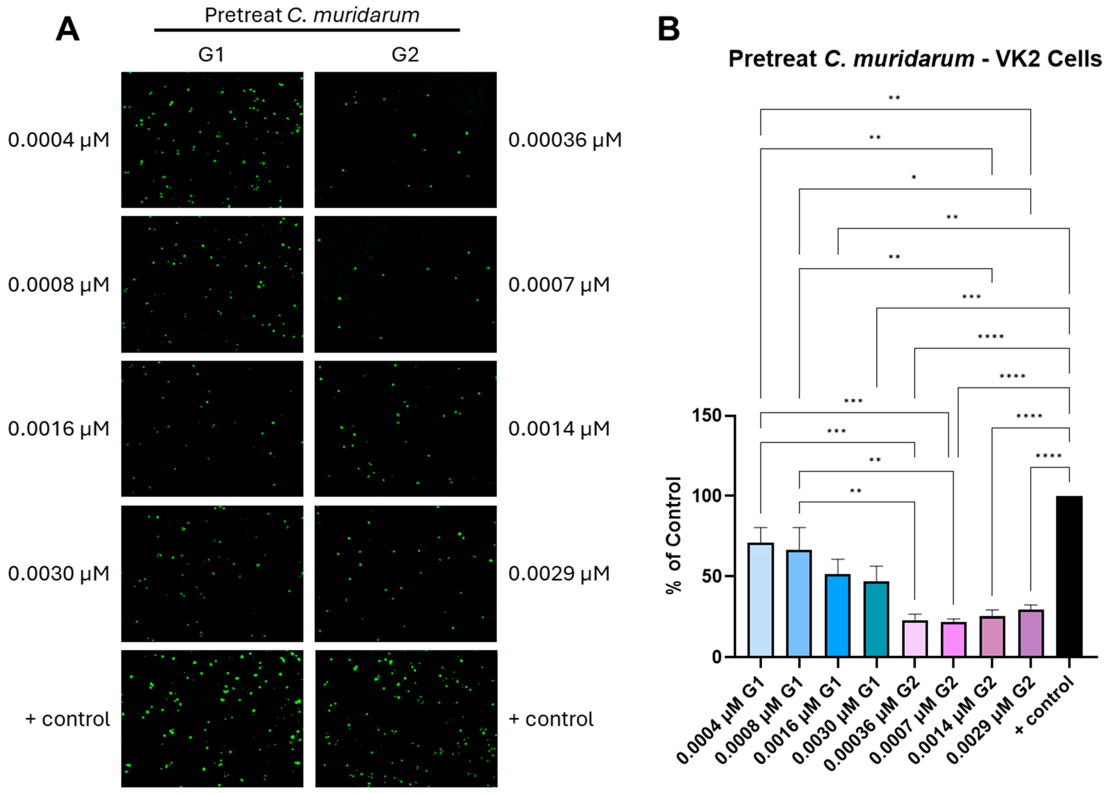
The impact of the G1 and G2 peptide ligand on the EBs’ pretreatment with infected vaginal epithelial cells (VK2 cells) was further determined to ascertain whether the EB neutralization effects were specific for HeLa cells. In addition, C. muridarum, being a murine pathogen similar to the human pathogen C. trachomatis, infects the vaginal epithelium [52]. Therefore, VK2 cells also served as an appropriate model to understand the effect of G1/G2 peptides targeting the HS and 3-OS HS receptors, improving the molecular basis of host–pathogen interactions. Interestingly, in contrast to our findings with HeLa cell infectivity, both the G1 and the G2 peptide ligands were highly effective in inhibiting C. muridarum infection in the VK2 cell model by significantly reducing the number of inclusion bodies in a dosage-dependent manner (Figure 4). Our results indicated that the G1 peptide ligand at the highest concentrations tested (0.003 and 0.0016 μM) significantly (p < 0.05) inhibited the EB infection of VK2 cells to 47% and 52% of the control, respectively. All of the G2 peptide ligand concentrations tested significantly (p < 0.001) inhibited Chlamydia cell infectivity by reducing the number of inclusion bodies  to that measured for untreated EBs control. This result indicates the significance of HS during EB’s infectivity in the two clinically relevant cell lines used in this study. Since the G1 and G2 peptide recognizes HS and 3-OS HS, it was logical to test whether the pre-treatment with the G1/G2 peptide for the host cells (HeLa and VK2) would have an impact on the infectivity of the C. muridarum. As indicated in Supplementary Figure S2, it was clear that pre-incubation of the G1 and G2 peptides at the majority of the tested concentrations did not affect the entry of the EBs into the HeLa cells, but the G2 peptide did show some blocking effects at a higher concentration. Similarly, the peptide pre-treatment in the VK2 cells had some blocking effects at a higher concentration. Because 3-OS HS expression alone was not utilized as an independent receptor for Chlamydia entry (Figure 2), it was expected that blocking of 3-OS HS by G2 peptide would not be sufficient to abolish the entry of EBs into the host cell. This data also opens the possibility that C. muridarum utilizes several other receptors, which could vary depending on cell types.

3.3. Investigating the Interaction of G1 and G2 Peptides with the Chlamydia Lipid Bilayer

Iterative Threading ASSEmbly Refinement (I-TASSER) generated five potential 3D structures for each G1 and G2 peptide, ranking them by confidence C-score, a measure of model confidence. The best-fit models were selected based on C-score, template modeling (TM) score and root mean square deviation (RMSD) (Table 1). Model 1 was chosen as the most suitable structure for both peptides based on its C-score (the higher the better). G2 peptide exhibited a higher C-score than G1, suggesting improved folding and structural stability.
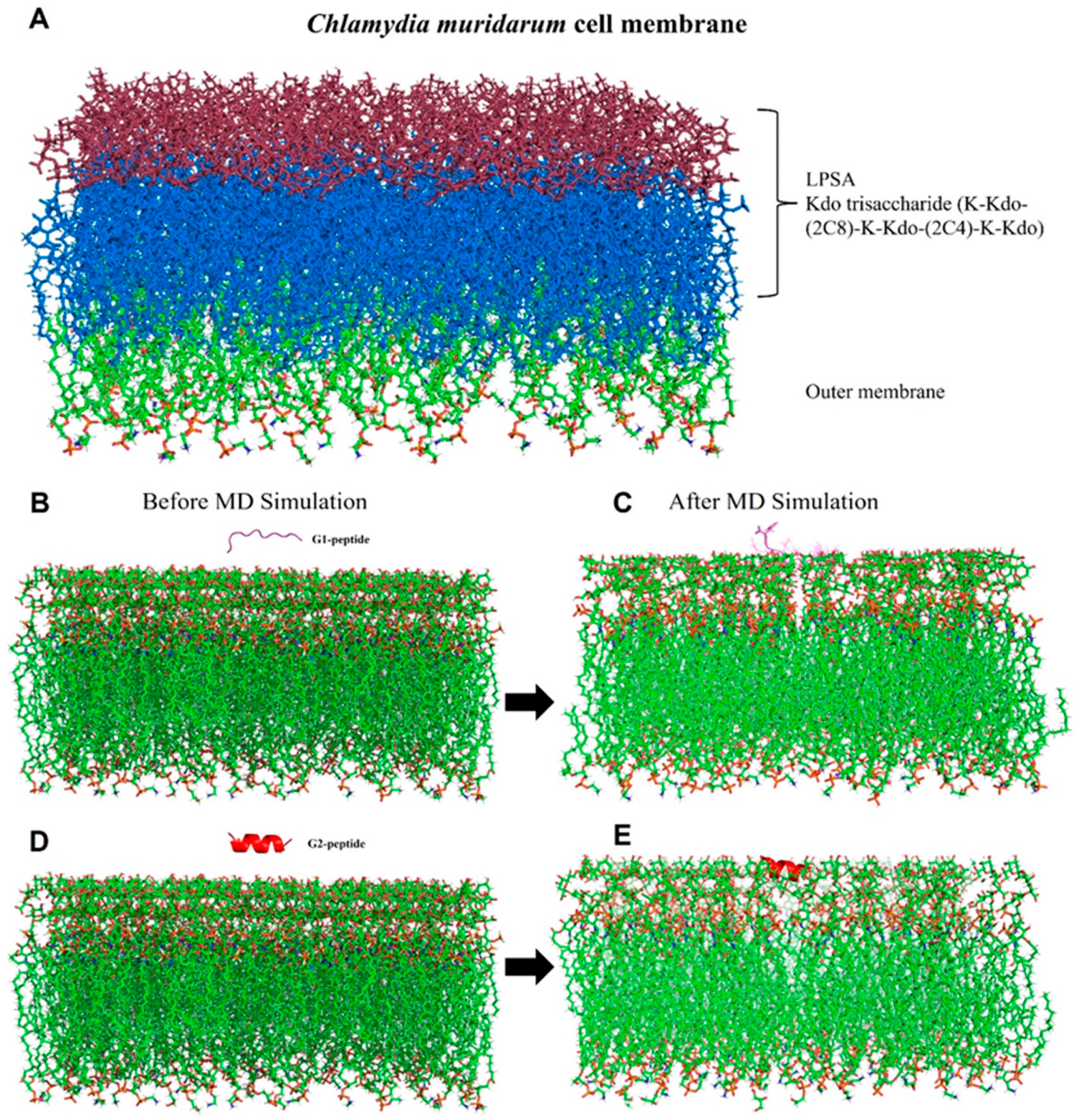
Molecular dynamics (MD) simulations were conducted to explore the interactions of the G1 and G2 peptides with the Chlamydia lipid bilayer, offering more profound insights into their binding modes and affinities (Figure 5B–E and Supplementary Figure S3). Molecular dynamics simulations were performed using Gromacs 2021.5 [26]. The stability of the simulated complexes was assessed by analyzing the RMSD plot, as shown in Figure 6A. The RMSD plot indicates that the G2 peptide reaches equilibrium after 150 ns in the presence of the lipid bilayer, suggesting that it achieves both conformational and structural stability, unlike the G1 peptide (Supplementary Movies S1 and S2). The G1 peptide exhibits significant conformational changes (Supplementary Movie S1) and also achieves equilibrium after 150 ns, as can be observed on the RMSD plot (Figure 6A). To further explore the conformational properties of the G1 and G2 peptides, we calculated the root mean square fluctuations (RMSFs), shown in Figure 6B. The RMSF plot measures the movement degree of Cα atoms around their average positions. Here, the G2 peptide generally exhibits lower RMSF values across the majority of the residues compared to the G1 peptide (Figure 6B), and this suggests that the residues in the G2 peptide experience less movement and are more constrained. The regions around residue numbers 4–5 and 6–7 for the G2 peptide show particularly low RMSF values (below 2 Å in most cases), indicating that these parts of the peptide are highly stable and likely deeply embedded or strongly interacting with the lipid bilayer of the membrane (Figure 6B and Supplementary Movie S2). Even where the RMSF values for G2 increase slightly (around residues 5 and 10–12), they generally remain lower than the corresponding residues in G1, suggesting a more consistently stable interaction profile across the entire peptide (Figure 6B). Conversely, the higher RMSF values observed for the G1 peptide indicate greater flexibility and weaker interactions with the membrane, with certain regions showing particularly high mobility (Figure 6B and Supplementary Movie S1). Furthermore, the conformation and folding state of the G1 and G2 peptides were analyzed using the radius of gyration (Rg). The G2 peptide generally fluctuates at a lower average Rg value (around 7–8 Å) throughout the simulation time compared to the G1 peptide. This suggests that, on average, the G2 peptide maintains a more compact conformation during the simulation (Figure 6C). By contrast, the G1 peptide exhibits a higher average Rg value (around 8–9 Å and sometimes exceeding 9 Å) compared to the G2 peptide throughout the simulation. This indicates that the G1 peptide, on average, adopts a less compact and more extended conformation, as shown in Figure 6C. Even though G1 has weaker interactions with the membrane overall, it undergoes a transient conformational change after 150 ns that leads to a more compact structure (Figure 6C). This compaction might not necessarily indicate a stronger or more stable interaction with the membrane, but rather G1 might have become more compact while still primarily residing on the surface of the membrane or interacting only with the LPSA, rather than inserting deeply into the hydrophobic core, like G2, as can be observed in Supplementary Movies S1 and S2.
To examine how these conformational effects influence binding affinity, we conducted binding energy calculations using the gmx_MMPBSA tool [32].

3.4. Binding Energy Calculations

To assess the binding affinity of G1 and G2 peptides with the lipid bilayer, we performed binding energy calculations using the MM-GBSA method through gmx_MMPBSA tool [32]. The binding energy data revealed that the G2 peptide had a higher affinity (−111.76 kcal/mol) with the lipid bilayer compared to the G1 peptide with the lipid bilayer (−40.61 kcal/mol), as shown in Table 2. Moreover, the G1- and G2-peptide-associated lipid bilayer complexes reveal negative van der Waals and electrostatic energy, indicating favorable interactions. The G1 peptide shows slightly higher van der Waal energy compared to the G2 peptide, but the G2 peptide has much stronger electrostatic interactions, suggesting that it has more charged or polar regions interacting with the membrane. Furthermore, the G2 peptide has a higher desolvation penalty (ΔEGB = 36001.98), reflecting its stronger initial solvation before interacting with the membrane as compared to the G1 peptide. Also, the G2 peptide with a more negative value (ΔGGAS = −36104.32) likely has a more hydrophobic surface exposed to the membrane as compared to the G1 peptide. The ΔGGAS values indicate favorable interactions in the gas phase, with the G2 peptide showing stronger interactions as compared to the G1 peptide. The interaction is more energetically favorable than the G1 peptide, with a total free energy of −111.76 kcal/mol. This is largely due to stronger electrostatic interactions, indicating a more stable or tighter binding with the membrane. The higher solvation energy value reflects the initial state of the peptide being well-solvated before binding. Taken together, the G2 peptide–membrane interactions are more stable and energetically favorable than in the G1 membrane complex (Table 2 and Supplementary Movies S1 and S2), primarily due to stronger non-bonded interactions. This suggests that the G2 peptide has more extensive or effective interactions with the membrane, which may be important for its function or stability in a biological context.

4. Discussion

Infection of host cells by Chlamydia is a multi-step dynamic process [50,51,52,53,54]. It is widely believed that C. muridarum first mediates a low-affinity interaction with HSPGs [6,8], followed by interacting specifically with high-affinity binding to host cell receptors. These receptors include a wide variety of proteins, ranging from cystic fibrosis transmembrane conductance regulator (CFTR), mannose 6-phosphate receptor (M6P), β1 integrin, epidermal growth factor receptor (EGFR), receptor tyrosine kinases, fibroblast growth factor (FGF) receptor and its ligand, FGF, platelet-derived growth factor receptor (PDGFR) and protein disulfide isomerase (PDI), a component of the estrogen receptor complex [9,55,56,57,58,59,60,61,62].
In this study, we assessed whether 3-O sulfation in the HS chain is preferentially utilized by C. muridarum during cell entry. Identifying the key sulfated residues involved during Chlamydia infection is an important step to better understand cell- and tissue-specific requirements towards the development of novel treatments. We first deployed a well-established CHO-K1 cell-based assay, which was adapted from the HSV-1 entry field to test the significance of 3-O sulfated heparan sulfate in Chlamydia infectivity. Our entry data with CHO-K1 cells expressing either empty pCDNA3.1 vector or 3-OST-3 enzyme showed no differences in the infectivity of C. muridarum, suggesting that 3-OS HS does not serve as an independent entry receptor. This result contrasts with previously established HSV entry data, according to which viruses use 3-OS HS to promote viral entry and cell-to-cell spread [23,33]. This data provided an early presumptive, yet critical clue that two independent pathogens such as HSV and Chlamydia perhaps do not compete for the same 3-OS HS receptor during either co- or super-infection. The expression of HS and 3-OS HS receptors is well-documented in multiple susceptible cell lines used by STI (sexually transmitted infection) pathogens, including dendritic cells, T helper cells and the genital epithelium [15,63,64]. Therefore, a preferential recognition of specific 3-OS HS receptors by HSV-1 glycoprotein D (gD) functions as a key determinant to allow the potential occurrence of HSV infection [16], while Chlamydia’s ability to use multiple cell surface receptors may provide unique flexibility to enter cells along with HSV. However, future studies will be needed to understand the significance of sulfated HS in a co- or super-infection model. Although the 3-OST-3-generated 3-OS HS receptor did not enhance C. muridarum cell infectivity, in the future, it will be worth investigating whether expression of other 3-OST enzymatic isoforms, such as 3-OST-1, -2, -4, -5 and -6, would have any impact on the infectivity of C. muridarum and, possibly, any associated inflammatory damage [45]. Similarly, other host genes, such as exostosin-1 (EXT-1) and N-deacetylase/N-sulfotransferase (NDST), which serve as critical players in HS biosynthesis, would also be interesting to test for their modulatory role in Chlamydia cell infectivity. Interestingly, a recent finding highlighted that EXT-1 plays a unique role in the life cycle of Zika virus infection [65]. Knocking down EXT1 at the early stage represses viral infection, whereas it increases virus infection when knockdown is performed at the late stage of viral infection [65]. In addition, since the HS chain is also modified by multiple isoforms of 2-O sulfotransferases (2-OST-1 and 2-OST-2) and 6-O sulfotransferases (6-OST-1, 6-OST-2 and 6-OST-3) before 3-O sulfotransferases (3-OST) [13], it remains unknown whether 2-OSTs and or 6-OSTs independently participate in the infectivity of C. muridarum. A previous study reported that the specific removal of 6-O sulfation by human endosulfatases diminished C. muridarum binding and decreased vacuole formation in the HeLa cell model, suggesting that 6-O sulfation is indeed a critical determinant of C. muridarum [22].
Our assessment of the impact of peptides targeting HS (G1) and 3-O sulfated HS (G2) on C. muridarum infectivity generated interesting findings. For instance, the pre-exposure of the G2 peptide to Chlamydia resulted in a significant blockage of infection in both the relevant cell lines used in this study. The above results are complemented by a previous finding that showed a heparan sulfate-specific monoclonal antibody that binds to glycosaminoglycans (GAGs) localized on the surface of the chlamydial organism and effectively neutralizes the infectivity of both C. trachomatis and C. pneumoniae [46]. Evidence of sugar being present on the outer surfaces of multiple other intracellular pathogens is also documented. For instance, Toxoplasma gondii displayed lectins and glycoconjugates on their surfaces [66]. In addition, the rhoptry and dense granule protein of Toxoplasma binding recognizes both N-sulfation and 6-O-sulfation on the glucosamine residue, with N-sulfation seeming to be more important than O-sulfation [67]. Similarly, the G protein of respiratory syncytial virus (RSV) possesses a short chain of oligosaccharide structures, which are involved in its infectivity in vitro [68]. Whether Toxoplasma and RSV carry the 3-O sulfation-like moieties on their surfaces remains to be determined. One major benefit of the presence of HS on various pathogens, including Chlamydia, is that it allows the use of ubiquitous multiple receptors for attachment and invasion. Furthermore, the use of HS is also advantageous because it presents a host-similar ligand on its surface, allowing it to exploit molecular mimicry and thereby prevent recognition by the host immune system [46]. However, the masking of EB ligands by HS may alter the display of alternative attachment ligands, which could alter infectivity in a manner that is host-cell-specific.
Interestingly, pre-treatment of Chlamydia with the G1 peptide uniquely promoted cell infectivity in HeLa cells, which was unusual. Since G1 peptide possesses affinity for HS, it is quite possible that sequestration or binding of HS by the G1 peptide resulted in an open state conformation of the sulfated moieties present on the HS chain, which provided easy access to the sulfated domains to promote the entry of C. muridarum. However, we did not notice a similar promoting effect of the G1 peptide in the VK2 cells, which could have been due to the differential expression of HS and the enzymatic isoforms of 3-OST-3. Another possibility is that G1 and G2 peptides also appear to modulate HS expression [69,70]. Similarly, other pathogens, such as HSV-2, have been shown to enhance the expression of HS and 3-OS HS upon exposure to the host cell, resulting in a higher affinity for the G2 peptide in virally infected cells compared to uninfected cells [32]. Further studies are needed to understand the glycans-based differences among cell types, as well as the independent effects of the peptides or Chlamydia exposure on HS/3-OS HS expression, which may influence Chlamydia–host cell interactions.
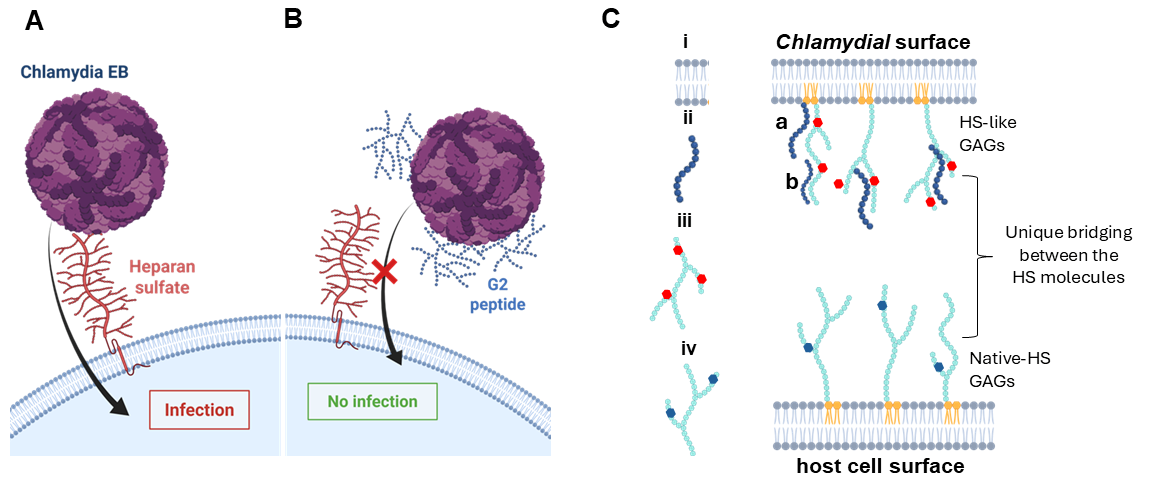
Taken together, our data demonstrate that the blocking of 3-OS HS moieties by the G2 peptide mediates the neutralization of C. muridarum cell infectivity. Based on our findings with the G2–lipid bilayer binding affinity analysis, we propose that the peptides targeting the lipid bilayer may have affected the cell dynamics and were able to prevent Chlamydia cell infection (Figure 7A,B) by having strong affinity towards the lipid membrane (Figure 7C). It is quite possible that the latter may also pose the 3-O sulfated HS-like domains on the polysaccharide, which could be the cause of the neuralization effect observed during the EBs’ exposure to the G2 peptide. Since the human strains of Chlamydia (C. trachomatis) and the associated serovars are known utilizers of a diverse subset of sulfated HS to promote cell entry [71], future studies will be needed to address whether the G2 peptide-mediated EB neutralization effect persists in other serovars, including assessing the impact of G1/G2 peptides on Chlamydia–HSV-associated co-infection and or super-infection in the murine model.

5. Conclusions

Our findings demonstrate that G2 peptide, which recognizes 3-O sulfation in HS, significantly inhibits C. muridarum entry into host cells, suggesting a strong interaction between the peptide and components of the chlamydial EBs. Molecular dynamics simulations further suggest that the G2 peptide interacts with the lipid bilayer, thereby disrupting the structural integrity and infective dynamics of C. muridarum. Collectively, these results emphasize the critical role of sulfated glycoconjugates in chlamydial pathogenesis and highlight them as promising targets for therapeutic intervention. Future studies should explore the serovar-specific nature of this interaction and evaluate its implications in co-infections with herpes simplex virus (HSV) and other sexually transmitted pathogens.

Supplementary Materials

The following supporting information can be downloaded at: https://www.mdpi.com/article/10.3390/biom15070999/s1. Figure S1. Graphic depicting the steps (a–f) involved during phage display library screening of M13 phage against heparan sulfate (HS) and 3-O sulfated heparan sulfate (3-OS HS) to isolate and characterize G1 and G2 peptides. Figure S2. Effect of the pretreatment with various concentrations of G1 and G2 peptides on HeLa cells (panel A) and VK2 (panel B) followed by infection with C. muridarum as determined by fluorescent microscopy. Inclusions were stained and representative images were shown. The number of inclusions were counted and the percentage of infected cells treated with G1 or G2 compared to non-treated infected cells only (positive control) was calculated (Mean ± SEM, * p ≤ 0.05, ** p < 0.005, *** p < 0.0005, and **** p < 0.0001). Data is a representative of the results of three independent experiments (n=3) performed in triplicates. Figure S3: The workflow representation of Chlamydia muridarum lipid bilayer building and simulation protocol. Supplementary Movie S1: Molecular dynamics simulation movie of G1 with lipid membrane complex. MD simulation movies were generated using Visual Molecular dynamics (VMD) software [42]. Supplementary Movie S2: Molecular dynamics simulation movie of G2 with lipid membrane complex. MD simulation movies were generated using Visual Molecular Dynamics (VMD) software [42].

Author Contributions

W.H.: Analysis and Data Curation. P.K.: In Silico Study, Analysis and Data Curation, Writing Draft. J.M.: Analysis and Data Curation. K.V.: Analysis and Data Curation; I.S.: Analysis and Data Curation, Funding acquisition. B.J.P.: Editing manuscript. A.K.: Analysis and Data Curation. J.E.: Cell culture. B.V.K.: Writing Original Draft, In Silico Methodology, Analysis and Data Curation. V.T.: Conceptualization, Supervision, Original Draft Preparation, Review and Editing of Manuscript, Funding Acquisition. All authors have read and agreed to the published version of the manuscript.

Funding

This study was supported by MWU Internal Grant Funding to I.S. and V.T.

Institutional Review Board Statement

Not applicable.

Informed Consent Statement

Not applicable.

Data Availability Statement

Data will be made available on request.

Acknowledgments

We sincerely thank Suvendu Biswas, Mark Richard Karver and E. Testa for peptide synthesis. Peptide synthesis was performed at the Peptide Synthesis Core Facility of the Simpson Querrey Institute for BioNanotechnology at Northwestern University, Chicago. B.V.K is grateful to SVKM’s NMIMS (Deemed-to-be) University, Mumbai (India) for providing the computational facility.

Conflicts of Interest

The authors declare no conflicts of interest.

Abbreviations

HS: heparan sulfate; 3-OST-3: 3-O sulfotransferase-3; 3-OS HS: 3-O sulfated heparan sulfate; EBs: elementary bodies; MD: molecular dynamics; RMSF: root mean square fluctuations; Rg: radius of gyration.

References

  1. Bachmann, N.L.; Polkinghorne, A.; Timms, P. Chlamydia genomics: Providing novel insights into chlamydial biology. Trends Microbiol. 2014, 22, 464–472. [Google Scholar] [CrossRef]
  2. Mandell, G.L.; Bennett, J.E.; Dolin, R. (Eds.) Mandell, Douglas, and Bennett’s Principles and Practice of Infectious Diseases, 7th ed.; Churchill Livingstone/Elsevier: London, UK, 2010; Volume 2. [Google Scholar]
  3. Malhotra, M.; Sood, S.; Mukherjee, A.; Muralidhar, S.; Bala, M. Genital Chlamydia trachomatis: An update. Indian J. Med. Res. 2013, 138, 303–316. [Google Scholar] [PubMed]
  4. Porritt, R.A.; Crother, T.R. Chlamydia pneumoniae Infection and Inflammatory Diseases. For. Immunopathol. Dis. Therap. 2016, 7, 237–254. [Google Scholar] [CrossRef] [PubMed]
  5. Dockterman, J.; Coers, J. Immunopathogenesis of genital Chlamydia infection: Insights from mouse models. Pathog. Dis. 2021, 79, ftab012. [Google Scholar] [CrossRef] [PubMed]
  6. Su, H.; Raymond, L.; Rockey, D.D.; Fischer, E.; Hackstadt, T.; Caldwell, H.D. A recombinant Chlamydia trachomatis major outer membrane protein binds to heparan sulfate receptors on epithelial cells. Proc. Natl. Acad. Sci. USA 1996, 93, 11143–11148. [Google Scholar] [CrossRef] [PubMed]
  7. Taraktchoglou, M.; Pacey, A.A.; Turnbull, J.E.; Eley, A. Infectivity of Chlamydia trachomatis serovar LGV but not E is dependent on host cell heparan sulfate. Infect. Immun. 2001, 69, 968–976. [Google Scholar] [CrossRef] [PubMed]
  8. Wuppermann, F.N.; Hegemann, J.H.; Jantos, C.A. Heparan sulfate-like glycosaminoglycan is a cellular receptor for Chlamydia pneumoniae. J. Infect. Dis. 2001, 184, 181–187. [Google Scholar] [CrossRef] [PubMed]
  9. Kim, J.H.; Jiang, S.; Elwell, C.A.; Engel, J.N. Chlamydia trachomatis co-opts the FGF2 signaling pathway to enhance infection. PLoS Pathog. 2011, 7, e1002285. [Google Scholar] [CrossRef] [PubMed]
  10. Elwell, C.; Mirrashidi, K.; Engel, J. Chlamydia cell biology and pathogenesis. Nat. Rev. Microbiol. 2016, 14, 385–400. [Google Scholar] [CrossRef] [PubMed]
  11. Sarrazin, S.; Lamanna, W.C.; Esko, J.D. Heparan sulfate proteoglycans. Cold Spring Harb. Perspect. Biol. 2011, 3, a004952. [Google Scholar] [CrossRef] [PubMed]
  12. Xu, D.; Arnold, K.; Liu, J. Using structurally defined oligosaccharides to understand the interactions between proteins and heparan sulfate. Curr. Opin. Struct. Biol. 2018, 50, 155–161. [Google Scholar] [CrossRef] [PubMed]
  13. Esko, J.D.; Lindahl, U. Molecular diversity of heparan sulfate. J. Clin. Investig. 2001, 108, 169–173. [Google Scholar] [CrossRef] [PubMed]
  14. Thacker, B.E.; Xu, D.; Lawrence, R.; Esko, J.D. Heparan sulfate 3-O-sulfation: A rare modification in search of a function. Matrix Biol. 2014, 35, 60–72. [Google Scholar] [CrossRef] [PubMed]
  15. Tiwari, V.; Maus, E.; Sigar, I.M.; Ramsey, K.H.; Shukla, D. Role of heparan sulfate in sexually transmitted infections. Glycobiology 2012, 22, 1402–1412. [Google Scholar] [CrossRef] [PubMed]
  16. Tiwari, V.; Tarbutton, M.S.; Shukla, D. Diversity of heparan sulfate and HSV entry: Basic understanding and treatment strategies. Molecules 2015, 20, 2707. [Google Scholar] [CrossRef]
  17. Darville, T.; Yedgar, S.; Krimsky, M.; Andrews, C.W., Jr.; Jungas, T.; Ojcius, D.M. Protection against Chlamydia trachomatis infection in vitro and modulation of inflammatory response in vivo by membrane-bound glycosaminoglycans. Microbes Infect. 2004, 6, 369–376. [Google Scholar] [CrossRef] [PubMed]
  18. Inic-Kanada, A.; Stein, E.; Stojanovic, M.; Schuerer, N.; Ghasemian, E.; Filipovic, A.; Marinkovic, E.; Kosanovic, D.; Barisani-Asenbauer, T. Effects of iota-carrageenan on ocular Chlamydia trachomatis infection in vitro and in vivo. J. Appl. Phycol. 2018, 30, 2601–2610. [Google Scholar] [CrossRef] [PubMed]
  19. Zaretzky, F.R.; Pearce-Pratt, R.; Phillips, D.M. Sulfated polyanions block Chlamydia trachomatis infection of cervix-derived human epithelia. Infect. Immun. 1995, 63, 3520–3526. [Google Scholar] [CrossRef] [PubMed]
  20. Chen, J.C.; Zhang, J.P.; Stephens, R.S. Structural requirements of heparin binding to Chlamydia trachomatis. J. Biol. Chem. 1996, 271, 11134–11140. [Google Scholar] [PubMed]
  21. Hou, S.; Lei, L.; Yang, Z.; Qi, M.; Liu, Q.; Zhong, G. Chlamydia trachomatis outer membrane complex protein B (OmcB) is processed by the protease CPAF. J. Bacteriol. 2013, 195, 951–957. [Google Scholar] [CrossRef] [PubMed]
  22. Kim, J.H.; Chan, C.; Elwell, C.; Singer, M.S.; Dierks, T.; Lemjabbar-Alaoui, H.; Rosen, S.D.; Engel, J.N. Endosulfatases SULF1 and SULF2 limit Chlamydia muridarum infection. Cell Microbiol. 2013, 15, 1560–1571. [Google Scholar] [CrossRef] [PubMed]
  23. Tiwari, V.; Liu, J.; Valyi-Nagy, T.; Shukla, D. Anti-heparan sulfate peptides that block herpes simplex virus infection in vivo. J. Biol. Chem. 2011, 286, 25406–25415. [Google Scholar] [CrossRef] [PubMed]
  24. Lourtet-Hascoet, J.; Mine, L.; Spindler, L.; Pilmis, B.; Aubert, M.; El Mituialy, A.; Vieillefond, V.; de Parades, V.; Le Monnier, A. Epidemiology of symptomatic infective anoproctitis in a population of men having sex with men (MSM). Infection 2022, 50, 933–940. [Google Scholar] [CrossRef] [PubMed]
  25. Larew, M.S.; Myers, M.G. Recovery of cytomegalovirus and Chlamydia trachomatis from vaginal tampons. J. Med. Virol. 1982, 9, 37–42. [Google Scholar] [CrossRef] [PubMed]
  26. Maïza, A.; Sidahmed-Adrar, N.; Michel, P.P.; Carpentier, G.; Habert, D.; Dalle, C.; Redouane, W.; Hamza, M.; van Kuppevelt, T.H.; Ouidja, M.O.; et al. 3-O-sulfated heparan sulfate interactors target synaptic adhesion molecules from neonatal mouse brain and inhibit neural activity and synaptogenesis in vitro. Sci. Rep. 2020, 10, 19114. [Google Scholar] [CrossRef] [PubMed]
  27. Wang, Z.; Patel, V.N.; Song, X.; Xu, Y.; Kaminski, A.M.; Doan, V.U.; Su, G.; Liao, Y.; Mah, D.; Zhang, F.; et al. Increased 3-O-sulfated heparan sulfate in Alzheimer’s disease brain is associated with genetic risk gene HS3ST1. Sci Adv. 2023, 9, eadf6232. [Google Scholar] [CrossRef] [PubMed]
  28. Vidaurre, M.D.P.H.; Osborn, B.K.; Lowak, K.D.; McDonald, M.M.; Wang, Y.W.; Pa, V.; Richter, J.R.; Xu, Y.; Arnold, K.; Liu, J.; et al. A 3-O-sulfated heparan sulfate dodecasaccharide (12-mer) suppresses thromboinflammation and attenuates early organ injury following trauma and hemorrhagic shock. Front. Immunol. 2023, 14, 1158457. [Google Scholar] [CrossRef] [PubMed]
  29. Ota, H.; Sato, H.; Mizumoto, S.; Wakai, K.; Yoneda, K.; Yamamoto, K.; Nakanishi, H.; Ikeda, J.I.; Sakamoto, S.; Ichikawa, T.; et al. Switching mechanism from AR to EGFR signaling via 3-O-sulfated heparan sulfate in castration-resistant prostate cancer. Sci. Rep. 2023, 13, 11618. [Google Scholar] [CrossRef] [PubMed]
  30. Mao, X.; Gauche, C.; Coughtrie, M.W.; Bui, C.; Gulberti, S.; Merhi-Soussi, F.; Ramalanjaona, N.; Bertin-Jung, I.; Diot, A.; Dumas, D.; et al. The heparan sulfate sulfotransferase 3-OST3A (HS3ST3A) is a novel tumor regulator and a prognostic marker in breast cancer. Oncogene 2016, 35, 5043–5055. [Google Scholar] [CrossRef] [PubMed]
  31. Tátrai, P.; Egedi, K.; Somorácz, A.; van Kuppevelt, T.H.; Ten Dam, G.; Lyon, M.; Deakin, J.A.; Kiss, A.; Schaff, Z.; Kovalszky, I. Quantitative and qualitative alterations of heparan sulfate in fibrogenic liver diseases and hepatocellular cancer. J. Histochem. Cytochem. 2010, 58, 429–441. [Google Scholar] [CrossRef] [PubMed]
  32. Ali, M.M.; Karasneh, G.A.; Jarding, M.J.; Tiwari, V.; Shukla, D. A 3-O-sulfated heparan sulfate binding peptide preferentially targets herpes simplex virus 2-infected cells. J. Virol. 2012, 86, 6434–6443. [Google Scholar] [CrossRef] [PubMed]
  33. Shukla, D.; Liu, J.; Blaiklock, P.; Shworak, N.W.; Bai, X.; Esko, J.D.; Cohen, G.H.; Eisenberg, R.J.; Rosenberg, R.D.; Spear, P.G. A novel role for 3-O-sulfated heparan sulfate in herpes simplex virus 1 entry. Cell 1999, 99, 13–22. [Google Scholar] [CrossRef] [PubMed]
  34. Yang, J.; Zhang, Y. I-TASSER server: New development for protein structure and function predictions. Nucleic Acids Res. 2015, 43, W174–W181. [Google Scholar] [CrossRef] [PubMed]
  35. Van Der Spoel, D.; Lindahl, E.; Hess, B.; Groenhof, G.; Mark, A.E.; Berendsen, H.J.C. GROMACS: Fast, flexible, and free. J. Comput. Chem. 2005, 26, 1501–1518. [Google Scholar] [CrossRef]
  36. Jo, S.; Kim, T.; Iyer, V.G.; Im, W. CHARMM-GUI: A web-based graphical user interface for CHARMM. J. Comput. Chem. 2008, 29, 1859–1865. [Google Scholar] [CrossRef] [PubMed]
  37. Lomize, M.A.; Pogozheva, I.D.; Joo, H.; Mosberg, H.I.; Lomize, A.L. OPM database and PPM web server: Resources for positioning of proteins in membranes. Nucleic Acids Res. 2012, 40, D350. [Google Scholar] [CrossRef]
  38. Kosma, P. Chlamydial lipopolysaccharide. Biochim. Biophys. Acta 1999, 1455, 385–402. [Google Scholar] [CrossRef]
  39. Huang, J.; Rauscher, S.; Nawrocki, G.; Ranadive, S.; Fastman, J.; Almond, D.E.; York, D.M.; MacKerell, A.D., Jr. CHARMM36m: An Improved Force Field for Folded and Intrinsically Disordered Proteins. Biomolecules 2017, 7, 45. [Google Scholar] [CrossRef] [PubMed]
  40. Bussi, G.; Donadio, D.; Parrinello, M. Canonical sampling through velocity rescaling. J. Chem. Phys. 2005, 126, 014101. [Google Scholar] [CrossRef]
  41. Berendsen, H.J.C.; Postma, J.P.M.; Van Gunsteren, W.F.; Dinola, A.; Haak, J.R. Molecular dynamics with coupling to an external bath. J. Chem. Phys. 1984, 81, 3684–3690. [Google Scholar] [CrossRef]
  42. Humphrey, W.; Dalke, A.; Schulten, K. VMD: Visual molecular dynamics. J. Mol. Graph. 1996, 14, 33–38. [Google Scholar] [PubMed]
  43. Valdés-Tresanco, M.S.; Valdés-Tresanco, M.E.; Valiente, P.A.; Moreno, E. Gmx_MMPBSA: A New Tool to Perform End-State Free Energy Calculations with GROMACS. J. Chem. Theory Comput. 2021, 17, 6281–6291. [Google Scholar] [CrossRef]
  44. Bhandare, V.V.; Kumbhar, B.V.; Kunwar, A. Differential binding affinity of tau repeat region R2 with neuronal-specific β-tubulin isotypes. Sci. Rep. 2019, 9, 10795. [Google Scholar] [CrossRef]
  45. Tiwari, V.; Clement, C.; Duncan, M.B.; Chen, J.; Liu, J.; Shukla, D. A role for 3-O-sulfated heparan sulfate in cell fusion induced by herpes simplex virus type 1. J. Gen. Virol. 2004, 85 Pt 4, 805–809. [Google Scholar] [CrossRef] [PubMed]
  46. Rasmussen-Lathrop, S.J.; Koshiyama, K.; Phillips, N.; Stephens, R.S. Chlamydia-dependent biosynthesis of a heparan sulphate-like compound in eukaryotic cells. Cell Microbiol. 2000, 2, 137–144. [Google Scholar] [CrossRef] [PubMed]
  47. Stephens, R.S. Molecular mimicry and Chlamydia trachomatis infection of eukaryotic cells. Trends Microbiol. 1994, 2, 99–101. [Google Scholar] [CrossRef] [PubMed]
  48. Zhang, J.P.; Stephens, R.S. Mechanism of C. trachomatis attachment to eukaryotic host cells. Cell 1992, 69, 861–869. [Google Scholar] [CrossRef] [PubMed]
  49. Moelleken, K.; Hegemann, J.H. The Chlamydia outer membrane protein OmcB is required for adhesion and exhibits biovar-specific differences in glycosaminoglycan binding. Mol. Microbiol. 2008, 67, 403–419. [Google Scholar] [CrossRef] [PubMed]
  50. Rosmarin, D.M.; Carette, J.E.; Olive, A.J.; Starnbach, M.N.; Brummelkamp, T.R.; Ploegh, H.L. Attachment of Chlamydia trachomatis L2 to host cells requires sulfation. Proc. Natl. Acad. Sci. USA 2012, 109, 10059–10064. [Google Scholar] [CrossRef] [PubMed]
  51. Wintgens, S.; Muller, J.; Drees, F.; Spona, D.; Bonda, L.; Hartmann, L.; Hegemann , J.H.; Schmidt, S. Sulfated Glycosaminoglycans as Inhibitors for Chlamydia Infections. Macromol. Biosci. 2025, 25, 2400443. [Google Scholar] [CrossRef] [PubMed]
  52. Shah, A.A.; Schripsema, J.H.; Imtiaz, M.T.; Sigar, I.M.; Kasimos, J.; Matos, P.G.; Inouye, S.; Ramsey, K.H. Histopathologic changes related to fibrotic oviduct occlusion after genital tract infection of mice with Chlamydia muridarum. Sex. Transm. Dis. 2005, 32, 49–56. [Google Scholar] [CrossRef]
  53. Hackstadt, T. Chapter 6—Initial Interactions of Chlamydiae with the Host Cell. In Intracellular Pathogens 1: Chlamydiales; Tan, M., Bavoil, P.M., Eds.; ASM Press: Washington, DC, USA, 2012; Volume 1, pp. 126–148. [Google Scholar]
  54. Mehlitz, A.; Rudel, T. Modulation of host signaling and cellular responses by Chlamydia. Cell Commun. Signal. 2013, 11, 90. [Google Scholar] [CrossRef]
  55. Ajonuma, L.C.; Fok, K.L.; Ho, L.S.; Chan, P.K.; Chow, P.H.; Tsang, L.L.; Wong, C.H.; Chen, J.; Li, S.; Rowlands, D.K.; et al. CFTR is required for cellular entry and internalization of Chlamydia trachomatis. Cell Biol. Int. 2010, 34, 593–600. [Google Scholar] [CrossRef] [PubMed]
  56. Puolakkainen, M.; Kuo, C.C.; Campbell, L.A. Chlamydia pneumoniae uses the mannose 6-phosphate/insulin-like growth factor 2 receptor for infection of endothelial cells. Infect. Immun. 2005, 73, 4620–4625. [Google Scholar] [CrossRef] [PubMed]
  57. Stallmann, S.; Hegemann, J.H. The Chlamydia trachomatis Ctad1 invasin exploits the human integrin beta1 receptor for host cell entry. Cell Microbiol. 2016, 18, 761–775. [Google Scholar] [CrossRef] [PubMed]
  58. Igietseme, J.U.; Partin, J.; George, Z.; Omosun, Y.; Goldstein, J.; Joseph, K.; Ellerson, D.; Eko, F.O.; Pohl, J.; Bandea, C.; et al. Epidermal Growth Factor Receptor and Transforming Growth Factor beta Signaling Pathways Cooperate To Mediate Chlamydia Pathogenesis. Infect. Immun. 2020, 88, e00819-19. [Google Scholar] [CrossRef] [PubMed]
  59. Birkelund, S.; Johnsen, H.; Christiansen, G. Chlamydia trachomatis serovar L2 induces protein tyrosine phosphorylation during uptake by HeLa cells. Infect. Immun. 1994, 62, 4900–4908. [Google Scholar] [CrossRef] [PubMed]
  60. Wang, A.; Johnston, S.C.; Chou, J.; Dean, D. A systemic network for Chlamydia pneumoniae entry into human cells. J. Bacteriol. 2010, 192, 2809–2815. [Google Scholar] [CrossRef] [PubMed]
  61. Abromaitis, S.; Stephens, R.S. Attachment and entry of Chlamydia have distinct requirements for host protein disulfide isomerase. PLoS Pathog. 2009, 5, e1000357. [Google Scholar] [CrossRef] [PubMed]
  62. Conant, C.G.; Stephens, R.S. Chlamydia attachment to mammalian cells requires protein disulfide isomerase. Cell Microbiol. 2007, 9, 222–232. [Google Scholar] [CrossRef] [PubMed]
  63. Cohen, E.; Mariotton, J.; Rozenberg, F.; Sams, A.; van Kuppevelt, T.H.; Barry Delongchamps, N.; Zerbib, M.; Bomsel, M.; Ganor, Y. CGRP inhibits human Langerhans cells infection with HSV by differentially modulating specific HSV-1 and HSV-2 entry mechanisms. Mucosal Immunol. 2022, 15, 762–771. [Google Scholar] [CrossRef] [PubMed]
  64. Vanpouille, C.; Deligny, A.; Delehedde, M.; Denys, A.; Melchior, A.; Liénard, X.; Lyon, M.; Mazurier, J.; Fernig, D.G.; Allain, F. The heparin/heparan sulfate sequence that interacts with cyclophilin B contains a 3-O-sulfated N-unsubstituted glucosamine residue. J. Biol. Chem. 2007, 282, 24416–24429. [Google Scholar] [CrossRef] [PubMed]
  65. Ling, J.; Khan, A.; Denkewitz, M.; Maccarana, M.; Lundkvist, Å.; Li, J.P.; Li, J. Dual roles of exostosin glycosyltransferase 1 in Zika virus infection. Virulence 2025, 16, 2458681. [Google Scholar] [CrossRef] [PubMed]
  66. Ortega-Barria, E.; Boothroyd, J.C. A Toxoplasma lectin-like activity specific for sulfated polysaccharides is involved in host cell infection. J. Biol. Chem. 1999, 274, 1267–1276. [Google Scholar] [PubMed]
  67. Azzouz, N.; Kamena, F.; Laurino, P.; Kikkeri, R.; Mercier, C.; Cesbron-Delauw, M.F.; Dubremetz, J.F.; De Cola, L.; Seeberger, P.H. Toxoplasma gondii secretory proteins bind to sulfated heparin structures. Glycobiology 2013, 23, 106–120. [Google Scholar] [CrossRef] [PubMed]
  68. Bourgeois, C.; Bour, J.B.; Lidholt, K.; Gauthray, C.; Pothier, P. Heparin-like structures on respiratory syncytial virus are involved in its infectivity in vitro. J. Virol. 1998, 72, 7221–7227. [Google Scholar] [PubMed]
  69. Suryawanshi, R.K.; Patil, C.D.; Koganti, R.; Singh, S.K.; Ames, J.M.; Shukla, D. Heparan Sulfate Binding Cationic Peptides Restrict SARS-CoV-2 Entry. Pathogens 2021, 10, 803. [Google Scholar] [CrossRef] [PubMed]
  70. Park, P.J.; Antoine, T.E.; Farooq, A.V.; Valyi-Nagy, T.; Shukla, D. An investigative peptide-acyclovir combination to control herpes simplex virus type 1 ocular infection. Invest. Ophthalmol. Vis. Sci. 2013, 54, 6373–6381. [Google Scholar] [CrossRef] [PubMed]
  71. Fechtner, T.; Stallmann, S.; Moelleken, K.; Meyer, K.L.; Hegemann, J.H. Characterization of the interaction between the chlamydial adhesin OmcB and the human host cell. J. Bacteriol. 2013, 195, 5323–5333. [Google Scholar] [CrossRef] [PubMed]
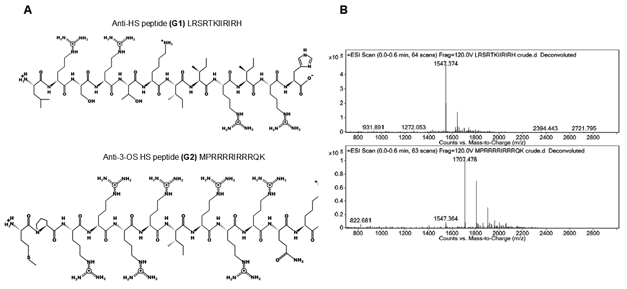
Figure 1. Characterization of the 12-mer HS (G1) peptide ligand and 3-OS HS (G2) peptide ligand. The sequence of HS (G1) peptide ligand and 3-OS HS (G2) peptide ligand (panel A), including the mass spec analysis of the purified peptides is shown (panel B). Purification of G1 and G2 peptides was achieved using preparative scale reverse-phase high-performance liquid chromatography (Waters Prep 150) with a Phenomenex Kinetex column (C-18 stationary phase, 5 μm, 100 Å pore size, 30 × 150 mm). The mobile phase consisted of acetonitrile and water with 0.1% trifluoroacetic acid (TFA). Fractions containing the desired peptides were confirmed using electrospray ionization mass spectrometry (ESI-MS) on an Agilent 6520 Quadrupole Time-of-Flight (Q-ToF) via direct injection. MassHunter Workstation Data Acquisition software operated the instrument, and MassHunter Qualitative Analysis software handled data analysis and processing. Excess acetonitrile was removed from pooled fractions by rotary evaporation. Samples were then freeze-dried and the resulting powders were stored at −20 °C. Purity was confirmed using liquid chromatography–mass spectrometry (LC-MS) on an Agilent 1200 system with a Phenomenex Jupiter C-12 column (1 × 100 mm; 4 µm). The mass detector (MS) was an Agilent 6520 Q-TOF MS.
Biomolecules 15 00999 g001
Figure 2. The impact of 3-O sulfotransferase-3 (3-OST-3) isoform on C. muridarum infectivity. (Panel A) shows that wild-type CHO-K1 cells, which lack endogenous HS modifying enzymes, were transfected with the human encoded 3-OST-3 isoform before infection with C. muridarum. The image in (Panels A,B) was created by BioRender. (Panel C,D) show the dosage-dependent (0.1 and 0.25 MOI) effect of C. muridarum on CHO-K1 cell infectivity in the presence and absence of the 3-OST-3 enzyme. Data is representative of the results of three independent experiments (n = 3) performed in triplicate.
Biomolecules 15 00999 g002
Figure 3. Effect of the pretreatment with various concentrations of G1 and G2 peptides on C. muridarum infection of HeLa cells, as determined by fluorescent microscopy. C. muridarum was pre-treated with G1 or G2 peptides before infecting HeLa cells. Inclusions were stained, and representative images are shown. The number of inclusions was counted and the percentage of infected cells treated with G1 or G2 compared to non-treated infected cells only (positive control) was calculated (Mean ± SEM, **** p < 0.0001). Data is representative of the results of three independent experiments (n = 3) performed in triplicates.
Biomolecules 15 00999 g003
Figure 4. (A) Effect of the pretreatment with various concentrations of G1 and G2 peptides on C. muridarum infection of VK2 cells, as determined by fluorescent microscopy. C. muridarum was pre-treated with G1 or G2 peptides before infecting VK2 cells. Inclusions were stained and representative images were shown. (B) The number of inclusions was counted and the percentage of infected cells treated with G1 or G2 compared to non-treated infected cells only (positive control) was calculated (Mean ± SEM, * p ≤ 0.05, ** p < 0.005, *** p < 0.0005 and **** p < 0.0001). Data is representative of the results of three independent experiments (n = 3) performed in triplicates.
Biomolecules 15 00999 g004
Figure 5. Molecular dynamics (MD) simulation of Chlamydia muridarum lipid bilayer and its interaction with G1 and G2 peptides. The interaction of G1 (purple) and G2 (red) peptides with the C. muridarum outer membrane is analyzed. (A) shows the representation of Chlamydia muridarum lipid bilayer building and simulation protocol, (B) shows the C. muridarum outer membrane composed of LPS-A. (C,E) Initial configurations of G1 and G2 peptides placed 20 Å above the membrane surface. (D,E) Post-MD simulation snapshots showing the peptide–membrane interactions, where G2 exhibits stable membrane association and deeper interaction compared to G1.
Biomolecules 15 00999 g005
Figure 6. Molecular dynamics (MD) simulation analysis of Chlamydia muridarum in complex with G1 and G2 peptides. The quantitative interactions of G1 (purple) and G2 (red) peptides with the C. muridarum membrane are illustrated. (A) Root Mean Square Deviation (RMSD) plot indicates that the G2 peptide achieves structural stability around the 150 ns time point, showing greater stability than G1. (B) Root Mean Square Fluctuation (RMSF) plot reflects residue-level flexibility. (C) Radius of gyration (Rg) analysis reveals that G2 maintains a more compact, folded conformation within the lipid membrane over 300 ns MD simulation.
Biomolecules 15 00999 g006
Figure 7. Significance of 3-O sulfation in C. muridarum entry. (A) C. muridarum infections occur in the absence of G2 peptide, (B) while a significant blockage in cell entry was noticed when C. muridarum was exposed to G2 peptide (which targets 3-O sulfation) before cell entry. (C). Proposed model for G2-mediated inhibition during cell entry of Chlamydia muridarum. Based on computational structural modeling data, the G2 peptide poses a significantly higher affinity towards the C. muridarum lipid membrane (a). It is also possible that the G2 peptides may also recognize the glycosaminoglycan (GAG)-like molecules with 3-O sulfation activity present on the OMP (b). Having GAG-like sulfated moieties on polysaccharides on the chlamydial surface along with HS-GAGs on the host cell provides critical bridging interactions between similar molecules, which offer a distinct advantage to Chlamydia in invading the immune system and remain persistent in the host. (i) OMP, (ii) G2 peptide, (iii) presence of 3-O sulfated moieties on OMP, (iv) native 3-O sulfation on the host cell. This image was created by BioRender. Tiwari V (2025) https://app.biorender.com/illustrations/canvas-beta/67f5cee0498da73002668e5f (accessed on 21 April 2025).
Biomolecules 15 00999 g007
Table 1. Selection parameters of the I-TASSER server with the best 3D structure of G1 and G2 peptides.
ParametersG1 PeptideG2 Peptide
Confidence score (C-score)−1.020.12
Template modelling score (TM score)0.59 ± 0.140.73 ± 0.11
Root mean square deviation (RMSD)1.9 ± 1.6 Å0.5 ± 0.5 Å
Table 2. Binding energy contribution of G1 and G2 peptides with C. muridarum lipid bilayer. All energies are given in kcal/mol. VDW = Van der Waals, ELE = Electron binding energy, GB = Generalized Born, GAS = Change in Gibbs free energy (ΔG), SURF = Surface binding energy, SOLV = Solvation.
Peptide–Membrane ComplexesΔEVDWΔEELEΔEGBΔESURFΔGGASΔGSOLVΔTotal
G1 membrane−44.66−20,879.9720,893.80−9.78−20,924.6320,884.02−40.61
G2 membrane−26.62−36,077.7036,001.98−9.42−36,104.3235,992.55−111.76
Disclaimer/Publisher’s Note: The statements, opinions and data contained in all publications are solely those of the individual author(s) and contributor(s) and not of MDPI and/or the editor(s). MDPI and/or the editor(s) disclaim responsibility for any injury to people or property resulting from any ideas, methods, instructions or products referred to in the content.