Review

The Multifaceted Role of VIRMA, a Core Component of the Methyltransferase Complex, in Cancer and Cancer Therapy

1
Laboratory of Molecular Pharmacology, Department of Pharmacology, School of Pharmacy, Southwest Medical University, Luzhou 646000, China
2
Cell Therapy & Cell Drugs of Luzhou Key Laboratory, Luzhou 646000, China
3
South Sichuan Institute of Translational Medicine, Luzhou 646000, China
*
Authors to whom correspondence should be addressed.
These authors contributed equally to this work.
Biomolecules 2025, 15(7), 912; https://doi.org/10.3390/biom15070912 (registering DOI)
Submission received: 26 May 2025 / Revised: 18 June 2025 / Accepted: 21 June 2025 / Published: 22 June 2025
(This article belongs to the Section Molecular Biology)

Abstract

:
VIRMA (also known as KIAA1429), as a core regulatory subunit of the m6A methyltransferase complex, plays a key role in tumorigenesis and progression by dynamically regulating RNA methylation modifications. Studies have shown that VIRMA is aberrantly overexpressed in more than 20 types of malignant tumors, including liver cancer, breast cancer, and lung cancer, and is significantly associated with chromosome 8q amplification and poor prognosis. Its mechanism of action involves regulating the expression of tumor-associated genes through both m6A-dependent and m6A-independent pathways, thereby promoting tumor proliferation, metastasis, and drug resistance. These findings suggest that VIRMA has the potential to serve as a pan-cancer diagnostic and prognostic biomarker. This review summarizes the role of VIRMA in malignant tumors from multiple perspectives and explores its potential applications in clinical diagnosis and treatment.

1. Introduction

According to the GLOBOCAN 2024 database, there were 20 million new cancer cases and 9.7 million cancer-related deaths globally in 2022. It is projected that by 2050, the number of new cancer cases will exceed 35 million, and cancer may become the leading cause of death in every country within the 21st century [1]. The occurrence and progression of tumors are often closely associated with changes in gene expression. In addition to DNA sequence alterations, gene expression is regulated at multiple levels, including epigenetic (such as DNA and histone modifications), transcriptional, epitranscriptome (RNA modification), and translational regulation [2]. In recent years, research on RNA modifications has rapidly advanced. Currently, more than 170 types of RNA post-transcriptional modifications are known, with N6-methyladenosine (m6A) being the most abundant endogenous RNA modification in eukaryotes [3,4].
m6A RNA methylation is a methylation reaction that occurs at the N6 position of adenosine. In mRNA, m6A sites are highly enriched in the 5′ untranslated region (5′-UTR), near the stop codon, in the 3′ untranslated region (3′-UTR), and in long introns [5,6]. This modification is dynamically reversible and is mediated by three classes of enzymes: “writers” (methyltransferases), “erasers” (demethylases), and “readers” (m6A-binding proteins). m6A deposition is primarily carried out by the “writer” methyltransferase complex [7,8]. “Erasers” are demethylases that make the m6A modification process reversible [9]. “Readers” are m6A-binding proteins that specifically recognize m6A-modified RNAs and regulate their function [10]. These proteins regulate RNA function and expression, significantly affecting cancer initiation and progression [11,12].
The methyltransferase complex consists of methyltransferase-like 3 (METTL3), methyltransferase-like 14 (METTL14), and auxiliary factors such as Wilms’ tumor 1-associating protein (WTAP), RNA-binding motif protein 15 (RBM15), ZC3H13, RBM, HAKAI, VIRMA, and others, with a molecular weight of approximately 1000 kDa [13]. Most of these components have been shown to participate in tumorigenesis and progression, such as METTL3 and WTAP [14,15,16]. As the largest structural component of the m6A methyltransferase complex, VIRMA (KIAA1429) serves as a scaffold protein that recruits the catalytic core (METTL3-METTL14) to RNA substrates and maintains methylation activity under specific conditions [13,17]. Given its central role in m6A modification, VIRMA has garnered significant attention in recent years. Accumulating evidence demonstrates that VIRMA is markedly overexpressed in multiple malignancies (e.g., hepatocellular carcinoma, breast cancer, colorectal cancer), where it drives tumor proliferation, invasion, and metastasis through m6A-dependent mechanisms [18,19,20]. Notably, emerging studies reveal m6A-independent functions of VIRMA, expanding our understanding of its oncogenic regulatory networks [21]. These findings position VIRMA not only as a promising diagnostic biomarker but also as a novel therapeutic target. This review systematically summarizes current advances in VIRMA research, focusing on its molecular functions, regulatory mechanisms, and clinical translation potential, thereby providing new perspectives for m6A-related research and precision cancer therapy.

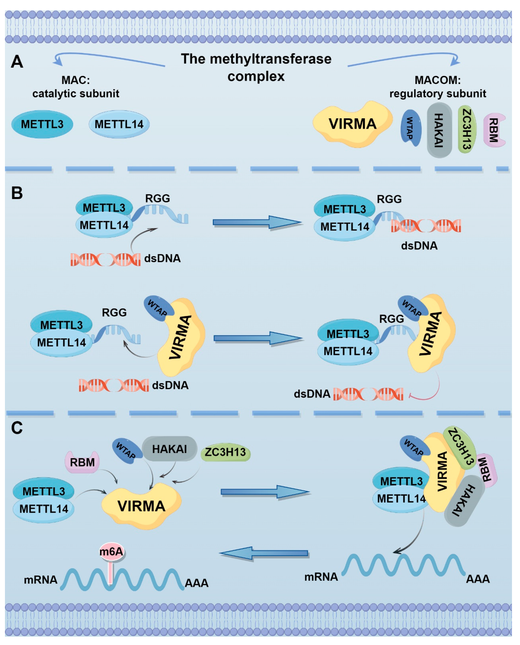
2. Localization, Structure, and Function of VIRMA

VIRMA (Vir-like m6A methyltransferase associated), also known as KIAA1429, is a gene located at 8q22.1 in humans and contains 25 exons. This gene encodes two distinct isoforms: a full-length isoform containing 1812 amino acids (202 kDa) and an N-terminal truncated isoform consisting of the first 1130 amino acids. Current evidence indicates that the full-length isoform predominantly mediates oncogenic functions in malignancies, while the biological role and regulatory mechanisms in polyadenylation site selection of the short isoform remain largely unexplored [22]. The subcellular distribution of VIRMA protein is closely related to tumor type and occurrence. In most malignancies (e.g., pancreatic ductal adenocarcinoma [PDAC] and hepatocellular carcinoma [HCC]), VIRMA primarily localizes to the nucleus. However, studies in breast cancer (BC) demonstrate predominant cytoplasmic localization, where it regulates the stability of downstream target HAS2. This dynamic nuclear-cytoplasmic distribution strongly correlates with tumor malignancy and metastatic potential, suggesting VIRMA localization may serve as a potential tumor grading biomarker [19,23,24]. However, the precise mechanisms governing VIRMA’s subcellular trafficking and its context-dependent functional variations in different tumor microenvironments require further investigation.
Current research on VIRMA primarily focuses on its role as a key component of the m6A methyltransferase complex. This complex structurally consists of two functional units: (1) the catalytic core MAC (m6A-METTL complex), formed by the METTL3-METTL14 heterodimer that directly catalyzes m6A modification; and (2) the regulatory complex MACOM (m6A-METTL associated complex), composed of WTAP, VIRMA, ZC3H13, and HAKAI, which mediates scaffold assembly, substrate recruitment, complex stabilization, and subcellular localization. As a core structural component of MACOM, VIRMA plays an essential role in bridging the catalytic core with regulatory subunits (Figure 1A) [25]. In cryo-EM structural analysis, VIRMA (residues 334–1585), WTAP (residues 64–247), and ZC3H13 (residues 1492–1643) together form the “war horse”-shaped human MACOM core. VIRMA adopts a horseshoe-like conformation and contains 20 ARML modules, with its surface charges predominantly negative and a few small areas carrying a positive charge. These regions are relatively conserved, confirming that VIRMA may play an important role in the interaction with other components of MACOM, particularly WTAP [26].
The RGG motif is a common protein sequence of arginine-glycine residues that mediates nucleic acid binding or protein-protein interactions [27]. Yan et al. found that the RGG motif can directly bind with WTAP-VIRMA and facilitate the binding of METTL3-METTL14 to RNA and double-stranded DNA (dsDNA). However, the presence of dsDNA significantly inhibits the RNA methyltransferase activity of METTL3-METTL14. As a regulatory subunit, WTAP-VIRMA maintains the RNA methylation activity of METTL3-METTL14 by preventing dsDNA binding through their interaction with the RGG motif. Consequently, the methylation activity of the METTL3-METTL14-WTAP-VIRMA quaternary complex (M3-M14-W-V) is significantly higher than that of the METTL3-METTL14 dimer (Figure 1B). Therefore, downregulation of either WTAP or VIRMA significantly affects the overall level of m6A in mRNA [17]. Additionally, there is evidence that VIRMA can serve as a scaffold for the methyltransferase complex, recruiting catalytic core components and linking them with RNA substrates (Figure 1C). After knockdown of VIRMA in A549 cells, the m6A peak score decreased by four times, far exceeding the reduction observed with METTL3 or METTL4 knockdown alone [13,28].
RNA-binding proteins (RBPs) are proteins with specific RNA-binding domains that can form various dynamic ribonucleoprotein complexes with RNA molecules, regulating multiple aspects of gene expression, including RNA splicing, mRNA stability, mRNA localization, and translation [29]. In Drosophila, the virilizer, which is homologous to KIAA1429, contains RBP structural domains similar to those found in RNA and DNA helicases, translation initiation factor 2 (IF2), and ribonucleoproteins [30]. Therefore, VIRMA may exert its biological functions independently of methylation modification.

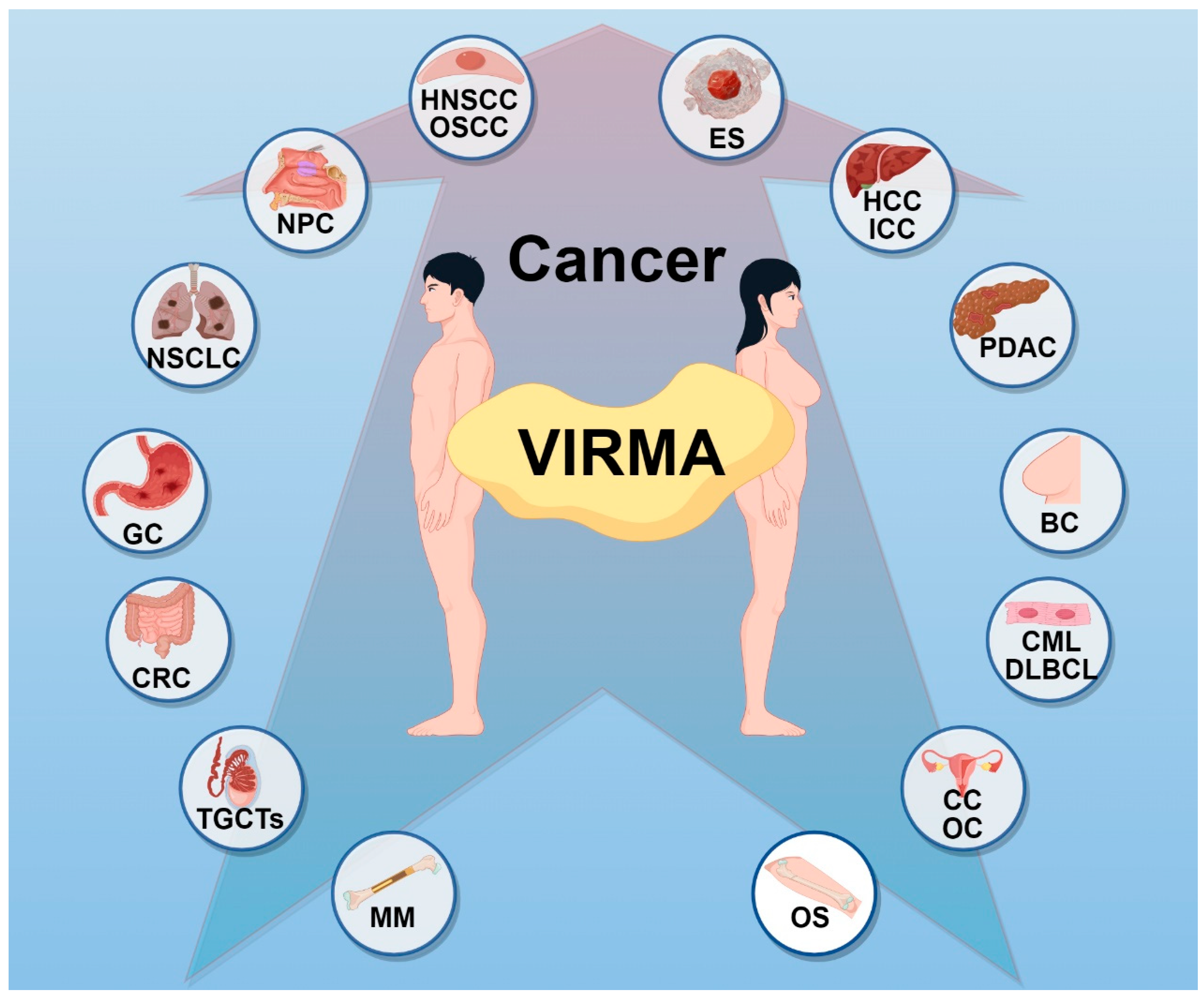
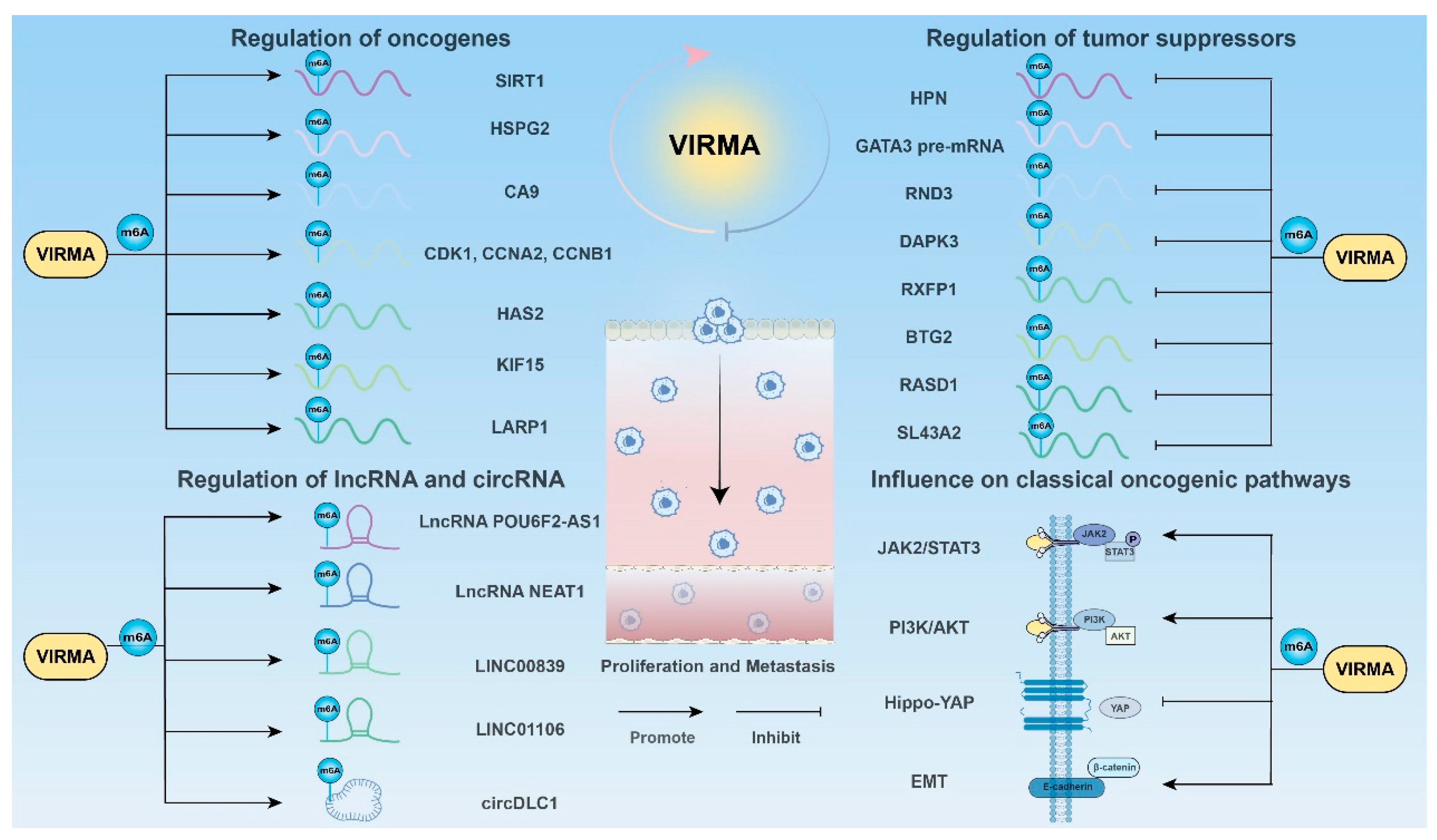
3. The Role of VIRMA in Cancer

Among all 15 core m6A genes, VIRMA exhibits the highest frequency of abnormalities, with 21.4% of tumor samples showing expression or copy number alterations (e.g., overexpression or amplification), significantly higher than METTL3 (6.2%) [31]. Numerous studies have shown that dysregulation of VIRMA is closely related to the initiation and progression of malignancies. Elevated VIRMA expression has been observed in eighteen types of malignancies, including head and neck squamous cell carcinoma (HNSCC), nasopharyngeal carcinoma (NPC), oral squamous cell carcinoma (OSCC), non-small cell lung cancer (NSCLC), hepatocellular carcinoma (HCC), intrahepatic cholangiocarcinoma (ICC), gastric cancer (GC), PDAC, colorectal cancer (CRC), multiple myeloma (MM), osteosarcoma (OS), diffuse large B-cell lymphoma (DLBCL), chronic myeloid leukemia (CML), BC, testicular germ cell tumors (TGCTs), cervical cancer (CC), ovarian cancer (OC), and Ewing’s sarcoma (ES) (Figure 2) [18,19,24,32,33,34,35,36,37,38,39,40,41,42,43,44,45]. In recent years, research on VIRMA has been increasing, making it particularly important to comprehensively summarize its role in cancer. The following sections will provide a detailed overview of the functions and mechanisms of VIRMA as an m6A writer, including promoting cancer cell proliferation and metastasis, influencing cell cycle, affecting cancer metabolism, mediating resistance to ferroptosis, mediating immune escape, and affecting drug resistance, as well as its involvement in tumor progression through m6A-independent mechanisms (Table 1).

3.1. Promoting Cancer Cell Proliferation and Metastasis

Tumor metastasis refers to the spread of tumor cells from the primary site to distant organs or tissues, leading to the formation of new tumor lesions. Invasion and metastasis are among the most lethal characteristics of cancer, with over 90% of cancer-related deaths being closely linked to tumor metastasis. Therefore, a deeper understanding of the molecular mechanisms under metastasis is of significant importance for preventing cancer-related death (Figure 3) [87,88,89].

3.1.1. Regulation of Oncogenes

CCL3 (Macrophage Inflammatory Protein-1 alpha, MIP-1α) belongs to the C-C chemokine family and is involved in the occurrence and development of various malignant tumors [90]. Zhou et al. discovered through co-culturing normal liver cells with ICC cells that CCL3 secreted by normal liver cells was significantly increased, further upregulating VIRMA expression, leading to an overall increase in m6A levels and enhancing the invasion and metastasis abilities of tumor cells. MeRIP-seq analysis showed that VIRMA enhanced the expression of SIRT1 through an m6A methylation-dependent mechanism, promoting the progression of ICC [69]. Chronic hepatitis B virus (HBV) infection plays a critical role in the initiation and progression of HCC. The hepatitis B virus X protein (HBx) is recognized as the sole regulatory protein encoded by the HBV genome, which promotes viral replication by stimulating HBV gene expression. In HCC cells with stable HBx expression, HBx upregulated VIRMA expression, thereby driving m6A methylation of its target gene HSPG2. This enhanced the stability of HSPG2 and subsequently promoted the proliferation, migration, and survival of HCC cells [66].
Inhibin A (INHBA) belongs to the transforming growth factor-β superfamily and is closely associated with cancer cell invasion, proliferation, and metastasis [91]. In OSCC, INHBA participates in tumorigenesis through VIRMA-regulated m6A modification [48]. CA9, a transmembrane zinc metalloprotease with cell adhesion functions, is crucial for tumor growth and survival. Overexpression of CA9 in various cancers is associated with poor prognosis. VIRMA upregulated CA9 expression in an m6A-dependent manner in OSCC, and inhibition of VIRMA suppressed tumor growth both in vitro and in vivo [49].
In OS, silencing VIRMA resulted in a reduction of overall m6A methylation level in SW1353 cells, inhibiting their proliferative capacity. Bioinformatic analysis indicated that VIRMA may promote OS progression by targeting CDK1, CCNA2, and CCNB1 [76].
In NPC, VIRMA mediated m6A modification of PTGS2, enhancing its mRNA stability and promoting cancer progression [46].
In HNSCC, VIRMA facilitated cancer progression by modulating the m6A levels of UBR5 [35]. The expression of the oncogene SIRT1 was positively correlated with VIRMA in CRC. VIRMA upregulated SIRT1 mRNA stability through an m6A-dependent mechanism, thereby promoting the growth of CRC cells [20].
Hyaluronan synthase 2 (HAS2) plays a critical role in both normal development and tumorigenesis. Studies have shown that VIRMA, by acting as a scaffold protein, recruited the m6A reader IGF2BP3 to increase the m6A modification level of HAS2, stabilizing its mRNA and mediating BC cell proliferation and migration [19]. Additionally, VIRMA enhanced the mRNA stability of the pro-oncogenic gene KIF15 through an m6A-dependent mechanism, thereby promoting BC progression [78].
In CC, VIRMA increased the mRNA stability of LARP1, contributing to the initiation and progression of CC [82].

3.1.2. Regulation of Tumor Suppressors

Hepsin (HPN) is a transmembrane serine protease, and its expression is decreased in HCC. A study by Meng et al. demonstrated that overexpressing HPN could effectively inhibit the invasion, migration, and proliferation of HCC cells. VIRMA inhibited the expression of HPN in an m6A-dependent manner, further promoting HCC progression [60]. Additionally, VIRMA could increase the m6A modification level of ID2 mRNA, suppress its expression, and facilitate migration and invasion in HCC [62]. GATA3 is a well-established tumor-suppressive transcription factor. Also in HCC, VIRMA also mediated the m6A modification of GATA3 pre-mRNA, inhibited its binding to the RNA-binding protein HuR, thus reduced the stability of GATA3 pre-mRNA, and ultimately led to downregulation of GATA3 expression. Moreover, the antisense long non-coding RNA GATA3-AS interacted with VIRMA and GATA3 pre-mRNA, guiding VIRMA to preferentially mediate the m6A modification of GATA3 pre-mRNA [23,92]. Another study demonstrated that VIRMA downregulated the mRNA stability of the tumor suppressor RND3 through an m6A-YTHDC1-dependent mechanism, thereby suppressing its expression and exerting oncogenic effects in HCC cells [18].
In NSCLC, VIRMA mediated the m6A modification of DAPK3, facilitating YTHDF2/3-mediated DAPK3 post-transcriptional degradation and promoting tumor growth. DAPK3 (Death-Associated Protein Kinase 3) silencing can reverse the invasion and migration suppression caused by VIRMA depletion [53].
RXFP1 (Relaxin Family Peptide Receptor 1) overexpression inhibits the proliferation, migration, and invasion of cancer cells. VIRMA could regulate the m6A modification level of RXFP1, suppressing its expression and exerting oncogenic effects in NSCLC [50]. VIRMA also regulated the mRNA stability of the tumor suppressor gene BTG2 through an m6A-YTHDF2-dependent mechanism, suppressing its expression and promoting the progression of lung adenocarcinoma (LUAD) [52].
A similar mechanism was observed in CC, where VIRMA negatively regulated the mRNA stability of BTG2 via an m6A-YTHDF2-dependent pathway, thereby facilitating malignant tumor progression [44]. RASD1 has been implicated as a potential tumor suppressor in GC. VIRMA reduced the mRNA stability of RASD1 through an m6A-YTHDF2-dependent mechanism, promoting GC development [36].
In PDAC, alcohol activated the transcription factor C/EBPβ, which bound to the promoter of VIRMA and enhanced its transcription. VIRMA further accelerated the degradation of SLC43A2 mRNA via the m6A-YTHDF2 pathway, reducing phenylalanine uptake and oxidative stress in cancer cells, ultimately leading to cancer progression [73].

3.1.3. Regulation of lncRNA

Long non-coding RNAs (lncRNAs) are defined as non-coding transcripts longer than 200 nucleotides. In recent years, the functions of lncRNAs in tumor initiation and progression have attracted extensive research interests [93]. Similar to mRNA, VIRMA can also mediate m6A methylation of lncRNA.
LncRNA POU6F2-AS1 is upregulated in CRC, and inhibiting this lncRNA suppresses the malignant phenotype of cancer cells. Studies have demonstrated that VIRMA increased the expression of POU6F2-AS1 through m6A modification, promoting the development of CRC [74]. LncRNA NEAT1 has oncogenic potential in various types of cancer. Lee et al. found that the full-length isoform of VIRMA (VIRMA FL) was primarily expressed in BC cells and significantly increased m6A levels. Overexpression of VIRMA FL enhanced the m6A methylation level of lncRNA NEAT1, promoting BC cell growth [80,94]. Zheng et al. demonstrated that LINC00839 directly interacted with transcription factor TAF15 and promoted the transcription of AOC1 by recruiting TAF15, thereby increasing the expression of AOC1 and promoting the growth and metastasis of NPC. VIRMA and IGF2BP1 mediated the m6A modification of LINC00839 and enhanced its expression and stability [47]. The level of LINC01106 is closely associated with clinical malignant features of cancer and can serve as a poor prognostic factor. VIRMA was found to enhance the m6A modification and expression of LINC01106 in LUAD, thereby promoting tumor progression [56,95].

3.1.4. Regulation of Circ RNA

Circular RNA (circRNA) is formed by exon or intron backsplicing, characterized by stability, sequence conservation, and expression specificity [96].
Liu et al. identified circDLC1 as the most significantly differentially expressed circRNA in HCC through RNA-seq and m6A-seq analysis of VIRMA-regulated circRNAs. Overexpression of circDLC1 inhibited the proliferation and metastasis of HCC cells. Moreover, circDLC1 was negatively correlated with VIRMA and positively correlated with patient prognosis. circDLC1 interacted with the RNA-binding protein HuR, preventing HuR from binding to MMP1 mRNA, thereby reducing the expression of MMP1 [61].

3.1.5. Influence on Classical Oncogenic Pathways

VIRMA not only regulates tumor-associated proliferation and metastasis factors but also participates in the activation of classical oncogenic signaling pathways, such as STAT3 and PI3K/AKT, which are crucial for cancer proliferation and metastasis. The JAK2/STAT3 signaling pathway, a member of the STAT family of transcription factors, is closely associated with the onset of various cancers and regulates cancer cell proliferation and migration [97].
ES is a highly invasive bone and soft tissue cancer that primarily affects children and young adults. Downregulation of VIRMA expression significantly inhibits ES proliferation. Bioinformatic analysis showed that NKX2-2 regulates VIRMA-associated m6A writers, promoting a positive feedback loop between VIRMA and STAT3, thus enhancing the malignant phenotype of ES [77,98].
The dysregulation of the PI3K/AKT pathway is closely linked to cancer progression [99]. VIRMA was found to enhance the activity of the PI3K/AKT pathway by upregulating the m6A modification of the tumor suppressor ARHGAP30, promoting lung cancer cell proliferation and migration [55]. In NPC, VIRMA maintained the stable expression of the transcription factor E2F7 through an m6A-dependent mechanism. E2F7 bound with CBFB, activating the PI3K-AKT signaling pathway and promoting tumorigenesis [32].
The Hippo-YAP signaling pathway is another important pathway involved in cancer. VIRMA was found to mediate the m6A modification of CHST11 mRNA and reduce its expression via an m6A-YTHDF2-dependent mechanism. The interaction between CHST11 and the core kinase MOB1B of the Hippo pathway was therefore disrupted, inhibiting the activation of the Hippo-YAP signaling pathway and thereby promoting tumor development [41].
Epithelial-mesenchymal transition (EMT) is a biological process defined by morphological changes and characteristic markers (such as E-cadherin and vimentin) and plays a critical role in the invasion and metastasis of tumors [100]. In ICC, VIRMA enhanced the expression of downstream targets TMED2 and PARD3B via an m6A-Hur-dependent mechanism. The increased levels of TMED2 and PARD3B led to activation of the AKT/GSK/β-catenin and MEK/ERK/Slug signaling pathways, promoting the expression of EMT markers vimentin and N-cadherin while inhibiting E-cadherin expression, thereby promoting the metastasis and EMT of ICC [68].

3.2. Influencing Cell Cycle

Cell cycle dysregulation is the foundation of abnormal cell proliferation in cancer. In most cancers, signaling pathways related to cell proliferation interfere with the normal progression of the cell cycle, thereby contributing to tumor initiation and progression [101].
In DLBCL, overexpression of VIRMA upregulated the anti-apoptotic protein Bcl-2 while inhibiting the expression of pro-apoptotic proteins (such as Cleaved-Caspase 3, Cleaved-Caspase 8, Cleaved-Caspase 9, and Cleaved-PARP), thereby suppressing apoptosis. After VIRMA downregulation, the proportion of DLBCL cells in the G2/M phase significantly increased, with higher expression of the cell cycle inhibitor p21 and lower expression of Cyclin B1. These results indicate that reducing VIRMA expression effectively suppresses DLBCL cell cycle progression [41]. In GC cells, knockdown of VIRMA led to cell cycle arrest in the S phase, increased apoptosis in AGS and HGC-27 cells, and promoted the transition to the G2/M phase [36,86]. In BC, reduced expression of VIRMA also caused a significant delay in the S phase in MCF-7 and SUM1315 cells [83]. Furthermore, in LUAD, VIRMA has been demonstrated to regulate the expression of the oncogene MUC3A through an m6A-dependent mechanism and promote cancer progression. The study also showed that downregulation of VIRMA can partially reverse the G1-phase arrest in A549 and H1299 cells with high MUC3A expression [54]. In HNSCC, after VIRMA knockout, most cancer cells were arrested in the S phase, indicating a reduction in the number of dividing tumor cells following VIRMA knockout [35]. The changes in the cell cycle are indirectly caused by the alteration of VIRMA, but the specific mechanism of interaction between VIRMA and cell cycle regulators remains unclear.
Future research may focus on exploring the direct interaction mechanisms between VIRMA and cell cycle-related proteins to deepen the understanding of the connection between VIRMA and cancer.

3.3. Affecting Cancer Metabolism

Aerobic glycolysis is the primary energy source for tumor cells’ rapid proliferation, invasion, and metastasis in various cancers and is closely associated with tumor malignancy [102,103]. This metabolic shift from oxidative phosphorylation to aerobic glycolysis is a hallmark of tumor cells and one of the most significant metabolic reprogramming phenomena in cancer cells [104].
VIRMA regulates tumor metabolic reprogramming by influencing key factors in tumor cell aerobic glycolysis. Glucose transporter-1 (GLUT1) plays a critical role in aerobic glycolysis and is closely related to tumor progression and tumor size [105]. In GC, VIRMA could interact with the m6A modification site of LINC00958, promoting its enrichment and enhancing its binding with GLUT1 mRNA, thereby increasing the stability of GLUT1 and ultimately promoting aerobic glycolysis in GC cells [70]. Hexokinase (HK) is the first rate-limiting enzyme in the glycolytic pathway, with major isoforms including HK1 and HK2 [106]. In liver cancer, upregulation of VIRMA significantly promoted lactate production, ATP synthesis, and glucose uptake. VIRMA enhanced HK1 mRNA stability through an m6A-dependent mechanism, increasing HK1 protein level and strengthening the first step of glycolysis, which provides the material basis for the Warburg effect. Inhibiting VIRMA reduced the Warburg effect and increased the sensitivity of liver cancer cells to sorafenib [63]. Furthermore, Li et al. found that VIRMA increased HK2 mRNA stability in an m6A-dependent manner, promoting aerobic glycolysis in CRC [107]. In PDAC, VIRMA cooperated with IGF2BP2 to enhance STRA6 mRNA stability via an m6A-dependent mechanism, which activates HIF-1α and upregulates GLUT1 and HK2, promoting aerobic glycolysis, cell proliferation, and metastasis [24]. Alpha-enolase (ENO1) is a key metabolic enzyme in the glycolytic pathway. In OC, VIRMA regulates ENO1 mRNA stability via an m6A-dependent mechanism, promoting glycolysis, proliferation, and metastasis in OC cells [45]. In OSCC, VIRMA regulated PGK1 via the YTHDF1/m6A-dependent pathway, leading to increased glucose uptake, lactate production, and extracellular acidification rate (ECAR), with decreased oxygen consumption rate (OCR), ultimately enhancing the glycolytic rate and capacity of cancer cells [33]. In MM, VIRMA expression is significantly higher than in normal tissue and is associated with poor prognosis in patients. The mRNA level of VIRMA positively correlated with the mRNA levels of HK2, ENO1, and lactate dehydrogenase A (LDHA), indicating that VIRMA plays a regulatory role in MM cell aerobic glycolysis. Moreover, VIRMA stabilized FOXM1, a player involved in aerobic glycolysis, in cooperation with YTHDF1, further emphasizing VIRMA’s importance in MM cell metabolism and biological behavior [39].

3.4. Mediating Resistance to Ferroptosis

Ferroptosis is a form of non-apoptotic, iron-dependent cell death triggered by the toxic accumulation of lipid peroxides in cell membranes [108]. Because ferroptosis is mechanistically and morphologically distinct from other forms of cell death, such as apoptosis, it has garnered significant attention for its role in cancer in recent years [109]. In cancer, epigenetic alterations can regulate the expression of ferroptosis regulators or related pathways, thus affecting the sensitivity of cells to ferroptosis [110].
The solute carrier family member SLC7A11, the 11th member of this family, can transport extracellular cysteine into cells for cysteine synthesis and the biosynthesis of glutathione. SLC7A11 counters cellular oxidative stress by maintaining intracellular glutathione (GSH) levels, thereby inhibiting ferroptosis [111]. In HCC, when VIRMA was inhibited, levels of lipid peroxides and oxidative C11-BODIPY were significantly increased, while VIRMA overexpression had the opposite effect. Mechanistically, KIAA1429 regulated SLC7A11 through m6A post-transcriptional modification, thereby exerting its ferroptosis-inhibiting role [64]. Wu et al. found that si-VIRMA significantly reduced the viability of NSCLC cells treated with the ferroptosis inducer erastin and increased the levels of reactive oxygen species (ROS), MDA, and Fe2+ in the cells while decreasing GSH levels. At the same time, ferroptosis-related proteins such as PTGS2, GPX4, and FTH1 also showed significant changes. These results indicate that VIRMA acts as a ferroptosis inhibitor in NSCLC, and its downregulation can promote erastin-mediated ferroptosis [57]. Similarly, in OSCC, silencing VIRMA upregulated the concentrations of Fe2+ and lipid ROS, promoting ferroptosis in cancer cells [33]. In CRC, VIRMA expression was significantly elevated in tumor cells, especially in radioresistant cell lines (such as HCT116R and SW620R), where its expression was further increased. Upon VIRMA knockdown, ferroptosis in tumor cells significantly increased, characterized by elevated intracellular Fe2+ levels, increased expression of ferroptosis-related protein ACSL4, and decreased expression of SLC7A11 and GPX4. In addition, intracellular ROS levels were increased, and GSH content was decreased. Mechanistically, VIRMA upregulated LncRNA EBLN3P expression in an m6A-dependent manner, and EBLN3P competed with miR-153-3p to increase VIRMA expression, thereby reducing ferroptosis in tumor cells and enhancing their radioresistance [75].
The m6A modification mediated by VIRMA in regulating ferroptosis is a promising area of further exploration in treating cancer and overcoming cancer resistance [112].

3.5. Mediating Immune Escape

The tumor microenvironment has a significant impact on the survival and function of CD8+ T cells. A metabolic environment unfavorable to the survival of CD8+ T cells accelerates their exhaustion, thus inhibiting tumor immunity, which is a key challenge in cancer immunotherapy [113].
Through analysis of TCGA BC data, Bin Lian et al. found that DDR1, a receptor tyrosine kinase (RTK) family member targeting collagen, is negatively correlated with the proportion of CD8+ T cells and CD4+ T cells. Silencing DDR1 in BC cells increased the infiltration of CD8+ T cells within the tumor by reducing the content of collagen IV in the extracellular matrix (ECM) and disrupting the alignment of collagen fibers. VIRMA mediated m6A modification of TFAP2A, a transcription factor for DDR1, thereby enhancing TFAP2A expression. The upregulation of TFAP2A promoted DDR1 expression by binding to the promoter region, inducing the alignment of collagen fibers, inhibiting the infiltration of immune cells, and accelerating immune escape in BC [79]. Additionally, the PD-1/PD-L1 checkpoint suppresses T cell receptor-mediated cytotoxicity and inhibits the proliferation of CD8+ T cells, thereby accelerating immune escape of tumor cells. KLF1, an important oncogene, is regulated by VIRMA in NSCLC through m6A modification. VIRMA could stabilize KLF1 mRNA, upregulate KLF1 expression, and consequently enhance PD-L1 expression and promote immune evasion by tumor cells [58].
Although there is limited research on VIRMA’s role in the immune microenvironment and immune infiltration, existing studies suggest the potential of targeting VIRMA to enhance the effectiveness of immunotherapy.

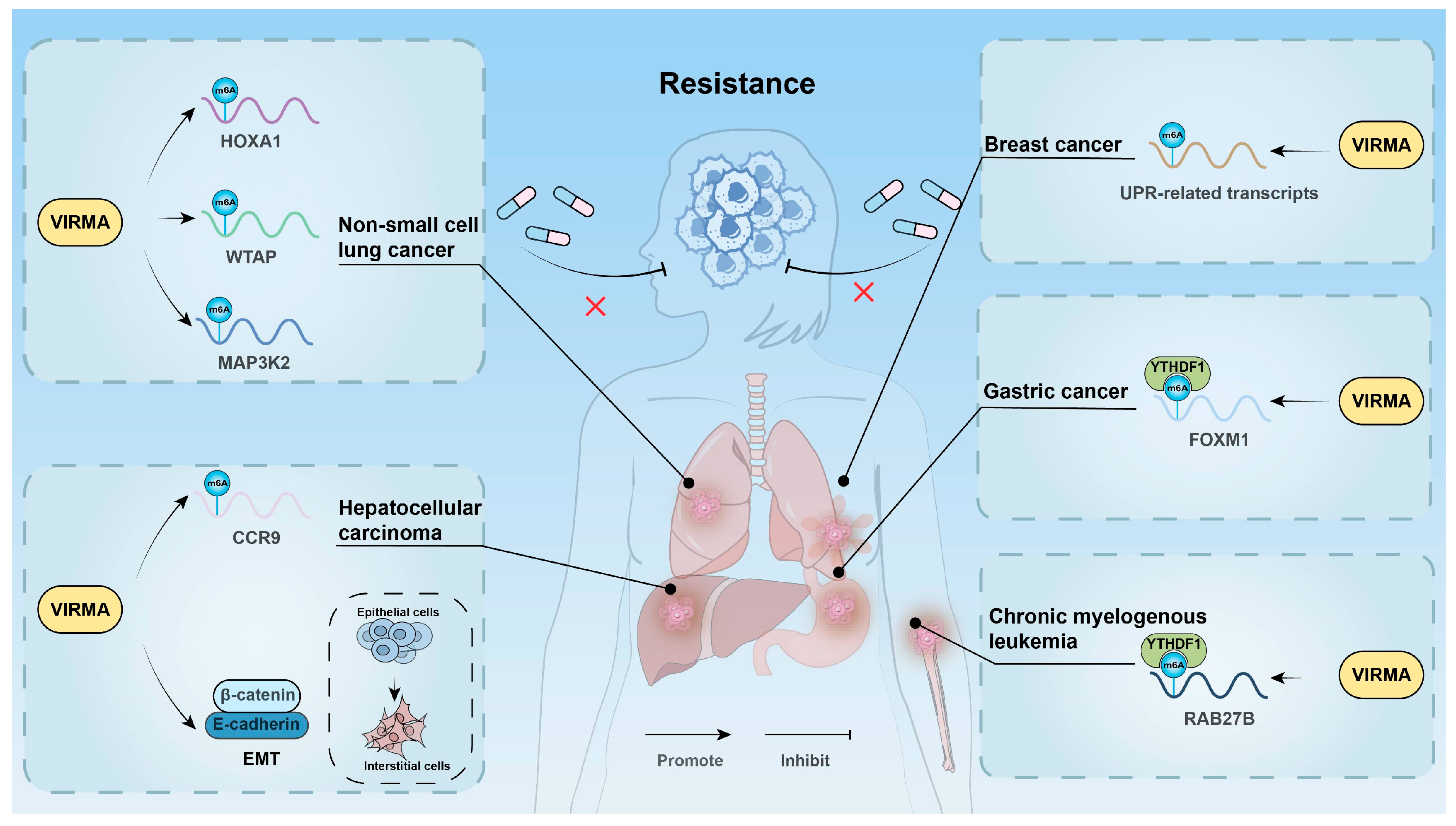
3.6. Affecting Drug Resistance

Chemotherapeutic drugs play a crucial role in treating various cancers by targeting rapidly proliferating tumor cells. However, chemotherapy resistance remains a major cause of poor prognosis in cancer patients [114]. Studies have shown that VIRMA regulates cell resistance to chemotherapy at multiple levels through m6A modification (Figure 4).
In CML, the expression of VIRMA was significantly higher in imatinib-resistant cell lines (K562/G01) compared with sensitive cell lines (K562 and KCL22). VIRMA enhanced the stability of RAB27B mRNA through m6A/YTHDF1, upregulating its expression and promoting CML cell proliferation, thereby inducing imatinib resistance [42]. In LUAD, VIRMA increased the m6A modification level of MAP3K2, activating the JNK/MAPK pathway and inducing gefitinib resistance [34]. Additionally, in non-small cell lung cancer, VIRMA enhanced the m6A modification level of HOXA1, stabilizing its mRNA and promoting tumor cell proliferation and gefitinib resistance [59]. VIRMA could also stabilize WTAP mRNA through an m6A-dependent mechanism, mediating the proliferation and gefitinib resistance of NSCLC cells via the VIRMA/WTAP axis [51]. In liver cancer, the EMT process in sorafenib-resistant cell lines was regulated by m6A methylation. Silencing VIRMA reduced m6A methylation levels, inhibited the invasion, migration, and EMT of sorafenib-resistant cells, and decreased angiogenesis [67]. In hepatitis B virus surface small antigen (SHBs)-induced liver cancer cells, SHBs upregulated VIRMA expression, mediating the m6A modification of CCR9 mRNA and enhancing its stability, thus promoting regorafenib resistance. The m6A modification at positions 1373 and 1496 of CCR9 mRNA is critical for this process [65]. In GC, VIRMA enhanced FOXM1 mRNA stability via an m6A-YTHDF1-dependent mechanism, inducing cisplatin resistance [71]. Additionally, the VIRMA/m6A/FOXM1 axis also played a crucial role in oxaliplatin resistance in GC [72]. In testicular germ cell cancer, knocking out VIRMA reduced the m6A modification level and the expression of components of the methyltransferase complex, significantly inhibiting the survival of cisplatin-resistant cells [43]. Endoplasmic reticulum (ER) stress, triggered by the unfolded protein response (UPR), plays a key role in maintaining tumor survival and chemotherapy resistance, but prolonged UPR activation may also induce tumor cell death [115,116]. In BC, VIRMA upregulation increased the m6A modification level of UPR-related transcripts, enhancing sensitivity to UPR and ER stress, thereby inducing tumor cell death. However, whether the association of VIRMA with UPR/ER stress can enhance chemotherapy sensitivity requires further investigation [80].
Targeting VIRMA in combination with chemotherapeutic drugs may significantly improve drug sensitivity and reverse tumor resistance. Further research into the mechanisms by which VIRMA regulates resistance-related genes through m6A modification will help uncover the underlying mechanisms of tumor resistance and provide new therapeutic strategies for chemotherapy-resistant patients.

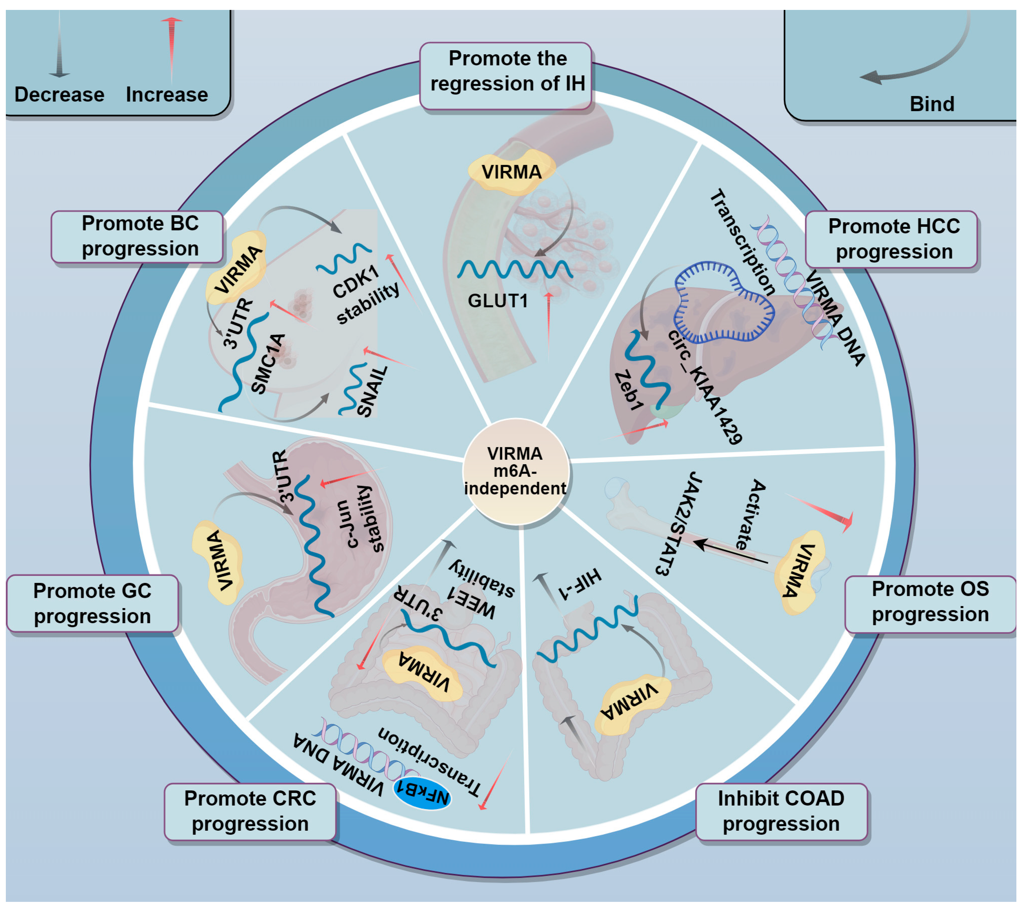
3.7. Involvement in Tumor Progression Through m6A-Independent Mechanisms

The function of VIRMA is not limited to regulating gene expression through m6A-dependent mechanisms (Figure 5). Its action as an RNA binding protein independent of m6A modification is also found in many cancers.
Infantile hemangioma (IH) is a common vascular tumor with a specific natural course, often involving the skin, subcutaneous tissues, and even internal organs. GLUT1, a marker of facultative stem cells, is closely related to the specific course of IH [117]. In IH, VIRMA could upregulate GLUT1, promoting the conversion of hemangioma endothelial cells into stem cells. Through RIP experiments, researchers found that VIRMA acts on GLUT1 in an m6A-independent manner. Moreover, the weakening of the endothelial cell phenotype and the promotion of conversion to stem cells align with the spontaneous regression process of IH to fibrofatty tissue, suggesting that VIRMA may promote the regression of IH [118]. CDK1 plays a key role in regulating the cell cycle and is closely related to cancer progression [119]. In BC, VIRMA directly interacted with CDK1 transcripts and prolonged their half-life, but VIRMA knockdown did not change m6A-modified CDK1 mRNA level, indicating VIRMA regulates CDK1 mRNA stability in an m6A-independent manner [83]. In another study, the VIRMA/SMC1A/SNAIL axis was found to regulate BC metastasis. VIRMA directly bound to the SMC1A mRNA 3′UTR in an m6A-independent manner, stabilizing its expression. SMC1A then positively regulated SNAIL expression, which triggered EMT in BC cells by inhibiting cell adhesion proteins, promoting BC metastasis. Notably, the short (N-terminal) isoform of VIRMA was found overexpressed in this study, rather than the full-length isoform [21,120]. Similar to the interaction with SMC1A, VIRMA was demonstrated to directly bind the c-Jun mRNA 3′UTR in an m6A-independent manner, stabilizing its mRNA and affecting the growth of GC cells [86]. Furthermore, VIRMA also promoted cancer progression in an m6A-independent manner in CRC. A study by Ma et al. found that VIRMA could bind the 3′UTR of the tumor suppressor gene WEE1 in an m6A-independent manner, decreasing the stability of WEE1 mRNA, thereby downregulating WEE1 expression and promoting CRC progression. NFκB1, a transcription factor in the NFκB1 family, was found to bind the VIRMA promoter. Interestingly, butyrate, a metabolite of the gut microbiota, can inhibit NFkB1 expression, further decreasing VIRMA levels [84,121].
However, in some studies, whether VIRMA functions dependent or independent of m6A is not clear. Hypoxia-inducible factor 1 (HIF-1), an oxygen-regulated transcription factor, plays a critical role in cancer progression. In colon adenocarcinoma (COAD), silencing VIRMA blocked the HIF-1 signaling pathway, inhibiting the proliferation, migration, invasion, and proptosis of colon cancer cells. However, whether VIRMA plays a role in this process through an m6A-independent mechanism remains to be further studied [37]. In OS, upregulation of VIRMA was significantly associated with poor prognosis in patients. The upregulation of VIRMA activated the JAK2/STAT3 signaling pathway, promoting the malignant phenotype progression of OS cells. However, whether this process involved m6A modification remains unclear [40]. Moreover, Wang et al. discovered that the circRNA hsa_circ_0084922 (named circ_KIAA1429), transcribed from VIRMA, was upregulated in liver cancer tissues and cell lines, promoting liver cancer cell migration, invasion, and EMT. The study showed that Zeb1 was a downstream target of circ_KIAA1429, and circ_KIAA1429 promoted liver cancer progression by targeting Zeb1 [85].
These studies indicate VIRMA’s dual role as both an m6A methylation regulator and a direct RNA-interacting factor in tumors, which offers key evidence for the multidimensional regulatory network in cancer development.

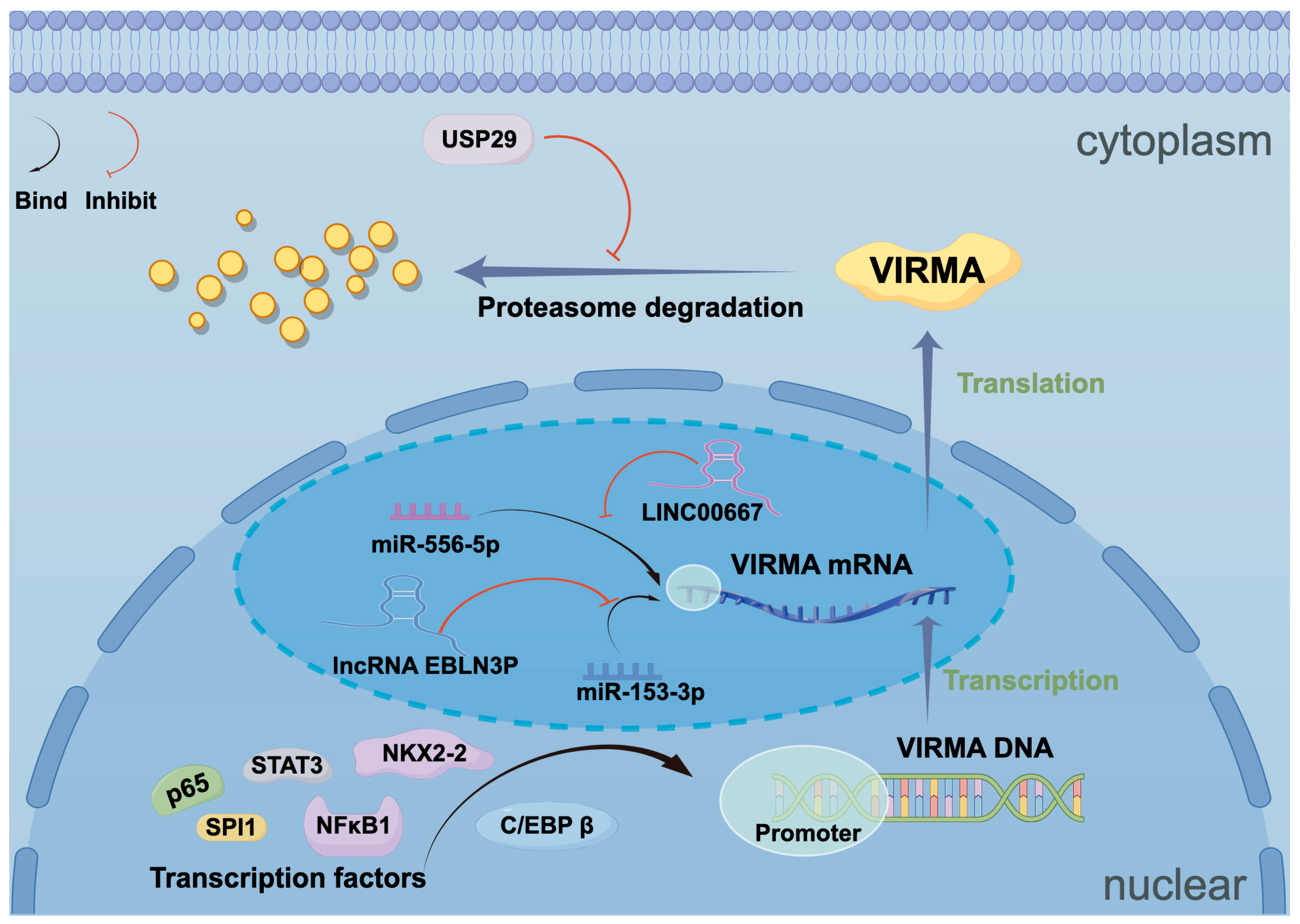
4. Upstream Regulatory Mechanisms of VIRMA

Transcription factors, non-coding RNAs, and ubiquitin-related enzymes can regulate the expression of VIRMA at multiple levels (Figure 6).

4.1. Transcription Factors

The study demonstrated that the transcription factor NKX2-2 is positively correlated with VIRMA and may serve as a specific upstream regulator of VIRMA in ES. Additionally, the study revealed that STAT3 can bind conservatively to the promoter of VIRMA [77]. Furthermore, the transcription factor NFκB1 could bind to the VIRMA promoter, upregulating its expression in CRC [84]. In OC, the transcription factor SPI1 regulated VIRMA transcription by binding to its DNA motif (AAGGAAGT) [45]. In PDAC, the transcription factor C/EBP β enhanced VIRMA promoter activity, thereby upregulating its expression [73]. The VIRMA promoter contains a p65 binding motif, and in gastric cancer, p65 upregulates VIRMA expression by binding to its promoter [71].

4.2. Non-Coding RNAs

LncRNA-miRNA networks also play a role in the regulation of VIRMA. In CRC, lncRNA EBLN3P competitively bound to miR-153-3p through a ceRNA network, promoting VIRMA expression, while VIRMA, in an m6A-dependent manner, enhanced the expression of lncRNA EBLN3P, forming a positive feedback loop. Similarly, in BC, a positive feedback regulatory mechanism also exists between lncRNA LINC00667, miR-556-5p, and VIRMA [75,81].

4.3. Ubiquitin-Related Enzymes

A study by Li et al. demonstrated that USP29, a member of the ubiquitin-specific protease (USP) family, stabilized VIRMA protein through deubiquitylation in CRC. VIRMA, in turn, enhanced SOX8 mRNA stability via m6A modification, promoting tumor progression [38].

5. The Potential of VIRMA in Cancer Diagnosis and Therapy

VIRMA serves as a core component of the m6A methyltransferase complex, demonstrating characteristic overexpression and tumor-specific subcellular localization across multiple malignancies. Its expression levels show strong correlations with tumor aggressiveness and poor clinical outcomes, positioning it as a promising diagnostic biomarker and therapeutic target (Table 2). Studies reveal VIRMA’s pan-cancer significance, with aberrant overexpression observed in over 20 cancer types that associate with chromosome 8q21.3-24.3 amplification [31]. Pathological analyses consistently demonstrate positive correlations between VIRMA expression and advanced TNM staging, lymph node metastasis, and distant metastasis, while showing negative associations with overall survival [24,39]. The protein exhibits distinct subcellular distribution patterns—predominantly nuclear in solid tumors such as hepatocellular and pancreatic carcinomas, yet cytoplasmically enriched in specific cancers including breast cancer, reflecting its functional heterogeneity across different tumor microenvironments and offering potential for molecular subtyping [19,23].
Extensive research has elucidated VIRMA’s dual regulatory mechanisms in tumor progression through both m6A-dependent and independent pathways. In the M6A-dependent pathway, VIRMA regulates key oncogenes (e.g., FOXM1, HK2) and tumor suppressor genes (e.g., RND3, BTG2), affecting the proliferation, metastasis, metabolic reprogramming, and drug resistance of tumor cells [18,24,39]. Additionally, VIRMA facilitates immune evasion within the tumor microenvironment via the KLF1-PD-L1 signaling axis [58]. The m6A-independent mechanism involves direct binding to 3′UTRs of specific mRNAs (CDK1, c-Jun) to regulate their stability [83,86]. These findings collectively demonstrate VIRMA’s multifaceted therapeutic potential across various intervention levels.
Current therapeutic strategies targeting VIRMA mainly focus on three aspects. One involves small-molecule inhibitor development, such as rucaparib, which binds to key residues LYS1029 and ASN1088 of VIRMA to inhibit its function [42]. Another strategy is gene therapy. CRISPR-Cas9-mediated knockout of VIRMA has shown anti-tumor effects in Ewing sarcoma models [77]. Future approaches may include targeted knockout or silencing of VIRMA using nanoparticle delivery systems, offering new possibilities for cancer treatment. In addition, combination therapies of VIRMA inhibition with ferroptosis inducers or immune checkpoint inhibitors have shown synergistic effects [57,58]. These preliminary findings lay a foundation for future exploration of VIRMA in cancer therapy.
Future studies should emphasize interdisciplinary collaboration. Integrating cutting-edge technologies such as single-cell sequencing and spatial transcriptomics may help to map the detailed regulatory role of VIRMA in tumor heterogeneity. AI-assisted molecular docking and virtual screening could accelerate the development of highly selective inhibitors. Organoid and patient-derived xenograft (PDX) models may better simulate clinical drug responses. It is also important to explore the potential of VIRMA in early cancer detection and treatment monitoring, including its value as a biomarker in exosomes or circulating tumor cells for liquid biopsy applications.
Table 2. Clinical significance of VIRMA and its relationship with patient survival.
Cancer TypeClinical
Significance
TargetsSurvival AssociationRef.
HNSCCOncogeneUBR5Not explored[35]
NPCOncogenePTGS2; LINC00839; E2F7Worst OS[32,46,47]
OSCCOncogeneinhibitor A; PGK1; CA9Worst OS[33,48,49]
NSCLCOncogenePXFP1; WTAP; BTG2; DAPK3; MUC3A; ARHGAP30; LINC01106; P53; KLF1; HOXA1;
MAP3K2
Worst OS[34,50,51,52,53,54,55,56,57,58,59]
HCCOncogeneRND3; HPN; circDLC1; GATA3 pre-mRNA; ID2; HK1; SLC7A11; CCR9; HSPG2; HBx; Zeb1Worst OS[18,23,60,61,62,63,64,65,66,67,85,122]
ICCOncogeneCCL3; TMED2P; ARD3B; SIRT1Worst OS[68,69]
GCOncogeneRASD1; LINC00958; FOXM1; P65; c-JunWorst OS[36,70,71,72,86]
PDACOncogeneSTRA6; SLC43A2; C/EBP βWorsetOS[24,73]
CRCOncogenelncRNA POU6F3-AS1; USP29; SOX8; SIRT1; miR-53-3p; lncRNA EBLN3P; HIF-1; NFκB1; WEE1Worst OS[20,37,38,74,75,84]
OSOncogeneCDK1; CCNA2; CCNB1; JAK-STATWorst OS[40,76]
ESOncogeneNKX2-2; STAT3Not explored[77]
MMOncogeneFOXM1Worst OS[39]
CMLOncogeneRAB27BNot explored[42]
DLBCLOncogeneCHST11Worst OS[41]
BCOncogeneHAS2; KIF15; TFAP2A; DDR1; NEAT1; miR-556-5p; LINC00667; SMC1A; CDK1Worse OS[19,21,78,79,80,81,83]
TGCTsOncogeneNot exploredNot explored[43]
CCOncogeneBTG2; LARP1Worst OS[44,82]
OCOncogeneSPI1; ENO1Worst OS[45]

6. Conclusions

In summary, VIRMA is a key molecule with both fundamental research value and strong potential for clinical translation. Advances in understanding its molecular mechanisms, along with continuous technological innovation, are expected to drive the development of VIRMA-based diagnostic tools and targeted therapies toward clinical application. These strategies may offer new options for cancer diagnosis and treatment. Progress in this field will require close collaboration among basic researchers, clinicians, and the pharmaceutical industry to bridge the gap between laboratory discoveries and clinical practice.

Author Contributions

J.L. and C.Z. contributed equally to this work. J.L., C.Z. and M.Y. wrote the main manuscript text and prepared figures. H.Y., C.X., J.W. and Y.G. assisted with language and figure quality improvement. Z.X. and J.S. conceptualized and edited the main manuscript text. All authors have read and agreed to the published version of the manuscript.

Funding

This work was supported by the National Natural Science Foundation of China (No. 82172962).

Institutional Review Board Statement

Not applicable.

Informed Consent Statement

Not applicable.

Data Availability Statement

Not applicable.

Acknowledgments

We thank Figdraw (www.figdraw.com (accessed on 25 March 2025)) for expert assistance in the pattern drawing.

Conflicts of Interest

The authors declare no conflicts of interest.

Abbreviations

m6AN6-methyladenosine
VIRMAVirus-like m6A methyltransferase associated
5′-UTR5′ untranslated region
3′-UTR3′ untranslated region
METTL3Methyltransferase-like 3
METTL14Methyltransferase-like 14
WTAPWilms’ tumor 1-associating protein
RBM15RNA-binding motif protein 15
BCBreast cancer
PDACPancreatic ductal adenocarcinoma
MACm6A-METTL complex
MACOMm6A-METTL- associated complex
dsDNAdouble-stranded DNA
M3-M14-W-VMETTL3-METTL14-WTAP-VIRMA quaternary complex
RBPsRNA-binding proteins
IF2Translation initiation factor 2
HNSCCHead and neck squamous cell carcinoma
NPCNasopharyngeal carcinoma
OSCCOral squamous cell carcinoma
NSCLCNon-small cell lung cancer
HCCHepatocellular carcinoma
ICCIntrahepatic cholangiocarcinoma
GCGastric cancer
CRCColorectal cancer
MMMultiple myeloma
OSOsteosarcoma
DLBCLDiffuse large B-cell lymphoma
CMLChronic myeloid leukemia
TGCTsTesticular germ cell tumors
CCCervical cancer
OCOvarian cancer
ESEwing’s sarcoma
CCL3Macrophage Inflammatory Protein-1 alpha, MIP-1α
HBVHepatitis B virus
HBxHepatitis B virus X protein
INHBAInhibin A
HAS2Hyaluronan synthase 2
HPNHepsin
DAPK3Death-Associated Protein Kinase 3
RXFP1Relaxin Family Peptide Receptor 1
LUADLung adenocarcinoma
LncRNAsLong non-coding RNAs
VIRMA FLFull-length isoform of VIRMA
circRNACircular RNA
EMTEpithelial-mesenchymal transition
GLUT1Glucose transporter-1
HKHexokinase
ENO1Alpha-enolase
ECARExtracellular acidification rate
OCROxygen consumption rate
LDHALactate dehydrogenase A
GSHGlutathione
ROSReactive oxygen species
ECMExtracellular matrix
RTKReceptor tyrosine kinase
SHBsHepatitis B virus surface small antigen
EREndoplasmic reticulum
UPRUnfolded protein response
IHInfantile Hemangioma
HIF-1Hypoxia-inducible factor 1
COADColon adenocarcinoma
USPsUbiquitin-specific protease
GDSCCancer drug sensitivity genomics

References

  1. Bray, F.; Laversanne, M.; Sung, H.; Ferlay, J.; Siegel, R.L.; Soerjomataram, I.; Jemal, A. Global cancer statistics 2022: GLOBOCAN estimates of incidence and mortality worldwide for 36 cancers in 185 countries. CA Cancer J. Clin. 2024, 74, 229–263. [Google Scholar] [CrossRef]
  2. Deng, X.; Qing, Y.; Horne, D.; Huang, H.; Chen, J. The roles and implications of RNA m(6)A modification in cancer. Nat. Rev. Clin. Oncol. 2023, 20, 507–526. [Google Scholar] [CrossRef]
  3. Gilbert, W.V.; Nachtergaele, S. mRNA Regulation by RNA Modifications. Annu. Rev. Biochem. 2023, 92, 175–198. [Google Scholar] [CrossRef] [PubMed]
  4. Dierks, D.; Garcia-Campos, M.A.; Uzonyi, A.; Safra, M.; Edelheit, S.; Rossi, A.; Sideri, T.; Varier, R.A.; Brandis, A.; Stelzer, Y.; et al. Multiplexed profiling facilitates robust m6A quantification at site, gene and sample resolution. Nat. Methods 2021, 18, 1060–1067. [Google Scholar] [CrossRef]
  5. Yue, S.W.; Liu, H.L.; Su, H.F.; Luo, C.; Liang, H.F.; Zhang, B.X.; Zhang, W. m6A-regulated tumor glycolysis: New advances in epigenetics and metabolism. Mol. Cancer 2023, 22, 137. [Google Scholar] [CrossRef] [PubMed]
  6. Uddin, M.B.; Wang, Z.; Yang, C. The m(6)A RNA methylation regulates oncogenic signaling pathways driving cell malignant transformation and carcinogenesis. Mol. Cancer 2021, 20, 61. [Google Scholar] [CrossRef] [PubMed]
  7. Zhou, H.; Yin, K.; Zhang, Y.; Tian, J.; Wang, S. The RNA m6A writer METTL14 in cancers: Roles, structures, and applications. Biochim. Biophys. Acta Rev. Cancer 2021, 1876, 188609. [Google Scholar] [CrossRef]
  8. Lence, T.; Paolantoni, C.; Worpenberg, L.; Roignant, J.Y. Mechanistic insights into m(6)A RNA enzymes. Biochim. Biophys. Acta Gene Regul. Mech. 2019, 1862, 222–229. [Google Scholar] [CrossRef]
  9. Zhang, X.L.; Chen, X.H.; Xu, B.; Chen, M.; Zhu, S.; Meng, N.; Wang, J.Z.; Zhu, H.; Chen, D.; Liu, J.B.; et al. K235 acetylation couples with PSPC1 to regulate the m(6)A demethylation activity of ALKBH5 and tumorigenesis. Nat. Commun. 2023, 14, 3815. [Google Scholar] [CrossRef]
  10. Jiang, X.; Liu, B.; Nie, Z.; Duan, L.; Xiong, Q.; Jin, Z.; Yang, C.; Chen, Y. The role of m6A modification in the biological functions and diseases. Signal Transduct. Target. Ther. 2021, 6, 74. [Google Scholar] [CrossRef]
  11. Wang, T.; Kong, S.; Tao, M.; Ju, S. The potential role of RNA N6-methyladenosine in Cancer progression. Mol. Cancer 2020, 19, 88. [Google Scholar] [CrossRef] [PubMed]
  12. Meyer, K.D.; Jaffrey, S.R. The dynamic epitranscriptome: N6-methyladenosine and gene expression control. Nat. Rev. Mol. Cell Biol. 2014, 15, 313–326. [Google Scholar] [CrossRef] [PubMed]
  13. Yue, Y.; Liu, J.; Cui, X.; Cao, J.; Luo, G.; Zhang, Z.; Cheng, T.; Gao, M.; Shu, X.; Ma, H.; et al. VIRMA mediates preferential m(6)A mRNA methylation in 3′UTR and near stop codon and associates with alternative polyadenylation. Cell Discov. 2018, 4, 10. [Google Scholar] [CrossRef]
  14. Chen, Y.; Peng, C.; Chen, J.; Chen, D.; Yang, B.; He, B.; Hu, W.; Zhang, Y.; Liu, H.; Dai, L.; et al. WTAP facilitates progression of hepatocellular carcinoma via m6A-HuR-dependent epigenetic silencing of ETS1. Mol. Cancer 2019, 18, 127. [Google Scholar] [CrossRef] [PubMed]
  15. Wang, J.; Zheng, F.; Wang, D.; Yang, Q. Regulation of ULK1 by WTAP/IGF2BP3 axis enhances mitophagy and progression in epithelial ovarian cancer. Cell Death Dis. 2024, 15, 97. [Google Scholar] [CrossRef]
  16. Zeng, C.; Huang, W.; Li, Y.; Weng, H. Roles of METTL3 in cancer: Mechanisms and therapeutic targeting. J. Hematol. Oncol. 2020, 13, 117. [Google Scholar] [CrossRef]
  17. Yan, X.; Liu, F.; Yan, J.; Hou, M.; Sun, M.; Zhang, D.; Gong, Z.; Dong, X.; Tang, C.; Yin, P. WTAP-VIRMA counteracts dsDNA binding of the m(6)A writer METTL3-METTL14 complex and maintains N(6)-adenosine methylation activity. Cell Discov. 2023, 9, 100. [Google Scholar] [CrossRef]
  18. Shan, M.; Liu, D.; Sun, L.; Yang, M.; He, M.; Zhang, Y.; Xiang, L.; Lu, L.; He, H.; Niu, D.; et al. KIAA1429 facilitates metastasis via m6A-YTHDC1-dependent RND3 down-regulation in hepatocellular carcinoma cells. Cancer Lett. 2024, 584, 216598. [Google Scholar] [CrossRef]
  19. Li, N.; Zhu, Z.; Deng, Y.; Tang, R.; Hui, H.; Kang, Y.; Rana, T.M. KIAA1429/VIRMA promotes breast cancer progression by m(6) A-dependent cytosolic HAS2 stabilization. EMBO Rep. 2023, 24, e55506. [Google Scholar] [CrossRef]
  20. Zhou, Y.; Pei, Z.; Maimaiti, A.; Zheng, L.; Zhu, Z.; Tian, M.; Zhou, Z.; Tan, F.; Pei, Q.; Li, Y.; et al. m(6)A methyltransferase KIAA1429 acts as an oncogenic factor in colorectal cancer by regulating SIRT1 in an m(6)A-dependent manner. Cell Death Discov. 2022, 8, 83. [Google Scholar] [CrossRef]
  21. Zhang, X.; Dai, X.Y.; Qian, J.Y.; Xu, F.; Wang, Z.W.; Xia, T.; Zhou, X.J.; Li, X.X.; Shi, L.; Wei, J.F.; et al. SMC1A regulated by KIAA1429 in m6A-independent manner promotes EMT progress in breast cancer. Mol. Ther. Nucleic Acids 2022, 27, 133–146. [Google Scholar] [CrossRef] [PubMed]
  22. Huang, J.; Guo, J.; Jia, R. N6-Methyladenosine Methyltransferase Component KIAA1429 Is a Potential Target of Cancer Therapy. Biomolecules 2024, 14, 1319. [Google Scholar] [CrossRef]
  23. Lan, T.; Li, H.; Zhang, D.; Xu, L.; Liu, H.; Hao, X.; Yan, X.; Liao, H.; Chen, X.; Xie, K.; et al. KIAA1429 contributes to liver cancer progression through N6-methyladenosine-dependent post-transcriptional modification of GATA3. Mol. Cancer 2019, 18, 186. [Google Scholar] [CrossRef] [PubMed]
  24. Yang, K.; Zhong, Z.; Zou, J.; Liao, J.Y.; Chen, S.; Zhou, S.; Zhao, Y.; Li, J.; Yin, D.; Huang, K.; et al. Glycolysis and tumor progression promoted by the m(6)A writer VIRMA via m(6)A-dependent upregulation of STRA6 in pancreatic ductal adenocarcinoma. Cancer Lett. 2024, 590, 216840. [Google Scholar] [CrossRef] [PubMed]
  25. Knuckles, P.; Lence, T.; Haussmann, I.U.; Jacob, D.; Kreim, N.; Carl, S.H.; Masiello, I.; Hares, T.; Villaseñor, R.; Hess, D.; et al. Zc3h13/Flacc is required for adenosine methylation by bridging the mRNA-binding factor Rbm15/Spenito to the m(6)A machinery component Wtap/Fl(2)d. Genes. Dev. 2018, 32, 415–429. [Google Scholar] [CrossRef]
  26. Su, S.; Li, S.; Deng, T.; Gao, M.; Yin, Y.; Wu, B.; Peng, C.; Liu, J.; Ma, J.; Zhang, K. Cryo-EM structures of human m(6)A writer complexes. Cell Res. 2022, 32, 982–994. [Google Scholar] [CrossRef]
  27. Thandapani, P.; O’Connor, T.R.; Bailey, T.L.; Richard, S. Defining the RGG/RG motif. Mol. Cell 2013, 50, 613–623. [Google Scholar] [CrossRef]
  28. Schwartz, S.; Mumbach, M.R.; Jovanovic, M.; Wang, T.; Maciag, K.; Bushkin, G.G.; Mertins, P.; Ter-Ovanesyan, D.; Habib, N.; Cacchiarelli, D.; et al. Perturbation of m6A writers reveals two distinct classes of mRNA methylation at internal and 5′ sites. Cell Rep. 2014, 8, 284–296. [Google Scholar] [CrossRef]
  29. Sun, Y.; Chen, D.; Sun, S.; Ren, M.; Zhou, L.; Chen, C.; Zhao, J.; Wei, H.; Zhao, Q.; Qi, Y.; et al. RBMS1 Coordinates with the m(6)A Reader YTHDF1 to Promote NSCLC Metastasis through Stimulating S100P Translation. Adv. Sci. 2024, 11, e2307122. [Google Scholar] [CrossRef]
  30. Niessen, M.; Schneiter, R.; Nothiger, R. Molecular identification of virilizer, a gene required for the expression of the sex-determining gene Sex-lethal in Drosophila melanogaster. Genetics 2001, 157, 679–688. [Google Scholar] [CrossRef]
  31. Destefanis, E.; Sighel, D.; Dalfovo, D.; Gilmozzi, R.; Broso, F.; Cappannini, A.; Bujnicki, J.M.; Romanel, A.; Dassi, E.; Quattrone, A. The three YTHDF paralogs and VIRMA are strong cross-histotype tumor driver candidates among m(6)A core genes. NAR Cancer 2024, 6, zcae040. [Google Scholar] [CrossRef]
  32. Zheng, Z.Q.; Huang, Z.H.; Liang, Y.L.; Zheng, W.H.; Xu, C.; Li, Z.X.; Liu, N.; Yang, P.Y.; Li, Y.Q.; Ma, J.; et al. VIRMA promotes nasopharyngeal carcinoma, tumorigenesis, and metastasis by upregulation of E2F7 in an m6A-dependent manner. J. Biol. Chem. 2023, 299, 104677. [Google Scholar] [CrossRef] [PubMed]
  33. Xu, K.; Dai, X.; Yue, J. m(6)A methyltransferase KIAA1429 accelerates oral squamous cell carcinoma via regulating glycolysis and ferroptosis. Transl. Oncol. 2023, 36, 101745. [Google Scholar] [CrossRef]
  34. Lin, X.; Ye, R.; Li, Z.; Zhang, B.; Huang, Y.; Du, J.; Wang, B.; Meng, H.; Xian, H.; Yang, X.; et al. KIAA1429 promotes tumorigenesis and gefitinib resistance in lung adenocarcinoma by activating the JNK/MAPK pathway in an m(6)A-dependent manner. Drug Resist. Updat. 2023, 66, 100908. [Google Scholar] [CrossRef]
  35. Zhu, C.; Cheng, Y.; Yu, Y.; Zhang, Y.; Ren, G. VIRMA promotes the progression of head and neck squamous cell carcinoma by regulating UBR5 mRNA and m6A levels. Biomol. Biomed. 2024, 24, 1244–1257. [Google Scholar] [CrossRef]
  36. Ren, M.; Pan, H.; Zhou, X.; Yu, M.; Ji, F. KIAA1429 promotes gastric cancer progression by destabilizing RASD1 mRNA in an m(6)A-YTHDF2-dependent manner. J. Transl. Med. 2024, 22, 584. [Google Scholar] [CrossRef] [PubMed]
  37. Ouyang, C.; Xu, G.; Xie, J.; Xie, Y.; Zhou, Y. Silencing of KIAA1429, a N6-methyladenine methyltransferase, inhibits the progression of colon adenocarcinoma via blocking the hypoxia-inducible factor 1 signalling pathway. J. Biochem. Mol. Toxicol. 2024, 38, e23829. [Google Scholar] [CrossRef] [PubMed]
  38. Li, J.; Yang, J.; Chen, Z.; Liu, L.; Wang, H.; Deng, Q.; Chen, Y.; Que, Y.; Fu, Z. Promotive role of USP29-mediated deubiquitination in malignant proliferation of colorectal cancer cells via the KIAA1429/SOX8 axis. Biomol. Biomed. 2023, 23, 483–495. [Google Scholar] [CrossRef]
  39. Wu, Y.; Luo, Y.; Yao, X.; Shi, X.; Xu, Z.; Re, J.; Shi, M.; Li, M.; Liu, J.; He, Y.; et al. KIAA1429 increases FOXM1 expression through YTHDF1-mediated m6A modification to promote aerobic glycolysis and tumorigenesis in multiple myeloma. Cell Biol. Toxicol. 2024, 40, 58. [Google Scholar] [CrossRef]
  40. Luo, J.; Wang, X.; Chen, Z.; Zhou, H.; Xiao, Y. The role and mechanism of JAK2/STAT3 signaling pathway regulated by m6A methyltransferase KIAA1429 in osteosarcoma. J. Bone Oncol. 2023, 39, 100471. [Google Scholar] [CrossRef]
  41. Chen, X.; Lu, T.; Cai, Y.; Han, Y.; Ding, M.; Chu, Y.; Zhou, X.; Wang, X. KIAA1429-mediated m6A modification of CHST11 promotes progression of diffuse large B-cell lymphoma by regulating Hippo-YAP pathway. Cell Mol. Biol. Lett. 2023, 28, 32. [Google Scholar] [CrossRef] [PubMed]
  42. Yao, F.; Zhong, F.; Jiang, J.; Cheng, Y.; Xu, S.; Liu, J.; Lin, J.; Zhang, J.; Li, S.; Li, M.; et al. The m(6)A regulator KIAA1429 stabilizes RAB27B mRNA and promotes the progression of chronic myeloid leukemia and resistance to targeted therapy. Genes Dis. 2024, 11, 993–1008. [Google Scholar] [CrossRef] [PubMed]
  43. Miranda-Gonçalves, V.; Lobo, J.; Guimarães-Teixeira, C.; Barros-Silva, D.; Guimarães, R.; Cantante, M.; Braga, I.; Maurício, J.; Oing, C.; Honecker, F.; et al. The component of the m(6)A writer complex VIRMA is implicated in aggressive tumor phenotype, DNA damage response and cisplatin resistance in germ cell tumors. J. Exp. Clin. Cancer Res. 2021, 40, 268. [Google Scholar] [CrossRef]
  44. Wang, M.; Wang, Z.; Zou, X.; Yang, D.; Xu, K. The regulatory role of KIAA1429 in epithelial-mesenchymal transition in cervical cancer via mediating m6A modification of BTG2. Cytotechnology 2025, 77, 34. [Google Scholar] [CrossRef]
  45. Gan, L.; Zhao, S.; Gao, Y.; Qi, Y.; Su, M.; Wang, A.; Cai, H. N6-methyladenosine methyltransferase KIAA1429 promoted ovarian cancer aerobic glycolysis and progression through enhancing ENO1 expression. Biol. Direct 2023, 18, 64. [Google Scholar] [CrossRef] [PubMed]
  46. Wu, L.; Zhou, Y.; Fu, J. KIAA1429 Promotes Nasopharyngeal Carcinoma Progression by Mediating m6A Modification of PTGS2. Crit. Rev. Immunol. 2023, 43, 15–27. [Google Scholar] [CrossRef] [PubMed]
  47. Zheng, W.H.; Long, Z.Q.; Zheng, Z.Q.; Zhang, L.L.; Liang, Y.L.; Li, Z.X.; Lv, J.W.; Kou, J.; Hong, X.H.; He, S.W.; et al. m6A-enriched lncRNA LINC00839 promotes tumor progression by enhancing TAF15-mediated transcription of amine oxidase AOC1 in nasopharyngeal carcinoma. J. Biol. Chem. 2023, 299, 104873. [Google Scholar] [CrossRef]
  48. Chen, X.; Fan, R. Inhibin A contributes to the tumorigenesis of oral squamous cell carcinoma by KIAA1429-mediated m6A modification. J. Oral. Pathol. Med. 2024, 53, 266–274. [Google Scholar] [CrossRef]
  49. Tu, J.; Feng, X.; Cao, Q.; Guan, Y. KIAA1429 promotes the malignancy of oral squamous cell carcinoma by regulating CA9 m6A methylation. Cytotechnology 2024, 76, 585–594. [Google Scholar] [CrossRef]
  50. Zhang, Z.; Guo, J.; Gong, C.; Wu, S.; Sun, Y. KIAA1429-mediated RXFP1 attenuates non-small cell lung cancer tumorigenesis via N6-methyladenosine modification. Cancer Biomark. 2024. Online ahead of print. [Google Scholar] [CrossRef]
  51. Ma, B.; Xiu, L.; Ding, L. The m6 RNA methylation regulator KIAA1429 is associated with autophagy-mediated drug resistance in lung cancer. FASEB Bioadv 2024, 6, 105–117. [Google Scholar] [CrossRef]
  52. Zhang, C.; Sun, Q.; Zhang, X.; Qin, N.; Pu, Z.; Gu, Y.; Yan, C.; Zhu, M.; Dai, J.; Wang, C.; et al. Gene amplification-driven RNA methyltransferase KIAA1429 promotes tumorigenesis by regulating BTG2 via m6A-YTHDF2-dependent in lung adenocarcinoma. Cancer Commun. 2022, 42, 609–626. [Google Scholar] [CrossRef]
  53. Xu, Y.; Chen, Y.; Yao, Y.; Xie, H.; Lu, G.; Du, C.; Cheng, J.; Zhou, J. VIRMA contributes to non-small cell lung cancer progression via N(6)-methyladenosine-dependent DAPK3 post-transcriptional modification. Cancer Lett. 2021, 522, 142–154. [Google Scholar] [CrossRef] [PubMed]
  54. Zhao, W.; Xie, Y. KIAA1429 promotes the progression of lung adenocarcinoma by regulating the m6A level of MUC3A. Pathol. Res. Pract. 2021, 217, 153284. [Google Scholar] [CrossRef]
  55. Guo, W.; Wang, T.; Huai, Q.; Guo, L.; Wang, X.; He, J. KIAA1429 regulates lung adenocarcinoma proliferation and metastasis through the PI3K/AKT pathway by modulating ARHGAP30 expression. Thorac. Cancer 2024, 15, 1397–1409. [Google Scholar] [CrossRef] [PubMed]
  56. Xu, D.; Wang, Z.; Li, F. KIAA1429 Induces m6A Modification of LINC01106 to Enhance the Malignancy of Lung Adenocarcinoma Cells via the JAK/STAT3 Pathway. Crit. Rev. Immunol. 2024, 44, 49–61. [Google Scholar] [CrossRef]
  57. Wu, Y.; Li, H.; Huang, Y.; Chen, Q. Silencing of m(6)A methyltransferase KIAA1429 suppresses the progression of non-small cell lung cancer by promoting the p53 signaling pathway and ferroptosis. Am. J. Cancer Res. 2023, 13, 5320–5333. [Google Scholar] [PubMed]
  58. Geng, R.; Ren, M.; Ma, Y.; Su, W. Mechanism of the KIAA1429/KLF1/PD-L1 Axis in Regulating Immune Escape in Non-small Cell Lung Cancer. Cell Biochem. Biophys. 2024, 82, 1835–1845. [Google Scholar] [CrossRef]
  59. Tang, J.; Han, T.; Tong, W.; Zhao, J.; Wang, W. N(6)-methyladenosine (m(6)A) methyltransferase KIAA1429 accelerates the gefitinib resistance of non-small-cell lung cancer. Cell Death Discov. 2021, 7, 108. [Google Scholar] [CrossRef]
  60. Meng, Y.; Yang, W.; Li, J.; Chai, W. KIAA1429 facilitates progression of hepatocellular carcinoma by modulating m6A levels in HPN. Heliyon 2023, 9, e22084. [Google Scholar] [CrossRef]
  61. Liu, H.; Lan, T.; Li, H.; Xu, L.; Chen, X.; Liao, H.; Chen, X.; Du, J.; Cai, Y.; Wang, J.; et al. Circular RNA circDLC1 inhibits MMP1-mediated liver cancer progression via interaction with HuR. Theranostics 2021, 11, 1396–1411. [Google Scholar] [CrossRef]
  62. Cheng, X.; Li, M.; Rao, X.; Zhang, W.; Li, X.; Wang, L.; Huang, G. KIAA1429 regulates the migration and invasion of hepatocellular carcinoma by altering m6A modification of ID2 mRNA. Onco Targets Ther. 2019, 12, 3421–3428. [Google Scholar] [CrossRef] [PubMed]
  63. Liu, D.; Shan, M.; Zeng, R.; He, M.; Dai, X.; Lu, L.; Yang, M.; He, H.; Zhang, Y.; Xiang, L.; et al. Inhibition of KIAA1429/HK1 axis enhances the sensitivity of liver cancer cells to sorafenib by regulating the Warburg effect. Biochem. Pharmacol. 2024, 227, 116419. [Google Scholar] [CrossRef]
  64. Wang, H.; Chen, W.; Cui, Y.; Gong, H.; Li, H. KIAA1429 protects hepatocellular carcinoma cells from ferroptotic cell death with a m(6) A-dependent posttranscriptional modification of SLC7A11. J. Cell Mol. Med. 2023, 27, 4118–4132. [Google Scholar] [CrossRef] [PubMed]
  65. Lv, Z.; Liu, L.; You, J.; Zhou, P.; Su, Y.; Zhao, K.; Zhang, J.; Zhu, F. Small HBV surface antigen drives regorafenib resistance in HCC via KIAA1429-dependent m6A modification of CCR9. J. Med. Virol. 2024, 96, e29894. [Google Scholar] [CrossRef]
  66. Sivasudhan, E.; Zhou, J.; Ma, J.; Wang, Y.; Liu, S.; Khan, F.I.; Lu, Z.; Meng, J.; Blake, N.; Rong, R. Hepatitis B Virus X Protein Contributes to Hepatocellular Carcinoma via Upregulation of KIAA1429 Methyltransferase and mRNA m6A Hypermethylation of HSPG2/Perlecan. Mol. Carcinog. 2025, 64, 108–125. [Google Scholar] [CrossRef] [PubMed]
  67. Kuang, Y.; Cheng, Y.; Wang, J.; Li, H.; Cao, X.; Wang, Y. KIAA1429 mediates epithelial mesenchymal transition in sorafenib-resistant hepatocellular carcinoma through m6A methylation modification. Cancer Med. 2023, 12, 7222–7233. [Google Scholar] [CrossRef]
  68. Xu, H.; Lin, X.; Li, Z.; He, X.; Li, Y.; Qiu, L.; Lu, L.; Liu, B.; Zhan, M.; He, K. VIRMA facilitates intrahepatic cholangiocarcinoma progression through epigenetic augmentation of TMED2 and PARD3B mRNA stabilization. J. Gastroenterol. 2023, 58, 925–944. [Google Scholar] [CrossRef]
  69. Zhou, S.; Yang, K.; Chen, S.; Lian, G.; Huang, Y.; Yao, H.; Zhao, Y.; Huang, K.; Yin, D.; Lin, H.; et al. CCL3 secreted by hepatocytes promotes the metastasis of intrahepatic cholangiocarcinoma by VIRMA-mediated N6-methyladenosine (m(6)A) modification. J. Transl. Med. 2023, 21, 43. [Google Scholar] [CrossRef]
  70. Yang, D.; Chang, S.; Li, F.; Ma, M.; Yang, J.; Lv, X.; Huangfu, L.; Jia, C. m(6) A transferase KIAA1429-stabilized LINC00958 accelerates gastric cancer aerobic glycolysis through targeting GLUT1. IUBMB Life 2021, 73, 1325–1333. [Google Scholar] [CrossRef]
  71. Zhu, Z.; Zhou, Y.; Chen, Y.; Zhou, Z.; Liu, W.; Zheng, L.; Pei, Q.; Tan, F.; Pei, H.; Li, Y. m(6)A Methyltransferase KIAA1429 Regulates the Cisplatin Sensitivity of Gastric Cancer Cells via Stabilizing FOXM1 mRNA. Cancers 2022, 14, 5025. [Google Scholar] [CrossRef]
  72. Tang, B.; Li, M.; Xu, Y.; Li, X. N(6)-methyladenosine (m(6)A) writer KIAA1429 accelerates gastric cancer oxaliplatin chemoresistance by targeting FOXM1. J. Cancer Res. Clin. Oncol. 2023, 149, 5037–5045. [Google Scholar] [CrossRef]
  73. Gao, L.; Lv, G.; Liu, Z.; Tian, Y.; Han, F.; Li, L.; Wang, G.; Zhang, Y. Alcohol-induced C/EBP β-driven VIRMA decreases oxidative stress and promotes pancreatic ductal adenocarcinoma growth and metastasis via the m6A/YTHDF2/SLC43A2 pathway. Oncogene 2025, 44, 1118–1132. [Google Scholar] [CrossRef] [PubMed]
  74. Lu, D.; Chen, A. lncRNA POU6F2-AS1 Regulated by KIAA1429 Contributes to Colorectal Cancer Progression in an m(6)A Modification Manner. Mol. Biotechnol. 2025, 67, 115–122. [Google Scholar] [CrossRef]
  75. Chen, H.; Zhu, P.; Zhu, D.; Jin, J.; Yang, Q.; Han, X. Role and mechanism of KIAA1429 in regulating cellular ferroptosis and radioresistance in colorectal cancer. Biomol. Biomed. 2024, 24, 1669–1681. [Google Scholar] [CrossRef]
  76. Sun, Y.; Lei, Y.W.; Zeng, J.X.; Zhong, L.Y.; Liu, J.W.; Man, Y.N.; He, M.L. Clinical Significance and Potential Mechanisms of the RNA Methyltransferase KIAA1429 in Osteosarcoma. J. Cancer 2024, 15, 126–139. [Google Scholar] [CrossRef]
  77. Tan, K.; Lu, W.; Chen, F.; Shi, H.; Ma, Y.; Chen, Z.; Wu, W.; Lv, Z.; Mo, J. CRISPR-Cas9 knockout screening identifies KIAA1429 as an essential gene in Ewing sarcoma. J. Exp. Clin. Cancer Res. 2023, 42, 250. [Google Scholar] [CrossRef] [PubMed]
  78. Chen, C.; Wang, Y.; Li, Y.; Zhang, C. VIRMA Facilitates Triple-Negative Breast Cancer Progression via Increasing m6A-Dependent KIF15 Expression. Discov. Med. 2023, 35, 787–795. [Google Scholar] [CrossRef] [PubMed]
  79. Lian, B.; Yan, S.; Li, J.; Bai, Z.; Li, J. HNRNPC promotes collagen fiber alignment and immune evasion in breast cancer via activation of the VIRMA-mediated TFAP2A/DDR1 axis. Mol. Med. 2023, 29, 103. [Google Scholar] [CrossRef]
  80. Lee, Q.; Song, R.; Phan, D.A.V.; Pinello, N.; Tieng, J.; Su, A.; Halstead, J.M.; Wong, A.C.H.; van Geldermalsen, M.; Lee, B.S.; et al. Overexpression of VIRMA confers vulnerability to breast cancers via the m(6)A-dependent regulation of unfolded protein response. Cell Mol. Life Sci. 2023, 80, 157. [Google Scholar] [CrossRef]
  81. Ren, S.; Zhang, Y.; Yang, X.; Li, X.; Zheng, Y.; Liu, Y.; Zhang, X. N6-methyladenine- induced LINC00667 promoted breast cancer progression through m6A/KIAA1429 positive feedback loop. Bioengineered 2022, 13, 13462–13473. [Google Scholar] [CrossRef]
  82. Feng, X.; Shu, L. The methyltransferase KIAA1429 potentiates cervical cancer tumorigenesis via modulating LARP1 mRNA m6A modification and stability. Histol. Histopathol. 2024, 18843. [Google Scholar] [CrossRef]
  83. Qian, J.Y.; Gao, J.; Sun, X.; Cao, M.D.; Shi, L.; Xia, T.S.; Zhou, W.B.; Wang, S.; Ding, Q.; Wei, J.F. KIAA1429 acts as an oncogenic factor in breast cancer by regulating CDK1 in an N6-methyladenosine-independent manner. Oncogene 2019, 38, 6123–6141. [Google Scholar] [CrossRef]
  84. Ma, L.; Lin, Y.; Sun, S.W.; Xu, J.; Yu, T.; Chen, W.L.; Zhang, L.H.; Guo, Y.C.; Wang, Y.W.; Chen, T.; et al. KIAA1429 is a potential prognostic marker in colorectal cancer by promoting the proliferation via downregulating WEE1 expression in an m6A-independent manner. Oncogene 2022, 41, 692–703. [Google Scholar] [CrossRef]
  85. Wang, M.; Yang, Y.; Yang, J.; Yang, J.; Han, S. circ_KIAA1429 accelerates hepatocellular carcinoma advancement through the mechanism of m(6)A-YTHDF3-Zeb1. Life Sci. 2020, 257, 118082. [Google Scholar] [CrossRef] [PubMed]
  86. Miao, R.; Dai, C.C.; Mei, L.; Xu, J.; Sun, S.W.; Xing, Y.L.; Wu, L.S.; Wang, M.H.; Wei, J.F. KIAA1429 regulates cell proliferation by targeting c-Jun messenger RNA directly in gastric cancer. J. Cell Physiol. 2020, 235, 7420–7432. [Google Scholar] [CrossRef]
  87. Gerstberger, S.; Jiang, Q.; Ganesh, K. Metastasis. Cell 2023, 186, 1564–1579. [Google Scholar] [CrossRef] [PubMed]
  88. Hanahan, D. Hallmarks of Cancer: New Dimensions. Cancer Discov. 2022, 12, 31–46. [Google Scholar] [CrossRef]
  89. Fares, J.; Fares, M.Y.; Khachfe, H.H.; Salhab, H.A.; Fares, Y. Molecular principles of metastasis: A hallmark of cancer revisited. Signal Transduct. Target. Ther. 2020, 5, 28. [Google Scholar] [CrossRef] [PubMed]
  90. Korbecki, J.; Grochans, S.; Gutowska, I.; Barczak, K.; Baranowska-Bosiacka, I. CC Chemokines in a Tumor: A Review of Pro-Cancer and Anti-Cancer Properties of Receptors CCR5, CCR6, CCR7, CCR8, CCR9, and CCR10 Ligands. Int. J. Mol. Sci. 2020, 21, 8421. [Google Scholar] [CrossRef]
  91. Singh, P.; Jenkins, L.M.; Horst, B.; Alers, V.; Pradhan, S.; Kaur, P.; Srivastava, T.; Hempel, N.; Győrffy, B.; Broude, E.V.; et al. Inhibin Is a Novel Paracrine Factor for Tumor Angiogenesis and Metastasis. Cancer Res. 2018, 78, 2978–2989. [Google Scholar] [CrossRef]
  92. Du, F.; Yuan, P.; Wang, T.; Zhao, J.; Zhao, Z.; Luo, Y.; Xu, B. The Significance and Therapeutic Potential of GATA3 Expression and Mutation in Breast Cancer: A Systematic Review. Med. Res. Rev. 2015, 35, 1300–1315. [Google Scholar] [CrossRef] [PubMed]
  93. Gao, N.; Li, Y.; Li, J.; Gao, Z.; Yang, Z.; Li, Y.; Liu, H.; Fan, T. Long Non-Coding RNAs: The Regulatory Mechanisms, Research Strategies, and Future Directions in Cancers. Front. Oncol. 2020, 10, 598817. [Google Scholar] [CrossRef] [PubMed]
  94. Knutsen, E.; Harris, A.L.; Perander, M. Expression and functions of long non-coding RNA NEAT1 and isoforms in breast cancer. Br. J. Cancer 2022, 126, 551–561. [Google Scholar] [CrossRef] [PubMed]
  95. Ma, R.T.; Wang, Y.; Ji, F.; Chen, J.N.; Wang, T.J.; Liu, Y.; Hou, M.X.; Guo, Z.G. YTHDF1’s grip on CRC vasculature: Insights into LINC01106 and miR-449b-5p-VEGFA axis. Cancer Cell Int. 2024, 24, 195. [Google Scholar] [CrossRef]
  96. Zhong, Y.; Du, Y.; Yang, X.; Mo, Y.; Fan, C.; Xiong, F.; Ren, D.; Ye, X.; Li, C.; Wang, Y.; et al. Circular RNAs function as ceRNAs to regulate and control human cancer progression. Mol. Cancer 2018, 17, 79. [Google Scholar] [CrossRef]
  97. Jin, W. Role of JAK/STAT3 Signaling in the Regulation of Metastasis, the Transition of Cancer Stem Cells, and Chemoresistance of Cancer by Epithelial-Mesenchymal Transition. Cells 2020, 9, 217. [Google Scholar] [CrossRef]
  98. Riggi, N.; Suvà, M.L.; Stamenkovic, I. Ewing’s Sarcoma. N. Engl. J. Med. 2021, 384, 154–164. [Google Scholar] [CrossRef]
  99. Liu, J.; Gu, X.; Guan, Z.; Huang, D.; Xing, H.; Zheng, L. Role of m6A modification in regulating the PI3K/AKT signaling pathway in cancer. J. Transl. Med. 2023, 21, 774. [Google Scholar] [CrossRef]
  100. Lu, H.; Cao, L.L.; Ballout, F.; Belkhiri, A.; Peng, D.; Chen, L.; Chen, Z.; Soutto, M.; Wang, T.C.; Que, J.; et al. Reflux conditions induce E-cadherin cleavage and EMT via APE1 redox function in oesophageal adenocarcinoma. Gut 2023, 73, 47–62. [Google Scholar] [CrossRef]
  101. Williams, G.H.; Stoeber, K. The cell cycle and cancer. J. Pathol. 2012, 226, 352–364. [Google Scholar] [CrossRef]
  102. You, Q.; Wang, J.; Yu, Y.; Li, F.; Meng, L.; Chen, M.; Yang, Q.; Xu, Z.; Sun, J.; Zhuo, W.; et al. The histone deacetylase SIRT6 promotes glycolysis through the HIF-1α/HK2 signaling axis and induces erlotinib resistance in non-small cell lung cancer. Apoptosis 2022, 27, 883–898. [Google Scholar] [CrossRef] [PubMed]
  103. Hay, N. Reprogramming glucose metabolism in cancer: Can it be exploited for cancer therapy? Nat. Rev. Cancer 2016, 16, 635–649. [Google Scholar] [CrossRef]
  104. Hanahan, D.; Weinberg, R.A. Hallmarks of cancer: The next generation. Cell 2011, 144, 646–674. [Google Scholar] [CrossRef] [PubMed]
  105. Yu, M.; Yongzhi, H.; Chen, S.; Luo, X.; Lin, Y.; Zhou, Y.; Jin, H.; Hou, B.; Deng, Y.; Tu, L.; et al. The prognostic value of GLUT1 in cancers: A systematic review and meta-analysis. Oncotarget 2017, 8, 43356–43367. [Google Scholar] [CrossRef]
  106. Guo, D.; Tong, Y.; Jiang, X.; Meng, Y.; Jiang, H.; Du, L.; Wu, Q.; Li, S.; Luo, S.; Li, M.; et al. Aerobic glycolysis promotes tumor immune evasion by hexokinase2-mediated phosphorylation of IκBα. Cell Metab. 2022, 34, 1312–1324.e6. [Google Scholar] [CrossRef]
  107. Li, Y.; He, L.; Wang, Y.; Tan, Y.; Zhang, F. N(6)-methyladenosine methyltransferase KIAA1429 elevates colorectal cancer aerobic glycolysis via HK2-dependent manner. Bioengineered 2022, 13, 11923–11932. [Google Scholar] [CrossRef] [PubMed]
  108. Dixon, S.J.; Lemberg, K.M.; Lamprecht, M.R.; Skouta, R.; Zaitsev, E.M.; Gleason, C.E.; Patel, D.N.; Bauer, A.J.; Cantley, A.M.; Yang, W.S.; et al. Ferroptosis: An iron-dependent form of nonapoptotic cell death. Cell 2012, 149, 1060–1072. [Google Scholar] [CrossRef]
  109. Lei, G.; Zhuang, L.; Gan, B. Targeting ferroptosis as a vulnerability in cancer. Nat. Rev. Cancer 2022, 22, 381–396. [Google Scholar] [CrossRef] [PubMed]
  110. Lee, J.; Roh, J.L. Epigenetic modulation of ferroptosis in cancer: Identifying epigenetic targets for novel anticancer therapy. Cell Oncol. 2023, 46, 1605–1623. [Google Scholar] [CrossRef]
  111. Lee, J.; Roh, J.L. SLC7A11 as a Gateway of Metabolic Perturbation and Ferroptosis Vulnerability in Cancer. Antioxidants 2022, 11, 2444. [Google Scholar] [CrossRef]
  112. Wang, Y.; Wu, X.; Ren, Z.; Li, Y.; Zou, W.; Chen, J.; Wang, H. Overcoming cancer chemotherapy resistance by the induction of ferroptosis. Drug Resist. Updat. 2023, 66, 100916. [Google Scholar] [CrossRef]
  113. Tharp, K.M.; Kersten, K.; Maller, O.; Timblin, G.A.; Stashko, C.; Canale, F.P.; Menjivar, R.E.; Hayward, M.K.; Berestjuk, I.; Ten Hoeve, J.; et al. Tumor-associated macrophages restrict CD8(+) T cell function through collagen deposition and metabolic reprogramming of the breast cancer microenvironment. Nat. Cancer 2024, 5, 1045–1062. [Google Scholar] [CrossRef]
  114. Mansoori, B.; Mohammadi, A.; Davudian, S.; Shirjang, S.; Baradaran, B. The Different Mechanisms of Cancer Drug Resistance: A Brief Review. Adv. Pharm. Bull. 2017, 7, 339–348. [Google Scholar] [CrossRef]
  115. Ojha, R.; Amaravadi, R.K. Targeting the unfolded protein response in cancer. Pharmacol. Res. 2017, 120, 258–266. [Google Scholar] [CrossRef] [PubMed]
  116. Iurlaro, R.; Muñoz-Pinedo, C. Cell death induced by endoplasmic reticulum stress. Febs J. 2016, 283, 2640–2652. [Google Scholar] [CrossRef]
  117. Huang, L.; Nakayama, H.; Klagsbrun, M.; Mulliken, J.B.; Bischoff, J. Glucose transporter 1-positive endothelial cells in infantile hemangioma exhibit features of facultative stem cells. Stem Cells 2015, 33, 133–145. [Google Scholar] [CrossRef] [PubMed]
  118. Wang, L.; Zou, Y.; Huang, Z.; Wang, W.; Li, J.; Bi, J.; Huo, R. KIAA1429 promotes infantile hemangioma regression by facilitating the stemness of hemangioma endothelial cells. Cancer Sci. 2023, 114, 1569–1581. [Google Scholar] [CrossRef] [PubMed]
  119. Zeng, K.; Li, W.; Wang, Y.; Zhang, Z.; Zhang, L.; Zhang, W.; Xing, Y.; Zhou, C. Inhibition of CDK1 Overcomes Oxaliplatin Resistance by Regulating ACSL4-mediated Ferroptosis in Colorectal Cancer. Adv. Sci. 2023, 10, e2301088. [Google Scholar] [CrossRef]
  120. Li, Y.; Ren, B.X.; Li, H.M.; Lu, T.; Fu, R.; Wu, Z.Q. Omeprazole suppresses aggressive cancer growth and metastasis in mice through promoting Snail degradation. Acta Pharmacol. Sin. 2022, 43, 1816–1828. [Google Scholar] [CrossRef]
  121. Patel, M.; Horgan, P.G.; McMillan, D.C.; Edwards, J. NF-κB pathways in the development and progression of colorectal cancer. Transl. Res. 2018, 197, 43–56. [Google Scholar] [CrossRef]
  122. Zhang, C.P.; Huang, X.Y. Circular RNA circ_KIAA1429 accelerates hepatocellular carcinoma progression via the miR-133a-3p/high mobility group AT-hook 2 (HMGA2) axis in an m6A-dependent manner. Hum. Cell 2023, 36, 1741–1754. [Google Scholar] [CrossRef] [PubMed]
Figure 1. The role of VIRMA in m6A methylation. (A) Components of the methyltransferase complex. (B) dsDNA inhibits the RNA methyltransferase activity of METTL3-METTL14. WTAP-VIRMA binds to the RGG motif, preventing dsDNA from interfering and thereby maintaining the RNA methylation activity of METTL3-METTL14. (C) VIRMA acts as a scaffold for the methyltransferase complex, recruiting the core catalytic components and linking them to the RNA substrate.
Biomolecules 15 00912 g001
Figure 2. VIRMA is upregulated in eighteen types of malignant tumors. Elevated VIRMA expression has been documented across a wide spectrum of malignancies. HNSCC, head and neck squamous cell carcinoma; NPC, nasopharyngeal carcinoma; OSCC, oral squamous cell carcinoma; NSCLC, non-small cell lung cancer; HCC, hepatocellular carcinoma; ICC, intrahepatic cholangiocarcinoma; GC, gastric cancer; PDAC, pancreatic ductal adenocarcinoma; CRC, colorectal cancer; MM, multiple myeloma; OS, osteosarcoma; DLBCL, diffuse large B-cell lymphoma; CML, chronic myeloid leukemia; BC, breast cancer; TGCTs, testicular germ cell tumors; CC, cervical cancer; OC, ovarian cancer; and ES, Ewing’s sarcoma.
Biomolecules 15 00912 g002
Figure 3. VIRMA promotes cancer cell proliferation and metastasis. VIRMA upregulates oncogenes, downregulates tumor suppressors, and modulating lncRNAs, circRNAs, and classical oncogenic pathways to promote cancer cell proliferation and metastasis in an m6A-dependent manner.
Biomolecules 15 00912 g003
Figure 4. VIRMA regulates cell resistance to chemotherapy through m6A modification.
Biomolecules 15 00912 g004
Figure 5. Involvement of VIRMA in tumor progression through m6A-independent mechanisms.
Biomolecules 15 00912 g005
Figure 6. Upstream regulatory mechanisms of VIRMA. Multiple transcription factors (P65, STAT3, NKX2-2, SPI1, and NFκB1, C/EBPβ) enhance VIRMA promoter activity and transcription. lncRNA EBLN3P promotes VIRMA expression by competitively binding miR-153-3p, while VIRMA reciprocally enhances EBLN3P expression in an m6A-dependent manner. A similar regulatory mechanism exists between LINC00667, miR-556-5p and VIRMA. At the protein level, USP29 stabilizes VIRMA through deubiquitylation.
Biomolecules 15 00912 g006
Table 1. Expression, regulatory mechanisms, and biological functions of VIRMA in various cancers.
Cancer TypeExpression of VIRMAm6A-Dependent RegulationUpstream RegulationDownstream TargetsFunctionIn Vivo
Study
Ref.
HNSCCUpYes./UBR5Promote cancer progression; influence mRNA stability/[35]
NPCUpYes./PTGS2Promote cancer progression; influence mRNA stabilityYes[46]
UpYes/LINC00839Promote cancer progressionYes[47]
UpYes/E2F7Promote cancer progressionYes[32]
OSCCUpYes/inhibitor APromote cancer progressionYes[48]
UpYes/PGK1Promote cancer progression; mediate resistance to ferroptosis; affect cancer metabolismYes[33]
UpYes/CA9Promote cancer progressionYes[49]
NSCLCUpYes/PXFP1Promote cancer progression/[50]
UpYes/WTAPPromote cancer progression, affecting drug resistance.Yes[51]
UpYes/BTG2Promote cancer progression; influence mRNA stabilityYes[52]
UpYes/DAPK3Promote cancer progressionYes[53]
UpYes/MUC3APromote cancer progression by influencing the cell cycle./[54]
UpYes/ARHGAP30Promote cancer progression/[55]
UpYes/LINC01106Promote cancer progressionYes[56]
UpYes/P53Promote cancer progression; mediating resistance to ferroptosisYes[57]
UpYes/KLF1promoting cancer cell proliferation and metastasis; mediating immune escape/[58]
UpYes/HOXA1Promote cancer progression, affecting drug resistance./[59]
UpYes/MAP3K2Promote cancer progression, affecting drug resistance.Yes[34]
HCCUpYes/RND3Promote cancer progression; influence mRNA stabilityYes[18]
UpYes/HPNPromote cancer progression/[60]
UpYes/circDLC1Promote cancer progressionYes[61]
UpYes/GATA3 pre-mRNAPromote cancer progression; influence mRNA stabilityYes[23]
UpYes/ID2Promote cancer progression/[62]
UpYes/HK1Promote cancer progression, affecting drug resistance and affecting cancer metabolism.Yes[63]
UpYes/SLC7A11Promote cancer progression; mediating resistance to ferroptosisYes[64]
UpYes/CCR9Promote cancer progression, affecting drug resistance.Yes[65]
UpYesHBxHSPG2Promote cancer progression; influence mRNA stability/[66]
UpYes//Promote cancer progression, affecting drug resistance.Yes[67]
ICCUpYes/TMED2P; ARD3BPromote cancer progressionYes[68]
UpYesCCL3SIRT1Promote cancer progression; influence mRNA stability and influencing cell cycle.Yes[69]
GCUpYes/RASD1Promote cancer progression; influence mRNA stability; influence cell cycleYes[36]
UpYes/LINC00958Promote cancer progression, affecting cancer metabolism.Yes[70]
UpYesP65FOXM1Promote cancer progression, affecting drug resistance.Yes[71]
UpYes/FOXM1Promote cancer progression, affecting drug resistance./[72]
PDACUpYes/STRA6Promote cancer progression, affecting cancer metabolism.Yes[24]
UpYesC/EBP βSLC43A2Promote cancer progression; influence mRNA stabilityYes[73]
CRCUpYes/lncRNA POU6F3-AS1Promote cancer progression/[74]
UpYesUSP29SOX8Promote cancer progressionYes[38]
UpYes/SIRT1Promote cancer progression; influence mRNA stabilityYes[20]
UpYesmiR-153-3plncRNA EBLN3PPromote cancer progression; mediating resistance to ferroptosis/[75]
OSUpYes/CDK1; CCNA2; CCNB1Promote cancer progressionYes[76]
ESUpYesNKX2-2
STAT3
STAT3Promote cancer progressionYes[77]
MMUpYes/FOXM1Promote cancer progression, affecting cancer metabolism.Yes[39]
CMLUpYes/RAB27BPromote cancer progression, affecting drug resistance.Yes[42]
DLBCLUpYes/CHST11Promote cancer progression by influencing the cell cycle.Yes[41]
BCUpYes/HAS2Promote cancer progression; influence mRNA stabilityYes[19]
UpYes/KIF15Promote cancer progression; influence mRNA stabilityYes[78]
UpYes/TFAP2A; DDR1Promote cancer progression, mediating immune escapeYes[79]
UpYes/NEAT1Promote cancer progressionYes[80]
UpYesmiR-556-5pLINC00667Promote cancer progression/[81]
TGCTsUpYes//Promote cancer progressionYes[43]
CCUpYes/BTG2Promote cancer progression; influence mRNA stability/[44]
UpYes/LARP1Promote cancer progression; influence mRNA stability.Yes[82]
OCUpYesSPI1ENO1Promote cancer progression, affecting cancer metabolism.Yes[45]
OSUpNot explored/JAK-STATPromote cancer progressionYes[40]
CRCUpNot explored/HIF-1Promote cancer progressionYes[83]
UpNoNFκB1WEE1Promote cancer progressionYes[84]
BCUpNo/SMC1APromote cancer progressionYes[21]
UpNo/CDK1Promote cancer progression, by influencing the cell cycle.Yes[83]
HCCUpNo/Zeb1Promote cancer progression/[85]
GCUpNo/c-JunPromote cancer progression, by influencing the cell cycle.Yes[86]
Disclaimer/Publisher’s Note: The statements, opinions and data contained in all publications are solely those of the individual author(s) and contributor(s) and not of MDPI and/or the editor(s). MDPI and/or the editor(s) disclaim responsibility for any injury to people or property resulting from any ideas, methods, instructions or products referred to in the content.

