Article

Genome-Wide Identification of DNA Methyltransferases (Dnmts) in Fish and Its Potential Roles During Sex Change in Blackhead Seabream

College of Life Sciences and Oceanography, Shenzhen University, Shenzhen 518057, China
*
Authors to whom correspondence should be addressed.
Biomolecules 2025, 15(6), 896; https://doi.org/10.3390/biom15060896
Submission received: 30 April 2025 / Revised: 5 June 2025 / Accepted: 6 June 2025 / Published: 18 June 2025
(This article belongs to the Section Molecular Genetics)

Abstract

:
DNA methylation, also known as 5-methylcytosine, is an epigenetic modification that has crucial functions in multiple important biological processes in fish, such as gonadal development. The cellular DNA methylation level is tightly regulated by DNA methyltransferases (Dnmt). However, detailed investigations of this family in fish are very scarce. In this study, our results confirmed that teleost genomes contain 4 to 16 Dnmt genes, with diversity likely resulting from a combination of whole-genome duplication (WGD), tandem duplication, and gene loss. Differences were observed in tissue distribution, transcription abundance, and protein structure of Dnmt duplicates, supporting their subfunctionalization or neofunctionalization after duplication. Interestingly, we found that fish Dnmt3b duplicates likely have acquired the functions of mammalian Dnmt3l, which may compensate for the absence of fish Dnmt3l. Furthermore, transcriptome analysis and qPCR results indicated that DNA methyltransferase genes (Dnmt1, Dnmt3aa, Dnmt3ab, Dnmt3ba, and Dnmt3bb.1) possibly play important roles in the natural sex change of protandrous hermaphrodite blackhead seabream (Acanthopagrus schlegelii) and inferred that global remodeling of gonadal DNA methylation, regulated by DNA methyltransferase genes, was closely associated with sex change in sequentially hermaphroditic fishes. Overall, our results may help provide a better understanding of the evolution and function of DNA methyltransferases in fish.

1. Introduction

DNA methylation, a crucial epigenetic regulatory mechanism, plays a key role in numerous biological processes, including gene expression regulation, gene imprinting, and preservation of chromosomal integrity [1,2,3]. This process is catalyzed by a group of enzymes called DNA methyltransferases (Dnmts), which covalently add methyl groups to specific DNA sequences [4]. Dnmt genes were first identified as part of the restriction/modification (RM) system in 1962 [5] and have since been extensively characterized in mammals. In mammals, five Dnmt genes have been identified: Dnmt1, Dnmt2, Dnmt3a, Dnmt3b, and Dnmt3l [6]. These Dnmts function in two distinct methylation processes: maintenance (Dnmt1) and de novo (Dnmt3 subfamily, including Dnmt3a, Dnmt3b, and Dnmt3l). Dnmt1, with its higher catalytic activity on hemi-methylated substrates, primarily functions as a maintenance methyltransferase essential for genome-wide methylation maintenance [6]. De novo DNA methylation is primarily performed by Dnmt3a and Dnmt3b. Although Dnmt3l lacks catalytic activity, it plays a crucial role in enhancing the methylation functions of Dnmt3a and Dnmt3b [7,8]. Dnmt2, an RNA methyltransferase, predominantly targets tRNA [9].
Whole genome duplication (WGD) events result in the multiplication of chromosomes within a species [10,11,12], leading to the formation of homologous gene copies and the retention or deletion of certain genes [13,14]. This process enhances genetic diversity and complexity in vertebrates, including mammals and teleosts, thereby driving phenotypic evolution [13]. The genomes of ancestral teleosts experienced two ancient WGD events (2R) shared among all vertebrates, as well as a teleost-specific WGD, referred to as the third round of genome duplication (3R) [14,15,16]. Moreover, certain teleost species, including salmonids and some cyprinids, underwent a fourth round of genome duplication (4R) [17,18,19,20]. Consequently, teleosts possess more Dnmt genes than other vertebrates, with eight different Dnmt genes identified in zebrafish (Dnmt1, Dnmt2, Dnmt3aa, Dnmt3ab, Dnmt3ba, Dnmt3bb.1, Dnmt3bb.2, and Dnmt3bb.3) [21]. However, comprehensive investigations of Dnmt genes in fish are limited.
Previous studies have suggested that Dnmts may be closely related to gonadal development in fish [22]. In Nile tilapia (Oreochromis niloticus) and bluehead wrasse (Thalassoma bifasciatum), the expression levels of Dnmt3aa and Dnmt3ab are notably elevated in the testes compared to the ovaries and significantly increase during the sex change from female to male triggered by fadrozole [23] or social cues [24,25]. In ricefield eel (Monopterus albus), Dnmt3aa and Dnmt3ab exhibit high expression levels in testicular spermatocytes, with Dnmt3aa expression significantly increasing during the transition from female to male sex reversal [26,27]. Although the expression patterns of Dnmts have been reported in the gonadal development of a few gonochoristic fishes, the functional mechanisms of Dnmts remain largely unknown. The identification of Dnmt genes and investigation of their transcription profiles in a broader range of fish species would be valuable for understanding the roles of Dnmts in gonadal development.
Blackhead seabream (A. schlegelii) is one of the most popular and economically significant commercial marine fish species in China and other East Asian countries. To enhance blackhead seabream production, effective management and control of the sex ratio are critical for ensuring a sustainable supply in aquaculture systems [28]. Interestingly, the blackhead seabream is a protandrous hermaphrodite with an impressive life cycle involving natural sex change from male to female, making it an excellent model for studying the molecular mechanisms of fish sex development [29]. Nevertheless, information on DNA methyltransferases in blackhead seabream and other protandrous hermaphrodite fish is currently unavailable.
In this study, we identified genes encoding DNA methyltransferases using high-quality genome data from representative fishes, including amphibious mudskippers, deep-sea snailfish (Careproctus pellucidus), tetraploid Sinocyclocheilus fishes, salmonids, lobe-finned fish (coelacanth), hermaphrodite blackhead seabream and yellowfin seabream (Acanthopagrus latus), cartilaginous sharks and skates, and jawless sea lamprey (Petromyzon marinus) and hagfish (Myxine glutinosa). We further investigated the diversity, structural differences, evolutionary selection, and tissue distribution of the Dnmt family. Additionally, we explored the potential roles of this gene family in the natural sex transition process of blackhead seabream using transcriptome sequencing and qPCR analysis. Our results provide valuable insights into the evolution and function of the Dnmt family in fish, particularly in the context of sex determination and development of hermaphroditic species.

2. Materials and Methods

2.1. Sequence Collection and Characterization of Dnmt Genes

The reference sequences of Dnmt1, Dnmt3ab, Dnmt3ba, Dnmt3aa, Dnmt3bb.1, Dnmt3bb.2, and Dnmt3bb.3 in zebrafish were downloaded from the National Center for Biotechnology Information (NCBI) to construct a local database. Whole genomes of 31 fish species were downloaded from the NCBI database. Nucleotide sequences of Dnmt1, Dnmt3ab, Dnmt3ba, Dnmt3aa, and Dnmt3bb.1 were extracted from the blackhead seabream genome using BLAST (version 2.2.31+) [30] and GeneWise (version 2.2.0) [31]. The physicochemical properties of Dnmt proteins in blackhead seabream were analyzed using the ExPASy ProtParam tool (https://web.expasy.org/protparam/, accessed on 11 April 2024). Subcellular localization of Dnmt proteins was predicted using Cell-PLoc 2.0 (http://www.csbio.sjtu.edu.cn/bioinf/euk-multi-2/, accessed on 26 April 2024).

2.2. Phylogenetic Analysis, Motif Identification, and Domain Prediction

We selected 31 representative fishes with high-quality genome assembly for comparative genomic analysis. These fish species include lineages that have experienced either the fourth WGD, the third WGD, or two WGD events. Dnmt gene sequences from 31 bony fishes and four mammalian species (Homo sapiens, Mus musculus, Sus scrofa, and Canis lupus familiaris) from the NCBI database were used to construct phylogenetic trees and to compare the number of Dnmt genes across species. Phylogenetic analysis was performed using PhyloSuite software (version 1.2.3). Multiple sequence alignment was conducted using MAFFT (version 7.526) [32], and the most appropriate model was selected using ModelFinder. A phylogenetic tree was constructed using the maximum likelihood method of the IQ-TREE (version 1.6.12) tool [33], with 1000 standard replicates in the MFP model for bootstrap analysis. Gene structure and domain analyses were performed using the ChiPlot (https://www.chiplot.online/, accessed on 4 June 2024). Conserved motifs of Dnmt genes were identified using MEME (version 5.5.7) [34]. Protein domains were predicted using the Batch CD-Search tool of NCBI (https://www.ncbi.nlm.nih.gov/Structure/bwrpsb/bwrpsb.cgi, accessed on 9 May 2024).

2.3. Synteny Analysis

To evaluate the conservation of Dnmt genes, the genes upstream and downstream of each Dnmt paralog were examined. The associated region information was obtained from the GenBank database. The chromosomal locations of Dnmt genes in 11 representative fishes were extracted using BLAST (version 2.2.31+) [35] and GeneWise (version 2.2.0) [31]. The zebrafish genome served as the reference standard for defining the upstream and downstream regions of Dnmt genes.

2.4. Evolutionary Homology and Protein Structure of Dnmt

A phylogenetic tree of the Dnmt sequences from A. schlegelii and A. latus was constructed using IQ-TREE (version 1.6.12) [33]. Protein domains were predicted using InterPro [36], and Pfam predictions were used for subsequent statistical analysis. The chromosomal positions of the CDS sequences were computed using Python 3.8.17. Collinearity analysis of the two species was performed using TBTools [37]. The secondary structure of A. schlegelii Dnmt protein was predicted using PSIPRED (http://bioinf.cs.ucl.ac.uk/psipred/SMART, accessed on 16 May 2024) and NovoPro (https://www.novopro.cn/tools/secondary-structure-prediction.html, accessed on 21 May 2024). The tertiary structure was predicted using AlphaFold3 [38], and structural models were generated using PyMOL [39].

2.5. Experimental Fish

Blackhead seabream individuals were obtained from Guangdong Marine Fisheries Experimental Centre, which is located at Daya Bay in Huizhou city, Guangdong province, China. These individuals were maintained in a circulating water system at 25–28 °C with a natural photoperiod. Gonadal tissues were collected during three distinct reproductive phases: (1) the functional male phase (characterized by testis-dominant morphology, January), (2) the intersexual transitional phase (featuring testis regression with concomitant ovarian development, May–June), and (3) the functional female phase (exhibiting ovary-dominant morphology, December). For each phase, gonads from three biologically independent individuals were collected as biological replicates.

2.6. RNA Extraction, Quantitative Reverse-Transcription PCR (qRT-PCR) Analysis, and Transcriptomic Analysis

Total RNA was isolated from the liver, gill, spleen, intestine, stomach, and gonad (testis, ovotestis, and ovary) tissues of blackhead seabream using a SteadyPure Quick RNA Extraction Kit (AG21017). Complimentary DNA was synthesized using the PrimeScript™ RT reagent kit (Takara, Code No. RR047A) according to the manufacturer’s instructions. qRT-PCR was performed using a SYBR Green Premix Pro Taq HS qPCR Kit (AG11701). β-actin was used as an internal control. The amplification parameters were as follows: 95 °C for 30 s, followed by 40 cycles of 95 °C for 5 s, and 60 °C for 30 s. All samples were measured in triplicates. Normalization and fold change were calculated using the 2−△△Ct method [40]. One-way ANOVA in GraphPad Prism 8.0 software [41] was used to analyze significant differences. The primers for all genes used in qRT-PCR are listed in Table 1. Related transcriptomic data of the tissues (testis, ovotestis, and ovary) from blackhead seabream were generated by our lab. TPM (Transcripts Per Kilobase per Million) values were used to quantify gene transcription levels.

3. Results

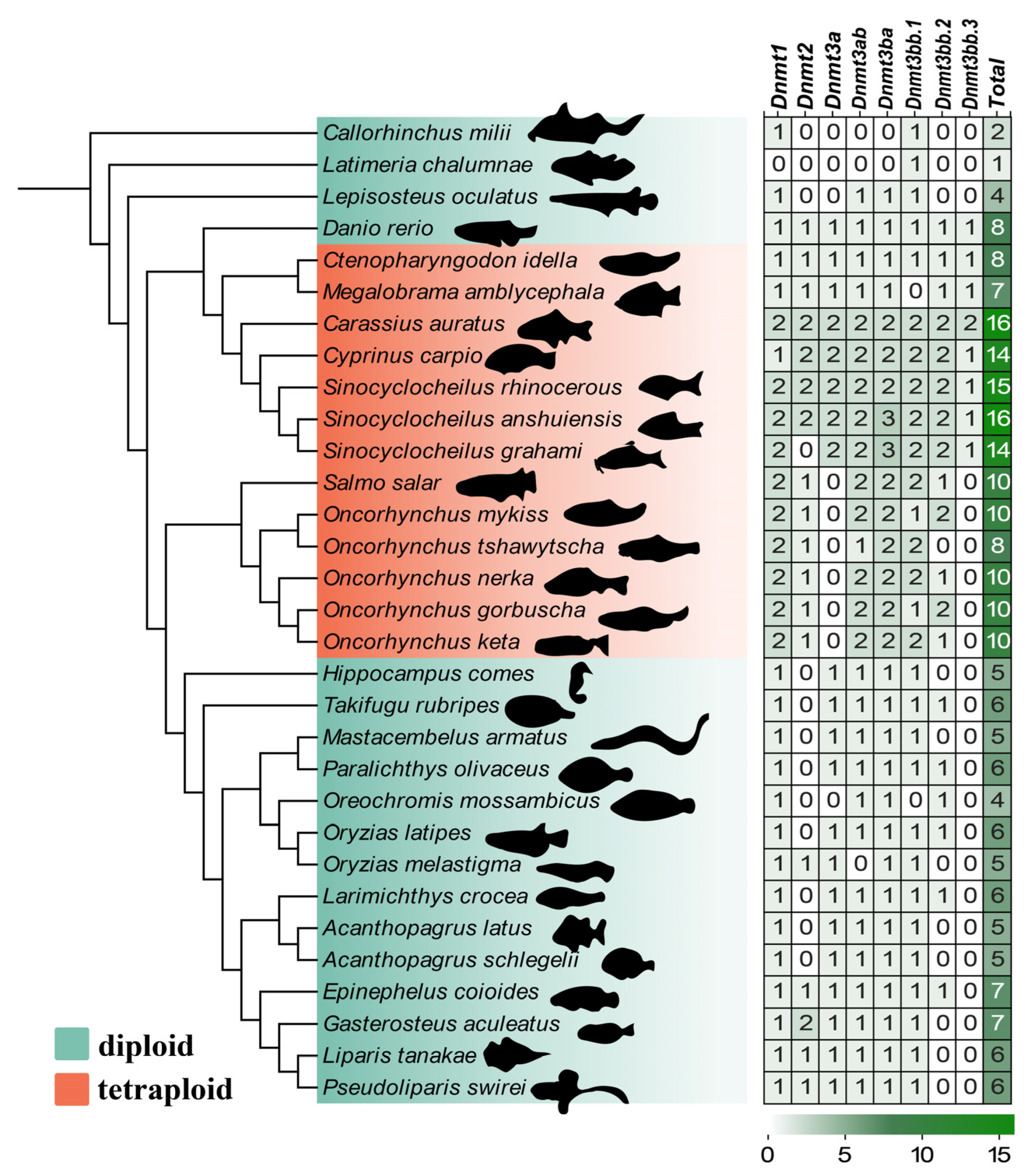
3.1. Genome-Wide Identification of the Dnmt Gene Family

In the present study, 242 Dnmt genes were identified in 31 species. We confirmed that the teleosts possessed 4 to 16 Dnmt copies (see more details in Figure 1). In diploid teleosts, their genomes have 4–8 Dnmt genes. Five candidate Dnmt genes were identified in the protandrous hermaphrodite A. schlegelii genome and designated as Dnmt1, Dnmt3bb.1, Dnmt3ab, Dnmt3ba, and Dnmt3aa. These genes were distributed across four chromosomes (chr 23, 7, 16, and 22), with Dnmt3bb.1 and Dnmt3ba sharing the same chromosomes. For the typical tetraploid teleost fishes, there are 7 to 16 Dnmt copies, such as river trout (Salmo trutta), rainbow trout (Oncorhynchus mykiss), and common carp (Cyprinus carpio), which underwent the fourth genome duplication.
Dnmt proteins ranged from 691 (Dnmt3aa) to 1508 (Dnmt1) amino acids in length. Their predicted molecular weights varied from 78.163 kDa (Dnmt3aa) to 170.325 kDa (Dnmt1), and all were classified as neutral (aliphatic index = 50–70) or hydrophobic (aliphatic index > 70). The predicted pI values ranged from 5.81 (Dnmt1) to 7.49 (Dnmt3bb.1). All five Dnmt proteins had an instability index of >40, indicating stability. Subcellular localization predictions suggested that all Dnmt members were primarily located in the nucleus and that Dnmt3ab and Dnmt3aa were also present in the cytoplasm (Table 2).

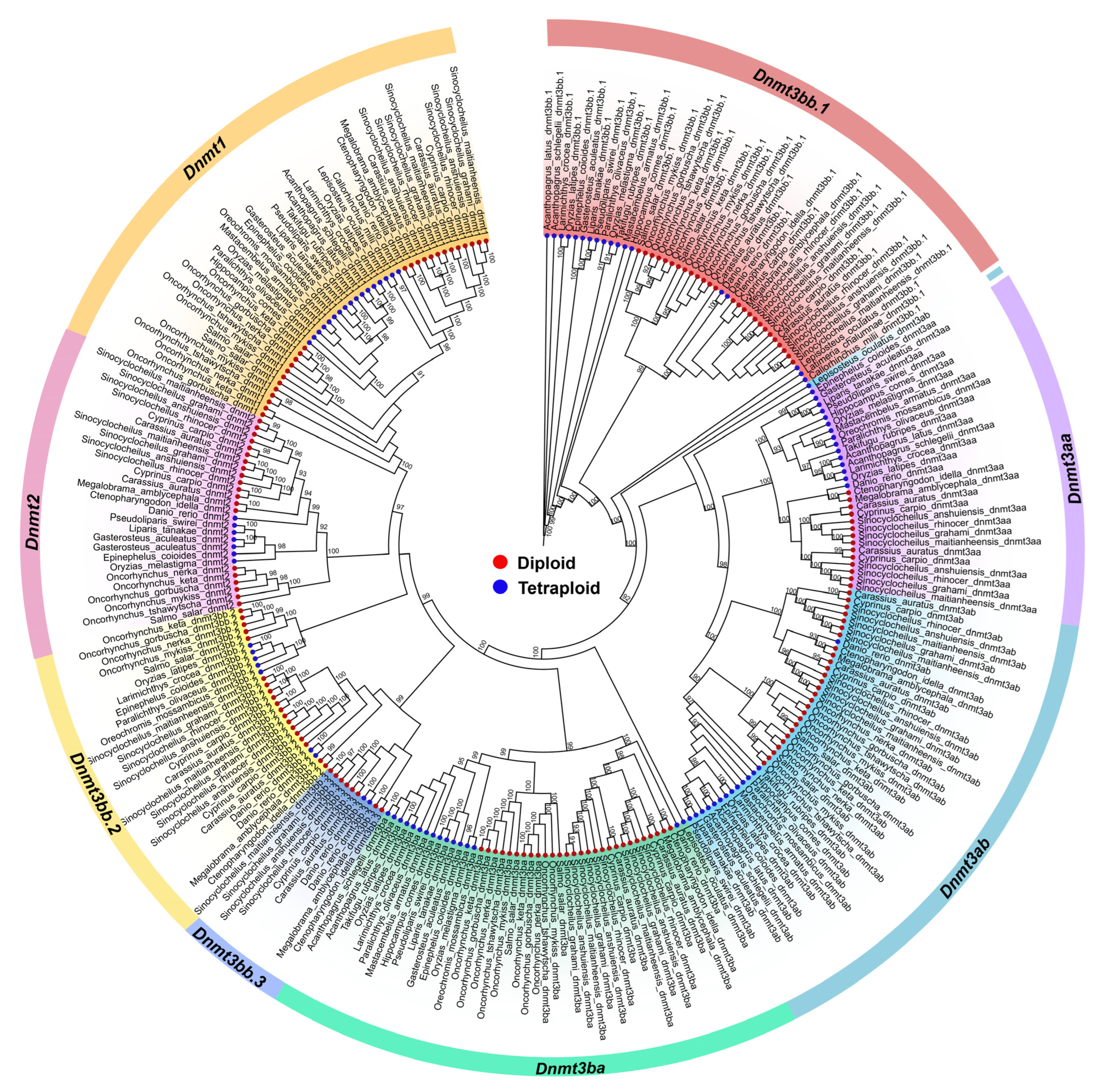
3.2. Phylogenetic Analysis of the Dnmt Family

A phylogenetic tree was constructed using Dnmt protein sequences from various fish species (Figure 2). Dnmt genes mainly clustered into eight groups (Dnmt1, Dnmt2, Dnmt3bb.1, Dnmt3bb.2, Dnmt3bb.3, Dnmt3ab, Dnmt3ba, and Dnmt3aa). Dnmt3bb.1, identified as a candidate gene shared among species that underwent two to three rounds of whole genome duplication (WGD) events early in vertebrate evolution (Callorhinchus milii, Latimeria chalumnae, and Lepisosteus oculatus) [42], formed the root of the phylogenetic tree (Figure 2). As shown in Figure 1, several species exhibited gene loss during evolution. Interestingly, Dnmt3bb.3 formed a distinct clade within Cyprinidae. Within each Dnmt subfamily, the Dnmt3a and Dnmt3b groups were generally clustered closely, suggesting similar biological functions consistent with previous studies [4,43]. In contrast, Dnmt1 was far more distantly related, indicating a functional divergence from Dnmt3a/3b.

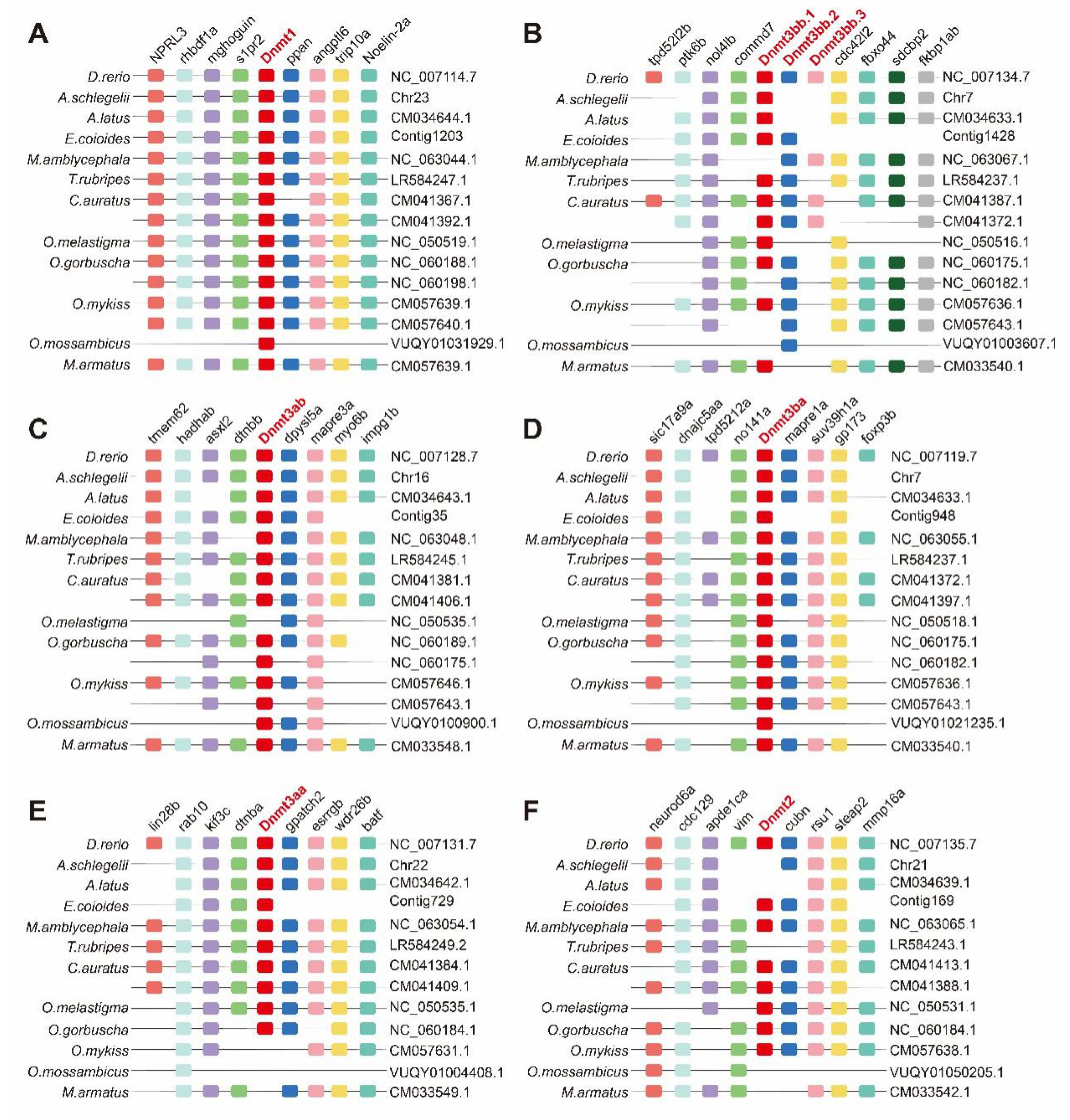
3.3. Synteny Analysis

To evaluate the synteny of conserved genes upstream and downstream of Dnmt in various fish species, we examined the collinear relationship between these conserved genes and Dnmt. As shown in Figure 3, Dnmt1 shared a conserved suite of flanking genes, whereas other Dnmt genes (Dnmt3bb.1, Dnmt3bb.2, Dnmt3bb.3, Dnmt3ab, Dnmt3ba, and Dnmt3aa) exhibited gene loss during evolution. O. mossambicus displayed extensive gene loss, including Dnmt genes and nearby loci, suggesting potential functional loss or compensation by other genes. Dnmt3bb.1, Dnmt3bb.2, and Dnmt3bb.3 were closely repeated on the same chromosome in some species such as D. rerio and C. auratus (Figure 3B). However, the partial loss of these genes and nearby loci was observed in other fish species. Among these fishes undergoing a fourth whole genome duplication event, C. auratus, O. gorbuscha, and O. mykiss possessed two copies of certain Dnmt genes (Dnmt1, Dnmt3bb.1, Dnmt3bb.2, Dnmt3bb.3, Dnmt3ab, and Dnmt3ba) on separate chromosomes (Figure 3A–D).

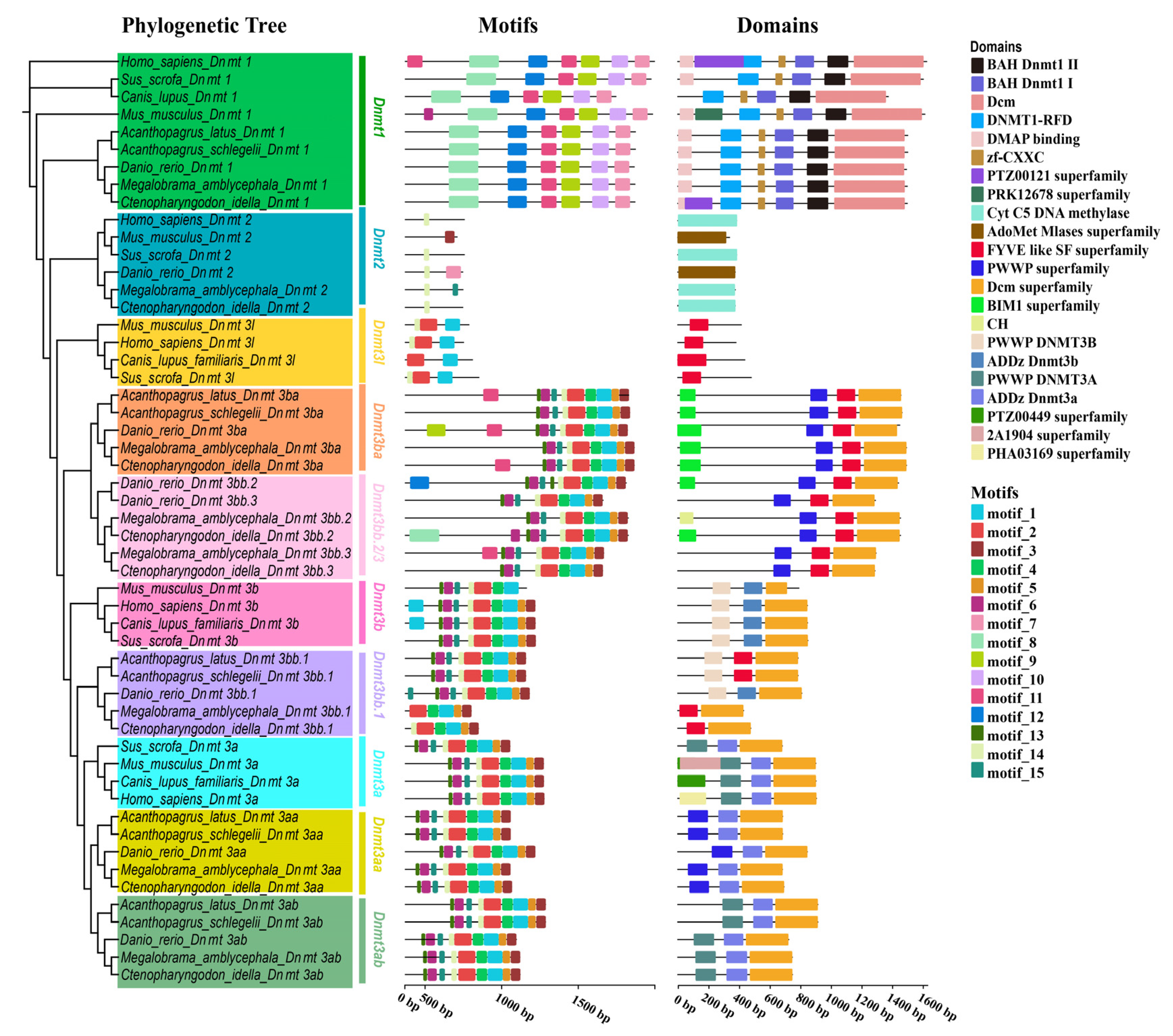
3.4. Structural Analysis of Dnmt Proteins

A phylogenetic tree was constructed using Dnmt proteins from four mammals (Homo sapiens, Mus musculus, Sus scrofa, and Canis lupus familiaris) and five bony fishes (Ctenopharyngodon idella, Megalobrama amblycephala, Danio rerio, Acanthopagrus schlegelii, and Acanthopagrus latus) (Figure 4). The analysis revealed that four Dnmt proteins (Dnmt1, Dnmt2, Dnmt3b, and Dnmt3a) were present in the four vertebrates, and five to eight Dnmt proteins were found in the five fish species. Shared motifs and domains showed slight differences among the species. All Dnmt1 proteins contained BAH_Dnmt1_I, BAH_Dnmt1_II, and Dcm domains, with conserved motifs 7–12. Dnmt2 proteins possessed motifs 3, 4, 7, and 14 and two domains (Cyt_C5_DNA_methylase and zf_CXCX). All Dnmt3 proteins (Dnmt3l, Dnmt3ba, Dnmt3bb.2, Dnmt3bb.3, Dnmt3a, Dnmt3aa, and Dnmt3ab) shared motifs 1–2. Except for Dnmt3l, the other Dnmt3 proteins contained motifs 1–5 and the Dcm superfamily. In summary, Dnmt proteins were classified into four types: Dnmt1, Dnmt2, Dnmt3l, and Dnmt3b/a. Within each type, the proteins exhibited highly similar structures, suggesting potentially similar biological functions.

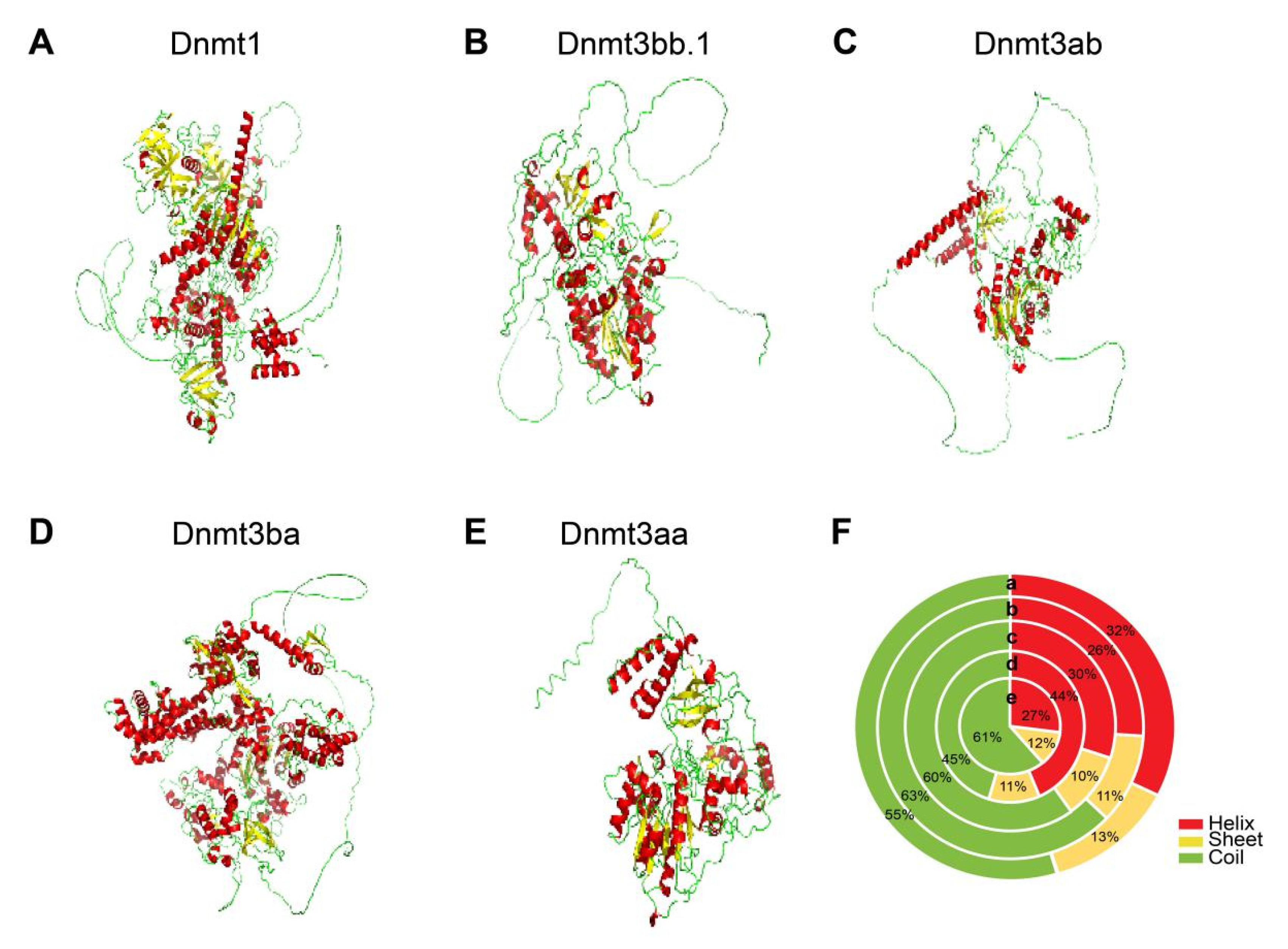
3.5. 3D Structures of Dnmts in Blackhead Seabream

The 3D structures of the five Dnmt proteins were predicted using AlphaFold3 (Figure 5). All modeled structures contained α-helices, β-sheets, and coils. The β-sheet content ranged from 10 to 13% for all five proteins. Except for β-sheet, Dnmt1 and Dnmt3ba showed a higher α-helix content and fewer coils, whereas Dnmt3bb.1, Dnmt3ab, and Dnmt3aa showed the opposite pattern.

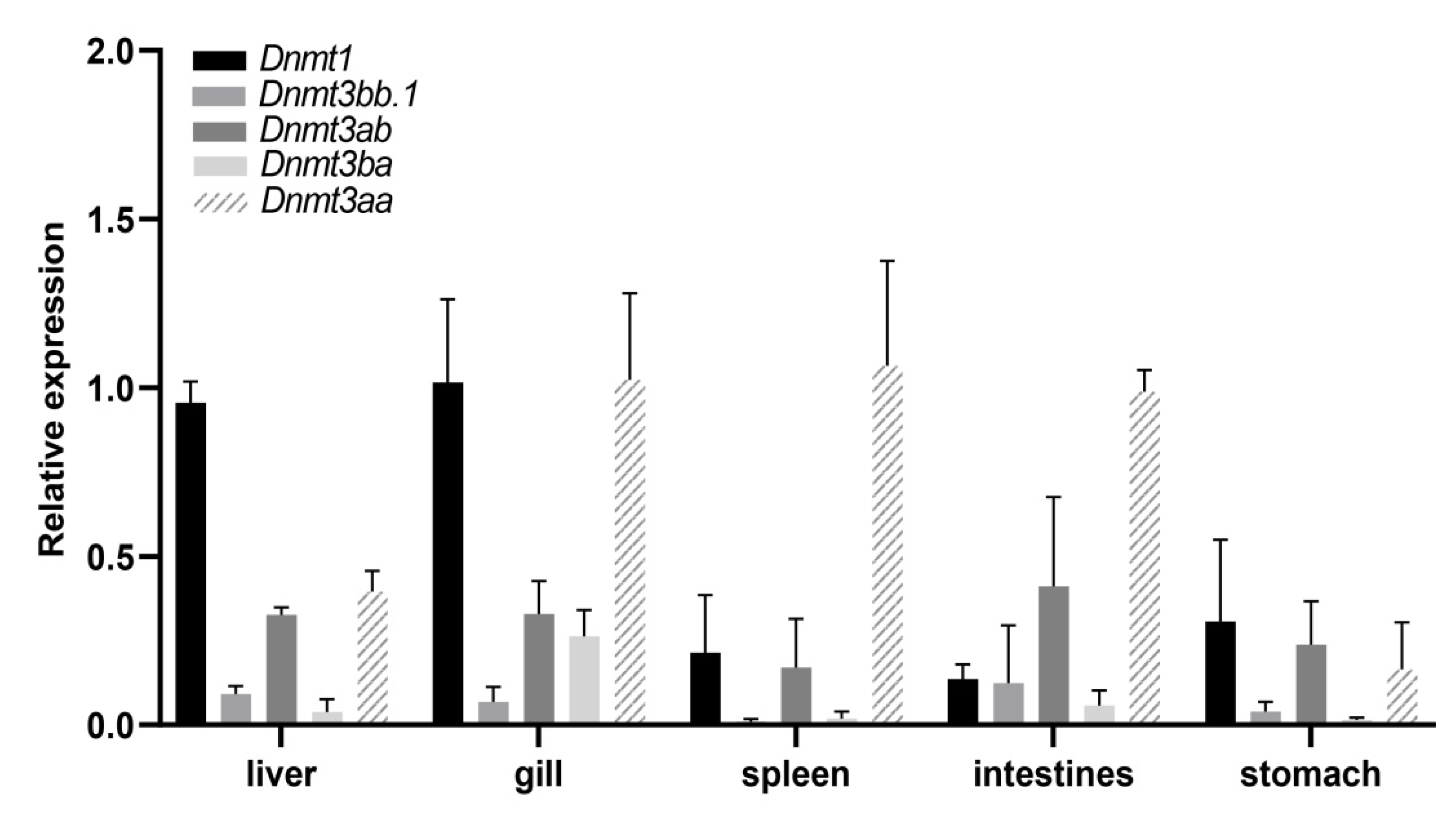
3.6. The Expression Profile of Dnmts in Different Tissues

The expression profile of Dnmt genes of blackhead seabream in five tissues (liver, gill, spleen, intestines, and stomach) was investigated by qRT-PCR analysis (Figure 6). Our results showed that the Dnmts were expressed in all the investigated tissues at varying levels. The Dnmt1 gene was highly expressed in the gill and liver but lowly expressed in the spleen, intestines, and stomach. The expression level of Dnmt3ba in the gill was higher than that in other tissues. Dnmt3ab was expressed in all five tissues at similar levels. Dnmt3bb.1 showed low expression across all five tissues. In addition, Dnmt3aa was highly expressed in the gills, spleen, and intestines but lowly expressed in the liver and stomach.

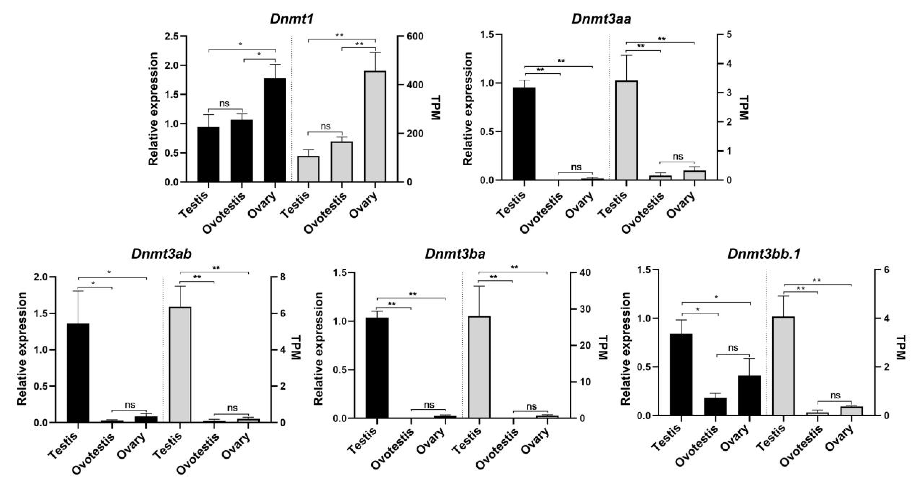
3.7. The Expression Profiles of Dnmts During the Natural Sex Change of Blackhead Seabream

We analyzed the expression profiles of dnmt genes during the natural sex change of blackhead seabream based on transcriptomic data and qPCR data. Our results demonstrated that these datasets generated by the two separate methods are typically in agreement (Figure 7). Intriguingly, all the Dnmt genes exhibited significant sex-biased expression patterns. Dnmt1 was significantly more highly expressed in the ovary compared to the ovotestis and testis (p < 0.01), whereas the other four Dnmt genes (Dnmt3bb.1, Dnmt3ab, Dnmt3ba, and Dnmt3aa) exhibited higher expression in the testis than in the other two developmental stages (p < 0.01). Notably, Dnmt1 displayed a progressive and significant upregulation in expression during the natural sex change of blackhead seabream. Moreover, Dnmt3ab, Dnmt3ba, and Dnmt3aa showed almost no expression in the ovotestis and ovary.

4. Discussion

Gene duplication not only plays a pivotal role in fish evolution but also provides raw material for functional innovation [17]. Two dominant mechanisms are considered to generate duplicated genes, including whole genome duplication (WGD) and tandem duplication resulting from unequal crossing over [44,45]. Mammals possess five Dnmt genes (Dnmt1, Dnmt2, Dnmt3l, Dnmt3a, and Dnmt3b) [6], whereas the investigated diploid fishes have up to eight Dnmt isoforms (Dnmt1, Dnmt2, Dnmt3aa, Dnmt3ab, Dnmt3ba, Dnmt3bb.1, Dnmt3bb.2, and Dnmt3bb.3), and tetraploid teleost fish like salmonids and certain cyprinids have even more copies of Dnmt genes resulting from fish-specific WGD events. Like mammals, most fish have only one Dnmt1 and one Dnmt2, while Dnmt3l is lost in the fish genome, suggesting that fish Dnmt1 and Dnmt2 are highly conserved during evolution. However, Dnmt3 underwent a lineage-specific evolution. Moreover, the ratio of the Dnmt gene number between diploids and tetraploids is not always 1:2, possibly due to selective gene loss. We analyzed gene duplication types among fish Dnmt genes and found that Dnmt3bb.1, Dnmt3bb.2, and Dnmt3bb.3 were tandemly and intrachromosomally duplicated, suggesting their origin of independent and continuous duplication. Our findings demonstrated the significance of tandem gene duplication as well as that of WGD in the course of Dnmt evolution, and copy number variations of the Dnmt gene likely resulted from WGD, tandem duplication and gene loss.
Duplicate genes typically follow two evolutionary trajectories: subfunctionalization (partitioning ancestral functions) or neofunctionalization (acquiring novel functions) [46]. For Dnmts, the presence of putative duplicates in most fish species suggests that some subfunctionalization or neofunctionalization occurred within the Dnmt family. Dnmt1 is localized to replicating DNA and heterochromatin through interactions with PCNA and UHRF1, as well as by direct binding to heterochromatic histone modifications H3K9me3 and H4K20me3 [41]. Dnmt3 enzymes bind to heterochromatin via protein multimerization and are targeted to chromatin by their ADD, PWWP, and UDR domains, which bind to unmodified H3K4, H3K36me2/3, and H2AK119ub1, respectively [41]. In the present study, differences in the number and type of domains were identified among fish Dnmt isotypes, potentially contributing to functional diversity and complexity. Furthermore, all the Dnmt3 duplications retained the ADD, PWWP, and Cyt_C5_DNA_methylase domains, suggesting similar functional capabilities. In mammals, de novo DNA methylation is mediated by Dnmt3a and Dnmt3b with the assistance of the stimulatory factor Dnmt3l [41]. Compared to mammals, the DNA methylation mechanism in fish is likely more complex because of the multiple Dnmt3 duplicates and the absence of Dnmt3l in their genome [45]. In mammals, the de novo methyltransferases Dnmt3a and Dnmt3b share similar structural compositions, both containing a PWWP domain, an ADD domain, and a C-terminal catalytic domain. In contrast, Dnmt3l lacks both the PWWP domain and the catalytic domain, while Dnmt1 possesses a C-terminal domain, two BAH domains, and a CXXC domain. Our findings demonstrate that mammalian Dnmt3 genes uniformly retain both PWWP and ADD domains, whereas teleost Dnmt3 and its homologs exhibit either one or both domains. This suggests lineage-specific diversification of Dnmt3 subfamily members during fish evolution, implying potential functional innovation or subfunctionalization in teleost DNA methylation catalysis. Interestingly, fish Dnmt3b duplicates contain the FYVE-like SF superfamily domain, which is the only domain present in mammalian Dnmt3l. We hypothesize that fish may have evolved a DNA methylation mechanism distinct from that of mammals, with Dnmt3 duplicates potentially replacing the function of mammalian Dnmt3l.
To better understand the functions of Dnmts in fish, we examined their distribution across various tissues in blackhead seabream. Dnmt1, Dnmt3aa, and Dnmt3bb genes were highly expressed in the liver, gill, spleen, intestine, and stomach, indicating their important roles in these tissues. Additionally, Dnmt copies exhibited different expression patterns among the examined tissues, which was consistent with previous studies in zebrafish [21] and tilapia [47]. These findings support the view that Dnmt copies have undergone subfunctionalization or neofunctionalization following duplication [48].
DNA methylation likely underwent remodeling at intermediate phases of sex change in the fish gonad, a process mediated by DNA methyltransferase [24]. In the present study, DNA methyltransferase genes exhibited a turnover in sex-specific expression during natural sex change in blackhead seabream gonads. Our results revealed that the maintenance methyltransferase gene Dnmt1 exhibited female-biased expression that significantly increased during the sex change from male to female, whereas de novo DNA methyltransferase genes (Dnmt3aa, Dnmt3ab, Dnmt3ba, and Dnmt3bb.1) have highest expression at the male stage in protandrous blackhead seabream. It was previously demonstrated that the sex transition from male to female in blackhead seabream occurred alongside a significant decline in cyp19a1a (a crucial female-related gene) methylation in the ovary [29,48]. Therefore, we speculated that the DNA methylation landscape in gonads was reconfigured as the expression of female-specific Dnmt superseded male-specific expression in blackhead seabream. A similarly distinct sex-specific expression pattern of Dnmt genes has recently been reported in gonadal transcriptomes of other teleosts undergoing sex change, whether through natural processes [24,27] or experimental induction [49,50]. Furthermore, earlier findings showed that changes in DNA methylation were closely related to sex change in sequentially hermaphroditic fishes, including protogynous orange-spotted grouper [51], rice-field eel [26,52], and protandrous barramundi [53]. Taken together, global reprogramming of gonadal DNA methylation, regulated by DNA methyltransferase genes, might be a convergent feature during sex change in hermaphroditic fishes.

5. Conclusions

This study offers a comprehensive analysis of the Dnmt gene family in fish and explores their roles in natural sex change in protandrous hermaphrodite blackhead seabream. We confirmed the presence of at least 21 Dnmt genes in fish, and a combination of WGD, tandem duplication, and gene loss was likely responsible for the diversity of Dnmt genes in fish. Differences were also observed in tissue distribution. Furthermore, protein sequence alignments and structural analysis of fish Dnmt duplicates supported their subfunctionalization or neofunctionalization. Our data revealed that Dnmt genes (Dnmt1, Dnmt3aa, Dnmt3ab, Dnmt3ba, and Dnmt3bb.1) possibly play important roles in the natural sex change process in blackhead seabream. We inferred that DNA methyltransferase genes likely regulated the remodeling of gonadal DNA methylation during sex change in sex-changing fishes. In summary, our findings will benefit further functional investigations of these fish genes.

Author Contributions

Conceptualization, L.D. and K.Z.; formal analysis, S.G., B.D. and K.Z.; funding acquisition, X.H., L.D. and K.Z.; investigation, K.Z.; project administration, L.D.; validation, S.G., B.D., J.C., M.C. and X.H.; visualization, S.G., B.D., J.C., M.C., C.Y., H.W. and K.Z.; writing—original draft, S.G.; writing—review and editing, L.D. and K.Z. All authors have read and agreed to the published version of the manuscript.

Funding

This research was funded by the Guangdong Basic and Applied Basic Research Foundation (no. 2023A1515110554), the Scientific Foundation for Youth Scholars of Shenzhen University (no. 000001032214 and no. 827-0001055), the National Key R&D Program of China (no. 2022YFE0139700), and the Shenzhen Science and Technology Program (no. 20231122124215001).

Institutional Review Board Statement

Not applicable.

Informed Consent Statement

Not applicable.

Data Availability Statement

The original contributions presented in this study are included in the article. Further inquiries can be directed to the corresponding authors.

Acknowledgments

We thank the Laboratory of Aquatic Genomics, College of Life Sciences and Oceanography, Shenzhen University, for providing the laboratory facilities essential to this study.

Conflicts of Interest

All authors declare no competing interests.

References

  1. Schubeler, D. Function and Information Content of DNA Methylation. Nature 2015, 517, 321–326. [Google Scholar] [CrossRef] [PubMed]
  2. Dura, M.; Teissandier, A.; Armand, M.; Barau, J.; Lapoujade, C.; Fouchet, P.; Bonneville, L.; Schulz, M.; Weber, M.; Baudrin, L.G.; et al. DNMT3A-Dependent DNA Methylation Is Required for Spermatogonial Stem Cells to Commit to Spermatogenesis. Nat. Genet. 2022, 54, 469–480. [Google Scholar] [CrossRef] [PubMed]
  3. Valdivieso, A.; Anastasiadi, D.; Ribas, L.; Piferrer, F. Development of Epigenetic Biomarkers for the Identification of Sex and Thermal Stress in Fish Using DNA Methylation Analysis and Machine Learning Procedures. Mol. Ecol. Resour. 2023, 23, 453–470. [Google Scholar] [CrossRef]
  4. Moore, L.D.; Le, T.; Fan, G. DNA Methylation and Its Basic Function. Neuropsychopharmacology 2013, 38, 23–38. [Google Scholar] [CrossRef]
  5. Dussoix, D.; Arber, W. Host Specificity of DNA Produced by Escherichia coli. II. Control over Acceptance of DNA from Infecting Phage Lambda. J. Mol. Biol. 1962, 5, 37–49. [Google Scholar] [CrossRef]
  6. Jurkowska, R.Z.; Jurkowski, T.P.; Jeltsch, A. Structure and Function of Mammalian DNA Methyltransferases. ChemBioChem 2011, 12, 206–222. [Google Scholar] [CrossRef] [PubMed]
  7. Okano, M.; Bell, D.W.; Haber, D.A.; Li, E. DNA Methyltransferases Dnmt3a and Dnmt3b Are Essential for de Novo Methylation and Mammalian Development. Cell 1999, 99, 247–257. [Google Scholar] [CrossRef]
  8. Kim, B.M.; Mirbahai, L.; Mally, A.; Chipman, J.K.; Rhee, J.S.; Lee, J.S. Correlation between the DNA Methyltransferase (Dnmt) Gene Family and Genome-Wide 5-Methylcytosine (5mC) in Rotifer, Copepod, and Fish. Genes Genom. 2016, 38, 13–23. [Google Scholar] [CrossRef]
  9. Lyko, F. The DNA Methyltransferase Family: A Versatile Toolkit for Epigenetic Regulation. Nat. Rev. Genet. 2018, 19, 81–92. [Google Scholar] [CrossRef]
  10. Wolfe, K.H. Yesterday’s Polyploids and the Mystery of Diploidization. Nat. Rev. Genet. 2001, 2, 333–341. [Google Scholar] [CrossRef]
  11. Ahn, D.; You, K.H.; Kim, C.H. Evolution of the Tbx6/16 Subfamily Genes in Vertebrates: Insights from Zebrafish. Mol. Biol. Evol. 2012, 29, 3959–3983. [Google Scholar] [CrossRef] [PubMed]
  12. Voldoire, E.; Brunet, F.; Naville, M.; Volff, J.N.; Galiana, D. Expansion by Whole Genome Duplication and Evolution of the Sox Gene Family in Teleost Fish. PLoS ONE 2017, 12, e0180936. [Google Scholar] [CrossRef] [PubMed]
  13. Ravi, V.; Venkatesh, B. Rapidly Evolving Fish Genomes and Teleost Diversity. Curr. Opin. Genet. Dev. 2008, 18, 544–550. [Google Scholar] [CrossRef] [PubMed]
  14. Glasauer, S.M.; Neuhauss, S.C. Whole-Genome Duplication in Teleost Fishes and Its Evolutionary Consequences. Mol. Genet. Genom. 2014, 289, 1045–1060. [Google Scholar] [CrossRef]
  15. Van de Peer, Y.; Maere, S.; Meyer, A. The Evolutionary Significance of Ancient Genome Duplications. Nat. Rev. Genet. 2009, 10, 725–732. [Google Scholar] [CrossRef]
  16. Ravi, V.; Venkatesh, B. The Divergent Genomes of Teleosts. Annu. Rev. Anim. Biosci. 2018, 6, 47–68. [Google Scholar] [CrossRef]
  17. Berthelot, C.; Brunet, F.; Chalopin, D.; Juanchich, A.; Bernard, M.; Noel, B.; Bento, P.; Da Silva, C.; Labadie, K.; Alberti, A.; et al. The Rainbow Trout Genome Provides Novel Insights into Evolution after Whole-Genome Duplication in Vertebrates. Nat. Commun. 2014, 5, 3657. [Google Scholar] [CrossRef] [PubMed]
  18. Xu, P.; Zhang, X.; Wang, X.; Li, J.; Liu, G.; Kuang, Y.; Xu, J.; Zheng, X.; Ren, L.; Wang, G.; et al. Genome Sequence and Genetic Diversity of the Common Carp, Cyprinus carpio. Nat. Genet. 2014, 46, 1212–1219. [Google Scholar] [CrossRef]
  19. Chen, D.; Zhang, Q.; Tang, W.; Huang, Z.; Wang, G.; Wang, Y.; Shi, J.; Xu, H.; Lin, L.; Li, Z.; et al. The Evolutionary Origin and Domestication History of Goldfish (Carassius auratus). Proc. Natl. Acad. Sci. USA 2020, 117, 29775–29785. [Google Scholar] [CrossRef]
  20. Guyomard, R.; Boussaha, M.; Krieg, F.; Hervet, C.; Quillet, E. A Synthetic Rainbow Trout Linkage Map Provides New Insights into the Salmonid Whole Genome Duplication and the Conservation of Synteny among Teleosts. BMC Genet. 2012, 13, 15. [Google Scholar] [CrossRef]
  21. Smith, T.H.; Collins, T.M.; McGowan, R.A. Expression of the Dnmt3 Genes in Zebrafish Development: Similarity to Dnmt3a and Dnmt3b. Dev. Genes Evol. 2011, 220, 347–353. [Google Scholar] [CrossRef] [PubMed]
  22. Zhou, R.; Shang, R.J.; Gong, D.B.; Xu, X.J.; Tang, Q.R.; Tao, M.; Zhao, R.R.; Liu, S.J. Characterization of De Novo DNA Methyltransferase Dnmt3 Regulating Sterility in Female Allotriploid Fish. Aquaculture 2019, 504, 345–353. [Google Scholar] [CrossRef]
  23. Sun, L.N.; Jiang, X.L.; Xie, Q.P.; Yuan, J.; Huang, B.F.; Tao, W.J.; Zhou, L.Y.; Nagahama, Y.; Wang, D.S. Transdifferentiation of Differentiated Ovary into Functional Testis by Long-Term Treatment of Aromatase Inhibitor in Nile Tilapia. Endocrinology 2014, 155, 1476–1488. [Google Scholar] [CrossRef] [PubMed]
  24. Todd, E.V.; Ortega-Recalde, O.; Liu, H.; Lamm, M.S.; Rutherford, K.M.; Cross, H.; Black, M.A.; Kardailsky, O.; Marshall Graves, J.A.; Hore, T.A.; et al. Stress, Novel Sex Genes, and Epigenetic Reprogramming Orchestrate Socially Controlled Sex Change. Sci. Adv. 2019, 5, eaaw7006. [Google Scholar] [CrossRef]
  25. Warner, R.R.; Swearer, S.E. Social Control of Sex Change in the Bluehead Wrasse, Thalassoma bifasciatum (Pisces: Labridae). Biol. Bull. 1991, 181, 199–204. [Google Scholar] [CrossRef] [PubMed]
  26. Wang, X.; Lai, F.; Xiong, J.; Zhu, W.; Yuan, B.; Cheng, H.; Zhou, R. DNA Methylation Modification Is Associated with Gonadal Differentiation in Monopterus albus. Cell Biosci. 2020, 10, 129. [Google Scholar] [CrossRef]
  27. Zhang, Y.; Sun, X.; Zhang, L.; Zhang, W. Testicular Dnmt3 Expression and Global DNA Methylation Are Down-Regulated by Gonadotropin Releasing Hormones in the Ricefield Eel Monopterus albus. Sci. Rep. 2017, 7, 43158. [Google Scholar] [CrossRef]
  28. Zhang, K.; Xu, J.; Zhang, Z.; Huang, Y.; Ruan, Z.; Chen, S.; Zhu, F.; You, X.; Jia, C.; Meng, Q.; et al. A Comparative Transcriptomic Study on Developmental Gonads Provides Novel Insights into Sex Change in the Protandrous Black Porgy (Acanthopagrus Schlegelii). Genomics 2019, 111, 277–283. [Google Scholar] [CrossRef]
  29. Wu, G.C.; Dufour, S.; Chang, C.F. Molecular and Cellular Regulation on Sex Change in Hermaphroditic Fish, with a Special Focus on Protandrous Black Porgy, Acanthopagrus schlegelii. Mol. Cell. Endocrinol. 2021, 520, 111069. [Google Scholar] [CrossRef]
  30. Zhang, K.; Guo, S.; Yang, S.; Zhou, W.; Wu, J.; Zhang, X.; Shi, Q.; Deng, L. A Telomere-to-Telomere Genome Assembly of the Protandrous Hermaphrodite Blackhead Seabream, Acanthopagrus schlegelii. Sci. Data 2025, 12, 350. [Google Scholar] [CrossRef]
  31. Birney, E.; Clamp, M.; Durbin, R. GeneWise and Genomewise. Genome Res. 2004, 14, 988–995. [Google Scholar] [CrossRef] [PubMed]
  32. Katoh, K.; Standley, D.M. MAFFT Multiple Sequence Alignment Software Version 7: Improvements in Performance and Usability. Mol. Biol. Evol. 2013, 30, 772–780. [Google Scholar] [CrossRef]
  33. Nguyen, L.T.; Schmidt, H.A.; von Haeseler, A.; Minh, B.Q. IQ-TREE: A Fast and Effective Stochastic Algorithm for Estimating Maximum-Likelihood Phylogenies. Mol. Biol. Evol. 2015, 32, 268–274. [Google Scholar] [CrossRef] [PubMed]
  34. Bailey, T.L.; Boden, M.; Buske, F.A.; Frith, M.; Grant, C.E.; Clementi, L.; Ren, J.; Li, W.W.; Noble, W.S. MEME SUITE: Tools for Motif Discovery and Searching. Nucleic Acids Res. 2009, 37, W202–W208. [Google Scholar] [CrossRef]
  35. Camacho, C.; Coulouris, G.; Avagyan, V.; Ma, N.; Papadopoulos, J.; Bealer, K.; Madden, T.L. BLAST+: Architecture and Applications. BMC Bioinform. 2009, 10, 421. [Google Scholar] [CrossRef]
  36. Paysan-Lafosse, T.; Blum, M.; Chuguransky, S.; Grego, T.; Pinto, B.L.; Salazar, G.A.; Bileschi, M.L.; Bork, P.; Bridge, A.; Colwell, L.; et al. InterPro in 2022. Nucleic Acids Res. 2023, 51, D418–D427. [Google Scholar] [CrossRef] [PubMed]
  37. Chen, C.; Chen, H.; Zhang, Y.; Thomas, H.R.; Frank, M.H.; He, Y.; Xia, R. TBtools: An Integrative Toolkit Developed for Interactive Analyses of Big Biological Data. Mol. Plant 2020, 13, 1194–1202. [Google Scholar] [CrossRef]
  38. Chen, L.; Li, Q.; Nasif, K.F.A.; Xie, Y.; Deng, B.; Niu, S.; Pouriyeh, S.; Dai, Z.; Chen, J.; Xie, C.Y. AI-Driven Deep Learning Techniques in Protein Structure Prediction. Int. J. Mol. Sci. 2024, 25, 8426. [Google Scholar] [CrossRef]
  39. Rosignoli, S.; Paiardini, A. Boosting the Full Potential of PyMOL with Structural Biology Plugins. Biomolecules 2022, 12, 1764. [Google Scholar] [CrossRef]
  40. Livak, K.J.; Schmittgen, T.D. Analysis of Relative Gene Expression Data Using Real-Time Quantitative PCR and the 2(-Delta Delta C(T)) Method. Methods 2001, 25, 402–408. [Google Scholar] [CrossRef]
  41. Abera, D.A.; Larbie, C.; Abugri, J.; Ofosu, M.; Mutocheluh, M.; Dongsogo, J. Prevalence and Predictors of Gestational Diabetes Mellitus in Sub-Saharan Africa: A 10-Year Systematic Review. Endocrinol. Diabetes Metab. 2024, 7, e00478. [Google Scholar] [CrossRef]
  42. Zhang, K.; Chen, M.; He, H.; Kou, H.; Lin, L.; Liang, R. Genome-Wide Identification and Characterization of Toll-like Receptor 5 (TLR5) in Fishes. Front. Genet 2022, 13, 1083578. [Google Scholar] [CrossRef] [PubMed]
  43. Jurkowska, R.Z.; Jeltsch, A. Enzymology of Mammalian DNA Methyltransferases. Adv. Exp. Med. Biol. 2022, 1389, 69–110. [Google Scholar] [CrossRef] [PubMed]
  44. Naseeb, S.; Ames, R.M.; Delneri, D.; Lovell, S.C. Rapid Functional and Evolutionary Changes Follow Gene Duplication in Yeast. Proc. Biol. Sci. 2017, 284, 20171393. [Google Scholar] [CrossRef]
  45. Maere, S.; De Bodt, S.; Raes, J.; Casneuf, T.; Van Montagu, M.; Kuiper, M.; Van de Peer, Y. Modeling Gene and Genome Duplications in Eukaryotes. Proc. Natl. Acad. Sci. USA 2005, 102, 5454–5459. [Google Scholar] [CrossRef]
  46. Liu, Y.; Li, M.; Zhang, M.; Yang, Z.; Chen, X.; Wu, X. Evolution and Expression Analysis of Carotenoid Cleavage Oxygenase Gene Family in Chinese Mitten Crab Eriocheir sinensis. Int. J. Biol. Macromol. 2024, 257, 128475. [Google Scholar] [CrossRef] [PubMed]
  47. Yang, Z.; Nielsen, R. Codon-Substitution Models for Detecting Molecular Adaptation at Individual Sites along Specific Lineages. Mol. Biol. Evol. 2002, 19, 908–917. [Google Scholar] [CrossRef]
  48. Wu, G.C.; Li, H.W.; Huang, C.H.; Lin, H.J.; Lin, C.J.; Chang, C.F. The Testis Is a Primary Factor That Contributes to Epigenetic Modifications in the Ovaries of the Protandrous Black Porgy, Acanthopagrus schlegelii. Biol. Reprod. 2016, 94, 132. [Google Scholar] [CrossRef]
  49. Wang, F.L.; Yan, L.X.; Shi, H.J.; Liu, X.Y.; Zheng, Q.Y.; Sun, L.N.; Wang, D.S. Genome-Wide Identification, Evolution of DNA Methyltransferases and Their Expression during Gonadal Development in Nile Tilapia. Comp. Biochem. Physiol. B Biochem. Mol. Biol. 2018, 226, 73–84. [Google Scholar] [CrossRef]
  50. Wang, F.; Qin, Z.; Li, Z.; Yang, S.; Gao, T.; Sun, L.; Wang, D. Dnmt3aa but Not Dnmt3ab Is Required for Maintenance of Gametogenesis in Nile Tilapia (Oreochromis niloticus). Int. J. Mol. Sci. 2021, 22, 10170. [Google Scholar] [CrossRef]
  51. Guo, C.Y.; Tseng, P.W.; Hwang, J.S.; Wu, G.C.; Chang, C.F. Potential Role of DNA Methylation of Cyp19a1a Promoter during Sex Change in Protogynous Orange-Spotted Grouper, Epinephelus coioides. Gen. Comp. Endocrinol. 2021, 311, 113840. [Google Scholar] [CrossRef] [PubMed]
  52. Hu, Q.; Lian, Z.; Xia, X.; Tian, H.; Li, Z. Integrated Chromatin Accessibility and DNA Methylation Analysis to Reveal the Critical Epigenetic Modification and Regulatory Mechanism in Gonadal Differentiation of the Sequentially Hermaphroditic Fish, Monopterus albus. Biol. Sex Differ. 2022, 13, 73. [Google Scholar] [CrossRef] [PubMed]
  53. Domingos, J.A.; Budd, A.M.; Banh, Q.Q.; Goldsbury, J.A.; Zenger, K.R.; Jerry, D.R. Sex-Specific Dmrt1 and Cyp19a1 Methylation and Alternative Splicing in Gonads of the Protandrous Hermaphrodite Barramundi. PLoS ONE 2018, 13, e0204182. [Google Scholar] [CrossRef] [PubMed]
Figure 1. The number of Dnmt family members in 31 vertebrate species genomes was compared between diploid species (green in the phylogenomic tree) and tetraploid species (red in the phylogenomic tree). The higher the number of Dnmt genes, the darker the histogram color.
Biomolecules 15 00896 g001
Figure 2. Phylogenetic tree of the Dnmt gene family in 31 fish species. Red and blue dots indicate diploid and tetraploid species, respectively. Orange, pink, yellow, blue, green, light blue, purple and red colors represent subfamilies Dnmt1, Dnmt2, Dnmt3bb.2, Dnmt3bb.3, Dnmt3ba, Dnmt3ab, Dnmt3aa, and Dnmt3bb.1, respectively.
Biomolecules 15 00896 g002
Figure 3. Syntenic analysis of chromosomal loci containing Dnmt genes in various fish species. Colored rectangles represent distinct loci, with some gene loci adjacent to Dnmt showing conserved synteny across the examined species. Panels (AF) correspond to dnmt1, dnmt3bb, dnmt3ab, dnmt3ba, dnmt3aa, and dnmt2, respectively. In panels (A,CF), red blocks represent different dnmt subtypes. In panel (B), the red, blue, and pink rectangles represent dnmt3bb.1, dnmt3bb.2, and dnmt3bb.3, respectively. Species names are shown on the left, and chromosome numbers are labeled on the right. (D. rerio, Danio rerio; A. schlegelii, Acanthopagrus schlegelii; A. latus, Acanthopagrus latus; E. coioides, Epinephelus coioides; M. amblycephala, Megalobrama amblycephala; T. rubripes, Takifugu rubripes; C. auratus, Carassius auratus; O. melastigma, Oryzias melastigma; O. gorbuscha, Oncorhynchus gorbuscha; O. mykiss, Oncorhynchus mykiss; O. mossambicus, Oreochromis mossambicus; M. amatus, Mastacembelus armatus).
Biomolecules 15 00896 g003
Figure 4. Phylogenetic relationships, gene structures, and motifs of Dnmt genes.
Biomolecules 15 00896 g004
Figure 5. The 3D structure modeling of Dnmt proteins. The red color represents α-helix, yellow represents β-sheet, and green represents coil. The protein structures were predicted by AlphaFhold3, and the structure image was generated using the pymol software (AE). The pie chart shows the statistical proportion of protein secondary structure (F) (a. Dnmt1, b. Dnmt3bb.1. c. Dnmt3ab, d. Dnmt3ba, and e. Dnmt3aa).
Biomolecules 15 00896 g005
Figure 6. Expression profiles of Dnmt genes in different tissues of A. schlegelii were analyzed by quantitative real-time PCR (qRT-PCR). Relative expression levels of Dnmt genes in the liver, gill, spleen, intestines and stomach, with β-actin as an internal control.
Biomolecules 15 00896 g006
Figure 7. The expression patterns in testis, ovotestis, and ovary of A. schlegelii based on qRT-PCR (left) and RNA-seq (right). Column charts and significance analysis were constructed by Prism 8.0. The * indicates significant differences by one-way ANOVA (ns: not significant, p > 0.05; * p  <  0.05; and ** p  <  0.01; n = 3).
Biomolecules 15 00896 g007
Table 1. The primer sequence used for qPCR.
PrimerForward Primer (5′-3′)Reverse Primer (5′-3′)
Dnmt1TCCCACAGCACAAGATTACAAGGAACACCACCATCCAAGC
Dnmt3bb.1CCGTTCTTCTGGCTGTTCGTTCTGGGAGGCTGTGATGG
Dnmt3abATGTCAGCCTTGAGCACCCGTCGTCCTCCGCAGCAGATAG
Dnmt3baCCCTGGCATGAACAGACCCCCATCTTGCCCTGCCGTAT
Dnmt3aaCTCCTGCGGAAGCCTCAATCTGGTAGCCGTCATCGTCAT
β-actinACAGGGAGAAGATGACCCAGATCACCGGAGTCCATGACGATA
Table 2. The Dnmt physical and chemical properties in blackhead seabream.
ProteinChrNumber of Amino AcidsMolecular WeightTheoretical pIInstability IndexAliphatic IndexGRAVYPredicted Location
Dnmt1231508170,325.945.8147.9565.79−0.701Nucleus
Dnmt3bb.1779289,338.447.4946.7370.29−0.509Nucleus
Dnmt3ab16921104,840.847.2461.3163.6−0.72Nucleus, cytoplasm
Dnmt3ba71471166,048.656.8744.8976.51−0.404Nucleus
Dnmt3aa2269178,163.046.0551.9369.29−0.431Nucleus, cytoplasm
Disclaimer/Publisher’s Note: The statements, opinions and data contained in all publications are solely those of the individual author(s) and contributor(s) and not of MDPI and/or the editor(s). MDPI and/or the editor(s) disclaim responsibility for any injury to people or property resulting from any ideas, methods, instructions or products referred to in the content.

