Article

Rapid Biochemical Analysis of Postmortem Serum and Myocardial Homogenates—An Exploratory Study

1
Swedish National Board of Forensic Medicine, 17165 Solna, Sweden
2
Department of Oncology-Pathology, Karolinska Institutet, Nobels v, 15A, 17177 Stockholm, Sweden
3
Division of Clinical Chemistry and Pharmacology, Department of Biomedical and Clinical Sciences, Faculty of Medicine, Linköping University, 58185 Linköping, Sweden
*
Author to whom correspondence should be addressed.
Biomolecules 2025, 15(10), 1483; https://doi.org/10.3390/biom15101483
Submission received: 13 September 2025 / Revised: 11 October 2025 / Accepted: 17 October 2025 / Published: 21 October 2025
(This article belongs to the Special Issue Molecular Biomarkers in Cardiology 2025)

Abstract

Postmortem diagnosis of sudden cardiac death (SCD) may escape detection due to the absence of thrombi and slow development of structural and immunohistochemical changes. Therefore, this study explores the possibility of analyzing relevant clinical chemistry biomarkers in myocardial homogenates and serum. Following an initial pilot study, myocardial samples from 113 autopsy cases were homogenized with distilled water, T-PER or 2 M urea. Aspartate aminotransferase (AST), alanine aminotransferase (ALT), creatine kinase (CK-MB), lactate dehydrogenase (LDH), orosomucoid and total protein were analyzed with an IndikoPlus and a subset was also analyzed with a Roche Cobas 8000 c701 analyzer, which also provided results for cardiac Troponin T, myoglobin and NT-proBNP. Although the yields varied with different extraction buffers depending on the analyte, distilled water was often as effective as T-PER and 2 M urea extraction for most analytes. Biomarker levels were consistently higher in the myocardial homogenates than in serum. Proteomic profiling on a subset confirmed higher concentrations of the cardiac markers in the tissue samples than in serum. Finally, we investigated whether selected markers could support the diagnosis of acute cardiac disease by classifying cases as sudden cardiac death (SCD) or controls. There was no significant difference in serum concentrations of the selected biomarkers between SCD cases and controls, whereas a significant loss of several markers was observed in SCD myocardial samples as compared to controls. Hence, our results suggest that analysis of tissue homogenates is likely better for detecting early ischemia, and we show that an in-house benchtop multi-analyzer can provide rapid results to assist the pathologist’s decision-making during autopsy.

1. Introduction

An autopsy has long been considered the gold standard for identifying the cause of death [1]. However, traditional autopsy work still focuses on morphological changes, which may fail to identify certain acute medical conditions that follow a rapid course since structural changes require time to develop [2]. In contrast to structural alterations, many biochemical changes occur rapidly, either through translocation of proteins and other molecules within cells and tissues or by leakage from damaged tissue into the bloodstream [3]. Clinical chemistry is used extensively in primary care and hospitals to detect biochemical changes, but access to tissue samples from living subjects is limited; therefore, reference levels in serum/plasma or other accessible fluids are used as proxies. Postmortem biochemistry has been studied for decades but remains underutilized in practical casework [4] and published biomarker levels are exclusively from body fluids, mainly serum, pericardial fluid, cerebrospinal fluid, and vitreous fluid. In addition, rapid analysis with point-of-care instruments may aid decision-making during autopsy [5,6]. While various options exist, including spot tests and handheld analyzers, these often analyze only one marker at a time [7,8]. Our focus is on benchtop analysis to provide rapid results for multiple markers simultaneously. That said, there is a need for more analytical tools to aid postmortem diagnostics, and naturally not all need to provide instant results; rather, some have to be very accurate, particularly if the results may be presented as evidence in court. Figure 1 represents a schematic illustration of a strategy to improve future autopsy work. In this article we focus on rapid analysis that can provide leads to the pathologist regarding which investigative tracks may be important to follow and which ones are not supported by quick tests.
At both clinical and forensic pathology departments, many deaths are unwitnessed and recent medical information is often lacking. A proportion of these cases are suspected sudden cardiac deaths (SCD) [9]. SCD is defined as an unexpected natural death from a cardiac cause occurring within a short time, usually within one hour from symptom onset [10,11,12]. In some cases, acute myocardial ischemia (AMI) can be detected macro- or microscopically, but if the survival time is short, these changes may not be visible [13]. In other instances, an unwitnessed acute cardiac death due to arrhythmia may not be detectable at all, and acute heart failure may also escape detection by traditional autopsy methods [14].
Clinical chemistry diagnostic tests are generally developed for living subjects, and their performance on postmortem samples requires further investigation. A study on marker combinations recently reported an excellent prediction of acute myocardial ischemia by analyzing a combination of biomarkers [15], but these results have not yet been confirmed by other studies. Since all organs are accessible at autopsy, early-phase changes are more likely to be detected by analyzing the affected tissue directly. We investigated the performance of a benchtop instrument, Indiko™ Plus (Thermo Fisher Scientific, Vantaa, Finland), on cases of SCD and deaths from other known causes. Due to its analytical principles, not all desired cardiac ischemia markers can be analyzed with this instrument, such as cTnT and myoglobin. We selected AST, ALT, CK-MB, and LDH, all of which will show changes upon cardiac ischemia. Using a different instrument, cTnT, myoglobin, and NT-proBNP were also analyzed in a subset of cases. The main objectives of this study were to determine: (a) whether the Indiko instrument can provide reliable results from postmortem serum and myocardial homogenates; (b) whether the degree of arteriosclerosis in the supporting artery is related to biomarker concentrations in the corresponding myocardial samples; (c) whether postmortem interval and cardiac massage affect biomarker levels; and (d) whether freeze-thawing of tissue causes changes in biomarker levels. Most importantly, we investigated whether certain preanalytical treatments could increase biomarker yields from myocardial homogenates and whether removal of hemoglobin from serum could improve analytical results in hemolyzed samples. Additionally, we aimed to compare the performance of the Indiko instrument with a standard clinical chemistry multi-analyzer, and to assess whether the selected biomarkers could discriminate SCD cases from those with other causes of death.

2. Materials and Methods

2.1. Study Design and Study Population

Two prospective, explorative studies were conducted from spring 2023 through spring 2024. First, a pilot study was performed on 43 autopsy subendocardial samples collected at the Department of Forensic Medicine in Stockholm, comprising 35 males and 9 females with an average age of 57.0 years (Table 1).
The main study was based on a separate cohort of 113 autopsy cases, from which subendocardial samples from the mid posterior wall of the left ventricle were collected at the same department, comprising 83 males and 30 females; average age 56.7 years (Table 2). In 91 of these (64 males and 27 females), a sample from the mid anterior wall was also collected.
It should be noted that both cohorts included cases with a variety of causes of death and several of them with additional medical conditions that in most cases were not considered to have contributed to death. This means we used an unselected sample for the studies of different preanalytical treatments to increase the likelihood that the results would be as generally applicable as possible to a typical forensic or clinical pathology autopsy population. Inclusion criteria for evaluating different extraction conditions were based solely on body freshness, i.e., absence of decomposition (although limited greenish discoloration of the skin on the lower abdomen was accepted). Hence, the main purpose of this study was to explore the possibility of analyzing available clinical chemistry markers in tissue homogenates, i.e., where a particular pathology may be present. Since we focused on myocardium, we selected cardiac markers that have been used for decades, although developed for blood (plasma or serum) samples, to determine whether these could be analyzed with a benchtop clinical chemistry multi-analyzer.
To further investigate if any of these markers may show a change in the myocardium before a rise in serum, we classified cases as SCD or control (or not included in any of these groups). Inclusion criteria for SCD were a cardiac cause of death determined by the forensic pathologist and circumstances supporting a cardiac death. Circumstances supporting SCD include cases where the subject had reported chest pain and patients with a known heart condition found dead after physical exertion. Hence, we did not classify cases as SCD based solely on a cardiac cause of death or solely on circumstances suggesting a sudden cardiac event. Equally important for the assessment was also the exclusion of other competing alternative causes of death. Autopsy reports, including microscopic examination, toxicological analytical results, police reports, and when available medical records, were perused. In a majority of cases, we retrieved and reviewed medical records, but most cases had no very recent visits. Probable SCD cases with competing causes of death were excluded. Cases were classified as controls when cause of death was unrelated to SCD; exclusion criteria were age <18 years, suspected homicides, severe decomposition, significant liver steatosis, liver cirrhosis, or chest trauma. Some cases could not be reliably classified, e.g., due to insufficiently detailed description in the autopsy protocol and/or (more often) unclear circumstances. This is why some cases in Table 1 and Table 2 with a cardiac death diagnosis, such as AMI, coronary arteriosclerosis, or enlarged heart were classified as SCD, whereas other cases with the same diagnosis were not included. Basically, we followed the SCD classification by Basso et al. [16], although two cases of pulmonary embolism (M06, M40) were included in the control group, since both had saddle emboli that most likely had caused a very rapid death with no chance to cause biochemical changes in the myocardium. We also included five cases (M38, M58, M62, M63, and M77) in the control group despite the presence of heart pathologies, since their deaths from other causes were rapid.

2.2. Postmortem Interval Estimation (PMI)

Police reports and medical records, when available, were perused to retrieve information about time found dead, last seen alive, or time of witnessed death. Most deaths occurred indoors, and a temperature of 22 °C was assumed, based on temperatures recorded at many previous scene investigations. All bodies were then stored in cold rooms at the forensic medicine department at a temperature of 6 °C before autopsy. A simple equation was used to generate temperature-corrected Postmortem Interval (tcPMI), which should reflect the time between death and sampling if the temperature had been constant at 22 °C: tcPMI = warm time + (6/22 × cold time). In a proportion of cases, the exact time of death was known. If death was unwitnessed, we accepted a time of death occurring during a time interval that was <10% of the warm time (until the body was placed in cold storage), and the midpoint of this interval was used as the estimated time of death.

2.3. Sample Collection and Preparation

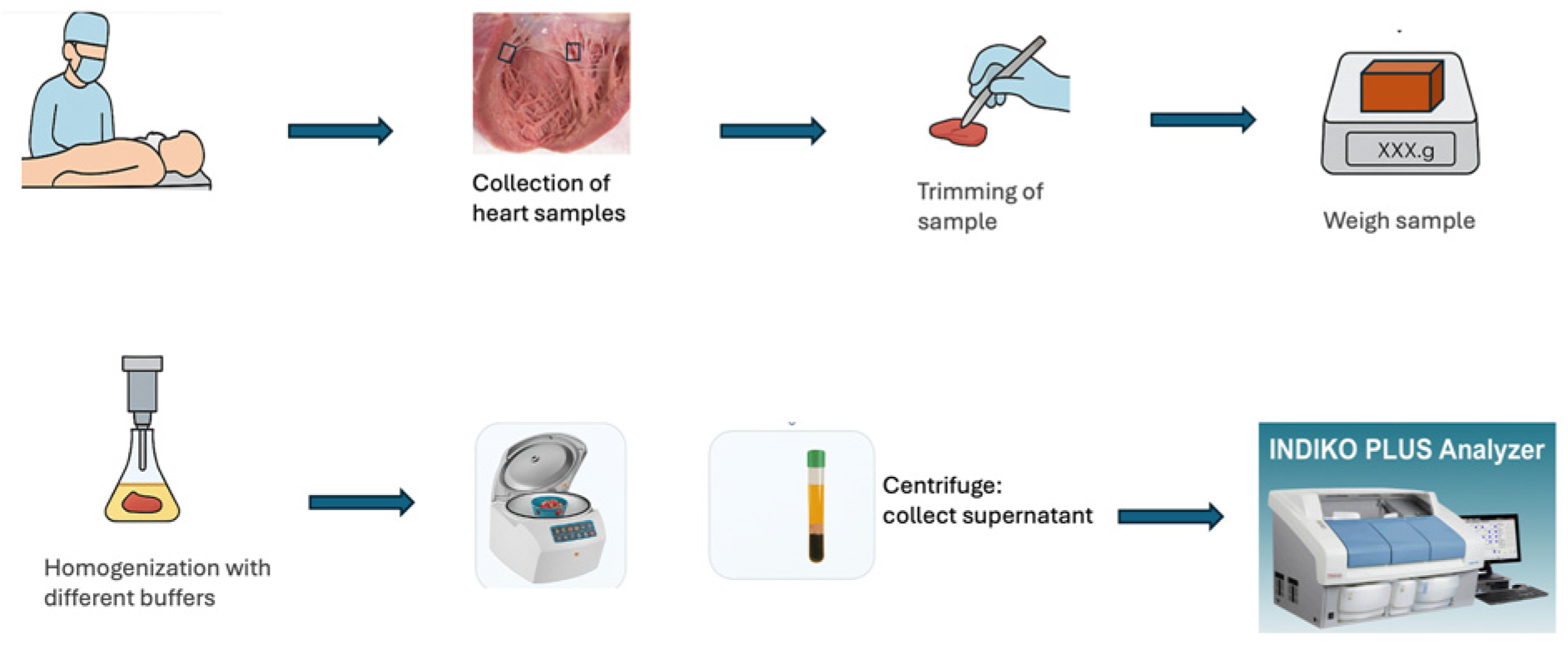
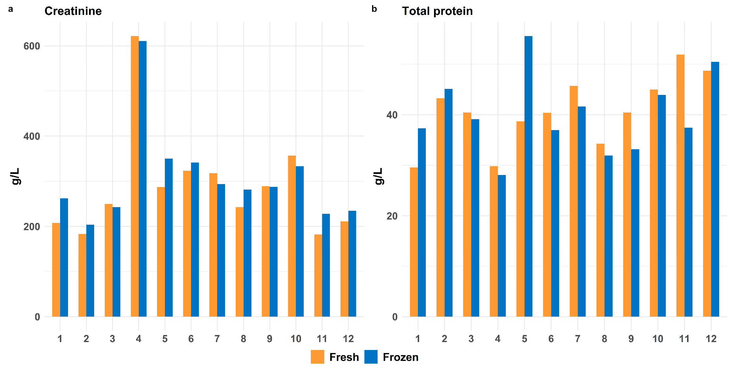
Figure 2 provides a summary of the workflow. All myocardial samples were collected approximately 0–6 mm from the endocardium, approximately 3 cm below the valves. From all cases, a femoral vein blood sample (for serum preparation) was also collected. In the pilot study, 1 g of myocardium was homogenized with 2 mL of the following solvents: distilled water (dH2O), PBS, 8 M urea in PBS, 0.1% tween in PBS, 8 M urea, and 20% SDS, using ULTRA-TURRAX homogenizer VDI 12 (IKA, Oxford, UK). Next, the homogenates were centrifuged at 3000× g for 20 min at 4 °C, and the supernatant was analyzed. To assess the effect of freeze-thawing on postmortem protein stability, one portion of 12 tissue samples was homogenized directly, and another portion was frozen for one week at −20 °C before homogenization. To this end, we chose to analyze creatinine and total protein, the former being a small molecule expected to remain stable, and the latter as a general marker of protein integrity. Pronounced hemolysis is quite common in serum from postmortem blood. To assess possible interference of hemoglobin in the serum with analysis, 23 femoral blood aliquots underwent hemoglobin removal with HemogloBind™ (Gentaur, 337-HO145-05, BSG, Monmouth Junction, NJ, USA). Briefly, equal parts of HemogloBind™ suspension and femoral blood were mixed, incubated for 10 min, and centrifuged at 3000× g for 2 min. For comparison, the same blood samples were mixed with dH2O and treated similarly.
In the main study, the myocardial homogenates were prepared at a 1:20 ratio (200 mg tissue: 4 mL buffer) in dH2O, T-PER (a proprietary detergent in 25 mM bicine, 150 mM NaCl; pH 7.6, Thermo Fisher Scientific, Vantaa, Finland, #78510), and Tris-Urea (25 mM Tris-HCl), or 2 M Urea (Thermo Fisher Scientific, #424585000; pH 7.4) buffers supplemented with HALT protease inhibitor cocktail (Thermo Fisher Scientific, #78438). T-PER is a mild detergent that maximizes protein solubilization without compromising enzymatic activity. After homogenization with ULTRA-TURRAX homogenizer VDI 12 (VWR), and centrifugation at 3000× g for 20 min at 4 °C, the supernatants were collected and stored at −20 °C. Serum was prepared by centrifugation of femoral vein blood at 3000× g for 20 min and stored at −20 °C.

2.4. Laboratory Assays

The biomarkers CK-MB, AST, total protein, creatinine, haptoglobin, fructose, and orosomucoid (alpha 1-acid glycoprotein) were analyzed in myocardial homogenates and serum in the pilot study using an Indiko™ (Thermo Fisher Scientific) instrument. In the main study, CK-MB, AST, ALT, LDH (all of which typically show temporal changes in serum concentration upon a significant myocardial injury), orosomucoid and total protein levels in tissue homogenates and serum samples were analyzed using an Indiko™ Plus (Thermo Fisher Scientific) instrument. The analytical principle for CK-MB, AST, ALT, and LDH is based on enzymatic reactions in which their specific substrates are converted through serial reactions that finally generate NADH/NADPH, and the spectrophotometrically measured absorbance at 340 nm is directly proportional to the enzyme activity in the samples. For determining orosomucoid concentration, Indiko utilizes a turbidimetric assay. To determine total protein concentration, the Indiko instrument uses a colorimetric method, in which the colored protein product is measured at 540 nm. The rationale for including creatinine, haptoglobin, and fructose (in the pilot study), and orosomucoid and total protein was that these are expected to be unaffected by an acute cardiac event, but also to determine whether different extraction alternatives affected the yields of the small molecules creatinine and fructose.
Measurements of a subgroup of sixteen samples were later independently analyzed at the Clinical Chemistry laboratory at Linköping University Hospital, Sweden, using a Roche Cobas 8000 c701 analyzer (Roche Diagnostics, West Sussex, UK). With this instrument, concentrations of cardiac cTnT, NT-proBNP, and myoglobin in tissue homogenates and serum were also determined, as these are also used for detecting cardiac conditions. The levels of CK-MB, cTnT, NT-proBNP, and myoglobin were detected using electrochemiluminescence immunoassay (ECLIA), which employs a sandwich immunoassay technique. The analysis of AST, ALT, LDH, and orosomucoid performed with the Roche Cobas 8000 c701 analyzer followed the same principles as those used with the Indiko instrument.

2.5. Mass Spectrometry Analysis (Proteomics)

Ten cardiac tissue homogenates and ten corresponding serum samples were analyzed in a buffer containing 10% β-octyl glucopyranoside. Serum samples were treated with a perchloric acid depletion protocol. For both sample types, proteins were reduced, alkylated, and subjected to on-filter digestion with trypsin using 3kDa centrifugal spin filters (Merck Millipore, Dublin, Ireland). Peptides were collected, dried using a Speedvac system, and reconstituted in 0.1% formic acid. Peptides were separated on a C18 reverse-phase column using a 150-min gradient. Analysis was performed on a QEx-Orbitrap mass spectrometer (Thermo Finnigan, San Jose, CA, USA) using HCD for tandem mass spectrometry [17]. Raw data were analyzed using MaxQuant 1.5.1.2 software. Proteins were identified using the Homo sapiens proteome from Uniprot [18].

2.6. Statistical Analysis

Due to the small sample sizes and the non-normal distribution of the data, non-parametric tests were used. For the freeze/thaw and Hemoglobind™ incubation serum experiments, differences between groups were assessed using the Wilcoxon signed-rank test. For the pretreatment optimization experiment, group differences were initially screened using the Kruskal–Wallis test. When a significant difference was found, this was followed by pairwise Wilcoxon rank-sum tests with Bonferroni–Holm correction for multiple comparisons. All tests were two-sided, and a p-value < 0.05 was considered statistically significant unless otherwise specified. Statistical analyses were conducted using R version 4.3.1. For the comparison between the SCD group and the control group, the Mann–Whitney U test was used with Bonferroni-Holm correction. With this test the two groups were compared for every measured analyte. For the comparisons regarding degree of arteriosclerosis and biomarker levels in the corresponding homogenates, the Wilcoxon signed-rank test was used. Comparisons between laboratories were analyzed with both Pearson and Spearman rank correlation tests for linear and non-linear correlations.

2.7. Ethical Considerations

All subjects were deceased, and therefore informed consent could not be obtained. In Sweden the Autopsy Law (1995:832) states that tissue samples may be procured by the forensic pathologist if the analytical results may be of importance for the purpose of the autopsy or for method development. This investigation was conducted in compliance with the abovementioned law and conformed to the principles of the Declaration of Helsinki. The study was approved by the Swedish Ethical Review Authority (No 2018/101-31/1 and 2023-03463-01). These approvals imply that the ethical review boards accepted the procedures described in the applications, where it is stated that the samples are not anonymized, but as soon as all necessary information about the deceased subjects has been retrieved, cases are assigned a code; hence, no person-identifiable results are generated during further processing and compilation of data.

3. Results

3.1. Pilot Study

3.1.1. HemogloBind Treatment

Creatinine levels remained largely unchanged between HemogloBind™-treated and dH2O-treated samples. Total protein levels decreased significantly after HemogloBind™ treatment (p < 0.001), which is explained by hemoglobin removal. Since simple dilution of samples with pronounced hemolysis with dH2O was as effective as HemogloBind™ treatment for obtaining analytical results, we decided not to use HemogloBind in these studies.

3.1.2. Impact of Freeze–Thaw Cycles on Postmortem Stability of Proteins

We compared biomarker levels in fresh tissue homogenates and samples stored for one week at −20 °C (Figure 3).
The Wilcoxon Signed-Rank Test showed no significant difference between fresh and frozen samples. Based on these findings, we decided to freeze samples before analysis to allow more samples to be analyzed in the same run.

3.1.3. Efficacy of Different Myocardial Tissue Homogenization Conditions

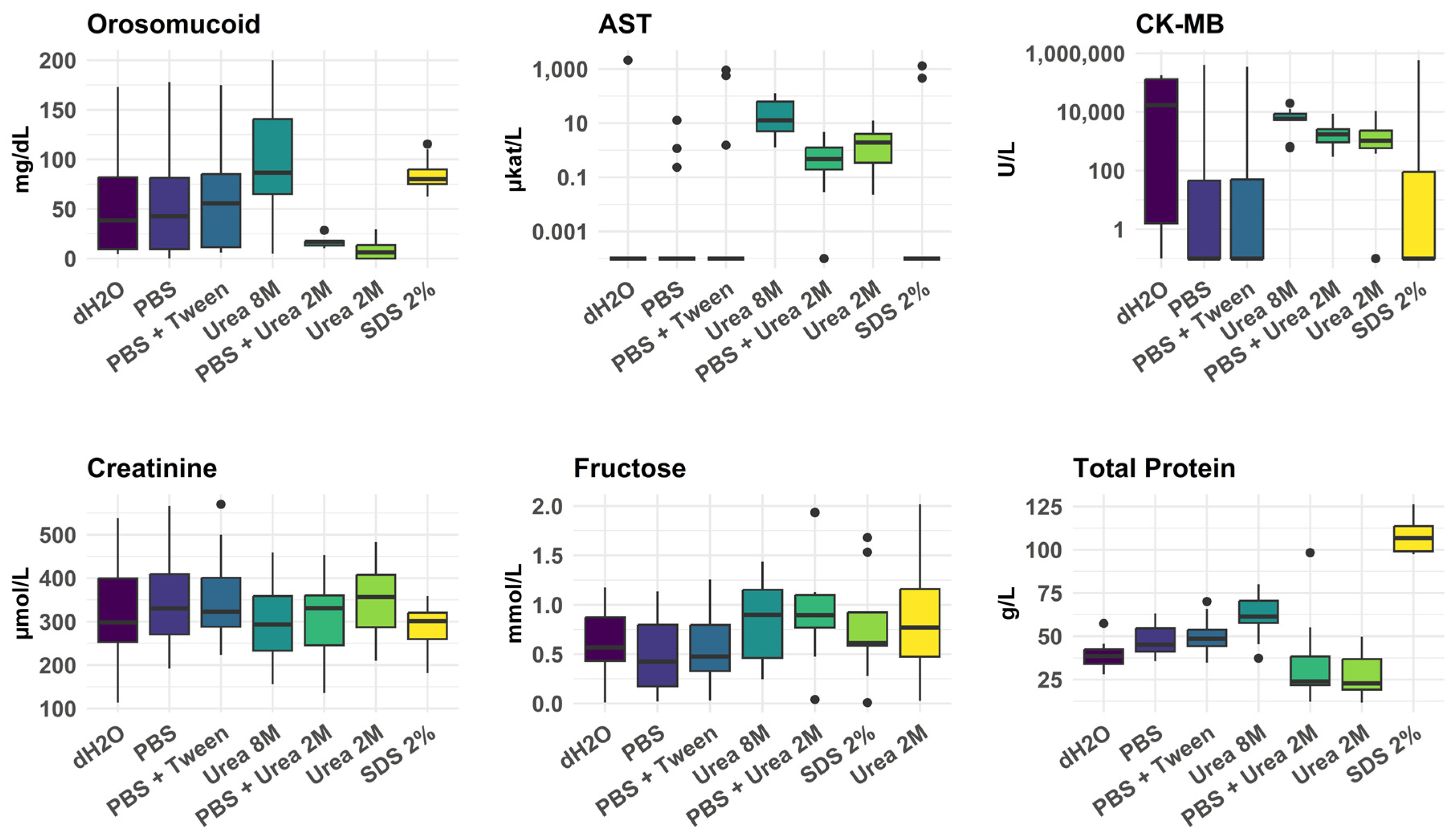
In the pilot study, we assessed the impact of different homogenization treatments (Figure 4). None of the preanalytical treatments was consistently most efficient. Although 8 M urea often gave a good yield, it also frequently produced a sample with high viscosity, preventing analysis.

3.2. Main Study

3.2.1. Evaluation of Extraction Methods for Effective Biomarker Yield from the Myocardium

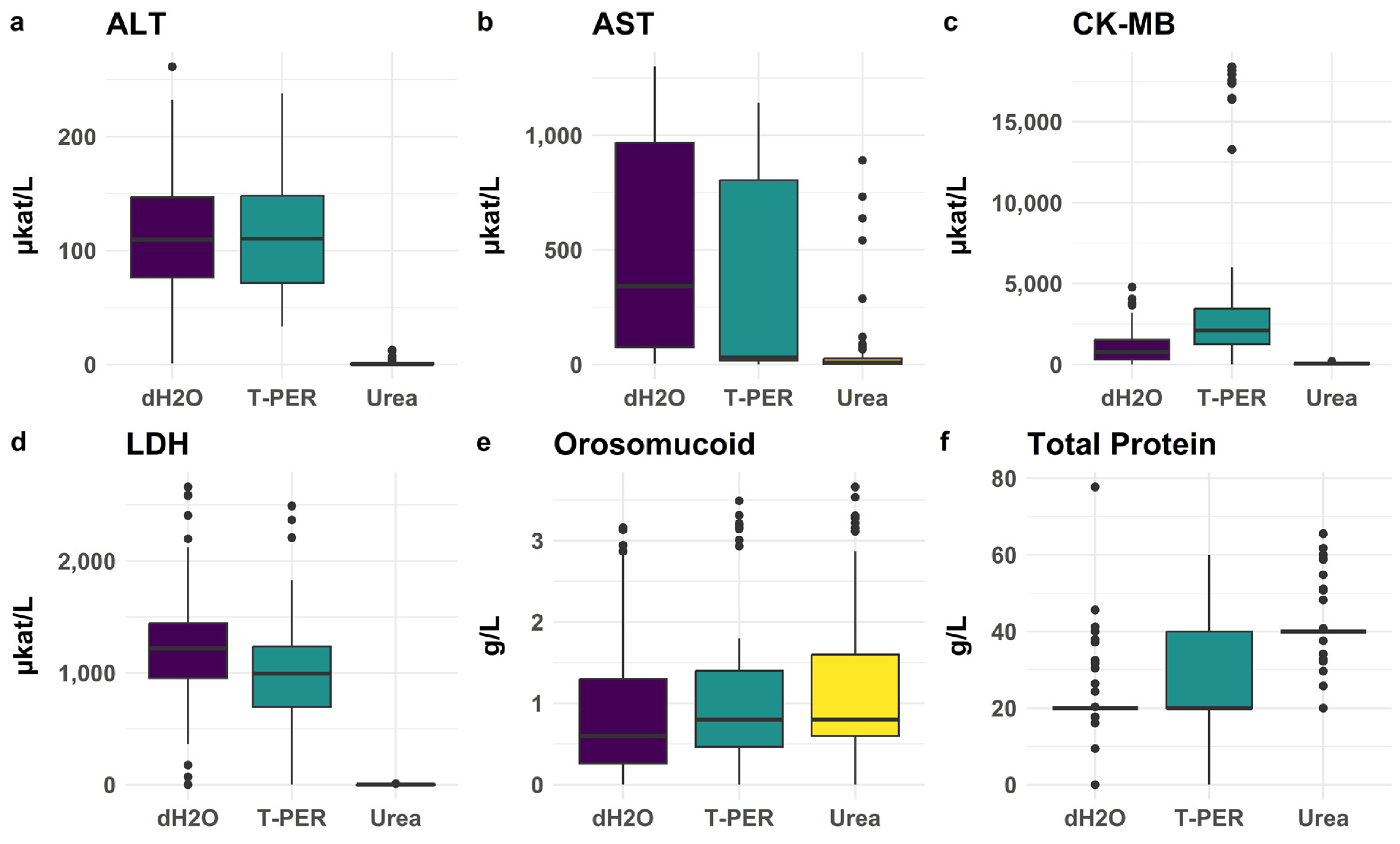
To further optimize the extraction conditions for the posterior wall myocardial samples, we examined the effectiveness of three extraction solvents: dH2O, T-PER, and urea 2 M, see Figure 5.
dH2O produced results similar to T-PER for most analytes, whereas urea often resulted in poor yields. All pairwise comparisons between serum and the preanalytical treatments for ALT, LDH, and CK-MB in homogenates showed p-values < 0.0001, with serum levels being consistently lower. The extraction efficiency of heart biomarkers in the anterior wall mirrored the efficiency observed in the posterior wall (see below).

3.2.2. Evaluation of Extraction Methods for Myoglobin, NT-proBNP and cTnT

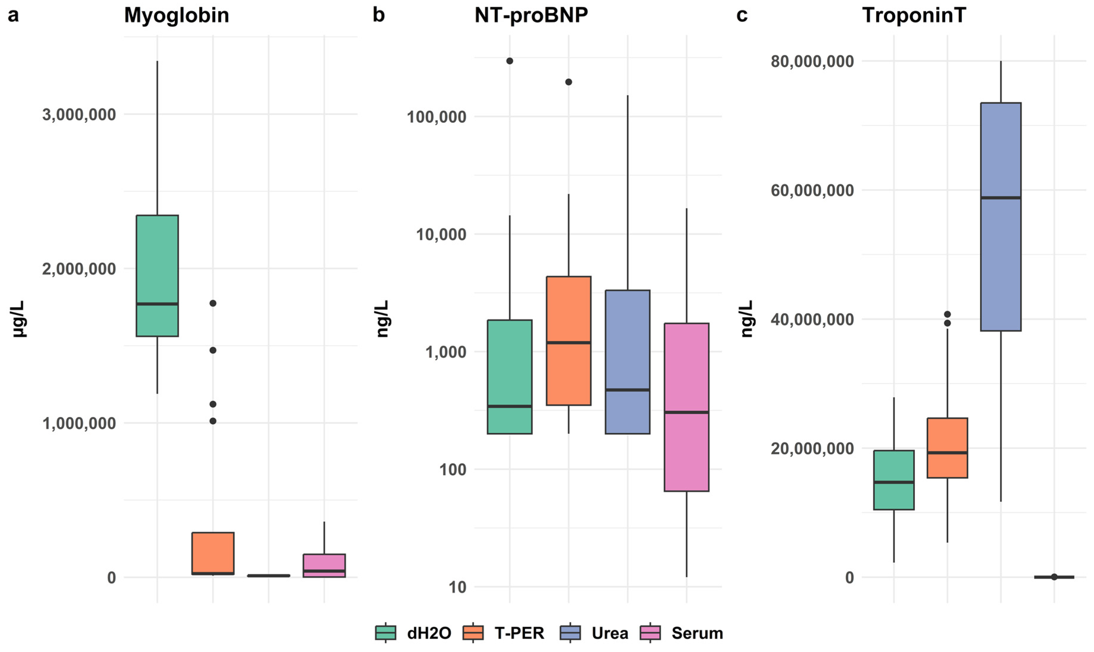
These biomarkers could not be analyzed with the Indiko instrument but were analyzed with the Cobas instrument at the Clinical Chemistry Laboratory in Linkoping, Sweden. The optimal extraction conditions varied for myoglobin, NT-proBNP, and cTnT, as shown in Figure 6.

3.2.3. Comparative Biochemical Analysis of Posterior and Anterior Wall

When examining results for all 113 cases, the anterior wall displayed almost consistently lower values than the posterior wall for most pretreatments. Although some differences were statistically significant, they were generally small (Figure 7).

3.2.4. Cross-Laboratory Validation of Extraction Methods

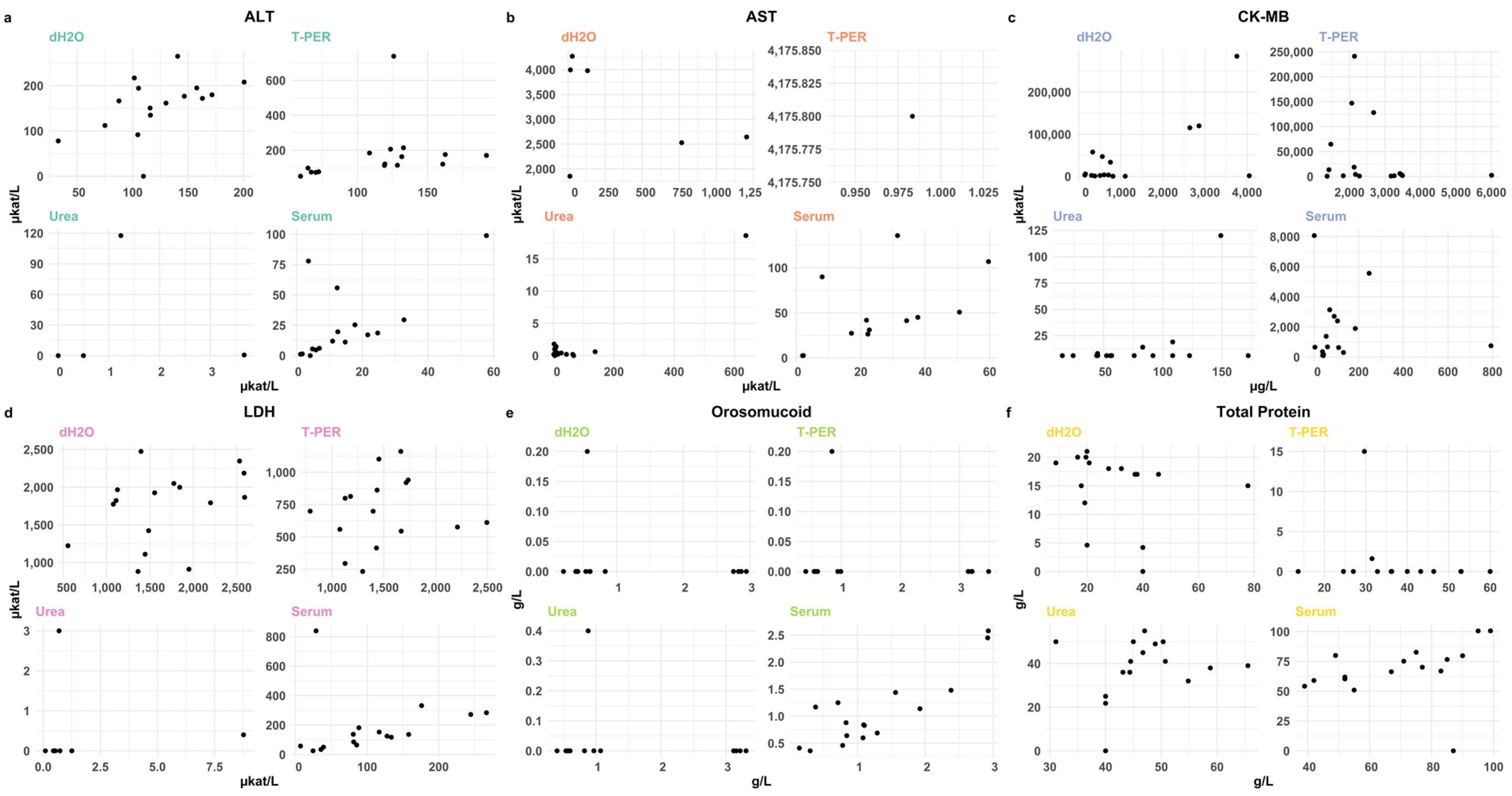
The results are shown in Figure 8. The Cobas instrument reported significantly higher median levels of ALT, AST, and CK-MB than the Indiko instrument for dH2O homogenates. The T-PER- and urea-treated homogenates showed no statistically significant differences. We then performed a regression analysis to examine whether the biomarker levels measured in two independent laboratories (Stockholm, Linköping) agreed, see Figure 8. The best correlations were observed for 2 M urea extractions of ALT (Spearman 0.803), but several correlations were low. Overall, positive correlations were observed for ALT across all treatments, and for AST, CK-MB, and LDH, under certain extraction conditions.

3.2.5. Comparison of Biomarkers Between SCD Cases and Controls

Comparisons between SCD cases and controls in posterior wall samples are shown in Table 3.
Cardiac biomarkers in posterior wall homogenates were generally lower in the SCD group compared to controls, except for CK-MB in T-PER extracts. With dH2O and urea extraction, CK-MB concentrations were significantly lower in SCD cases than in controls, and with dH2O and T-PER, LDH concentrations were also lower than in controls (p < 0.05 for these comparisons). In the anterior wall, AST, CK-MB, and LDH concentrations in SCD cases were significantly lower than in controls (p < 0.05) (see Table 3).

3.2.6. Impact of CPR on Analytical Results

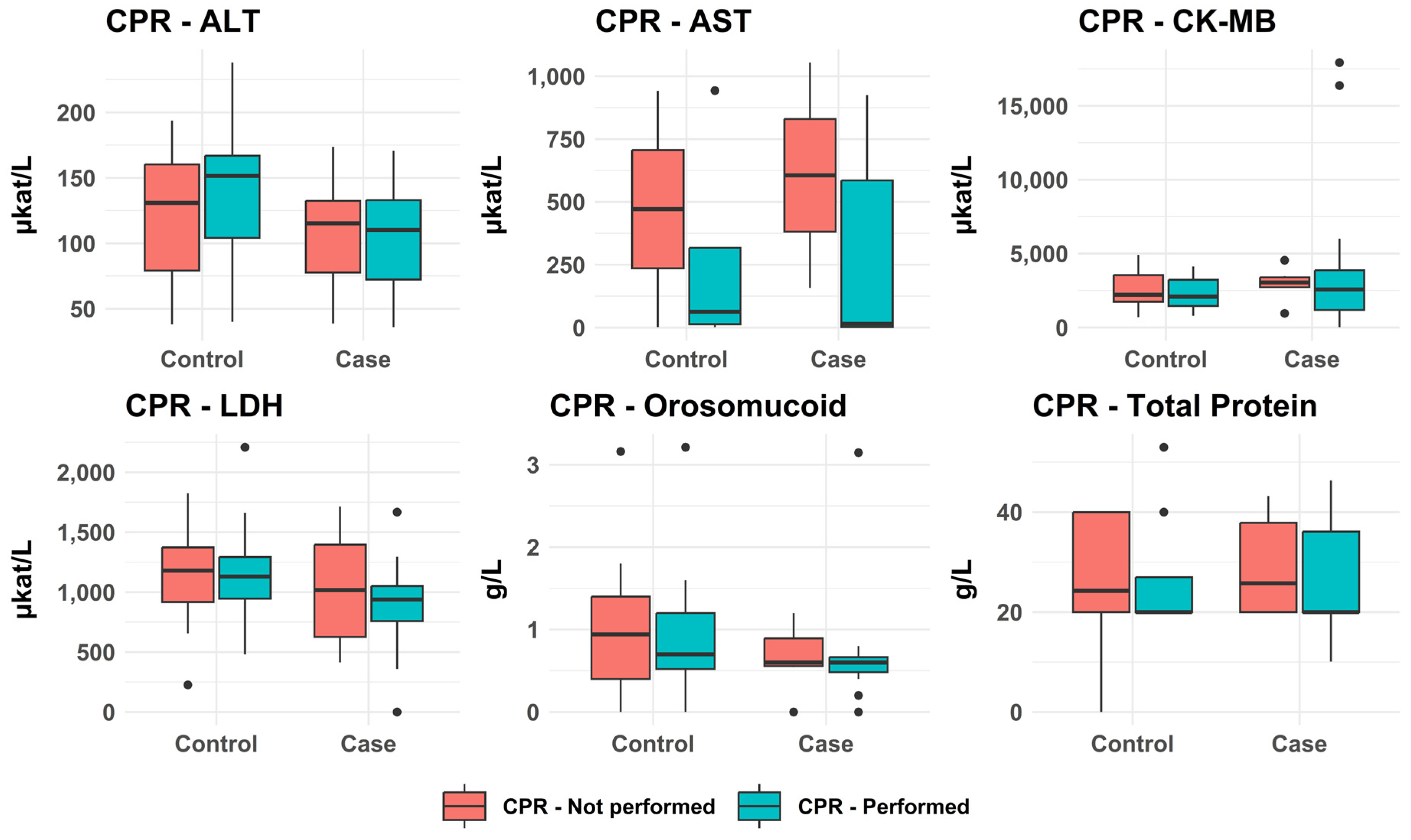
As shown in Figure 9, median concentrations of all markers were generally lower in subjects who received CPR compared to those who did not receive CPR. However, the differences were usually small, and no differences reached statistical significance.

3.2.7. PMI—tcPMI—Postmortem Interval Correlation with Biomarkers Levels

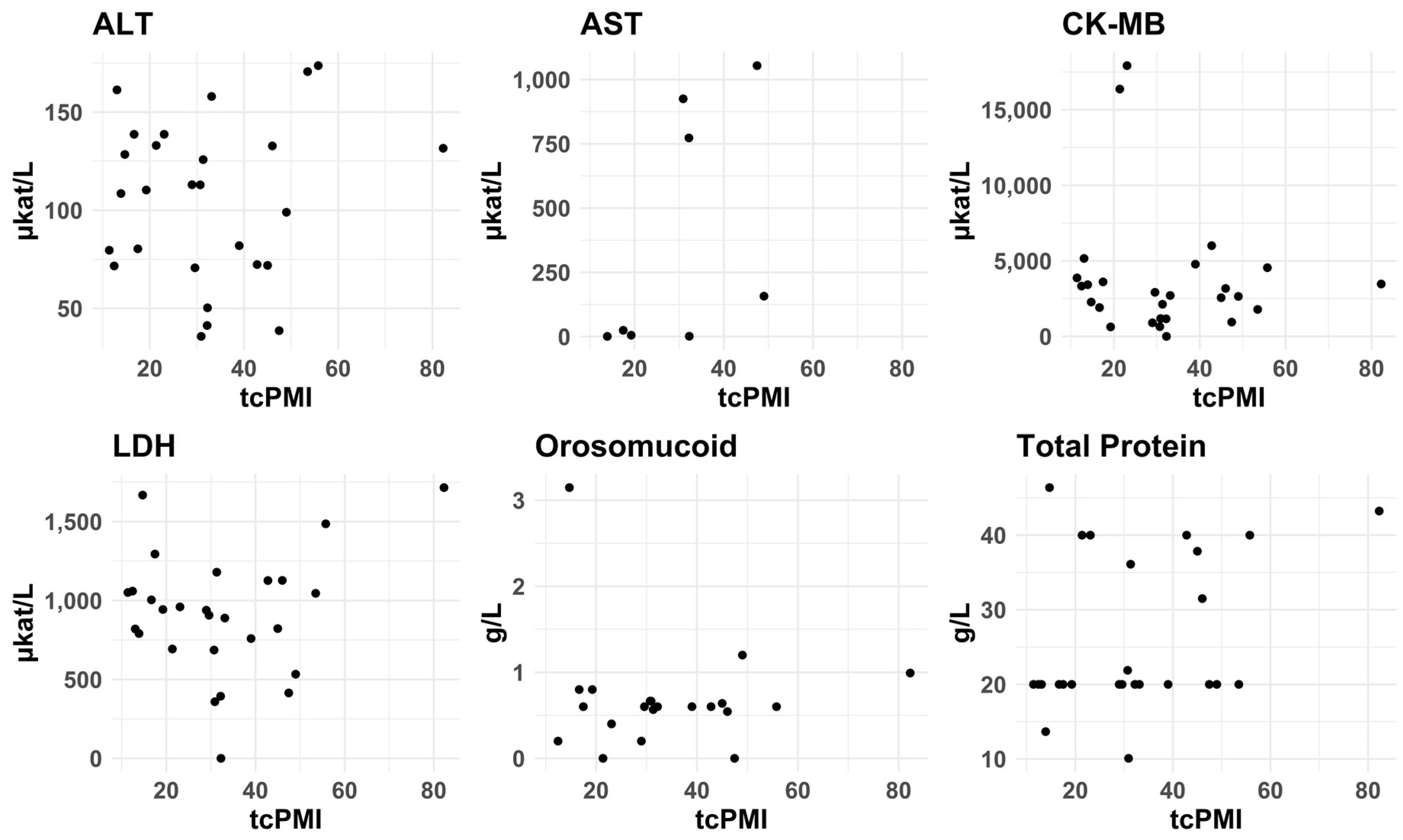
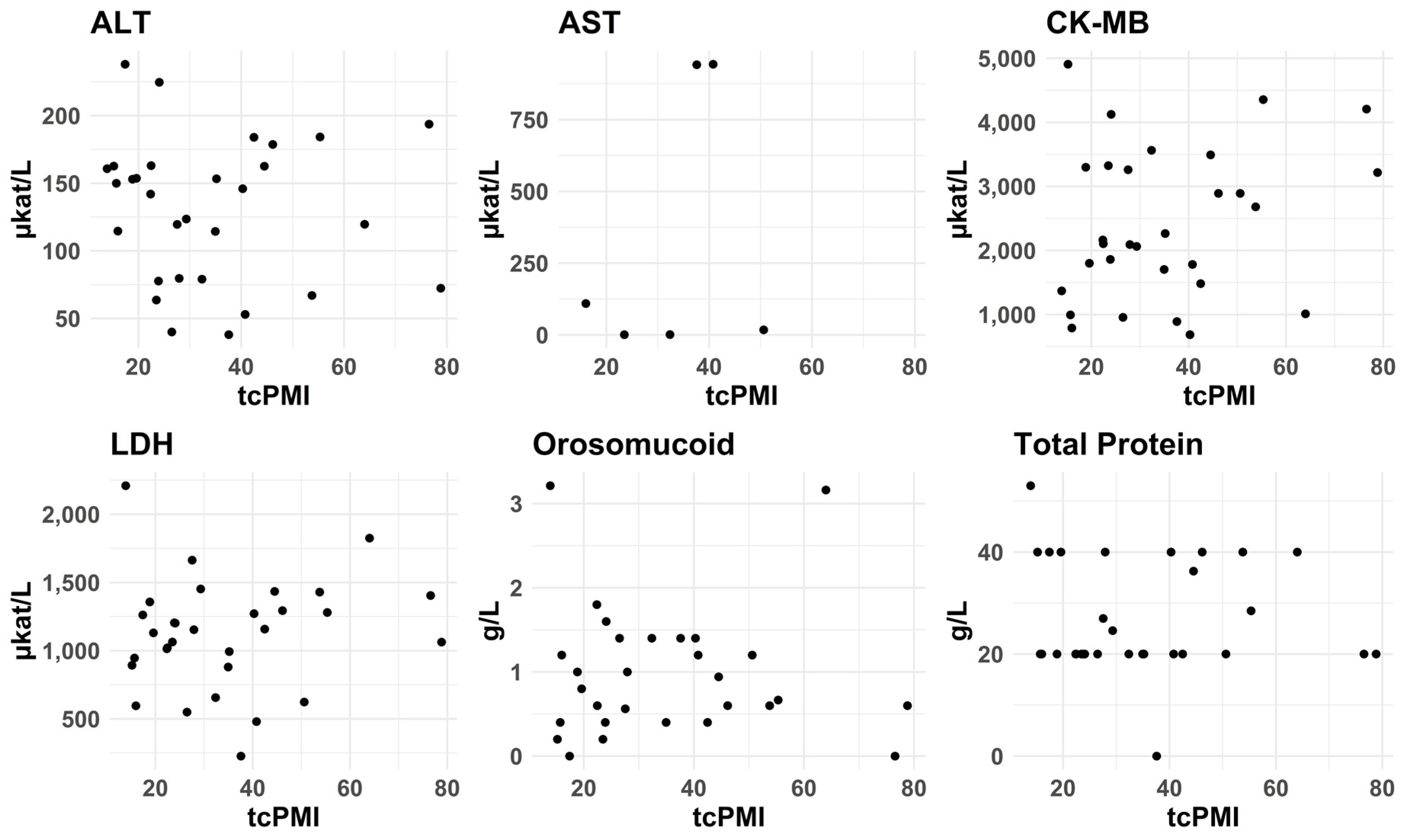
We conducted Pearson and Spearman correlation analyses to assess the possible linear and non-linear relationships between analyte values and postmortem crude interval (PMI), cold time (CT), and warm time (WT) for case and control groups from the left posterior wall, as well as temperature-corrected PMI (ctPMI). There were no significant correlations between the levels of the different biomarkers and any of these measures. For ctPMI, see Figure 10 and Figure 11. It should be noted, though, that we did not include decomposed cases.

3.2.8. Comparison of Coronary Arteriosclerosis and Biomarker Levels

We compared the degree of arteriosclerosis of the left anterior descending coronary artery (LAD) and the right coronary artery with concentrations of the selected biomarkers in the anterior and posterior wall samples, respectively, but we did not find significant changes in the biomarker levels in the corresponding homogenates (Table 4).

3.2.9. Proteomics

We performed proteomics analysis of 10 randomly selected cases to confirm the presence and concentrations of the cardiac biomarkers in myocardial samples and serum. In the cardiac tissue homogenates, a total of 1355 proteins were identified across all 10 samples. The number of identified proteins per sample ranged from 256 to 864, with an average of 589 proteins per sample. In the serum samples, a total of 1062 proteins were identified across all 10 samples. The number of identified proteins per sample ranged from 338 to 608, with an average of 434 proteins per sample. The concentrations of all analytes were much higher in the homogenates than in serum.
In fact, serum cTnT, CK-MB and AST were under the limit of quantitation, and only orosomucoid and myoglobin were consistently detected in serum from all cases, see Table 5.

4. Discussion

The Indiko instrument successfully delivered results for all selected biomarkers in most cases, both for serum samples and myocardial tissue homogenates. Homogenization, centrifugation and analysis could be completed within one hour, implying the possibility of implementing analysis of samples collected at autopsy and obtaining results before the examination of the body is completed. Furthermore, hemolysis of serum did not pose any issues, and freeze-thawing had no significant effect on concentrations of the examined biomarkers, which is in accordance with previous studies [19,20]. Cardiopulmonary resuscitation (CPR) did not affect biomarker levels, which aligns with previous studies [21,22,23]. Additionally, we did not find a correlation between postmortem interval and biomarker concentrations, which is also is consistent with results from other studies [22,24]. However, we excluded cases with significant decomposition, since there is an obvious risk for marker degradation, or at least a loss of enzymatic activity for those markers that are enzymes and are detected by their ability to catalyze reactions in the instrument. Hence, we do not know how well these markers will perform on decomposed cases. This will be an important objective for future studies.
The choice of method for extracting proteins and other components from a tissue sample is dependent on which molecules one wishes to obtain for analysis. To this end, different methods provide variable yields dependent on the characteristics of the target molecules, e.g., their size, physicochemical properties, and attachment to various cellular structures. In this context, the pioneering work by Fred S Apple and co-workers must be acknowledged. In the paper by Voss et al. [25], they showed that cTnT, CK-MB, and myoglobin concentrations in 14 regions of the healthy human heart were similar and that CK-MB and myoglobin were 99% cytosolic whereas 92% of troponin T was myofibril-bound. They further reported that the increase in troponins in serum paralleled a decrease in troponins in ischemic areas of the myocardium after coronary artery occlusion in dogs [26]. Another aspect to consider is that pretreatment may interfere with the function of the marker molecules. Several of the markers used in this study are enzymes, and the analytical methods of these depend on a preserved enzymatic activity of the protein. An additional aspect is the solubility of the marker in the extract. For instance, urea has been used extensively because it interacts both with polar and nonpolar residues, thereby stabilizing the solvation of the unfolded protein state [27], but excessively high urea concentration caused a high viscosity of the sample that prevented analysis in the pilot study. T-PER is a standard buffer for protein extraction from tissue samples [28]. When comparing pretreatment methods, extraction with dH2O was usually as effective as other extractions for most analytes. IndikoPlus generally reported higher concentrations of CK-MB extracted with dH2O than the Cobas instrument, but we have not been able to identify the explanation for this, since the analytical principle is the same in both instruments, although the calibration protocols are somewhat different. It is also possible that the instruments may have different sensitivities to matrix effects, given that both are designed to analyze serum rather than tissue homogenates. However, it should be kept in mind that this is an exploratory study, and repeated analysis on larger samples might not confirm this difference.
Urea generally provided the lowest yields, although it was most efficient for cTnT, whereas dH2O gave the best yields for myoglobin. This means that it is important for further studies to evaluate extraction alternatives for each new analyte. Such studies may include combinations of extraction agents to improve the yields; for instance, Frostadottir et al. [29] reported that addition of 8 M urea to a RIPA buffer increased protein yields from fresh frozen human peripheral nerve samples. Dilution of samples before analysis is a classical measure that often is automatically activated in standard clinical chemistry multi-analyzers, but since biomarker levels in myocardial homogenates differ from those in serum, testing different dilutions may be necessary to match the calibration range of the instrument’s analytical methods. Serum samples provided reliable results for nearly all analytes, with only rare failures. In contrast, heart homogenates showed occasional failures across all tested pretreatment methods, particularly in the pilot study.
Results for different analytes differed between analysis with Indiko and Cobas. One reason for the difference is that the Indiko results were obtained from analysis of the fresh samples, whereas the Cobas results were obtained from one of several aliquots of extracts that had been frozen for several months, and which might not have been perfectly mixed before or after freezing.
Sacco et al. [24] and Kutlu et al. [30] recently reported that a combination of cardiac injury serum markers could be used and demonstrated high sensitivity and specificity. However, confirmatory studies are needed, since several previous studies have not shown such good separation of cases and controls. Most likely, case selection is the critical factor, i.e., that SCD cases with certainty had extensive area(s) of cardiomyocyte necrosis and that controls had perfectly healthy hearts. In the study by Kutlu et al. [30], the control group subjects were significantly younger and had significantly lower heart weights, whereas in our study, the groups were much more similar in these and other respects, which we believe is more relevant in practical casework if one wishes to apply biochemistry as a tool to distinguish cases with similar characteristics. Furthermore, in addition to a loss of cardiac biomarkers from injured cardiomyocytes, it has also been observed that there are dynamic changes in levels over time, most likely due to de-novo production by cells in the penumbra zone; this temporal variation was already reported in experiments by Sharkey in 1991 [31]. The lower amounts of several analytes in the anterior left ventricular wall sample compared to the posterior wall sample may be explained by postmortem redistribution when most victims are placed in a supine position [26,27,28,32,33,34], implying sedimentation of blood and lymph possibly rich in leaked cardiomyocyte contents.
Today, repeated measurements of cTnT are used extensively in hospitals upon suspicion of myocardial ischemia. Even if there are sensitive immunoassays for troponin analysis, the most reliable method for analysis of cTnT is by electrochemiluminescence, which the Indiko instrument cannot offer. We therefore decided to analyze AST, ALT, CK-MB, and LDH. The pattern of these biomarkers in serum was used extensively in the past to confirm/assess AMI [35,36]. The advantage of analyzing the biomarkers also in cardiac homogenates is that it is likely that the decrease in concentrations in the myocardium will precede the increase in serum levels, since losses from the necrotic cardiomyocytes first will enter the local blood and lymph vessels in the heart before reaching the large volume of blood in the circulation.
Following an acute ischemic heart event, serum levels of CK-MB and cTnT will not increase immediately [37,38,39]; hence, there is a potential to discover acute myocardial ischemia by measuring the loss of biomarkers in myocardial homogenates. For AST, ALT and LDH, their rise in serum is even more delayed. This delay may explain why we did not find increased serum levels of these markers if many of the SCD cases died early after symptom onset (Table 3).
Whereas we failed to find increased serum levels of the selected biomarkers, in the tissue homogenates, CK-MB and LDH, and even ALT levels were significantly lower in SCD cases than in controls (Table 3), which lends support for analysis of biomarkers in myocardial homogenates.
Several factors limited the classification accuracy, including (a) lack of precise information of ischemic myocardial area; (b) arrhythmia as the cause of death; and (c) non-cardiac causes of death where the circumstances surrounding death mimicked SCD. The inclusion of controls also provided some challenges, but we selected cases where there was an obvious non-cardiac-related cause of death and where death was either immediate or followed a rapid course. As pointed out in Section 2, five of the controls did show heart pathology (enlarged heart or arteriosclerosis), which may be considered a limitation. However, all these deaths had another explanation, and we consider it very unlikely that their pre-existent heart pathologies would coincidentally have resulted in a myocardial injury shortly prior to death. Another limitation is that genetic testing had not been requested by the responsible pathologist in these cases. Although such testing will not prove that a functional deficit caused death, we acknowledge that this type of information is important, just like other forms of medical history that can imply a risk for sudden cardiac death. Another drawback was that we collected samples after the pathologist had collected samples for the death investigation; hence, some of our samples were obviously not taken from the center of visibly suspected myocardial areas. Furthermore, even if we performed proteomics analysis on a subset of cases, the purpose was not to compare SCD and controls but to determine whether we could confirm higher concentrations of the investigated cardiac markers in the myocardium than in serum. Indeed, we observed that biomarker levels, as measured with this quantitative mass spectrometry, were consistently higher in the myocardial homogenates than in serum, which aligns with the protein expression data reported in the Human Protein Atlas (www.proteinatlas.org) [40]. We believe that data from existing and additional proteomics analyses of myocardial homogenates can be used to identify more suitable markers of ischemia and inflammation for analysis in myocardial homogenates than the currently used cardiac injury markers, which are all intended for serum analysis. In such efforts, it will be important to consider the size, shape and localization of candidate molecules, e.g., whether they are bound to myofibrils, integrated in the cell membrane or are cytosolic. These and other factors, such as the myocard:serum concentration ratio, will impact how easily molecules leak into the blood. Further, proteomics analysis may also identify metabolites that are formed in small amounts and therefore difficult to analyze in serum, and there may be reactive products in surviving cells associated with ischemic injury that are not lost to any significant extent to the circulation, but which might be suitable targets in myocardial extracts.
In summary, we show that clinical chemistry cardiac markers can be analyzed in myocardial homogenates and that, with a benchtop instrument, results can be obtained in less than an hour, thus assisting the pathologist in the decision-making regarding which further investigative tracks to follow before the autopsy is completed.

5. Conclusions

Analysis of cardiac markers in myocardial samples collected at autopsy is seemingly a feasible approach to detect early myocardial ischemia, and possibly also acute inflammation. To this end, a systematic search for, and testing of, promising target molecules present in the myocardium, is warranted. Once suitable target molecules are identified, it will be important to examine the possible need for pre-treatment, since concentrations, both pathological and normal, in the extract into the instrument must match the calibration range of each particular method. To determine the sensitivity and specificity of different cardiac markers, carefully designed studies are essential; ideally, samples should be collected precisely from ischemic areas of the myocardium, and cases should be carefully selected to ensure they have a well-defined acute ischemic area. This can be accomplished by selecting cases with acute myocardial ischemia confirmed antemortem by clinical observations, ECG, and Troponin T and/or I measurements, and yet who underwent autopsy for some reason. Control cases should include both rapid deaths and those with prolonged agony to allow for fair comparisons. Additionally, the impact of PMI on biomarker stability in cases with significant decomposition warrants further studies, as does the reproducibility of analytical results through repeated analyses to ensure the reliability of internal controls.

Author Contributions

N.S. (Niki Sarri): methodology development for the main study, original draft preparation, review, and editing; H.D.: conceptualization, original draft preparation, review, editing, methodology, and supervision; K.A.: original draft preparation, review, editing, methodology, and supervision; A.-R.R.: statistical analysis and calculations; K.O.: methodology development for the pilot study; N.S. (Nargis Sultana): lead for methodology development and validation. All authors have read and agreed to the published version of the manuscript.

Funding

This research was funded by Swedish National Board of Forensic Medicine, Stockholm, Sweden.

Institutional Review Board Statement

This investigation was conducted in compliance with the abovementioned law and conformed to the principles of the Declaration of Helsinki. The study was approved by the Swedish Ethical Review Authority (2018/101-31/1, 7 February 2018; and 2023-03463-01, 7 July 2023). These approvals imply that the ethical review boards accepted the procedures described in the applications, where it is stated that the samples are not anonymized, but as soon as all necessary information about the deceased subjects has been retrieved, cases are assigned a code; hence, no person-identifiable results are generated during further processing and compilation of data.

Informed Consent Statement

All subjects were deceased, and therefore informed consent could not be obtained. In Sweden the Autopsy Law (1995:832) states that tissue samples may be procured by the forensic pathologist if the analytical results may be of importance for the purpose of the autopsy or for method development.

Data Availability Statement

The data presented in this study are available on request from the corresponding author due to access restrictions by the owner of the original data, the Swedish National Board of Forensic Medicine. Although the cases are coded, there is linked information to each case, such as date of death and circumstances, which may disclose the identity of the subject, so before disclosure of data upon request, a filtering of certain information in the research data master file will be necessary.

Conflicts of Interest

The authors declare no conflicts of interest.

Abbreviations

The following abbreviations are used in this manuscript:
SCDSudden Cardiac Death
ALTAlanine aminotransferase
ASTAspartate aminotransferase
CK-MBCreatine Kinase-MB
LDHLactate Dehydrogenase
NT-proBNPN-terminal pro-B-type Natriuretic Peptide
PMIPost-Mortem Interval 
CRPC-Reactive Protein
AMIAcute Myocardial Infarction
MRMagnetic Resonance 
tcPMITemperature Corrected Post-Mortem Interval
PBSPhosphate-Buffered Saline
SDSSodium Dodecyl Sulfate
NADHNicotinamide Adenine Dinucleotide
NADPHNicotinamide Adenine Dinucleotide Phosphate
ECLIAElectrochemiluminescence Immunoassay
cTnTCardiac Troponin T
ECGElectrocardiogram
CTComputed Tomography

References

  1. Kuijpers, C.C.; Fronczek, J.; van de Goot, F.R.; Niessen, H.W.; van Diest, P.J.; Jiwa, M. The value of autopsies in the era of high-tech medicine: Discrepant findings persist. J. Clin. Pathol. 2014, 67, 512–519. [Google Scholar] [CrossRef]
  2. Dettmeyer, R.B. The role of histopathology in forensic practice: An overview. Forensic Sci. Med. Pathol. 2014, 10, 401–412. [Google Scholar] [CrossRef]
  3. Thygesen, K.; Alpert, J.S.; Jaffe, A.S.; Chaitman, B.R.; Bax, J.J.; Morrow, D.A.; White, H.D.; Executive Group on behalf of the Joint European Society of Cardiology/American College of Cardiology/American Heart Association/World Heart Federation Task Force for the Universal Definition of Myocardial Infarction. Fourth Universal Definition of Myocardial Infarction (2018). J. Am. Coll. Cardiol. 2018, 72, 2231–2264. [Google Scholar] [CrossRef]
  4. Palmiere, C.; Mangin, P. Postmortem chemistry update part I. Int. J. Legal Med. 2012, 126, 187–198. [Google Scholar] [CrossRef]
  5. Astrup, B.S.; Thomsen, J.L. The routine use of C-reactive protein in forensic investigations. Forensic Sci. Int. 2007, 172, 49–55. [Google Scholar] [CrossRef]
  6. Zilg, B.; Alkass, K.; Kronstrand, R.; Berg, S.; Druid, H. A Rapid Method for Postmortem Vitreous Chemistry-Deadside Analysis. Biomolecules 2021, 12, 32. [Google Scholar] [CrossRef] [PubMed]
  7. Johns, S.H.; Wist, A.A.; Najam, A.R. Spot Tests: A Color Chart Reference for Forensic Chemists. J. Forensic Sci. 1979, 24, 631–649. [Google Scholar] [CrossRef]
  8. Yagoda, H. Applications of Confined Spot Tests in Analytical Chemistry: Preliminary Paper. Ind. Eng. Chem. Anal. Ed. 1937, 9, 79–82. [Google Scholar] [CrossRef]
  9. Eckart, R.E.; Shry, E.A.; Burke, A.P.; McNear, J.A.; Appel, D.A.; Castillo-Rojas, L.M.; Avedissian, L.; Pearse, L.A.; Potter, R.N.; Tremaine, L.; et al. Sudden death in young adults: An autopsy-based series of a population undergoing active surveillance. J. Am. Coll. Cardiol. 2011, 58, 1254–1261. [Google Scholar] [CrossRef]
  10. Al-Khatib, S.M.; Stevenson, W.G.; Ackerman, M.J.; Bryant, W.J.; Callans, D.J.; Curtis, A.B.; Deal, B.J.; Dickfeld, T.; Field, M.E.; Fonarow, G.C.; et al. 2017 AHA/ACC/HRS guideline for management of patients with ventricular arrhythmias and the prevention of sudden cardiac death: Executive summary: A Report of the American College of Cardiology/American Heart Association Task Force on Clinical Practice Guidelines and the Heart Rhythm Society. Heart Rhythm. 2018, 15, e190–e252. [Google Scholar] [CrossRef]
  11. Goldstein, S. The necessity of a uniform definition of sudden coronary death: Witnessed death within 1 hour of the onset of acute symptoms. Am. Heart J. 1982, 103, 156–159. [Google Scholar] [CrossRef]
  12. Priori, S.G.; Blomstrom-Lundqvist, C.; Mazzanti, A.; Blom, N.; Borggrefe, M.; Camm, J.; Elliott, P.M.; Fitzsimons, D.; Hatala, R.; Hindricks, G.; et al. 2015 ESC Guidelines for the management of patients with ventricular arrhythmias and the prevention of sudden cardiac death: The Task Force for the Management of Patients with Ventricular Arrhythmias and the Prevention of Sudden Cardiac Death of the European Society of Cardiology (ESC). Endorsed by: Association for European Paediatric and Congenital Cardiology (AEPC). Eur. Heart J. 2015, 36, 2793–2867. [Google Scholar] [CrossRef]
  13. Aljakna, A.; Fracasso, T.; Sabatasso, S. Molecular tissue changes in early myocardial ischemia: From pathophysiology to the identification of new diagnostic markers. Int. J. Legal Med. 2018, 132, 425–438. [Google Scholar] [CrossRef]
  14. Isbister, J.; Semsarian, C. Sudden cardiac death: An update. Intern. Med. J. 2019, 49, 826–833. [Google Scholar] [CrossRef]
  15. Carvajal-Zarrabal, O.; Hayward-Jones, P.M.; Nolasco-Hipolito, C.; Barradas-Dermitz, D.M.; Calderón-Garcidueñas, A.L.; López-Amador, N. Use of Cardiac Injury Markers in the Postmortem Diagnosis of Sudden Cardiac Death. J. Forensic Sci. 2017, 62, 1332–1335. [Google Scholar] [CrossRef] [PubMed]
  16. Basso, C.; Aguilera, B.; Banner, J.; Cohle, S.; d’Amati, G.; de Gouveia, R.H.; di Gioia, C.; Fabre, A.; Gallagher, P.J.; Leone, O.; et al. Guidelines for autopsy investigation of sudden cardiac death: 2017 update from the Association for European Cardiovascular Pathology. Virchows Arch. 2017, 471, 691–705. [Google Scholar] [CrossRef]
  17. Hawkes, J.A.; Dittmar, T.; Patriarca, C.; Tranvik, L.; Bergquist, J. Evaluation of the Orbitrap Mass Spectrometer for the Molecular Fingerprinting Analysis of Natural Dissolved Organic Matter. Anal. Chem. 2016, 88, 7698–7704. [Google Scholar] [CrossRef] [PubMed]
  18. UniProt Tools. Proteomes · Homo sapiens (Human). Available online: https://www.uniprot.org/proteomes/UP000005640 (accessed on 12 September 2025).
  19. Abraham, R.A.; Rana, G.; Agrawal, P.K.; Johnston, R.; Sarna, A.; Ramesh, S.; Acharya, R.; Khan, N.; Porwal, A.; Kurundkar, S.B.; et al. The Effects of a Single Freeze-Thaw Cycle on Concentrations of Nutritional, Noncommunicable Disease, and Inflammatory Biomarkers in Serum Samples. J. Lab. Physicians 2021, 13, 6–13. [Google Scholar] [CrossRef]
  20. Reimers, T.J.; McCann, J.P.; Cowan, R.G.; Concannon, P.W. Effects of Storage, Hemolysis, and Freezing and Thawing on Concentrations of Thyroxine, Cortisol, and Insulin in Blood Samples. Proc. Soc. Exp. Biol. Med. 1982, 170, 509–516. [Google Scholar] [CrossRef]
  21. Mattana, J.; Singhal, P.C. Determinants of elevated creatine kinase activity and creatine kinase MB-fraction following cardiopulmonary resuscitation. Chest 1992, 101, 1386–1392. [Google Scholar] [CrossRef] [PubMed]
  22. Palmiere, C.; Tettamanti, C.; Bonsignore, A.; De Stefano, F.; Vanhaebost, J.; Rousseau, G.; Scarpelli, M.P.; Bardy, D. Cardiac troponins and NT-proBNP in the forensic setting: Overview of sampling site, postmortem interval, cardiopulmonary resuscitation, and review of the literature. Forensic Sci. Int. 2018, 282, 211–218. [Google Scholar] [CrossRef] [PubMed]
  23. Polena, S.; Shen, K.H.; Mamakos, E.; Chuang, P.J.; Sharma, M.; Griciene, P.; Ponomarev, A.A.; Gintautas, J.; Maniar, R. Correlation between cardiac enzyme elevation and the duration of cardiopulmonary resuscitation. Proc. West. Pharmacol. Soc. 2005, 48, 136–138. [Google Scholar] [PubMed]
  24. Sacco, M.A.; Aquila, V.R.; Gualtieri, S.; Raffaele, R.; Verrina, M.C.; Tarda, L.; Gratteri, S.; Aquila, I. Quantification of Myocardial Biomarkers in Sudden Cardiac Deaths Using a Rapid Immunofluorescence Method for Simultaneous Biomarker Analysis. Biomedicines 2025, 13, 193. [Google Scholar] [CrossRef]
  25. Voss, E.M.; Sharkey, S.W.; Gernert, A.E.; Murakami, M.M.; Johnston, R.B.; Hsieh, C.C.; Apple, F.S. Human and canine cardiac troponin T and creatine kinase-MB distribution in normal and diseased myocardium. Infarct sizing using serum profiles. Arch. Pathol. Lab. Med. 1995, 119, 799–806. [Google Scholar]
  26. Ricchiuti, V.; Sharkey, S.W.; Murakami, M.M.; Voss, E.M.; Apple, F.S. Cardiac troponin I and T alterations in dog hearts with myocardial infarction: Correlation with infarct size. Am. J. Clin. Pathol. 1998, 110, 241–247. [Google Scholar] [CrossRef]
  27. Rossky, P.J. Protein denaturation by urea: Slash and bond. Proc. Natl. Acad. Sci. USA 2008, 105, 16825–16826. [Google Scholar] [CrossRef]
  28. Wright, M.C.; Mi, R.; Connor, E.; Reed, N.; Vyas, A.; Alspalter, M.; Coppola, G.; Geschwind, D.H.; Brushart, T.M.; Hoke, A. Novel roles for osteopontin and clusterin in peripheral motor and sensory axon regeneration. J. Neurosci. 2014, 34, 1689–1700. [Google Scholar] [CrossRef]
  29. Frostadottir, D.; Welinder, C.; Perez, R.; Dahlin, L.B. Refinement of Protein Extraction Protocols for Human Peripheral Nerve Tissue. ACS Omega 2025, 10, 5111–5118. [Google Scholar] [CrossRef]
  30. Kutlu, E.; Cil, N.; Avci, E.; Bir, F.; Kilic, I.D.; Dereli, A.K.; Acar, K. Significance of postmortem biomarkers and multimarker strategy in sudden cardiac death. Leg. Med. 2023, 61, 102212. [Google Scholar] [CrossRef]
  31. Sharkey, S.W.; Murakami, M.M.; Smith, S.A.; Apple, F.S. Canine myocardial creatine kinase isoenzymes after chronic coronary artery occlusion. Circulation 1991, 84, 333–340. [Google Scholar] [CrossRef] [PubMed]
  32. Maeda, H.; Zhu, B.L.; Ishikawa, T.; Quan, L.; Michiue, T. Significance of postmortem biochemistry in determining the cause of death. Leg. Med. 2009, 11 (Suppl. 1), S46–S49. [Google Scholar] [CrossRef]
  33. Pélissier-Alicot, A.L.; Gaulier, J.M.; Champsaur, P.; Marquet, P. Mechanisms underlying postmortem redistribution of drugs: A review. J. Anal. Toxicol. 2003, 27, 533–544. [Google Scholar] [CrossRef]
  34. Zilg, B.; Thelander, G.; Giebe, B.; Druid, H. Postmortem blood sampling-Comparison of drug concentrations at different sample sites. Forensic Sci. Int. 2017, 278, 296–303. [Google Scholar] [CrossRef]
  35. Matsui, Y.; Hashimoto, H.; Tsukamoto, H.; Okumura, K.; Ito, T.; Ogawa, K.; Satake, T. Disappearance and appearance of isoenzymes of creatine kinase, lactate dehydrogenase and aspartate aminotransferase in the myocardium undergoing infarction. Cardiovasc. Res. 1989, 23, 249–253. [Google Scholar] [CrossRef]
  36. Van der Laarse, A.; Dijkshoorn, N.J.; Hollaar, L.; Caspers, T. The (iso)enzyme activities of lactate dehydrogenase, alpha-hydroxybutyrate dehydrogenase, creatine kinase and aspartate aminotransferase in human myocardial biopsies and autopsies. Clin. Chim. Acta 1980, 104, 381–391. [Google Scholar] [CrossRef]
  37. Ali, M.; Laraia, S.; Angeli, R.; Fayemi, A.O.; Braun, E.V.; Davis, E.; Palladino, P.H. Immunochemical CK-MB assay for myocardial infarction. Am. J. Clin. Pathol. 1982, 77, 573–579. [Google Scholar] [CrossRef] [PubMed]
  38. Gerhardt, W.; Ljungdahl, L.; Herbert, A.K. Troponin-T and CK MB (mass) in early diagnosis of ischemic myocardial injury. The Helsingborg Study, 1992. Clin. Biochem. 1993, 26, 231–240. [Google Scholar] [CrossRef] [PubMed]
  39. Shah, H.; Haridas, N. Evaluation of clinical utility of serum enzymes and troponin-T in the early stages of acute myocardial infarction. Indian. J. Clin. Biochem. 2003, 18, 93–101. [Google Scholar] [CrossRef] [PubMed]
  40. Uhlen, M.; Fagerberg, L.; Hallstrom, B.M.; Lindskog, C.; Oksvold, P.; Mardinoglu, A.; Sivertsson, A.; Kampf, C.; Sjostedt, E.; Asplund, A.; et al. Proteomics. Tissue-based map of the human proteome. Science 2015, 347, 1260419. [Google Scholar] [CrossRef]
Figure 1. Proposed workflow for improved autopsy investigation; the upper panel represents analyses that can be performed in-house and provide rapid results. The lower panel includes analyses that may need to be performed at external laboratories. Abbreviations: V = vitreous, B = blood, U = urine, T = tissue, CT = computer tomography; AI = artificial intelligence.
Biomolecules 15 01483 g001
Figure 2. A schematic illustration of the workflow. At autopsy, blood and myocardial samples from the posterior wall were consistently collected. In 91 cases from the main study, an additional sample from the anterior wall was also collected. Myocardial samples were homogenized with UltraTurrax with different buffers and subsequently centrifuged. The supernatant (diluted 1:20 in the main study) was analyzed using the Indiko multi-analyzer.
Biomolecules 15 01483 g002
Figure 3. Effect of freezing and thawing on heart tissue levels of the selected biomarkers. (a) creatinine and (b) total protein. The x-axis shows case numbers, and the y-axis displays the measured values. Fresh tissue (orange) and tissue frozen at −20 °C for one week (blue) showed no significant differences (Wilcoxon Signed-Rank Test).
Biomolecules 15 01483 g003
Figure 4. Effects of different homogenization treatments on the concentrations of the selected biomarkers in the pilot study. Results are presented for orosomucoid, AST, CK-MB, creatinine, fructose, and total protein. All 43 cases were analyzed. However, with the 8 M urea treatment, the Indiko instrument failed to report a result for many samples due to excessive viscosity of the extract.
Biomolecules 15 01483 g004
Figure 5. Impact of different homogenization treatments on the concentrations of the selected biomarkers (posterior wall), (a) ALT, (b) AST; (c) CK-MB, (d) LDH; (e) orosomucoid; (f) total protein. All 113 cases in the main cohort were included regardless of classification as SCD or controls (or neither). ALT activity showed no significant difference between dH2O and T-PER treatments (p = 0.954). dH2O yielded the highest median activity for AST and LDH (p = 0.013 and p < 0.0001, respectively), while T-PER produced the highest CK-MB activity (p < 0.0001). Significantly lower enzyme levels were obtained with 2 M urea compared to dH2O and T-PER (p < 0.0001).
Biomolecules 15 01483 g005
Figure 6. Biomarker levels, (a) myoglobin; (b) NT-proBNP; (c) troponinT, in myocardial tissue extracts from all cases in the main study were analyzed using the Cobas 8000 c701 platform. dH2O yielded the highest myoglobin levels (p < 0.001 vs. other conditions) but with a broad interquartile range. NT-proBNP levels were slightly higher with T-PER, although the differences were not statistically significant (p > 0.05). Extraction with 2 M urea resulted in markedly higher cardiac troponin T (cTnT) concentrations compared with dH2O and T-PER homogenates (p < 0.001), whereas dH2O and T-PER did not differ significantly (p = 0.056).
Biomolecules 15 01483 g006
Figure 7. Concentrations of the selected biomarkers, (a) ALT, (b) AST, (c) CK-MB, (d) LDH, (e) orosomucoid, (f) total protein, in subendocardial samples from the anterior and posterior left ventricular walls. Using dH2O extraction, significantly lower concentrations were observed in anterior wall samples for ALT and CK-MB (p < 0.001). With T-PER, total protein levels were also significantly lower in the anterior wall compared with the posterior wall (p = 0.019). Using urea extraction, AST, CK-MB, and LDH showed significantly lower concentrations in the anterior wall (p < 0.005). For all three pretreatments, orosomucoid levels were consistently lower in the anterior wall than in the posterior wall (p < 0.001).
Biomolecules 15 01483 g007
Figure 8. Comparison of selected biomarker levels of (a) ALT, (b) AST, (c) CK-MB, (d) LDH, (e) orosomucoid, (f) total protein, between the Indiko Plus and Cobas 8000 c701 analyzers. A total of 64 samples were analyzed. For dH2O-treated samples, median enzyme levels of ALT (p = 0.011), AST (p < 0.001), CK-MB (p < 0.001), and LDH (p < 0.001) were significantly higher with the Cobas instrument. Other treatments (T-PER and urea) and serum samples showed either similar or non-significant differences between instruments. Regression analysis demonstrated variable results for the homogenates, whereas most serum measurements showed fair correlations between the two systems.
Biomolecules 15 01483 g008
Figure 9. Effect of CPR on the concentrations of the selected analytes in the posterior myocardial homogenates. There were no significant differences in biomarkers between subjects who received CPR and those who did not.
Biomolecules 15 01483 g009
Figure 10. Effects of temperature-corrected postmortem interval (tcPMI) on concentration of the selected biomarkers in SCD cases. We did not observe any significant correlation between these measures of postmortem time and biomarker concentrations.
Biomolecules 15 01483 g010
Figure 11. Effects of temperature-corrected postmortem interval (tcPMI) on concentration of the selected biomarkers in control cases. We conducted the same Pearson and Spearman correlation analyses as in Figure 10. We did not observe any significant correlation between these measures of postmortem time and biomarker concentration in this group either.
Biomolecules 15 01483 g011
Table 1. Description of cases, treatment, and analyses in the pilot study. The columns Heart and Serum list the cases in which myocardial homogenates and serum, respectively, were analyzed with the Indiko instrument.
CaseAgeSexCause of DeathFreeze ThawHemogloBindHeartSerum
P0177MBronchopneumonia11  
P0247MLobar pneumonia11  
P0320FHanging11  
P0426MIntoxication drugs11  
P0554MHanging11  
P0618MUndetermined11  
P0755MLung embolism1   
P0864FFracture complication1   
P0968FAMI with rupture11 1
P1066MBronchopneumonia11  
P1150MHanging11  
P1250MHanging11  
P1359MAnoxic brain injury due to AMI 11 
P1445MUndetermined 1  
P1555MRupture of esophageal varices    
P1660MBlood loss due to gastric ulcer 1  
P1799MCoronary arteriosclerosis    
P1857MHanging 11 
P1961MHanging 11 
P2052MAspiration pneumonia  1 
P2141MHanging 11 
P2258MAMI 111
P2378MDrowning 11 
P2467MAcute pyelonephritis 111
P2584FBronchopneumonia 111
P2653MTraumatic subarachnoid bleeding    
P2764MHanging 11 
P2860MIntoxication drugs and ethanol 1  
P2956FMultiple trauma    
P3068MIntoxication ethanol 1  
P3176MCoronary arteriosclerosis  1 
P3252MIntoxication drugs  11
P3359FIntoxication drugs  11
P3492MMultiple trauma  11
P3540MDissecting aortic aneurysm  11
P3681MCervical spine injury  11
P3727MIntoxication drugs  1 
P3840FTraumatic brain injury  11
P3919MDrowning  11
P4083MTraumatic brain injury  11
P4183MLobar pneumonia  11
P4249FIntoxication carbon monoxide  11
P4340MTraumatic brain injury  11
Table 2. Description of subjects in the main study. Asc LAD and Asc Post = arteriosclerosis in left anterior descending coronary artery and right coronary artery, respectively. BW = body weight. Fibrosis = diffuse or patchy scars in the left ventricular wall. CKD = Chronic Kidney Disease.
CaseAgeSexCause of DeathGroupHeight (cm)BW (kg)Heart (g)Asc LADAsc PostFibrosisCKD
M0188MDrowning 165824921110
M0251MHemopericardium 181925600000
M0320FAnoxic brain injury 162422050000
M0455MBronchopneumonia 171664001200
M0554MAlcoholic ketoacidosis 1861003940000
M0673MLung embolismControl1791145040001
M0771MCoronary arteriosclerosisCase177573302200
M0875MAMICase190805052000
M0966FHangingControl158483200000
M1038MAshyxia plastic bagControl175693750000
M1164MCardiomegalyCase1821387901000
M1273MAMICase186855402221
M1329FHangingControl165492180000
M1453MMultiple trauma 1861195860002
M1549MTBI 1881285440000
M1655MIntoxication opioids 1971366421100
M1738MHangingControl181753350000
M1858MLung cancer 183953720000
M1952MAMICase1881044901100
M2070MBlood lossControl166824500000
M2183MFractures with fat embolism 183625500100
M2249MTBIControl179904700000
M2344MAMICase1851055001020
M2477MDrowning 186725562110
M2568MIntox. drugs and ethanol 1851276050100
M2658MCoronary arteriosclerosisCase178854281100
M2764MIntoxication drugs 174733552100
M2852MHangingControl180814740000
M2919FHangingControl159421850000
M3076FHeart failureCase164715301110
M3164MPossibly SCD 183743950000
M3265MCoronary arteriosclerosis 1931055821210
M3369MAMICase183885502111
M3432MBrain hemorrhageControl176733450000
M3536MCausa ignota 177733500000
M3653MCoronary arteriosclerosisCase1841184651110
M3723MIntoxication drugs 1831074700000
M3863MGunshot in the headControl1741185220000
M3955FCoronary arteriosclerosis 178984501000
M4029FLung embolismControl1651314140000
M4143MCardiomegalyCase1711125960020
M4267MAMI 167563402221
M4356MAcute pleuritis 170884500000
M4458FAnoxic brain injury 162844401200
M4557FIntox. drugs and ethanolControl171794050000
M4621MIntoxication drugsControl180593100000
M4777MCardiomegaly 171765200000
M4886FBolus deathControl171844820000
M4966FAlcoholic ketoacidosis 165473521100
M5072FAMICase168495002200
M5133MHangingControl167772900000
M5239MHangingControl170753800000
M5367MCoronary arteriosclerosisCase175934000001
M5418MHangingControl185883240000
M5582MTBI 175684402100
M5649MAcute tonsillitis 180885301100
M5779MAMI 183815202210
M5867MBrain hemorrhageControl165493100001
M5953MHangingControl181764200000
M6034FHangingControl185664400000
M6122MTBIControl165623140000
M6277MHangingControl174774302200
M6343MBurnsControl174805100000
M6430MIntoxication drugs 193984300000
M6520FIntoxication drugs 181903620000
M6642MIntoxication drugs 2001828580000
M6770FAlcoholic ketoacidosis 159755140000
M6871FDiabetic coma 163452701100
M6934MIntoxication drugsControl178803350000
M7073MIntoxication drugs 1911584760000
M7168FAlcoholic ketoacidosis 160663650000
M7261MIntoxication COControl176893950000
M7376MCoronary arteriosclerosis 173775602211
M7471MCoronary arteriosclerosis 174703882200
M7576FCardiac arrhythmia 168703020000
M7663MAMICase174874651200
M7770FIntoxication drugsControl151834251100
M7884FIntoxication drugs 170745162210
M7954MIntoxication drugsControl168683200000
M8032MKetoacidosis NOS 191713420000
M8167FAMICase159685152200
M8257FFat embolism 164433150000
M8365MEsophageal bleeding 178582950000
M8432MIntoxication drugs 185994600000
M8548MHypothermia 167623200000
M8681FHangingControl157413420000
M8764MAMICase177846252222
M8867FLobar pneumonia—sepsis 170 2900000
M8965MAMICase175455742210
M9075FSpleen injury 157452750000
M9159MCardiomegaly 1891035780000
M9264MDrowningControl1771033800000
M9319FIntox. drugs + hypothermia 171491701111
M9482FCoronary arteriosclerosisCase152616952211
M9576FCardiomegaly 1731006351100
M9641MCardiomegaly 1831075500000
M9759MAMICase185994651110
M9853MCardiomegaly 1791166000000
M9956MCoronary arteriosclerosis 1821224700000
M10056MAMICase180714822100
M10143MAMI 179664800000
M10259MIntoxication CO 189774762110
M10346FMulti-organ failure 1701214980001
M10458MAMICase1861205941100
M10562MMyocardial fibrosisCase189915160020
M10676MCoronary arteriosclerosisCase175904962210
M10789MStab wound in heart 175763800000
M10854MAMICase174753601100
M10922MIntoxication drug 182794120000
M11055FAMICase175685200000
M11176MAMICase192967402210
M11241MLung embolismControl183844200000
M11377MAMICase177714202220
Footnote: Coronary arteriosclerosis: grade 0 = absent or soft atheromatosis without luminal narrowing; grade 1 = moderate number and sizes of plaques, occupying <50% of transection area; grade 2 = severe and/or widespread arteriosclerosis with >50% luminal narrowing in at least one of the three main branches. Fibrosis: grade 0 = no fibrosis, or only minute scars; grade 1 = one or more medium-sized scar(s); grade 2 = larger scars or widespread patchy fibrosis, or microscopically significant diffuse myocardial fibrosis. Chronic kidney disease (CKD): grade 0 = no pathologies; grade 1 = macro- and/or microscopic severe nephrosclerosis with reduction of the cortex, macro- and/or microscopic signs of chronic pyelonephritis or other significant kidney pathology indicating reduced renal function. AMI = acute myocardial infarction. TBI = traumatic brain injury. CO = carbon monoxide.
Table 3. Concentrations of selected analytes in serum and homogenates. The table shows the levels in both the posterior and anterior myocardial wall in subjects classified as SCD and controls.
Posterior Myocardial Samples
AnalayteExtractionSCD CasesControls
RangeQ1:Q3MedianNRangeQ1:Q3MedianN
ALTSerum0.0–984.11.9; 19.86.5270.1–343.55.8; 25.416.831
ASTSerum0.0–102.12.2; 31.48.5250.0–196.88.5; 42.421.826
CK-MBSerum0.0–790114.4; 85.041.7270.3–981.636.1; 128.466.930
LDHSerum0.0–374517.9; 133.172.4270.1–157586.6; 209.3133.831
OrosomucoidSerum0.0–7.40.4; 1.10.7270.1–8.50.1; 1.30.931
Total ProteinSerum0.0–686.044.0; 79.071.0276.0–117657.0; 92.075.031
ALTdH2O1.0–171.082.2; 138.9109.72742.7–261.097; 171.0132.031
ASTdH2O8.0–129942.0; 1000429.3174.3–4632296; 2697755.313
CK-MB *dH2O6.7–3664179.3; 650.6449.72765.0–120,000499.3; 2051108831
LDHdH2O69.7–2198958.2; 1339.71157270.3–25851116; 1520132931
OrosomucoiddH2O0.0–2.80.2; 0.50.4210.0–3.20.2; 1.30.627
Total ProteindH2O0.0–40.020.0; 20.020.0270.0–45.620; 20.220.027
ALTT-PER35.8–73.772.1; 132.9110.3838.0–238.079.2; 163.0144.030
ASTT-PER1.0–10544.3; 811.091.0270.7–943.05.1; 733.063.26
CK-MBT-PER4.0–17,9251487.1; 3740271527685.0–49061537; 3289213430
LDHT-PER0.4–1715726; 1093938.727227.0–2209920; 1325115431
OrosomucoidT-PER0.0–3.10.5; 0.70.6210.0–3.20.4; 1.40.829
Total ProteinT-PER10.0–46.420.0; 37.020.0270.0–53.020.0; 40.020.031
Anterior myocardial samples
ALTSerum0.0–984.01.9; 19.86.5270.1–343.55.8; 25.416.831
ASTSerum0.0–102.12.2; 31.48.5270.0–196.88.5; 42.421.831
CK-MBSerum0.0–790114.4; 85.041.7270.3–981.636.1; 128.466.931
LDHSerum0.0–374517.9; 133.172.4270.1–157586.6; 209.3133.831
OrosomucoidSerum0.0–7.40.4; 1.10.7270.1–8.50.1; 1.30.931
Total ProteinSerum0.0–686.044.0; 79.071.0276.0–117657.0; 92.075.031
ALTdH2O1.3–223.725.7; 81.355.7210.3–211.043.3; 139.093.7.022
ASTdH2O3.3–139041.0; 703.3199.0190.7–121565.3; 998.7215.318
CK-MB *dH2O4.7–182242.2; 388.268.22222.7–2439193.3; 1082559.322
LDHdH2O2.0–2515982.7; 1977.31407210.3–2098899.3; 1143152122
OrosomucoiddH2O0.0–0.00.0; 0.00.040.0–0.60.0; 0.20.219
Total ProteindH2O0.0–40.020.0; 40.020.0220.0–40.020.0; 20.020.025
ALT **T-PER39.7–236.088.6; 179.3152.52210.7–189.043.3; 112.062.025
ASTT-PER0.0–11710.3; 855.792,350.3–121714.3; 470.065.717
CK-MB **T-PER5.7–17,6501627; 12,631376822327.7–4405851.7; 2455126225
LDH **T-PER254.7–1735919; 1534137122187.0–1413436; 784.7624.725
OrosomucoidT-PER0.0–0.40.2; 0.20.2100.0–1.00.0; 0.40.220
Total ProteinT-PER20.0–60.040.0; 40.040.02220.0–40.020.0; 40.040.025
Concentration ranges, first (Q1) and third (Q3) quartiles, median and number of observations (N) for the biomarkers measured in postmortem serum and homogenates. * With dH2O extraction CK-MB levels were significantly lower in SCD cases than in controls in both posterior and anterior wall samples (p = 0.010 and p = 0.0061, respectively). ** With T-PER extraction, ALT, CK-MB, and LDH levels were significantly lower in SCD cases than in controls in anterior wall samples (p = 0.0029, p = 0.026 and p = 0.00038, respectively). All comparisons were made with a Mann–Whitney U test with Bonferroni–Holm correction. No significant differences in serum concentrations were seen between cases and controls. With urea pretreatment, too few analytical results were obtained for the cardiac biomarkers to allow for a reliable comparison of groups.
Table 4. Degree of coronary arteriosclerosis in the LAD and A. coronary dexter artery, and the levels of select biomarkers in the corresponding myocardial area.
AnalytePositionASC Grade 0 + 1ASC Grade 2
RangeQ1:Q3MedianNRangeQ1:Q3MedianN
ALTLAD vs. anterior wall4.7–22459.3; 146102280.3–26534.3; 13587.725
ASTLAD vs. anterior wall8.0–3980123; 1020469203.3–429936.8; 124231622
CK-MBLAD vs. anterior wall14.7–366496.3; 3664425284.7–47,34060.2; 65121027
LDHLAD vs. anterior wall363.0–2499838; 24991278282.0–25151111; 1498125126
ALTA cor. dexter vs. posterior wall4.7–22456.6; 141104300.3–26537.0; 13087.623
ASTA cor. dexter vs. posterior wall8.0–398074.8; 1000317223.3–429940.3; 126651320
CK-MBA cor. dexter vs. posterior wall14.7–366499.2; 805425304.7–47,34059.7; 70215425
LDHA cor. dexter vs. posterior wall362.7–24991080; 17621278302.0–2515957.0; 1526121524
Table 5. Results of proteomics analysis of serum and myocardial homogenates from 10 randomly selected cases, based on mass spectrometry with label-free quantification (LFQ).
CaseTissueASTMyoglobinTcnTCK-M-TypeLDHOrosomucoidALTCK-B-Type
N001Homogenates2.8 × 1092.1 × 10104.8 × 1085.1 × 1096.3 × 1091.1 × 1091.6 × 1088.4 × 108
N003Homogenates6.2 × 1091.3 × 10102.8 × 1089.3 × 1098.5 × 1094.0 × 1095.0 × 1086.0 × 107
N008Homogenates1.9 × 1091.9 × 10101.3 × 1094.1 × 1094.5 × 1092.2 × 1091.3 × 1086.1 × 107
N009Homogenates2.8 × 1092.3 × 10109.1 × 1085.7 × 1095.1 × 1095.3 × 1082.6 × 1083.2 × 107
N011Homogenates1.1 × 1084.2 × 10101.4 × 1093.1 × 1081.0 × 1081.2 × 1094.6 × 107ND
N013Homogenates2.7 × 1094.1 × 10105.7 × 1084.1 × 1091.1 × 1098.9 × 1083.7 × 1086.3 × 108
N040Homogenates2.7 × 1084.9 × 10101.5 × 1096.6 × 1088.0 × 1082.3 × 109NDND
N044Homogenates3.9 × 1084.9 × 10101.3 × 1097.7 × 1088.5 × 1084.1 × 1094.1 × 107ND
N085Homogenates1.4 × 1074.1 × 10103.6 × 1081.7 × 1085.1 × 1073.0 × 109ND4.2 × 107
N086Homogenates1.7 × 1092.6 × 10101.2 × 1085.3 × 1093.1 × 1092.0 × 1091.0 × 1082.1 × 109
Mean 1.89 × 1093.24 × 10108.24 × 1083.55 × 1093.05 × 1092.13 × 1092.01 × 1085.33 × 108
N001SerumND1.4 × 109NDNDND5.0 × 1010NDND
N003SerumND1.7 × 109NDNDND3.6 × 1010NDND
N008SerumND9.4 × 107NDNDND5.4 × 1010NDND
N009SerumND2.8 × 108NDND4.0 × 1073.1 × 1010NDND
N011SerumND2.9 × 108NDNDND3.4 × 1010NDND
N013Serum1.3 × 1079.4 × 108ND1.2 × 1082.4 × 1072.8 × 1010NDND
N040SerumND7.6 × 108ND4.8 × 1071.5 × 1072.6 × 1010NDND
N044SerumND3.4 × 109ND8.9 × 107ND2.5 × 1010NDND
N085SerumND4.7 × 109ND9.0 × 107ND2.5 × 1010NDND
N086Serum8.6 × 1071.4 × 109ND5.3 × 1074.1 × 1072.0 × 1010NDND
MeanSerum4.93 × 1071.49 × 109ND7.98 × 1072.939 × 1073.31 × 1010NDND
Disclaimer/Publisher’s Note: The statements, opinions and data contained in all publications are solely those of the individual author(s) and contributor(s) and not of MDPI and/or the editor(s). MDPI and/or the editor(s) disclaim responsibility for any injury to people or property resulting from any ideas, methods, instructions or products referred to in the content.