

Currently Used Methods to Evaluate the Efficacy of Therapeutic Drugs and Kidney Safety

 , , and
Abstract
:1. Introduction
2. Protein Biomarkers of Kidney Safety to Predict and Diagnose Kidney Disease in Preclinical and Clinical Studies
2.1. Protein Biomarkers for the Prediction, Diagnosis, and Prognosis of Kidney Injury
2.2. Biomarkers of Inflammation
2.3. Biomarkers of Nephrotoxicity
3. Currently Used Experimental Models to Assess the Drug Mechanism in Kidney Therapy
3.1. Sepsis-Induced Models
3.2. Chemical-Induced AKI
4. In Vitro and In Silico Alternative Models to Animal Tests for Drug Safety Assessment
4.1. In Vitro Renal Models
4.2. Organoid Models of Kidney Diseases
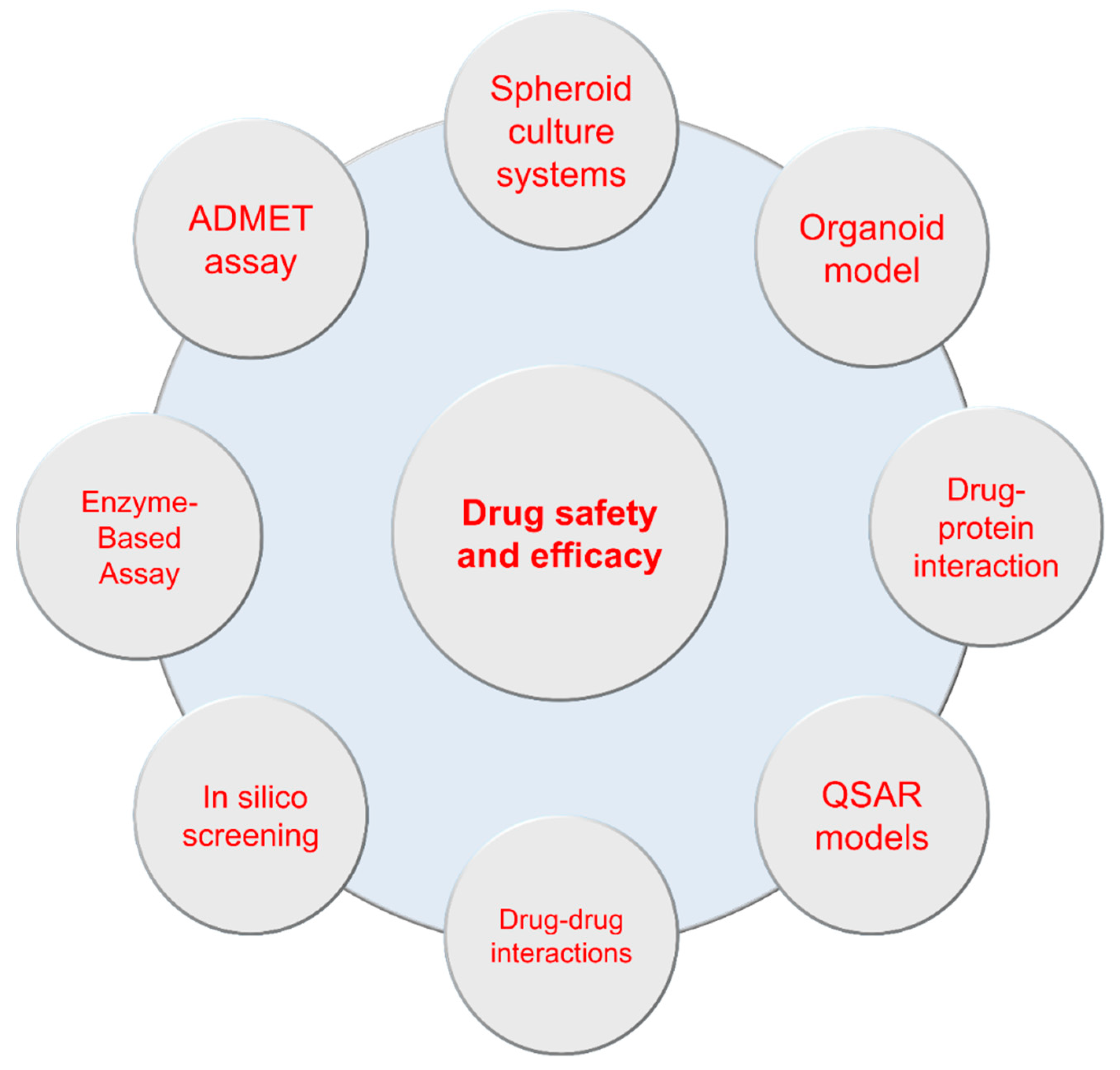
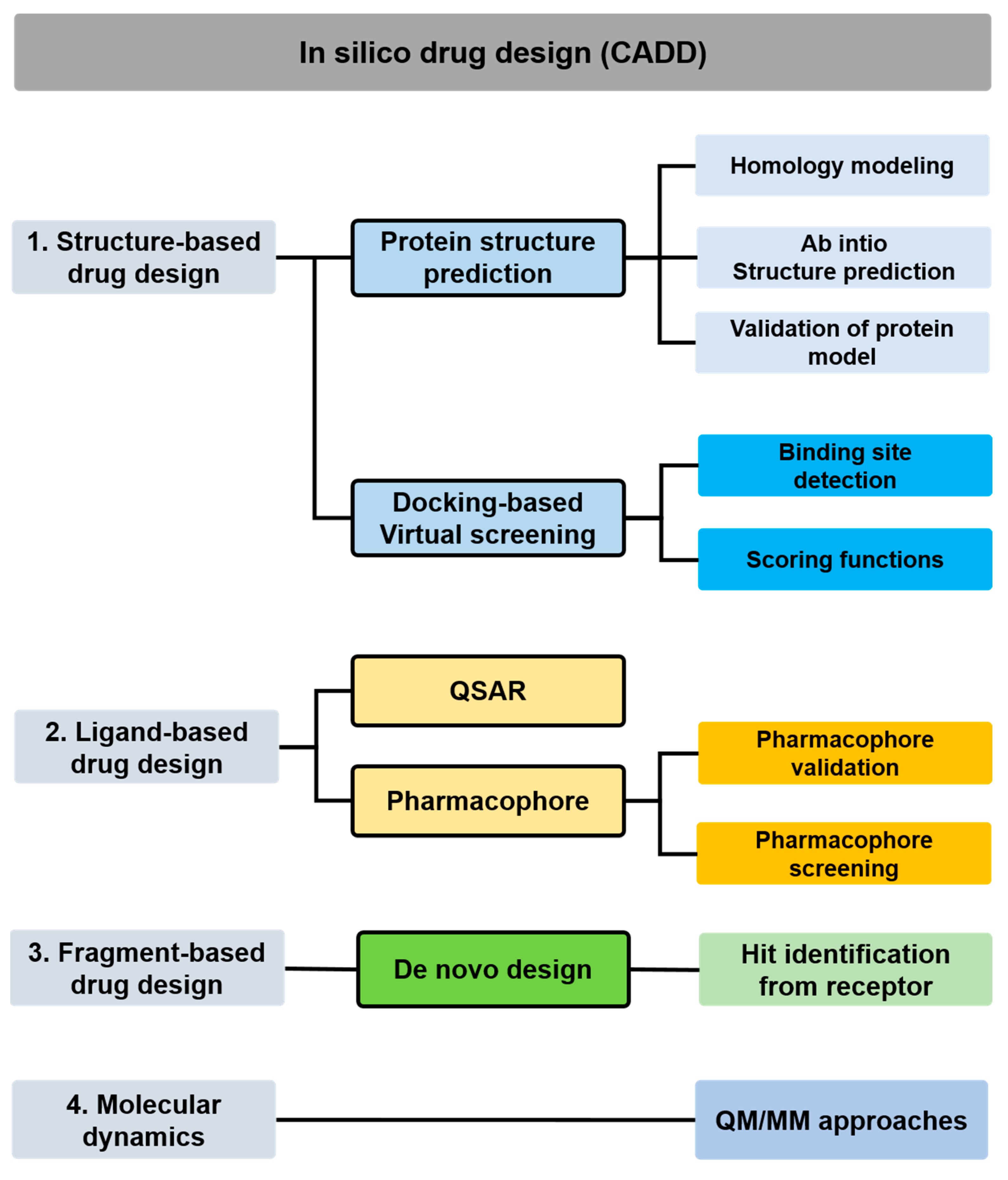
5. In Silico Models for Predicting Drug Safety and Efficacy
5.1. Structure-Based In Silico Methods
5.2. Ligand-Based In Silico Methods
6. Conclusions
Author Contributions
Funding
Institutional Review Board Statement
Informed Consent Statement
Data Availability Statement
Conflicts of Interest
References
- Zabka, T.S.; Burkhardt, J.; Reagan, W.J.; Gautier, J.-C.; Glaab, W.E.; Guffroy, M.; Harding, J.; Brees, D.; McDuffie, E.; Ramaiah, L.; et al. The use of emerging safety biomarkers in nonclinical and clinical safety assessment—The current and future state: An IQ DruSafe industry survey. Regul. Toxicol. Pharmacol. 2021, 120, 104857. [Google Scholar] [CrossRef] [PubMed]
- McDermott, J.E.; Wang, J.; Mitchell, H.; Webb-Robertson, B.J.; Hafen, R.; Ramey, J.; Rodland, K.D. Challenges in Biomarker Discovery: Combining Expert Insights with Statistical Analysis of Complex Omics Data. Expert Opin. Med. Diagn. 2013, 7, 37–51. [Google Scholar] [CrossRef] [PubMed]
- Conway, S.R.; Wong, H.R. Biomarker Panels in Critical Care. Crit. Care Clin. 2020, 36, 89–104. [Google Scholar] [CrossRef] [PubMed]
- Friedman, L.S.; Ostermeyer, E.A.; Lynch, E.D.; Szabo, C.I.; Anderson, L.A.; Dowd, P.; Lee, M.K.; Rowell, S.E.; Boyd, J.; King, M.C. The search for BRCA1. Cancer Res. 1994, 54, 6374–6382. [Google Scholar]
- Provenzano, M.; Rotundo, S.; Chiodini, P.; Gagliardi, I.; Michael, A.; Angotti, E.; Borrelli, S.; Serra, R.; Foti, D.; De Sarro, G.; et al. Contribution of Predictive and Prognostic Biomarkers to Clinical Research on Chronic Kidney Disease. Int. J. Mol. Sci. 2020, 21, 5846. [Google Scholar] [CrossRef]
- Wang, J.; Yin, Y.; Lu, Q.; Zhao, Y.-r.; Hu, Y.-j.; Hu, Y.-Z.; Wang, Z.-Y. Identification of Important Modules and Hub Gene in Chronic Kidney Disease Based on WGCNA. J. Immunol. Res. 2022, 2022, 4615292. [Google Scholar] [CrossRef]
- Sauriasari, R.; Safitri, D.D.; Azmi, N.U. Current updates on protein as biomarkers for diabetic kidney disease: A systematic review. Ther. Adv. Endocrinol. Metab. 2021, 12, 20420188211049612. [Google Scholar] [CrossRef]
- Condrat, C.E.; Thompson, D.C.; Barbu, M.G.; Bugnar, O.L.; Boboc, A.; Cretoiu, D.; Suciu, N.; Cretoiu, S.M.; Voinea, S.C. miRNAs as Biomarkers in Disease: Latest Findings Regarding Their Role in Diagnosis and Prognosis. Cells 2020, 9, 276. [Google Scholar] [CrossRef]
- Ferro, C.J.; Mark, P.B.; Kanbay, M.; Sarafidis, P.; Heine, G.H.; Rossignol, P.; Massy, Z.A.; Mallamaci, F.; Valdivielso, J.M.; Malyszko, J.; et al. Lipid management in patients with chronic kidney disease. Nat. Rev. Nephrol. 2018, 14, 727–749. [Google Scholar] [CrossRef]
- Peng, H.; Liu, X.; Aoieong, C.; Tou, T.; Tsai, T.; Ngai, K.; Cheang, H.I.; Liu, Z.; Liu, P.; Zhu, H. Identification of Metabolite Markers Associated with Kidney Function. J. Immunol. Res. 2022, 2022, 6190333. [Google Scholar] [CrossRef]
- Adam, A.M.; Nasir, S.A.R.; Merchant, A.Z.; Rizvi, A.H.; Rehan, A.; Shaikh, A.T.; Abbas, A.H.; Godil, A.; Khetpal, A.; Mallick, M.S.A.; et al. Efficacy of serum blood urea nitrogen, creatinine and electrolytes in the diagnosis and mortality risk assessment of patients with acute coronary syndrome. Indian Heart J. 2018, 70, 353–359. [Google Scholar] [CrossRef] [PubMed]
- Duan, S.; Li, Y.; Yang, P. Predictive value of blood urea nitrogen in heart failure: A systematic review and meta-analysis. Front. Cardiovasc. Med. 2023, 10, 1189884. [Google Scholar] [CrossRef] [PubMed]
- Lewandowska, L.; Małyszko, J.; Matuszkiewicz-Rowińska, J. Urinary and serum biomarkers for prediction of acute kidney injury in patients undergoing liver transplantation. Ann. Transplant. 2019, 24, 291. [Google Scholar] [CrossRef] [PubMed]
- Groothof, D.; Post, A.; Polinder-Bos, H.A.; Erler, N.S.; Flores-Guerrero, J.L.; Kootstra-Ros, J.E.; Pol, R.A.; de Borst, M.H.; Gansevoort, R.T.; Gans, R.O.B.; et al. Muscle mass and estimates of renal function: A longitudinal cohort study. J. Cachexia Sarcopenia Muscle 2022, 13, 2031–2043. [Google Scholar] [CrossRef] [PubMed]
- Liu, H.; Feng, J.; Tang, L. Early renal structural changes and potential biomarkers in diabetic nephropathy. Front. Physiol. 2022, 13, 1020443. [Google Scholar] [CrossRef]
- Mizdrak, M.; Kumrić, M.; Kurir, T.T.; Božić, J. Emerging Biomarkers for Early Detection of Chronic Kidney Disease. J. Pers. Med. 2022, 12, 548. [Google Scholar] [CrossRef]
- Calvillo, L. Editorial: 3Rs approach (replace, reduce and refine animal models) to improve preclinical research. Front. Physiol. 2022, 13, 1040575. [Google Scholar] [CrossRef]
- Bassan, A.; Alves, V.M.; Amberg, A.; Anger, L.T.; Beilke, L.; Bender, A.; Bernal, A.; Cronin, M.T.D.; Hsieh, J.-H.; Johnson, C.; et al. In silico approaches in organ toxicity hazard assessment: Current status and future needs for predicting heart, kidney and lung toxicities. Comput. Toxicol. 2021, 20, 100188. [Google Scholar] [CrossRef]
- Bufi, R.; Korstanje, R. The impact of genetic background on mouse models of kidney disease. Kidney Int. 2022, 102, 38–44. [Google Scholar] [CrossRef]
- Nangaku, M.; Kitching, A.R.; Boor, P.; Fornoni, A.; Floege, J.; Coates, P.T.; Himmelfarb, J.; Lennon, R.; Anders, H.-J.; Humphreys, B.D.; et al. International Society of Nephrology first consensus guidance for preclinical animal studies in translational nephrology. Kidney Int. 2023, 104, 36–45. [Google Scholar] [CrossRef]
- Bartochowski, P.; Gayrard, N.; Bornes, S.; Druart, C.; Argilés, A.; Cordaillat-Simmons, M.; Duranton, F. Gut–Kidney Axis Investigations in Animal Models of Chronic Kidney Disease. Toxins 2022, 14, 626. [Google Scholar] [CrossRef]
- Mukherjee, P.; Roy, S.; Ghosh, D.; Nandi, S.K. Role of animal models in biomedical research: A review. Lab. Anim. Res. 2022, 38, 18. [Google Scholar] [CrossRef]
- Barroca, N.C.B.; Della Santa, G.; Suchecki, D.; García-Cairasco, N.; de Lima Umeoka, E.H. Challenges in the use of animal models and perspectives for a translational view of stress and psychopathologies. Neurosci. Biobehav. Rev. 2022, 140, 104771. [Google Scholar] [CrossRef]
- Caloni, F.; De Angelis, I.; Hartung, T. Replacement of animal testing by integrated approaches to testing and assessment (IATA): A call for in vivitrosi. Arch. Toxicol. 2022, 96, 1935–1950. [Google Scholar] [CrossRef] [PubMed]
- Zevnik, B.; Jerchow, B.; Buch, T. 3R measures in facilities for the production of genetically modified rodents. Lab. Anim. 2022, 51, 162–177. [Google Scholar] [CrossRef] [PubMed]
- Madani, S.; Machaty, Z.; Vajta, G. An Alternative Way to Improve Mammalian Embryo Development In Vitro: Culture of Zona Pellucida-Free Embryos. Cell. Reprogramming 2022, 24, 111–117. [Google Scholar] [CrossRef] [PubMed]
- Mayasich, S.A.; Goldsmith, M.R.; Mattingly, K.Z.; LaLone, C.A. Combining In Vitro and In Silico New Approach Methods to Investigate Type 3 Iodothyronine Deiodinase Chemical Inhibition Across Species. Environ. Toxicol. Chem. 2023, 42, 1032–1048. [Google Scholar] [CrossRef]
- Thakkar, Y.; Moustakas, H.; Moelijker, N.; Hendriks, G.; Brandsma, I.; Pfuhler, S.; Api, A.M. Utility of ToxTracker in animal alternative testing strategy for fragrance materials. Environ. Mol. Mutagen. 2023, 64, 234–243. [Google Scholar] [CrossRef]
- Krishna, G.; Goel, S.; Krishna, K.A. Chapter 7—Alternative animal toxicity testing and biomarkers. In Biomarkers in Toxicology; Gupta, R.C., Ed.; Academic Press: Boston, MA, USA, 2014; pp. 129–147. [Google Scholar]
- Strimbu, K.; Tavel, J.A. What are biomarkers? Curr. Opin. HIV AIDS 2010, 5, 463–466. [Google Scholar] [CrossRef]
- Goutman, S.A.; Hardiman, O.; Al-Chalabi, A.; Chió, A.; Savelieff, M.G.; Kiernan, M.C.; Feldman, E.L. Recent advances in the diagnosis and prognosis of amyotrophic lateral sclerosis. Lancet Neurol. 2022, 21, 480–493. [Google Scholar] [CrossRef]
- Faham, S.; Salimi, A.; Ghavami, R. Electrochemical-based remote biomarker monitoring: Toward Internet of Wearable Things in telemedicine. Talanta 2023, 253, 123892. [Google Scholar] [CrossRef] [PubMed]
- Bakker, E.; Hendrikse, N.M.; Ehmann, F.; van der Meer, D.S.; Llinares Garcia, J.; Vetter, T.; Starokozhko, V.; Mol, P.G.M. Biomarker Qualification at the European Medicines Agency: A Review of Biomarker Qualification Procedures from 2008 to 2020. Clin. Pharmacol. Ther. 2022, 112, 69–80. [Google Scholar] [CrossRef] [PubMed]
- Califf, R.M. Biomarker definitions and their applications. Exp. Biol. Med. 2018, 243, 213–221. [Google Scholar] [CrossRef] [PubMed]
- Allinovi, M.; Sessa, F.; Villa, G.; Cocci, A.; Innocenti, S.; Zanazzi, M.; Tofani, L.; Paparella, L.; Curi, D.; Cirami, C.L.; et al. Novel Biomarkers for Early Detection of Acute Kidney Injury and Prediction of Long-Term Kidney Function Decline after Partial Nephrectomy. Biomedicines 2023, 11, 1046. [Google Scholar] [CrossRef]
- Wang, Q.; Gümüş, Z.H.; Colarossi, C.; Memeo, L.; Wang, X.; Kong, C.Y.; Boffetta, P. SCLC: Epidemiology, Risk Factors, Genetic Susceptibility, Molecular Pathology, Screening, and Early Detection. J. Thorac. Oncol. 2023, 18, 31–46. [Google Scholar] [CrossRef] [PubMed]
- Saadeh, R.S.; Ramos, P.A.; Algeciras-Schimnich, A.; Flanagan, E.P.; Pittock, S.J.; Willrich, M.A. An Update on Laboratory-Based Diagnostic Biomarkers for Multiple Sclerosis and Beyond. Clin. Chem. 2022, 68, 1134–1150. [Google Scholar] [CrossRef]
- Ding, L.; Gosh, A.; Lee, D.J.; Emri, G.; Huss, W.J.; Bogner, P.N.; Paragh, G. Prognostic biomarkers of cutaneous melanoma. Photodermatol. Photoimmunol. Photomed. 2022, 38, 418–434. [Google Scholar] [CrossRef]
- Pourani, M.R.; Abdollahimajd, F.; Zargari, O.; Shahidi Dadras, M. Soluble biomarkers for diagnosis, monitoring, and therapeutic response assessment in psoriasis. J. Dermatol. Treat. 2022, 33, 1967–1974. [Google Scholar] [CrossRef]
- Salawu, A.; Hernando-Calvo, A.; Chen, R.Y.; Araujo, D.V.; Oliva, M.; Liu, Z.A.; Siu, L.L. Impact of pharmacodynamic biomarkers in immuno-oncology phase 1 clinical trials. Eur. J. Cancer 2022, 173, 167–177. [Google Scholar] [CrossRef]
- Sauer, J.-M.; Porter, A.C. Qualification of translational safety biomarkers. Exp. Biol. Med. 2021, 246, 2391–2398. [Google Scholar] [CrossRef]
- Priyadarshini, G.; Rajappa, M. Predictive markers in chronic kidney disease. Clin. Chim. Acta 2022, 535, 180–186. [Google Scholar] [CrossRef] [PubMed]
- Griffin, B.R.; Faubel, S.; Edelstein, C.L. Biomarkers of Drug-Induced Kidney Toxicity. Ther. Drug Monit. 2019, 41, 213–226. [Google Scholar] [CrossRef] [PubMed]
- Tanase, D.M.; Gosav, E.M.; Radu, S.; Costea, C.F.; Ciocoiu, M.; Carauleanu, A.; Lacatusu, C.M.; Maranduca, M.A.; Floria, M.; Rezus, C. The Predictive Role of the Biomarker Kidney Molecule-1 (KIM-1) in Acute Kidney Injury (AKI) Cisplatin-Induced Nephrotoxicity. Int. J. Mol. Sci. 2019, 20, 5238. [Google Scholar] [CrossRef] [PubMed]
- Srisawat, N.; Kellum, J.A. The Role of Biomarkers in Acute Kidney Injury. Crit. Care Clin. 2020, 36, 125–140. [Google Scholar] [CrossRef] [PubMed]
- Chen, Y.-T.; Jenq, C.-C.; Hsu, C.-K.; Yu, Y.-C.; Chang, C.-H.; Fan, P.-C.; Pan, H.-C.; Wu, I.W.; Cherng, W.-J.; Chen, Y.-C. Acute kidney disease and acute kidney injury biomarkers in coronary care unit patients. BMC Nephrol. 2020, 21, 207. [Google Scholar] [CrossRef]
- Tajima, S.; Yamamoto, N.; Masuda, S. Clinical prospects of biomarkers for the early detection and/or prediction of organ injury associated with pharmacotherapy. Biochem. Pharmacol. 2019, 170, 113664. [Google Scholar] [CrossRef]
- El Alam, S.; Pena, E.; Aguilera, D.; Siques, P.; Brito, J. Inflammation in Pulmonary Hypertension and Edema Induced by Hypobaric Hypoxia Exposure. Int. J. Mol. Sci. 2022, 23, 12656. [Google Scholar] [CrossRef]
- Singh, V.; Kaur, R.; Kumari, P.; Pasricha, C.; Singh, R. ICAM-1 and VCAM-1: Gatekeepers in various inflammatory and cardiovascular disorders. Clin. Chim. Acta 2023, 548, 117487. [Google Scholar] [CrossRef]
- Mills, K.H.G. IL-17 and IL-17-producing cells in protection versus pathology. Nat. Rev. Immunol. 2023, 23, 38–54. [Google Scholar] [CrossRef]
- Aliyu, M.; Zohora, F.T.; Anka, A.U.; Ali, K.; Maleknia, S.; Saffarioun, M.; Azizi, G. Interleukin-6 cytokine: An overview of the immune regulation, immune dysregulation, and therapeutic approach. Int. Immunopharmacol. 2022, 111, 109130. [Google Scholar] [CrossRef]
- Wang, Y.; Zhang, Y.; Shou, S.; Jin, H. The role of IL-17 in acute kidney injury. Int. Immunopharmacol. 2023, 119, 110307. [Google Scholar] [CrossRef] [PubMed]
- Habanjar, O.; Bingula, R.; Decombat, C.; Diab-Assaf, M.; Caldefie-Chezet, F.; Delort, L. Crosstalk of Inflammatory Cytokines within the Breast Tumor Microenvironment. Int. J. Mol. Sci. 2023, 24, 4002. [Google Scholar] [CrossRef] [PubMed]
- Siegmund, D.; Wajant, H. TNF and TNF receptors as therapeutic targets for rheumatic diseases and beyond. Nat. Rev. Rheumatol. 2023, 19, 576–591. [Google Scholar] [CrossRef] [PubMed]
- Matsumori, A. Nuclear Factor-κB is a Prime Candidate for the Diagnosis and Control of Inflammatory Cardiovascular Disease. Eur. Cardiol. 2023, 18, e40. [Google Scholar] [CrossRef] [PubMed]
- Prasad, S.; Kumar, V.; Singh, C.; Singh, A. Crosstalk between phytochemicals and inflammatory signaling pathways. Inflammopharmacology 2023, 31, 1117–1147. [Google Scholar] [CrossRef]
- Baião, V.M.; Cunha, V.A.; Duarte, M.P.; Andrade, F.P.; Ferreira, A.P.; Nóbrega, O.T.; Viana, J.L.; Ribeiro, H.S. Effects of Exercise on Inflammatory Markers in Individuals with Chronic Kidney Disease: A Systematic Review and Meta-Analysis. Metabolites 2023, 13, 795. [Google Scholar] [CrossRef]
- Singh, S.; Anshita, D.; Ravichandiran, V. MCP-1: Function, regulation, and involvement in disease. Int. Immunopharmacol. 2021, 101, 107598. [Google Scholar] [CrossRef]
- Lousa, I.; Reis, F.; Santos-Silva, A.; Belo, L. The Signaling Pathway of TNF Receptors: Linking Animal Models of Renal Disease to Human CKD. Int. J. Mol. Sci. 2022, 23, 3284. [Google Scholar] [CrossRef]
- Lousa, I.; Reis, F.; Viana, S.; Vieira, P.; Vala, H.; Belo, L.; Santos-Silva, A. TNFR2 as a Potential Biomarker for Early Detection and Progression of CKD. Biomolecules 2023, 13, 534. [Google Scholar] [CrossRef]
- Khanijou, V.; Zafari, N.; Coughlan, M.T.; MacIsaac, R.J.; Ekinci, E.I. Review of potential biomarkers of inflammation and kidney injury in diabetic kidney disease. Diabetes Metab. Res. Rev. 2022, 38, e3556. [Google Scholar] [CrossRef]
- Gohda, T.; Yanagisawa, N.; Murakoshi, M.; Ueda, S.; Nishizaki, Y.; Nojiri, S.; Ohashi, Y.; Ohno, I.; Shibagaki, Y.; Imai, N.; et al. Association between Kidney Function Decline and Baseline TNFR Levels or Change Ratio in TNFR by Febuxostat Chiefly in Non-diabetic CKD Patients with Asymptomatic Hyperuricemia. Front. Med. 2021, 8, 634932. [Google Scholar] [CrossRef] [PubMed]
- Troth, S.P.; Vlasakova, K.; Amur, S.; Amin, R.P.; Glaab, W.E. Translational Safety Biomarkers of Kidney Injury. Semin. Nephrol. 2019, 39, 202–214. [Google Scholar] [CrossRef] [PubMed]
- Akcay, A.; Nguyen, Q.; Edelstein, C.L. Mediators of Inflammation in Acute Kidney Injury. Mediat. Inflamm. 2009, 2009, 137072. [Google Scholar] [CrossRef] [PubMed]
- Jana, S.; Mitra, P.; Roy, S. Proficient Novel Biomarkers Guide Early Detection of Acute Kidney Injury: A Review. Diseases 2023, 11, 8. [Google Scholar] [CrossRef] [PubMed]
- Shimosawa, T. How animal models can be utilized to find new biomarkers for cardiovascular diseases. Clin. Sci. 2023, 137, 527–535. [Google Scholar] [CrossRef]
- Li, Y.; Ma, J.; Diao, J.; Chen, W.; Wang, Z. Esmolol inhibits cognitive impairment and neuronal inflammation in mice with sepsis-induced brain injury. Transl. Neurosci. 2023, 14, 20220297. [Google Scholar] [CrossRef]
- Grande, M.T.; Pérez-Barriocanal, F.; López-Novoa, J.M. Role of inflammation in tubulo-interstitial damage associated to obstructive nephropathy. J. Inflamm. 2010, 7, 19. [Google Scholar] [CrossRef]
- Burek, M.; Burmester, S.; Salvador, E.; Möller-Ehrlich, K.; Schneider, R.; Roewer, N.; Nagai, M.; Förster, C.Y. Kidney Ischemia/Reperfusion Injury Induces Changes in the Drug Transporter Expression at the Blood–Brain Barrier in vivo and in vitro. Front. Physiol. 2020, 11, 569881. [Google Scholar] [CrossRef]
- Perše, M.; Večerić-Haler, Ž. Cisplatin-Induced Rodent Model of Kidney Injury: Characteristics and Challenges. BioMed Res. Int. 2018, 2018, 1462802. [Google Scholar] [CrossRef]
- Zaghloul, M.S.; Abdelrahman, R.S. Nilotinib ameliorates folic acid-induced acute kidney injury through modulation of TWEAK and HSP-70 pathways. Toxicology 2019, 427, 152303. [Google Scholar] [CrossRef]
- Al Asmari, A.K.; Al Sadoon, K.T.; Obaid, A.A.; Yesunayagam, D.; Tariq, M. Protective effect of quinacrine against glycerol-induced acute kidney injury in rats. BMC Nephrol. 2017, 18, 41. [Google Scholar] [CrossRef] [PubMed]
- Udupa, V.; Prakash, V. Gentamicin induced acute renal damage and its evaluation using urinary biomarkers in rats. Toxicol. Rep. 2019, 6, 91–99. [Google Scholar] [CrossRef] [PubMed]
- Brodsky, S.V. Anticoagulants and acute kidney injury: Clinical and pathology considerations. Kidney Res. Clin. Pract. 2014, 33, 174–180. [Google Scholar] [CrossRef] [PubMed]
- van Koppen, A.; Verhaar, M.C.; Bongartz, L.G.; Joles, J.A. 5/6th Nephrectomy in Combination with High Salt Diet and Nitric Oxide Synthase Inhibition to Induce Chronic Kidney Disease in the Lewis Rat. J. Vis. Exp. 2013, 77, e50398. [Google Scholar] [CrossRef]
- Maranduca, M.A.; Clim, A.; Pinzariu, A.C.; Statescu, C.; Sascau, R.A.; Tanase, D.M.; Serban, D.N.; Branisteanu, D.C.; Branisteanu, D.E.; Huzum, B.; et al. Role of arterial hypertension and angiotensin II in chronic kidney disease (Review). Exp. Ther. Med. 2023, 25, 153. [Google Scholar] [CrossRef]
- Deeds, M.C.; Anderson, J.M.; Armstrong, A.S.; Gastineau, D.A.; Hiddinga, H.J.; Jahangir, A.; Eberhardt, N.L.; Kudva, Y.C. Single dose streptozotocin-induced diabetes: Considerations for study design in islet transplantation models. Lab. Anim. 2011, 45, 131–140. [Google Scholar] [CrossRef]
- Jefferson, J.A.; Pippin, J.W.; Shankland, S.J. Experimental models of membranous nephropathy. Drug Discov. Today Dis. Models 2010, 7, 27–33. [Google Scholar] [CrossRef]
- Kuwabara, S.; Goggins, E.; Okusa, M.D. The Pathophysiology of Sepsis-Associated AKI. Clin. J. Am. Soc. Nephrol. 2022, 17, 1050–1069. [Google Scholar] [CrossRef]
- Yu, X.; Yao, H.; Zhang, X.; Liu, L.; Liu, S.; Dong, Y. Comparison of LPS and MS-induced depressive mouse model: Behavior, inflammation and biochemical changes. BMC Psychiatry 2022, 22, 590. [Google Scholar] [CrossRef]
- Drechsler, S.; Osuchowski, M. Cecal Ligation and Puncture. In Sepsis: Methods and Protocols; Walker, W.E., Ed.; Springer: New York, NY, USA, 2021; pp. 1–8. [Google Scholar] [CrossRef]
- Thomas, R.C.; Bath, M.F.; Stover, C.M.; Lambert, D.G.; Thompson, J.P. Exploring LPS-induced sepsis in rats and mice as a model to study potential protective effects of the nociceptin/orphanin FQ system. Peptides 2014, 61, 56–60. [Google Scholar] [CrossRef]
- Kim, Y.K.; Hwang, J.H.; Lee, H.T. Differential susceptibility to lipopolysaccharide affects the activation of toll-like-receptor 4 signaling in THP-1 cells and PMA-differentiated THP-1 cells. Innate Immun. 2022, 28, 122–129. [Google Scholar] [CrossRef] [PubMed]
- Schabbauer, G. Polymicrobial sepsis models: CLP versus CASP. Drug Discov. Today Dis. Models 2012, 9, e17–e21. [Google Scholar] [CrossRef]
- Al-Githmi, I.S.; Abdulqader, A.A.; Alotaibi, A.; Aldughather, B.A.; Alsulami, O.A.; Wali, S.M.; Alghamdi, M.S.; Althabaiti, T.S.; Melebary, T.B. Acute Kidney Injury After Open Heart Surgery. Cureus 2022, 14, e25899. [Google Scholar] [CrossRef] [PubMed]
- Han, L.; Fan, L.; Xingyu, J.; Weijing, Z.; Tong, X.; Qiuzhong, P.; Liming, C.; Jingjing, Z.; Desheng, W.; Yue, L.; et al. CircITGB6 promotes ovarian cancer cisplatin resistance by resetting tumor-associated macrophage polarization toward the M2 phenotype. J. Immunother. Cancer 2022, 10, e004029. [Google Scholar] [CrossRef]
- Koshkin, V.S.; Barata, P.C.; Rybicki, L.A.; Zahoor, H.; Almassi, N.; Redden, A.M.; Fergany, A.F.; Kaouk, J.; Haber, G.-P.; Stephenson, A.J.; et al. Feasibility of Cisplatin-Based Neoadjuvant Chemotherapy in Muscle-Invasive Bladder Cancer Patients with Diminished Renal Function. Clin. Genitourin. Cancer 2018, 16, e879–e892. [Google Scholar] [CrossRef] [PubMed]
- Li, Z.; Li, S.; Wen, Y.; Chen, J.; Liu, K.; Jia, J. MiR-495 Inhibits Cisplatin Resistance and Angiogenesis in Esophageal Cancer by Targeting ATP7A. Technol. Cancer Res. Treat. 2021, 20, 15330338211039127. [Google Scholar] [CrossRef]
- Zhang, Q.; Luo, P.; Chen, J.; Yang, C.; Xia, F.; Zhang, J.; Tang, H.; Liu, D.; Gu, L.; Shi, Q.; et al. Dissection of Targeting Molecular Mechanisms of Aristolochic Acid-induced Nephrotoxicity via a Combined Deconvolution Strategy of Chemoproteomics and Metabolomics. Int. J. Mol. Sci. 2022, 18, 2003–2017. [Google Scholar] [CrossRef]
- Ponce de León-Rodríguez, M.d.C.; Guyot, J.-P.; Laurent-Babot, C. Intestinal in vitro cell culture models and their potential to study the effect of food components on intestinal inflammation. Crit. Rev. Food Sci. Nutr. 2019, 59, 3648–3666. [Google Scholar] [CrossRef]
- Kicha, A.A.; Kalinovsky, A.I.; Malyarenko, T.V.; Malyarenko, O.S.; Ermakova, S.P.; Popov, R.S.; Stonik, V.A.; Ivanchina, N.V. Disulfated ophiuroid type steroids from the Far Eastern starfish Pteraster marsippus and their cytotoxic activity on the models of 2D and 3D cultures. Mar. Drugs 2022, 20, 164. [Google Scholar] [CrossRef]
- Mittal, R.; Woo, F.W.; Castro, C.S.; Cohen, M.A.; Karanxha, J.; Mittal, J.; Chhibber, T.; Jhaveri, V.M. Organ-on-chip models: Implications in drug discovery and clinical applications. J. Cell. Physiol. 2019, 234, 8352–8380. [Google Scholar] [CrossRef]
- Huang, S.-M.; Strong, J.M.; Zhang, L.; Reynolds, K.S.; Nallani, S.; Temple, R.; Abraham, S.; Habet, S.A.; Baweja, R.K.; Burckart, G.J.; et al. New Era in Drug Interaction Evaluation: US Food and Drug Administration Update on CYP Enzymes, Transporters, and the Guidance Process. J. Clin. Pharmacol. 2008, 48, 662–670. [Google Scholar] [CrossRef] [PubMed]
- Hall, A.M.; Trepiccione, F.; Unwin, R.J. Drug toxicity in the proximal tubule: New models, methods and mechanisms. Pediatr. Nephrol. 2022, 37, 973–982. [Google Scholar] [CrossRef] [PubMed]
- Petreski, T.; Varda, L.; Gradišnik, L.; Maver, U.; Bevc, S. Renal Proximal Tubular Epithelial Cells: From Harvesting to Use in Studies. Nephron 2023, 7, 1–5. [Google Scholar] [CrossRef] [PubMed]
- Syed Mohamed, S.M.D.; Welsh, G.I.; Roy, I. Renal tissue engineering for regenerative medicine using polymers and hydrogels. Biomater. Sci. 2023, 11, 5706–5726. [Google Scholar] [CrossRef] [PubMed]
- Shapiro, D.D.; Virumbrales-Muñoz, M.; Beebe, D.J.; Abel, E.J. Models of Renal Cell Carcinoma Used to Investigate Molecular Mechanisms and Develop New Therapeutics. Front. Oncol. 2022, 12, 871252. [Google Scholar] [CrossRef]
- Ziegler, W.H.; Lüdiger, S.; Hassan, F.; Georgiadis, M.E.; Swolana, K.; Khera, A.; Mertens, A.; Franke, D.; Wohlgemuth, K.; Dahmer-Heath, M.; et al. Primary URECs: A source to better understand the pathology of renal tubular epithelia in pediatric hereditary cystic kidney diseases. Orphanet J. Rare Dis. 2022, 17, 122. [Google Scholar] [CrossRef]
- Yu, P.; Duan, Z.; Liu, S.; Pachon, I.; Ma, J.; Hemstreet, G.P.; Zhang, Y. Drug-Induced Nephrotoxicity Assessment in 3D Cellular Models. Micromachines 2022, 13, 3. [Google Scholar] [CrossRef]
- Lake, B.B.; Menon, R.; Winfree, S.; Hu, Q.; Melo Ferreira, R.; Kalhor, K.; Barwinska, D.; Otto, E.A.; Ferkowicz, M.; Diep, D.; et al. An atlas of healthy and injured cell states and niches in the human kidney. Nature 2023, 619, 585–594. [Google Scholar] [CrossRef]
- Perazella, M.A. Drug-induced acute kidney injury: Diverse mechanisms of tubular injury. Curr. Opin. Crit. Care 2019, 25, 550–557. [Google Scholar] [CrossRef]
- Le, T.A.; Hiba, T.; Chaudhari, D.; Preston, A.N.; Palowsky, Z.R.; Ahmadzadeh, S.; Shekoohi, S.; Cornett, E.M.; Kaye, A.D. Aminoglycoside-Related Nephrotoxicity and Ototoxicity in Clinical Practice: A Review of Pathophysiological Mechanism and Treatment Options. Adv. Ther. 2023, 40, 1357–1365. [Google Scholar] [CrossRef]
- Bartlett, C.S.; Jeansson, M.; Quaggin, S.E. Vascular Growth Factors and Glomerular Disease. Annu. Rev. Physiol. 2016, 78, 437–461. [Google Scholar] [CrossRef] [PubMed]
- Soo, J.Y.C.; Jansen, J.; Masereeuw, R.; Little, M.H. Advances in predictive in vitro models of drug-induced nephrotoxicity. Nat. Rev. Nephrol. 2018, 14, 378–393. [Google Scholar] [CrossRef] [PubMed]
- Ryan, S.-L.; Baird, A.-M.; Vaz, G.; Urquhart, A.J.; Senge, h.; Richard, D.J.; O’Byrne, K.J.; Davies, A.M. Drug Discovery Approaches Utilizing Three-Dimensional Cell Culture. Assay Drug Dev. Technol. 2016, 14, 19–28. [Google Scholar] [CrossRef] [PubMed]
- Musah-Eroje, A.; Watson, S. A novel 3D in vitro model of glioblastoma reveals resistance to temozolomide which was potentiated by hypoxia. J. Neurooncol. 2019, 142, 231–240. [Google Scholar] [CrossRef]
- Huang, X.; Huang, Z.; Gao, W.; Gao, W.; He, R.; Li, Y.; Crawford, R.; Zhou, Y.; Xiao, L.; Xiao, Y. Current Advances in 3D Dynamic Cell Culture Systems. Gels 2022, 8, 829. [Google Scholar] [CrossRef]
- Suarez-Martinez, E.; Suazo-Sanchez, I.; Celis-Romero, M.; Carnero, A. 3D and organoid culture in research: Physiology, hereditary genetic diseases and cancer. Cell Biosci. 2022, 12, 39. [Google Scholar] [CrossRef]
- Bas-Cristóbal Menéndez, A.; Du, Z.; van den Bosch, T.P.P.; Othman, A.; Gaio, N.; Silvestri, C.; Quirós, W.; Lin, H.; Korevaar, S.; Merino, A.; et al. Creating a kidney organoid-vasculature interaction model using a novel organ-on-chip system. Sci. Rep. 2022, 12, 20699. [Google Scholar] [CrossRef]
- Safi, W.; Marco, A.; Moya, D.; Prado, P.; Garreta, E.; Montserrat, N. Assessing kidney development and disease using kidney organoids and CRISPR engineering. Front. Cell Dev. Biol. 2022, 10, 948395. [Google Scholar] [CrossRef]
- Dvela-Levitt, M.; Kost-Alimova, M.; Emani, M.; Kohnert, E.; Thompson, R.; Sidhom, E.-H.; Rivadeneira, A.; Sahakian, N.; Roignot, J.; Papagregoriou, G.; et al. Small Molecule Targets TMED9 and Promotes Lysosomal Degradation to Reverse Proteinopathy. Cell 2019, 178, 521–535.e523. [Google Scholar] [CrossRef]
- Forbes, T.A.; Howden, S.E.; Lawlor, K.; Phipson, B.; Maksimovic, J.; Hale, L.; Wilson, S.; Quinlan, C.; Ho, G.; Holman, K.; et al. Patient-iPSC-Derived Kidney Organoids Show Functional Validation of a Ciliopathic Renal Phenotype and Reveal Underlying Pathogenetic Mechanisms. Am. J. Hum. Genet. 2018, 102, 816–831. [Google Scholar] [CrossRef]
- Low, J.H.; Li, P.; Chew, E.G.Y.; Zhou, B.; Suzuki, K.; Zhang, T.; Lian, M.M.; Liu, M.; Aizawa, E.; Rodriguez Esteban, C.; et al. Generation of Human PSC-Derived Kidney Organoids with Patterned Nephron Segments and a De Novo Vascular Network. Cell Stem Cell 2019, 25, 373–387.e379. [Google Scholar] [CrossRef] [PubMed]
- Kim, Y.K.; Refaeli, I.; Brooks, C.R.; Jing, P.; Gulieva, R.E.; Hughes, M.R.; Cruz, N.M.; Liu, Y.; Churchill, A.J.; Wang, Y.; et al. Gene-Edited Human Kidney Organoids Reveal Mechanisms of Disease in Podocyte Development. Stem Cells 2017, 35, 2366–2378. [Google Scholar] [CrossRef]
- Little, M.H.; Combes, A.N. Kidney organoids: Accurate models or fortunate accidents. Genes Dev. 2019, 33, 1319–1345. [Google Scholar] [CrossRef] [PubMed]
- Nicholson, M.W.; Ting, C.-Y.; Chan, D.Z.H.; Cheng, Y.-C.; Lee, Y.-C.; Hsu, C.-C.; Huang, C.-Y.; Hsieh, P.C.H. Utility of iPSC-Derived Cells for Disease Modeling, Drug Development, and Cell Therapy. Cells 2022, 11, 1853. [Google Scholar] [CrossRef] [PubMed]
- Sterling, T.; Irwin, J.J. ZINC 15—Ligand Discovery for Everyone. J. Chem. Inf. Model. 2015, 55, 2324–2337. [Google Scholar] [CrossRef] [PubMed]
- Wishart, D.S.; Feunang, Y.D.; Guo, A.C.; Lo, E.J.; Marcu, A.; Grant, J.R.; Sajed, T.; Johnson, D.; Li, C.; Sayeeda, Z.; et al. DrugBank 5.0: A major update to the DrugBank database for 2018. Nucleic Acids Res. 2018, 46, D1074–D1082. [Google Scholar] [CrossRef]
- Douguet, D. Data Sets Representative of the Structures and Experimental Properties of FDA-Approved Drugs. ACS Med. Chem. Lett. 2018, 9, 204–209. [Google Scholar] [CrossRef]
- Meisburger, S.P.; Case, D.A.; Ando, N. Robust total X-ray scattering workflow to study correlated motion of proteins in crystals. Nat. Commun. 2023, 14, 1228. [Google Scholar] [CrossRef]
- Vant, J.W.; Sarkar, D.; Nguyen, J.; Baker, A.T.; Vermaas, J.V.; Singharoy, A. Exploring cryo-electron microscopy with molecular dynamics. Biochem. Soc. Trans. 2022, 50, 569–581. [Google Scholar] [CrossRef]
- Oxenfarth, A.; Kümmerer, F.; Bottaro, S.; Schnieders, R.; Pinter, G.; Jonker, H.R.A.; Fürtig, B.; Richter, C.; Blackledge, M.; Lindorff-Larsen, K.; et al. Integrated NMR/Molecular Dynamics Determination of the Ensemble Conformation of a Thermodynamically Stable CUUG RNA Tetraloop. J. Am. Chem. Soc. 2023, 145, 16557–16572. [Google Scholar] [CrossRef]
- Bogetti, X.; Saxena, S.K. Integrating Electron Paramagnetic Resonance Spectroscopy and Computational Modeling to Measure Protein Structure and Dynamics. ChemPlusChem 2023, e202300506. [Google Scholar] [CrossRef] [PubMed]
- Hirashima, S.; Park, S.; Sugiyama, H. Evaluation by Experimentation and Simulation of a FRET Pair Comprising Fluorescent Nucleobase Analogs in Nucleosomes. Chem. A Eur. J. 2023, 29, e202203961. [Google Scholar] [CrossRef] [PubMed]
- Mayanja, R.; Kintu, C.; Diabate, O.; Soremekun, O.; Oluwagbemi, O.O.; Wele, M.; Kalyesubula, R.; Jjingo, D.; Chikowore, T.; Fatumo, S. Molecular Dynamic Simulation Reveals Structure Differences in APOL1 Variants and Implication in Pathogenesis of Chronic Kidney Disease. Genes 2022, 13, 1460. [Google Scholar] [CrossRef] [PubMed]
- Loew, G.H.; Villar, H.O.; Alkorta, I. Strategies for Indirect Computer-Aided Drug Design. Pharm. Res. 1993, 10, 475–486. [Google Scholar] [CrossRef] [PubMed]
- Koehn, F.E.; Carter, G.T. The evolving role of natural products in drug discovery. Nat. Rev. Drug Discov. 2005, 4, 206–220. [Google Scholar] [CrossRef]
- Huang, H.-J.; Lee, Y.-H.; Chou, C.-L.; Zheng, C.-M.; Chiu, H.-W. Investigation of potential descriptors of chemical compounds on prevention of nephrotoxicity via QSAR approach. Comput. Struct. Biotechnol. J. 2022, 20, 1876–1884. [Google Scholar] [CrossRef]
- Huang, H.-J.; Lee, Y.-H.; Sung, L.-C.; Chen, Y.-J.; Chiu, Y.-J.; Chiu, H.-W.; Zheng, C.-M. Drug repurposing screens to identify potential drugs for chronic kidney disease by targeting prostaglandin E2 receptor. Comput. Struct. Biotechnol. J. 2023, 21, 3490–3502. [Google Scholar] [CrossRef]


| The Functional Unit of the Kidney | Biomarkers | Nephrotoxic Drug |
|---|---|---|
| Proximal tubule | Interferons, Interleukins, TNF, CSFs, KIM-1, NGAL, Clusterin, Osteopontin, NAG, Beta-2 microglobulin, Albumin | Aminoglycoside, Antibiotics, Amphotericin B, Adefovir, Cyclosporine, Cisplatin, Foscarnet Contrast stain, Cocaine, Heroin, Methadone Methamphetamine Gentamicin, Vancomycin |
| Distal tubule | Serum cystatin C, NGAL, Clusterin, Osteopontin | Aminoglycosides, Amphoterin B, Radiocontrast dye, Methotrexate, Vancomycin |
| Glomerulus | Interferons, Interleukins, TNF, CSFs, Type IV collagen | ACE inhibitor, ARB, NSAIDs, Mitomycin-C, Antiplatelet, Cyclosporin, Quinone |
| Pathology | Experimental Models | Experimental Strategy | References |
|---|---|---|---|
| Sepsis-induced AKI | Sepsis | Sepsis induced by intraperitoneal injection of 10 mg/kg LPS | [67] |
| Obstructive AKI | Unilateral ureteric obstruction | 1–2 weeks of induction for renal fibrosis | [68] |
| Ischemia-reperfusion injury | Bilateral renal ischemia for 30 min and then treatment with reperfusion for 24 h | [69] | |
| Chemical induced AKI | Cisplatin | 20 mg/kg of cisplatin within 72 h | [70] |
| Folic acid | A single dose of 250 mg/kg Folic acid injection for AKI induction | [71] | |
| Glycerol | 24 h of water deprivation and a single injection of 3–30 mg/kg 25% glycerol | [72] | |
| Gentamicin | Dose range 30–200 mg/kg for 4–8 days | [73] | |
| Warfarin | 5/6 nephrectomy rats for 3 weeks and then treated with warfarin for 8 days | [74] | |
| CKD caused by mass reduction | 5/6 nephrectomy (rats) | Renal mass reduction by one-sided nephrectomy after removal of the right kidney 1 week | [75] |
| CKD induced by hypertension | Angiotensin II infusion models | Induction of fibrosis by the increase in the circulating level of angiotensin II | [76] |
| Diabetic nephropathy | Streptozotocin mice/rats | A single dose of 200 mg/kg Streptozotocin is directly toxic to pancreatic beta-cells | [77] |
| Membranous nephropathy | Heymann nephritis rats | Anti- fraction 1A (Fx1A) antiserum in Sprague-Dawley rats | [78] |
Disclaimer/Publisher’s Note: The statements, opinions and data contained in all publications are solely those of the individual author(s) and contributor(s) and not of MDPI and/or the editor(s). MDPI and/or the editor(s) disclaim responsibility for any injury to people or property resulting from any ideas, methods, instructions or products referred to in the content. |
© 2023 by the authors. Licensee MDPI, Basel, Switzerland. This article is an open access article distributed under the terms and conditions of the Creative Commons Attribution (CC BY) license (https://creativecommons.org/licenses/by/4.0/).
Share and Cite
Huang, H.-J.; Chou, C.-L.; Sandar, T.T.; Liu, W.-C.; Yang, H.-C.; Lin, Y.-C.; Zheng, C.-M.; Chiu, H.-W. Currently Used Methods to Evaluate the Efficacy of Therapeutic Drugs and Kidney Safety. Biomolecules 2023, 13, 1581. https://doi.org/10.3390/biom13111581
Huang H-J, Chou C-L, Sandar TT, Liu W-C, Yang H-C, Lin Y-C, Zheng C-M, Chiu H-W. Currently Used Methods to Evaluate the Efficacy of Therapeutic Drugs and Kidney Safety. Biomolecules. 2023; 13(11):1581. https://doi.org/10.3390/biom13111581
Chicago/Turabian StyleHuang, Hung-Jin, Chu-Lin Chou, Tin Tin Sandar, Wen-Chih Liu, Hsiu-Chien Yang, Yen-Chung Lin, Cai-Mei Zheng, and Hui-Wen Chiu. 2023. "Currently Used Methods to Evaluate the Efficacy of Therapeutic Drugs and Kidney Safety" Biomolecules 13, no. 11: 1581. https://doi.org/10.3390/biom13111581
APA StyleHuang, H.-J., Chou, C.-L., Sandar, T. T., Liu, W.-C., Yang, H.-C., Lin, Y.-C., Zheng, C.-M., & Chiu, H.-W. (2023). Currently Used Methods to Evaluate the Efficacy of Therapeutic Drugs and Kidney Safety. Biomolecules, 13(11), 1581. https://doi.org/10.3390/biom13111581