Brief Report

HOXA Amplification Defines a Genetically Distinct Subset of Angiosarcomas

by
Hongbo M. Xie
1,2 and
Kathrin M. Bernt
1,3,4,*
1
Division of Pediatric Oncology, Department of Pediatrics, Center for Childhood Cancer Research, Children’s Hospital of Philadelphia, 3501 Civic Center Boulevard, CTRB 3064, Philadelphia, PA 19104, USA
2
Department of Bioinformatics and Health Informatics (DBHI), Children’s Hospital of Philadelphia, Philadelphia, PA 19104, USA
3
Department of Pediatrics, Perelman School of Medicine at the University of Pennsylvania, Philadelphia, PA 19104, USA
4
Abramson Cancer Center, Philadelphia, PA 19106, USA
*
Author to whom correspondence should be addressed.
Biomolecules 2022, 12(8), 1124; https://doi.org/10.3390/biom12081124
Submission received: 5 July 2022 / Revised: 14 August 2022 / Accepted: 15 August 2022 / Published: 16 August 2022

Abstract

:
Angiosarcoma is a rare, devastating malignancy with few curative options for disseminated disease. We analyzed a recently published genomic data set of 48 angiosarcomas and noticed recurrent amplifications of HOXA-cluster genes in 33% of patients. HOXA genes are master regulators of embryonic vascular development and adult neovascularization, which provides a molecular rationale to suspect that amplified HOXA genes act as oncogenes in angiosarcoma. HOXA amplifications typically affected multiple pro-angiogenic HOXA genes and co-occurred with amplifications of CD36 and KDR, whereas the overall mutation rate in these tumors was relatively low. HOXA amplifications were found most commonly in angiosarcomas located in the breast and were rare in angiosarcomas arising in sun-exposed areas on the head, neck, face and scalp. Our data suggest that HOXA-amplified angiosarcoma is a distinct molecular subgroup. Efforts to develop therapies targeting oncogenic HOX gene expression in AML and other sarcomas may have relevance for HOXA-amplified angiosarcoma.

Graphical Abstract

1. Introduction

Angiosarcoma is a rare malignancy with a poor prognosis. The 5-year survival rates are about 30% with conventional approaches, and survival is almost entirely dependent on surgical resection of low-stage disease [1,2].
Due to the low incidence of angiosarcoma, large genomic studies have been challenging. One of the largest cohorts reported to date includes 48 samples from 36 patients who underwent comprehensive genomic profiling of archived tumor samples [3]. This study reported substantial heterogeneity in the mutational landscape of angiosarcoma related to location. Tumors from sun-exposed sites (head, neck, face and scalp (HNFS)) showed a pattern of high mutational burden that was associated with sensitivity to checkpoint blockade in two patients. The study also confirmed common alterations of the p53 and RAS pathways, consistent with previous reports [1,4,5,6], as well as a known risk of angiosarcoma in Li Fraumeni patients. The combination of p53 mutations and Ras pathway alterations in angiosarcoma was modeled in recent independent studies, and these tumors may be sensitive to mTOR or Ras pathway inhibition [6,7,8,9]. Recurrent mutations were also found in PIK3CA, MAPK signaling, PTPRB, PLCG1, KDM6A and amplification of FLT4 and MYC [4,5,10,11,12].
We reanalyzed the data from the study by Painter and colleagues [3] available in cBioPortal [13,14] in order to determine if additional subsets could be identified that might have potential therapeutic implications. We noticed an unexpectedly high frequency of HOXA-cluster alterations. The HOX genes are a set of 39 evolutionary conserved transcription factors that are arranged in 4 clusters (HOXA, HOXB, HOXC and HOXD) and determine body patterning during embryogenesis (Figure 1A) (reviewed in [15]). Paralogs within the different clusters exhibit a certain degree of functional redundancy, and in the right context, they can be swapped with only minor phenotypic consequences. HOX clusters are enriched for ultraconserved elements, which are frequently amplified in cancer and may contribute to oncogenesis through a variety of mechanisms [16,17]. The HOXA cluster in particular plays a role in the emergence of the blood-forming and vascular systems, which arise from a common embryonic progenitor [15,18]. Postnatally, the later HOXA-cluster genes (HOXA7-13) are expressed and required for the maintenance and growth of hematopoietic stem and uncommitted progenitor cells. Individual HOXA genes also play a role in regulating the emergence of endothelial progenitor cells (EPCs) from the bone marrow, neovascularization and wound healing [15]. This suggests a potential mechanism for HOXA amplifications to act as an oncogene in angiosarcoma. In this study, we report the molecular and clinical features of HOXA-amplified angiosarcomas in the patient cohort reported by Painter and colleagues [3].

Methodology

Previously published and openly available data from the Angiosarcoma Project (Provisional, September 2018) were retrieved and analyzed based on Painter et al. 2020 [3]. HOXA cluster, KDR and CD36 gene copy number (CN) alterations were inferred using ReCapSeg, as detailed by Painter et al. 2020 [3]. Clinical annotation data of angiosarcoma patients were also obtained from cBioPortal. Integrative genome viewer (IGV) tracks of the HOXA cluster in all 48 samples in Figure 1B,C show patients ordered by HOXA3 sequence abundance.
Significance testing for HOXA gene enrichment analyses in Table 1 used the Fisher’s exact test implemented in R (version 3.5). Significance was tested both on a sample level and a patient level, and results were similar for both. The more stringent patient-level analysis is shown. Bonferroni correction method as implemented in R (p.adjust() function) was applied to correct for multiple testing. Significance testing for CN fold-change (downloaded from cBioPortal) of HOXA3 (as a surrogate for HOXA cluster amplification), KDR and CD36 was performed using the Mann–Whitney U test.
BioVenn (biovenn.nl) was used to visualize the overlap of HOXA, KDR and CD36 amplifications in angiosarcoma patients.

2. Results

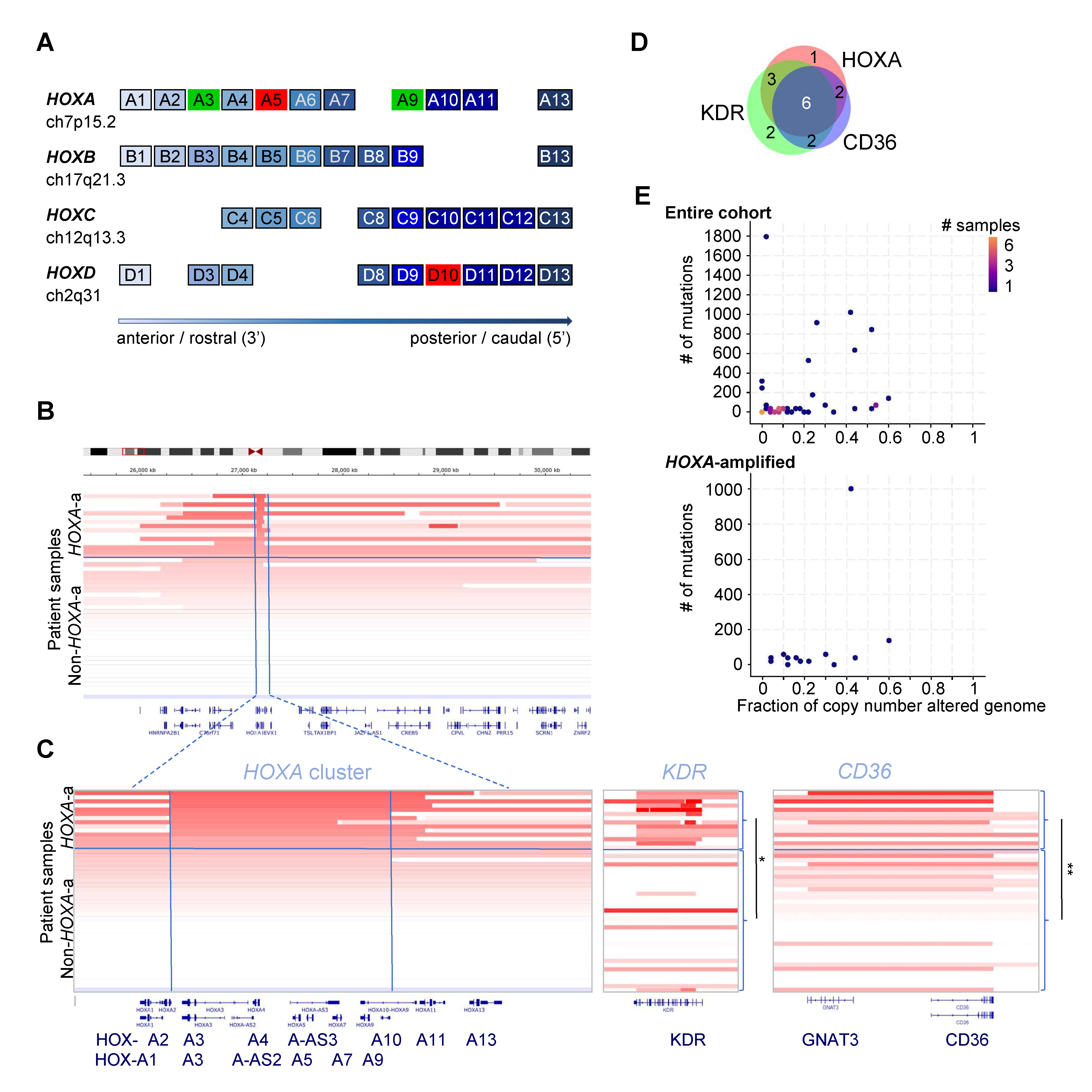
Overall, amplification of at least one pro-angiogenic HOXA-cluster gene was present in 12/36 (33%) patients. The most frequently amplified HOXA-cluster gene with a documented role in vasculogenesis/angiogenesis was HOXA3. HOXA9, HOXA11 and HOXA13 were significantly co-amplified with HOXA3 as part of a larger amplification (Figure 1B,C, left panel). In four patients, the amplification occurred within the HOXA-cluster encompassing a minimal region including HOXA3-HOXA9.
HOXA amplification was significantly associated with amplifications of the vascular endothelial marker CD36 (p = 5.59 × 10⁻⁴, Fisher’s exact test, remaining significant after Bonferroni correction at 1.62 × 10⁻²), and KDR (p = 1.07 × 10⁻³, Fisher’s exact test, remaining significant after Bonferroni correction at 3.11 × 10⁻²) (Table 1). The CN fold-change for both KDR and CD36 was significantly higher in HOXA-amplified samples at p < 1 × 10⁵ and p = 3.6 × 10⁴, respectively (Figure 1C, Mann–Whitney U test). Similarly, the CN fold-change of HOXA3 (as a marker for HOXA-cluster amplification) was significantly higher in KDR (p = 3.6 × 10³) and CD36 (p = 4 × 10³) amplified samples (Mann–Whitney U test). There was substantial overlap between HOXA, KDR and CD36 amplifications (Figure 1D). KDR encodes the VEGFR2, and KDR mutation/amplification has previously been identified by other groups [3,10,11]. Overall, HOXA-amplified tumors had a comparatively low mutation burden, although the difference failed to reach statistical significance (Figure 1E, Table 1).
The percent of co-occurring signaling pathway mutations (PIK3CA, MAP3K13, NF1, NRAS, HRAS, BRAF, RAF1) was not substantially different from non-HOXA-amplified samples. RAS pathway mutations occur with similar frequencies in HOXA-cluster dysregulated AML, where they are usually subclonal. TP53 alterations were also similar between the two groups (Table 1).
We did not observe any statistically significant differences in the clinical presentation of HOXA-cluster-amplified angiosarcomas compared to those without HOXA amplification, although we noted several interesting trends that may warrant further study. We observed a trend toward a less frequent location in the sun-exposed HNFS area compared to the whole cohort (17 vs. 33% in those with and without HOXA amplification, respectively), and a more frequent location in the breast (67 vs. 46%). There was also a trend toward a lower percentage of male patients with HOXA amplifications (male: 17 vs. 33%). We observed a trend toward a higher proportion of patients receiving tumor-directed therapy at the time of sample submission (58 vs. 42%), which could be a surrogate for higher-stage disease linked to worse outcomes. However, reliable outcomes data for this cohort were not available. Based on the available clinical annotation in cBioPortal, only two patients with HOXA-amplified angiosarcoma had prior radiation. This seems low based on the clinical experience of breast angiosarcomas as secondary malignancies after irradiation for breast cancer, but is not significantly different from the entire cohort (17 vs. 21%). HOXA-amplified samples had the same age distribution, with two peaks: one around age 30, and a second peak around age 65 (Table 1).
Figure 1. (A) Schematic of the human HOX clusters. Paralogs have the same number and coloring. The two key HOX-cluster genes that control EPC fate decisions and vasculogenesis, HOXA3 and HOXA9, are marked in green. Anti-angiogenic HOXA5 and HOXD10 are marked in red. (B) HOXA-cluster amplifications in angiosarcoma. Out of 48 patient samples (12 out of 36 patients), 13 had amplifications in HOXA3. Most HOXA3 amplifications also encompassed additional 5′ HOXA genes, including HOXA9 (HOXA3 to HOXA9 marked by vertical lines in (B), p-value: 1.9 × 10⁸, p-adjust: 1.398 × 10⁷), HOXA 11 (p-value: 2.596515 × 10⁷, p-adjust: 1.817561 × 10⁶) and HOXA13 (p-value: 9.487666 × 10⁵, p-adjust: 6.641366 × 10⁴). Copy numbers (CNs) were inferred using ReCapSeg, as described by Painter et al. [3]. Statistical significance was calculated using Fisher’s exact test (Bonferroni correction). Visualization was performed using the IVG in cBioPortal sorting on fold-change of the HOXA3 copy number. (C) HOXA-cluster, KDR and CD36 amplifications in angiosarcoma patient samples. Visualization of CN fold-change of the HOXA cluster, KDR and CD36 sorted on HOXA3-CN. * p < 1 × 10⁵, ** p = 3.6 × 10⁴, Mann–Whitney U test. (D) Venn diagram of HOXA-cluster, KDR and CD36 amplifications in angiosarcoma patients. (E) # of mutations (y-axis) and copy number alterations (x-axis) of the entire cohort (top panel) compared to HOXA-amplified angiosarcoma (bottom panel) using the visualization tools in cBioPortal. The difference was not statistically significant.
Biomolecules 12 01124 g001
Table 1. Patient clinical characteristics and recurrent amplifications/mutation of HOXA-amplified and non-HOXA-amplified angiosarcomas. HOXA amplifications were significantly associated with amplifications of KDR and CD36. There is also a non-statistically significant trend toward lower mutational burden and location in the breast versus HNSF. FE: fixed effect; MV: multi-variant.
Patient Characteristics
(36 Patients Total)
HOXA-Amplified (12)
# (%)
Non-HOXA-Amp (24)
# (%)
Significance
p-Value FE (MV)
Gender
Male2 (16.7)8 (33.3)ns
Female10 (83.3)16 (66.7)ns
Location
HNFS2 (16.7)8 (33.3)ns
Breast8 (66.7)11 (45.8)ns
intrathoracic/abd2 (16.7)4 (16.7)ns
LIMB0 (0)1 (4.2)ns
Prior Radiation
Yes2 (16.7)5 (20.8)ns
Age
Peak 1: 26–40 Years5 (41.6)12 (50)ns
41–50 Years0 (0)2 (8.3)ns
Peak 2: 51–70 Years6 (50)8 (33.3)ns
>70 Years1 (8.3)2 (8.3)ns
Pathology
Vasoformative10 (83.3)24 (100)ns
Epithelioid7 (58.3)8 (33.3)ns
Spindle Cell6 (50)17 (70.8)ns
Mutation Burden
>200 Mutations1 (8.3)7 (29.2)ns
Amplifications
CD368 (66.7)2 (8.3)5.59 × 10⁴ (1.62 × 10²)
KDR9 (75)4 (16.7)1.07 × 10³ (3.11 × 10²)
PHF19(75)10 (41.7)ns
Mutations
TP534 (33.3)7 (29.2)ns
KDR3 (25)5 (20.8)ns
PIK3CA2 (16.7)4 (16.7)ns
NRAS03 (12.5)ns
HRAS1 (8.3)1 (4.1)ns
BRAF02 (8.3)ns
RAF101 (4.1)ns
NF12 (16.7)2 (8.3)ns
MAP3K1301 (4.1)ns

3. Discussion

Here, we report a not previously recognized, high percentage of HOXA amplifications in angiosarcoma. Dysregulation of HOXA-cluster genes is a common feature of acute myeloid leukemia (AML). Distinct HOXA-cluster genes also play critical roles in regulating bone-marrow-derived endothelial precursor cells (EPCs) and in situ neovascularization [15]. Specifically, HOXA3 and HOXA9 are the two key angiogenic/vasculogenic homeobox transcription factors during development and in wound healing [19,20,21,22,23,24,25,26,27]. Furthermore, HOXA11 is expressed in vascular smooth muscle cells in fetal but not adult tissue [28]. HOXA genes are separated into two distinct groups, the anterior (HOXA 1–5) and posterior (HOXA 7–13) clusters. All HOX genes within each group are regulated coordinately in muscle development (controlled by the anterior HOXA cluster) and hematopoiesis (controlled by the posterior HOXA cluster). In contrast, individual HOXA genes within the anterior HOXA cluster have opposing functions in angiogenesis: HOXA3 is a key pro-angiogenic transcription factor and is not expressed in adult quiescent vascular endothelium. Conversely, its close neighbor HOXA5 is highly expressed in quiescent endothelium, and HOXA5 expressing endothelial cells fail to respond to angiogenic stimuli [29,30]. In addition, HOXD10 expression has been shown to inhibit angiogenesis [15,31].
HOXA genes play a critical role in the transformation of several subtypes or AML, suggesting that HOXA amplifications could also act as an oncogene in angiosarcoma. This is supported by the following observations: HOXA amplifications are common in angiosarcoma. Several of the amplified HOXA genes have well-described roles in driving blood vessel growth, suggesting a molecular mechanism for how HOXA amplifications could induce aberrant vascular proliferation. Finally, in a subset of patients, the amplified domain did not encompass the entire HOXA cluster, but there was consistent focal amplification of a segment that included pro-angiogenic members of the HOXA cluster (HOXA 3–9).
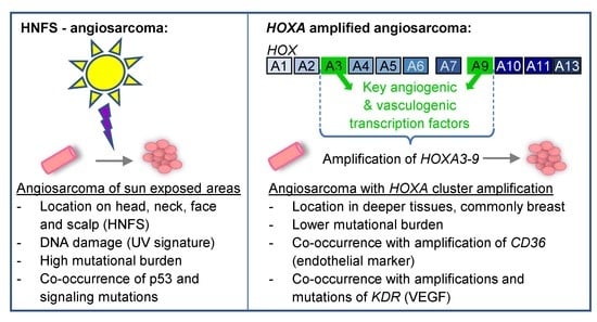
Our analysis also suggests that HOXA-amplified angiosarcomas may have a different etiology from the more common angiosarcomas of the head, neck, face and scalp [2]. The latter carries a mutational signature that is consistent with UV-induced DNA damage [3]. In contrast, HOXA-amplified angiosarcomas may be caused by the amplification of developmental master regulators of vasculogenesis and angiogenesis. The observed trend of different presentations (deeper tumors, commonly in the breast versus sun-exposed areas) and a distinct genomic landscape (strong association with KDR and CD36 amplification, as well as lower mutational burden) support the notion of a different origin.
Finally, our study raises the possibility that therapies developed to modulate HOX-cluster expression in other malignancies could have efficacy in this subtype of angiosarcoma. Regulators of HOXA transcription in other malignancies include Menin [32] and DOT1L [33,34,35]. Menin inhibitors in particular have reported pre-clinical efficacy in AML, Ewing sarcoma [36] and hepatocellular carcinomas [37], and are currently in clinical trials. Direct small-molecule inhibitors of HOX transcription factors are also an active area of research.
Our study has several important limitations. Although this is the largest cohort of angiosarcoma patients reported to date, the number of patients is still quite small, and we were unable to conduct an independent validation. It will be critical to confirm HOXA amplifications in a second, independent, patient cohort. This may also allow confirming the amplification using a second independent method such as fluorescence in situ hybridization (FISH), and to better delineate differences in the mutational landscape, clinical characteristics, and outcomes of HOXA-amplified angiosarcoma. Functional mechanistic studies will be required to determine whether HOXA amplifications are in fact oncogenic, whether HOX gene expression is required for tumor growth, and whether it can be targeted for therapeutic purposes.

Author Contributions

K.M.B. designed the study and wrote the manuscript. H.M.X. conducted data analysis and statistical analysis and co-wrote the manuscript. All authors have read and agreed to the published version of the manuscript.

Funding

This work was supported by the Emerson Collective Count-Me-In challenge.

Institutional Review Board Statement

The original study by Painter and colleagues [3] was approved by the Dana-Farber/Harvard Cancer Center Institutional Review Board (DF/HCC Protocol 15-057B). Deidentified data from this study is publicly available without additional IRB review.

Informed Consent Statement

Patients provided informed consent that included sharing de-identified linked, clinical, genomic, and patient-reported data publicly [3].

Data Availability Statement

Acknowledgments

We thank Nikhil Wagle for early access to results from the Count-Me-In Angiosarcoma project and helpful discussions.

Conflicts of Interest

Both authors declare no conflict of interest.

Abbreviations

HNFShead, neck, face and scalp
HOXhomeobox

References

  1. Weidema, M.E.; Versleijen-Jonkers, Y.M.H.; Flucke, U.E.; Desar, I.M.E.; van der Graaf, W.T.A. Targeting angiosarcomas of the soft tissues: A challenging effort in a heterogeneous and rare disease. Crit. Rev. Oncol. Hematol. 2019, 138, 120–131. [Google Scholar] [CrossRef] [PubMed]
  2. Weidema, M.E.; Flucke, U.E.; van der Graaf, W.T.A.; Ho, V.K.Y.; Hillebrandt-Roeffen, M.H.S.; Dutch Nationwide Network and Registry of Histo- and Cytopathology (PALGA)-Group; Versleijen-Jonkers, Y.M.H.; Husson, O.; Desar, I.M.E. Prognostic Factors in a Large Nationwide Cohort of Histologically Confirmed Primary and Secondary Angiosarcomas. Cancers 2019, 11, 1780. [Google Scholar] [CrossRef] [PubMed]
  3. Painter, C.A.; Jain, E.; Tomson, B.N.; Dunphy, M.; Stoddard, R.E.; Thomas, B.S.; Damon, A.L.; Shah, S.; Kim, D.; Gomez Tejeda Zanudo, J.; et al. The Angiosarcoma Project: Enabling genomic and clinical discoveries in a rare cancer through patient-partnered research. Nat. Med. 2020, 26, 181–187. [Google Scholar] [CrossRef]
  4. Behjati, S.; Tarpey, P.S.; Sheldon, H.; Martincorena, I.; Van Loo, P.; Gundem, G.; Wedge, D.C.; Ramakrishna, M.; Cooke, S.L.; Pillay, N.; et al. Recurrent PTPRB and PLCG1 mutations in angiosarcoma. Nat. Genet. 2014, 46, 376–379. [Google Scholar] [CrossRef]
  5. Murali, R.; Chandramohan, R.; Moller, I.; Scholz, S.L.; Berger, M.; Huberman, K.; Viale, A.; Pirun, M.; Socci, N.D.; Bouvier, N.; et al. Targeted massively parallel sequencing of angiosarcomas reveals frequent activation of the mitogen activated protein kinase pathway. Oncotarget 2015, 6, 36041–36052. [Google Scholar] [CrossRef]
  6. Italiano, A.; Chen, C.L.; Thomas, R.; Breen, M.; Bonnet, F.; Sevenet, N.; Longy, M.; Maki, R.G.; Coindre, J.M.; Antonescu, C.R. Alterations of the p53 and PIK3CA/AKT/mTOR pathways in angiosarcomas: A pattern distinct from other sarcomas with complex genomics. Cancer 2012, 118, 5878–5887. [Google Scholar] [CrossRef]
  7. Andersen, N.J.; Boguslawski, E.B.; Kuk, C.Y.; Chambers, C.M.; Duesbery, N.S. Combined inhibition of MEK and mTOR has a synergic effect on angiosarcoma tumorgrafts. Int. J. Oncol. 2015, 47, 71–80. [Google Scholar] [CrossRef]
  8. Chadwick, M.L.; Lane, A.; Thomas, D.; Smith, A.R.; White, A.R.; Davidson, D.; Feng, Y.; Boscolo, E.; Zheng, Y.; Adams, D.M.; et al. Combined mTOR and MEK inhibition is an effective therapy in a novel mouse model for angiosarcoma. Oncotarget 2018, 9, 24750–24765. [Google Scholar] [CrossRef]
  9. Rothweiler, S.; Dill, M.T.; Terracciano, L.; Makowska, Z.; Quagliata, L.; Hlushchuk, R.; Djonov, V.; Heim, M.H.; Semela, D. Generation of a murine hepatic angiosarcoma cell line and reproducible mouse tumor model. Lab. Investig. 2015, 95, 351–362. [Google Scholar] [CrossRef]
  10. Guo, T.; Zhang, L.; Chang, N.E.; Singer, S.; Maki, R.G.; Antonescu, C.R. Consistent MYC and FLT4 gene amplification in radiation-induced angiosarcoma but not in other radiation-associated atypical vascular lesions. Genes Chromosomes Cancer 2011, 50, 25–33. [Google Scholar] [CrossRef]
  11. Kunze, K.; Spieker, T.; Gamerdinger, U.; Nau, K.; Berger, J.; Dreyer, T.; Sindermann, J.R.; Hoffmeier, A.; Gattenlohner, S.; Brauninger, A. A recurrent activating PLCG1 mutation in cardiac angiosarcomas increases apoptosis resistance and invasiveness of endothelial cells. Cancer Res. 2014, 74, 6173–6183. [Google Scholar] [CrossRef] [PubMed]
  12. Shon, W.; Sukov, W.R.; Jenkins, S.M.; Folpe, A.L. MYC amplification and overexpression in primary cutaneous angiosarcoma: A fluorescence in-situ hybridization and immunohistochemical study. Mod. Pathol. 2014, 27, 509–515. [Google Scholar] [CrossRef] [PubMed]
  13. Cerami, E.; Gao, J.; Dogrusoz, U.; Gross, B.E.; Sumer, S.O.; Aksoy, B.A.; Jacobsen, A.; Byrne, C.J.; Heuer, M.L.; Larsson, E.; et al. The cBio cancer genomics portal: An open platform for exploring multidimensional cancer genomics data. Cancer Discov. 2012, 2, 401–404. [Google Scholar] [CrossRef] [PubMed]
  14. Gao, J.; Aksoy, B.A.; Dogrusoz, U.; Dresdner, G.; Gross, B.; Sumer, S.O.; Sun, Y.; Jacobsen, A.; Sinha, R.; Larsson, E.; et al. Integrative analysis of complex cancer genomics and clinical profiles using the cBioPortal. Sci. Signal. 2013, 6, pl1. [Google Scholar] [CrossRef]
  15. Kachgal, S.; Mace, K.A.; Boudreau, N.J. The dual roles of homeobox genes in vascularization and wound healing. Cell Adhes. Migr. 2012, 6, 457–470. [Google Scholar] [CrossRef]
  16. Terracciano, D.; Terreri, S.; de Nigris, F.; Costa, V.; Calin, G.A.; Cimmino, A. The role of a new class of long noncoding RNAs transcribed from ultraconserved regions in cancer. Biochim. Biophys. Acta Rev. Cancer 2017, 1868, 449–455. [Google Scholar] [CrossRef]
  17. Bejerano, G.; Pheasant, M.; Makunin, I.; Stephen, S.; Kent, W.J.; Mattick, J.S.; Haussler, D. Ultraconserved elements in the human genome. Science 2004, 304, 1321–1325. [Google Scholar] [CrossRef]
  18. Collins, E.M.; Thompson, A. HOX genes in normal, engineered and malignant hematopoiesis. Int. J. Dev. Biol. 2018, 62, 847–856. [Google Scholar] [CrossRef]
  19. Chisaka, O.; Capecchi, M.R. Regionally restricted developmental defects resulting from targeted disruption of the mouse homeobox gene hox-1.5. Nature 1991, 350, 473–479. [Google Scholar] [CrossRef]
  20. Bahrami, S.B.; Veiseh, M.; Dunn, A.A.; Boudreau, N.J. Temporal changes in Hox gene expression accompany endothelial cell differentiation of embryonic stem cells. Cell Adhes. Migr. 2011, 5, 133–141. [Google Scholar] [CrossRef]
  21. Iacovino, M.; Chong, D.; Szatmari, I.; Hartweck, L.; Rux, D.; Caprioli, A.; Cleaver, O.; Kyba, M. HoxA3 is an apical regulator of haemogenic endothelium. Nat. Cell Biol. 2011, 13, 72–78. [Google Scholar] [CrossRef] [PubMed]
  22. Rossig, L.; Urbich, C.; Bruhl, T.; Dernbach, E.; Heeschen, C.; Chavakis, E.; Sasaki, K.; Aicher, D.; Diehl, F.; Seeger, F.; et al. Histone deacetylase activity is essential for the expression of HoxA9 and for endothelial commitment of progenitor cells. J. Exp. Med. 2005, 201, 1825–1835. [Google Scholar] [CrossRef]
  23. Crompton, M.R.; Bartlett, T.J.; MacGregor, A.D.; Manfioletti, G.; Buratti, E.; Giancotti, V.; Goodwin, G.H. Identification of a novel vertebrate homeobox gene expressed in haematopoietic cells. Nucleic Acids Res. 1992, 20, 5661–5667. [Google Scholar] [CrossRef] [PubMed]
  24. Mace, K.A.; Hansen, S.L.; Myers, C.; Young, D.M.; Boudreau, N. HOXA3 induces cell migration in endothelial and epithelial cells promoting angiogenesis and wound repair. J. Cell Sci. 2005, 118, 2567–2577. [Google Scholar] [CrossRef]
  25. Mace, K.A.; Restivo, T.E.; Rinn, J.L.; Paquet, A.C.; Chang, H.Y.; Young, D.M.; Boudreau, N.J. HOXA3 modulates injury-induced mobilization and recruitment of bone marrow-derived cells. Stem Cells 2009, 27, 1654–1665. [Google Scholar] [CrossRef]
  26. Mahdipour, E.; Charnock, J.C.; Mace, K.A. Hoxa3 promotes the differentiation of hematopoietic progenitor cells into proangiogenic Gr-1+CD11b+ myeloid cells. Blood 2011, 117, 815–826. [Google Scholar] [CrossRef] [PubMed]
  27. Bruhl, T.; Urbich, C.; Aicher, D.; Acker-Palmer, A.; Zeiher, A.M.; Dimmeler, S. Homeobox A9 transcriptionally regulates the EphB4 receptor to modulate endothelial cell migration and tube formation. Circ. Res. 2004, 94, 743–751. [Google Scholar] [CrossRef]
  28. Miano, J.M.; Firulli, A.B.; Olson, E.N.; Hara, P.; Giachelli, C.M.; Schwartz, S.M. Restricted expression of homeobox genes distinguishes fetal from adult human smooth muscle cells. Proc. Natl. Acad. Sci. USA 1996, 93, 900–905. [Google Scholar] [CrossRef]
  29. Rhoads, K.; Arderiu, G.; Charboneau, A.; Hansen, S.L.; Hoffman, W.; Boudreau, N. A role for Hox A5 in regulating angiogenesis and vascular patterning. Lymphat. Res. Biol. 2005, 3, 240–252. [Google Scholar] [CrossRef]
  30. Arderiu, G.; Cuevas, I.; Chen, A.; Carrio, M.; East, L.; Boudreau, N.J. HoxA5 stabilizes adherens junctions via increased Akt1. Cell Adhes. Migr. 2007, 1, 185–195. [Google Scholar] [CrossRef]
  31. Chen, A.; Cuevas, I.; Kenny, P.A.; Miyake, H.; Mace, K.; Ghajar, C.; Boudreau, A.; Bissell, M.J.; Boudreau, N. Endothelial cell migration and vascular endothelial growth factor expression are the result of loss of breast tissue polarity. Cancer Res. 2009, 69, 6721–6729. [Google Scholar] [CrossRef] [PubMed]
  32. Kuhn, M.W.; Song, E.; Feng, Z.; Sinha, A.; Chen, C.W.; Deshpande, A.J.; Cusan, M.; Farnoud, N.; Mupo, A.; Grove, C.; et al. Targeting Chromatin Regulators Inhibits Leukemogenic Gene Expression in NPM1 Mutant Leukemia. Cancer Discov. 2016, 6, 1166–1181. [Google Scholar] [CrossRef] [PubMed]
  33. Bernt, K.M.; Zhu, N.; Sinha, A.U.; Vempati, S.; Faber, J.; Krivtsov, A.V.; Feng, Z.; Punt, N.; Daigle, A.; Bullinger, L.; et al. MLL-rearranged leukemia is dependent on aberrant H3K79 methylation by DOT1L. Cancer Cell 2011, 20, 66–78. [Google Scholar] [CrossRef] [PubMed]
  34. Daigle, S.R.; Olhava, E.J.; Therkelsen, C.A.; Majer, C.R.; Sneeringer, C.J.; Song, J.; Johnston, L.D.; Scott, M.P.; Smith, J.J.; Xiao, Y.; et al. Selective killing of mixed lineage leukemia cells by a potent small-molecule DOT1L inhibitor. Cancer Cell 2011, 20, 53–65. [Google Scholar] [CrossRef] [PubMed]
  35. Stein, E.M.; Garcia-Manero, G.; Rizzieri, D.A.; Tibes, R.; Berdeja, J.G.; Savona, M.R.; Jongen-Lavrenic, M.; Altman, J.K.; Thomson, B.; Blakemore, S.J.; et al. The DOT1L inhibitor pinometostat reduces H3K79 methylation and has modest clinical activity in adult acute leukemia. Blood 2018, 131, 2661–2669. [Google Scholar] [CrossRef]
  36. Svoboda, L.K.; Bailey, N.; Van Noord, R.A.; Krook, M.A.; Harris, A.; Cramer, C.; Jasman, B.; Patel, R.M.; Thomas, D.; Borkin, D.; et al. Tumorigenicity of Ewing sarcoma is critically dependent on the trithorax proteins MLL1 and menin. Oncotarget 2017, 8, 458–471. [Google Scholar] [CrossRef]
  37. Kempinska, K.; Malik, B.; Borkin, D.; Klossowski, S.; Shukla, S.; Miao, H.; Wang, J.; Cierpicki, T.; Grembecka, J. Pharmacologic Inhibition of the Menin-MLL Interaction Leads to Transcriptional Repression of PEG10 and Blocks Hepatocellular Carcinoma. Mol. Cancer Ther. 2018, 17, 26–38. [Google Scholar] [CrossRef]
  38. The Angiosarcoma Project—Count Me In (Nature Medicine, 2020). Available online: https://www.cbioportal.org/study/summary?id=angs_project_painter_2018 (accessed on 4 July 2022).
Publisher’s Note: MDPI stays neutral with regard to jurisdictional claims in published maps and institutional affiliations.