Article

Decoding the Role of Melatonin Structure on Plasmodium falciparum Human Malaria Parasites Synchronization Using 2-Sulfenylindoles Derivatives

by
Lenna Rosanie Cordero Mallaupoma
1,2,
Bárbara Karina de Menezes Dias
2,3,
Maneesh Kumar Singh
2,
Rute Isabel Honorio
2,
Myna Nakabashi
2,
Camila de Menezes Kisukuri
4,
Márcio Weber Paixão
4 and
Celia R. S. Garcia
2,*
1
Departamento de Química, Instituto de Química, Universidade de São Paulo, São Paulo 05508-000, Brazil
2
Departamento de Análises Clínicas e Toxicológicas, Faculdade de Ciências Farmacêuticas, Universidade de São Paulo, São Paulo 05508-000, Brazil
3
Departamento de Parasitologia, Instituto de Ciências Biomédicas, Universidade de São Paulo, São Paulo 05508-000, Brazil
4
Centro de Excelência para Pesquisa em Química Sustentável (CERSusChem), Departamento de Química, Universidade Federal de São Carlos, São Carlos 13565-905, Brazil
*
Author to whom correspondence should be addressed.
Biomolecules 2022, 12(5), 638; https://doi.org/10.3390/biom12050638
Submission received: 16 March 2022 / Revised: 12 April 2022 / Accepted: 14 April 2022 / Published: 26 April 2022

Abstract

:
Melatonin acts to synchronize the parasite’s intraerythrocytic cycle by triggering the phospholipase C-inositol 1,4,5-trisphosphate (PLC-IP3) signaling cascade. Compounds with an indole scaffold impair in vitro proliferation of blood-stage malaria parasites, indicating that this class of compounds is potentially emerging antiplasmodial drugs. Therefore, we aimed to study the role of the alkyl and aryl thiol moieties of 14 synthetic indole compounds against chloroquine-sensitive (3D7) and chloroquine-resistant (Dd2) strains of Plasmodium falciparum. Four compounds (3, 26, 18, 21) inhibited the growth of P. falciparum (3D7) by 50% at concentrations below 20 µM. A set of 2-sulfenylindoles also showed activity against Dd2 parasites. Our data suggest that Dd2 parasites are more susceptible to compounds 20 and 28 than 3D7 parasites. These data show that 2-sulfenylindoles are promising antimalarials against chloroquine-resistant parasite strains. We also evaluated the effects of the 14 compounds on the parasitemia of the 3D7 strain and their ability to interfere with the effect of 100 nM melatonin on the parasitemia of the 3D7 strain. Our results showed that compounds 3, 7, 8, 10, 14, 16, 17, and 20 slightly increased the effect of melatonin by increasing parasitemia by 8–20% compared with that of melatonin-only-treated 3D7 parasites. Moreover, we found that melatonin modulates the expression of kinase-related signaling components giving additional evidence to investigate inhibitors that can block melatonin signaling.

Graphical Abstract

1. Introduction

Malaria is a parasitic disease of importance to public health that affects millions of people worldwide, and Plasmodium falciparum is responsible for most deaths from malaria that occur annually around the world. Artemisinin-based combination therapy (ACT), which consists of artemisinin, or a derivative combined with other compounds with different action mechanisms and a longer half-life, is the first-line treatment for uncomplicated malaria. However, there is a threat of parasites becoming resistant to ACT. Therefore, antimalarial treatment failure rates are monitored to avoid the high mortality rates of the 1980s, when chloroquine-resistant parasites appeared in Africa [1].
In the scenario of the global burden of the spread of chloroquine- and artemisinin-resistant P. falciparum, the search for new molecules to replace those used in classical malaria therapy is important. Compounds with distinct chemical architectures have shown antimalarial activity and different mechanisms of action [2,3], e.g., 4-aminoquinolines, antifolates, aryl-amino alcohols, naphthoquinones, antibiotics, and endoperoxides. In addition to traditional antimalarial drugs, other compounds have also displayed potential antiplasmodial activity; among these drugs, those containing an indole moiety are of high importance [4,5].
The host hormone melatonin is an indole derivative that synchronizes P. falciparum and P. chabaudi parasites [6]. Strikingly, the nonsynchronous murine parasites P. berghei and P. yoelii do not display melatonin-dependent synchronization in vitro [7]. Moreover, some indole derivatives, such as N-acetyl-serotonin and serotonin, and synthetic indole analogs also modulate parasitemia in vitro [8,9,10], and others, such as 8-oxo-tryptamine, show antiplasmodial activity against P. falciparum [11]. It was shown that melatonin receptor antagonist, luzindole, disrupts Ca2+ oscillation and cAMP increase in asexual P. falciparum, causing severe growth defects in intraerythrocytic maturation [12]. Other indole compounds that inhibited melatonin-induced synchronization and exhibited antimalarial activity against 3D7 parasites are the C2-arylalkanimino tryptamine derivatives [13]. These studies showed that compounds resembling the melatonin structure might disrupt the parasite life cycle. Using a rational indole synthesis strategy, several research groups have independently reported that indole derivatives are able to impair the intraerythrocytic development of Plasmodium [10,11,13,14,15,16,17,18,19]. Additionally, reports on 2- and 3-functionalized indoles with biological properties have led biofunctionalized indole compounds to be seen as promising bioactive molecules against malaria parasites, such as Flinderoles [20] or Borreverines [21]. In addition, among these analogs, spiroindolone KAE609 has high antimalarial activity and is currently in phase II clinical trials [22].
In this study, we aimed to investigate the antiplasmodial activity of a series of 14 structurally divergent 2-sulfenylindoles [23] against chloroquine-sensitive (CQS) 3D7 and chloroquine-resistant (CQR) Dd2 P. falciparum strains. In addition, we also investigated the cumulative effects of the compounds and melatonin on the parasitemia of 3D7 and Dd2 in vitro. Additional evidence of melatonin on the expression of signaling components was also investigated.

2. Materials and Methods

2.1. Plasmodium Falciparum Culture

The parasites 3D7 and Dd2 were cultured at 37 °C in RPMI-1640 medium (Gibco, Waltham, MA, USA) with 0.21% sodium bicarbonate (Sigma, Burlington, MA, USA) and 50 mg/L hypoxanthine (Sigma) supplemented with 0.5% AlbuMAX I (Gibco). The cultures were maintained under an atmosphere of 5% CO2, 5% O2, and 90% N2. Fresh complete medium was provided daily, and rapid-stained (Panotico) blood smears were examined.

2.2. In Vitro Drug Susceptibility Assay

To evaluate the effects of synthetic compounds on the intraerythrocytic development of P. falciparum, we analyzed parasitemia by flow cytometry with 0.3% initial parasitemia and 1% hematocrit in the assay. Plates were incubated with the compounds at concentrations ranging from 0.10 to 100 µM for 72 h. Dual staining with SYBR Green I (SG-I) (Invitrogen, Waltham, MA, USA) and MitoTracker Deep Red (MT-Red) (Invitrogen) were used to label nucleic acids and mitochondria, respectively. Approximately 10,000 cells were counted by an Accuri C6 (Becton Dickinson, San Jose, CA, USA) and analyzed by FlowJo 5 software. Parasitemia was determined from the analysis of dot plots (side scatter versus fluorescence). DMSO-treated parasites (0.125%) were used as a negative control.

2.3. Cytotoxicity Assay

HEK293T (human embryonic kidney) cells were cultured in 75 cm2 vented tissue culture flasks (Greiner Bio-One, Frickenhausen, Germany) at 37 °C in a humidified atmosphere containing 5% CO2 in Dulbecco’s modified essential medium (Gibco) supplemented with 10% (v/v) fetal bovine serum (Gibco), 100 U/mL penicillin, and 100 μg/mL streptomycin (Sigma). Cytotoxicity was evaluated with the 4,5-dimethylthiazol-2-yl-2,5-diphenyltetrazolium bromide (MTT) cell proliferation assay [24,25]. Briefly, 104 cells/well were seeded in flat-bottom 96-well plates in complete DMEM medium for 24 h, followed by incubation with different concentrations of each compound (0.10–100 µM) for 72 h. DMSO (0.13%) and digitonin were used as the negative and positive controls, respectively. The concentrations of DMSO chosen as control were the concentration present in the tested compounds (from 0.0001% to 0.12%, two-fold dilution). After 72 h, cells were then incubated with the MTT reagent for 3 h, and absorbance was measured at 570 nm using a FlexStation® 3 Multi-Mode Microplate Reader (Molecular Devices, Sunnyvale, CA, USA). Moreover, we calculated the selectivity indexes (SI) to understand whether the compounds were more toxic to parasites than cells, or vice versa, according to the ratio of the CC50 value to the IC50 value for each compound. The data are shown in Figure S1.

2.4. Effects of Indole Derivatives on Parasitemia

To evaluate the role of the compounds in parasitemia, 1% 3D7 parasites in 2% hematocrit were incubated for 48 h with 500 nM of the 2-sulfenylindole derivatives. Melatonin (100 nM) was used as a positive control, and DMSO (0.0013% v/v) was used as a negative control. The concentration of DMSO chosen as control was the concentration present in the tested compounds at 500 nM. The activity of the compounds was measured by SG-I and MT-Red staining. The data obtained were normalized to those of the control (DMSO). Additionally, to test the ability of the compounds to affect melatonin activity in parasitemia, 3D7 parasites were incubated with 500 nM indole compounds along with 100 nM melatonin for 48 h.

2.5. Blood-Stage Development Evaluation by Microscopy

Ring-stage parasites (10–14 h) at 1% parasitemia in 2% hematocrit were treated with compound 20 and compound 28 at concentrations of 1 and 30 µM in 24-well plates for 48 h. Treatment with chloroquine (20 nM) and DMSO (0.1%) was used as positive and negative controls. Smears were rapid stained (Panotico) every 12 h after treatment (12, 24, 36, 48, and 60 h time points), and we took images of parasites to assess the blood-stage development (Zeiss, Oberkochen, Germany).

2.6. Real-Time PCR and Data Analysis

P. falciparum trophozoites (approximately 30–34 h post invasion) were treated with 100 nM and 1 µM melatonin (SIGMA-Aldrich, Burlington, MA, USA). RNA was extracted with Trizol (Life Technologies, Carlsbad, CA, USA) 1 and 3 h post treatment. cDNA synthesis was carried out using random primers and Superscript IV reverse transcriptase (Life Technologies) according to the manufacturer’s protocol. Oligonucleotides used are listed in Supplementary Table S1. SYBR Green incorporation was measured during PCR amplification performed on the 7300 Real-Time PCR System (Applied Biosystems, Foster City, CA, USA). The statistical analysis was performed by applying Student’s t-test on the log2 values of relative expression (delta delta Ct method) of the genes (normalized with the seryl t-RNA synthase housekeeping gene).

2.7. Data Analysis

The experiments were performed independently at least three times, and each experiment was analyzed in triplicate unless mentioned otherwise. Data analyses were performed with GraphPad Prism 8, and the results are expressed as the mean and standard deviation (SD) of three independent experiments. p < 0.05 was considered significant. One-way analysis of variance (ANOVA) followed by Dunnett’s post-hoc test was used to determine the statistical significance of the comparison of parasitemia among groups.

3. Results

3.1. Antiplasmodial Activity of 2-sulfenylindoles against CQS (3D7) and CQR (Dd2) Parasites

We determined the susceptibility of P. falciparum to the indole compounds and investigated whether CQR parasites responded differently to the compounds. Asynchronous parasite cultures were incubated with different concentrations of the compounds (0.1 to 100 µM) for 72 h, followed by labeling of active mitochondria in parasites with MT-Red and DNA with SG-I to identify viable parasites. We determined the IC50 values for each compound by analyzing viable and non-viable parasites after compound treatment, represented in dot plots (Figure 1H).
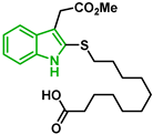
Figure 1 shows the survival curves obtained for compounds 16, 17, 18, 19, 20, and 21 for the 3D7 and Dd2 strains. Compounds 16, 17, and 21 have an S-alkyl side chain attached to C2 of the indole ring, tert-butylthio, ethylthio, and dodecylthio, respectively. Compound 18 has an adamantanethio group at C2 of the indole ring, 19 has an S-alkyl chain containing a hydroxy group, and 20 has an S-alkyl chain containing a terminal carboxylic acid.
Compound 20 was the sulfenylindole studied with the most potent antiplasmodial activity against CQR. In addition, 18 and 21 suggest that a bulky group or a long alkyl side chain at C2 of the indole ring is likely important for the antiplasmodial activity of 2-sulfenylindoles (Table 1).
In Figure 2, similar survival curves were obtained using compounds 3, 7, 8, 10, 14, 25, 26, and 28. Compounds 3, 7, 8, and 25 have a phenylthio group attached to C2 of the indole ring and an ester component attached to C3. Compound 10 has a pyrimidinylthio group at C2 of the indole ring, 14 has a benzylthio at C2 of the indole ring, 26 has a phenylthiol group at C2 and a benzyl group attached to C3 of the indole ring, and 28 has a phenylthiol group at C2 and an amide group attached at C3 of the indole ring.
We observed that compound 3 was the most effective against the 3D7 strain (IC50 = 10.58 µM). Compound 3 was more active against 3D7 parasites than its 3-acetamide counterpart (28, IC50 = 31.75 µM), and their activities against Dd2 parasites were not the same. In addition, the hydroxy substituent present in 7 had a negative effect on the antiplasmodial activity of the thioindole in vitro (Table 1).
Five compounds had IC50 values lower than 20 µM against Dd2 (3, 18, 20, 21, and 28). In addition, we found four compounds with IC50 values lower than 20 µM against 3D7 (3, 18, 21, and 26). We also examined the impact of a methoxy group at C5 of the indole ring in indole analogs presenting similar substituents. Compounds 25 and 3 show that a methoxy group at C5 results in lower antiplasmodial activity, IC503D7 = 26.47 ± 4.87 µM and IC503D7 = 10.58 ± 5.19 µM, respectively.
In general, sulfenylindoles are more active against the CQS parasites than the CQR strain. The compounds more active against 3D7 parasites are 3, 26, and 18; the two first ones are arylthioindoles, and the other one is an alkylthioindole. However, 20 and 28 are more active against CQR strain, and the first one is an alkylthioindole and the other an arylthioindole. Their resistant indexes are 0.30 and 0.39, respectively. Moreover, the selectivity index for these compounds was calculated using 100 as CC50 since performing a CC50 assay with higher concentrations could be challenging due to the compound’s solubility. Compounds 3, 26, and 18 exhibited low selectivity to Plasmodium parasites, while compounds 20 and 28 were modestly more selective, exhibiting selectivity index values >3.15.

3.2. Effect of 2-sulfenylindoles on the Parasitemia of 3D7 Parasites

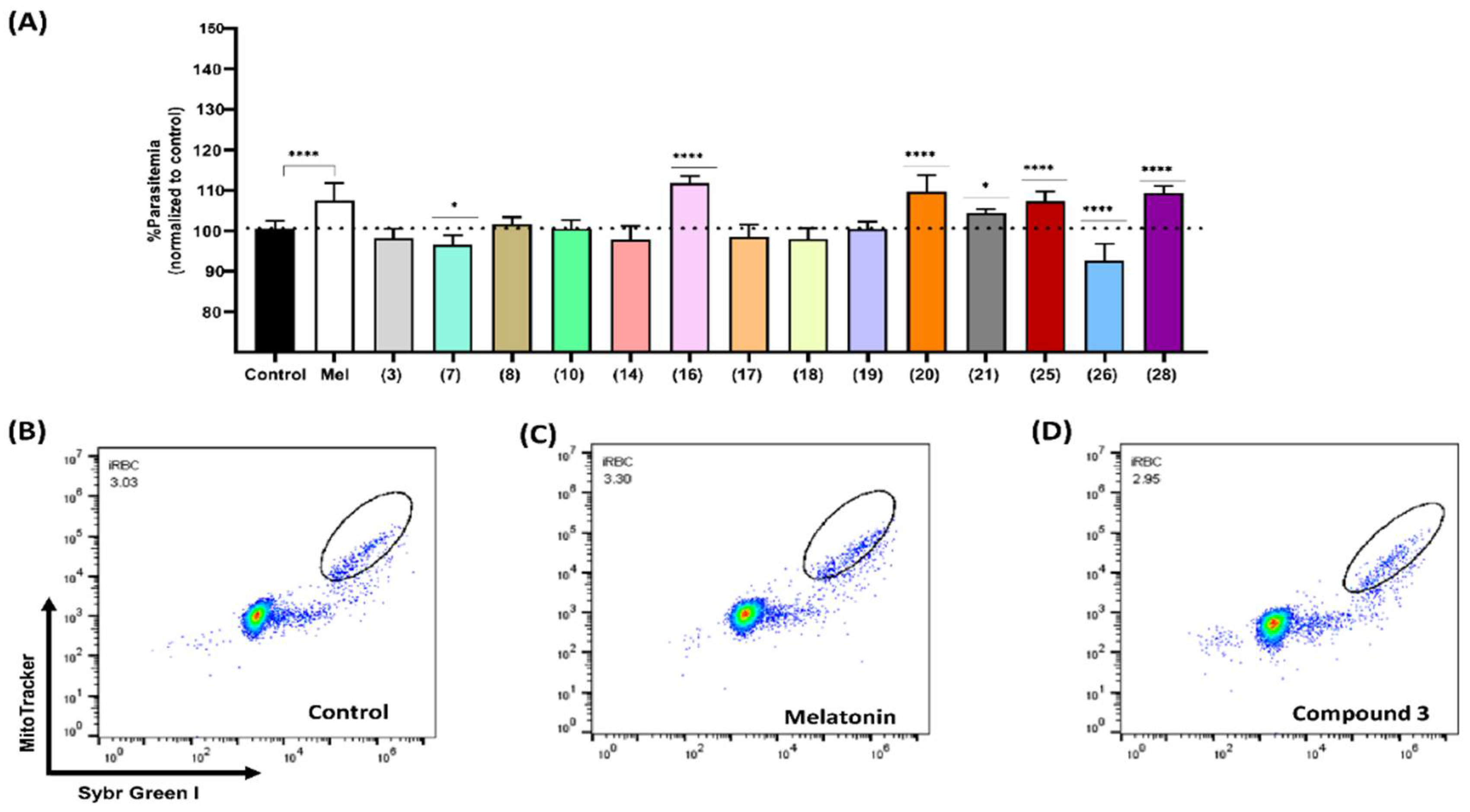
Previous studies have reported that melatonin promotes parasite growth in the nanomolar range [6]. Hence, we assessed the effect of compounds on parasitemia by incubating asynchronous 3D7 parasites with the set of 2-sulfenylindoles at 500 nM for 48 h. After incubation, the final parasitemia was determined using flow cytometry. The results are shown in Figure 3. Compounds 7 and 26 exhibited slight inhibitory effects on parasite growth, as parasitemia decreased by 4.05% and 7.94%, respectively. Moreover, compounds 16, 20, 21, 25, and 28 led to slight increases in parasitemia of 11.2%, 9.1%, 3.8%, 6.6%, and 8.7%, respectively, compared to the control group treated with DMSO (100.6%).
Our results showed that 2-sulfenylindoles could inhibit intraerythrocytic development of parasites in vitro after 72 h incubation at the micromolar concentrations. At a lower range and after 48 h incubation, five of the compounds (16, 20, 21, 25, and 28) led to an increase in parasitemia, in a similar way as that observed for the host hormone melatonin (a rise of 6.9% from 100.6% to 107.5%) and its derivatives [8].

3.3. Effect of 2-sulfenylindoles in Combination with Melatonin on the Parasitemia

Following the assumption that compounds resembling melatonin structure might interact through the melatonin signaling pathways, we evaluated the effect of the compounds on melatonin action on parasitemia. Our next step was to study the combination of 2-sulfenylindoles with melatonin in asynchronous 3D7 by incubating parasites simultaneously with 100 nM melatonin and 500 nM of each compound.
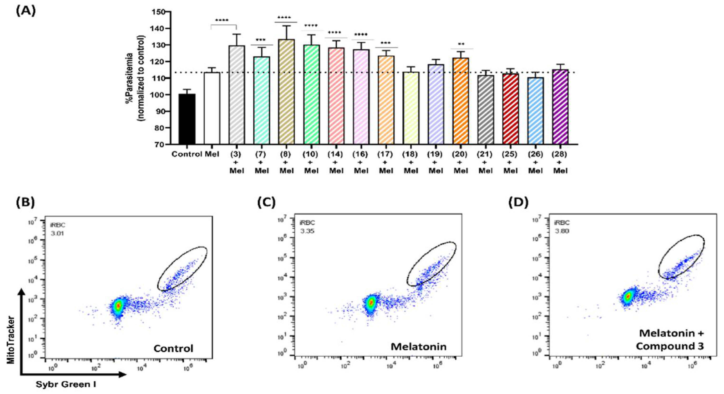
The combination of compound 3 (500 nM) with melatonin (100 nM) had a synergistic effect, enhancing parasitemia by 16.27% compared with that of parasites exposed only to 100 nM melatonin (113.5%). Similar responses were observed for compounds 8, 14, 17, 10, 7, 16, and 20 (Figure 4). After simultaneous exposure to melatonin and compound 8, the parasitemia of 3D7 parasites increased by 20% compared to that of melatonin-only-treated parasites. Similarly, a synergistic effect on parasitemia was observed after incubation with melatonin along with some compounds, leading to an increase of 16.63% (10), 14.88% (14), 13.88% (16), 10.05% (17), 9.42% (7), and 8.81% (20) (Table S1).
As mentioned above, at micromolar concentrations, we observed an inhibitory effect for each compound. However, compound 7 (500 nM) alone slightly decreased parasitemia by 4.084%, whereas in combination with melatonin, it increased parasitemia by 22.2% compared to that of the control group (100.7%).

3.4. Cytotoxic Activity of 2-sulfenylindoles on Mammalian Cells

We further assessed the toxicity of 2-sulfenylindole compounds against HEK293 cells using the MTT assay with different concentrations of the compounds for 72 h. The data are shown in Figure S1. Table 1 presents all the toxicity data and summarizes the IC50 values of the compounds evaluated in this study for Dd2 and 3D7 strains. All the compounds exhibited IC50 values in the micromolar range.
Compound 25 was the least non-toxic with a CC50 of 13.14 ± 3.74 µM in HEK293 cells, whereas compound 21 was the least toxic with a CC50 of >100 µM (Figure S1). Some of the 2-sulfenylindoles displayed activity against HEK293 cells, and selectivity indexes to Plasmodium parasites over mammalian cells of between 0.28 and 2.58 times were identified (Table 1). The CC50 values for five compounds were not determined, and we speculated that these compounds exhibit more selective toxicity to the Plasmodium parasites than to HEK293 cells because the highest test concentration was not able to kill 100 of the mammalian cells and is impossible to evaluate higher concentrations due to compounds solubility. Therefore, we considered that compounds 8, 10, 20, 21, and 28 exhibit more selective toxicity to the Plasmodium parasites than to HEK293 cells.

3.5. Effect on Blood-Stage Growth Progression of the Most Active Compounds

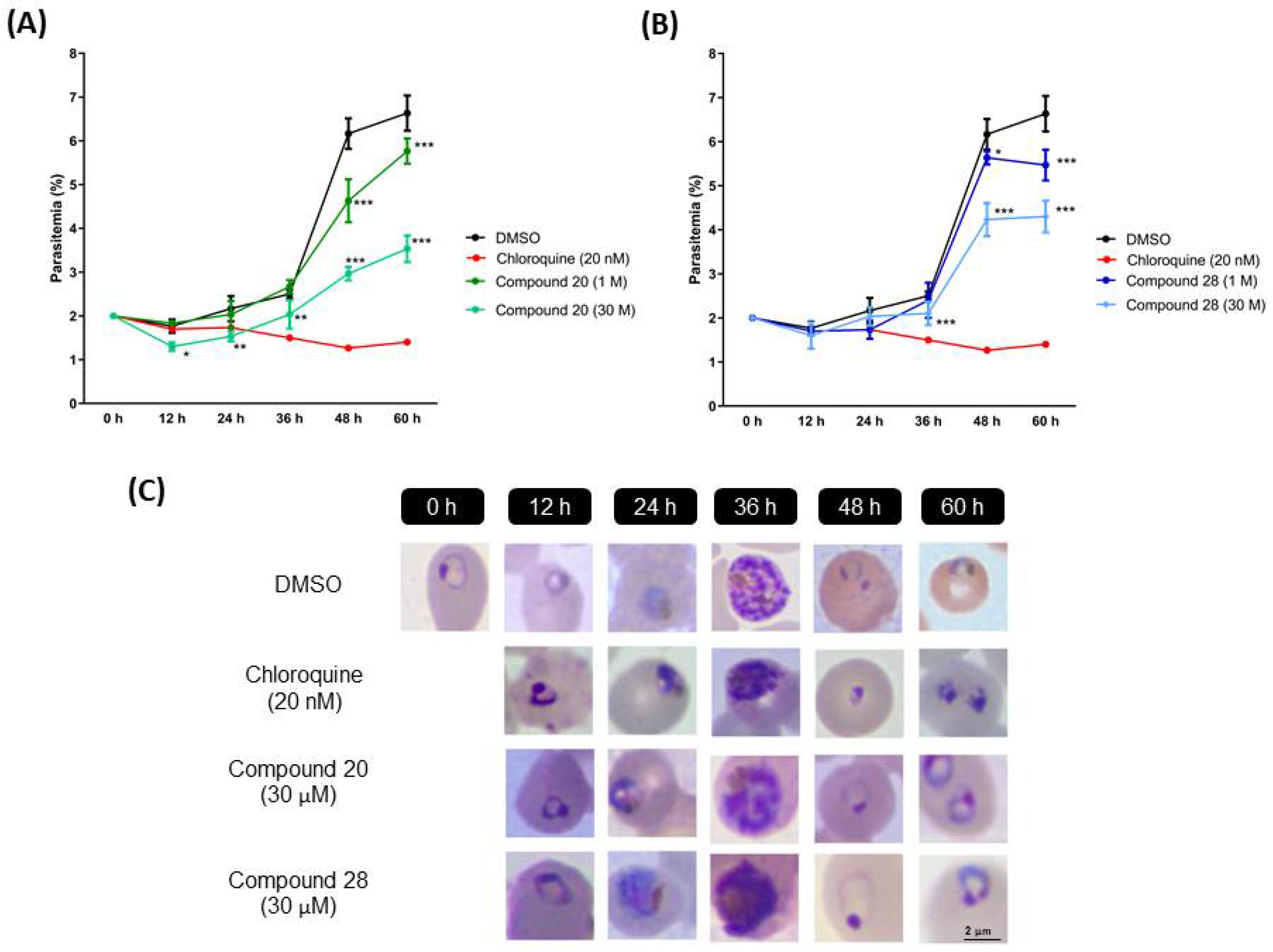
Analyses with microscopy reveal the blood-stage development inhibition in 3D7 parasites treated with compounds 20 and 28 at IC50 (Figure 5C) and compound 3, 18, and 26 at IC50 (Figure S2). During monitoring, parasites continued to develop throughout the cycle in the presence of both compounds. However, compounds 20 and 28 significantly affect parasitemia (Figure 5A,B, respectively). To examine its stage specificity, we monitored development every 12 h. Compound 28 had no effect on parasite growth at the schizont stage (36–48 h time points) but stalled ring to trophozoite progression. On the other hand, compound 20 affects RBC reinvasion, evidenced by the reduction in ring formation at the 48 h time point.
Treatment with compounds 3, 18, and 26 allowed parasites’ development continues. However, in the second cycle of proliferation, these compounds reduced the progress from rings to trophozoites (Figure S2).

3.6. Indole Compound Melatonin Alters the Expression of Plasmodium Transcript

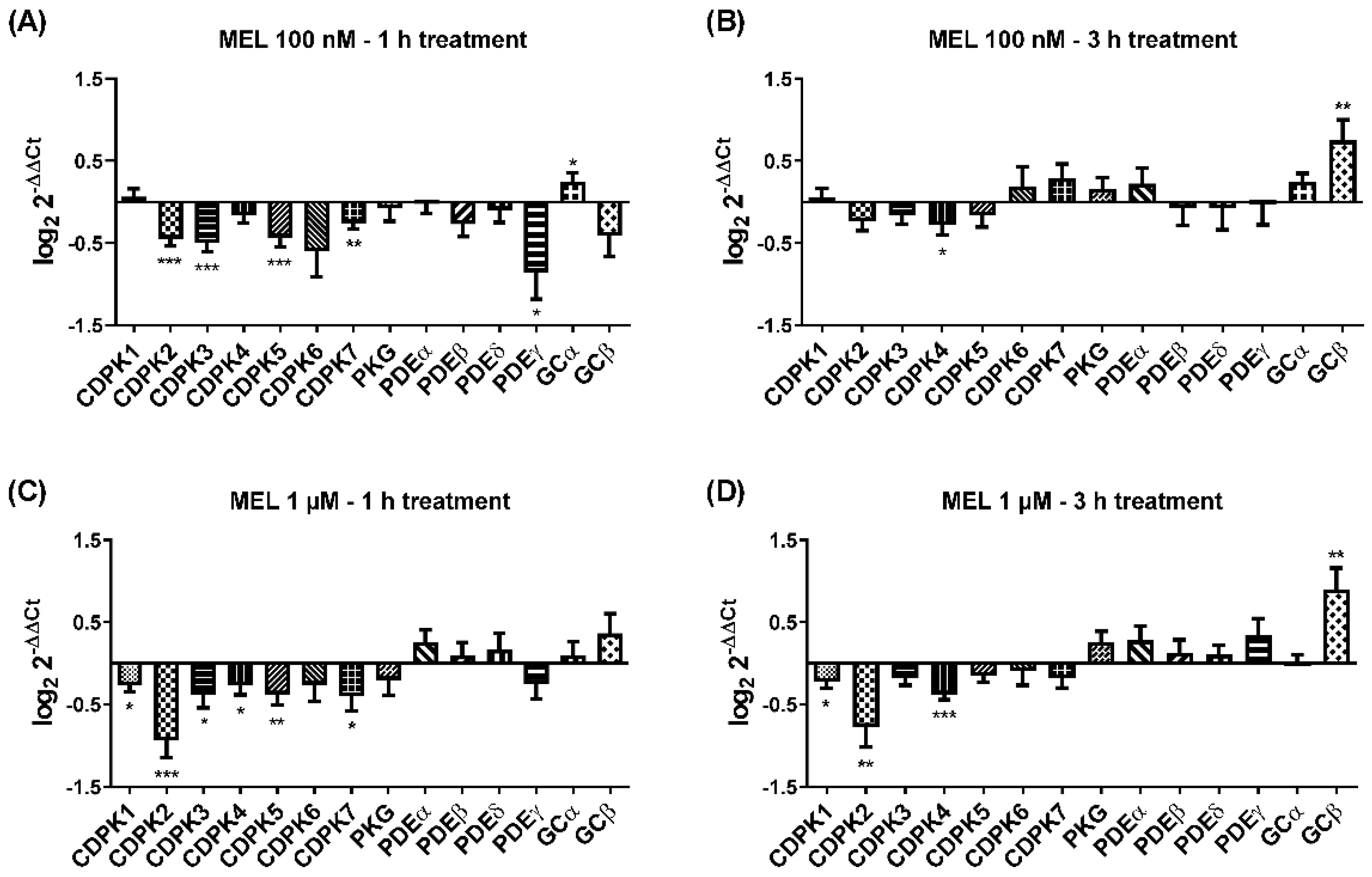
Previous studies have shown that melatonin alters the expression of the ubiquitin/proteasome system [26], mitochondrial FIS1, DYN2 [27], and nuclear PfMORC [28]. These findings lead us to investigate the effect of melatonin on kinases, phosphodiesterase, and cyclase for two reasons; first, kinases and their components are required for rupturing the parasites to facilitate egress in both asexual and sexual stages. Second, in the asexual blood stage, melatonin has been shown to mobilize calcium from internal stores [29,30], and prior to egress, calcium is mobilized by PKG-mediated breakdown of PIP2 [31]. We treated the late-stage trophozoite with two different melatonin concentrations (100 nM and 1 µM) for two time points (1 and 3 h). In our result (Figure 6), we found that 100 nM melatonin for 1 h has a very minor effect on change in gene expression for CDPK1 and GCα; however, after 3 h, increased expression can be seen in additional CDPK6, CDPK7, PKG, PDEα, and GCβ. Interestingly, 1 µM melatonin for 3 h treatment has the most prominent effect on change in gene expression for cGMP-dependent signaling component suggesting melatonin role in the asexual cycle. It is possible that melatonin may act upstream of PKG and modulates cytosolic calcium, but this hypothesis requires more experimental validation.

4. Discussion

The parasite maturation during the intraerythrocytic cycle is extremely coordinated in which schizonts rupture and invade new erythrocytes in a highly synchronized manner at intervals usually multiples of 24 h [32,33,34]. Our lab has shown that the developmental synchrony of the parasite was lost in pinealectomized mice [6]. Calcium is well known to be central in controlling the molecular processes of the cell cycle in parasites [35,36]. The downstream molecular mechanism of melatonin involves the PLC-IP3 signaling pathways [30,37,38] through increases of Ca2+ and cAMP, leading to the activation of PKA [8,39].
Several studies have shown that melatonin modulates the cell cycle of the human malaria parasite P. falciparum, determining the synchrony of the invasion of red blood cells by intraerythrocytic Plasmodium parasites and their asexual reproduction [40,41]. Therefore, the potential for using melatonin derivatives containing an indole moiety to treat malaria has been reported previously [10,13,19].
Antiplasmodial compounds containing a benzenesulfonamide moiety [19] or a carboxamide group [10] at the C3 position of the indole ring have also been reported. Additionally, some marine-indole alkaloids with substitutions at the C3-position of the indole ring are active against P. falciparum, with IC50 values of 4.44 to 200 µM. In addition, evidence suggests that bromide and hydroxyl substitutions at the C7 and C4 positions of the indole ring, respectively, increase activity against parasites [16,17].
In our study, sulfenyl indoles showed inhibitory activity ranging from 8 to 59 µM for both 3D7 and Dd2 parasites. Compound 21 is a 6-chloro analog of meridoquin (IC50 = 200 µM) [16] and showed modest antiplasmodial activity compared to that of 2-sulfenyl indoles in our study, suggesting that the presence of the S-alkyl chain at the C2 position of the indole ring resulted in greater improvement of antiplasmodial activity than the presence of a pyrimidine group at C3 of the indole ring.
We observed that compounds 20 and 28, which had significantly lower IC50 values against the Dd2 strain, had a low cytotoxic effect against HEK293 cells. In addition, these compounds alone at a nanomolar concentration (500 nM) were able to significantly increase the parasitemia of 3D7 asynchronous cultures, indicating that although they can increase parasitemia at low concentrations, they can selectively affect parasite growth at higher concentrations. Previous studies have reported that a 4-aminoquinoline-piperidine is less effective against a wild-type NF54 strain than against a CQR strain K1 [42]. Similarly, some chloroquine analogs have been shown to have higher activity against W2 than 3D7 parasites [43]. Furthermore, β-benzoylstyrene derivatives of acridine with better potency against Dd2 than the 3D7 have been reported [44]. Compounds with better activity against CQR strains are a promising avenue of research, as reported by Ngemenya et al. [45].
It has been reported that an indole compound with a methoxy group at C5, melatotosil, can increase parasitemia and interfere with the action of melatonin [19]. Our results also suggested that the presence of a methoxy group at C5 in compound 25 led to a slight increase in parasitemia of 6.66%, while no increase was observed after exposure to its counterpart (compound 3). However, treatment with compound 25 in combination with melatonin did not potentiate or block the effect of melatonin on parasitemia. On the other hand, a slight increase in parasitemia was observed after exposure to compound 3 in combination with melatonin compared with that after exposure to melatonin alone (113.5%). Compounds 25 and 3, unlike melatotosil, showed antimalarial activity (Table 1).
Since compounds 16 and 21 were able to slightly increase parasitemia at 500 nM and since both have an S-alkyl group at C2 of the indole ring, a small alkyl substituent might favor an increase in parasitemia since compound 16 contains a tert-butylthio group and led to an increase in parasitemia of 11.2%. Compound 21, which has a long alkyl side chain, led to an increase in parasitemia of 3.8%. However, a compound with a smaller alkyl group (compound 17) did not affect parasitemia.
Moreover, real-time data with melatonin implicates the alteration in kinases and their components related to egress and/or invasion. It is more evident that the development of more indole-related compounds may exhibit an antimalarial effect by affecting genes that modulate both egress and invasion.

5. Conclusions

Two compounds have shown slightly more activity on the resistant Dd2 strain with lower IC50 values; these results suggest the potential of sulfenylindoles as antiplasmodial. Compounds 3, 18, 26, and 28 affect ring to trophozoite progression, whereas compound 20 affects RBC reinvasion. In addition, aiming to obtain a compound that blocks parasite development, we evaluated the action of 14 compounds on parasitemia at nanomolar concentrations. Compound 3, which has a phenylthio group at C2 of the indole ring, presented the lower IC50 value in 3D7 (IC50 = 10.58 µM). However, the response was not the same against Dd2 parasites. Compound 20, an S-alkyl chain at C2, was the most active against Dd2 parasites (IC50 = 8.72 µM). Moreover, five indole compounds (16, 20, 21, 25, and 28) increase parasitemia at low concentrations, whereas at high ones, they reduce parasitemia. These findings suggest sulfenylindoles as potential antimalarials against parasites resistant and susceptible to chloroquine despite some of the compounds tested can increase parasitemia, similar to the host hormone melatonin.

Supplementary Materials

The following supporting information can be downloaded at: https://www.mdpi.com/article/10.3390/biom12050638/s1, Figure S1: Effect of indole derivatives in HEK293 cells. Figure S2: Parasitemia reduction obtained after incubation with compound 3 (A), compound 18 (B), or compound 26 (C). Table S1: Oligonucleotides used in real-time PCR experiment for melatonin treatment. Table S2: Effects of 2-sulfenylindoles on the parasitemia of P. falciparum after 48 h of incubation.

Author Contributions

L.R.C.M., B.K.d.M.D., R.I.H., M.K.S., M.N. and C.R.S.G. designed the experiments. L.R.C.M., B.K.d.M.D., R.I.H., M.K.S., C.d.M.K. and M.N. performed the experiments. C.R.S.G. and M.W.P. are the principal investigators who contributed to the analysis of the data and managed the project. All the authors contributed to the writing of the paper and the interpretation of the data. All authors have read and agreed to the published version of the manuscript.

Funding

This work was supported by the Fundação de Amparo à Pesquisa do Estado de São Paulo (FAPESP) 2017/08684-7 provided to C.R.S.G., 2019/09490-7 to M.K.S., Conselho Nacional de Desenvolvimento Científico e Tecnológico (CNPq) 142188/2017-4 provided to B.K.M.D. and 131146/2020-3 provided to L.R.C-M, and CAPES 88887.614839/2021-00 provided to R.I.H. We are also grateful to CNPq (INCT Catálise), FAPESP-2018/12986-1, CAPES and GSK for their financial support.

Institutional Review Board Statement

Not applicable.

Informed Consent Statement

Not applicable.

Data Availability Statement

Not applicable.

Acknowledgments

We are grateful to Colsan for providing blood and plasma for the parasite cultures. We thank David Fidock for providing us with the chloroquine-resistant strain Dd2.

Conflicts of Interest

The authors declare no conflict of interest.

References

  1. WHO. World Malaria Reports 2016; World Health Organization: Geneva, Switzerland, 2020. [Google Scholar]
  2. Blasco, B.; Leroy, D.; Fidock, D.A. Antimalarial drug resistance: Linking Plasmodium falciparum parasite biology to the clinic. Nat. Med. 2017, 23, 917–928. [Google Scholar] [CrossRef]
  3. Tse, E.G.; Korsik, M.; Todd, M.H. The past, present and future of anti-malarial medicines. Malar. J. 2019, 18, 93. [Google Scholar] [CrossRef] [PubMed] [Green Version]
  4. Chadha, N.; Silakari, O. Indoles as therapeutics of interest in medicinal chemistry: Bird’s eye view. Eur. J. Med. Chem. 2017, 134, 159–184. [Google Scholar] [CrossRef] [PubMed]
  5. Garg, V.; Maurya, R.K.; Thanikachalam, P.V.; Bansal, G.; Monga, V. An insight into the medicinal perspective of synthetic analogs of indole: A review. Eur. J. Med. Chem. 2019, 180, 562–612. [Google Scholar] [CrossRef] [PubMed]
  6. Hotta, C.T.; Gazarini, M.L.; Beraldo, F.H.; Varotti, F.P.; Lopes, C.; Markus, R.P.; Pozzan, T.; Garcia, C.R. Calcium-dependent modulation by melatonin of the circadian rhythm in malarial parasites. Nat. Cell Biol. 2000, 2, 466–468. [Google Scholar] [CrossRef]
  7. Bagnaresi, P.; Alves, E.; da Silva, H.B.; Epiphanio, S.; Mota, M.M.; Garcia, C.R. Unlike the synchronous Plasmodium falciparum and P. chabaudi infection, the P. berghei and P. yoelii asynchronous infections are not affected by melatonin. Int. J. Gen. Med. 2009, 2, 47–55. [Google Scholar] [CrossRef] [Green Version]
  8. Beraldo, F.H.; Almeida, F.M.; da Silva, A.M.; Garcia, C.R. Cyclic AMP and calcium interplay as second messengers in melatonin-dependent regulation of Plasmodium falciparum cell cycle. J. Cell Biol. 2005, 170, 551–557. [Google Scholar] [CrossRef] [Green Version]
  9. Budu, A.; Peres, R.; Bueno, V.B.; Catalani, L.H.; Garcia, C.R. N1-acetyl-N2-formyl-5-methoxykynuramine modulates the cell cycle of malaria parasites. J. Pineal Res. 2007, 42, 261–266. [Google Scholar] [CrossRef]
  10. Schuck, D.C.; Jordao, A.K.; Nakabashi, M.; Cunha, A.C.; Ferreira, V.F.; Garcia, C.R. Synthetic indole and melatonin derivatives exhibit antimalarial activity on the cell cycle of the human malaria parasite Plasmodium falciparum. Eur. J. Med. Chem. 2014, 78, 375–382. [Google Scholar] [CrossRef]
  11. Campos, P.E.; Pichon, E.; Moriou, C.; Clerc, P.; Trepos, R.; Frederich, M.; De Voogd, N.; Hellio, C.; Gauvin-Bialecki, A.; Al-Mourabit, A. New Antimalarial and Antimicrobial Tryptamine Derivatives from the Marine Sponge Fascaplysinopsis reticulata. Mar. Drugs 2019, 17, 167. [Google Scholar] [CrossRef] [Green Version]
  12. Furuyama, W.; Enomoto, M.; Mossaad, E.; Kawai, S.; Mikoshiba, K.; Kawazu, S. An interplay between 2 signaling pathways: Melatonin-cAMP and IP3-Ca2+ signaling pathways control intraerythrocytic development of the malaria parasite Plasmodium falciparum. Biochem. Biophys. Res. Commun. 2014, 446, 125–131. [Google Scholar] [CrossRef]
  13. Luthra, T.; Nayak, A.K.; Bose, S.; Chakrabarti, S.; Gupta, A.; Sen, S. Indole based antimalarial compounds targeting the melatonin pathway: Their design, synthesis and biological evaluation. Eur. J. Med. Chem. 2019, 168, 11–27. [Google Scholar] [CrossRef] [PubMed]
  14. Agarwal, A.; Srivastava, K.; Puri, S.K.; Chauhan, P.M. Synthesis of substituted indole derivatives as a new class of antimalarial agents. Bioorg. Med. Chem. Lett. 2005, 15, 3133–3136. [Google Scholar] [CrossRef] [PubMed]
  15. Zhu, J.; Chen, T.; Chen, L.; Lu, W.; Che, P.; Huang, J.; Li, H.; Li, J.; Jiang, H. 2-amido-3-(1H-indol-3-yl)-N-substituted-propanamides as a new class of falcipain-2 inhibitors. 1. Design, synthesis, biological evaluation and binding model studies. Molecules 2009, 14, 494–508. [Google Scholar] [CrossRef] [Green Version]
  16. Lebar, M.D.; Hahn, K.N.; Mutka, T.; Maignan, P.; McClintock, J.B.; Amsler, C.D.; van Olphen, A.; Kyle, D.E.; Baker, B.J. CNS and antimalarial activity of synthetic meridianin and psammopemmin analogs. Bioorg. Med. Chem. 2011, 19, 5756–5762. [Google Scholar] [CrossRef]
  17. Bharate, S.B.; Yadav, R.R.; Khan, S.I.; Tekwani, B.L.; Jacob, M.R.; Khan, I.A.; Vishwakarma, R.A. Meridianin G and its analogs as antimalarial agents. MedChemComm 2013, 4, 1042–1048. [Google Scholar] [CrossRef]
  18. Devender, N.; Gunjan, S.; Tripathi, R.; Tripathi, R.P. Synthesis and antiplasmodial activity of novel indoleamide derivatives bearing sulfonamide and triazole pharmacophores. Eur. J. Med. Chem. 2017, 131, 171–184. [Google Scholar] [CrossRef]
  19. Dias, B.K.; Nakabashi, M.; Alves, M.R.R.; Portella, D.P.; Dos Santos, B.M.; Costa da Silva Almeida, F.; Ribeiro, R.Y.; Schuck, D.C.; Jordão, A.K.; Garcia, C.R. The Plasmodium falciparum eIK1 kinase (PfeIK1) is central for melatonin synchronization in the human malaria parasite. Melatotosil blocks melatonin action on parasite cell cycle. J. Pineal Res. 2020, 69, e12685. [Google Scholar] [CrossRef]
  20. Fernandez, L.S.; Buchanan, M.S.; Carroll, A.R.; Feng, Y.J.; Quinn, R.J.; Avery, V.M. Flinderoles A-C: Antimalarial bis-indole alkaloids from Flindersia species. Org. Lett. 2009, 11, 329–332. [Google Scholar] [CrossRef]
  21. Fernandez, L.S.; Sykes, M.L.; Andrews, K.T.; Avery, V.M. Antiparasitic activity of alkaloids from plant species of Papua New Guinea and Australia. Int. J. Antimicrob. Agents 2010, 36, 275–279. [Google Scholar] [CrossRef]
  22. Turner, H. Spiroindolone NITD609 is a novel antimalarial drug that targets the P-type ATPase PfATP4. Future Med. Chem. 2016, 8, 227–238. [Google Scholar] [CrossRef] [PubMed]
  23. Santos, M.S.; Betim, H.L.I.; Kisukuri, C.M.; Campos Delgado, J.A.; Correa, A.G.; Paixao, M.W. Photoredox Catalysis toward 2-Sulfenylindole Synthesis through a Radical Cascade Process. Org. Lett. 2020, 22, 4266–4271. [Google Scholar] [CrossRef] [PubMed]
  24. Carmichael, J.; DeGraff, W.G.; Gazdar, A.F.; Minna, J.D.; Mitchell, J.B. Evaluation of a tetrazolium-based semiautomated colorimetric assay: Assessment of chemosensitivity testing. Cancer Res. 1987, 47, 936–942. [Google Scholar] [PubMed]
  25. Schuck, D.C.; Ferreira, S.B.; Cruz, L.N.; Da Rocha, D.R.; Moraes, M.S.; Nakabashi, M.; Rosenthal, P.J.; Ferreira, V.F.; Garcia, C.R. Biological evaluation of hydroxynaphthoquinones as anti-malarials. Malar. J. 2013, 12, 234. [Google Scholar] [CrossRef] [Green Version]
  26. Koyama, F.C.; Azevedo, M.F.; Budu, A.; Chakrabarti, D.; Garcia, C.R. Melatonin-induced temporal up-regulation of gene expression related to ubiquitin/proteasome system (UPS) in the human malaria parasite Plasmodium falciparum. Int. J. Mol. Sci. 2014, 15, 22320–22330. [Google Scholar] [CrossRef] [PubMed] [Green Version]
  27. Scarpelli, P.H.; Tessarin-Almeida, G.; Viçoso, K.L.; Lima, W.R.; Borges-Pereira, L.; Meissner, K.A.; Wrenger, C.; Raffaello, A.; Rizzuto, R.; Pozzan, T.; et al. Melatonin activates FIS1, DYN1, and DYN2 Plasmodium falciparum related-genes for mitochondria fission: Mitoemerald-GFP as a tool to visualize mitochondria structure. J. Pineal Res. 2019, 66, e12484. [Google Scholar] [CrossRef] [Green Version]
  28. Singh, M.K.; Tessarin-Almeida, G.; Dias, B.K.; Pereira, P.S.; Costa, F.; Przyborski, J.M.; Garcia, C.R. A nuclear protein, PfMORC confers melatonin dependent synchrony of the human malaria parasite P. falciparum in the asexual stage. Sci. Rep. 2021, 11, 2057. [Google Scholar] [CrossRef]
  29. Hotta, C.T.; Markus, R.P.; Garcia, C.R. Melatonin and N-acetyl-serotonin cross the red blood cell membrane and evoke calcium mobilization in malarial parasites. Braz. J. Med. Biol. Res. 2003, 36, 1583–1587. [Google Scholar] [CrossRef] [Green Version]
  30. Alves, E.; Bartlett, P.J.; Garcia, C.R.; Thomas, A.P. Melatonin and IP3-induced Ca2+ release from intracellular stores in the malaria parasite Plasmodium falciparum within infected red blood cells. J. Biol. Chem. 2011, 286, 5905–5912. [Google Scholar] [CrossRef] [Green Version]
  31. Collins, C.R.; Hackett, F.; Strath, M.; Penzo, M.; Withers-Martinez, C.; Baker, D.A.; Blackman, M.J. Malaria parasite cGMP-dependent protein kinase regulates blood stage merozoite secretory organelle discharge and egress. PLoS Pathog. 2013, 9, e1003344. [Google Scholar] [CrossRef] [Green Version]
  32. Hawking, F.; Gammage, K.; Worms, M.J. The asexual and sexual circadian rhythms of Plasmodium vinckei chabaudi, of P. berghei and of P. gallinaceum. Parasitology 1972, 65, 189–201. [Google Scholar] [CrossRef] [PubMed]
  33. Garcia, C.R.; Markus, R.P.; Madeira, L. Tertian and quartan fevers: Temporal regulation in malarial infection. J. Biol. Rhythms 2001, 16, 436–443. [Google Scholar] [CrossRef] [PubMed]
  34. Mideo, N.; Reece, S.E.; Smith, A.L.; Metcalf, C.J. The Cinderella syndrome: Why do malaria-infected cells burst at midnight? Trends Parasitol. 2013, 29, 10–16. [Google Scholar] [CrossRef] [PubMed] [Green Version]
  35. Borges-Pereira, L.; Budu, A.; McKnight, C.A.; Moore, C.A.; Vella, S.A.; Triana, M.A.H.; Liu, J.; Garcia, C.R.; Pace, D.A.; Moreno, S.N. Calcium Signaling throughout the Toxoplasma gondii Lytic Cycle: A study using genetically encoded calcium indicators. J. Biol. Chem. 2015, 290, 26914–26926. [Google Scholar] [CrossRef] [PubMed] [Green Version]
  36. Canepa, G.E.; Degese, M.S.; Budu, A.; Garcia, C.R.; Buscaglia, C.A. Involvement of TSSA (trypomastigote small surface antigen) in Trypanosoma cruzi invasion of mammalian cells. Biochem. J. 2012, 444, 211–218. [Google Scholar] [CrossRef] [Green Version]
  37. Beraldo, F.H.; Mikoshiba, K.; Garcia, C.R. Human malarial parasite, Plasmodium falciparum, displays capacitative calcium entry: 2-aminoethyl diphenylborinate blocks the signal transduction pathway of melatonin action on the P. falciparum cell cycle. J. Pineal Res. 2007, 43, 360–364. [Google Scholar] [CrossRef] [PubMed]
  38. Pecenin, M.F.; Borges-Pereira, L.; Levano-Garcia, J.; Budu, A.; Alves, E.; Mikoshiba, K.; Thomas, A.; Garcia, C.R. Blocking IP3 signal transduction pathways inhibits melatonin-induced Ca2+ signals and impairs P. falciparum development and proliferation in erythrocytes. Cell Calcium. 2018, 72, 81–90. [Google Scholar] [CrossRef]
  39. Gazarini, M.L.; Beraldo, F.H.; Almeida, F.M.; Bootman, M.; Da Silva, A.M.; Garcia, C.R. Melatonin triggers PKA activation in the rodent malaria parasite Plasmodium chabaudi. J. Pineal Res. 2011, 50, 64–70. [Google Scholar] [CrossRef]
  40. Koyama, F.C.; Ribeiro, R.Y.; Garcia, J.L.; Azevedo, M.F.; Chakrabarti, D.; Garcia, C.R. Ubiquitin proteasome system and the atypical kinase PfPK7 are involved in melatonin signaling in Plasmodium falciparum. J. Pineal Res. 2012, 53, 147–153. [Google Scholar] [CrossRef]
  41. Greischar, M.A.; Read, A.F.; Bjornstad, O.N. Synchrony in malaria infections: How intensifying within-host competition can be adaptive. Am. Nat. 2014, 183, E36–E49. [Google Scholar] [CrossRef]
  42. Van de Walle, T.; Boone, M.; Van Puyvelde, J.; Combrinck, J.; Smith, P.J.; Chibale, K.; Mangelinckx, S.; D’hooghe, M. Synthesis and biological evaluation of novel quinoline-piperidine scaffolds as antiplasmodium agents. Eur. J. Med. Chem. 2020, 198, 112330. [Google Scholar] [CrossRef]
  43. De Souza, N.B.; Carmo, A.M.; da Silva, A.D.; Franca, T.C.; Krettli, A.U. Antiplasmodial activity of chloroquine analogs against chloroquine-resistant parasites, docking studies and mechanisms of drug action. Malar. J. 2014, 13, 469. [Google Scholar] [CrossRef] [PubMed] [Green Version]
  44. Prajapati, S.P.; Kaushik, N.K.; Zaveri, M.; Mohanakrishanan, D.; Kawathekar, N.; Sahal, D. Synthesis, characterization and antimalarial evaluation of new β-benzoylstyrene derivatives of acridine. Arab. J. Chem. 2017, 10, S274–S280. [Google Scholar] [CrossRef] [Green Version]
  45. Ngemenya, M.N.; Abwenzoh, G.N.; Ikome, H.N.; Zofou, D.; Ntie-Kang, F.; Efange, S.M.N. Structurally simple synthetic 1, 4-disubstituted piperidines with high selectivity for resistant Plasmodium falciparum. BMC Pharmacol. Toxicol. 2018, 19, 42. [Google Scholar] [CrossRef] [PubMed]
Figure 1. Determination of the antiplasmodial activity of 2-sulfenyl indoles in the 3D7 and Dd2 strains. Asynchronous parasites were exposed to compounds 16, 17, 18, 19, 20, and 21 for 72 h in concentrations ranging from 0.1 to 100 µM. Parasitemia was calculated using a flow cytometer by double staining parasites with SYBR and MitoTracker. Dot plot analysis of each drug concentration tested allowed us to determine parasitemia. Growth survival curves of blood-stage P. falciparum of compounds 16 (A), 17 (B), 18 (C), 19 (D), 20 (E), and 21 (F) for 3D7 (black) or Dd2 (green). Chloroquine (G) was administered at concentrations from 0 to 250 nM to 3D7 parasites and from 0 to 6 µM to Dd2 parasites. Data represent three independent experiments performed in triplicate. (H) Dot plots representing SG-I/MT-Red staining of control and chloroquine-treated asynchronous 3D7 (left) and Dd2 (right) parasites after 72 h of treatment. The bar graphs represent the SD. Statistical significance was calculated using Student’s t-test: ns = not significant, * p ≤ 0.05, ** p ≤ 0.01.
Biomolecules 12 00638 g001
Figure 2. Determination of the antiplasmodial effects of 2-sulfenyl indoles on the 3D7 and Dd2 strains. Asynchronous parasites exposed to compounds 3, 7, 8, 10, 14, 25, 26, and 28 for 72 h in the range of 0.1–100 µM. Parasitemia was obtained using a flow cytometer by double staining parasites with SYBR and MitoTracker. Dot plot analysis of each drug concentration tested allowed us to determine parasitemia. Growth survival curves of blood-stage P. falciparum of compounds. Growth survival curves of blood-stage P. falciparum of compounds 3 (A), 7 (B), 8 (C), 10 (D), 14 (E), 25 (F), 26 (G), and 28 (H) for 3D7 (black) or Dd2 (green). Data represent three independent experiments performed in triplicate. The bar graphs represent the SD. Statistical significance was calculated using Student’s t-test: ns = not significant, * p ≤ 0.05, ** p ≤ 0.01.
Biomolecules 12 00638 g002
Figure 3. Effects of 2-sulfenyl indoles on the parasitemia of P. falciparum after 48 h of incubation with 500 nM compounds 3, 7, 8, 10, 14, 16, 17, 18, 19, 20, 21, 25, 26, and 28 (A). Dot plots showing SG-I/MT-Red-labeled parasites detected by flow cytometry after 48 h of treatment with DMSO (B), 100 nM melatonin (C), and 500 nM compound 3 (D). Data are presented as the percentage of parasitemia normalized to that of the control group treated with solvent (DMSO). Experiments were performed 3 independent times in triplicate. The error bars represent the SD. * indicates a significant difference from the control group. Statistical significance was calculated using one-way ANOVA followed by Dunnett’s test: * p ≤ 0.05, **** p ≤ 0.0001.
Biomolecules 12 00638 g003
Figure 4. Effects of 2-sulfenyl indoles on the parasitemia of P. falciparum after 48 h of incubation with 500 nM compounds 3, 7, 8, 10, 14, 16, 17, 18, 19, 20, 21, 25, 26, and 28 in combination with 100 nM melatonin (Mel) (A). Dot plots showing SG-I/MT-Red-labeled parasites detected by flow cytometry after 48 h of treatment with DMSO (B), 100 nM melatonin (C), and a combination of 100 nM melatonin and 500 nM compound 3 (D). Data are presented as the percentage of parasitemia normalized to that of the control group treated with solvent (DMSO). Experiments were performed 3 independent times in triplicate. The error bars represent the SD. * indicates a significant difference compared to the melatonin treatment (Mel). Statistical significance was calculated using one-way ANOVA followed by Dunnett’s test: ** p ≤ 0.01, *** p ≤ 0.001, **** p ≤ 0.0001.
Biomolecules 12 00638 g004
Figure 5. Parasitemia reduction with compound 20 (A) and compound 28 (B) to determine the stage activity. Data are means ± SD. Ring-stage synchronized parasites with sorbitol 5% were treated with chloroquine (20 nM), compound 20 (30 µM), and compound 28 (30 µM). The chosen treatment concentration was the IC50 values obtained for these compounds against the 3D7 parasites. (C) After drug administration, smears of P. falciparum 3D7 strain were taken at 0, 12, 24, 36, 48, and 60 h after drug administration, observing the growth development of treated parasites. * p ≤ 0.05, ** p ≤ 0.01, *** p ≤ 0.001.
Biomolecules 12 00638 g005
Figure 6. Differential transcription of CDPKs, PKG, and its accessory genes from parasites treated with melatonin for 1 and 3 h. Sorbitol synchronized parasites were treated with melatonin, and RNA samples were extracted, purified, and transcript level was investigated by RT-qPCR. Expression of genes has been normalized by housekeeping seryl-tRNA Synthetase expression for (A) 100 nM melatonin for 1 h; (B) 100 nM melatonin for 3 h; (C) 1 μM melatonin for 1 h; and (D) 1 μM melatonin for 3 h. Each figure represents three independent experiments in triplicate. * p ≤ 0.05, ** p ≤ 0.01, *** p ≤ 0.001.
Biomolecules 12 00638 g006
Table 1. In vitro activity of the compounds against asexual blood stages of Plasmodium falciparum and cytotoxicity in the HEK293 cell line.
CompoundIC50 ± SD
Pf3D7
IC50 ± SD
PfDd2
CC50 ± SD
HEK293
SI
3D7
SI
Dd2
(3)
Biomolecules 12 00638 i001
10.58 ± 5.19 µM17.97 ± 1.72 µM15.24 ± 1.36 µM1.440.84
(7)
Biomolecules 12 00638 i002
58.18 ± 2.45 µM55.42 ± 2.96 µM31.59 ± 4.03 µM0.540.57
(8)
Biomolecules 12 00638 i003
37.29 ± 3.58 µM43.29 ± 6.51 µM>100 µM>2.68> 2.31
(10)
Biomolecules 12 00638 i004
54.86 ± 2.25 µM43.62 ± 3.060 µM>100 µM>1.82> 2.29
(14)
Biomolecules 12 00638 i005
20.86 ± 2.97 µM37.08 ± 7.664 µM23.69 ± 8.540 µM1.140.63
(16)
Biomolecules 12 00638 i006
31.69 ± 5.09 µM48.50 ± 2.88 µM26.52 ± 8.70 µM0.840.55
(17)
Biomolecules 12 00638 i007
24.99 ± 2.90 µM35.37 ± 1.56 µM26.49 ± 4.67 µM1.060.75
(18)
Biomolecules 12 00638 i008
13.52 ± 3.24 µM15.72 ± 4.87 µM16.82 ± 2.63 µM1.241.07
(19)
Biomolecules 12 00638 i009
27.78 ± 2.03 µM39.92 ± 1.61 µM28.14 ± 5.46 µM1.010.70
(20)
Biomolecules 12 00638 i010
28.78 ± 4.86 µM8.72 ± 3.36 µM>100 µM>3.47>11.47
(21)
Biomolecules 12 00638 i011
19.10 ±1.78 µM16.86 ± 4.66 µM>100 µM>5.24>5.93
(25)
Biomolecules 12 00638 i012
26.47 ± 4.87 µM45.53 ± 4.84 µM13.14 ± 3.74 µM0.50 0.28
(26)
Biomolecules 12 00638 i013
13.48 ± 2.37 µM35.80 ± 4.64 µM34.72 ± 8.44 µM2.580.97
(28)
Biomolecules 12 00638 i014
31.75 ± 5.02 µM12.39 ± 0.59 µM>100 µM>3.15>8.07
Chloroquine22.33 ± 4.17 nM199.05 ± 26.23 nMNDNDND
ND: not determined, SI: selectivity index (CC50 value of HEK293 cell/IC50 value of Pf3D7 or PfDd2).
Publisher’s Note: MDPI stays neutral with regard to jurisdictional claims in published maps and institutional affiliations.