Communication

Plg-RKT Expression in Human Breast Cancer Tissues

1
Department of Molecular Medicine, Scripps Research Institute, La Jolla, CA 92037, USA
2
Cellstan-Immunoquant Inc., Oceanside, CA 92054, USA
3
Department of Medicine, Veterans Administration San Diego Healthcare System, University of California San Diego, San Diego, CA 92161, USA
4
San Diego Biomedical Research Institute, San Diego, CA 92121, USA
*
Author to whom correspondence should be addressed.
Biomolecules 2022, 12(4), 503; https://doi.org/10.3390/biom12040503
Submission received: 26 February 2022 / Revised: 21 March 2022 / Accepted: 23 March 2022 / Published: 26 March 2022
(This article belongs to the Special Issue Plasminogen, Plasminogen Receptors and Binding Mechanisms in Cancer)

Abstract

:
The plasminogen activation system regulates the activity of the serine protease, plasmin. The role of plasminogen receptors in cancer progression is being increasingly appreciated as key players in modulation of the tumor microenvironment. The interaction of plasminogen with cells to promote plasminogen activation requires the presence of proteins exposing C-terminal lysines on the cell surface. Plg-RKT is a structurally unique plasminogen receptor because it is an integral membrane protein that is synthesized with and binds plasminogen via a C-terminal lysine exposed on the cell surface. Here, we have investigated the expression of Plg-RKT in human breast tumors and human breast cancer cell lines. Breast cancer progression tissue microarrays were probed with anti-Plg-RKT mAB and we found that Plg-RKT is widely expressed in human breast tumors, that its expression is increased in tumors that have spread to draining lymph nodes and distant organs, and that Plg-RKT expression is most pronounced in hormone receptor (HR)-positive tumors. Plg-RKT was detected by Western blotting in human breast cancer cell lines. By flow cytometry, Plg-RKT cell surface expression was highest on the most aggressive tumor cell line. Future studies are warranted to address the functions of Plg-RKT in breast cancer.

1. Introduction

The plasminogen activation system regulates the activity of the serine protease, plasmin. Plasminogen activation to plasmin is carried out by plasminogen activators, notably the urokinase-type plasminogen activator (uPA). The plasminogen activation system is critical for the dissolution of fibrin clots, plays a major role in processes of tissue remodeling and cell migration [1,2,3], and has been compellingly implicated in the biology of breast cancer [4,5]. Indeed, several components of this system including the protease uPA, its receptor, uPAR, and the plasminogen activator inhibitor-1 (PAI-1) are strong, independent markers of a poor prognosis in breast cancer [6,7,8,9].
Efficient plasminogen activation requires coordinated activity on the cell surface [10,11]. The interaction of plasminogen with cells to promote plasminogen activation requires the presence of proteins exposing C-terminal lysines on the cell surface [12]. The role of plasminogen receptors in cancer progression is being increasingly appreciated (reviewed in [13,14,15,16,17,18]).
Previously, using a proteomics-based approach, we discovered the plasminogen receptor, Plg-RKT. Among cell surface plasminogen binding proteins Plg-RKT is structurally unique in that it is an integral membrane protein that is synthesized with and binds plasminogen via a C-terminal lysine exposed on the cell surface [19,20]. Plg-RKT is highly colocalized with uPAR [19,21] and promotes uPA-dependent plasminogen activation [22]. Furthermore, Plg-RKT regulates fibrinolysis, pro-MMP cleavage [22], and leukocyte recruitment [22,23,24,25], all of which may play a role in breast cancer progression. Here, we have investigated the expression of Plg-RKT in human breast tumors. Our results suggest that Plg-RKT is expressed on human breast tumors and expression increases with tumor progression.

2. Materials and Methods

2.1. Tissue Microarrays

We used the human breast (cancer) tissue microarray IMH 364 from Novus, Centennial, CO, USA. We also used the National Cancer Institute (NCI) Cancer Diagnosis Program (CDP) 2nd Generation Breast Cancer Progression (BCP) Tissue Microarrays (CDP-BCP-TMA) case sets 4, 6, and 8, from University of Virginia, CHTN (Cooperative Human Tissue Network), Charlottesville, VA 22908, USA, http://www.chtn.org (Accessed on 20 February 2022).

2.2. Antibodies

Anti-Plg-RKT (mAb 7H1) is a pan-specific antibody that we raised in mice against a synthetic peptide corresponding to the 9 C-terminal amino acids of rat Plg-RKT [22]. Anti-β actin antibody was from LI-COR (Lincoln, NE, USA). Normal mouse IgG2a was from Southern Biotech (Birmingham, AL, USA).

2.3. Immunohistochemistry and Calculation of Immunoscores

Immunohistochemistry was performed as described [26]. Briefly, immunostaining of TMAs was carried out using the Anti-Mouse Envision-Plus-HRP system (Dako, Troy, MI, USA) with a Dako Universal Staining System automated immunostainer. In controls, the immunostaining procedure was performed in parallel by absorbing the antibody with the synthetic peptide used for immunization. To quantify Plg-RKT expression we calculated anti-Plg-RKT immunoscores as described [27].

2.4. Cell Lines

The MCF-7 hormone receptor positive human cancer cell line was obtained from the America Type Culture Collection (ATCC, Manassas, VA, USA) and cultured in DMEM containing 10% fetal bovine serum (FBS), 1% L-Glutamine, 1% sodium pyruvate, 1% nonessential amino acids (NEAA) and 2% vitamins. The MCF-10A normal human breast cell line was from the University of Colorado and cells were cultured in DMEM:F12 containing 5% horse serum, 1% L-Glutamine, 20 ng/mL EGF, 0.5 µg/mL hydrocortisone, 100 ng/mL cholera toxin and 10 µg/mL insulin. The triple negative SUM159PT human breast cancer cell line was from BioIVT (Westbury, NY, USA) and was cultured in Ham’s F-12 media containing 5% FBS, 10 mM HEPES, 1 µg/mL hydrocortisone and 5 µg/mL insulin. The MDA-MB-231 triple negative human breast cancer cell line was obtained from ATCC and was cultured in Leibovitz’s L-15 media containing 10% FBS and 10 mM HEPES without CO2. The MDA-MB-231 mfp line was obtained by establishing orthotopic xenograft tumors of the invasive human breast cancer line, MDA-MB-231, in the mammary fat pad of SCID mice. Tumor cells were recovered and cultured to provide a propagatable subpopulation, referred to as 231mfp cells [28].

2.5. Western Blotting

Cells were lysed in radio-immunoprecipitation assay (RIPA) buffer containing an anti-protease and anti-phosphate cocktail (Thermo Fisher Scientific, Waltham, MA, USA) and electrophoresed on 4–12% polyacrylamide gels under reducing conditions followed by electrotransfer to nitrocellulose membranes. The membranes were incubated with primary antibodies, then washed with phosphate buffered saline (PBS) containing 0.1% Tween-20 and then incubated with species specific IRDye® 680RD/800CW-conjugated second antibodies. Immunoreactive bands were visualized with the Odyssey Imaging System (LI-COR). Densitometric analysis was performed using the software Image Studio™ Lite Software 5.2 (LI-COR).

2.6. Flow Cytometric Analysis

Cell lines were stained with either anti-Plg-RKT mAB, normal mouse IgG2a or unstained. The anti-Plg-RKT and isotype control antibody were directly labeled with PE-Cy7 Conjugation Kit from Abcam (Boston, MA, USA). The stained cells were acquired in a NovoCyte (ACEA Biosciences, San Diego, CA, USA) and analyzed with FlowJo software (Tree Star Inc., Ashland, OR, USA). Viable cells (propidium iodide and annexin V negative) were gated from nonviable cells.
Quantitative flow cytometric equilibrium binding of fluorescein isothiocyanate (FITC)-plasminogen to cells was analyzed using beads impregnated with FITC as described [19]. Nonspecific (FITC)-plasminogen binding in the presence of epsilon amino caproic acid (EACA) was subtracted from total binding to obtain specific binding for Scatchard analysis.

2.7. Statistical Analysis

Data are as mean ± SEM. Significance was determined by ANOVA with Tukey’s multiple comparison test. Statistical calculations were performed using the Prism 5.0 software program (GraphPad Software, San Diego, CA, USA).

3. Results

3.1. Plg-RKT Expression in Human Breast Cancer Tissue Microarrays

To determine Plg-RKT expression in human breast cancer, we initially probed the human breast tissue (cancer) tissue microarray IMH 364 from Novus. We found faint to moderate expression of Plg-RKT in human ductal carcinoma in situ (Figure 1A,B) and highest expression in invasive ductal carcinoma (Figure 1C) compared to normal adjacent tissue. Weak expression of Plg-RKT was detected in normal breast ducts and lobules from a healthy control subject (Figure 1D,E). No staining was detectable after preabsorbing the antibody with the Plg-RKT peptide used for immunization (Figure 1F). In addition, in normal noncancerous breast tissue, periacinar cells exhibiting macrophage morphology also stained with anti Plg-RKT antibody (Figure 1E, arrows).
To further explore Plg-RKT expression during progression of human breast cancer we used the NCI Cancer Diagnosis Program Breast Cancer Progression Tissue Microarray (BCP-TMA), which has tissues and associated pathological and clinical outcome data from the Cooperative Breast Cancer Tissue Resource. Three nonoverlapping case sets of the BCP-TMA were probed using immunohistochemistry with anti-Plg-RKT mAb. Staining of representative tissues is shown in Figure 2. Plg-RKT was weakly expressed in ductal epithelial cells in normal breast tissue with light granular Plg-RKT staining (Figure 2A). Plg-RKT expression increased with anatomical stage of breast tumors. Ductal carcinoma in situ showed faint to moderate staining (Figure 2B). Invasive, lymph node (LN)-positive hormone receptor (HR)-positive breast tumor (Figure 2C) and invasive, HR-positive tumors with distant metastasis showed moderate (Figure 2D) to strong granular Plg-RKT staining (Figure 2E). In contrast, an invasive, LN-positive triple-negative (lacking estrogen receptor (ER), progesterone receptor (PR) and human EGF receptor 2 (HER2)) breast tumor showed very light granular Plg-RKT staining of tumor cells and light to moderate staining of the reactive stroma (Figure 2F). In addition, strong Plg-RKT staining was observed on structures with morphologies consistent with tumor associated macrophages (TAMs) (Figure 2G).
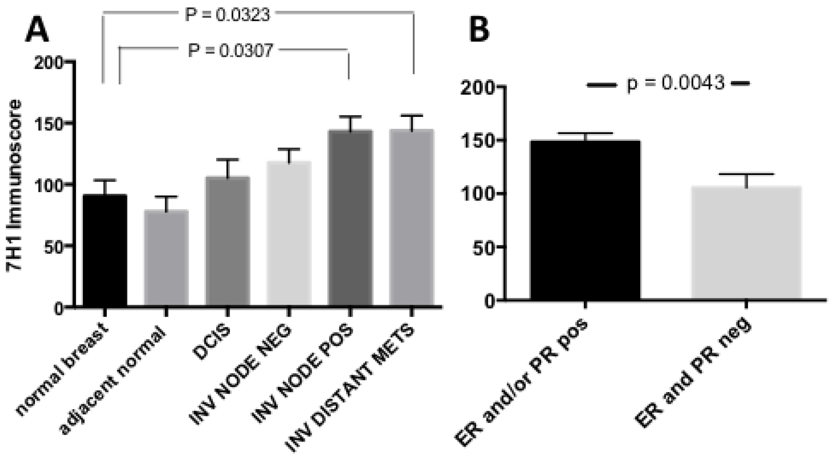
To quantify Plg-RKT expression we calculated anti-Plg-RKT immunoscores [27]. Briefly the number of Plg-RKT positive cells and the total number of cells were counted in 10 random high-power fields. The intensity of immunostaining was scored as 0 = negative; 1+ = weak; 2+ = moderate; and 3+ = strong. Immunoscores were calculated as the percentage of positive cells (0 to 100) multiplied by staining intensity score (0/1/2/3) to yield scores ranging from 0 to 300. Plg-RKT immunoscores increased with anatomical stage (Figure 3A) and were highest in invasive tumors with lymph node (LN) involvement and/or distant metastasis. Plg-RKT expression was also compared with expression of the biomarkers ER, PR, and HER2, as annotated in the BCP-TMA. For analysis, ER and/or PR-positive tumors were treated as one group, (hormone receptor (HR) positive tumors). We found that Plg-RKT expression was significantly higher in HR-positive tumors than in HR-negative tumors (Figure 3B). Immunoscores for Plg-RKT in ER/PR/HER2-negative (triple negative) tumors were low and were not significantly different from HR-negative, HER2 positive tumors (Supplementary Figure S1). Plg-RKT expression in HER2-positive tumors was moderate to high (Supplementary Figure S1), however the majority of Plg-RKT high scoring HER2 tumors were also HR-positive (e.g., Supplementary Figure S1). It was also noteworthy that only four tumors were negative for Plg-RKT expression and these were also HR-negative. Together, these results demonstrate that Plg-RKT is widely expressed in human breast tumors, that its expression is increased in tumors that have spread to draining lymph nodes and distant organs, and that Plg-RKT expression is most pronounced in HR-positive tumors.

3.2. Plg-RKT Expression in Human Breast Cancer Cell Lines

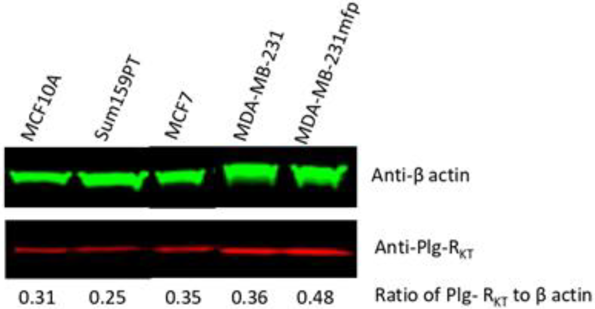
We assessed Plg-RKT expression on a number of established human cell lines. Using Western blotting we found the receptor to be expressed in all cell lines tested, including the HR-positive normal breast cell line MCF10A and the tumorigenic HR-positive MCF7 line as well as tumorigenic triple negative breast cancer cell lines, SUM159PT and MDA-MB-231 (Figure 4). These data show that the receptor is expressed on human breast cancer cell lines including triple-negative cell lines which are examples of highly aggressive tumors. By flow cytometry, positive signals were obtained for all cell lines tested (data not shown).
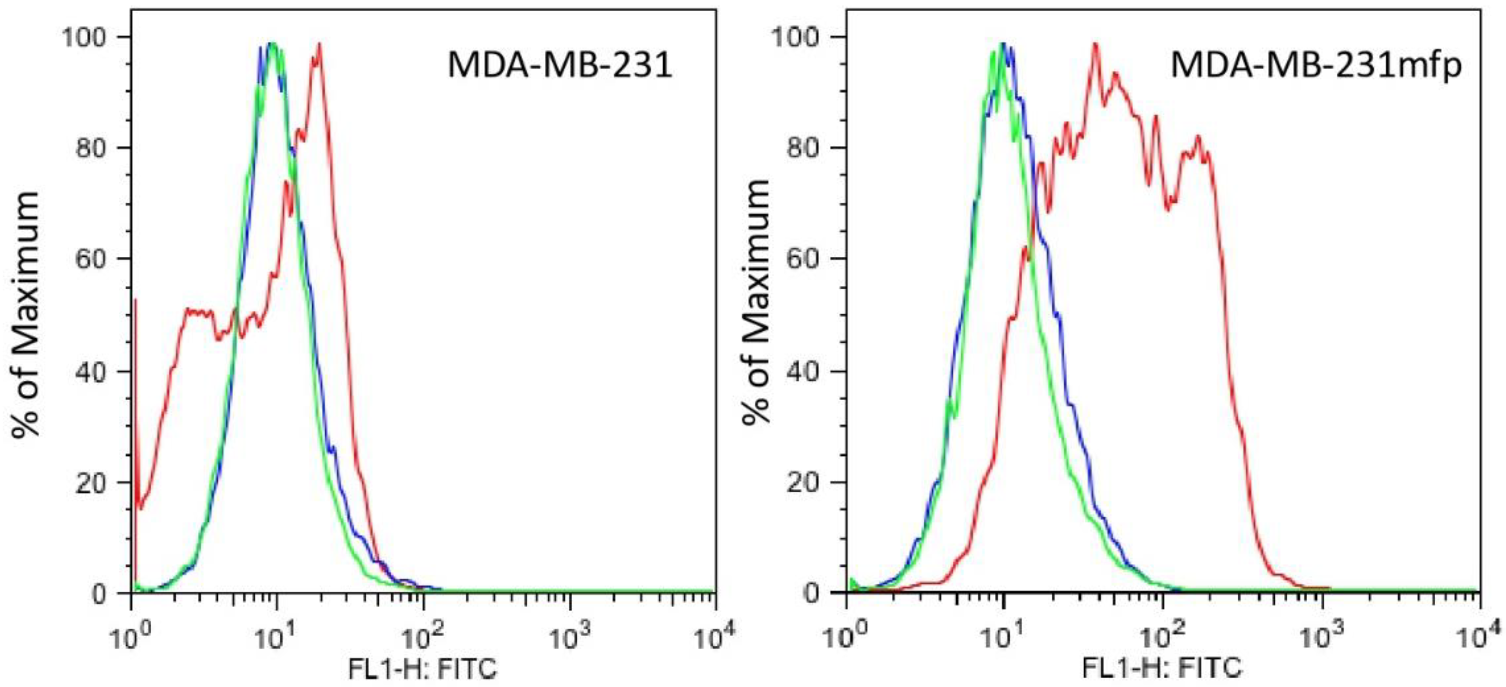
We have previously examined the impact of the microenvironment on breast cancer by establishing orthotopic xenograft tumors of the invasive human breast cancer line, MDA-MB-231, in the mammary fat pad (mfp) of SCID mice. Tumor cells were recovered and cultured to provide a propagatable subpopulation, here referred to as MDA-MB-231mfp cells [28,29]. A total of 231mfp cells exhibited a dramatic increase in tumor growth in vivo. The 231mfp tumors also produced significantly more lung and lymph node metastases than tumors from the parental cell line, MDA-MB-231 [28,29]. In the MDA-MB-231 model we found that the highly metastatic subline MDA-MD-231mfp expressed more Plg-RKT than the parental MDA-MB-231 line (Figure 4 and Figure 5). The MDA-MB-231 mfp cell line also exhibited markedly enhanced plasminogen binding ability compared to the parental MDA-MB-231 cells. In FACS analysis specific plasminogen binding to MDA-MB-231mfp cells was increased 3-fold, compared to MDA-MB-231 cells (Supplementary Figure S2). Quantitative FACS analysis yielded a Bmax of 3.8 ± 0.5 × 10⁵ plasminogen binding sites with a Kd of 1.2 μM for the MDA-MB-231mfp cells and a Bmax of 1.2 ± 0.3 × 10⁵ sites with a Kd of 1.5 μM for the parental cells. Comparing the FACS data for plasminogen binding and anti-Plg-RKT mAB binding, we reason that most of the increased plasminogen binding on the MDA-MB-231mfp cells is to Plg-RKT. However, other plasminogen receptors/binding proteins have been described [16,17] and some plasminogen binding to the surfaces of the cells may be due to other receptors. This will be addressed in future studies. Together, these data show that the receptor is expressed on human breast cancer cell lines and receptor expression appears to increase with tumor progression.

4. Discussion

The interaction of plasminogen with cancer cells plays a key role in progression of breast cancer and other cancers [13,14,15,16,17,18]. Here, we provide the first report that the plasminogen receptor, Plg-RKT, is widely expressed in human breast tumors, that its expression is increased in tumors that have spread to draining lymph nodes and distant organs, and that Plg-RKT expression is most pronounced in HR-positive tumors. In addition, we provide the first evidence that Plg-RKT is present on the surface of breast cancer cell lines.
Triple negative breast cancer TMA samples had low Plg-RKT expression whereas triple negative human breast cancer cell lines (MDA-MB-231 and MDA-MB-231mfp) expressed higher levels of Plg-RKT than the HR-positive MCF7 cell line. It is important to note that the TMA was constructed using primary breast tumor tissues from women that were diagnosed and underwent breast cancer surgery. Tissues from advanced tumors including tumors from metastatic sites were not represented in the tumor microarray (because these tumors are not subjected to surgery and such tissues are rarely available). A likely reason for the discrepancy in Plg-RKT expression in the TMA and established cancer cell lines is that the MDA-MB-231 cell line was established from a metastatic tumor (pleural effusion) and cell lines can also become more aggressive following multiple passages.
Our results suggest that Plg-RKT could provide a new marker for staging of human breast tumors. Notably, a recent report identified over expression of Plg-RKT as a novel marker in inflammatory breast cancer [30]. Interestingly, Plg-RKT mRNA expression is increased in gliomas of higher grades [17]. Future studies are warranted to investigate the potential of Plg-RKT as a marker in other cancers.
In the MDA-MB-231 model we found that the highly metastatic subline MDA-MB-231mfp expressed more Plg-RKT than the parental MDA-MB-231 line, suggesting that Plg-RKT may promote breast cancer progression. Furthermore, the MDA-MB-231mfp cell line exhibited markedly enhanced plasminogen binding ability compared to the parental MDA-MB-231 cells. In FACS analysis specific plasminogen binding to 231mfp was increased 3-fold, compared to MDA-MB-231 cells (Supplementary Figure S2). This increase in plasminogen binding is very significant because each plasminogen binding site stimulates plasminogen activation 11-60-fold [10] and the Kd for the interaction indicates that these binding sites will be substantially occupied at the plasminogen concentration in the interstitial fluid in the tumor microenvironment (2 μM [31]).
Mechanistically, Plg-RKT may regulate breast cancer progression by binding plasminogen on cancer cell surfaces to promote its activation to plasmin, leading to fibrinolysis, extracellular matrix degradation and activation of proenzymes of MMPs. We also found Plg-RKT expression on tumor associated macrophages (TAMs). Plg-RKT regulates macrophage recruitment [22,23,24] and also promotes plasminogen-dependent polarization to the M2 like phenotype and macrophage intracellular signaling [25]. Thus, regulation of TAM function [32] is an additional mechanism by which Plg-RKT may regulate tumor progression.
Plg-RKT may also have protective functions in breast cancer independent of its plasminogen binding capacity. Plg-RKT deletion in mice results in complete inability to lactate, due to decreased proliferation of epithelial cells [33]. A preponderance of evidence from epidemiological studies points to an inverse relationship between the length of the breastfeeding period and breast cancer risk, suggesting that breastfeeding protects mothers from development of breast cancer (reviewed in [34,35,36,37]). One proposed mechanism is that differentiation of epithelial cells to generate milk diminishes the vulnerability of breast tissue toward the carcinogenic effects of estrogens [38]. Thus, Plg-RKT-dependent lactational development may prevent the development of breast cancer. Future studies are warranted to address the functions of Plg-RKT in breast cancer.

Supplementary Materials

The following supporting information can be downloaded at: https://www.mdpi.com/article/10.3390/biom12040503/s1, Figure S1: Plg-RKT in human breast cancer progression, Figure S2, Plasminogen binding is enhanced on MDA-MB-231mfp cells.

Author Contributions

Conceptualization, L.A.M., R.J.P. and B.M.M.; methodology, S.K. and N.B.; formal analysis, S.K. and B.M.M.; investigation, S.K. and N.B.; data curation, S.K., L.A.M., R.J.P. and B.M.M.; writing—original draft preparation, L.A.M. and B.M.M.; writing—review and editing, L.A.M., S.K., N.B., R.J.P. and B.M.M.; funding acquisition, L.A.M., R.J.P. and B.M.M. All authors have read and agreed to the published version of the manuscript.

Funding

This research was funded by National Institutes of Health (grants HD101133 and HL 081046 to L.A.M., CA 166473 to B.M.M. and L.A.M.) and by Merit Review Award I01BX003933 from the U.S. Department of Veterans Affairs (to R.J.P.).

Institutional Review Board Statement

Not applicable.

Informed Consent Statement

Not applicable.

Data Availability Statement

Please contact Stan Krajewski at [email protected] or Barbara Mueller at [email protected] for IHC data.

Conflicts of Interest

The authors declare no conflict of interest.

References

  1. Fay, W.P.; Garg, N.; Sunkar, M. Vascular functions of the plasminogen activation system. Arterioscler. Thromb. Vasc. Biol. 2007, 27, 1231–1237. [Google Scholar] [CrossRef] [PubMed]
  2. Kwaan, H.C.; McMahon, B. The role of plasminogen-plasmin system in cancer. Cancer Treat. Res. 2009, 148, 43–66. [Google Scholar] [CrossRef] [PubMed]
  3. Andreasen, P.A.; Egelund, R.; Petersen, H.H. The plasminogen activation system in tumor growth, invasion, and metastasis. Cell. Mol. Life Sci. 2000, 57, 25–40. [Google Scholar] [CrossRef] [PubMed]
  4. Dano, K.; Behrendt, N.; Hoyer-Hansen, G.; Johnsen, M.; Lund, L.R.; Ploug, M.; Romer, J. Plasminogen activation and cancer. Thromb. Haemost. 2005, 93, 676–681. [Google Scholar] [CrossRef] [PubMed]
  5. Han, B.; Nakamura, M.; Mori, I.; Nakamura, Y.; Kakudo, K. Urokinase-type plasminogen activator system and breast cancer (Review). Oncol. Rep. 2005, 14, 105–112. [Google Scholar]
  6. Harbeck, N.; Kates, R.E.; Schmitt, M.; Gauger, K.; Kiechle, M.; Janicke, F.; Thomassen, C.; Look, M.P.; Foekens, J.A. Urokinase-type plasminogen activator and its inhibitor type 1 predict disease outcome and therapy response in primary breast cancer. Clin. Breast Cancer 2004, 5, 348–352. [Google Scholar] [CrossRef]
  7. Duffy, M.J.; McGowan, P.M.; Harbeck, N.; Thomssen, C.; Schmitt, M. uPA and PAI-1 as biomarkers in breast cancer: Validated for clinical use in level-of-evidence-1 studies. Breast Cancer Res. 2014, 16, 428. [Google Scholar] [CrossRef] [Green Version]
  8. Gouri, A.; Dekaken, A.; El Bairi, K.; Aissaoui, A.; Laabed, N.; Chefrour, M.; Ciccolini, J.; Milano, G.; Benharkat, S. Plasminogen Activator System and Breast Cancer: Potential Role in Therapy Decision Making and Precision Medicine. Biomark. Insights 2016, 11, 105–111. [Google Scholar] [CrossRef]
  9. Li Santi, A.; Napolitano, F.; Montuori, N.; Ragno, P. The Urokinase Receptor: A Multifunctional Receptor in Cancer Cell Biology. Therapeutic Implications. Int. J. Mol. Sci. 2021, 22, 4111. [Google Scholar] [CrossRef]
  10. Miles, L.A.; Hawley, S.B.; Baik, N.; Andronicos, N.M.; Castellino, F.J.; Parmer, R.J. Plasminogen receptors: The sine qua non of cell surface plasminogen activation. Front. Biosci. 2005, 10, 1754–1762. [Google Scholar]
  11. Miles, L.A.; Parmer, R.J. Plasminogen receptors: The first quarter century. Semin. Thromb. Hemost. 2013, 39, 329–337. [Google Scholar] [CrossRef] [PubMed] [Green Version]
  12. Félez, J.; Miles, L.A.; Fábregas, P.; Jardi, M.; Plow, E.F.; Lijnen, R.H. Characterization of cellular binding sites and interactive regions within reactants required for enhancement of plasminogen activation by tPA on the surface of leukocytic cells. Thromb. Haemost. 1996, 76, 577–584. [Google Scholar] [CrossRef] [PubMed]
  13. Godier, A.; Hunt, B.J. Plasminogen receptors and their role in the pathogenesis of inflammatory, autoimmune and malignant disease. J. Thromb. Haemost. 2013, 11, 26–34. [Google Scholar] [CrossRef]
  14. Didiasova, M.; Wujak, L.; Wygrecka, M.; Zakrzewicz, D. From plasminogen to plasmin: Role of plasminogen receptors in human cancer. Int. J. Mol. Sci. 2014, 15, 21229–21252. [Google Scholar] [CrossRef] [PubMed] [Green Version]
  15. Rein-Smith, C.M.; Church, F.C. Emerging pathophysiological roles for fibrinolysis. Curr. Opin. Hematol. 2014, 21, 438–444. [Google Scholar] [CrossRef] [PubMed]
  16. Kumari, S.; Malla, R. New Insight on the Role of Plasminogen Receptor in Cancer Progression. Cancer Growth Metastasis 2015, 8, 35–42. [Google Scholar] [CrossRef] [Green Version]
  17. Gonias, S.L.; Zampieri, C. Plasminogen Receptors in Human Malignancies: Effects on Prognosis and Feasibility as Targets for Drug Development. Curr. Drug Targets 2020, 21, 647–656. [Google Scholar] [CrossRef]
  18. Bharadwaj, A.G.; Holloway, R.W.; Miller, V.A.; Waisman, D.M. Plasmin and Plasminogen System in the Tumor Microenvironment: Implications for Cancer Diagnosis, Prognosis, and Therapy. Cancers 2021, 13, 1838. [Google Scholar] [CrossRef]
  19. Andronicos, N.M.; Chen, E.I.; Baik, N.; Bai, H.; Parmer, C.M.; Kiosses, W.B.; Kamps, M.P.; Yates, J.R., III; Parmer, R.J.; Miles, L.A. Proteomics-based discovery of a novel, structurally unique, and developmentally regulated plasminogen receptor, Plg-RKT, a major regulator of cell surface plasminogen activation. Blood 2010, 115, 1319–1330. [Google Scholar] [CrossRef] [Green Version]
  20. Miles, L.A.; Andronicos, N.M.; Chen, E.I.; Baik, N.; Bai, H.; Parmer, C.M.; Lighvani, S.; Nangia, S.; Kiosses, W.B.; Kamps, M.P.; et al. Identification of the novel plasminogen receptor, Plg-RKT. In Proteomics/Book 1: Human Diseases and Protein Functions; Man, T.K., Flores, R.J., Eds.; Intech: Rijeka, Croatia, 2012; pp. 219–238. ISBN 978-953-307-832-8. [Google Scholar]
  21. Bai, H.; Baik, N.; Kiosses, W.B.; Krajewski, S.; Miles, L.A.; Parmer, R.J. The novel plasminogen receptor, plasminogen receptor(KT) (Plg-R(KT)), regulates catecholamine release. J. Biol. Chem 2011, 286, 33125–33133. [Google Scholar] [CrossRef] [Green Version]
  22. Lighvani, S.; Baik, N.; Diggs, J.E.; Khaldoyanidi, S.; Parmer, R.J.; Miles, L.A. Regulation of macrophage migration by a novel plasminogen receptor Plg-RKT. Blood 2011, 118, 5622–5630. [Google Scholar] [CrossRef] [PubMed] [Green Version]
  23. Miles, L.A.; Baik, N.; Lighvani, S.; Khaldoyanidi, S.; Varki, N.M.; Bai, H.; Mueller, B.M.; Parmer, R.J. Deficiency of plasminogen receptor, Plg-RKT, causes defects in plasminogen binding and inflammatory macrophage recruitment in vivo. J. Thromb. Haemost. 2017, 15, 155–162. [Google Scholar] [CrossRef] [PubMed] [Green Version]
  24. Thaler, B.; Baik, N.; Hohensinner, P.J.; Baumgartner, J.; Panzenbock, A.; Stojkovic, S.; Demyanets, S.; Huk, I.; Rega-Kaun, G.; Kaun, C.; et al. Differential expression of Plg-RKT and its effects on migration of proinflammatory monocyte and macrophage subsets. Blood 2019, 134, 561–567. [Google Scholar] [CrossRef] [PubMed] [Green Version]
  25. Vago, J.P.; Sugimoto, M.A.; Lima, K.M.; Negreiros-Lima, G.L.; Baik, N.; Teixeira, M.M.; Perretti, M.; Parmer, R.J.; Miles, L.A.; Sousa, L.P. Plasminogen and the Plasminogen receptor, Plg-RKT, regulate macrophage phenotypic and functional changes. Front. Immunol. 2019, 10, 1458. [Google Scholar] [CrossRef]
  26. Krajewski, S.; Krajewska, M.; Ellerby, L.M.; Welsh, K.; Xie, Z.; Deveraux, Q.L.; Salvesen, G.S.; Bredesen, D.E.; Rosenthal, R.E.; Fiskum, G.; et al. Release of caspase-9 from mitochondria during neuronal apoptosis and cerebral ischemia. Proc. Natl. Acad. Sci. USA 1999, 96, 5752–5757. [Google Scholar] [CrossRef] [Green Version]
  27. Krajewska, M.; Zapata, J.M.; Meinhold-Heerlein, I.; Hedayat, H.; Monks, A.; Bettendorf, H.; Shabaik, A.; Bubendorf, L.; Kallioniemi, O.P.; Kim, H.; et al. Expression of Bcl-2 family member Bid in normal and malignant tissues. Neoplasia 2002, 4, 129–140. [Google Scholar] [CrossRef] [PubMed] [Green Version]
  28. Jessani, N.; Humphrey, M.; McDonald, W.H.; Niessen, S.; Masuda, K.; Gangadharan, B.; Yates, J.R., III; Mueller, B.M.; Cravatt, B.F. Carcinoma and stromal enzyme activity profiles associated with breast tumor growth in vivo. Proc. Natl. Acad. Sci. USA 2004, 101, 13756–13761. [Google Scholar] [CrossRef] [Green Version]
  29. Jessani, N.; Niessen, S.; Mueller, B.M.; Cravatt, B.F. Breast cancer cell lines grown in vivo: What goes in isn’t always the same as what comes out. Cell Cycle 2005, 4, 253–255. [Google Scholar] [CrossRef] [Green Version]
  30. Suarez-Arroyo, I.J.; Feliz-Mosquea, Y.R.; Perez-Laspiur, J.; Arju, R.; Giashuddin, S.; Maldonado-Martinez, G.; Cubano, L.A.; Schneider, R.J.; Martinez-Montemayor, M.M. The proteome signature of the inflammatory breast cancer plasma membrane identifies novel molecular markers of disease. Am. J. Cancer Res. 2016, 6, 1720–1740. [Google Scholar]
  31. Collen, D. On the regulation and control of fibrinolysis. Thromb. Haemost. 1980, 43, 77–89. [Google Scholar] [CrossRef]
  32. Amens, J.N.; Bahçecioglu, G.; Zorlutuna, P. Immune System Effects on Breast Cancer. Cell. Mol. Bioeng. 2021, 14, 279–292. [Google Scholar] [CrossRef] [PubMed]
  33. Miles, L.A.; Baik, N.; Bai, H.; Makarenkova, H.P.; Kiosses, W.B.; Krajewski, S.; Castellino, F.J.; Valenzuela, A.; Varki, N.M.; Mueller, B.M.; et al. The Plasminogen Receptor, Plg-RKT, is Essential for Mammary Lobuloalveolar Development and Lactation. J. Thromb. Haemost. 2018, 16, 919–932. [Google Scholar] [CrossRef] [PubMed] [Green Version]
  34. Breast cancer and breastfeeding: Collaborative reanalysis of individual data from 47 epidemiological studies in 30 countries, including 50302 women with breast cancer and 96973 women without the disease. Lancet 2002, 360, 187–195. [CrossRef]
  35. Gonzalez-Jimenez, E.; Garcia, P.A.; Aguilar, M.J.; Padilla, C.A.; Alvarez, J. Breastfeeding and the prevention of breast cancer: A retrospective review of clinical histories. J. Clin. Nurs. 2013, 23, 2397–2403. [Google Scholar] [CrossRef]
  36. Bartick, M.C.; Stuebe, A.M.; Schwarz, E.B.; Luongo, C.; Reinhold, A.G.; Foster, E.M. Cost analysis of maternal disease associated with suboptimal breastfeeding. Obstet. Gynecol 2013, 122, 111–119. [Google Scholar] [CrossRef] [PubMed] [Green Version]
  37. Qiu, R.; Zhong, Y.; Hu, M.; Wu, B. Breastfeeding and Reduced Risk of Breast Cancer: A Systematic Review and Meta-Analysis. Comput. Math. Methods Med. 2022, 2022, 8500910. [Google Scholar] [CrossRef] [PubMed]
  38. Xing, P.; Li, J.; Jin, F. A case-control study of reproductive factors associated with subtypes of breast cancer in Northeast China. Med. Oncol. 2010, 27, 926–931. [Google Scholar] [CrossRef]
Figure 1. Plg-RKT is highly expressed in human ductal carcinoma in situ and invasive ductal carcinoma. A core from a patient with ductal carcinoma in situ was stained with anti-Plg-RKT mAb using paraffin immunocytochemistry (A) and enlarged in (B). Cores from the same patient showing invasive ductal carcinoma (C) and from a 60-year-old healthy female control subject ((D) and enlarged in panel (E)) were stained in the same way. Green arrows in panel E indicate cells with macrophage morphology that also stain with anti-Plg-RKT mAb. A specificity control in which the core shown in Panel C was stained with anti-Plg-RKT mAb that had been absorbed with the Plg-RKT peptide used for immunization is shown in Panel F. Original magnifications, ×100 (A,D), ×200 (B) to ×400 (C,E,F).
Biomolecules 12 00503 g001
Figure 2. Plg-RKT expression in human mammary tissues. (A) Ductal epithelial cells in normal breast with light granular Plg-RKT staining, (B) ductal carcinoma in situ with faint to moderate staining, (C) invasive, LN-positive HR-positive breast tumor, (D) invasive, HR-positive tumors with distant metastases show moderate and (E) strong granular Plg-RKT staining. (F) Invasive, LN-positive triple-negative breast tumor exhibits very fine granular Plg-RKT staining of tumor cells and faint to moderate staining of the reactive stroma. (G) Ductal carcinoma in situ showing staining of TAMs (identified by morphology and indicated with green arrows). Plg-RKT by IHC using anti-Plg-RKT monoclonal antibody 7H1. Shown are representative tissues from the CDP breast cancer progression TMA. Original magnifications, ×100 (A,E), ×200 (C,D,F) to ×400 (B,G).
Biomolecules 12 00503 g002
Figure 3. Plg-RKT in human breast cancer progression. Anti-Plg-RKT mAB immunoscores for tissues presented in the CDP breast cancer progression TMA. (A) By anatomical stage: normal breast from nonbreast cancer (n = 23), normal adjacent breast from breast cancer patient (n = 12), DCIS (n = 22), invasive tumor without lymph node involvement (n = 48), invasive tumor with lymph node involvement (n = 50) and invasive tumor with distant metastasis (n = 45). (B) By hormone receptor status: ER and/or PR-positive (n = 95), ER and PR-negative (n = 43). Values are means and SEM, significance by ANOVA with Tukey’s multiple comparison test.
Biomolecules 12 00503 g003
Figure 4. Western blot analysis of expression of Plg-RKT in human breast cancer cell lines. The cells were lysed in RIPA buffer and 30 µg were electrophoresed on 4–12% gradient gels under reducing conditions and Western blotted with anti-Plg-RKT mAB and anti-actin.
Biomolecules 12 00503 g004
Figure 5. Plg-RKT expression is enhanced on MDA-MB-231mfp cells. MDA-MB-231mfp cells and MDA-MB-231 cells were analyzed, using FACS analysis, for staining with anti-Plg-RKT mAb7H1 (red) compared to isotype control (blue) or unstained (green).
Biomolecules 12 00503 g005
Publisher’s Note: MDPI stays neutral with regard to jurisdictional claims in published maps and institutional affiliations.