Article

Characterization of SLC34A2 as a Potential Prognostic Marker of Oncological Diseases

by
Ramilia Vlasenkova
1,
Alsina Nurgalieva
1,
Natalia Akberova
1,
Mikhail Bogdanov
1,2 and
Ramziya Kiyamova
1,*
1
Department of Biochemistry, Biotechnology and Pharmacology, Institute of Fundamental Medicine and Biology, Kazan Federal University, 420008 Kazan, Russia
2
Department of Biochemistry and Molecular Biology, McGovern Medical School, The University of Texas Health Science Center, Houston, TX 77030, USA
*
Author to whom correspondence should be addressed.
Biomolecules 2021, 11(12), 1878; https://doi.org/10.3390/biom11121878
Submission received: 3 October 2021 / Revised: 10 December 2021 / Accepted: 10 December 2021 / Published: 14 December 2021

Abstract

:
The main goal of this study is to consider SLC34A2 as a potential prognostic marker of oncological diseases using the mutational, expression, and survival data of cancer studies which are publicly available online. We collected data from four databases (cBioPortal, The Cancer Genome Atlas; cBioPortal, Genie; International Cancer Genome Consortium; ArrayExpress). In total, 111,283 samples were categorized according to 27 tumor locations. Ninety-nine functionally significant missense mutations and twelve functionally significant indel mutations in SLC34A2 were found. The most frequent mutations were SLC34A2-ROS1, p.T154A, p.P506S/R/L, p.G257A/E/R, p.S318W, p.A396T, p.P410L/S/H, p.S461C, p.A473T/V, and p.Y503H/C/F. The upregulation of SLC34A2 was found in samples of myeloid, bowel, ovarian, and uterine tumors; downregulation was found in tumor samples of breast, liver, lung, and skin cancer tumors. It was found that the life expectancy of breast and thymus cancer patients with an SLC34A2 mutation is lower, and it was revealed that SLC34A2 overexpression reduced the life span of patients with brain, ovarian, and pancreatic tumors. Thereby, for these types of oncological diseases, the mutational profile of SLC34A2 can be a potential prognostic marker for breast and thymus cancers, and the upregulation of SLC34A2 can be a potential prognostic marker for brain, ovarian, and pancreatic cancers.

1. Introduction

The search for and the characterization of new molecular markers of oncological diseases are important for the prediction of the prognosis of the disease and the response of the tumor to chemotherapy. The majority of these tumor-targeting compounds are directed against cell membrane-bound proteins [1]. However, there is surprisingly little knowledge about which targets should be used for optimal results per the tumor type, or even better, per the individual tumor or patient. Cell membrane transporter proteins such as transporters belonging to glucose transporter GLUT, ATP-binding cassette transporter ABC, and solute carrier transporter SLC families are upregulated on cancer cells, compared to adjacent normal cells. High levels of transporters are found in a wide range of solid tumors, correlating with poor survival [1]. One of the potential molecular tumor markers may be the sodium-dependent phosphate transporter NaPi2b encoded by the SLC34A2 gene. SLC34A2, belonging to the solute carrier gene family, encodes the type II Na/Pi co-transporter (NaPi2b) [2]. NaPi2b is a multitransmembrane sodium-dependent phosphate transporter responsible for transcellular inorganic phosphate absorption [3]. NaPi2b is highly abundant in the brush-border membrane (BBM) of the small intestine, where it is involved in the transcellular flux of inorganic phosphates via the apical membrane of epithelial cells [4,5]. An altered expression of sodium-dependent phosphate transporter NaPi2b has been reported in ovarian cancer [6], lung cancer [7,8,9,10], gastric cancer [11], thyroid cancer [12], and other cancers [13]. The phosphate transporter NaPi2b was identified as a target for MX35 monoclonal antibodies using modified SEREX (the serological analysis of recombinant cDNA expression libraries) technology [14,15]. Currently, NaPi2b is a target for therapeutic antibodies XMT-1536 and XMT-1592, which are in clinical trials for the treatment of ovarian and lung cancers [16]. Despite the fact that NaPi2B is expressed in normal tissues, therapeutic MX35 and Rebmab200 mAbs [17] are accumulated predominantly in cancer tissues. Therefore, NaPi2b could be considered as a potential molecular marker of several types of cancer, and represents a new family of potential cell-surface targets for the immunotherapy of cancer.
At the moment, there is limited evidence that NaPi2b has potential as a prognostic tumor marker. It has been shown that the high expression of NaPi2b may confer resistance to cancer chemotherapy, and may increase the metastatic potential in non-small cell lung cancer [10]. It was revealed that in lung cancer, SLC34A2 induces resistance to crizotinib [18] when it undergoes molecular re-arrangement with ROS-1, a tyrosine kinase receptor. This re-arrangement creates a constitutively active tyrosine kinase that also increases the oncogenicity in lung cancer [19].
In order to consider SLC34A2 as a potential prognostic and predictive marker of oncological diseases, we performed an analysis of the mutation and expression of SLC34A2, and assessed their effect on the life expectancy of patients from cancer studies which are publicly available online.

2. Materials and Methods

2.1. Data Collection and Preparation

The data were obtained from 4 open-access databases: cBioPortal, AACR Project Genie, The Insertional Cancer Genome Consortium (ICGC), and ArrayExpress (data downloaded on 21 December 2020). The mutational and the expression data were collected for the SLC34A2 gene and mRNA. cBioPortal [20] was used to access the data from the 32 most recent TCGA (The Cancer Genome Atlas) studies. The TCGA data consisted of mutational, expression, and clinical data. From the AACR Project Genie database [21], 33 studies were utilized; only mutational data were collected. The ICGC data portal [22] was used to obtain 6 studies, and only mutational data were collected. From the ArrayExpress database [23], the E-MTAB-3732 study [24] was used to obtain expression data from tumor and relatively healthy samples divided into 24 groups. All of the collected studies and ArrayExpress samples were categorized by 27 tumor locations: adrenal gland, biliary tract, bladder, bone, bowel, breast, CNS and brain, cervix, esophagus and stomach, eye, head and neck, kidney, liver, lung, lymphoid, myeloid, ovary, pancreas, pleura, prostate, skin, soft tissue, testis, thymus, thyroid, uterus, and various tumors. The sample counts and study grouping are summarized in Supplementary Table S1. Different IDs other than the HGNC (HUGO Gene Nomenclature Committee) symbol (SLC34A2) were used: AFFY HG U133A 2 probe ID—204124_at, and Ensemble Protein ID—ENSP00000371483, TIGRFAMs ID—TIGR01013.

2.2. Prediction of the Functional Impact of Mutation

An analysis was conducted on the SLC34A2 mutational data from 3 databases: cBioPortal, AACR Project Genie, and ICGC. Only missense and indel (insertion or deletion) SLC34A2 missense and indel mutations were analyzed with tools for the prediction of the functional significance of mutations. The following tools were used to analyze missense mutations: PROVEAN [25], SIFT [26], PolyPhen-2 [27], Panther-PSEP [28], FATHMM [29], and Mutation Assessor [30]. In order to consider a missense mutation as pathogenic, it needed to be reported by at least five of the variant effect prediction tools. Indel mutations were analyzed with the PROVEAN tool. The determination of highly conserved regions of the SLC34A2 protein was performed with the Conserved Domains and Protein Classification resource [31].

2.3. Analysis of the Expression Levels in Various Tumors

A comparison of the expression levels of SLC34A2 in relatively healthy and tumor samples was carried out on the ArrayExpress SLC34A2 expression data. A Wilcoxon test (p < 0.05) was performed in order to compare healthy and tumor samples.

2.4. Survival Analysis

A survival analysis was performed using the Kaplan–Meyer estimate (p < 0.05). For this analysis, only the cBioPortal TCGA studies were used. The tumor samples were divided into groups according to the level of SLC34A2 mRNA expression (upregulation was considered as two standard deviations above the mean of the samples in each dataset) and by the presence or absence of an SLC34A2 mutation.
For each study in which we found significant differences between the patient groups by the Kaplan–Meyer estimate, multivariate survival analysis was performed using Cox regression (p < 0.05). This analysis was carried out in order to determine whether the level of expression of SLC34A2 or mutations in SLC34A2 is one of the key factors linked to the survivability of patients. For this analysis, we used the clinical parameters which were available for each study.

3. Results

Mutational, expression and clinical data from 95 studies of oncological diseases were collected from four databases (cBioPortal, TCGA; cBioPortal, Genie; ICGC; and ArrayExpress). A total of 111283 samples was gathered and categorized by 27 tumor locations: adrenal gland (2 studies), biliary tract (1 study), bladder (3 studies), bone (2 studies), bowel (5 studies), breast (4 studies), CNS and brain (8 studies), cervix (2 studies), esophagus and stomach (9 studies), eye (1 studies), head and neck (5 studies), kidney (5 studies), liver (4 studies), lung (6 studies), lymphoid (5 studies), myeloid (4 studies), ovary (3 studies), pancreas (3 studies), pleura (2 studies), prostate (3 studies), skin (4 studies), soft tissue (3 studies), testis (1 studies), thymus (1 studies), thyroid (3 studies), uterus (4 studies), and various tumors (2 studies). The summary table can be found in Table 1, and the detailed information is located in Supplementary Table S1.

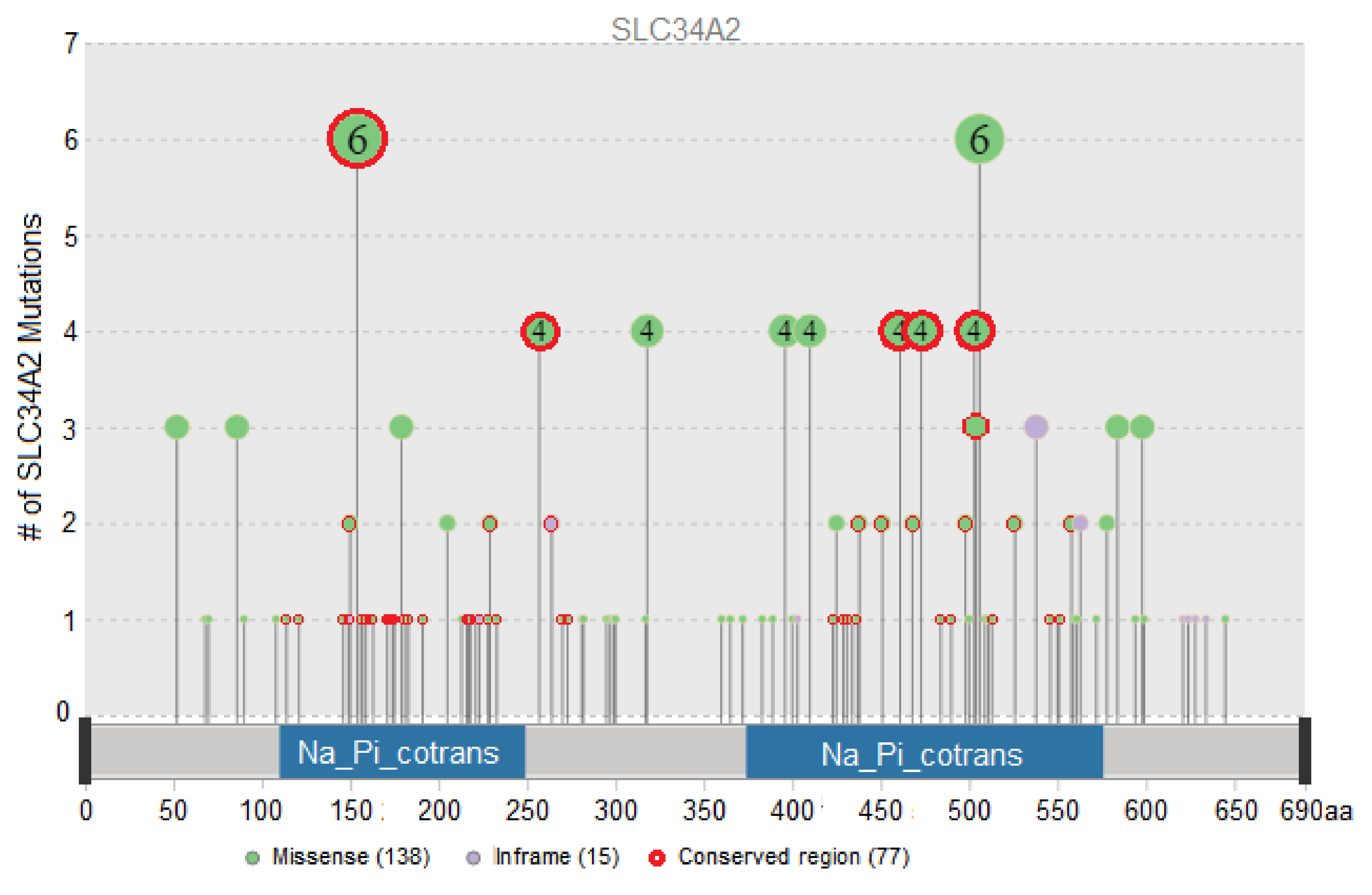
3.1. Evaluation of the Functional Impact of Missense and Indel Mutations in SLC34A2

Five hundred and twenty-one mutations in the SLC34A2 gene were detected in the samples of three databases (cBioPortal, TCGA, n = 10,967; Genie, n = 85,369; ICGC, n = 2197): 441 missense mutations, 17 in-frame deletions, 14 out-of-frame mutations, three intergenic mutations, seven splice site mutations, seven nonsense mutations, four same-sense mutations, and 28 mutations of unknown origin.
Out of the entire mutational dataset, 99 functionally significant missense mutations and 12 functionally significant insertion–deletion mutations in the SLC34A2 gene were determined using tools for the prediction of the functional significance of mutations: PROVEAN, SIFT, PolyPhen-2, Panther-PSEP, FATHMM, and Mutation Assessor (Figure 1). In addition, three intergenic mutations were added to the list of functionally significant mutations. The list of functionally significant mutations, allele frequencies, rsIDs (identification number from dbSNP database), tumor locations, sample counts, and the indication of highly conserved regions are stated in Supplementary Table S2.
The most frequent mutations by the sample count in total (the numbers are given in total for all studies, n = 98,533) are intergenic mutation SLC34A2-ROS1 (10 samples), which was found only in lung cancer samples; p.T154A (six samples); p.P506S/R/L (six samples); p.G257A/E/R (four samples); p.S318W (four samples); p.A396T (four samples); p.P410L/S/H (four samples); p.S461C (four samples); p.A473T/V (four samples); and p.Y503H/C/F (four samples). The sample counts according to the tumor locations are displayed in Table 2.
Twenty-one functionally significant mutations in the SLC34A2 gene were found in two or more tumor locations (Supplementary Table S3). Several mutations were found in more than three locations: p.T154A (breast, esophagus and stomach, lymphoid, ovary, pleura), p.A396T (lung, thymus, uterus), p.A473T (esophagus and stomach, head and neck, lung), and p.F538del (lung, lymphoid, kidney). The most frequently occurring tumor locations (with the highest number of functionally significant mutations in the SLC34A2 gene) were the skin, lung, bowel, and uterus, with 23, 22, 17, and 17 unique mutations respectively (Supplementary Table S4).

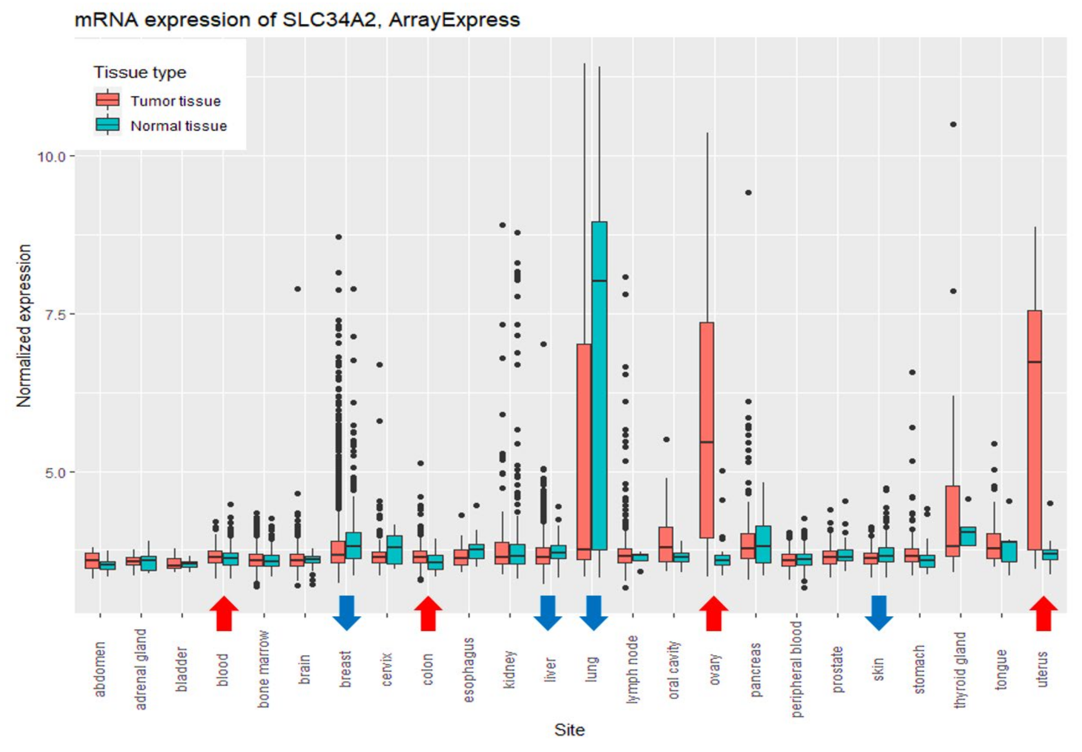
3.2. Comparison of the Expression Levels of SLC34A2 between Relatively Healthy and Tumor Tissues

The expression data used for comparison between relatively healthy and tumor tissue samples were gathered from the ArrayExpress E-MTAB-3732 study (n = 12,750). It should be mentioned that the data we used did not contain methylation information that could influence the expression of SLC34A2, and we have not taken this factor into account in our analysis.
Comparing the levels of SLC34A2 expression between relatively healthy and tumor tissues (Wilcoxon test, p < 0.05), the higher levels of expression were found in tumor tissues of the myeloid, bowel, ovary, and uterus, and the lower levels were found in samples of breast, liver, lung, and skin tumors (Figure 2).

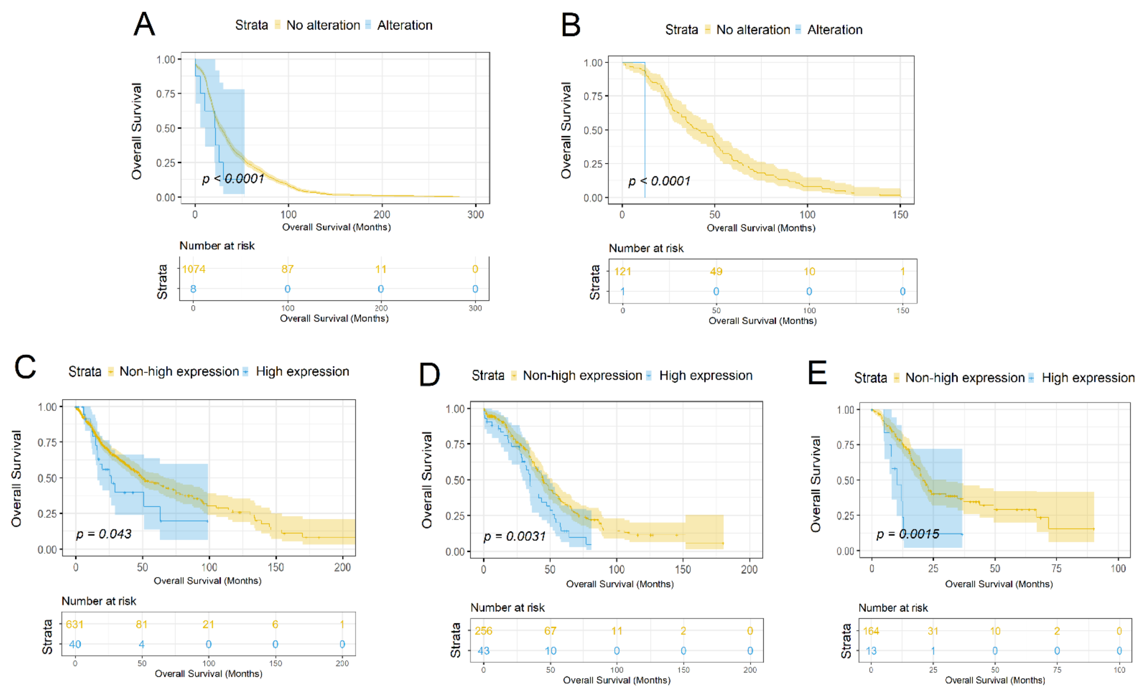
3.3. Characterization of the SLC34A2 Gene as a Prognostic Marker

A survival analysis was conducted on 32 cBioportal TCGA studies using the Kaplan-Meier estimator. The tumor samples were divided into groups in two ways: by the level of SLC34A2 mRNA expression and by the presence or absence of an SLC34A2 mutation. It must be mentioned that not all of the cBioportal TCGA samples had lifespan information; thus, the datasets may be smaller (Supplementary Table S1).
The life expectancy of patients with mutations in the SLC34A2 gene is significantly lower than in patients without gene alterations in studies of breast (p < 0.042) and thymus (p < 0.0008) tumors (cBioPortal, TCGA: Breast invasive carcinoma, n = 1082, Thymoma, n = 122; Figure 3A-B).
The life expectancy of patients with a high expression of SLC34A2 is significantly lower than that of patients with a lower expression of the gene in studies of brain (p < 0.043), ovarian (p < 0.0031), and pancreatic (p < 0.0015) cancer (cBioPortal, TCGA: Brain Lower Grade Glioma, n = 514, Glioblastoma multiforme, n = 592, Ovarian serous cystadenocarcinoma—n = 299, Pancreatic adenocarcinoma, n = 179, p < 0.05; Figure 3C–E).
Multivariate survival analysis was performed using Cox regression (p < 0.05) for each dataset in which we found significant differences between the patient groups according to the Kaplan–Meyer estimate.
For the breast cancer dataset, the results of the multivariate survival analysis showed that the hazard ratio for the group with a mutation of SLC34A2 gene is 14.42 (CI: 4.24–49.05; Supplementary Figure S1A); the parameters with a higher hazard ratio were Neoplasm Disease Lymph Node Stage N3, Neoplasm Disease Stage III and IV. The result for the thymoma dataset indicated as significant only the presence of the mutation of SLC34A2 (HR = 24.57, CI: 1.87–323.65; Supplementary Figure S1B). The multivariate analysis of the CNS and brain cancer dataset showed that the level of SLC34A2 mRNA expression has a hazard ratio of 2.07 (CI: 0.99–4.3; Supplementary Figure S1C); the parameter with a higher hazard ratio was only Neoplasm Disease Lymph Node Stage N3. For the ovary and pancreas datasets, the level of SLC34A2 expression had the highest hazard ratios—1.77 (CI: 1.19–2.63; Supplementary Figure S1D) and 3.51 (CI: 1.6–7.71; Supplementary Figure S1E) respectively—and for the pancreas dataset, the level of expression was the only significant parameter in the analysis.

4. Discussion

In order to assess the possible role of SLC34A2as a prognostic marker of cancer, we performed the analysis of the SLC34A2 mutational data, the SLC34A2 mRNA expression data, and the survival data of cancer patients which were publicly available online.
The analysis of mutations in the SLC34A2 gene revealed that most of them are associated with skin, lung, bowel, and uterine cancer types. The most frequent functionally significant mutation is the intergenic mutation SLC34A2-ROS1, which was found only in lung cancer samples, but in other studies, this mutation was connected not only with lung cancer [19] but also with bowel [32], stomach [33], and ovary [34] cancers. We found no association between intergenic mutation SLC34A2-ROS1 and the survivability of lung cancer patients in our study. Among the functionally significant single nucleotide polymorphisms, several frequent mutations should be mentioned: p.T154A, p.P506S/R/L, p.G257A/E/R, p.S318W, p.A396T, p.P410L/S/H, p.S461C, p.A473T/V, and p.Y503H/C/F.
The comparisons of SLC34A2 expression between healthy and tumor tissues showed a distinction in their expression level: upregulation was found in samples of myeloid, bowel, ovarian, and uterine tumors; downregulation was found in tumor samples of breast, liver, lung, and skin cancer tumors. Our results are in accordance with the published data. Upregulation in myeloid and bowel tumors was found in several studies [35,36]; the overexpression of SLC34A2 was found in endometrioid and papillary serous ovarian carcinomas [14,15,16,37,38], and it was shown that in uterine (endometrial) cancer, SLC34A2 expression appeared relatively increased [14,16]. Downregulation in breast [39], liver [40], and lung [7,8,9] tumors was discovered in several studies. It should be noted that the research also shows the upregulation of SLC34A2 in lung tumor samples [7,41]. Our analysis for the first time showed that the expression of SLC34A2 is downregulated in skin cancer.
We conducted a survival analysis of cancer patients, taking into account the obtained mutational data and the expression profile of SLC34A2. The analysis of the mutational data showed the lower life expectancy of patients with SLC34A2 alterations in the studies of breast and thymus cancers. The results of the multivariate survival analysis showed that the mutational profile of SLC34A2 plays a significant role in the survivability of breast and thymus cancer patients. Both studies contained the discovered functionally significant mutations. Breast samples contained the following functionally significant mutations: p.D68G and p.P504L (the latter is located in a conserved region). These alterations could be a reason for poor prognosis. It was mentioned that SLC34A2 could influence chemotherapy [42] and could establish reasoning for a targeting pathway [43], and the downregulation of SLC34A2 could play a role in breast cancer progression [39]. In thymus samples, we found one functionally significant mutation: p.A396T. Our research, for the first time, showed the correlation between patient survivability and SLC34A2 alterations in thymus cancer.
The analysis of the SLC34A2 expression data showed the potential impact of SLC34A2 upregulation being related to poor survival prognosis in ovarian, pancreatic, and brain tumors. The multivariate survival analysis demonstrated that the level of SLC34A2 expression is one of the key parameters for ovarian, pancreatic, and brain cancer patient survival. We confirmed the published data that SLC34A2 is upregulated in ovarian tumors compared to relatively healthy ovarian tissues [37]. Besides this, we showed that patients with the higher expression of SLC34A2 had a lower life expectancy. Interestingly, we have shown previously that SLC34A2 is overexpressed in well-differentiated papillary serous and endometrioid ovarian carcinomas which usually have a good prognosis [44]. This inconsistency may be related to the molecular heterogeneity of ovarian tumors, and it requires further consideration. The expression of SLC34A2 in pancreatic cancer has not yet been researched, and our study for the first time showed the correlation between patient survivability and SLC34A2 expression. Concerning brain cancer, it has been shown previously that SLC34A2 is overexpressed in glioma [45], but the relation of increased expression with the life expectancy of patients has not been studied.
In conclusion, we analyzed a significantly large dataset of the SLC34A2 mutational data, the SLC34A2 expression data, and the survival data of cancer patients (n = 111,283). One hundred and eleven functionally significant mutations were discovered, and it was found that functionally significant mutations of SLC34A2 might be involved in the reduction of the life expectancy of breast (p.D68G, p.P504L) and thymus (p.A396T) cancer patients. Thus, for these types of oncological diseases, the mutational profile of SLC34A2 can be a potential prognostic marker. It was revealed that SLC34A2 mRNA overexpression decreases the lifespan of patients with brain, ovarian, and pancreatic tumors; therefore, the upregulation of SLC34A2 can be a prognostic marker for these tumors. Furthermore, we suggest that SLC34A2 upregulation—not only for ovary but also for myeloid, bowel, and uterine tumors—can be considered as a potential predictive marker for targeted therapy with monoclonal antibodies, including XMT-1536 and XMT-1592. Taking into account the molecular heterogeneity of tumors, we concluded that the obtained data require further investigation considering the molecular subtypes of these tumors.

Supplementary Materials

The following are available online at https://www.mdpi.com/article/10.3390/biom11121878/s1. Table S1: Overview of the collected data—95 studies across 4 databases (cBioPortal, TCGA; cBioPortal, Genie; ICGC; ArrayExpress). Table S2: List of functionally significant missense and indel mutations in SLC34A2 gene. Table S3: Functionally significant mutations in the SLC34A2 gene found in two or more tumor locations. Table S4: Tumor locations with the highest numbers of functionally significant mutations in the SLC34A2 gene. Figure S1: Multivariate survival analysis using clinical parameters and the SLC34A2 group.

Author Contributions

Conceptualization, R.K.; methodology, R.V., N.A., R.K.; validation, R.V., R.K.; formal analysis, R.V.; investigation, R.V., A.N., R.K.; data curation, R.V.; writing—original draft preparation, R.V.; writing—review and editing, R.K., M.B., N.A.; visualization, R.V.; project administration, R.K., M.B.; supervision, R.K. All authors have read and agreed to the published version of the manuscript.

Funding

The work was performed with Russian Science Foundation support (grant#20-14-00166).

Institutional Review Board Statement

Not applicable.

Informed Consent Statement

Not applicable.

Data Availability Statement

Publicly available datasets were analyzed in this study. These datasets can be found here (the data was downloaded on 21 December 2020): http://www.cbioportal.org, https://genie.cbioportal.org/, https://dcc.icgc.org/, and https://www.ebi.ac.uk/arrayexpress/experiments/E-MTAB-3732.

Acknowledgments

The work was performed under the Kazan Federal University Strategic Academic Leadership Program.

Conflicts of Interest

The authors declare that they have no known competing financial interest or personal relationships that could have appeared to influence the work reported in this paper.

References

  1. Boonstra, M.C.; De Geus, S.W.; Prevoo, H.A.; Hawinkels, L.J.; Van De Velde, C.J.; Kuppen, P.J.; Vahrmeijer, A.L.; Sier, C.F. Selecting Targets for Tumor Imaging: An Overview of Cancer-Associated Membrane Proteins. Biomark. Cancer 2016, 8, 119. [Google Scholar] [CrossRef] [PubMed] [Green Version]
  2. Xu, H.; Bai, L.; Collins, J.F.; Ghishan, F.K. Molecular Cloning, Functional Characterization, Tissue Distribution, and Chromosomal Localization of a Human, Small Intestinal Sodium-Phosphate (Na+-Pi) Transporter (SLC34A2). Genomics 1999, 62, 281–284. [Google Scholar] [CrossRef] [PubMed]
  3. Rangel, L.B.; Sherman-Baust, C.A.; Wernyj, R.P.; Schwartz, D.R.; Cho, K.R.; Morin, P.J. Characterization of Novel Human Ovarian Cancer-Specific Transcripts (HOSTs) Identified by Serial Analysis of Gene Expression. Oncogene 2003, 22, 7225–7232. [Google Scholar] [CrossRef] [Green Version]
  4. Murer, H.; Forster, I.; Biber, J. The Sodium Phosphate Cotransporter Family SLC34. Pflug. Arch. Eur. J. Physiol. 2004, 447, 763–767. [Google Scholar] [CrossRef] [PubMed]
  5. Gupta, A.; Tenenhouse, H.S.; Hoag, H.M.; Wang, D.; Khadeer, M.A.; Namba, N.; Feng, X.; Hruska, K.A. Identification of the Type II Na(+)-Pi Cotransporter (Npt2) in the Osteoclast and the Skeletal Phenotype of Npt2-/- Mice. Bone 2001, 29, 467–476. [Google Scholar] [CrossRef]
  6. Sabbagh, Y.; O’Brien, S.P.; Song, W.; Boulanger, J.H.; Stockmann, A.; Arbeeny, C.; Schiavi, S.C. Intestinal Npt2b Plays a Major Role in Phosphate Absorption and Homeostasis. J. Am. Soc. Nephrol. JASN 2009, 20, 2348–2358. [Google Scholar] [CrossRef]
  7. Kopantzev, E.P.; Monastyrskaya, G.S.; Vinogradova, T.V.; Zinovyeva, M.V.; Kostina, M.B.; Filyukova, O.B.; Tonevitsky, A.G.; Sukhikh, G.T.; Sverdlov, E.D. Differences in Gene Expression Levels between Early and Later Stages of Human Lung Development Are Opposite to Those between Normal Lung Tissue and Non-Small Lung Cell Carcinoma. Lung Cancer 2008, 62, 23–34. [Google Scholar] [CrossRef] [PubMed]
  8. Yang, W.; Wang, Y.; Pu, Q.; Ye, S.; Ma, Q.; Ren, J.; Zhong, G.; Liu, L.; Zhu, W. Elevated Expression of SLC34A2 Inhibits the Viability and Invasion of A549 Cells. Mol. Med. Rep. 2014, 10, 1205. [Google Scholar] [CrossRef] [Green Version]
  9. Zhao, Y.; Zhu, Z.; Shi, S.; Wang, J.; Li, N. Long Non-Coding RNA MEG3 Regulates Migration and Invasion of Lung Cancer Stem Cells via MiR-650/SLC34A2 Axis. Biomed. Pharmacother. 2019, 120, 109457. [Google Scholar] [CrossRef]
  10. Jiang, Z.; Hao, Y.; Ding, X.; Zhang, Z.; Liu, P.; Wei, X.; Xi, J. The Effects and Mechanisms of SLC34A2 on Tumorigenicity in Human Non-Small Cell Lung Cancer Stem Cells. Tumour Biol. J. Int. Soc. Oncodevelopmental Biol. Med. 2016, 37, 10383–10392. [Google Scholar] [CrossRef] [PubMed]
  11. Zhang, L.; Guo, X.; Zhang, L.; Yang, F.; Qin, L.; Zhang, D.; Qin, Y. SLC34A2 Regulates MiR-25-Gsk3β Signaling Pathway to Affect Tumor Progression in Gastric Cancer Stem Cell-like Cells. Mol. Carcinog. 2018, 57, 440–450. [Google Scholar] [CrossRef] [PubMed]
  12. Huang, F.; Wang, H.; Xiao, J.; Shao, C.; Zhou, Y.; Cong, W.; Gong, M.; Sun, J.; Shan, L.; Hao, Z.; et al. SLC34A2 Up-Regulation And SLC4A4 Down-Regulation Correlates With Invasion, Metastasis, And The MAPK Signaling Pathway In Papillary Thyroid Carcinomas. Int. Publ. J. Cancer 2021, 12, 5439–5453. [Google Scholar] [CrossRef] [PubMed]
  13. Kiyamova, R.; Shyian, M.; Lyzogubov, V.V.; Usenko, V.S.; Gout, T.; Filonenko, V. Immunohistochemical Analysis of NaPi2b Protein (MX35 Antigen) Expression and subcellular Localization in Human Normal and Cancer Tissues. Exp. Oncol. 2011, 33, 157–161. [Google Scholar] [PubMed]
  14. Yin, B.; Kiyamova, R.; Chua, R.; Caballero, O.L.; Gout, I.; Gryshkova, V.; Bhaskaran, N.; Souchelnytskyi, S.; Hellman, U.; Filonenko, V.; et al. Monoclonal Antibody MX35 Detects the membrane Transporter NaPi2b (SLC34A2) in Human Carcinomas. Cancer Immun. J. Acad. Cancer Immunol. 2008, 8, 3. [Google Scholar]
  15. Kiyamova, R.G.; Gryshkova, V.S.; Usenko, V.S.; Khozaenko, Y.S.; Gurtovyy, V.A.; Yin, B.; Ritter, G.; Old, L.; Gout, I.T.; Filonenko, V.V. Identification of Phosphate Transporter Napi2b as MX35 Cancer Antigen by Modified SEREX Approach. Biopolym. Cell 2008, 24, 218–224. [Google Scholar] [CrossRef] [Green Version]
  16. Bodyak, N.D.; Mosher, R.; Yurkovetskiy, A.V.; Yin, M.; Bu, C.; Conlon, P.R.; Demady, D.R.; DeVit, M.J.; Gumerov, D.R.; Gurijala, V.R.; et al. The Dolaflexin-Based Antibody–Drug Conjugate XMT-1536 Targets the Solid Tumor Lineage Antigen SLC34A2/NaPi2b. Mol. Cancer Ther. 2021, 20, 896–905. [Google Scholar] [CrossRef]
  17. Dos Santos, M.L.; Yeda, F.P.; Tsuruta, L.R.; Horta, B.B.; Pimenta, A.A.; Degaki, T.L.; Soares, I.C.; Tuma, M.C.; Okamoto, O.K.; Alves, V.A.F.; et al. Rebmab200, a Humanized Monoclonal Antibody Targeting the Sodium Phosphate Transporter NaPi2b Displays Strong Immune Mediated Cytotoxicity against Cancer: A Novel Reagent for Targeted Antibody Therapy of Cancer. PLoS ONE 2013, 8, e70332. [Google Scholar] [CrossRef] [Green Version]
  18. Kato, Y.; Ninomiya, K.; Ohashi, K.; Tomida, S.; Makimoto, G.; Watanabe, H.; Kudo, K.; Matsumoto, S.; Umemura, S.; Goto, K.; et al. Combined Effect of Cabozantinib and Gefitinib in Crizotinib-Resistant Lung Tumors Harboring ROS1 Fusions. Cancer Sci. 2018, 109, 3149–3158. [Google Scholar] [CrossRef] [Green Version]
  19. Yin, X.; Wang, H.; Wu, D.; Zhao, G.; Shao, J.; Dai, Y. SLC34A2 Gene Mutation of Pulmonary Alveolar Microlithiasis: Report of Four Cases and Review of Literatures. Respir. Med. 2013, 107, 217–222. [Google Scholar] [CrossRef] [Green Version]
  20. Cerami, E.; Gao, J.; Dogrusoz, U.; Gross, B.E.; Sumer, S.O.; Aksoy, B.A.; Jacobsen, A.; Byrne, C.J.; Heuer, M.L.; Larsson, E.; et al. The CBio Cancer Genomics Portal: An Open Platform for Exploring Multidimensional Cancer Genomics Data. Cancer Discov. 2012, 2, 401–404. [Google Scholar] [CrossRef] [Green Version]
  21. AACR Project GENIE AACR Project GENIE: Powering Precision Medicine through an International Consortium. Cancer Discov. 2017, 7, 818–831. [CrossRef] [Green Version]
  22. Hudson, T.J.; Anderson, W.; Artez, A.; Barker, A.D.; Bell, C.; Bernabé, R.R.; Bhan, M.K.; Calvo, F.; Eerola, I.; Gerhard, D.S.; et al. International Network of Cancer Genome Projects. Nature 2010, 464, 993–998. [Google Scholar] [CrossRef] [Green Version]
  23. Athar, A.; Füllgrabe, A.; George, N.; Iqbal, H.; Huerta, L.; Ali, A.; Snow, C.; Fonseca, N.A.; Petryszak, R.; Papatheodorou, I.; et al. ArrayExpress Update—From Bulk to Single-Cell Expression Data. Nucleic Acids Res. 2019, 47, D711–D715. [Google Scholar] [CrossRef] [PubMed]
  24. Torrente, A.; Lukk, M.; Xue, V.; Parkinson, H.; Rung, J.; Brazma, A. Identification of Cancer Related Genes Using a Comprehensive Map of Human Gene Expression. PLoS ONE 2016, 11, e0157484. [Google Scholar] [CrossRef] [Green Version]
  25. Choi, Y.; Sims, G.E.; Murphy, S.; Miller, J.R.; Chan, A.P. Predicting the Functional Effect of Amino Acid Substitutions and Indels. PLoS ONE 2012, 7, e46688. [Google Scholar] [CrossRef] [Green Version]
  26. Sim, N.L.; Kumar, P.; Hu, J.; Henikoff, S.; Schneider, G.; Ng, P.C. SIFT Web Server: Predicting Effects of Amino Acid Substitutions on Proteins. Nucleic Acids Res. 2012, 40, W452–W457. [Google Scholar] [CrossRef]
  27. Adzhubei, I.A.; Schmidt, S.; Peshkin, L.; Ramensky, V.E.; Gerasimova, A.; Bork, P.; Kondrashov, A.S.; Sunyaev, S.R. A Method and Server for Predicting Damaging Missense Mutations. Nat. Methods 2010, 7, 248–249. [Google Scholar] [CrossRef] [Green Version]
  28. Mi, H.; Muruganujan, A.; Thomas, P.D. PANTHER in 2013: Modeling the Evolution of Gene Function, and Other Gene Attributes, in the Context of Phylogenetic Trees. Nucleic Acids Res. 2013, 41, D377–D386. [Google Scholar] [CrossRef] [PubMed] [Green Version]
  29. Shihab, H.A.; Gough, J.; Cooper, D.N.; Stenson, P.D.; Barker, G.L.; Edwards, K.J.; Day, I.N.; Gaunt, T.R. Predicting the Functional, Molecular, and Phenotypic Consequences of Amino Acid Substitutions Using Hidden Markov Models. Hum. Mutat. 2013, 34, 57–65. [Google Scholar] [CrossRef] [PubMed]
  30. Reva, B.; Antipin, Y.; Sander, C. Predicting the Functional Impact of Protein Mutations: Application to Cancer Genomics. Nucleic Acids Res. 2011, 39, e118. [Google Scholar] [CrossRef] [PubMed] [Green Version]
  31. Lu, S.; Wang, J.; Chitsaz, F.; Derbyshire, M.K.; Geer, R.C.; Gonzales, N.R.; Gwadz, M.; Hurwitz, D.I.; Marchler, G.H.; Song, J.S.; et al. CDD/SPARCLE: The Conserved Domain Database in 2020. Nucleic Acids Res. 2020, 48, D265. [Google Scholar] [CrossRef] [PubMed] [Green Version]
  32. Lee, J.; Lee, S.E.; Kang, S.Y.; Do, I.G.; Lee, S.; Ha, S.Y.; Cho, J.; Kang, W.K.; Jang, J.; Ou, S.H.; et al. Identification of ROS1 Rearrangement in Gastric Adenocarcinoma. Cancer 2013, 119, 1627–1635. [Google Scholar] [CrossRef]
  33. Li, X.; Xing, J.; Wang, H.; Yu, E. The SLC34A2-ROS-HIF-1-Induced up-Regulation of EZH2 Expression Promotes Proliferation and Chemo-Resistance to Apoptosis in Colorectal Cancer. Biosci. Rep. 2019, 39, BSR20180268. [Google Scholar] [CrossRef] [Green Version]
  34. Lin, K.; Rubinfeld, B.; Zhang, C.; Firestein, R.; Harstad, E.; Roth, L.; Tsai, S.P.; Schutten, M.; Xu, K.; Hristopoulos, M.; et al. Preclinical Development of an Anti-NaPi2b (SLC34A2) Antibody-Drug Conjugate as a Therapeutic for Non-Small Cell Lung and Ovarian Cancers. Clin. Cancer Res. Off. J. Am. Assoc. Cancer Res. 2015, 21, 5139–5150. [Google Scholar] [CrossRef] [Green Version]
  35. Davis, L.; Mills, K.I.; Orchard, K.H.; Guinn, B.-A. Identification of Genes Whose Expression Overlaps Age Boundaries and Correlates with Risk Groups in Paediatric and Adult Acute Myeloid Leukaemia. Cancers 2020, 12, 2769. [Google Scholar] [CrossRef]
  36. Liu, L.; Yang, Y.; Zhou, X.; Yan, X.; Wu, Z. Solute Carrier Family 34 Member 2 Overexpression Contributes to Tumor Growth and Poor Patient Survival in Colorectal Cancer. Biomed. Pharmacother. Biomed. Pharmacother. 2018, 99, 645–654. [Google Scholar] [CrossRef]
  37. Shyian, M.; Gryshkova, V.; Kostianets, O.; Gorshkov, V.; Gogolev, Y.; Goncharuk, I.; Nespryadko, S.; Vorobjova, L.; Filonenko, V.; Kiyamova, R. Quantitative Analysis of SLC34A2 Expression in Different Types of Ovarian Tumors—PubMed. Available online: https://pubmed.ncbi.nlm.nih.gov/21716206/ (accessed on 28 July 2021).
  38. Kiyamova, R.; Gryshkova, V.; Ovcharenko, G.; Lituyev, D.; Malyuchik, S.; Usenko, V.; Khozhayenko, Y.; Gurtovyy, V.; Yin, B.; Ritter, G.; et al. Development of Monoclonal Antibodies Specific for the Human Sodium-Dependent Phosphate Co-Transporter NaPi2b. Hybridoma 2008, 27, 277–284. [Google Scholar] [CrossRef] [PubMed]
  39. Chen, D.R.; Chien, S.Y.; Kuo, S.J.; Teng, Y.H.; Tsai, H.T.; Kuo, J.H.; Chung, J.G. SLC34A2 as a Novel Marker for Diagnosis and Targeted Therapy of Breast Cancer—PubMed. Available online: https://pubmed.ncbi.nlm.nih.gov/21036732/ (accessed on 28 July 2021).
  40. Li, Y.; Chen, X.; Lu, H. Knockdown of SLC34A2 Inhibits Hepatocellular Carcinoma Cell Proliferation and Invasion. Oncol. Res. 2016, 24, 511. [Google Scholar] [CrossRef]
  41. Zhang, Z.; Ye, S.; Zhang, M.; Wu, J.; Yan, H.; Li, X.; He, J. High Expression of SLC34A2 Is a Favorable Prognostic Marker in Lung Adenocarcinoma Patients. Tumor Biol. 2017, 39, 1010428317720212. [Google Scholar] [CrossRef] [Green Version]
  42. Ge, G.; Zhou, C.; Ren, Y.; Tang, X.; Wang, K.; Zhang, W.; Niu, L.; Zhou, Y.; Yan, Y.; He, J. Enhanced SLC34A2 in Breast Cancer Stem Cell-like Cells Induces Chemotherapeutic Resistance to Doxorubicin via SLC34A2-Bmi1-ABCC5 Signaling. Tumour Biol. J. Int. Soc. Oncodevelopmental Biol. Med. 2016, 37, 5049–5062. [Google Scholar] [CrossRef] [PubMed]
  43. Lv, Y.; Wang, T.; Fan, J.; Zhang, Z.; Zhang, J.; Xu, C.; Li, Y.; Zhao, G.; He, C.; Meng, H.; et al. The Effects and Mechanisms of SLC34A2 on Maintaining Stem Cell-like Phenotypes in CD147 + Breast Cancer Stem Cells. Tumour Biol. J. Int. Soc. Oncodevelopmental Biol. Med. 2017, 39, 1010428317695927. [Google Scholar] [CrossRef] [PubMed] [Green Version]
  44. Gryshkova, V.; Goncharuk, I.; Gurtovyy, V.; Khozhayenko, Y.; Nespryadko, S.; Vorobjova, L.; Usenko, V.; Gout, I.; Filonenko, V.; Kiyamova, R. The Study of Phosphate Transporter NAPI2B Expression in Different Histological Types of Epithelial Ovarian Cancer. Available online: https://pubmed.ncbi.nlm.nih.gov/19300415/ (accessed on 3 August 2021).
  45. Bao, Z.; Chen, L.; Guo, S. Knockdown of SLC34A2 Inhibits Cell Proliferation, Metastasis, and Elevates Chemosensitivity in Glioma. J. Cell. Biochem. 2019, 120, 10205–10214. [Google Scholar] [CrossRef] [PubMed]
Figure 1. Functionally significant missense and indel mutations in the SLC34A2 gene detected in the tumor samples of three databases (cBioPortal, TCGA, Genie, ICGC; n = 98,533). The x-axis shows the position of the amino acid in the SLC34A2 protein sequence, the y-axis and the number of the dot represent the number of mutations found in the overall dataset, and the red outline of a dot indicates the conserved regions in the protein.
Biomolecules 11 01878 g001
Figure 2. Comparison of the mRNA expression levels of SLC34A2 between relatively healthy and tumor tissues (ArrayExpress: n = 12,873; Wilcoxon test, p < 0.05).
Biomolecules 11 01878 g002
Figure 3. Survival analysis based on mutational and expression profiles of SLC34A2 (cBioPortal, TCGA; Kaplan–Meier estimator, p < 0.05). The tumor samples were divided into groups according to the presence or absence of SLC34A2 mutation: (A) breast: breast invasive carcinoma, n = 1082; (B) thymus: thymoma, n = 122. The tumor samples were divided into groups according to the level of SLC34A2 mRNA expression: (C) CNS and brain: brain lower grade glioma, n = 514; glioblastoma multiforme, n = 592; (D) ovary: ovarian serous cystadenocarcinoma, n = 299; (E) pancreas: pancreatic adenocarcinoma, n = 179).
Biomolecules 11 01878 g003
Table 1. Summary of the collected data representing the number of samples for every tumor location and every database.
LocationcBioPortal, TCGAICGCcBioPortal, GenieArrayExpress
Adrenal gland92--36
Biliary Tract36---
Bladder411-211820
Bone--7181987
Bowel5944119682947
Breast108456911,7421505
CNS and Brain1106-6609791
Cervix297--292
Esophagus and Stomach622-4419259
Eye80---
Head and neck523-2223120
Kidney860-1665293
Liver372773-636
Lung105317014,844794
Lymphoid482742625565
Myeloid200-28202431
Ovary585-3527573
Pancreas184-3613235
Pleura87-620-
Prostate494-3490314
Skin448-4537647
Soft Tissue433-2752-
Testis149---
Thymus123---
Thyroid500-139830
Uterus586-2905275
Various tumors--3065-
Sample Count10,967219785,36912,750
Table 2. The most frequent mutations in SLC34A2 with the sample count according to the tumor location and total sample count, and the indication of highly conserved regions.
Protein ConsequenceTumor Locations and Sample CountTotal Sample CountConserved Region
SLC34A2-ROS1Lung (10)10
p.T154ABreast (1), Esophagus and Stomach (2), Lymphoid (1), Ovary (1), Pleura (1)6+
p.P506S/R/L Bowel (1), Liver (2), Lung (1), Skin (2)6
p.G257A/E/RLung (1), Ovary (1), Skin (2)4+
p.S318WBreast (2), Kidney (2)4
p.A396TLung (1), Thymus (1), Uterus (2)4
p.P410L/S/HSkin (3), Uterus (1)4
p.S461CLung (4)4+
p.A473T/VEsophagus and Stomach (1), Head and neck (1), Lung (1), Uterus (1)4+
p.Y503H/C/FBowel (2), Esophagus and Stomach (1), Head and neck (1)4+
Publisher’s Note: MDPI stays neutral with regard to jurisdictional claims in published maps and institutional affiliations.

