Article

Assessment of Fatty Acids Profile and Omega-3 Polyunsaturated Fatty Acid Production by the Oleaginous Marine Thraustochytrid Aurantiochytrium sp. T66 Cultivated on Volatile Fatty Acids

Biochemical Process Engineering, Division of Chemical Engineering, Department of Civil, Environmental, and Natural Resources Engineering, Luleå University of Technology, SE-971 87 Luleå, Sweden
*
Author to whom correspondence should be addressed.
Biomolecules 2020, 10(5), 694; https://doi.org/10.3390/biom10050694
Submission received: 27 January 2020 / Revised: 27 April 2020 / Accepted: 28 April 2020 / Published: 29 April 2020
(This article belongs to the Special Issue Lipids of Marine Algae)

Abstract

:
Thraustochytrids are considered natural producers of omega-3 fatty acids as they can synthesize up to 70% docosahexaenoic acids (DHA) of total lipids. However, commercial and sustainable production of microbial DHA is limited by elevated cost of carbon substrates for thraustochytrids cultivation. This problem can be addressed by utilizing low-cost renewable substrates. In the present study, growth, lipid accumulation and fatty acid profiles of the marine thraustochytrid Aurantiochytrium sp. T66 (ATCC-PRA-276) cultivated on volatile fatty acids (C1, formic acid; C2, acetic acid; C3, propionic acid; C4, butyric acid; C5, valeric acid and C6, caproic acid) and glucose as control were evaluated for the first time. This strain showed an inability to utilize C3, C5 and C6 as a substrate when provided at >2 g/L, while efficiently utilizing C2 and C4 up to 40 g/L. The highest cell dry weight (12.35 g/L) and total lipid concentration (6.59 g/L) were attained when this strain was cultivated on 40 g/L of butyric acid, followed by cultivation on glucose (11.87 g/L and 5.34 g/L, respectively) and acetic acid (8.70 g/L and 3.43 g/L, respectively). With 40 g/L butyric acid, the maximum docosahexaenoic acid content was 2.81 g/L, corresponding to 42.63% w/w of total lipids and a yield of 0.23 g/gcell dry weight (CDW). This marine oleaginous microorganism showed an elevated potential for polyunsaturated fatty acids production at higher acetic and butyric acid concentrations than previously reported. Moreover, fluorescence microscopy revealed that growth on butyric acid caused cell size to increase to 45 µm, one of the largest values reported for oleaginous microorganisms, as well as the presence of numerous tiny lipid droplets.

1. Introduction

Omega-3 and omega-6 fatty acids are considered essential fatty acids in the human diet because humans are unable to synthesize them in sufficient amounts [1]. Biosynthesis of eicosapentaenoic acid (C20:5n−3; EPA), docosapentaenoic acid (C22:5n−3, DPA) and docosahexaenoic acid (C22:6n−3, DHA) occurs from the parent omega-3 fatty acid α-linolenic acid (C18:3n−3), however, the conversion rates are too low to meet daily requirements [2,3]. Therefore, it is recommended to consume EPA and DHA through the diet. DHA plays an important role in the function and responsiveness of cell membranes, tissue metabolism and hormonal and other signaling pathways [4,5]. It also constitutes the major fatty acid in the brain and retina [6]. Significant quantities of DHA need to be supplemented by the diet during early brain development due to rapid neurogenesis [6,7]. DHA is the main fatty acid in the brain’s gray matter and its insufficiency has been linked to several major depressive and bipolar disorders, Alzheimer’s disease, schizophrenia and other types of dementia [8]. Notably, increasing omega-3 fatty acids (EPA and DHA) intake reduces also the risk of cardiovascular disease [9]. Randomized controlled trials have revealed that intake of fish and fish oil reduces the risk of fatal myocardial infarction (heart attack) and coronary heart disease mortality and may reduce the complications derived from elevated serum triacylglycerol in individuals with type-2 diabetes [10,11]. Marine fish belonging to the Salmonidae, Scombridae and Clupeidae families are an important source of omega-3 fatty acids, but, due to increasing demand for these lipids and a diminishing aquatic ecosystem, they are no longer a sustainable source of omega-3 polyunsaturated fatty acids (PUFAs) [12]. Plants can produce certain PUFAs, such as linolenic acid (18:2n−6), γ-linolenic acid (18:3n−6), α-linolenic acid (18:3n−3) and octadecatetraenoic acid (18:4n−3), [13,14] but they lack essential enzymes for the biosynthesis of EPA and DHA [15,16]. Finally, increasing concerns about the presence of contaminants in fish, which renders fish oil unsuitable for some applications such as infant formula, has led the quest for alternative sources of DHA [17].
Oleaginous thraustochytrid promise a potentially sustainable option for the production of microbial omega-3 fatty acids [18]. Thraustochytrids, a group of marine protists belonging to the Labyrinthula class of the Chromista kingdom, can grow heterotrophically and can synthesize high amounts of DHA. These properties make them excellent candidates for commercial DHA production, with the Aurantiochytrium/Schizochytrium genera being of particular interest [19]. In 2012, the global market for microbial DHA was estimated to be nearly $350 million and the value was revised up to nearly USD $4212 million in 2017, indicating a clear spike in demand for superior-quality microbial DHA [20,21,22]. Despite the advantages offered by heterotrophic production of DHA by oleaginous microorganisms, a major challenge is posed by the elevated cost of cultivation substrates, such as glucose, organic acids, yeast extract and corn steep liquor [22]. The carbon source (i.e., refined glucose) alone accounts for almost 80% of the total cost of cultivation media. This makes the overall production cost of microbial DHA highly dependent on the price of the carbon source and higher than fish oil [23]. Due to a continuous rise in the price of refined sugar over the past years, efforts have been made to explore alternative, less expensive carbon sources for DHA production [24,25]. Some studies have focused on the use of feedstocks from renewable and waste sources [26]. In this respect, biologic hydrogen production through dark fermentation under anaerobic conditions has attracted particular attention due to the simultaneous production of other valuable secondary metabolites know as volatile fatty acids (VFAs). VFAs contain six or fewer carbon chain fatty acids that can be easily distilled at atmospheric pressure. VFAs can be applied for the production of biofuels [27] and bioplastic [28]. In recent years, commercial-scale production of VFAs has favored the biologic route over chemical synthesis, as it allows the use of renewable substrates [29,30]. Nevertheless, because in the biologic route pure sources of sugar, such as glucose and sucrose, are utilized for the production of VFAs [31], this raises ethical issue about the utilization of food for chemicals. This concern can be eliminated by employing organic-rich waste materials, such as sludge derived from food waste, municipal solid waste and industrial water, for VFA production.
Several reports have been published to show the capability of heterotrophic freshwater microalgae to utilize VFAs as substrate, but they can assimilate only small quantity of VFAs (up to 2 g/L) as substrate [32]. Oleaginous yeast hardly utilizes more than 5 g/L of VFAs and showed an inhibitory effect on growth [33,34]. Only certain marine microalgae can utilize high concentration of VFAs [35]. In our previous reports, several species of thraustochytrids such as Aurantiochytrium sp. and Schizochytrium limacinum SR21 were cultivated for the production of DHA on either pure glucose or glucose derived from renewable substrates or waste stream [18,36].
However, marine thraustochytrid Aurantiochytrium sp. T66 (ATCC-PRA-276) was explored for the first time to cultivate on VFAs. The aim of the present work was to evaluate the ability of the marine thraustochytrid Aurantiochytrium sp. to utilize a range of different VFAs for growth and lipid production. Moreover, the effect of the different VFAs on fatty acid composition and particularly on DHA content was also studied. By understanding which specific VFAs promote high growth and DHA production, it will be possible to fine-tune the dark fermentation process and deliver a mixture of VFAs suitable for DHA production. Ultimately, the current work aims to give additional value to the traditional bio-hydrogen production process, by providing a nutraceutical compound (DHA) alongside bioenergy (hydrogen), while simultaneously treating organic waste streams.

2. Materials and Methods

2.1. Microorganism and Cultivation

The marine thraustochytrid Aurantiochytrium sp. T66 (ATCC-PRA-276) was procured from the ATCC. It was initially cultured on ATCC® 790 By + medium containing yeast extract (1 g/L), peptone (1 g/L), glucose (5 g/L) and seawater (1000 mL) at 25 °C with 180 rpm for 48 h. After that, the thraustochytrid was cultivated at 25 °C with 180 rpm on seed culture medium containing glucose (20 g/L), yeast extract and 50% artificial seawater for 24 h. Artificial seawater composition was similar to that reported previously [36].

2.2. Batch Cultivation on Different Volatile Fatty Acids

The commercial VFAs formic acid (C1), acetic acid (C2), propionic acid (C3), butyric acid (C4), caproic acid (C5) and valeric acid (C6) were purchased from Sigma Aldrich (St. Louis, MO, USA). All VFAs were used as sole carbon source without any further sugar addition. Initially, 2 g/L of each VFA were mixed with an appropriate amount of yeast extract to maintain a C/N ratio of 10, followed by addition of 50% v/v artificial seawater. Tris (hydroxymethyl) aminomethane was used as buffer at a concentration of 100 mM. The volume was adjusted to 90% of the final volume and the pH was set to 6.8 with 3 M NaOH and 3 M HCl. Cultivation experiments were carried out in 250-mL Erlenmeyer flasks with 100 mL of working solution. Seed culture (10% v/v) was used to inoculate the medium and flasks were incubated in an orbital shaker with 180 rpm at 25 °C until stationary phase was reached. Growth was monitored by taking optical density readings at 680 nm (OD680) with a UV-Vis spectrophotometer (Spectra Max M2; Molecular Devices, San Jose, CA, USA). For VFAs that Aurantiochytrium sp. T66 could grow on, the concentration was gradually increased up to the point where the strain was not able to consume it. The C/N ratio for each trial was adjusted to 10 with yeast extract.

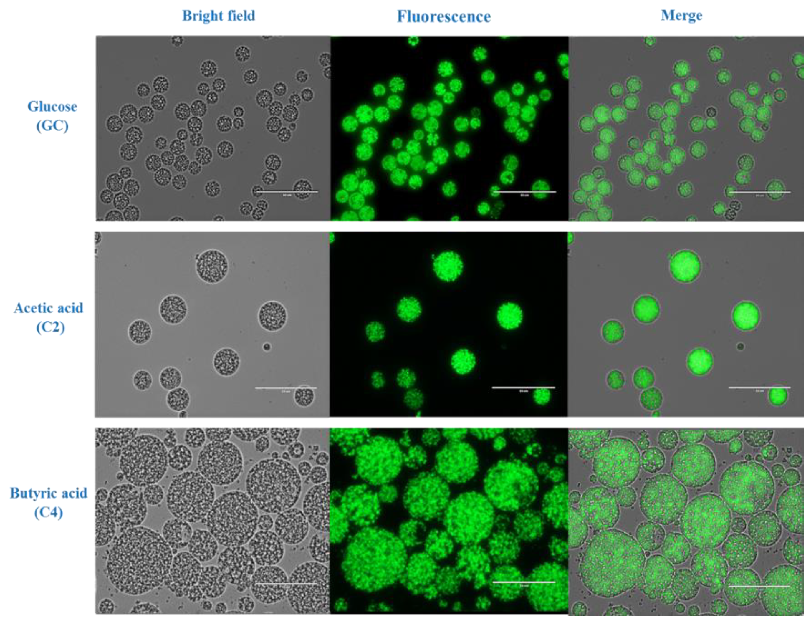
2.3. Morphologic Analysis and Estimation of Lipid Accumulation by Fluorescence Microscopy

Aliquots of 1 mL were harvested from the culture at early stationary phase, centrifuged and resuspended in 100 µL of 0.9% saline solution. 4,4-difluoro-1,3,5,7,8-pentamethyl-4-bora-3a,4a-diaza-s-indacene (BODIPY493/503) stock solution was prepared in DMSO (0.1 mg/mL) and 2 μL of it was added to the 100-µL samples. The samples were incubated for 5 min in the dark and imaging was carried out on a digital inverted fluorescence microscope equipped with a GFP light cube (EVOS-FL, (Thermo Fisher Scientific, Waltham, MA, USA). Cell size was determined by ImageJ 1.8.0 software (NIH, Bethesda, MD, USA).

2.4. Cell Dry Weight (CDW) Determination

At the end of each cultivation the cells were harvested to determine the cell dry weight. The culture was harvested into 50-mL tubes as soon as it reached stationary phase, as determined by daily OD680 measurements and centrifuged at 8000 rpm (7881× g) for 10 min in an Eppendorf 5804 R (Eppendorf Nordic A/S, Sverige Filial, Gulbackegatan, Malmö, Sweden) centrifuge with an F-34-6-38 rotor. Next, 2 mL of the supernatant was used to determine the residual carbon source (see Section 2.5). The cell pellet was washed three times with deionized (DI)-water, resuspended in DI-water, and finally pipetted into a preweighed metallic pan. The pans were dried in an oven at 50 °C until constant weight was attained. Cell dry weight was then determined gravimetrically.

2.5. Total Lipid Determination

To determine the amount of lipids in the cells, the dried biomass was milled with a mortar and pestle into a fine powder according to our previous report [37]. The powder was blended with chloroform:methanol (2:1, v/v) and incubated for 2 h under shaking. Subsequently, DI-water was added to the slurry at a volume equal to the volume of the slurry. The mixture was mixed thoroughly and afterwards centrifuged at 8000 rpm (7881× g) for 10 min. The bottom clear phase was aspirated into a preweighed glass plate and placed into a hot-air oven at 50 °C to evaporate the solvent. Lipid weight was then determined gravimetrically, and lipids were stored in a freezer at −20 °C until further analysis.

2.6. Residual Carbon Estimation by High-Performance Liquid Chromatography (HPLC)

Residual carbon source concentration was determined at the end of the cultivation by high-performance liquid chromatography (HPLC) analysis of the supernatant. Samples were prepared by filtration through a 0.2-μm syringe filter (SartoriusTM MinisartTM RC) into HPLC vials. A PerkinElmer (Waltham, Massachusetts, USA) Series 200 HPLC instrument equipped with a Bio-Rad Aminex HPX-87H Column (#1250140) was used to analyze C1, C2, C3, C4 and glucose. The analysis was programmed for 30 min and the column temperature was set to 65 °C with 5-mM H2SO4 as mobile solvent. A refractive index (RI) detector was used. Determination of residual C5 and C6 concentrations was performed as described above, with the difference that analysis time was extended to 50 min and the mobile phase was 90% v/v 5-mM H2SO4 and 10% v/v acetonitrile. Detection took place with a UV-Vis detector PerkinElmer (Waltham, Massachusetts, USA) set to 210 nm.

2.7. Analysis of Fatty Acid Profile by Gas Chromatography-Mass Spectrometry (GC-MS)

Lipids were transesterified as described by Wychen et al. (2013) [38] with some modifications. Specifically, a 50 to 100 mg lipid sample was dissolved in 2 mL of chloroform:methanol solution (2:1, v/v) in a screw-capped glass vial and 3 mL of 0.6 M HCl:methanol was added to it. The liquid was mixed and placed in a preheated water bath at 85 °C for 1 h. After that, it was cooled at room temperature to avoid extensive evaporation. The cooled liquid was transferred into 50-mL tubes and 3 mL HPLC-grade hexane was added. The tubes were mixed well and centrifuged at 8000 rpm (7881× g) for 10 min. The upper hexane layer was transferred to new glass vials. The fatty acid profile was determined by GC (Clarus 690; PerkinElmer) with MS (Clarus SQ8; PerkinElmer) using a capillary column (Elite 5MS; 30 m, 0.25 mm ID, 0.25 µm df, Cat. # N9316282; PerkinElmer). The oven was programmed to 50 °C for 0.5 min; temperature was then ramped to 194 °C at 30 °C/min for 3.5 min and then to 240 °C at 5 °C/min, where it was held for 10 min. The injection port temperature was adjusted to 250 °C and 1 µL of sample was injected with He as carrier gas in split mode (10:1). Solvent delay time was 3 min. The MS transfer line temperature was adjusted to 250 °C with 170 °C source temperature. Mass spectra (mass range 50 to 400 m/z) were recorded at 3 scans/s with electron ionization at 70 eV. Individual fatty acid methyl ester (FAME) peaks were identified by their mass spectra and further confirmed by a standard (Supelco 37 Component FAME Mix: 47885-U; Sigma-Aldrich (St. Louis, MO, USA). DHA concentration (g/L) and yield (mg/gCDW) were estimated by the following equations:
DHA   concentration   ( g L ) = T o t a l   l i p i d   c o n c e n t r a t i o n   ( g / L ) × %   o f   D H A   i n   t o t a l   l i p i d 100
DHA   yield   ( mg / gCDW ) = D H A   c o n c e n t r a t i o n   ( m g / L ) C D W   ( g / L )

2.8. Statistical Analysis

All experiments in this study were conducted in triplicate. All data are expressed as a mean ± standard deviation. The experimental data were subjected to one-way analysis of variance (ANOVA) as implemented in the Graph Pad Prism 8.4.2, 676 (GraphPad Software, San Diego, CA, USA) statistics platform. Tukey simultaneous tests were conducted to determine the statistical differences between the results obtained with various carbon sources. To ascertain that the observed variations were statistically significant, the probability (p) values were determined. A 95% confidence level (p < 0.05) was applied for all analyses.

3. Results and Discussion

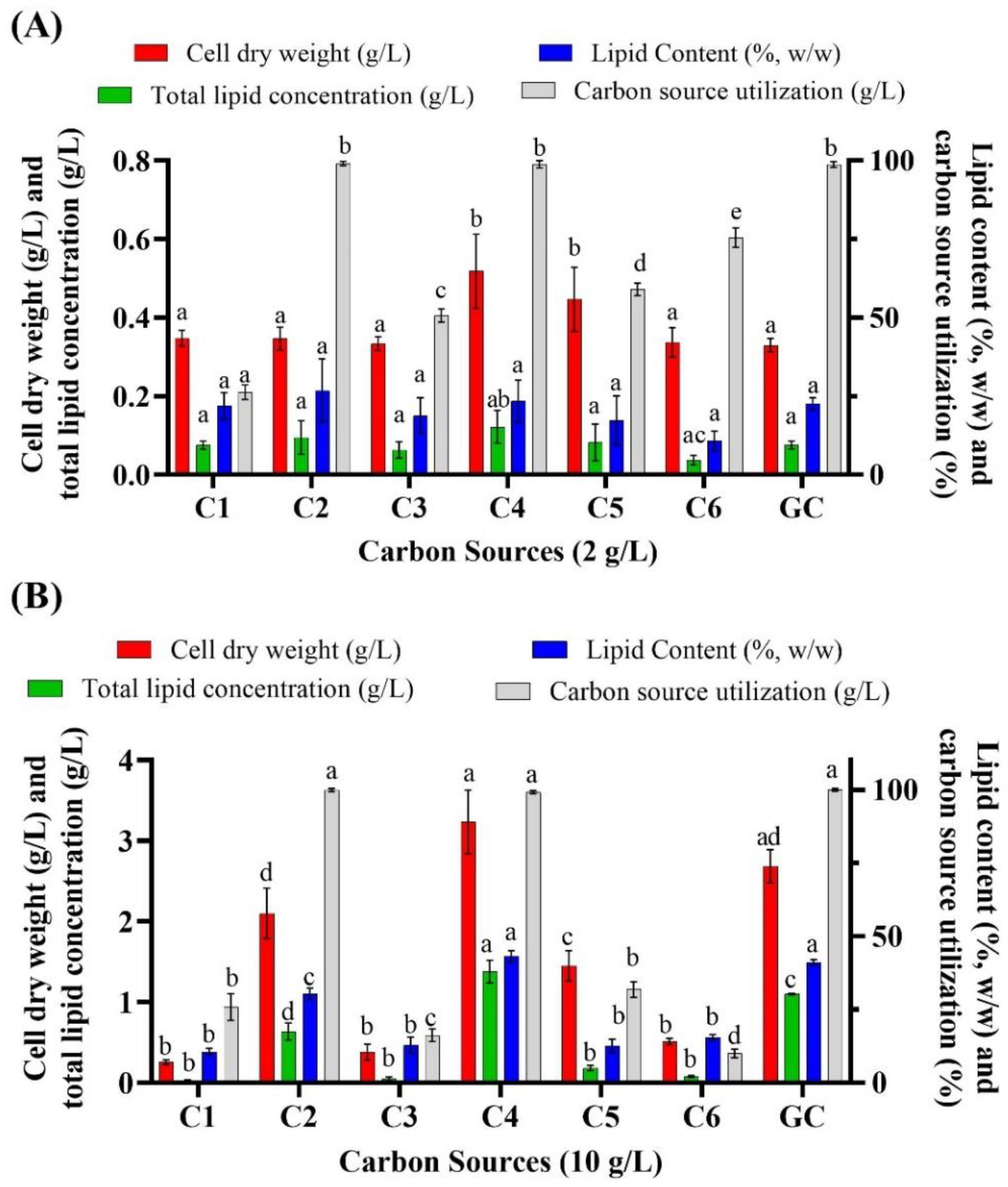
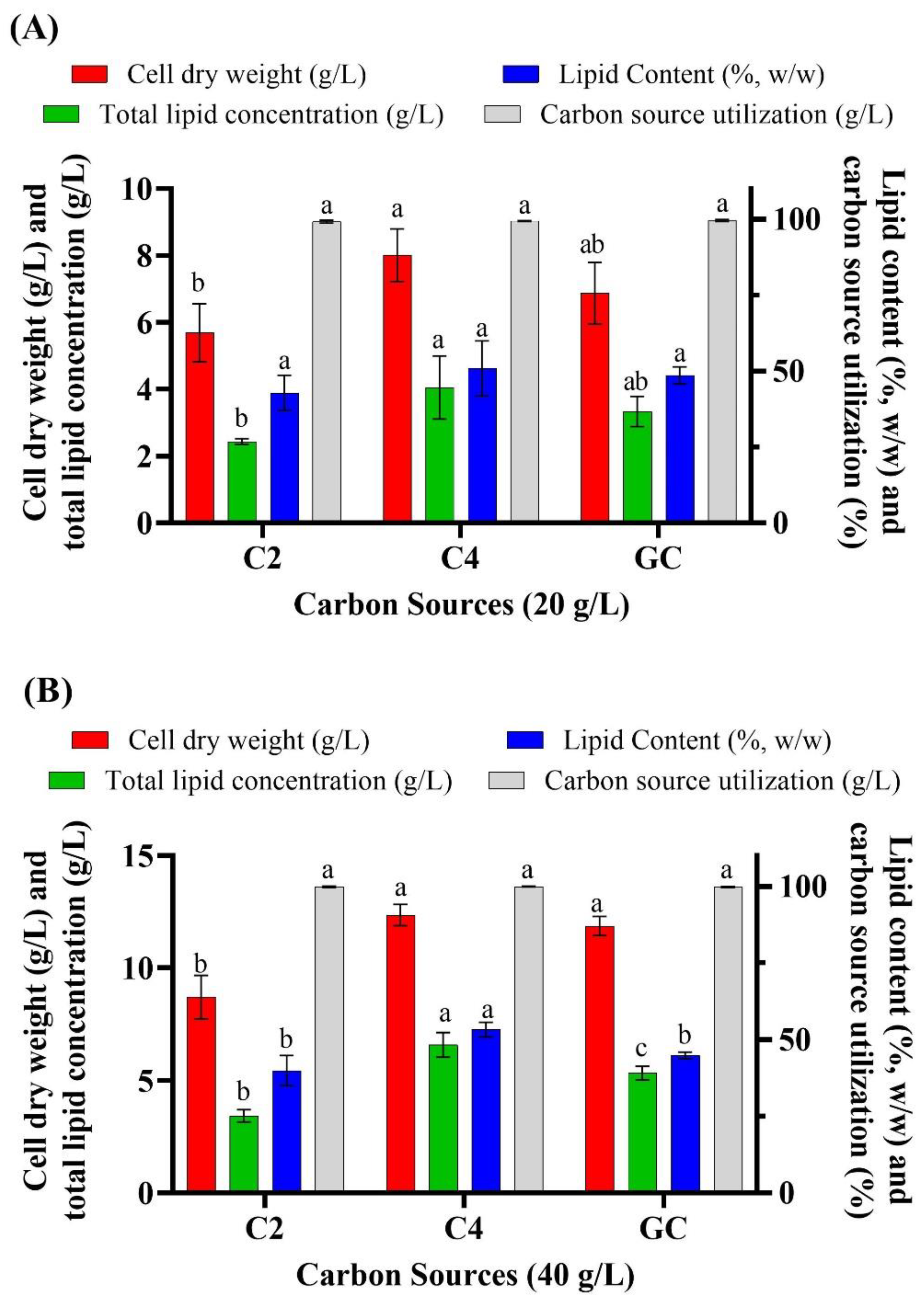
3.1. Batch Cultivation of Aurantiochytrium sp. T66 on Different Volatile Fatty Acids

In the present study, the marine oleaginous thraustochytrid Aurantiochytrium sp. T66 was grown on artificial medium containing various concentrations of VFAs. When Aurantiochytrium sp. T66 was cultivated in 2 g/L of C1, C2, C3, C4, C5 and glucose (Figure 1A), cell dry weight was 0.35 g/L, 0.35 g/L, 0.34 g/L, 0.52 g/L, 0.45 g/L, 0.34 g/L and 0.33 g/L, respectively. The corresponding total lipid concentration was 0.07 g/L, 0.10 g/L, 0.06 g/L, 0.12 g/L, 0.08 g/L, 0.04 g/L and 0.08 g/L (Figure 1). Total carbon source consumption was complete only when cultivations were carried out in C2, C4 and glucose. The value of 2 g/L was chosen because it had been reported as the upper limit to avoid inhibition in other oleaginous microorganisms. One of them, the heterotrophic microalga Chlorella protothecoides (UTEX 25), exhibited optimal growth at 2 g/L VFAs (acetic acid:propionic acid:butyric acid; 6:1:3), resulting in 0.58 g/L cell dry weight and 0.278 g/L lipids [32]. Ramping the concentration of VFAs from 2 g/L to 4 g/L, severely inhibited growth and lipid accumulation by this microalga, which dropped to 0.33 g/L and 0.136 g/L, respectively [32]. In an effort to identify whether Aurantiochytrium sp. T66 could tolerate higher VFA concentrations, 10 g/L of carbon source was used (Figure 1B). Cell dry weight was now 0.25 g/L, 2.10 g/L, 0.38 g/L, 3.23 g/L, 1.46 g/L, 0.51 g/L and 2.68 g/L in C1, C2, C3, C4, C5 and glucose, respectively. The respective lipid content was 10.57%, 30.32%, 12.90%, 43.11%, 12.60%, 15.45% and 41.13% (Figure 1B).
The above findings showed that C2 and C4 were suitable substrates, with C4 in particular yielding better results than glucose. Almost complete consumption of C2 and C4 was observed at the end of cultivation. In contrast, thraustochytrids cultivated on C1, C3 and C6 exhibited growth inhibition and final cell dry weights below 1 g/L at both 2 g/L and 10 g/L of the substrate (Figure 1). Growth on C5 was partially inhibited and C5 consumption was 31.85%.
Based on the results obtained so far, the most suitable VFAs appeared to be acetic acid (C2) and butyric acid (C4); hence, their concentration was increased to 20 g/L (Figure 2A). This resulted in a cell dry weight of 5.68 g/L, 8.00 g/L and 6.87 g/L when the microorganism was cultivated on C2, C4 and glucose, respectively, and a corresponding lipid concentration of 2.44 g/L, 4.05 g/L and 3.34 g/L. Because the cells showed no growth inhibition under these conditions, the concentration of VFAs was further increased to 40 g/L (Figure 2B). The highest cell dry weight (12.35 g/L) and total lipid concentration (6.59 g/L) was obtained with C4, followed by glucose (11.87 g/L and 5.34 g/L, respectively), whereas C2 resulted in lower biomass and lipid accumulation. Further increasing the concentration of VFAs (C2 and C4) to 60 g/L resulted in growth inhibition, which was not observed with 60 g/L of glucose (data not shown). Notably, 40 g/L of VFAs is higher than what was reported as tolerable by other oleaginous microorganisms. However, no study was reported to utilize VFAs as a sole carbon source by thraustochytrids. Among various types of oleaginous microorganisms only the oleaginous microalgae Crypthecodinium cohnii could utilize a high concentration of acetic acid and butyric acid as sole carbon sources for PUFA production. When Chalima et al. (2019) cultivated C. cohnii on various VFAs at an initial concentration of 5 to 50 g/L, the highest amount of biomass was obtained on 30 g/L of acetate (~6 g/L), 10 g/L of propionate (~3.8 g/L) and 15 g/L of butyrate (~3.9 g/L), while further increasing the concentration of substrates inhibited microbial growth [35]. Likewise, Ratledge et al. (2001) suggested that C. cohnii could grow on up to 1–16 g/L sodium acetate as a sole carbon source in a pH auxostat with acetate used to control the pH of the medium [39]. In another study, the freshwater microalga C. protothecoides was cultivated heterotrophically in waste activated sludge whose total chemical oxygen demand was 3840 mg/L, including acetic acid (1.2 g/L), propionic acid (0.45 g/L), butyric acid (0.23 g/L), isobutyric acid (0.24 g/L), valeric acid (0.36 g/L) and isovaleric acid (0.14 g/L) [40]. Waste activated sludge yielded a cell dry weight of 0.34 g/L; which was only slightly lower than that obtained on 15 g/L of pure glucose (0.63 g/L) or waste activated sludge enriched with selenite (0.50 g/L) [40]. Another freshwater microalga, Chlorella sorokiniana, was also heterotrophically cultivated on acetate, butyrate and a mixture of butyrate and acetate, but the concentration of these substrates was considerably lower (500-mM acetate and butyrate) than in the present study [41].
Cells grown on glucose, acetic acid and butyric acid were observed under a fluorescence microscope. The cells cultivated on butyric acid had a significantly larger size (41.23 ± 6.75 µm) and more lipid droplets compared to those cultivated on acetic acid, which was of intermediate size (20.09 ± 3.38 µm), and those grown on glucose, which were significantly smaller (11.11 ± 2.31 µm) (Figure 3). The cell number were high in the case of cells grown in glucose than the C2 and C4 cultivated cells while the lipid droplets per cell were high in the case of C4 grown cells. The largest cells were estimated to be almost 40 to 45 µm in diameter and, to the best of our knowledge, this is the largest oleaginous microorganism ever reported. While the exact mechanism for such a dramatic increase in size remains unknown, a possible reason may be the profound effect of butyric acid on metabolic flux and fatty acid synthesis [42].

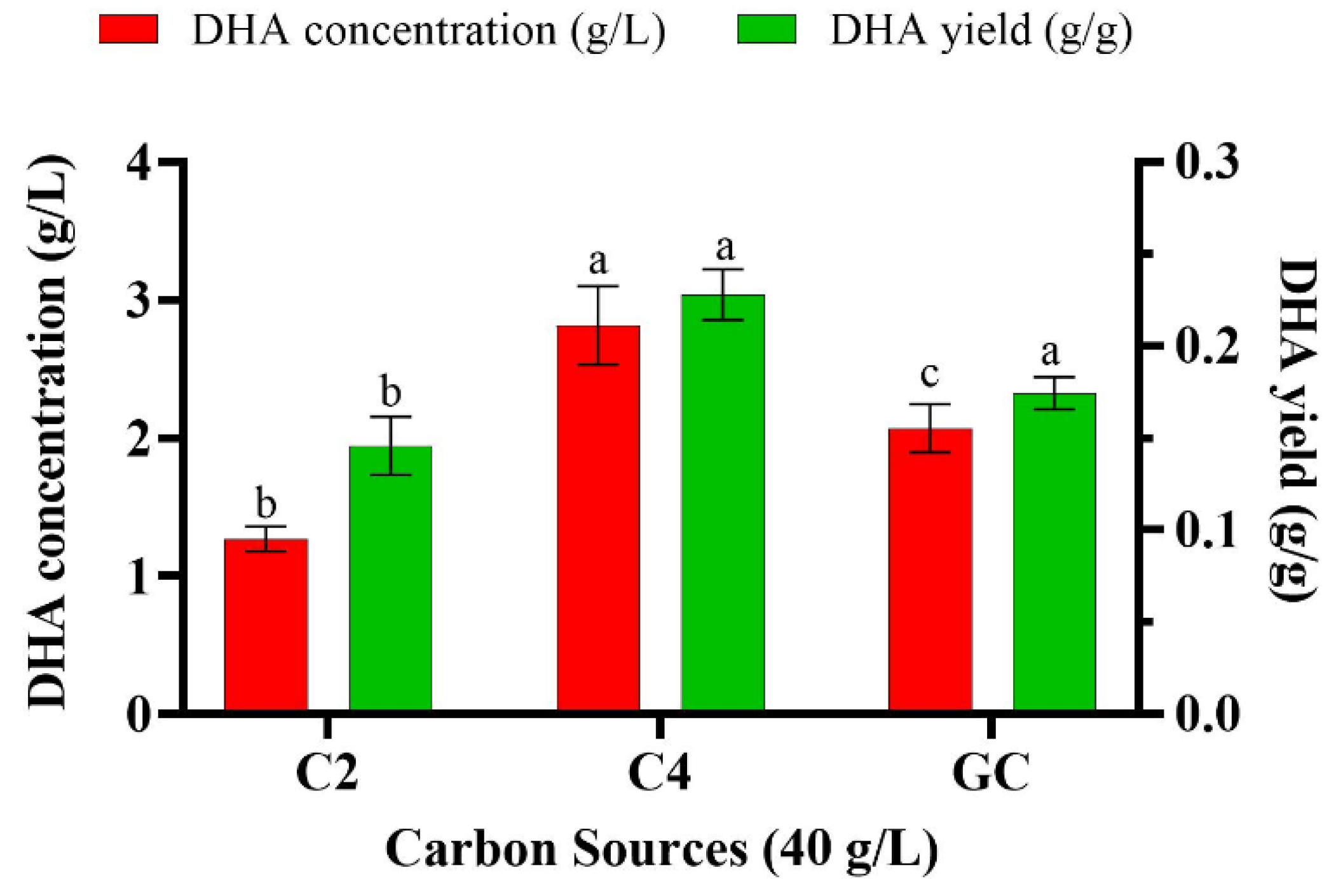
3.2. Effect of Various Volatile Fatty Acids on the Fatty Acid Profile of Aurantiochytrium sp. T66

According to a literature survey by Morabito et al. (2019), more than 65% w/w of total lipids accumulated by thraustochytrids is made of C16:0 (palmitic acid) and C22:6 (DHA), which corresponds to one of the highest DHA contents among PUFA-producing microorganisms [43]. The total amount of lipids and their profile varies among thraustochytrid species, with the genera Aurantiochytrium, Thraustochytrium and Schizochytrium considered the most productive for DHA [44].
In the present study, when Aurantiochytrium sp. T66 was cultivated on 2 g/L of VFAs, it synthesized mainly C 14:0, C14:1, C15:0, C16:0, C16:1, C17:0, C18:0 and C18:1 (Table 1). Long-chain PUFAs (>18:3), including DPA and DHA, were absent from the total lipids, which may be due to insufficient amounts of desaturases and elongases at very low substrate concentrations. Two different pathways exist for PUFA synthesis: the oxygen-dependent aerobic fatty acid synthase pathway (FAS), also known as elongase–desaturase pathway, and the oxygen-independent anerobic polyketide synthase pathway (PKS)—also designated as the PUFA synthase pathway [45]. In the FAS pathway, the starting point for PUFA synthesis is C18:3–9,12,15, which is converted by Δ6 desaturase into C18:4–6,9,12,15 and by elongase into C20:4–8,11,14,17. The latter is then converted into C20:5–5,8,11,14,17 by Δ5 desaturase, followed by elongation to 22:5–7,10,13,16,19 and, finally, conversion into DHA (22:6–4,7,10,13,16,19) by Δ4 desaturase [43,45,46,47]. Hence, PUFA synthesis through the FAS pathway can only proceed with an adequate supply of C18:3 as a substrate. Insufficient C18:3 may explain the poor production of DHA at low concentrations of VFAs. However, in the PKS pathway, acyl carrier protein (ACP), generated by CoA, is used as a covalent attachment point for chain synthesis, which proceeds with reiterative cycles. During the full fatty acid synthesis process, a series of enzymes including 3-ketoacyl synthase (KS), 3-ketoacyl-ACP reductase (KR), enoyl reductase (ER), and dehydrase/isomerase (DH) are necessary [48]. However, the whole genome annotation results showed that S. limacinum does not contain Δ4 desaturase, in agreement with literature reports. On the other hand, some ORFs in S. limacinum were predicted to be potential PKS proteins [48]. This means S. limacinum could not synthesize DHA by the FAS route, but rather employs the PKS system for PUFA biosynthesis [45,49,50]. Contrasting to this, some thraustochytrids such as Thraustochytrium sp., synthesize DHA via Δ4 desaturation where desaturase introduces Δ4 bond into 22:5(n−3) that resulted into DHA [51,52,53].
When cultivation was shifted from 2 to 10 g/L of VFAs, the main fatty acids were C14:0, C15:0, C16:0, C17:0, C18:0, C18:1, as well as long-chain fatty acids such as C20:4n6, DPA and DHA (Table 2). The DHA content (w/wtotal lipids) was 9.57%, 28.75%, 7.28%, 18.64%, 7.88%, 6.01% and 4.14% when cultivation was carried out in C1, C2, C3, C4, C5, C6 and glucose, respectively (Table 2). The highest DHA content was observed with C2 as substrate followed by C4. A possible reason is the direct production of acetyl-CoA, a key intermediate in fatty acid synthesis, from VFA via a non-conventional route [54]. When oleaginous microorganisms are grown on glucose as sole carbon source, the cells start assimilating glucose for glycolysis and form pyruvate, which is transported to mitochondria and fed into the tricarboxylic acid cycle. This yields citrate, which is transported back to the cytosol, where it is converted into acetyl-CoA by ATP:citrate lyase [55]. In contrast, when oleaginous cells assimilate acetic acid, this is converted directly into acetyl-CoA in the cytosol by acetyl-CoA synthetase in a single step [47,54].
Finally, Aurantiochytrium sp. T66 was shifted to an even higher concentration of the substrate to study the effect on DHA production of 20 g/L and 40 g/L of C2 or C4. At 20 g/L of C2, C4 or glucose, DHA content increased to 30.40%, 36.25% and 29.58%, respectively (Table 3); the other major fatty acids being produced were C15:0, C16:0, C17:0, C18:0, C18:3n3 and DPA. When cells are cultivated on C2 compounds such as acetic acid, the tricarboxylic acid cycle cannot provide sufficient carbon skeleton building blocks, ATP and reducing power (NADH). This shortage prompts the induction of another metabolic pathway, the glyoxylate cycle, and the activation of enzymes, such as isocitrate lyase and malate synthase [56,57]. It has been suggested that the oleaginous microalgae Chlorella vulgaris can express the glyoxylate cycle enzymes even when cultivated on glucose, however they are not active unless the microorganism is grown on acetate [58]. The activity and concentration of isocitrate lyase was found to be increased in another microalga, Scenedesmus obliquus, after being cultivated on acetate in the dark; moreover, the concentration of this enzyme increased with increasing concentration of acetate [59]. In the present study, when cells were grown in 40 g/L C2 and C4, DHA content increased to 36.93% and 42.63%, respectively, while on glucose the DHA concentration was 38.72% (Table 4). The highest DHA concentration (2.81 g/L) and DHA yield (0.23 g/gCDW) were observed when cells were cultivated in 40 g/L of C4, followed by 40 g/L of glucose (2.07 g/L and 0.17 g/gCDW, respectively) (Figure 4). Addition of 1% v/v n-dodecane (C12) to the medium of the marine microalga C. cohnii was shown to enhance cell dry weight and lipid accumulation, including DHA content (51% w/w of total lipids) [60]. The authors suggested that the hydrocarbon in the medium acted as an oxygen vector and could improve oxygen availability to the microorganisms, thus enhancing DHA productivity. At the same time, the total content of unsaturated fatty acids (16:1n7, 18:1n9 and 22:5n3) remained unaffected, suggesting that the increase in DHA was independent of the oxygen-dependent desaturase [60].

4. Conclusions

The present study indicates that VFAs can be utilized efficiently as a sole carbon source for the cultivation of oleaginous marine thraustochytrids. Cell dry weight and lipid production were particularly high when Aurantiochytrium sp. T66 was cultivated on butyric acid as sole carbon source. The fatty acid profile of this strain revealed up to 42.63% of DHA in total lipids after cultivation on VFAs. The maximum DHA concentration (2.81 g/L) and DHA yield (0.23 g/gCDW) were attained when cells were cultivated in 40 g/L butyric acid, and were even higher than those observed in 40 g/L glucose (2.07 g/L and 0.17 g/gCDW, respectively). The important finding of this study was the dramatic increment in cell size after cultivation on 40 g/L of butyric acid that provides a foundation for the further exploration of the assimilation pathway and metabolic flux in thraustochytrids. Moreover, replacing costly refined sugars as carbon source with VFAs derived from abundant waste streams will make microbial PUFA synthesis more economical at an industrial scale.

Author Contributions

Conceptualization, A.P., U.R., P.C. and L.M.; methodology, A.P. and L.M.; investigation, A.P.; data analysis, A.P. and L.M.; writing—original draft preparation, A.P.; writing—review and editing, U.R., P.C. and L.M.; funding acquisition, L.M. All authors have read and agreed to the published version of the manuscript.

Funding

This work was part of the project ‘Tuned volatile fatty acids production from organic waste for biorefinery platforms (VFA biorefinery)’ funded by the Swedish Research Council (FORMAS) with reference number 2018–00818.

Conflicts of Interest

The authors declare no conflict of interest.

References

  1. Kaur, G.; Cameron-Smith, D.; Garg, M.; Sinclair, A.J. Docosapentaenoic acid (22:5n-3): A review of its biological effects. Prog. Lipid Res. 2011, 50, 28–34. [Google Scholar] [CrossRef] [PubMed] [Green Version]
  2. Chiu, C.C.; Su, K.P.; Cheng, T.C.; Liu, H.C.; Chang, C.J.; Dewey, M.E.; Stewart, R.; Huang, S.Y. The effects of omega-3 fatty acids monotherapy in Alzheimer’s disease and mild cognitive impairment: A preliminary randomized double-blind placebo-controlled study. Prog. Neuro Psychopharmacol. Biol. Psychiatry 2008, 32, 1538–1544. [Google Scholar] [CrossRef] [PubMed]
  3. Saini, R.K.; Keum, Y.S. Omega-3 and omega-6 polyunsaturated fatty acids: Dietary sources, metabolism, and significance—A review. Life Sci. 2018, 203, 255–267. [Google Scholar] [CrossRef] [PubMed]
  4. Lazzarin, N.; Vaquero, E.; Exacoustos, C.; Bertonotti, E.; Romanini, M.E.; Arduini, D. Low-dose aspirin and omega-3 fatty acids improve uterine artery blood flow velocity in women with recurrent miscarriage due to impaired uterine perfusion. Fertil. Steril. 2009, 92, 296–300. [Google Scholar] [CrossRef]
  5. Kiecolt-Glaser, J.K.; Belury, M.A.; Andridge, R.; Malarkey, W.B.; Hwang, B.S.; Glaser, R. Omega-3 supplementation lowers inflammation in healthy middle-aged and older adults: A randomized controlled trial. Brain. Behav. Immun. 2012, 26, 988–995. [Google Scholar] [CrossRef] [Green Version]
  6. Lauritzen, L.; Brambilla, P.; Mazzocchi, A.; Harsløf, L.B.S.; Ciappolino, V.; Agostoni, C. DHA effects in brain development and function. Nutrients 2016, 8, 6. [Google Scholar] [CrossRef] [Green Version]
  7. Innis, S.M. Dietary omega 3 fatty acids and the developing brain. Brain Res. 2008, 1237, 35–43. [Google Scholar] [CrossRef]
  8. Guesnet, P.; Alessandri, J. Biochimie Docosahexaenoic acid (DHA) and the developing central nervous system (CNS) e implications for dietary recommendations. Biochimie 2011, 93, 7–12. [Google Scholar] [CrossRef]
  9. Elagizi, A.; Lavie, C.J.; Marshall, K.; DiNicolantonio, J.J.; O’Keefe, J.H.; Milani, R.V. Omega-3 polyunsaturated fatty acids and cardiovascular health: A comprehensive review. Prog. Cardiovasc. Dis. 2018, 61, 76–85. [Google Scholar] [CrossRef] [Green Version]
  10. Harris, W.S.; Mozaffarian, D.; Rimm, E.; Kris-Etherton, P.; Rudel, L.L.; Appel, L.J.; Engler, M.M.; Engler, M.B.; Sacks, F. Omega-6 fatty acids and risk for cardiovascular disease: A science advisory from the American heart association nutrition subcommittee of the council on nutrition, physical activity, and metabolism; council on cardiovascular nursing; and council on epidem. Circulation 2009, 119, 902–907. [Google Scholar] [CrossRef] [Green Version]
  11. Kris-Etherton, P.; Fleming, J.; Harris, W.S. The debate about n-6 polyunsaturated fatty acid recommendations for cardiovascular health. J. Am. Diet. Assoc. 2010, 110, 201–204. [Google Scholar] [CrossRef] [PubMed]
  12. Shahidi, F.; Wanasundara, U.N. Omega-3 fatty acid concentrates: Nutritional aspects and production technologies. Trends Food Sci. Technol. 1998, 9, 230–240. [Google Scholar] [CrossRef]
  13. Pejin, B.; Bianco, A.; Newmaster, S.; Sabovljevic, M.; Vujisic, L.; Tesevic, V.; Vajs, V.; De Rosa, S. Fatty acids of Rhodobryum ontariense (Bryaceae). Nat. Prod. Res. 2012, 26, 696–702. [Google Scholar] [CrossRef] [PubMed]
  14. Pejin, B.; Vujisic, L.; Sabovljevic, M.; Tesevic, V.; Vajs, V. The moss mnium hornum, a promising source of arachidonic acid. Chem. Nat. Compd. 2012, 48, 120–121. [Google Scholar] [CrossRef]
  15. Ward, O.P.; Singh, A. Omega-3/6 fatty acids: Alternative sources of production. Process Biochem. 2005, 40, 3627–3652. [Google Scholar] [CrossRef]
  16. Alonso, D.L.; Maroto, F.G. Plants as “chemical factories” for the production of polyunsaturated fatty acids. Biotechnol. Adv. 2000, 18, 481–497. [Google Scholar] [CrossRef]
  17. Domingo, J.L.; Bocio, A.; Falcó, G.; Llobet, J.M. Benefits and risks of fish consumption. Part I. A quantitative analysis of the intake of omega-3 fatty acids and chemical contaminants. Toxicology 2007, 30, 219–226. [Google Scholar] [CrossRef]
  18. Patel, A.; Liefeldt, S.; Rova, U.; Christakopoulos, P.; Matsakas, L. Co-production of DHA and squalene by thraustochytrid from forest biomass. Sci. Rep. 2020, 10, 1–12. [Google Scholar] [CrossRef]
  19. Raghukumar, S. Thraustochytrid marine protists: Production of PUFAs and other emerging technologies. Mar. Biotechnol. 2008, 631–640. [Google Scholar] [CrossRef]
  20. Vigani, M.; Parisi, C.; Rodríguez-Cerezo, E.; Barbosa, M.J.; Sijtsma, L.; Ploeg, M.; Enzing, C. Food and feed products from micro-algae: Market opportunities and challenges for the EU. Trends Food Sci. Technol. 2015, 42, 81–92. [Google Scholar] [CrossRef]
  21. Bannenberg, G.; Mallon, C.; Edwards, H.; Yeadon, D.; Yan, K.; Johnson, H.; Ismail, A. Omega-3 long-chain polyunsaturated fatty acid content and oxidation state of fish oil supplements in New Zealand. Sci. Rep. 2017, 7, 1–13. [Google Scholar] [CrossRef] [PubMed] [Green Version]
  22. Subhadra, B.G.; Edwards, M. Coproduct market analysis and water footprint of simulated commercial algal biorefineries. Appl. Energy 2011, 88, 3515–3523. [Google Scholar] [CrossRef]
  23. Li, X.; Xu, H.; Wu, Q. Large-scale biodiesel production from microalga Chlorella protothecoides through heterotrophic cultivation in bioreactors. Biotechnol. Bioeng. 2007, 98, 764–771. [Google Scholar] [CrossRef] [PubMed]
  24. Park, G.W.; Fei, Q.; Jung, K.; Chang, H.N.; Kim, Y.C.; Kim, N.J.; Choi, J.d.R.; Kim, S.; Cho, J. Volatile fatty acids derived from waste organics provide an economical carbon source for microbial lipids/biodiesel production. Biotechnol. J. 2014, 9, 1536–1546. [Google Scholar] [CrossRef] [PubMed]
  25. Leong, H.Y.; Su, C.A.; Lee, B.S.; Lan, J.C.W.; Law, C.L.; Chang, J.S.; Show, P.L. Development of Aurantiochytrium limacinum SR21 cultivation using salt-rich waste feedstock for docosahexaenoic acid production and application of natural colourant in food product. Bioresour. Technol. 2019, 271, 30–36. [Google Scholar] [CrossRef]
  26. Ferreira, G.F.; Ríos Pinto, L.F.; Maciel Filho, R.; Fregolente, L.V. A review on lipid production from microalgae: Association between cultivation using waste streams and fatty acid profiles. Renew. Sustain. Energy Rev. 2019, 109, 448–466. [Google Scholar] [CrossRef]
  27. Choi, J.D.R.; Chang, H.N.; Han, J.I. Performance of microbial fuel cell with volatile fatty acids from food wastes. Biotechnol. Lett. 2011, 33, 705–714. [Google Scholar] [CrossRef]
  28. Mengmeng, C.; Hong, C.; Qingliang, Z.; Shirley, S.N.; Jie, R. Optimal production of polyhydroxyalkanoates (PHA) in activated sludge fed by volatile fatty acids (VFAs) generated from alkaline excess sludge fermentation. Bioresour. Technol. 2009, 100, 1399–1405. [Google Scholar] [CrossRef]
  29. Huang, Y.L.; Wu, Z.; Zhang, L.; Ming Cheung, C.; Yang, S.T. Production of carboxylic acids from hydrolyzed corn meal by immobilized cell fermentation in a fibrous-bed bioreactor. Bioresour. Technol. 2002, 82, 51–59. [Google Scholar] [CrossRef]
  30. Akaraonye, E.; Keshavarz, T.; Roy, I. Production of polyhydroxyalkanoates: The future green materials of choice. J. Chem. Technol. Biotechnol. 2010, 85, 732–743. [Google Scholar] [CrossRef]
  31. Kondo, T.; Kondo, M. Efficient production of acetic acid from glucose in a mixed culture of Zymomonas mobilis and Acetobacter sp. J. Ferment. Bioeng. 1996, 81, 42–46. [Google Scholar] [CrossRef]
  32. Fei, Q.; Fu, R.; Shang, L.; Brigham, C.J.; Chang, H.N. Lipid production by microalgae Chlorella protothecoides with volatile fatty acids (VFAs) as carbon sources in heterotrophic cultivation and its economic assessment. Bioprocess Biosyst. Eng. 2015, 38, 691–700. [Google Scholar] [CrossRef] [PubMed] [Green Version]
  33. Fei, Q.; Chang, H.N.; Shang, L.; Choi, J.d.R.; Kim, N.J.; Kang, J.W. The effect of volatile fatty acids as a sole carbon source on lipid accumulation by Cryptococcus albidus for biodiesel production. Bioresour. Technol. 2011, 102, 2695–2701. [Google Scholar] [CrossRef] [PubMed]
  34. Fontanille, P.; Kumar, V.; Christophe, G.; Nouaille, R.; Larroche, C. Bioconversion of volatile fatty acids into lipids by the oleaginous yeast Yarrowia lipolytica. Bioresour. Technol. 2012, 114, 443–449. [Google Scholar] [CrossRef] [PubMed]
  35. Chalima, A.; Hatzidaki, A.; Karnaouri, A.; Topakas, E. Integration of a dark fermentation effluent in a microalgal-based biorefinery for the production of high-added value omega-3 fatty acids. Appl. Energy 2019, 241, 130–138. [Google Scholar] [CrossRef]
  36. Patel, A.; Rova, U.; Christakopoulos, P.; Matsakas, L. Simultaneous production of DHA and squalene from Aurantiochytrium sp. grown on forest biomass hydrolysates. Biotechnol. Biofuels 2019, 12, 1–12. [Google Scholar] [CrossRef]
  37. Patel, A.; Matsakas, L.; Rova, U.; Christakopoulos, P. Heterotrophic cultivation of Auxenochlorella protothecoides using forest biomass as a feedstock for sustainable biodiesel production. Biotechnol. Biofuels 2018, 11, 169. [Google Scholar] [CrossRef]
  38. Van Wychen, S.; Ramirez, K.; Laurens, L.M. Determination of total lipids as fatty acid methyl esters (FAME) by in situ transesterification. Contract 2013, 303, 275–3000. [Google Scholar]
  39. Ratledge, C.; Kanagachandran, K.; Anderson, A.J.; Grantham, D.J.; Stephenson, J.C. Production of docosahexaenoic acid by Crypthecodinium cohnii grown in a pH-auxostat culture with acetic acid as principal carbon source. Lipids 2001, 36, 1241–1246. [Google Scholar] [CrossRef]
  40. Wen, Q.; Chen, Z.; Li, P.; Duan, R.; Ren, N. Lipid production for biofuels from hydrolyzate of waste activated sludge by heterotrophic Chlorella protothecoides. Bioresour. Technol. 2013, 143, 695–698. [Google Scholar] [CrossRef]
  41. Turon, V.; Trably, E.; Fouilland, E.; Steyer, J.P. Growth of Chlorella sorokiniana on a mixture of volatile fatty acids: The effects of light and temperature. Bioresour. Technol. 2015, 198, 852–860. [Google Scholar] [CrossRef] [PubMed]
  42. Marshall, W.F.; Young, K.D.; Swaffer, M.; Wood, E.; Nurse, P.; Kimura, A.; Frankel, J.; Wallingford, J.; Walbot, V.; Qu, X.; et al. What determines cell size? BMC Biol. 2012, 10, 101. [Google Scholar] [CrossRef] [PubMed] [Green Version]
  43. Morabito, C.; Bournaud, C.; Maës, C.; Schuler, M.; Aiese Cigliano, R.; Dellero, Y.; Maréchal, E.; Amato, A.; Rébeillé, F. The lipid metabolism in thraustochytrids. Prog. Lipid Res. 2019, 76, 101007. [Google Scholar] [CrossRef] [PubMed]
  44. Fossier, M.L.; Lee, C.K.J.; Nichols, P.D.; Mitchell, W.J.; Polglase, J.L.; Gutierrez, T. Taxonomy, ecology and biotechnological applications of thraustochytrids: A review. Biotechnol. Adv. 2018, 36, 26–46. [Google Scholar] [CrossRef]
  45. Qiu, X. Biosynthesis of docosahexaenoic acid (DHA, 22:6-4, 7,10,13,16,19): Two distinct pathways. Prostaglandins Leukot. Essent. Fat. Acids 2003, 68, 181–186. [Google Scholar] [CrossRef]
  46. Monroig, Ó.; Wang, S.; Zhang, L.; You, C.; Tocher, D.R.; Li, Y. Elongation of long-chain fatty acids in rabbitfish Siganus canaliculatus: Cloning, functional characterisation and tissue distribution of Elovl5- and Elovl4-like elongases. Aquaculture 2012, 350–353, 63–70. [Google Scholar] [CrossRef] [Green Version]
  47. Sijtsma, L.; De Swaaf, M.E. Biotechnological production and applications of the -3 polyunsaturated fatty acid docosahexaenoic acid. Appl. Microbiol. Biotechnol. 2004, 64, 146–153. [Google Scholar] [CrossRef]
  48. Ye, C.; Qiao, W.; Yu, X.; Ji, X.; Huang, H.; Collier, J.L.; Liu, L. Reconstruction and analysis of the genome-scale metabolic model of Schizochytrium limacinum SR21 for docosahexaenoic acid production. BMC Genom. 2015, 16, 1–11. [Google Scholar] [CrossRef] [Green Version]
  49. Wallis, J.G.; Watts, J.L.; Browse, J. Polyunsaturated fatty acid synthesis: What will they think of next? Trends Biochem. Sci. 2002, 27, 467–473. [Google Scholar] [CrossRef]
  50. Metz, J.G.; Roessler, P.; Facciotti, D.; Levering, C.; Dittrich, F.; Lassner, M.; Valentine, R.; Lardizabal, K.; Domergue, F.; Yamada, A.; et al. Production of polyunsaturated fatty acids by potyketide synthases in both prokaryotes and eukaryotes. Science 2001, 293, 290–293. [Google Scholar] [CrossRef]
  51. Qiu, X.; Hong, H.; Mackenzie, S.L. Identification of a Δ4 Fatty Acid Desaturase from Thraustochytrium sp. Involved in the Biosynthesis of Docosahexanoic Acid by Heterologous Expression in Saccharomyces cerevisiae and Brassica juncea *. J. Biol. Chem. 2001, 276, 31561–31566. [Google Scholar] [CrossRef] [PubMed] [Green Version]
  52. Weete, J.D.; Kim, H.; Gandhi, S.R.; Wang, Y.; Dute, R. Lipids and Ultrastructure of Thraustochytrium sp. ATCC 26185. Lipids 1997, 839–845. [Google Scholar] [CrossRef] [PubMed]
  53. Lewis, T.E.; Nichols, P.D.; Mcmeekin, T.A. The biotechnological potential of Thraustochytrids. Mar. Biotechnol. 1999, 580–587. [Google Scholar] [CrossRef] [PubMed]
  54. De Swaaf, M.E.; Sijtsma, L.; Pronk, J.T. High-cell-density fed-batch cultivation of the docosahexaenoic acid producing marine alga Crypthecodinium cohnii. Biotechnol. Bioeng. 2003, 81, 666–672. [Google Scholar] [CrossRef]
  55. Patel, A.; Arora, N.; Sartaj, K.; Pruthi, V.; Pruthi, P.A. Sustainable biodiesel production from oleaginous yeasts utilizing hydrolysates of various non-edible lignocellulosic biomasses. Renew. Sustain. Energy Rev. 2016, 62, 836–855. [Google Scholar] [CrossRef]
  56. Perez-Garcia, O.; Escalante, F.M.E.; de-Bashan, L.E.; Bashan, Y. Heterotrophic cultures of microalgae: Metabolism and potential products. Water Res. 2011, 45, 11–36. [Google Scholar] [CrossRef]
  57. Boyle, N.R.; Morgan, J.A. Flux balance analysis of primary metabolism in Chlamydomonas reinhardtii. BMC Syst. Biol. 2009, 3, 1–14. [Google Scholar] [CrossRef] [Green Version]
  58. Harrop, L.C.; Kornberg, H.L. The role of isocitrate lyase in the metabolism of algae. Proc. R. Soc. Lond. Ser. B Biol. Sci. 1966, 166, 11–29. [Google Scholar]
  59. Combres, C.; Laliberté, G.; Reyssac, J.S.; de la Noüe, J. Effect of acetate on growth and ammonium uptake in the microalga Scenedesmus obliquus. Physiol. Plant. 1994, 91, 729–734. [Google Scholar] [CrossRef]
  60. Lopes Da Silva, T.; Reis, A. The use of multi-parameter flow cytometry to study the impact of n-dodecane additions to marine dinoflagellate microalga Crypthecodinium cohnii batch fermentations and DHA production. J. Ind. Microbiol. Biotechnol. 2008, 35, 875–887. [Google Scholar] [CrossRef]
Figure 1. Batch cultivation of Aurantiochytrium sp. T66 on various volatile fatty acids (VFAs) at 2 g/L (A) and 10 g/L (B). Glucose (GC) was used as control. Error bars express the standard deviation of the mean (n = 3). Different letters (a,b,c,d,e) above the bars in each group of results indicate significant difference (p < 0.05), based on one-way ANOVA with Tukey’s test.
Biomolecules 10 00694 g001
Figure 2. Batch cultivation of Aurantiochytrium sp. T66 on acetic acid (C2) and butyric acid (C4) at 20 g/L (A) and 40 g/L (B). Glucose (GC) was used as control. Error bars express the standard deviation of the mean Error bars express the standard deviation of the mean (n = 3). Different letters (a,b,c) above the bars in each group of results indicate significant difference (p < 0.05), based on one-way ANOVA with Tukey’s test.
Biomolecules 10 00694 g002
Figure 3. Morphologic analysis of cells and lipid droplets of Aurantiochytrium sp. T66 grown on 40 g/L of acetic acid, butyric acid and glucose. The cells were stained with 4,4-difluoro-1,3,5,7,8-pentamethyl-4-bora-3a,4a-diaza-s-indacene (BODIPY493/503) and observed by live fluorescence microscopy. Scale bars corresponds to 50 µm.
Biomolecules 10 00694 g003
Figure 4. DHA concentration and yield of Aurantiochytrium sp. T66 cultivated in 40 g/L of C2, C4 or glucose. Error bars express the standard deviation of the mean (n = 3). Different letters (a,b,c) above the bars in each group of results indicate significant difference (p < 0.05), based on one-way ANOVA with Tukey’s test.
Biomolecules 10 00694 g004
Table 1. Fatty acid composition (wt% of total fatty acids) of Aurantiochytrium sp. T66 cultivated on 2 g/L of various VFAs (C1:0, formic acid; C2:0, acetic acid; C3:0, propionic acid; C4:0, butyric acid; C5:0, valeric acid and C6:0, caproic acid). Glucose (GC) was used as control.
Fatty Acids (%) in Total LipidC1C2C3C4C5C6GC
C14:06.04 ± 0.32 b7.50 ± 0.28 c7.26 ± 0.12 c,d6.46 ± 0.57 b,c5.44 ± 0.16 b6.90 ± 0.52 b,c21.59 ± 0.30 a
C14:112.12 ± 0.58 b14.89 ± 0.63 c17.60 ± 0.81 d17.30 ± 0.41 d12.64 ± 0.42 b25.93 ± 1.14 aND
C15:0NDNDNDNDNDND18.60 ± 0.73
C15:13.67 ± 0.61 b3.42 ± 0.42 b4.14 ± 0.13 b4.44 ± 0.65 b3.09 ± 0.23 b8.38 ± 0.34 aND
C 16:035.44 ± 0.82 b37.72 ± 0.94 c32.90 ± 1.23 f38.46 ± 0.60 c,d36.44 ± 0.73 b,c32.63 ± 0.42 e,f44.31 ± 0.63 a
C16:18.54 ± 1.17 a8.17 ± 0.64 a8.18 ± 0.82 a7.97± 0.35 a8.68 ± 1.00 a7.73 ± 0.92 aND
C 18:018.66 ± 0.92 b22.45 ± 0.58 c25.58 ± 1.08 d23.23 ±1.19 c,e27.79 ± 0.24 a,d17.736 ± 0.89 b9.64 ± 0.35 f
C18:114.88 ± 1.40 d5.75 ± 0.48 a,b4.37 ± 0.73 a,b2.16 ± 0.41 b,c5.81 ± 0.89 aNDND
DPANDNDNDNDNDND2.65 ± 0.37
DHANDNDNDNDNDND2.48 ± 0.52
ND = not detected; Results are presented as mean ± SD, n = 3. Different superscript small letters (a,b,c,d,e,f) of each fatty acid obtained with various carbon sources indicate significant difference (Tukey’s test, p < 0.05).
Table 2. Fatty acid composition (wt% of total fatty acids) of Aurantiochytrium sp. T66 cultivated on 10 g/L of various VFAs (C1:0, formic acid; C2:0, acetic acid; C3:0, propionic acid; C4:0, butyric acid; C5:0, valeric acid and C6:0, caproic acid). Glucose (GC) was used as control.
Fatty Acids (%) in Total LipidC1C2C3C4C5C6GC
C14:06.92 ± 0.42 b4.79 ± 0.32 c9.00 ± 0.40 d15.08 ± 0.37 a7.74 ± 0.35 b,d5.09 ± 0.30 c14.99 ± 1.13 a
C14:1NDNDNDNDND17.37 ± 0.72ND
C15:018.54 ± 0.49 d3.49 ± 0.03 c19.59 ± 0.90 a10.25 ± 0.28 b22.56 ± 1.28 a8.39 ± 0.82 b21.37 ± 0.85 a
C 16:028.43 ± 0.85 b23.22 ± 0.32 c29.58 ± 0.82 b34.19 ± 1.25 d30.19 ± 1.05 b31.29 ± 1.01 b,e42.79 ± 0.47 a
C16:1NDNDNDNDND5.10 ± 0.49ND
C17:09.22 ± 0.69 aND9.27 ± 0.45 a5.77 ± 0.55 b8.92 ± 0.38 aND8.56 ± 0.93 a
C 18:014.44 ± 0.75 a8.90 ± 0.52 b14.89 ± 1.37 a6.74 ± 0.49 c14.32 ± 0.55 a8.63 ± 0.78 b5.79 ± 0.33 c
C18:1ND1.84 ± 0.49NDNDND9.46 ± 0.89ND
C20:0NDNDNDNDND5.58 ± 0.81ND
C20:42.31 ± 0.27 b5.13 ± 0.57 a1.03 ± 0.14 cND0.96 ± 0.05 c,dNDND
EPA2.32 ± 0.16 b5.24 ± 0.05 a1.60 ± 0.27 cND1.94 ± 0.14 b,cNDND
DPA7.52 ± 0.60 b18.29 ± 0.78 a7.27 ± 0.78 b8.88 ± 0.41 b4.87 ± 0.41 d2.41± 0.12 c2.20 ± 0.13 c
DHA9.57± 0.68 b28.73 ± 1.28 a7.28 ± 0.21 c18.64 ± 0.59 d7.88 ± 0.54 b,c6.01 ± 0.53 c4.14 ± 0.28 c
ND = not detected; Results are presented as mean ± SD, n = 3. Different superscript small letters (a,b,c,d,e) of each fatty acid obtained with various carbon sources indicate significant difference (Tukey’s test, p < 0.05).
Table 3. Fatty acid composition (wt% of total fatty acids) of Aurantiochytrium sp. T66 cultivated on 20 g/L of acetic acid (C2) or butyric acid (C4). Glucose (GC) was used as control.
Fatty Acids (%) in Total LipidC2C4GC
C14:02.85 ± 0.35 b6.67 ± 0.31 a8.33 ± 0.58 a
C15:010.32 ± 0.40 a2.60 ± 0.18 b8.03 ± 0.10 c
C 16:025.59 ± 0.43 a19.79 ± 0.77 b18.54 ± 1.04 b
C16:15.48 ± 0.682.37 ± 0.35ND
C 18:03.67 ± 0.17 b3.00 ± 0.53 b7.99 ± 0.40 a
C18:1ND1.33 ± 0.16ND
C20:41.74 ± 0.175.23 ± 0.53ND
EPA0.72 ± 0.13 b1.89 ± 0.48 a1.59 ± 0.43 a
DPA12.16 ± 0.14 c13.93 ± 0.48 b17.00 ± 0.62 a
DHA36.93 ± 0.69 a42.63 ± 1.08 b38.72 ± 1.04 a
ND = not detected; Results are presented as mean ± SD, n = 3. Different superscript small letters (a,b,c) of each fatty acid obtained with various carbon sources indicate significant difference (Tukey’s test, p < 0.05).
Table 4. Fatty acid composition (wt% of total fatty acids) of Aurantiochytrium sp. T66 cultivated on 40 g/L of acetic acid (C2) or butyric acid (C4). Glucose (GC) was used as control.
Fatty Acids (%) in Total LipidC2C4GC
C14:07.65 ± 0.23 a2.45 ± 0.17 b5.31 ± 0.04 b
C15:08.93 ± 0.28 b3.43 ± 0.18 c18.3 ± 0.76 a
C 16:022.78 ± 0.46 a20.34 ± 0.48 b15.58 ± 1.02 c
C17:07.11 ± 0.26 b1.02 ± 0.13 c11.74 ± 0.37 a
C 18:02.45 ± 0.91 b4.47 ± 0.73 a4.043 ± 0.40 a,b
C20:41.17 ± 0.145.28 ± 0.37ND
EPA1.65 ± 0.37 b3.12 ± 0.16 a0.82 ± 0.03 c
DPA17.94 ± 0.29 b23.35 ± 0.81 a14.54 ± 0.72 c
DHA30.40 ± 0.64 b36.25 ± 1.17 a29.58 ± 0.95 b
ND = not detected; Results are presented as mean ± SD, n = 3. Different superscript small letters (a,b,c) of each fatty acid obtained with various carbon sources indicate significant difference (Tukey’s test, p < 0.05).

