Article

Differences in Metabolite Profiles and Bioactivities of Intra-Strain Variants of Marine Fungus Penicillium antarcticum KMM 4668

by
Gleb V. Borkunov
1,2,*,
Natalya N. Kirichuk
1,
Viktoria E. Chausova
1,
Roman S. Popov
1,
Olesya I. Zhuravleva
1,
Ekaterina A. Chingizova
1,
Ekaterina A. Yurchenko
1,
Marina P. Isaeva
1 and
Anton N. Yurchenko
1,*
1
G.B. Elyakov Pacific Institute of Bioorganic Chemistry, Far Eastern Branch of the Russian Academy of Sciences, 159 Prospect 100-Letiya Vladivostoka, Vladivostok 690022, Russia
2
Laboratory of Biologically Active Compounds, Institute of High Technologies and Advanced Materials, Far Eastern Federal University, Vladivostok 690922, Russia
*
Authors to whom correspondence should be addressed.
Metabolites 2025, 15(2), 77; https://doi.org/10.3390/metabo15020077
Submission received: 29 November 2024 / Revised: 20 January 2025 / Accepted: 22 January 2025 / Published: 27 January 2025
(This article belongs to the Section Microbiology and Ecological Metabolomics)

Abstract

:
Background: During the cultivation of the marine fungus KMM 4668 on solid agar medium, the morphological instability of the strain was observed. As a result of the selection work, five intra-strain variants, named KMM 4711, KMM 4712, KMM 4713, KMM 4714, and KMM 4715, were obtained. Methods: The main objectives of this work were to compare the parent strain and its intra-strain variants using multi-locus phylogenetic analysis and to study the UPLC MS metabolite profiles and cytotoxic activities of their extracts. Results: A study of the original strain, KMM 4668, and its intra-strain variants using multi-locus phylogenetic analysis showed that they are sequence identical and belong to Penicillium antarcticum. UPLC MS metabolite profiling of fungal extracts revealed 20 compounds, including cladosporin-related polyketides, carotane sesquiterpenoids, andrastine meroterpenoids, and alkaloids. It was shown that the intra-strain variants KMM 4713 and KMM 4714 differed most strongly from the others in the increased production of cladosporin-related compounds, carotanoids, and the alkaloid chrysogin. In addition, the influence of fungal extracts on the viability of four mammalian cell lines was investigated. Conclusions: It has been shown that the intra-strain variants of P. antarcticum KMM 4668 may be promising sources of bioactive secondary metabolites.

1. Introduction

Marine-derived fungi are rich sources of promising molecules with diverse bioactive properties [1,2]. Most of them belong to 590 genera of Ascomycota, including globally occurring Penicillium, Aspergillus, and Cladosporium. Penicillium species are among the most widespread fungal organisms worldwide. Penicillium antarcticum is a common micromycete species belonging to the subgenus Aspergilloides, section Canescentia, and Atroveneta series [3]. Currently, the section Canescentia includes 23 species, 6 of which belong to the series Atroveneta, including P. antarcticum A.D. Hocking & C.F. McRae [4]. Along with other members of the section Canescentia, P. antarcticum is a characteristic species of terrestrial and marine fungal communities and is frequently isolated from soils and substrates of plant and animal origin [5]. According to the Global Biodiversity Information Facility (https://www.gbif.org, accessed on 24 July 2025), strains belonging to the P. antarcticum species were isolated from samples collected worldwide, but are mainly distributed in temperate and polar latitudes [4,6,7].
Various compounds have been reported from fungi of the section Canescentia, including the following polyketides: cladosporin (asperentin) and its various derivatives [8,9], penicanesins [10], atrovenetin [11], antarones [12]; diketopiperazines—cis-cyclo(4R-Hyp, L-Leu), trans-cyclo(4R-Hyp, L-Leu), and cis-cyclo(4R-Hyp, L-Phe) [13]; alkaloids—aurantioclavine [14] and chrysogine [15]; and andrastin-type meroterpenoids [16].
Previously, our research team isolated a range of β-resorcylic acid and cladosporin derivatives [17], as well as andrastin-type meroterpenoid meroantarctines A-C [18], from the fungus P. antarcticum KMM 4685, isolated from brown algae Sargassum miyabei (the Sea of Japan). Cyclopiane diterpenes, together with a new precursor of cladosporin, were obtained from the fungus P. antarcticum KMM 4670 (previously identified as P. ochotense) isolated from marine sediments [19], and carotane sesquiterpenoid piltunines A-F were isolated from the marine sediment-derived fungus P. antarcticum KMM 4668 (previously identified as P. piltunense) [20].
It has been reported that ten single-spore isolates were obtained from the aflatoxin-producing Aspergillus parasiticus strain M-3, and some of these isolates had a higher production of aflatoxin [21]. In another study, intra-strain variants of Cryptococcus neoformans with different colors were obtained using phloxin B medium [22]. At the same time, changes in the morphology of fungal strains of biotechnological importance, and the degeneration of bioactive compound production, are known [23], which is a matter of particular interest in relation to strains that produce important compounds.
Thus, the aim of the present work was to investigate the original P. antarcticum KMM 4668 and its intra-strain variants using multi-locus phylogenetic analysis and to study their UPLC MS metabolite profiles and the cytotoxic activities of their extracts against cancer and normal cells.

2. Materials and Methods

2.1. Fungal Intra-Strain Variant Selection

To obtain monoconidial cultures, a suspension of conidia of the original strain was prepared at a concentration of 4 × 10² conidia/mL. Several drops of the suspension were evenly spread over the surface of wort agar in Petri dishes. The dishes were incubated at room temperature until colony growth was visible. Individual colonies were plated in a pure culture on slanted wort agar. The obtained cultures were sorted by colony appearance, and five of them, characterized by different phenotypic features, were selected for further investigation.
To study the phenotypic features, the obtained variants were plated on Petri dishes containing Czapek Yeast Autolysate agar medium (CYA) [24].
The fungal strains were stored in the Collection of Marine Microorganisms (PIBOC FEB RAS, Vladivostok, Russia) under the codes KMM 4668, KMM 4711, KMM 4712, KMM 4713, KMM 4714, and KMM 4715.

2.2. DNA Extraction, Amplification, and Sequencing

Genomic DNAs of strain KMM 4668 and its variants, KMM 4711, KMM 4712, KMM 4713, KMM 4714, and KMM 4715, were isolated from fungal mycelium grown on malt extract agar (MEA) at 25 °C for 7 days using a MagJET Plant Genomic DNA Kit (Thermo Fisher Scientific, Waltham, MA, USA) following the manufacturer’s protocol. PCR was performed using GoTaq Flexi DNA Polymerase (Promega, Madison, WI, USA). Amplification and sequencing of the ITS region, of the partial BenA, CaM, and RPB2 genes, were performed as described in [19]. Approximately 600 bp fragments of the ITS region, about 500 bp fragments of BenA, and about 550 bp fragments of the CaM genes, as well as about 900 bp fragments of the RPB2 gene (only for KMM 4668), were amplified. The fragments were purified and sequenced with gene-specific primers using a SeqStudio™ genetic analyzer (Thermo Fisher Scientific, Waltham, MA, USA). The obtained gene sequences were deposited in GenBank under the accession numbers indicated in Table S1.

2.3. Multi-Locus Phylogenetic Analysis

The ITS region, the partial BenA and CaM gene sequences of the fungal strains KMM 4668 and KMM 4711–KMM 4715, and members of the genus Penicillium section Canescentia, series Atroveneta, according to [3,25], were aligned using MEGA X software version 11.0.9 [26] using the Clustal W algorithm. A search for ITS, BenA, and CaM sequences of ex-type strains was performed in the GenBank database using the BLASTn algorithm (http://www.ncbi.nlm.nih.gov/BLAST, accessed on 10 September 2024). Multiple alignment of ITS, BenA, and CaM sequences of these strains and ex-type strains of the genus Penicillium section Canescentia, series Atroveneta, and their phylogenetic analysis were carried out using MEGA X software version 11.0.9 [26]. The phylogenetic tree was built based on the aligned combined sequences of ITS, BenA, and CaM using the ML algorithm and the selected optimal evolutionary model: the Kimura 2-parameter model [27]. A bootstrap test based on 1000 replicates was used to statistically estimate branch support. The sequences of the strain Talaromyces marneffei CBS 388.87T were used as an outgroup for the phylogenetic analysis (Table S1).

2.4. Cultivation of Fungi

Each fungal strain (KMM 4668, KMM 4711, KMM 4712, KMM 4713, KMM 4714, and KMM 4715) was cultured on a wort agar medium at 22 °C for three weeks in one test tube (20 mL), containing 10 mL of the medium (2 mL of wort, 8 mL of natural seawater, 0.18 g of agar).

2.5. Extraction and HPLC MS Analysis

2.5.1. Extraction of Fungal Cultures

Each fungal culture with medium was extracted with EtOAc (100 mL) and then evaporated in vacuo to yield a crude extract (Table S2). Each extract was then dissolved in methanol and passed through a column with C18-SiO2 (YMC Gel ODS-A). The masses of purified extracts are presented in Table S2.

2.5.2. HPLC MS Analysis of Fungal Extracts

HPLC MS analysis was performed using a Bruker Elute UHPLC chromatograph (Bruker Daltonics, Bremen, Germany) connected to a Bruker Impact II Q-TOF mass spectrometer (Bruker Daltonics, Bremen, Germany). An InfinityLab Poroshell 120 SB-C18 column (2.1 × 150 mm, 2.7 m, Agilent Technologies, Santa Clara, CA, USA) was used for chromatographic separation. Chromatographic separation and mass spectrometric detection were performed as previously described [28].

2.5.3. UHPLC-Q-TOF Data Analysis

UHPLC-Q-TOF was performed according to the method described in the Supplementary Materials.

2.6. Principal Component Analysis (PCA)

PCA, hierarchical dendrogram, and visualization of the resulting graphs were performed with MZMine (version 2.53) [29].

2.7. Cytotoxic Activity of Extracts

The human cervical cancer HeLa, prostate cancer PC-3, and breast cancer MCF-7 cells were purchased from ATCC (Manassas, VA, USA). Rat cardiomyocyte H9c2 cells were kindly provided by Prof. Dr. Gunhild von Amsberg from the Martini-Klinik Prostate Cancer Center, University Hospital Hamburg-Eppendorf, Hamburg, Germany. The cells were cultured in DMEM with 10% fetal bovine serum (BioloT, St. Petersburg, Russia) and 1% penicillin/streptomycin (BioloT, St. Petersburg, Russia). For the experiments, HeLa, PC-3, and MCF-7 cells were seeded at concentrations of 5 × 10³ cell/well, and H9c2 cells were seeded at concentrations of 3 × 10³ cell/well. Then, the experiments started after 24 h.
The cells were treated with the extracts at a concentration of 10 μg/mL for 24 h, and cell viability was measured using an MTT (3-(4,5-dimethylthiazol-2-yl)-2,5-diphenyltetrazolium bromide) assay, which was performed according to the manufacturer’s instructions (Sigma-Aldrich, St.-Louis, MO, USA). Culture plates were incubated for 24 h, and the OD570 was measured using a Multiskan FS spectrophotometer (Thermo Scientific Inc., Beverly, MA, USA). The results were calculated as a percentage of the control.

3. Results

3.1. Morphological Study

During the study of the cultural and morphological characteristics of the KMM 4668 strain and its subsequent cultivation on solid agar medium in Petri dishes, the separation of recessive phenotypes in the form of sectors was observed. Because of the instability of the original KMM 4668 strain, selection work was carried out, and as a result, five intra-strain variants with various phenotypic features (color of sporulation and mycelium, sporulation level, presence of pigment) were isolated and named KMM 4711, KMM 4712, KMM 4713, KMM 4714, and KMM 4715 (Figure 1).

3.2. Molecular Identification of the Fungal Strain Variants

The original strain KMM 4668 and its intra-strain variants, KMM 4711 and KMM 4715, were identified using molecular markers such as ITS regions and partial BenA and CaM genes. Additionally, the RPB2 gene was used to identify the original strain KMM 4668 (GenBank accession number: PQ336934). A BLAST search showed that the partial BenA, as well as the RPB2 gene sequences of strain KMM 4668, were 100% identical to the sequences of the ex-type strain P. antarcticum CBS 100492T, whereas the ITS regions and partial CaM genes were more than 99% identical. Moreover, all KMM 4711-KMM 4715 had 100% sequence identity of ITS, BenA, and CaM with those of the original strain KMM 4668. The phylogenetic ML tree of the concatenated ITS-BenA-CaM gene sequences clearly showed that the strain KMM 4668 and its intra-strain variants cluster with the ex-type strain P. antarcticum CBS 100492T (Figure 2).

3.3. PCA of UHPLC MS Chromatogramms of Fungal Extracts

To study the differences in the secondary metabolite profiles of the P. antarcticum strains, the extracts of KMM 4668 (Pa1), KMM 4711 (Pa2), KMM 4712 (Pa3), KMM 4713 (Pa4), KMM 4714 (Pa5), and KMM 4715 (Pa6) fungal cultures were prepared and analyzed using UPLC MS.
To show the statistical differences between the extracts, a PCA and a dendrogram were used. The optimal number of principal components (PCs) turned out to be two; therefore, two PCs (PC1 and PC2) were chosen to describe 67.69% of the variations in the samples. The PC1 describes 49.33% of the variance, and the PC2 describes 18.36% of the variations (Figure 3).
Extracts Pa1–Pa3 and Pa6 showed minimal differences between them in both components and were located in the same cluster. At the same time, the Pa4 and Pa5 extracts differed only in the second component and differed in the first component with the first cluster.
The dendrogram confirmed the relationships between the extracts visualized on the PCA plot, showing maximum similarity between the Pa4 and Pa5 extracts.

3.4. Bioassay

The influence of fungal extracts on the viability of four mammalian cell lines was investigated, and the data are presented in Table 1.
The extracts Pa1, Pa2, Pa3, and Pa6 decreased the viability of HeLa cells by 90.9–91.2%, PC-3 cells by 80.1–86.7%, MCF-7 cell by 87.3–87.9%, and H9c2 cells by 77.1–80.7%. At the same time, Pa4 and Pa5 extracts decreased the viability of HeLa cells by 66.9% and 72.6%, PC-3 cells by 37.7% and 27.2%, MCF-7 cells by 63.8% and 68.5%, and H9c2 cells by 68.4% and 58.1%, respectively.

3.5. Identification of the Compounds in UHPLC MS Chromatogramms of P. antarcticum Strains

The UPLC MS chromatogram of the P. antarcticum KMM 4668 culture extract (Pa1) is shown in Figure 4.
The GNPS database, MetFrag service, and in-house database were used to annotate the low-molecular-weight compounds in the extract.
Peak #2, detected at 3.15 min with m/z 191.0805, corresponded with the molecular formula C10H10N2O2, the same as chrysogine, which was previously isolated from the fungus Penicillium chrysogenum [15]. The peak was associated with the compound using the GNPS database (MQScore 0.87).
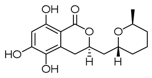
Peak #3, detected at 5.05 min (m/z 325.1285), corresponded with the molecular formula C16H20O7, the same as 14-hydroxyasperentin B, which was previously isolated from the fungus P. antarcticum KMM 4685 [17]. The compound was identified using an in-house database.
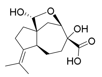
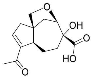
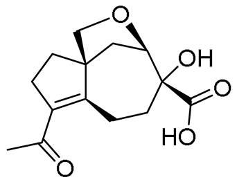
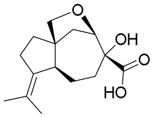
The peaks #4 and #5, detected at 5.18 and 6.08 min, respectively, with m/z 283.1552, corresponded to the molecular formula C15H22O5. These peaks were suggested as piltunine A, piltunine C, and penigrisacid D, which were previously isolated from this strain [20].
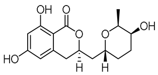
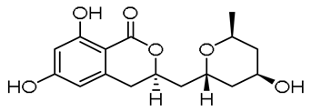
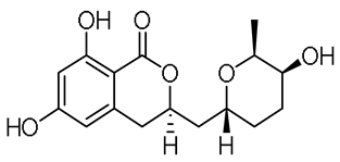
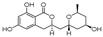
Peak #12, detected at 10.76 min at m/z 293.1377, corresponded with the molecular formula C16H20O5, and was assigned to cladosporin, the usual polyketide of P. antarcticum. The compound was identified based on its exact mass value and RT using an in-house database. The peaks #6 and #10, detected at 6.28 and 9.23 min at m/z 309.1332, corresponded with the molecular formula C16H20O6. These peaks showed characteristic MS/MS spectra for the monohydroxylated derivative of cladosporin, such as cladomarin and 5′-hydroxyasperentin, which were isolated from the fungus of this species.
The peaks #7 and #8, detected at 7.12 and 7.37 min with m/z 267.1227, corresponded with the molecular formula C14H18O5 that can be annotated with piltunins E and F, metabolites of this strain.
Peak #11, detected at 9.32 min with m/z 267.1585, corresponded with the molecular formula C15H22O4 that can be annotated with aspterric acid, a possible biosynthetic precursor of piltunins.
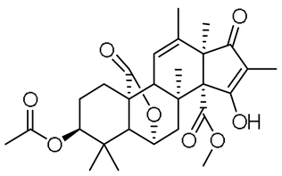
Peak #13, detected at 11.69 min with m/z 501.2126, corresponded with the molecular formula C27H32O9, which was the same as austin, the characteristic meroterpenoid of P. antarcticum. The compound was identified using the GNPS database (MQScore 0.95).
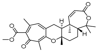
Peak #14, detected at 11.69 min with m/z 523.2281, corresponded with the molecular formula C28H36O8, the same as citreohybridonol, a common androstane-type meroterpenoid of Penicillium fungi. The compound was identified using the GNPS database (MQScore 0.95). Peak #15, detected at 11.69 min with m/z 441.2272, corresponded with the molecular formula C26H32O6, the same as tropolactone C, a widespread fungal meroterpenoid. The compound was annotated based on the exact mass value and MetFrag service.
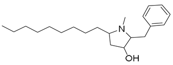
Peak #16 was detected at 12.72 min with m/z 318.2789, corresponding to the molecular formula C21H35NO, the same as preussin, a hydroxyl pyrrolidine derivative from the sponge-associated fungus Aspergillus candidus. The compound was identified using the GNPS database (MQScore 0.81).
Peak #17 was detected at 14.48 min with m/z 507.2282, corresponding to the molecular formula C32H30N2O4, the same as asperphenamate, a linear amino acid ester from fungi P. astrolabium. The compound was identified using the GNPS database (MQScore 0.91).
Peak #18 was detected at 14.92 min with m/z 279.2320, corresponding to the molecular formula C18H30O2. The peak was suggested to be an unidentified fatty acid with four degrees of unsaturation based on tandem MS data and exact mass.
Peak #19 was detected at 18.87 min with m/z 282.2793, corresponding to the molecular formula C18H35NO. The compound was annotated as an amide of octadecenoic fatty acid based on its exact mass and fragmentation pattern.
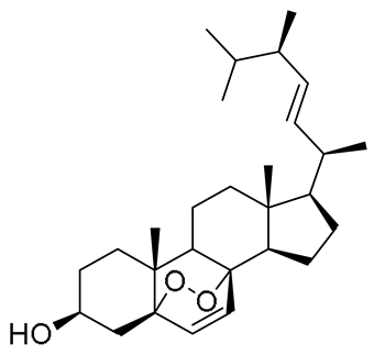
Peak #20 was detected at 19.85 min with m/z 411.3259, corresponding to the molecular formula C28H44O3, the same as ergosterol peroxide, a usual derivative of the main fungal triterpenoid ergosterol. The compound was suggested based on an exact mass value and RT with an in-house database, and using the GNPS database (MQScore 0.82).
Peak #1, detected at 2.03 min (m/z 195.1008) based on the fragmentation pattern, was assumed to be an unknown polyketide derivative with the molecular formula C11H14O3. Peak #9, detected at 8.75 min (m/z 535.2716), can be assigned to an unknown alkaloid with the molecular formula C33H34N4O3.
In total, 20 compounds were annotated using the GNPS database, the in-house database, or the MetFrag service. Detailed characteristics of the identified compounds are presented in the Appendix A (Table A1).
It should be noted that we have not been able to find convincing evidence for the production of cyclopiane diterpenes that are characteristic of another P. antarcticum strain [19].
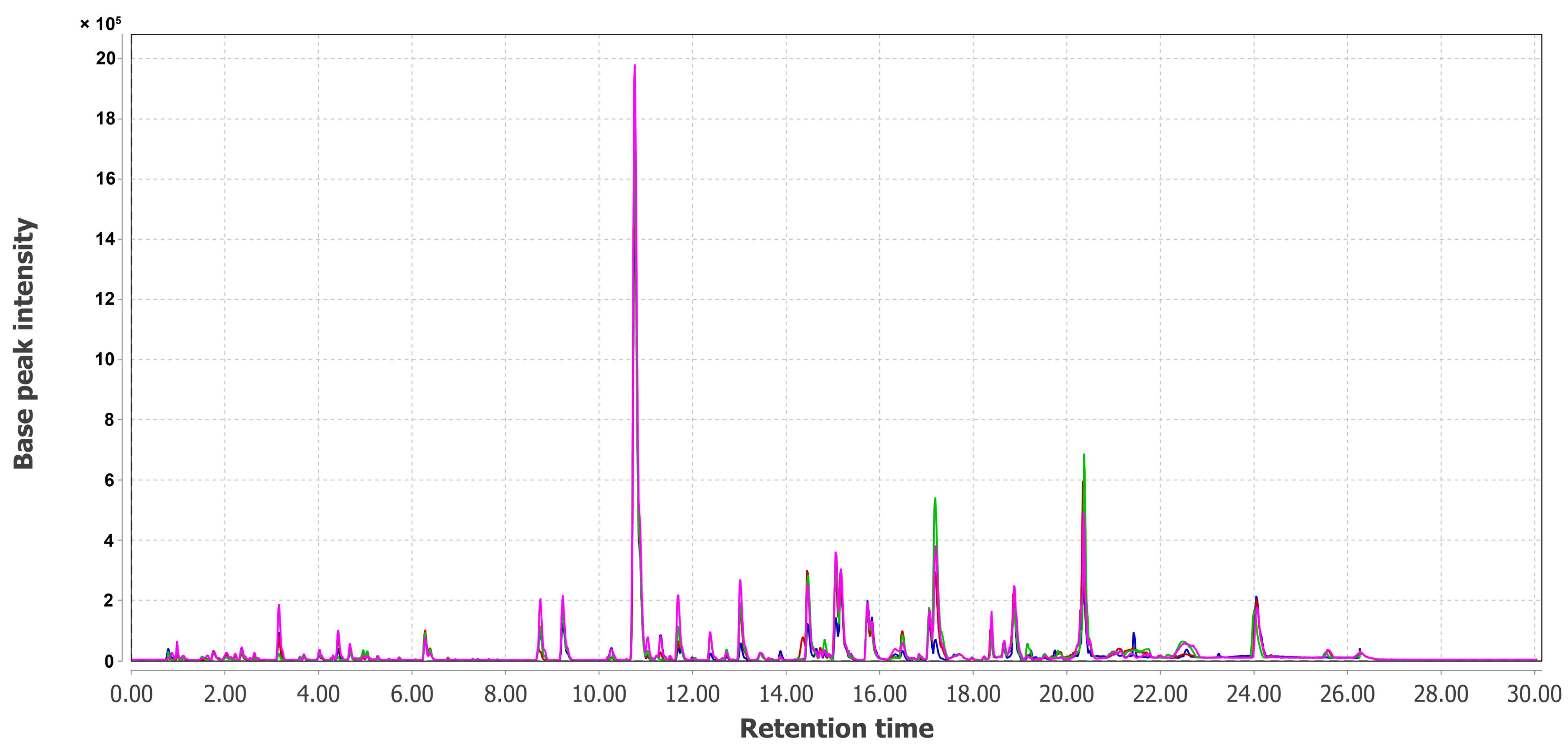
The combined UPLC MS chromatograms of KMM 4668 (Pa1), KMM 4711 (Pa2), KMM 4712 (Pa3), KMM 4713 (Pa4), KMM 4714 (Pa5), and KMM 4715 (Pa6) extracts are presented in Figure 5 and Figure 6.
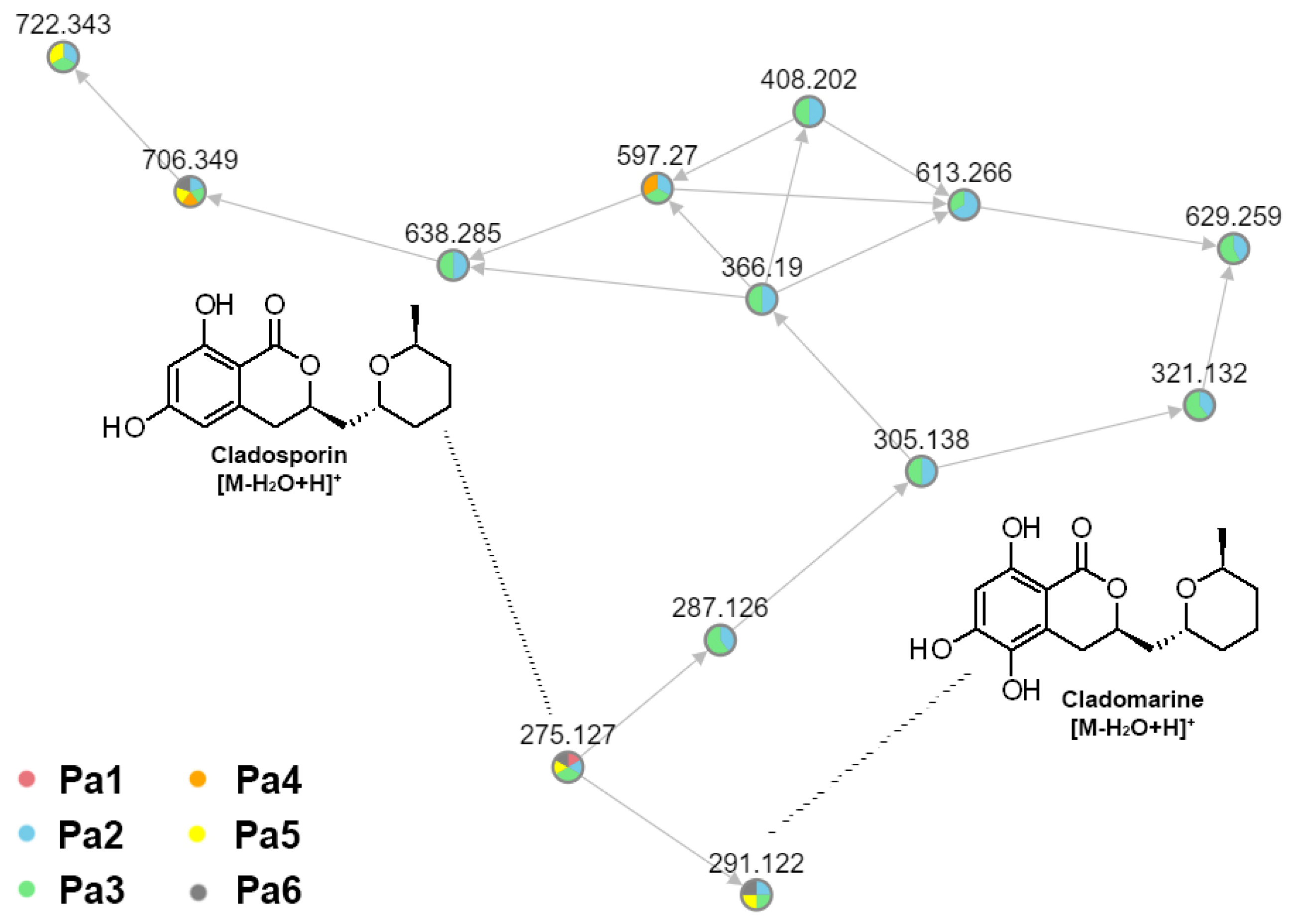
The use of a FBMN, together with the in-house database, made it possible to reveal a cluster of related features, which were assigned to cladosporin-related compounds. Unfortunately, we were only able to assign two compounds in the cluster, and most of the features remained unidentified (Figure 7).
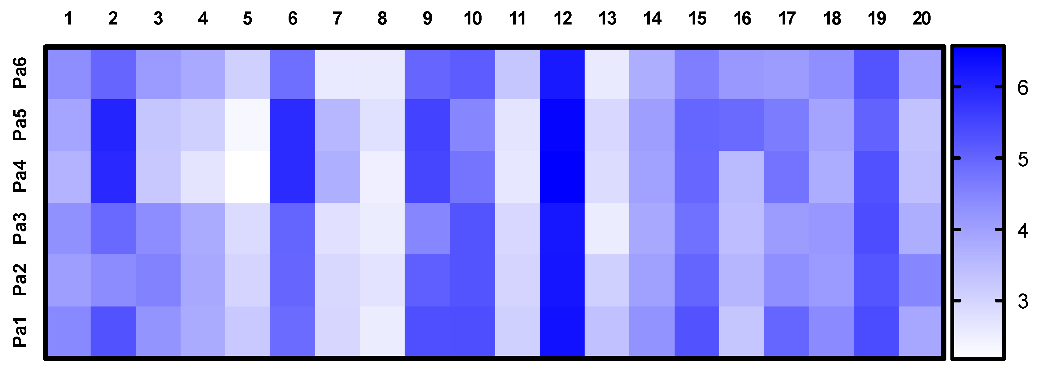
The relative contents of the announced compounds calculated as a decimal logarithm of the peak area of these peaks in the UPLC-MS chromatograms of Pa1, Pa2, Pa3, Pa4, Pa5, and Pa6 extracts were visualized in the heatmap (Figure 8). Cladosporin (#12) was the main component of each extract. Extracts Pa4 and Pa5 were different from other extracts, with a large content of chrysogine (#2) and an unidentified cladosporin derivative (#6).

4. Discussion

Phenotypic diversity is one of the ways that fungi adapt to changing environments. Three types of phenotypic changes have been reported: morphological transitions (MT) induced by environmental signals, phenotypic switching (PS) that occurs in a small fraction of the population and is not necessarily induced by external signals, and antigenic variation (AV), which involves alternating the expression of surface proteins (or carbohydrates) [30]. Phenotypic switching is associated with metabolic changes [31], which are a part of the environmental adaptation of microorganisms. For example, aflatoxin production by Aspergillus flavus was reduced over several generations of laboratory fermentation owing to the absence of natural competition, and was recovered in co-cultivation with other fungi [32].
Various strains of a microbial species may exhibit different phenotypic variations. Using single-cell measurements, the phenotypic diversity of 37 natural strains of Saccharomyces cerevisiae yeasts was investigated, and these wild strains displayed different levels of noise in single-cell traits [33]. This suggests a different level of phenotypic diversity in different fungal strains, which can lead to the isolation of single-cell isolates with different secondary metabolites [21]. The biosynthesis of secondary metabolites is controlled by biosynthetic gene clusters (BGCs), and changes in these clusters can result in a change in compound yield [34]. From an evolutionary perspective, new metabolic capabilities typically emerge through some form of gene duplication, acquisition of novel genes through horizontal gene transfer, and, in addition, novelty might emerge through the establishment of a new regulatory network [35].
It is known that the phenotypic diversity of fungi, expressed both at the level of morphology and production of secondary metabolites, is due to the phenomenon of heterokaryosis, which provides haploid organisms with significant adaptive advantages [36]. Heterocaryosis is of great importance to fungi, whose life cycle is exclusively represented by the anamorphic stage [36]. Most fungi, including Penicillium species, primarily exist as heterokaryons in nature. They are more flexible and viable than homokaryons, which play a significant role in the colonization of a variety of natural substrates [37]. However, the heterokaryotic state of most fungi that reproduce via mononuclear conidia is unstable, and when such conidia are dispersed, the heterokaryon disintegrates easily into the original homokaryons. Different phenotypic variants representing the original parental forms were isolated during a study of the KMM 4668 strain. The latter differed not only in terms of cultural and morphological characteristics, but also in the ability to produce biologically active compounds.
In our study, we observed differences in chrysogine and cladosporin-related compounds, as well as sesquiterpenes piltunin production in KMM 4713 and KMM 4714 intra-strain variants of P. antarcticum KMM 4668. The biosynthesis of cladosporin and related compounds is well known and carried out under the sequential action of two type I polyketide synthases [38]. It was found earlier that biosynthesis of chrysogine is carried out by gene clusters from seven genes [15]. Piltunins are the closest derivatives of the well-known herbicide carotanoid aspterric acid. In particular, piltunins A and C are products of the Asp hydroxylation of aspterric acid. It is known that biosynthesis of aspterric acid is controlled by a BGC, including genes encoding a sesquiterpene cyclase, two P450 monooxygenases, and the self-resistance gene astD [39]. The production of these compounds was enhanced in the KMM 4713 and KMM 4714 strains, which may be the result of changes in the described BGCs. However, further investigations are required.
At the same time, the cytotoxic effects of the KMM 4713 and KMM 4714 extracts were lower than those of the other extracts (Table 1), which may be the result of a decrease in the concentration of cytotoxic components or an increase in cytoprotective compounds. As shown in the heatmap (Figure 8), the contents of #3 (14-hydroxyasperentin B), #4–5 (piltunine A, piltunine C, and penigrisacid D), #10 (cladosporin derivative), and #18 (unsaturated fatty acid) were lower in the KMM 4713 and KMM 4714 extracts. However, the cytotoxic activity of 14-hydroxyasperentin B has not been studied. Piltunine A, piltunine C, and penigrisacid D showed weak cytotoxic activity against prostate cancer 22Rv1 cells and normal prostate PNT-2 cells, and anti-inflammatory activity in LPS-stimulated RAW264.7 macrophages [20]. Cladosporin and its derivatives have various bioactivities, including potent antimalarial activity [40,41].
Based on the outcome of our research, the intra-strain variants of P. antarcticum may be a promising source of bioactive secondary metabolites.

5. Conclusions

The intra-strain variants of P. antarcticum KMM 4668 were isolated and labelled KMM 4711, KMM 4712, KMM 4713, KMM 4714, and KMM 4715. The investigation of molecular markers such as ITS regions, partial BenA, CaM, and RPB2 gene sequences indicated that KMM 4668 belongs to P. antarcticum. A comparison of the first three phylogenetic makers between the original strain KMM 4668 and its variants KMM 4711–KMM 4715 showed their 100% identity. Differences in the UPLC MS metabolite profiles of these variants were observed in cladosporin, chrysogine, and piltunin production. The variants KMM 4713 and KMM 4714 were characterized by increased production of most of the cladosporin-related compounds, alkaloid chrysogine, and piltunin sesquiterpenoids. Thus, these variants could be used to obtain these metabolites.

Supplementary Materials

The following supporting information can be downloaded at https://www.mdpi.com/article/10.3390/metabo15020077/s1, Figure S1: HR (+)ESI MS/MS spectrum of 1; Figure S2: HR (+)ESI MS/MS spectrum of 2; Figure S3: HR (+)ESI MS/MS spectrum of 3; Figure S4: HR (+)ESI MS/MS spectrum of 4; Figure S5: HR (+)ESI MS/MS spectrum of 6; Figure S6: UPLC MS chromatogram of Penicillium antarcticum KMM 4668 extract (Pa1); Figure S7: UPLC MS chromatogram of Penicillium antarcticum KMM 4711 extract (Pa2); Figure S8: UPLC MS chromatogram of Penicillium antarcticum KMM 4712 extract (Pa3); Figure S9: UPLC MS chromatogram of Penicillium antarcticum KMM 4713 extract (Pa4); Figure S10: UPLC MS chromatogram of Penicillium antarcticum KMM 4714 extract (Pa5); Figure S11: UPLC MS chromatogram of Penicillium antarcticum KMM 4715 extract (Pa6); Table S1: The strains used in multi-locus phylogenetic analysis and GenBank accession numbers; Table S2: Amounts of the extracts of the fungal cultures; Supplementary Text: UHPLC-Q-TOF Data Analysis. References [42,43,44,45,46] are cited in the Supplementary Materials.

Author Contributions

Data curation, E.A.Y. and A.N.Y.; Formal analysis, R.S.P. and E.A.Y.; Funding acquisition, M.P.I.; Investigation, G.V.B., N.N.K., V.E.C., R.S.P., O.I.Z., E.A.C. and E.A.Y.; Methodology, N.N.K. and E.A.Y.; Project administration, M.P.I.; Resources, M.P.I. and A.N.Y.; Supervision, M.P.I. and A.N.Y.; Validation, R.S.P., E.A.Y., M.P.I. and A.N.Y.; Visualization, G.V.B., V.E.C. and E.A.C.; Writing—original draft, G.V.B., N.N.K., V.E.C. and E.A.Y.; Writing—review and editing, M.P.I. and A.N.Y. All authors have read and agreed to the published version of the manuscript.

Funding

This research was funded by a grant from the Ministry of Science and Higher Education of Russian Federation 15.BRK.21.0004 (Contract No.: 075-15-2021-1052/9).

Institutional Review Board Statement

Not applicable.

Informed Consent Statement

Not applicable.

Data Availability Statement

The original contributions presented in this study are included in the article and Supplementary Material. Further inquiries can be directed to the corresponding authors.

Acknowledgments

This study was carried out using the Collective Facilities Center “Collection of Marine Microorganisms PIBOC FEB RAS” and on the equipment of the Collective Facilities Center “The Far Eastern Center for Structural Molecular Research (NMR/MS) PIBOC FEB RAS”.

Conflicts of Interest

The authors declare no conflicts of interest.

Appendix A

Table A1. The list of annotated compounds.
#RT, minMeasured m/zMFCalculated m/zError, ppmMQScore (GNPS)StructureName and
Identification Level
Ref.
12.03195.1008
[M + H]+
C11H14O3195.1016−3.95----
23.15191.0805
[M + H]+
C10H10N2O2191.0815−5.250.87Metabolites 15 00077 i001Chrysogine b[15]
35.05325.1285
[M + H]+
C16H20O7325.12820.99-Metabolites 15 00077 i00214-hydroxyasperentin B a[17]
45.18283.1552
[M + H]+
C15H22O5283.15404.24-Metabolites 15 00077 i003Penigrisacid D c[47]
-Metabolites 15 00077 i004Piltunine C c[20]
56.08283.1552
[M + H]+
C15H22O5283.15404.24-Metabolites 15 00077 i005Piltunine A c[20]
66.28309.1332
[M + H]+
C16H20O6309.1333−0.21-Metabolites 15 00077 i006Hydroxyasperentin B c
(Cladomarin)
[8]
-Metabolites 15 00077 i00714-hydroxyasperentin c
(14-hydroxycladosporin)
[17]
-Metabolites 15 00077 i00813-hydroxyasperentin c
(13-hydroxycladosporin)
[48]
77.12267.1227
[M + H]+
C14H18O5267.12270.00-Metabolites 15 00077 i009Piltunine E c[20]
87.37267.1227
[M + H]+
C14H18O5267.12270.00-Metabolites 15 00077 i010Piltunine F c[20]
98.75535.2716
[M + H]+
C33H34N4O3535.27042.24--
109.23309.1336
[M + H]+
C16H20O6309.13331.08-Metabolites 15 00077 i011Cladomarin c[8]
-Metabolites 15 00077 i01214-hydroxyasperentin c
(14-hydroxycladosporin)
[17]
-Metabolites 15 00077 i01313-hydroxyasperentin c
(13-hydroxycladosporin)
[48]
119.32267.1585
[M + H]+
C15H22O4267.1591−2.25-Metabolites 15 00077 i014Aspterric acid c-
1210.76293.1377
[M + H]+
C16H20O5293.1384−2.39-Metabolites 15 00077 i015Cladosporin a
(Asperentin)
[19]
1311.69501.2126
[M + H]+
C27H32O9501.21191.380.95Metabolites 15 00077 i016Austine b[49]
1411.69523.2281
[M + Na]+
C28H36O8523.2302−4.090.95Metabolites 15 00077 i017Citreohybridonol b[16]
1511.69441.2272
[M + H]+
C26H32O6441.22720.08-Metabolites 15 00077 i018Tropolactone C c[50]
1612.72318.2789
[M + H]+
C21H35NO318.2791−0.760.81Metabolites 15 00077 i019Preussin b[51]
1714.48507.2282
[M + H]+
C32H30N2O4507.22780.720.91Metabolites 15 00077 i020Asperphenamate b[52]
1814.92279.2320
[M + H]+
C18H30O2279.23190.51-- -
1918.87282.2793
[M + H]+
C18H35NO282.27910.56-- -
2019.85411.3259
[M–H2O + H]+
C28H44O3411.32580.350.82Metabolites 15 00077 i021Ergosterol peroxide a[53]
a—identification was made by comparing the RT, MS, and MS/MS data with those of authentic standards; b—structure was annotated by comparing the obtained MS/MS data with available data in the in-house database or in the GNPS; c—structure was assigned based on MF and literature data.

References

  1. Xiliang, Y.; Jinping, L.; Jiahui, M.; Rui, J.; Shizheng, T.; Huafeng, D.; Jing, L.; Sumei, Y.; Juan, L. Origins, Structures, and Bioactivities of Secondary Metabolites from Marine-derived Penicillium Fungi. Mini-Rev. Med. Chem. 2021, 21, 2000–2019. [Google Scholar] [CrossRef]
  2. Yurchenko, A.N.; Girich, E.V.; Yurchenko, E.A. Metabolites of Marine Sediment-Derived Fungi: Actual Trends of Biological Activity Studies. Mar. Drugs 2021, 19, 88. [Google Scholar] [CrossRef]
  3. Visagie, C.M.; Frisvad, J.C.; Houbraken, J.; Visagie, A.; Samson, R.A.; Jacobs, K. A re-evaluation of Penicillium section Canescentia, including the description of five new species. Persoonia Mol. Phylogeny Evol. Fungi 2021, 46, 163–187. [Google Scholar] [CrossRef]
  4. McRae, C.F.; Seppelt, R.D.; Hocking, A.D. Penicillium species from terrestrial habitats in the Windmill Islands, East Antarctica, including a new species, Penicillium antarcticum. Polar Biol. 1999, 21, 97–111. [Google Scholar] [CrossRef]
  5. Kirichuk, N.N.; Pivkin, M.V.; Matveeva, T.V. Three new Penicillium species from marine subaqueous soils. Mycol. Prog. 2017, 16, 15–26. [Google Scholar] [CrossRef]
  6. Park, M.S.; Lee, E.J.; Fong, J.J.; Sohn, J.H.; Lim, Y.W. A new record of Penicillium antarcticum from marine environments in Korea. Mycobiology 2014, 42, 109–113. [Google Scholar] [CrossRef]
  7. Vansteelandt, M.; Kerzaon, I.; Blanchet, E.; Fossi Tankoua, O.; Robiou Du Pont, T.; Joubert, Y.; Monteau, F.; Le Bizec, B.; Frisvad, J.C.; Pouchus, Y.F.; et al. Patulin and secondary metabolite production by marine-derived Penicillium strains. Fungal Biol. 2012, 116, 954–961. [Google Scholar] [CrossRef]
  8. Takahashi, K.; Sakai, K.; Nagano, Y.; Orui Sakaguchi, S.; Lima, A.O.; Pellizari, V.H.; Iwatsuki, M.; Takishita, K.; Nonaka, K.; Fujikura, K.; et al. Cladomarine, a new anti-saprolegniasis compound isolated from the deep-sea fungus, Penicillium coralligerum YK-247. J. Antibiot. 2017, 70, 911–914. [Google Scholar] [CrossRef]
  9. Wiese, J.; Aldemir, H.; Schmaljohann, R.; Gulder, T.A.M.; Imhoff, J.F. Asperentin B, a New Inhibitor of the Protein Tyrosine Phosphatase 1B. Mar. Drugs 2017, 15, 191. [Google Scholar] [CrossRef]
  10. Zang, Y.; Gong, Y.; Shi, Z.; Qi, C.; Chen, C.; Tong, Q.; Liu, J.; Wang, J.; Zhu, H.; Zhang, Y. Multioxidized aromatic polyketides produced by a soil-derived fungus Penicillium canescens. Phytochemistry 2022, 193, 113012. [Google Scholar] [CrossRef] [PubMed]
  11. Ishikawa, Y.; Morimoto, K.; Iseki, S. Atrovenetin as a potent antioxidant compound from Penicillium species. J. Am. Oil Chem. Soc. 1991, 68, 666–668. [Google Scholar] [CrossRef]
  12. Shiono, Y.; Seino, Y.; Koseki, T.; Murayama, T.; Kimura, K.-i. Antarones A and B, Two Polyketides from an Endophytic Penicillium antarcticum. Z. Für Naturforschung B 2008, 63, 909–914. [Google Scholar] [CrossRef]
  13. Marchese, P.; Mahajan, N.; O’Connell, E.; Fearnhead, H.; Tuohy, M.; Krawczyk, J.; Thomas, O.P.; Barry, F.; Murphy, M.J. A Novel High-Throughput Screening Platform Identifies Itaconate Derivatives from Marine Penicillium antarcticum as Inhibitors of Mesenchymal Stem Cell Differentiation. Mar. Drugs 2020, 18, 192. [Google Scholar] [CrossRef]
  14. Ravunni, M.; Benjamin, P.S. Penicillium verruculosum strain BS3 produces aurantioclavine and rugulosuvine B Alkaloids. Electron. J. Biol. 2016, 12, 484–489. [Google Scholar]
  15. Viggiano, A.; Salo, O.; Ali, H.; Szymanski, W.; Lankhorst Peter, P.; Nygård, Y.; Bovenberg Roel, A.L.; Driessen Arnold, J.M. Pathway for the Biosynthesis of the Pigment Chrysogine by Penicillium chrysogenum. Appl. Environ. Microbiol. 2018, 84, e02246-17. [Google Scholar] [CrossRef] [PubMed]
  16. Kosemura, S. Meroterpenoids from Penicillium citreo-viride B. IFO 4692 and 6200 hybrid. Tetrahedron 2003, 59, 5055–5072. [Google Scholar] [CrossRef]
  17. Leshchenko, E.V.; Antonov, A.S.; Borkunov, G.V.; Hauschild, J.; Zhuravleva, O.I.; Khudyakova, Y.V.; Menshov, A.S.; Popov, R.S.; Kim, N.Y.; Graefen, M.; et al. New Bioactive β-Resorcylic Acid Derivatives from the Alga-Derived Fungus Penicillium antarcticum KMM 4685. Mar. Drugs 2023, 21, 178. [Google Scholar] [CrossRef] [PubMed]
  18. Leshchenko, E.V.; Antonov, A.S.; Dyshlovoy, S.A.; Berdyshev, D.V.; Hauschild, J.; Zhuravleva, O.I.; Borkunov, G.V.; Menshov, A.S.; Kirichuk, N.N.; Popov, R.S.; et al. Meroantarctines A–C, meroterpenoids with rearranged skeletons from the alga-derived fungus Penicillium antarcticum KMM 4685 with potent p-glycoprotein inhibitory activity. J. Nat. Prod. 2022, 85, 2746–2752. [Google Scholar] [CrossRef]
  19. Yurchenko, A.N.; Zhuravleva, O.I.; Khmel, O.O.; Oleynikova, G.K.; Antonov, A.S.; Kirichuk, N.N.; Chausova, V.E.; Kalinovsky, A.I.; Berdyshev, D.V.; Kim, N.Y.; et al. New Cyclopiane Diterpenes and Polyketide Derivatives from Marine Sediment-Derived Fungus Penicillium antarcticum KMM 4670 and Their Biological Activities. Mar. Drugs 2023, 21, 584. [Google Scholar] [CrossRef]
  20. Afiyatullov, S.S.; Zhuravleva, O.I.; Antonov, A.S.; Leshchenko, E.V.; Pivkin, M.V.; Khudyakova, Y.V.; Denisenko, V.A.; Pislyagin, E.A.; Kim, N.Y.; Berdyshev, D.V.; et al. Piltunines A-F from the marine-derived fungus Penicillium piltunense KMM 4668. Mar. Drugs 2019, 17, 647. [Google Scholar] [CrossRef]
  21. Mayne, R.F.; Bennett, J.W.; Tallant, J. Instability of an aflatoxin-producing strain of Aspergillus parasiticus. Mycologia 1971, 63, 644–648. [Google Scholar] [CrossRef] [PubMed]
  22. Kucsera, J.; Yarita, K.; Takeo, K.; Yoshida, S.; Gácser, A.; Hamari, Z.; Avasi, Z.; Kevei, F. Intra-strain variability of Cryptococcus neoformans can be detected on Phloxin B medium. J. Basic Microbiol. 2002, 42, 111–119. [Google Scholar] [CrossRef]
  23. Pazouki, M.; Panda, T. Understanding the morphology of fungi. Bioprocess Eng. 2000, 22, 127–143. [Google Scholar] [CrossRef]
  24. Visagie, C.M.; Houbraken, J.; Frisvad, J.C.; Hong, S.B.; Klaassen, C.H.W.; Perrone, G.; Seifert, K.A.; Varga, J.; Yaguchi, T.; Samson, R.A. Identification and nomenclature of the genus Penicillium. Stud. Mycol. 2014, 78, 343–371. [Google Scholar] [CrossRef]
  25. Visagie, C.M.; Yilmaz, N.; Kocsubé, S.; Frisvad, J.C.; Hubka, V.; Samson, R.A.; Houbraken, J. A review of recently introduced Aspergillus, Penicillium, Talaromyces and other Eurotiales species. Stud Mycol. 2024, 107, 1–66. [Google Scholar] [CrossRef] [PubMed]
  26. Kumar, S.; Stecher, G.; Li, M.; Knyaz, C.; Tamura, K. MEGA X: Molecular Evolutionary Genetics Analysis across Computing Platforms. Mol. Biol. Evol. 2018, 35, 1547–1549. [Google Scholar] [CrossRef] [PubMed]
  27. Kimura, M. A simple method for estimating evolutionary rates of base substitutions through comparative studies of nucleotide sequences. J. Mol. Evol. 1980, 16, 111–120. [Google Scholar] [CrossRef]
  28. Nesterenko, L.; Popov, R.; Zhuravleva, O.; Kirichuk, N.; Chausova, V.; Krasnov, K.; Mikhail, P.; Yurchenko, E.; Isaeva, M.; Yurchenko, A. A study of the metabolic profiles of Penicillium dimorphosporum KMM 4689 which led to its re-identification as Penicillium hispanicum. Fermentation 2023, 9, 337. [Google Scholar] [CrossRef]
  29. Pluskal, T.; Castillo, S.; Villar-Briones, A.; Orešič, M. MZmine 2: Modular framework for processing, visualizing, and analyzing mass spectrometry-based molecular profile data. BMC Bioinform. 2010, 11, 395. [Google Scholar] [CrossRef]
  30. Jain, N.; Fries, B.C. Antigenic and phenotypic variations in fungi. Cell. Microbiol. 2009, 11, 1716–1723. [Google Scholar] [CrossRef]
  31. Lan, C.-Y.; Newport, G.; Murillo, L.A.; Jones, T.; Scherer, S.; Davis, R.W.; Agabian, N. Metabolic specialization associated with phenotypic switching in Candida albicans. Proc. Natl. Acad. Sci. USA 2002, 99, 14907–14912. [Google Scholar] [CrossRef]
  32. Horn, B.W.; Dorner, J.W. Effect of competition and adverse culture conditions on aflatoxin production by Aspergillus flavus through successive generations. Mycologia 2002, 94, 741–751. [Google Scholar] [CrossRef]
  33. Yvert, G.; Ohnuki, S.; Nogami, S.; Imanaga, Y.; Fehrmann, S.; Schacherer, J.; Ohya, Y. Single-cell phenomics reveals intra-species variation of phenotypic noise in yeast. BMC Syst. Biol. 2013, 7, 54. [Google Scholar] [CrossRef] [PubMed]
  34. Zhgun, A.A. Fungal BGCs for Production of Secondary Metabolites: Main Types, Central Roles in Strain Improvement, and Regulation According to the Piano Principle. Int. J. Mol. Sci. 2023, 24, 11184. [Google Scholar] [CrossRef] [PubMed]
  35. Naranjo-Ortiz, M.A.; Gabaldón, T.A.-O. Fungal evolution: Cellular, genomic and metabolic complexity. Biol. Rev. Camb. Philos Soc. 2020, 95, 1198–1232. [Google Scholar] [CrossRef]
  36. Strom, N.B.; Bushley, K.E. Two genomes are better than one: History, genetics, and biotechnological applications of fungal heterokaryons. Fungal Bio. Biotechnol. 2016, 3, 4. [Google Scholar] [CrossRef]
  37. Jinks, J.L. Heterocaryosis in wild Penicillium. Heredity 1952, 6, 77–87. [Google Scholar] [CrossRef]
  38. Cochrane, R.V.K.; Sanichar, R.; Lambkin, G.R.; Reiz, B.; Xu, W.; Tang, Y.; Vederas, J.C. Production of New Cladosporin Analogues by Reconstitution of the Polyketide Synthases Responsible for the Biosynthesis of this Antimalarial Agent. Angew. Chem. Int. Ed. 2016, 55, 664–668. [Google Scholar] [CrossRef] [PubMed]
  39. Yan, Y.; Liu, Q.; Zang, X.; Yuan, S.; Bat-Erdene, U.; Nguyen, C.; Gan, J.; Zhou, J.; Jacobsen, S.E.; Tang, Y. Resistance-gene-directed discovery of a natural-product herbicide with a new mode of action. Nature 2018, 559, 415–418. [Google Scholar] [CrossRef]
  40. Wang, X.; Wedge, D.E.; Cutler, S.J. Chemical and Biological Study of Cladosporin, an Antimicrobial Inhibitor: A Review. Nat. Prod. Commun. 2016, 11, 1595–1600. [Google Scholar] [CrossRef] [PubMed]
  41. Hou, A.; Li, B.; Deng, Z.; Xu, M.; Dickschat, J.S. Cladosporin, A Highly Potent Antimalaria Drug? Chem. Bio. Chem. 2023, 24, e202300154. [Google Scholar] [CrossRef]
  42. Chambers, M.C.; MacLean, B.; Burke, R.; Amodei, D.; Ruderman, D.L.; Neumann, S.; Gatto, L.; Fischer, B.; Pratt, B.; Egertson, J.; et al. A cross-platform toolkit for mass spectrometry and proteomics. Nat. Biotechnol. 2012, 30, 918–920. [Google Scholar] [CrossRef]
  43. Myers, O.D.; Sumner, S.J.; Li, S.; Barnes, S.; Du, X. One Step Forward for Reducing False Positive and False Negative Compound Identifications from Mass Spectrometry Metabolomics Data: New Algorithms for Constructing Extracted Ion Chromatograms and Detecting Chromatographic Peaks. Anal. Chem. 2017, 89, 8696–8703. [Google Scholar] [CrossRef]
  44. Nothias, L.F.; Petras, D.; Schmid, R.; Dührkop, K.; Rainer, J.; Sarvepalli, A.; Protsyuk, I.; Ernst, M.; Tsugawa, H.; Fleischauer, M.; et al. Feature-based molecular networking in the GNPS analysis environment. Nat. Methods 2020, 17, 905–908. [Google Scholar] [CrossRef]
  45. Christiansen, J.V.; Larsen, T.O.; Frisvad, J.C. Production of Fungal Quinones: Problems and Prospects. Biomolecules 2022, 12, 1041. [Google Scholar] [CrossRef]
  46. Ruttkies, C.; Schymanski, E.L.; Wolf, S.; Hollender, J.; Neumann, S. MetFrag relaunched: Incorporating strategies beyond in silico fragmentation. J. Cheminf. 2016, 8, 3/1. [Google Scholar] [CrossRef] [PubMed]
  47. Xing, C.-P.; Xie, C.-L.; Xia, J.-M.; Liu, Q.-M.; Lin, W.-X.; Ye, D.-Z.; Liu, G.-M.; Yang, X.-W. Penigrisacids A–D, Four New Sesquiterpenes from the Deep-Sea-Derived Penicillium griseofulvum. Mar. Drugs 2019, 17, 507. [Google Scholar] [CrossRef]
  48. Grove, J.F. New metabolic products of Aspergillus flavus. Part IV. 4′-Hydroxyasperentin and 5′-hydroxyasperentin 8-methyl ether. J. Chem. Soc. Perkin Trans. 1 1973, 22, 2704–2706. [Google Scholar] [CrossRef] [PubMed]
  49. Chexal, K.K.; Springer, J.P.; Clardy, J.; Cole, R.J.; Kirksey, J.W.; Dorner, J.W.; Cutler, H.G.; Strawter, B.J. Austin, a novel polyisoprenoid mycotoxin from Aspergillus ustus. J. Am. Chem. Soc. 1976, 98, 6748–6750. [Google Scholar] [CrossRef]
  50. Cueto, M.; MacMillan, J.B.; Jensen, P.R.; Fenical, W. Tropolactones A–D, four meroterpenoids from a marine-derived fungus of the genus Aspergillus. Phytochemistry 2006, 67, 1826–1831. [Google Scholar] [CrossRef]
  51. Malhão, F.; Ramos, A.A.; Buttachon, S.; Dethoup, T.; Kijjoa, A.; Rocha, E. Cytotoxic and Antiproliferative Effects of Preussin, a Hydroxypyrrolidine Derivative from the Marine Sponge-Associated Fungus Aspergillus candidus KUFA 0062, in a Panel of Breast Cancer Cell Lines and Using 2D and 3D Cultures. Mar. Drugs 2019, 17, 448. [Google Scholar] [CrossRef]
  52. Subko, K.; Wang, X.; Nielsen, F.H.; Isbrandt, T.; Gotfredsen, C.H.; Ramos, M.C.; Mackenzie, T.; Vicente, F.; Genilloud, O.; Frisvad, J.C.; et al. Mass Spectrometry Guided Discovery and Design of Novel Asperphenamate Analogs from Penicillium astrolabium Reveals an Extraordinary NRPS Flexibility. Front Microbiol. 2021, 11, 618730. [Google Scholar] [CrossRef] [PubMed]
  53. Lobo, A.M.; De Abreu, M.; Prabhakar, S.; Godinho, L.S.; Jones, R.; Rzepa, H.S.; Williams, D.J. Triterpenoids of the fungus Pisolithus tinctorius. Phytochemistry 1988, 27, 3569–3574. [Google Scholar] [CrossRef]
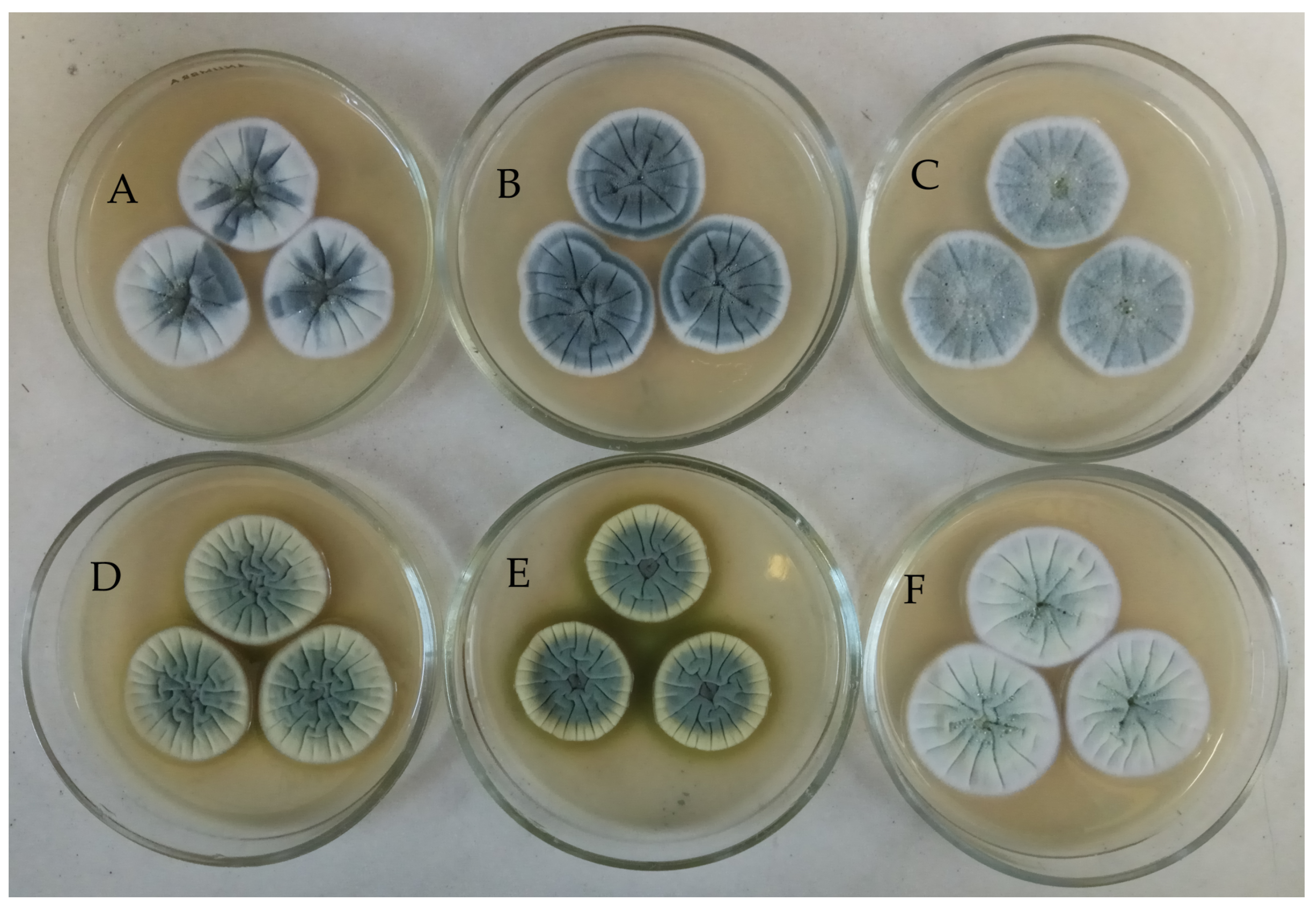
Figure 1. Morphology of intra-strain variants of Penicillium antarcticum KMM 4668 obtained by selection work ((A)—KMM 4668, (B)—KMM 4711, (C)—KMM 4712, (D)—KMM 4713, (E)—KMM 4714, (F)—KMM 4715).
Metabolites 15 00077 g001
Figure 2. ML tree based on concatenated ITS-BenA-CaM gene sequences showing phylogenetic positions of the original strain KMM 4668, and its intra-strain variants KMM 4711-KMM 4715 among members of the genus Penicillium section Canescentia, series Atroveneta. Bootstrap values (%) of 1000 replications and nodes with confidence values greater than 50% are indicated. The scale bar represents 0.1 substitutions per site.
Metabolites 15 00077 g002
Figure 3. Principal component analysis plot (a) and dendrogram (b) of UPLC-MS data of the extracts of Penicillium antarcticum KMM 4668 (Pa1), KMM 4711 (Pa2), KMM 4712 (Pa3), KMM 4713 (Pa4), KMM 4714 (Pa5), and KMM 4715 (Pa6).
Metabolites 15 00077 g003
Figure 4. UPLC MS chromatogram of Penicillium antarcticum KMM 4668 extract.
Metabolites 15 00077 g004
Figure 5. UPLC-MS chromatogram comparison of extracts of KMM 4668 (Pa1, green) and its intra-strain variants (Pa4, blue and Pa5, red).
Metabolites 15 00077 g005
Figure 6. UPLC-MS chromatogram comparison of extracts of KMM 4668 (Pa1, magenta) and its intra-strain variants (Pa2, green, Pa3, red and Pa6, blue).
Metabolites 15 00077 g006
Figure 7. Feature-based molecular network (FBMN) of extracts of intra-strain variants of KMM 4668. Feature identity is annotated with the results from the in-house library. The precursor mass of all annotated compounds corresponds to the [M + H]+ adduct of these compounds. A pie chart shows the distribution of the metabolite extract according to the legend.
Metabolites 15 00077 g007
Figure 8. The heatmap of a related content of compounds identified in the extracts of Penicillium antarcticum KMM 4668 (Pa1), KMM 4711 (Pa2), KMM 4712 (Pa3), KMM 4713 (Pa4), KMM 4714 (Pa5), and KMM 4715 (Pa6). Each cell represents a decimal logarithm of the peak area in the UPLC-MS chromatogram.
Metabolites 15 00077 g008
Table 1. The cytotoxic activity of the Penicillium antarcticum extracts.
ExtractsCell Viability, % of Control
HeLaPC-3MCF-7H9c2
Pa18.8 ± 0.117.1 ± 4.612.1 ± 0.119.4 ± 0.6
Pa28.7 ± 0.413.9 ± 3.912.7 ± 0.620.7 ± 4.4
Pa39.1 ± 0.513.3 ± 1.012.5 ± 0.219.3 ± 1.0
Pa433.1 ± 3.462.3 ± 2.436.2 ± 2.931.6 ± 1.5
Pa527.4 ± 3.672.8 ± 2.731.5 ± 0.641.9 ± 0.7
Pa68.9 ± 0.119.9 ± 0.912.5 ± 0.622.9 ± 0.9
Penicillium antarcticum KMM 4668 (Pa1), KMM 4711 (Pa2), KMM 4712 (Pa3), KMM 4713 (Pa4), KMM 4714 (Pa5), and KMM 4715 (Pa6). All extracts were used at a concentration of 10 mkg/mL. The assays were carried out in triplicate and the data are presented as the mean ± standard error of mean.
Disclaimer/Publisher’s Note: The statements, opinions and data contained in all publications are solely those of the individual author(s) and contributor(s) and not of MDPI and/or the editor(s). MDPI and/or the editor(s) disclaim responsibility for any injury to people or property resulting from any ideas, methods, instructions or products referred to in the content.