Article

Betaxanthin Profiling in Relation to the Biological Activities of Red and Yellow Beta vulgaris L. Extracts

by
Aneta Spórna-Kucab
1,*,
Anna Tekieli
1,*,
Agnieszka Grzegorczyk
2,
Łukasz Świątek
3,
Anastazja Boguszewska
3 and
Krystyna Skalicka-Woźniak
4
1
Department of Chemical Technology and Environmental Analytics, Faculty of Chemical Engineering and Technology, Cracow University of Technology, Warszawska 24, 31-155 Krakow, Poland
2
Department of Pharmaceutical Microbiology, Faculty of Pharmacy, Medical University of Lublin, Chodźki 1, 20-093 Lublin, Poland
3
Department of Virology with SARS Laboratory, Faculty of Medicine, Medical University of Lublin, Chodźki 1, 20-093 Lublin, Poland
4
Department of Natural Products Chemistry, Medical University of Lublin, Chodźki 1, 20-093 Lublin, Poland
*
Authors to whom correspondence should be addressed.
Metabolites 2023, 13(3), 408; https://doi.org/10.3390/metabo13030408
Submission received: 19 February 2023 / Revised: 2 March 2023 / Accepted: 6 March 2023 / Published: 9 March 2023
(This article belongs to the Special Issue Bioactive Metabolites from Natural Sources)

Abstract

:
Beta vulgaris L. is an edible plant with health-beneficial activities. The profile of betaxanthins is more complex than previously described in beetroot cultivars. Twenty-four betaxanthins were detected in extracts of the peel and flesh of five cultivars by HPLC-DAD-ESI-MS, of which two new betaxanthins (arginine-Bx and ornithine-Bx) were detected for the first time in B. vulgaris cultivars. The content of betaxanthins in the studied cultivars decreased in the Tytus > Ceryl > Chrobry > Forono > Boldor sequence. The highest content of compounds (1231 mg/100 g DE) was observed in the Tytus cultivar (peel). The peel of B. vulgaris, which is often considered a waste, appeared to be a richer source of betaxanthins compared to its flesh. Antibacterial and antifungal activities were determined against twenty-three microorganisms. Tytus (peel) showed a moderate or good bactericidal effect, especially against the majority of Gram-positive bacteria as well as against most of the tested fungi (MIC = 0.125–0.5 mg/mL) and additionally characterized by low cytotoxicity towards non-cancerous cells (CC50 = 405 μg/mL, CC50—50% cytotoxic concentration). Tytus flesh also showed a high cytotoxicity value against human cervical adenocarcinoma (HeLa), with CC50 of 282 μg/mL. Correlation analysis was used to determine the relationship between the betaxanthin profiles and antimicrobial and anticancer activities. Arginine-Bx, proline-Bx, and tryptophan-Bx were indicated as active against HeLa and the colon cancer cell line (RKO), while asparagine-Bx and phenylalanine-Bx was responsible for activity against all tested bacterial and yeast species. The significant effectiveness and safety of these beetroots make indicated compounds promising applicants as antimicrobial and anticancer agents.

1. Introduction

Numerous natural compounds isolated from plant material are screened in order to check their pharmacological activities. Extensive research on these compounds demonstrates that they exhibit a wide spectrum of actions against human diseases as well as biological activities [1,2,3,4,5].
One of the plants with health-beneficial properties is beetroot (Beta vulgaris L.), usually cultivated as a vegetable with edible roots and leaves [6,7,8]. Recently, we have noticed an increase in interest in beetroot, especially due to its biological activity, including its positive effects on gastrointestinal health [7,9]. The consumption of beets supports the treatment of many diseases due to the presence of compounds that are very strong antioxidants and have anti-inflammatory effects [7]. The health-promoting effect of beets is often associated with the presence of betalains (betacyanins and betaxanthins), which have shown anti-inflammatory, antioxidant, chemopreventive [7,10], antimicrobial [11], antimalarial actions [12,13,14,15,16], as well as hepatoprotective, hypolipidemic and neuroprotective activities [14,15,16].
Betalains are derived from tyrosine via the immonium conjugates of betalamic acid with cyclo-DOPA (red-violet betacyanins) and amino acids or amines (yellow-orange betaxanthins) (Figure 1) [17].
The simplest betacyanin is betanidin (betalamic acid + cyclo-DOPA). The most widespread betacyanin among plants is betanidin-5-O-β-glucoside, commonly named betanin [17,18,19,20,21,22]. Betaxanthins are condensation products of betalamic acid with amines or amino acids. The most common betaxanthins are vulgaxanthin I (glutamine-Bx) and indicaxanthin (proline-Bx), found in yellow beet and cactus pear, respectively. Different amino acid or amine side chains determine the color of betaxanthins [12,23].
More than 90 betalains (60 betacyanins and 33 betaxanthins) have been reported to occur naturally in plants of approximately 17 families within the order Caryophyllales [15,24,25]. Most plants contain mainly betacyanins, but in some sources, such as yellow beet, cactus pear, and Portulaca grandiflora, betaxanthins are the dominant pigments [9,10,11,26,27]. The yellow or red beet color is influenced by both the total amount and the prevalence of different betalains (betaxanthins or betacyanins). White beet has overall very low levels of betalains [28,29].
The dominance of betacyanins in the world of plants may be associated with much greater interest in these compounds [11,12,18,19,23,30]. Betaxanthins can be synthesized, which significantly increases their possibility of application in the future; however, the synthesis of selected betaxanthins requires the development of appropriate procedures [31,32]. Therefore, plants containing a variety of betaxanthins are a valuable source for gaining those bioactive compounds.
This report aims to explore the correlations between the betaxanthin profiles in yellow and red B. vulgaris (flesh and peel) and the antimicrobial and anticancer properties they exhibit. To the best of our knowledge, so far, the comprehensive betaxanthin profiles in different varieties of B. vulgaris as well as their potential antimicrobial and anticancer activities, have not been reported.

2. Materials and Methods

2.1. Plant Material

The experiments were conducted on the red beetroot cultivars: Ceryl, Chrobry, Forono, and Tytus, as well as the yellow cultivar Boldor. Red B. vulgaris were harvested by the Spójnia company (Nochowo, Poland), while the yellow cultivar was by the Bejo company (Ożarów Mazowiecki, Poland) in 2019. The fresh beetroots were washed under running water and peeled to obtain the peel and flesh. Then the peel and flesh were weighted separately before extraction according to a procedure described in Section 2.3.

2.2. Reagents and Reference Compounds

For the identification of betaxanthins, authentic standards from previous betalain studies were used [33] or semi-synthesized [34].
Ethyl alcohol pure p.a. was purchased from Avantor Performance Materials Poland S.A. (Gliwice, Poland). LC-MS-grade methanol and formic acid (purity ≥ 98%) were obtained from Sigma-Aldrich (St. Louis, MO, USA).
Reference strains of bacteria and fungi from the American Type Culture Collection (ATCC, LGC Standards, Teddington, UK) and Centers for Disease Control and Prevention (CDC, Atlanta, GA, USA) were used in the study. These strains included nine Gram-positive bacteria (Staphylococcus aureus ATCC 25923, and 29213—methicillin-sensitive strains, Staphylococcus aureus ATCC 43300 and BAA-1707—methicillin-resistant strains, Staphylococcus epidermidis ATCC 12228, Enterococcus faecalis ATCC 29212, Micrococcus luteus ATCC 10240, Bacillus subtilis ATCC 6633, Bacillus cereus ATCC 10876,), six Gram-negative bacteria (Salmonella Typhimurium ATCC 14028, Proteus mirabilis ATCC 12453, Bordetella bronchiseptica ATCC 4617, Escherichia coli ATCC 25922, Klebsiella pneumoniae ATCC 13883, Pseudomonas aeruginosa ATCC 27853), and eight fungal species belonging to the genus Candida (C. parapsilosis ATCC 22019, C. albicans ATCC 10231 and 2091, C. glabrata ATCC 90030, C. krusei ATCC 14243, C. auris CDC B11903, C. lusitaniae ATCC 3449, C. tropicalis ATCC 1369).
Cell lines used for in vitro experiments included VERO (ATCC, CCL-81, monkey kidney), HeLa (ATCC, CCL-2, human cervical adenocarcinoma), and RKO (ATCC, CRL-2577, human colon cancer). The cells were cultured using MEM (cancer cells) or DMEM (VERO) media (Corning, Tewksbury, MA, USA) containing penicillin–streptomycin solution (Corning). The phosphate-buffered saline (FBS) and trypsin were obtained from Corning, while fetal bovine serum (FBS) was from Capricorn Scientific (Ebsdorfergrund, Germany). Sodium dodecyl-sulfate (SDS) was acquired from PanReac Applichem (Darmstadt, Germany), dimethylformamide (DMF), and dimethyl sulfoxide (DMSO, p.a.) from Avantor Performance Materials (Gliwice, Poland), while 3-(4,5-dimethylthiazol-2-yl)-2,5-diphenyltetrazolium bromide (MTT) from Sigma-Aldrich (St. Louis, MO, USA).

2.3. Sample Preparation

The 100 g sample (fresh peels and flesh from five Beta vulgaris L. cv.) was ground into small pieces using a blender (thermomix, Vorwerk, Wuppertal, Germany) and subjected to maceration three times for 30 min using 300 mL of 80% ethanol at room temperature each time. Three extractions were performed for each cultivar. Obtained extracts were transferred to the volumetric flask, and the solutions were made up to 1 L. Next, 200 μL of every extract was centrifuged at 4000 rpm for 5 min, and quantitative analysis of total betaxanthin was performed according to a procedure described in Section 2.4.
The remaining extracts were partially evaporated at 25 °C under reduced pressure (rotary evaporator, Heidolph, Schwabach, Germany) and frieze-dried (Christ, Osterode am Harz, Germany). The dried extracts were weighed and used in further research on their betaxanthin profiles (Section 2.5).

2.4. Quantitation and Qualitation of Compounds

The content of betaxanthins in the fresh peels and flesh of all cultivar extracts dissolved in the extraction solvents was determined spectrophotometrically with a microplate reader (Tecan Infinite 200, Grödig, Salzburg, Austria), respectively, according to the methods of Stintzing et al. [35]. Measurements of the absorption values for the extracts (200 μL) were performed in the range of λ 300–700 nm with a wavelength step of 1 nm step at 25 °C in triplicate. The absorbance reading was used to calculate the betaxanthin content of each sample. The betaxanthin content (BC) was calculated as BC (mg/L) = ([A × DF × MW × 1000]/[e × l]), where A is the absorption, DF is the dilution factor, and l is the path length (0.53 cm) of the microplate. For the quantification of betaxanthins, the molecular weights (MW = 339 g/mol) and molar extinction coefficients (e = 48.000 cm−1 mol−1 L) were applied. Fresh extracts (each 1 L) were dried and weight. The betaxanthin content was expressed as mg of pigment in 100 g of dried flesh or peel extracts of B. vulgaris.
The quantitative determination of single betaxanthins was estimated from the peak areas using MS chromatograms of studied B. vulgaris extracts. P. grandiflora extract [33], including 19 identified betaxanthins, was used as the reference standard for the description of individual betaxanthins in B. vulgaris extracts. All samples (30 mg/1 mL) were analyzed in triplicate. For the evaluation of the instrumental precision for each sample, three independent LC-MS runs were conducted. It was shown that with repetition of the same sample (n = 3), the relative standard deviation was 6.3–7.4%.

2.5. HPLC-MS Analysis

The qualitative and quantitative determination of single betaxanthins in yellow and red B. vulgaris were determined by HPLC-DAD-ESI-MS (LCMS-8030 system, Shimadzu, Japan). Quantitative analysis was performed as previously described [33]. The LC-MS system consisted of SIL-20ACXR autosampler, degasser, and LC-20ADXR Nexera binary pump. Samples were eluted through a 100 mm × 4.6 mm i.d., 5.0 μm Kinetex C18 chromatographic column (Phenomenex, Torrance, CA, USA) protected by a 4 mm × 2 mm, i.d., 5.0 μm guard column of the same material (Phenomenex). The column was thermostated at 40 °C.
All samples before analysis were diluted in demineralized water and centrifuged at 4000 rpm for 5 min. The analyses were performed with a binary gradient. The mobile phase was composed of methanol (A) and 2% aqueous formic acid (B). A flow rate of 0.5 mL/min was used, and 15 μL of the sample was injected. The solvent gradient system for extracts was: 1% A in B at 0 min, a gradient to 11% A in B at 12.0 min and 60% A in B at 24 min, then a gradient to 90% A in B at 24.01 min. The UV/Vis spectra were collected using a DAD detector.
In ESI-MS experiments, the capillary voltage used in the positive electrospray ionization mode was 4.5 kV at a capillary temperature of 250 °C. ESI-MS data were recorded using scan mode with m/z ranging from 100 to 2000 Da and selected ion monitoring (SIM). For data acquisition in HPLC-DAD-ESI-MS, the LabSolutions version 5.91 SP1 software was used.

2.6. Antimicrobial Activity

The tests were performed using the microdilution broth method according to the guidelines of the European Committee on Antimicrobial Susceptibility Testing (EUCAST) [36]. The minimum inhibitory concentration (MIC) of the selected extracts was determined for twenty-three reference strains of the American Type Culture Collection (ATCC). All the microbial strains used were first subcultured on Mueller-Hinton Agar for bacteria or RPMI 1640 Agar for yeasts and incubated at 35 °C for 24 h (bacteria) and 30 °C for 24 h (fungi). Microbial colonies were collected and suspended in sterile physiological saline to obtain an inoculum of 0.5 McFarland standard, corresponding to 1.5 × 10⁸ CFU/mL (colony forming units) for bacteria and 5 × 10⁶ CFU/mL for yeasts. The extracts were dissolved in pure DMSO to obtain the final concentration of 500 mg/mL. The concentration of DMSO was 6.4% in the final stock solution in Mueller-Hinton Broth (MHB) or RPMI 1640 medium at a concentration of extracts of 32 mg/mL and decreased two-fold with each serial dilution. The two-fold dilutions of extracts in MHB for bacteria or RPMI 1640 for fungi were prepared in 96-well polystyrene plates to obtain final concentrations tested were 32, 16, 8, 4, 2, 1, 0.5, 0.25, 0.125, 0.06 and 0.03 mg/mL. At the same time, tests were always performed: control of the sterility of the medium (MHB and RPMI 1640), control of the viability of bacterial and fungal strains, control of extracts, and control of DMSO. The antimicrobial studies were performed as previously described [37]. To determine the MIC (minimum inhibitory concentration), the absorbance was measured in a spectrophotometer at a wavelength of 600 nm. Minimum bactericidal concentration (MBC) or minimum fungicidal concentration (MFC) was also determined. The experiments were performed in triplicate. Based on each MIC, MBC, and MFC value, the most common representative value, i.e., mode, was presented. The standard chemotherapeutics agents: vancomycin (range of 0.06–16 µg/mL), ciprofloxacin (range of 0.015–16 µg/mL), and fluconazole (range of 0.06–16 µg/mL) were used as antimicrobial substances active against Gram-positive bacteria, Gram-negative bacteria, and fungi, respectively.

2.7. Cytotoxicity Evaluation and Anticancer Selectivity

Cell line subculturing and experiments were performed at 37 °C in the 5% CO2 atmosphere (CO2 incubator, Panasonic Healthcare Co., Ltd., Tokyo, Japan). The extracts of B. vulgaris were dissolved (100 mg/mL) in DMSO to obtain the stock solution for the evaluation of cytotoxicity.
The cytotoxicity was tested using the microculture tetrazolium assay as previously described [38]. Briefly, cell monolayers in 96-well plates were treated for 24 h with serial dilutions of test extracts. After the incubation, the media was removed, plates were washed with PBS, MTT-supplemented media was added, and incubation continued for another 4 h. The precipitated formazan crystals were then dissolved using SDS/DMF/DMSO, and after overnight incubation, the absorbance (540 and 620 nm) was tested using Synergy H1 Multi-Mode Microplate Reader (BioTek Instruments, Inc., Winooski, VT, USA). Data were analyzed using GraphPad Prism (version 7.04), and concentrations reducing the cellular viability by 50% (CC50) were calculated from dose-response curves (curve fit–nonlinear regression). Moreover, the anticancer selectivity was evaluated by calculating the selectivity indices (SI) (SI = CC50VERO/CC50CancerCells, SI > 1 suggests anticancer selectivity).

2.8. Statistical Evaluation

The data are presented as mean ± standard deviation (SD) for triplicate analysis. Results were statistically analyzed with Statistica version 7.1 (StatSoft, TIBCO Software Inc., Palo Alto, CA, USA), using one-way variance analysis (ANOVA) of the average of five cultivars of B. vularis (Ceryl, Chrobry, Forono, Tytus, and Boldor). The results were analyzed by ANOVA and Tukey’s Post Hoc test to determine the differences between samples. Significance was assessed at α level 0.05 to find out how many and which B. vulgaris cultivars have different contents. p-values less than 0.05 were considered statistically significant. The statistical analysis of correlations was done with Pearson’s test for significance of correlation coefficient.

3. Results and Discussion

3.1. The Profile and Content of Betaxanthins in B. vulgaris

Currently, the most widespread and rich source of betalains is red beet, which the composition of betacyanins has been thoroughly characterized [23,39,40,41,42,43,44]. On the contrary, the profile of betaxanthins in red and yellow beetroots has been described only to a small extent in the literature [34,39,40,42,45].
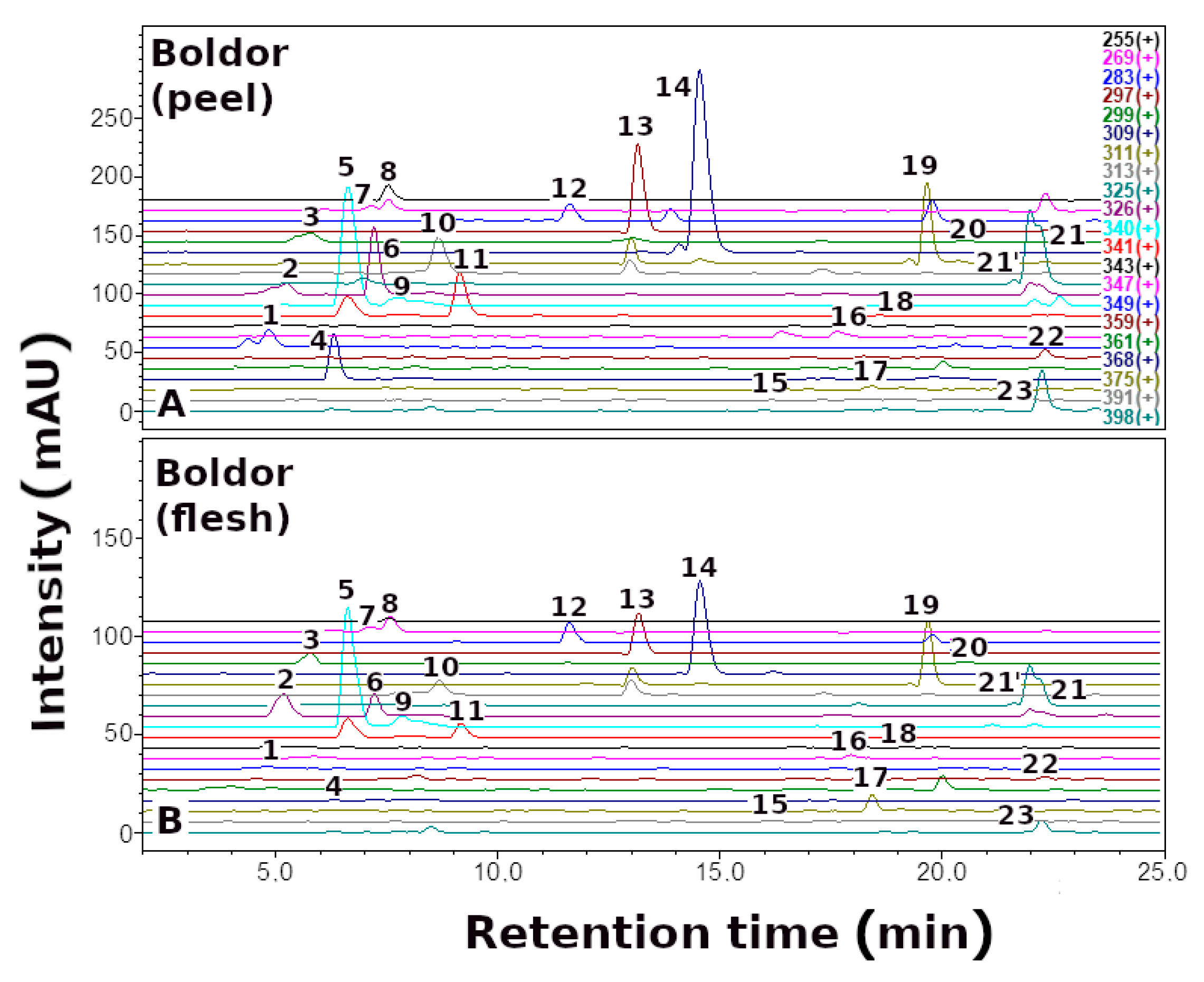
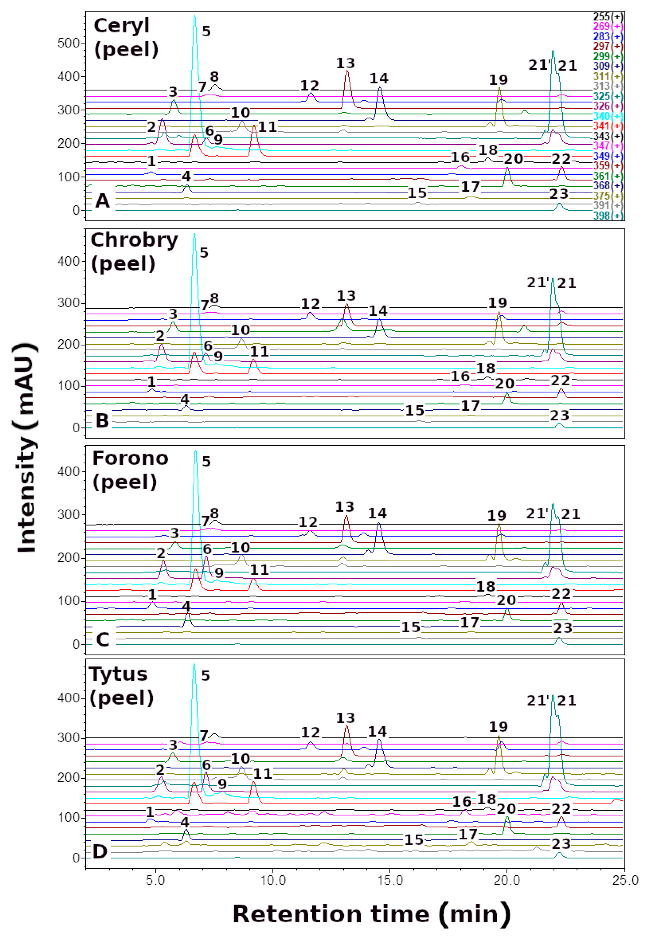
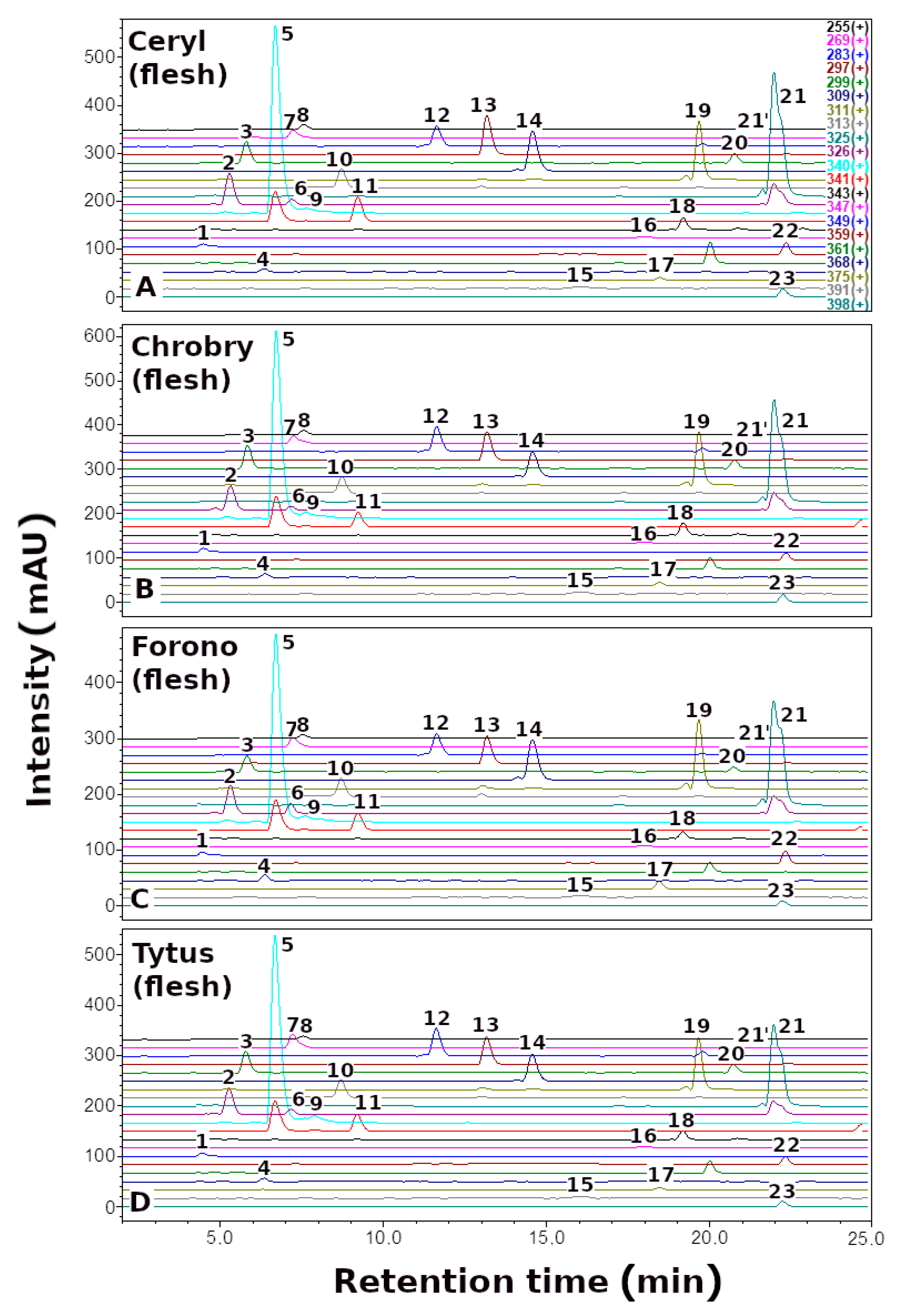
Here, we report the quantitative and qualitative (Figure 2, Figure 3 and Figure 4, Table 1) profile of betaxanthins of the peel and flesh extracts of four red (Ceryl, Chrobry, Forono, Tytus) and one yellow (Boldor) Beta vulgaris L. cultivars. These are popular edible beet cultivars.
The highest concentration of betaxanthins was noticed in the red variety Tytus peel (1231 mg/100 g Dry Extract (DE)), while the yellow variety Boldor flesh accumulated the lowest content of betaxanthins (317 mg/100 g DE). It might seem that yellow beetroot reflecting the yellow-orange color of betaxanthins will be their richer source than red beetroot. However, it is worth noting that in red beets, the yellow-orange betaxanthins are visually overpowered by the red-violet betacyanins, reflecting the color of the red beet but not its actual composition. The total concentration of betaxanthins in the extracts of B. vulgaris decreased in the following order, depending on the cultivar: Tytus > Ceryl > Chrobry > Forono > Boldor.
The peel of B. vulgaris, which is often considered a waste, appeared to be a richer source of betaxanthins compared to its flesh [46]. The highest concentration of betaxanthins in the peel extracts of B. vulgaris was observed in Tytus (1231 mg/100 g DE), while the lowest was noticed in Boldor (574 mg/100 g DE). In contrast, the lowest concentration of these pigments in B. vulgaris flesh extracts was also recorded in Boldor (317 mg/100 g DE) and the highest was in Chrobry (609 mg/100 g DE) (Table 2 and Table 3).
In addition to B. vulgaris, Portulaca grandiflora Hook. is also a proven rich source of betaxanthins. Our research team [33] investigated the quantitative and qualitative profile of betaxanthins from yellow, orange, red, and purple flowers of P. grandiflora. The content of betaxanthins decreased according to the following flower color order: orange > yellow > purple > red (982, 417, 323, and 162 mg/100 g DE, respectively) [33]. It should be noted that a comparable source of these pigments to the peel extract of the Ceryl cultivar (919 mg/100 g DE) is the orange flower extract of P. grandiflora (982 mg/100 g DE).
Qualitative analysis of B. vulgaris extracts by LC-DAD-ESI-MS showed the presence of 24 yellow-orange betaxanthins. The compounds identified, as well as their retention times, λmax values, and (M + H)+ data, are summarized in Table 1. Reference compounds semi-synthesized [34] or isolated of P. grandiflora fruit extracts [23] were used to identify betaxanthins.
Betaxanthins analysis in B. vulgaris peel and flesh extracts revealed an identical profile in red (Ceryl, Chrobry, Forono, and Tytus) and yellow (Boldor) cultivars; however, the profile of betaxanthins is more complex than previously described in yellow beetroot (Boldor) [42] and other varieties of B. vulgaris [39,40,45], comprising additionally arginine-Bx (4) and ornithine-Bx (6).
Kugler et al. [40] also identified a total of 24 betaxanthins in the yellow Boldor beet variety, including tyramine-Bx and aspartic-Bx, which were not identified in our study [40]. Our research team [33] identified 19 betaxanthins in P. grandiflora that are included in the B. vulgaris cultivars tested, with the exception of histamine-Bx [33]. It can be concluded that the tested cultivars of B. vulgaris are a much richer source of betaxanthins than the inedible P. grandiflora.
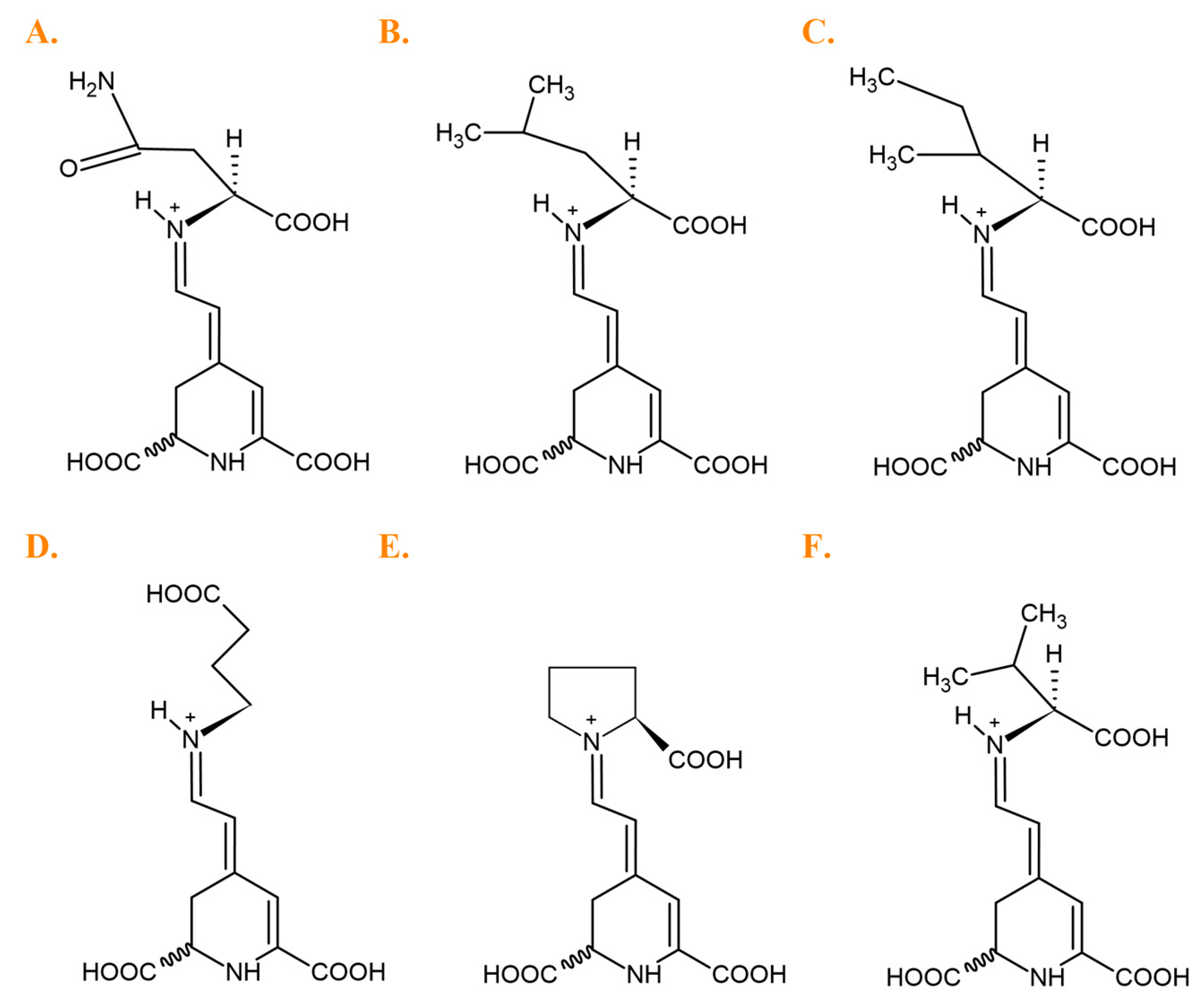
The dominant compounds in the B. vulgaris cultivars were glutamine-Bx (5), isoleucine-Bx (21′), leucine-Bx (21), γ-aminobutyric acid-Bx (13), proline-Bx (14) and valine-Bx (19) (Figure 5).
Definitely, the extract obtained from the peel of the Tytus variety is the best source of dominant compounds such as glutamine-Bx (5) (350.82 mg/100 g DE), isoleucine-Bx (21′) (159.96 mg/100 g DE), leucine-Bx (21) (150.47 mg/100 g DE), γ-aminobutyric acid-Bx (13) (70.06 mg/100 g DE) and valine-Bx (19) (74.29 mg/100 g DE), while yellow beet Boldor proline-Bx (14) (140.95 mg/100 g DE). The second best source of the above compounds is the extract obtained from the peel of the red beet of the Ceryl cultivars.

3.2. Antimicrobial Activity

Results of the antibacterial analysis of extracts obtained from five varieties of beetroot—Ceryl, Chrobry, Forono, Tytus, and Boldor are presented in Table 4, Table 5 and Table 6. It can be seen that the extracts showed diverse activity against the tested reference bacteria (MIC = 0.06–32 mg/mL) and Candida species (MIC = 0.125–8 mg/mL). The most sensitive strain was Micrococcus luteus ATCC 10240 (MIC = 0.06–8 mg/mL). Overall, only Gram-positive bacteria were susceptible to B. vulgaris extracts. This is also confirmed by studies by other authors from Canada who write that Gram-positive bacteria generally show higher sensitivity to red beetroot than Gram-negative ones [47].
According to our observations, it should be noted that the Tytus (peel) extract showed the highest activity (MIC = 0.125–0.5 mg/mL) against all Gram-positive bacteria except for Bacillus subtilis ATCC 6633 and B. cereus ATCC 10876, but according to other authors, MIC values of 0.100–0.625 mg/mL indicated moderate activity [48]. The pulp extract of this variety showed moderate activity against M. luteus ATCC 10240 (MIC = 0.5 mg/mL).
The extract of the Ceryl variety (peel) also showed significant activity (MIC = 0.06–0.125 mg/mL) against Staphylococcus aureus ATCC 25923 and M. luteus ATCC 10240 and B. subtilis ATCC 6633. It is worth noting that also the extract of Chrobry (peel) showed good activity (MIC = 0.125–0.5 mg/mL) against all Gram-positive bacteria except B. cereus ATCC 10876 (Table 4). The similarity results had the other author, who tested the in vitro antibacterial activity of the ethanol extract of beetroot against the food pathogens: S. aureus, B. cereus, and Escherichia coli by two methods: disc diffusion method and microdilution methods using selected Gram-positive and Gram-negative [49]. According to their results, Gram-positive bacteria: S. aureus and B. cereus demonstrated higher susceptibility than Gram-negative—E. coli. Their extract showed antibacterial activity against S. aureus (MIC = 0.75 mg/mL), one of the most common Gram-positive bacteria causing food poisoning. On the other hand, weak antimicrobial activity was found against E. coli (MIC = 1.5 mg/mL) [49].
The peel extracts of the Tytus, Chrobry, and Ceryl varieties were the only ones that showed activity against most of the Candida species tested, with MIC values ranging from 0.125 to 1 mg/mL. Candida parapsilosis ATCC 22019, C. albicans ATCC 10231, and C. albicans ATCC 2091 were the most sensitive to Tytus and Ceryl extracts (MIC = 0.25 mg/mL), while C. lusitaniae ATCC 3449 and C. tropicalis ATCC 1369 were only susceptible to Tytus peel extract (MIC = 0.125–0.25 mg/mL) (Table 6). Tenor et al. [50] undertook to investigate the antifungal activity of beetroot against two Candida species, C. albicans ATCC 10231, Rhizoctonia solani ATCC 13048; four fungi: Fusarium oxysporum ATCC 695, Cladosporium herbarum ATCC 11281, Botrytis cinerea ATCC 11542, Aspergillus flavus ATCC 15517. They showed, as in this work, that betalains-rich extracts from red pitahaya (Hylocereus polyrhizus) were active against fungi (C. albicans, R. solani) at 0.125–0.25 mg/mL and fungi (F. oxysporum, C. herbarum, B. cinerea, A. flavus) at 0.5 mg/mL. The results of these researchers indicated that the peel, which is inedible and waste from these plants, can be subjected to simple purification methods to obtain extracts and/or various fractions and can be further used for the preparation of health products and supplements, e.g., for food preservation.
As shown in Table 4, the extract of the Forono variety (peel) showed versatile activity against Gram-positive bacteria (MIC = 0.006–16 mg/mL) with the highest activity against B. subtilis ATCC 6633 and B. cereus ATCC 10876 with MIC = 0.06 mg/mL, one of the most common Gram-positive bacteria causing food poisoning. In addition, it showed activity against fungi with MIC values ranging from 0.5 to 4 mg/mL, with the highest activity against C. parapsilosis ATCC 22019 (MIC = 0.5 mg/mL) (Table 6).
On the other hand, extracts from the Boldor variety, both from the peel and pulp, showed weak activity against all tested microorganisms (MIC = 2–16 mg/mL). In general, extracts obtained from beetroot peels were characterized by higher antimicrobial activity compared to extracts obtained from the pulp. All extracts showed low activity against all Gram-negative bacteria, with MIC values ranging from 2 to 32 mg/mL (Table 5).
Antimicrobial substances are usually considered bactericidal or fungicidal if the MBC/MIC or MFC/MIC ratio is ≤4. If the MBC/MIC or MFC/MIC ratio is >4, antimicrobial substances are usually considered to be bacteriostatic or fungistatic [37]. Based on the results presented, it can be concluded that most of the beetroot extracts showed bactericidal activity (MBC/MIC = 1–4) against most bacteria and fungi, and the extracts of the Tytus, Chrobry, Ceryl, and Forono varieties showed bacteriostatic activity (MBC/MIC = 8–64) and fungistatic (MFC/MIC = 8–32) to selected microorganisms with low MIC values.
The MIC values for the reference antimicrobials were 1 µg/mL fluconazole for C. albicans ATCC 10231, 1 µg/mL vancomycin for S. aureus ATCC 29213 and 0.015 µg/mL ciprofloxacin for E. coli ATCC 25922.

3.3. Correlation between Phytochemical Composition and Antimicrobial Activity

Correlations between identified betaxanthins and antimicrobial activity were checked for all extracts obtained from five varieties of B. vulgaris. The values of the correlation coefficient (R) were calculated based on the obtained MIC values and the absolute peak area of each assigned peak from the chromatograms of the individual betaxanthins. A negative R-value refers to decreasing in the MIC parameters with an increase in the component peak value (negative correlation), which proves the positive impact on antimicrobial properties.
All identified betaxanthins showed a negative correlation against one strain of Gram-positive bacteria S. epidermidis ATCC 12228 and two strains of Gram-negative bacteria P. mirabilis ATCC 1,453 and K. pneumoniae ATCC 13883. S. epidermidis are natural pathogens found on human skin, increasingly resistant to antibiotics and contributing to the occurrence of infections [51]. P. mirabilis is the cause of 90% of Proteus infections and occurs mainly in people with weakened immune systems [52]. K. pneumoniae is the fourth most common cause of Gram-negative hospital-associated infections, including urinary tract infections, pneumonia, and wound infections [53]. Therefore, it is worth noting that the betaxanthins identified in B. vulgaris extracts inhibit the activity of the above bacterial strains to some extent and may be a good candidate for further research in this respect.
Asparagine-Bx (2), ethanolamine-Bx (8), and phenylalanine-Bx (22) exhibited negative correlation against all strains of Gram-positive bacteria and fungi, while asparagine-Bx (2), glutamine-Bx (5), threonine-Bx (10), isoleucine-Bx (21′), leucine-Bx (21) and phenylalanine-Bx (22) against all strains of Gram-negative bacteria. On the other hand, in relation to all tested strains of fungi, a negative correlation was also shown by compounds such as arginine-Bx (2), ornithine-Bx (6), glutamic acid-Bx (11), γ-aminobutyric acid-Bx (13), proline-Bx (14), dopamine-Bx (16), 3-methoxytyramine-Bx (20), isoleucine-Bx (21′), leucine-Bx (21), and tryptophan-Bx (23).
It is also worth mentioning that glutamic acid-Bx (11), γ-aminobutyric acid-Bx (13), and 3-methoxytyramine-Bx (20) exhibited negative correlation against all strains of Gram-positive bacteria except B. cereus ATCC 10876. In contrast, tryptophan-Bx (23) and proline-Bx (14) showed positive effects against all strains of Gram-positive bacteria except M. luteus ATCC 10240. The negative correlation against all strains of Gram-positive bacteria was also exhibited arginine-Bx (2) and ornithine-Bx (6) except E. faecalis ATCC 29212.
The dominant betaxanthins (5, 13, 14, 21′, and 21) in B. vulgaris extracts showed a positive effect against all Gram-negative bacteria or fungi except valine-Bx (19). Nevertheless, other identified betaxanthins also had a positive effect on microbial activity against selected microorganisms. This shows that probably the concentration of individual betaxanthins did not significantly affect their potency against microorganisms.
A positive correlation against all strains of Gram-positive bacteria was exhibited by histidine-Bx (1), glycine-Bx (7), alanine-Bx (12), and methionine-Bx (18) except S. epidermidis ATCC 12228 and M. luteus ATCC 10240. Histidine-Bx (1), glycine-Bx (7), and additionally tyrosine-Bx (17) also showed a positive correlation with all strains of Gram-negative bacteria except P. mirabilis ATCC 12453 and K. pneumoniae ATCC 13883. The above five betaxanthins (1, 7, 12, 17, and 18) and serine-Bx (3) also did not affect activity against all fungal strains.
Previously we showed six betaxanthins from Portulaca grandiflora Hook., such as glutamine-Bx (5), ethanolamine-Bx (8), glutamic acid-Bx (11), γ-aminobutyric acid-Bx (13), proline-Bx (14), and phenylalanine-Bx (22), also identified in B. vulgaris, showed negative correlation against the same Gram-positive bacterial strains, confirming the effect of these compounds on their antimicrobial potential [33]. Glutamine-Bx (5) showed a positive effect against three fungal strains such as C. albicans ATCC 10231, C. glabrata ATCC 90,030, and C. krusei ATCC 14243. However, ten betaxanthins (5, 8, 10, 11, 13, 14, 19, 21′, 21, and 22) found in P. grandiflora and B. vulgaris also showed a positive effect on activity against C. albicans ATCC 10231.
Recently, an interesting group of antimicrobial agents with strong activity against resistant bacteria and fungi are antimicrobial peptides (AMP) consisting of various amino acids. The designed sequence with strong antimicrobial activity against the gram-positive bacteria S. aureus ATCC 25923 and E. faecalis ATCC 29212 contains 24 amino acid residues, in which the lysine, histidine, and serine residues were replaced with arginine, and also the hydrophobic phenylalanine was replaced with tryptophan [54]. Such amino acids are present in the betaxanthins (4 and 23) in B. vulgaris, with a significant effect on the activity against these two strains of Gram-positive bacteria.
An amino acid inhibitor consisting of glutamic acid sulfonamides was also synthesized, which showed good inhibitory activity against E. coli ATCC 25922 [55]. In B. vulgaris, glutamic acid-Bx (11) consisting of the above amino acid also showed a strong effect on the activity against this strain of Gram-negative bacteria.

3.4. Cytotoxicity and Anticancer Selectivity

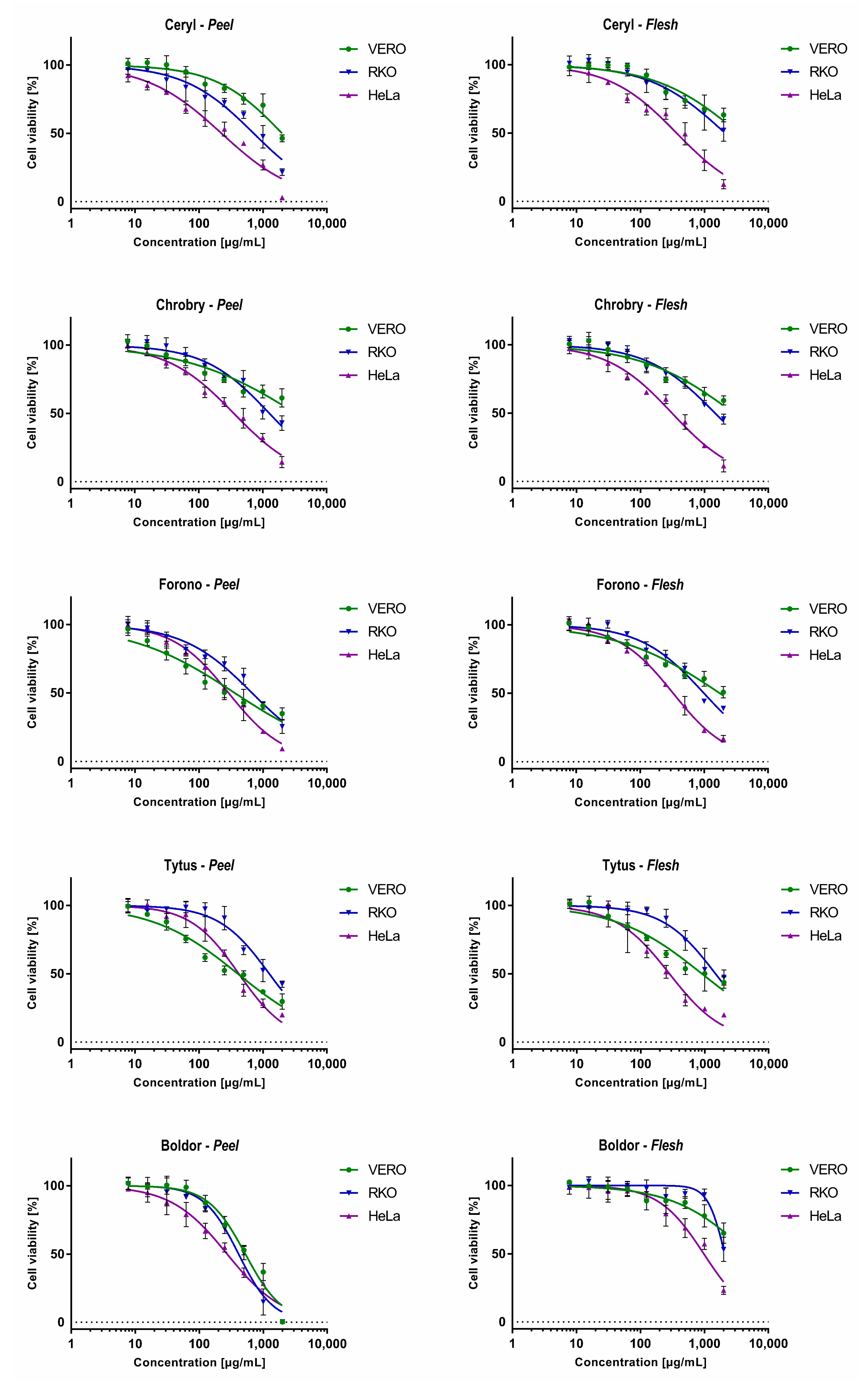
The cytotoxicity was evaluated using an MTT-based assay towards non-cancerous VERO cells and cancer cell lines originating from colon cancer (RKO) and cervical adenocarcinoma (HeLa). The calculated CC50 (50% cytotoxic concentration) values are presented in Table 7, and the dose-response effects are in Figure 6.
Overall, the extracts obtained from the flesh of tested cultivars were less toxic to non-cancerous cells than the peel extracts obtained from the same plant material, except the Chrobry cultivar. Interestingly, the extracts of the peel and flesh of the Ceryl and Chrobry cultivars showed lower toxicity on VERO cells than those obtained from the Forono and Tytus cultivars. In the case of the Boldor cultivar, extracts from flesh showed the lowest observed toxicity among all tested samples with CC50 of 4948.6 μg/mL, and the extracts from peel were significantly more toxic (CC50 527.7 μg/mL). Taking into account the classification of plant extracts cytotoxicity [56,57] extracts from tested cultivars can be regarded as non-cytotoxic (CC50 > 500 μg/mL) to VERO cells, except peel extracts from Forono and Tytus cultivars where weak cytotoxic activity (CC50 201–500 μg/mL) was observed. The peel extract from the Boldor cultivar showed weak cytotoxicity towards both cancer cell lines without showing a cytotoxic effect on non-cancerous cells. HeLa cells were noticeably more sensitive to most of the extracts tested than VERO and RKO.
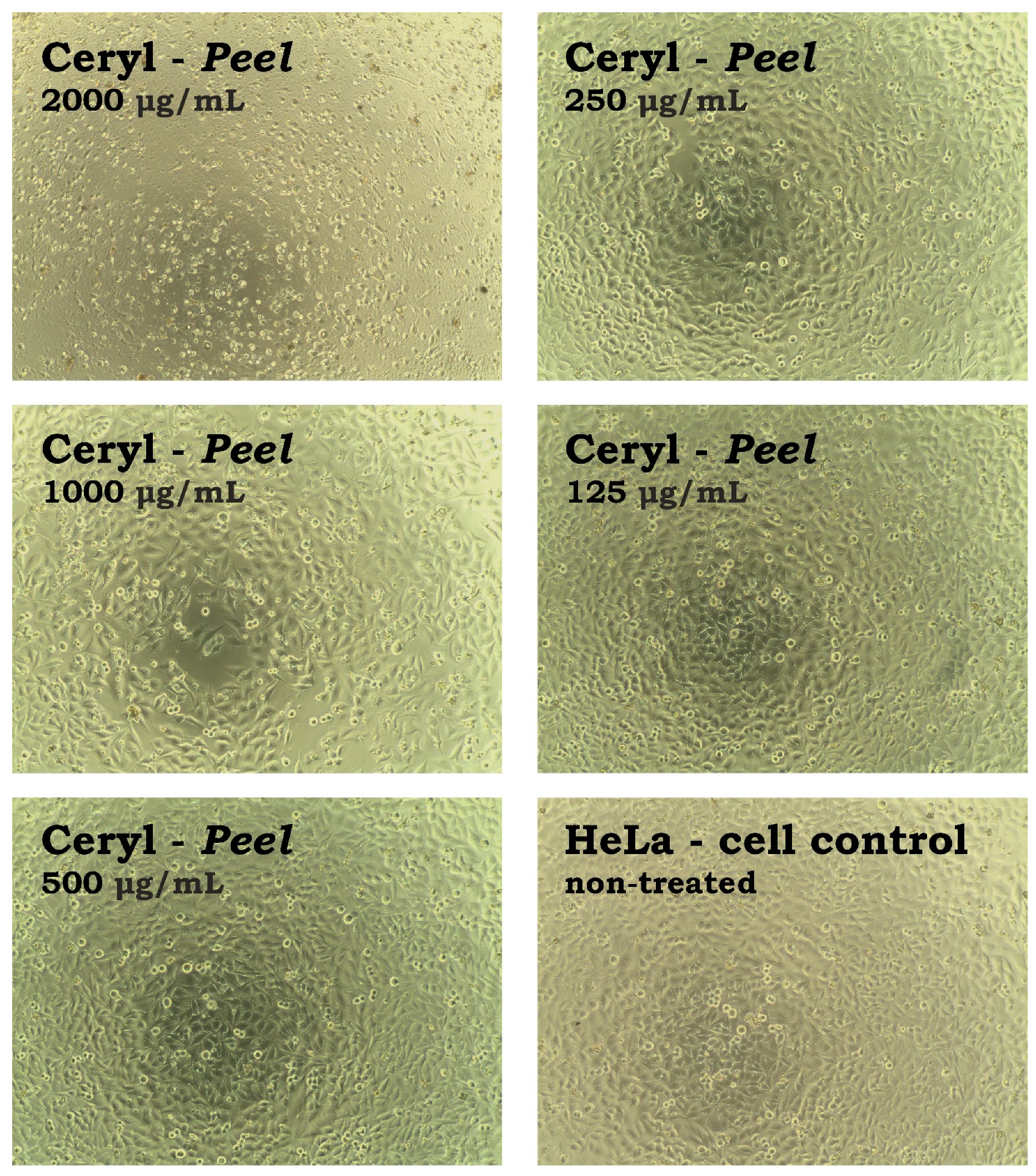
The anticancer potential was evaluated by comparing the CC50 values obtained on cancer cells with those observed on VERO cells. The calculated SI (selectivity index) is shown in Table 7. Extracts from the peel of Ceryl and Chrobry cultivars and flesh of Boldor cultivar showed potential selectivity towards colon cancer cells with SI between 2.37 and 2.78. Additionally, extracts from both the peel and flesh of the Ceryl and Chrobry cultivars showed highly selective cytotoxicity to cervical cancer cells with SI between 8.84 and 9.39. The influence of extract from the peel of the Ceryl cultivar on the morphology of the HeLa cell monolayer is presented in Figure 7.
At the concentration of 2000 μg/mL, the monolayer was destroyed, whereas, at half-lower concentration, there were some HeLa cells present, but the monolayer was noticeably less dense than the cell control. Further dilutions resulted in the increase of the confluence of the HeLa monolayer. Selectivity towards HeLa was also exerted by extracts from the flesh of Forono, Tytus, and Boldor cultivars (SI 3.22–5.35). Despite the potential anticancer selectivity, it must be underlined that this selectivity was observed at relatively high 50% cytotoxic concentrations. According to published literature [56,57], high cytotoxic activity and anticancer potential can be reported for plant extracts showing CC50 < 20 μg/mL.
Hence, the B. vulgaris extracts studied herein, despite showing potential selectivity towards cancer cell lines, failed to exert significant anticancer potential. Interestingly, Romero et al. [58] reported that the beetroot and leaf extracts decreased the viability of HeLa cells and potentiated the anticancer effects of cisplatin and rapamycin. Additionally, the beetroot extract 100 μg/mL induced early apoptosis in HeLa cells, decreased the cell size, and promoted cell death [58].
Clement et al. [59] reported that beetroot juice, or blended beetroot, was the second most popular functional food used by patients suffering from prostate, breast, and colorectal cancer, in combination with anticancer drugs [59]. Thanks to the multi-target action of B. vulgaris phytochemicals, including antioxidant activity, direct inhibition of proliferation, downregulation of the cancer pro-survival genes, ant-angiogenic activity, and inhibition of anti-apoptotic genes, they can be used in combination with conventional anticancer drugs to increase their efficacy and reduce toxicity, and overcome the multidrug resistance of cancer cells [60]. Moreover, B. vulgaris root extracts were shown to exert a chemopreventative role in cancer development [1,61,62].

3.5. Correlation between Phytochemical Composition and Anticancer Activity

Table 8 presents the correlation coefficients (R) calculated on the basis of the absolute peak area of each assigned peak from the chromatograms of the individual betaxanthins and the anticancer activity expressed by the CC50 parameter. The positive effect of the compounds on anticancer activity is represented by the negative value of the R coefficient, which refers to the decrease in the CC50 parameter with the increase in the component peak value (negative correlation).
The strongest positive effect on the activity against cancer cells derived from colon cancer (RKO) and cervical adenocarcinoma (HeLa) cell lines was shown by arginine-Bx (4), proline-Bx (14) and tryptophan-Bx (23). It should be noted that almost all identified betaxanthins in B. vulgaris exhibited a negative correlation against HeLa cell lines, with the exception of dopamine-Bx (16) and tyrosine-Bx (17). In contrast, negative correlation against RKO cell lines showed only ten betaxanthins such as asparagine-Bx (2), arginine-Bx (4), ornithine-Bx (6), ethanolamine-Bx (8), glutamic acid-Bx (11), γ-aminobutyric acid-Bx (13), proline -Bx (14), leucine-Bx (21), phenylalanine-Bx (22), and tryptophan-Bx (23).
Dopamine-Bx (16) and tyrosine-Bx (17) were the only ones to show a positive correlation against the two tested cell lines, RKO and HeLa, which suggests that they do not affect the anticancer activity against these cancer cells. Positive correlation against RKO cell lines was shown by as many as fourteen betaxanthins from B. vulgaris. In conclusion, identified betaxanthins definitely showed a stronger effect on anticancer activity against HeLa cell lines than against RKO cell lines.
According to the literature, among the betalains tested, tryptophan-Bx (23) showed the greatest effect on the size of the tumor by reducing its size by 56.4% in the animal model of Caenorhabditis elegans (cancer strain JK1466) and extended the animal’s lifespan by 9.3%, which indicates high effectiveness and low toxicity [63]. According to our results, tryptophan-Bx (23) showed strong activity against HeLa and RKO cell lines, so further in vitro and in vivo studies in this direction on the pure compound are also worth considering. It is worth noting that the above compound (23) may prove to be a potential chemotherapeutic with possible importance in chemoprevention and treatment strategies for colon cancer and cervical adenocarcinoma.
Arginine-Bx (4) is a product of betalamic acid with the amino acid (arginine). L-arginine is a substrate of nitric oxide synthase. Macrophages associated with early-stage cancer act to inhibit tumor growth by secreting nitric oxide. Nitric oxide produced from L-arginine by macrophages has been confirmed to work to enhance the effects of the anticancer drug (doxorubicin). This suggests that co-administration of L-arginine with doxorubicin would be an effective treatment to enhance chemotherapy [64]. Many studies confirm the anticancer activity of arginine and often use it as an additive to enhance the effects of potential anticancer drugs [64,65,66,67,68]. In B. vulgaris, arginine-Bx (4) showed a strong effect on the RKO and HeLa cell lines; therefore, it may also be a potential anticancer drug candidate like L-alanine.
According to the literature, proline-Bx (14), more popular under the name of indicaxanthin, can act as a neuromodulator, anti-inflammatory and anticancer agent in the prevention or treatment of neurological diseases and related cancer pathologies [69]. Combining phytochemicals with chemotherapeutic agents has become very popular recently as a new cancer treatment strategy to overcome drug toxicity and resistance to natural compounds. It has also been shown that proline-Bx (14) individually exhibits growth inhibitory effects on HeLa cervical cancer cells and in combination with cisplatin, collaborates in producing enhanced anticancer activity against these cells [2]. In conclusion, these studies confirm previous reports on the anticancer activity of this compound.

4. Conclusions

Obtained results enabled the characterization of 24 betaxanthins in all studied samples, of which two new betaxanthins (arginine-Bx and ornithine-Bx) were detected for the first time in Beta vulgaris L. cultivars. Glutamine-Bx, isoleucine-Bx, leucine-Bx, γ-aminobutyric acid-Bx, proline-Bx, and valine-Bx were dominant compounds in the cultivars studied. The content of betaxanthins was noticed in the peel and flesh of five cultivars of B. vulgaris for the first time and decreased in the following order, depending on the cultivar: Tytus > Ceryl > Chrobry > Forono > Boldor. The highest total content of betaxanthins (1231 mg/100 g Dry Extract) was observed for the Tytus (peel) cultivar. The Boldor (flesh) accumulated the lowest betaxanthin content (317 mg/100 g Dry Extract). Importantly, the peel of B. vulgaris, which are often considered waste, appeared to be a richer source of betaxanthins compared to its flesh, indicating wastes as a valuable and rich source of active compounds.
Tytus and Chrobry showed a moderate or good bactericidal effect, especially against the majority of Gram-positive bacteria. Ceryl peel extract was most active against Staphylococcus aureus and Micrococcus luteus at minimal inhibitory concentration (MIC) = 0.06 mg/mL and fungi with MIC = 0.25–0.5 mg/mL and was not cytotoxic (CC50 =1992 μg/mL, CC50 – 50% cytotoxic concentration). Forono peel showed the highest activity against Micrococcus luteus and Bacillus subtilis at MIC = 0.06 mg/mL. The good and moderate activity against most of the tested fungi was shown by the peel of Tytus and Ceryl (MIC = 0.125–0.5 mg/mL).
It should be noted that Gram-positive bacteria were more sensitive to the antibacterial effect of beetroot extracts, which is related to the structure of their cell wall consisting of a thick permeable layer of murein. However, it was noted that Gram-negative bacteria were resistant to the antimicrobial effect of beetroot extracts. This may be related not only to the content of active substances in the tested extracts but may also be influenced by the external structure of this group of bacteria. Namely, the cell wall of gram-negative bacteria consists of a thin layer of murein, over which there is an additional thick impermeable outer membrane made of phospholipids and hydrophilic lipopolysaccharides. This makes it difficult for extracts to overcome such a barrier of macromolecules and hydrophobic compounds.
All identified betaxanthins might have possessed bactericidal effect against one strain of Gram-positive bacteria Staphylococcus epidermidis ATCC 12228 and two strains of Gram-negative bacteria Proteus mirabilis ATCC 12453 and Klebsiella pneumoniae ATCC 13883, respectively. Asparagine-Bx and phenylalanine-Bx might be active against all bacteria and yeast tested. The remaining betaxanthins were sensitive to selected strains.
B. vulgaris extracts are safe and has weak or no cytotoxic activity. Our results indicate that studied extracts show stronger activity against cervical adenocarcinoma (HeLa) cells than the colon cancer cell line (RKO). Similarly, betaxanthins show a stronger effect on anticancer activity against HeLa cell lines than against RKO cell lines. Arginine-Bx, proline-Bx and tryptophan-Bx might be considered as betaxanthins with the highest anticancer potential due to the strongest activity against RKO and HeLa cell lines, what was indicated by correlation analysis.
Studies indicate that betaxanthins have significant potential application, so it is worth continuing research on selected compounds.

Author Contributions

Conceptualization, A.S.-K. and A.T.; methodology, A.S.-K. and A.T.; software, A.S.-K., A.T., A.G. and Ł.Ś.; validation, A.S.-K., A.T., A.G. and Ł.Ś.; formal analysis, A.S.-K. and A.T.; investigation, A.S.-K., A.T., A.G., Ł.Ś. and A.B.; resources, A.S.-K., A.T., A.G., Ł.Ś. and A.B.; data curation, A.S.-K. and A.T.; writing—original draft preparation, A.S.-K., A.T., A.G. and Ł.Ś.; writing—review and editing, A.S.-K., A.T., A.G., Ł.Ś. and K.S.-W.; visualization, A.S.-K., A.T., A.G. and Ł.Ś.; supervision, A.S.-K.; project administration, A.S.-K.; funding acquisition, A.S.-K. All authors have read and agreed to the published version of the manuscript.

Funding

This research was financed by the Polish National Science Centre for years 2019–2020; Project No. 2019/03/X/ST4/00968.

Institutional Review Board Statement

Not applicable.

Informed Consent Statement

Not applicable.

Data Availability Statement

Data is contained within the article.

Conflicts of Interest

The authors declare no conflict of interest.

References

  1. Tan, M.L.; Hamid, S.B.S. Beetroot as a potential functional food for cancer chemoprevention, a narrative review. J. Cancer Prev. 2021, 26, 1. [Google Scholar] [CrossRef] [PubMed]
  2. Allegra, M.; D’Anneo, A.; Frazzitta, A.; Restivo, I.; Livrea, M.A.; Attanzio, A.; Tesoriere, L. The phytochemical indicaxanthin synergistically enhances cisplatin-induced apoptosis in Hela cells via oxidative stress-dependent P53/P21waf1 axis. Biomolecules 2020, 10, 994. [Google Scholar] [CrossRef]
  3. Attanzio, A.; Restivo, I.; Tutone, M.; Tesoriere, L.; Allegra, M.; Livrea, M.A. Redox properties, bioactivity and health effects of indicaxanthin, a Tablebioavailable phytochemical from Opuntia ficus indica, L.: A critical review of accumulated evidence and perspectives. Antioxidants 2022, 11, 2364. [Google Scholar] [CrossRef] [PubMed]
  4. Stücheli, P.; Sieber, S.; Fuchs, D.W.; Scheller, L.; Strittmatter, T.; Saxena, P.; Gademann, K.; Fussenegger, M. Genetically encoded betaxanthin-based small-molecular fluorescent reporter for Mammalian cells. Nucleic Acids Res. 2020, 48, 1–12. [Google Scholar] [CrossRef] [PubMed]
  5. Allegra, M.; Tutone, M.; Tesoriere, L.; Almerico, A.M.; Culletta, G.; Livrea, M.A.; Attanzio, A. Indicaxanthin, a multi-target natural compound from Opuntia ficus-indica fruit: From its poly-pharmacological effects to biochemical mechanisms and molecular modelling studies. Eur. J. Med. Chem. 2019, 179, 753–764. [Google Scholar] [CrossRef] [PubMed]
  6. Shinde, P.R.; Wagh, K.R.; Patil, P.S.; Bairagi, V.A. Pharmacognostic standardization and antibacterial potential of aerial herbs of Portulaca grandiflora Hooker (Portulaceae). World J. Pharm. Sci. 2014, 2, 1871–1885. [Google Scholar]
  7. De Oliveira, S.P.A.; de Nascimento, H.M.A.; Sampaio, K.B.; de Souza, E.L. A review on bioactive compounds of beet (Beta vulgaris L. subsp. vulgaris) with special emphasis on their beneficial effects on gut microbiota and gastrointestinal health. Crit. Rev. Food Sci. Nutr. 2021, 61, 2022–2033. [Google Scholar]
  8. Chavalittumrong, P.; Sriwanthana, B.; Rojanawiwat, A.; Kijphati, R.; Jitjuk, B.; Treesangsri, W.; Phadungpat, S.; Bansiddhi, J.; Bunjob, M. Safety of the aqueous extract of Portulaca grandiflora Hook in healthy volunteers. Safety 2007, 29, 1. [Google Scholar]
  9. Dos Santos Baião, D.; Silva de Freitas, C.; Paes Gomes, L.; da Silva, D.; Carolina, N.T.F.; Correa, A.; Ribeiro Pereira, P.; Mere del Aguila, E.; Margaret Flosi Paschoalin, V. Polyphenols from root, tubercles and grains cropped in Brazil: Chemical and nutritional characterization and their effects on human health and diseases. Nutrients 2017, 9, 1044. [Google Scholar] [CrossRef]
  10. Kumorkiewicz-Jamro, A.; Świergosz, T.; Sutor, K.; Spórna-Kucab, A.; Wybraniec, S. Multi-Colored shades of betalains: Recent advances in betacyanin chemistry. Nat. Prod. Rep. 2021, 38, 2315–2346. [Google Scholar] [CrossRef]
  11. Spórna-Kucab, A.; Bernaś, K.; Grzegorczyk, A.; Malm, A.; Skalicka-Woźniak, K.; Wybraniec, S. Liquid chromatographic techniques in betacyanin isomers separation from Gomphrena globosa L. flowers for the determination of their antimicrobial activities. J. Pharm. Biomed. Anal. 2018, 161, 83–93. [Google Scholar] [CrossRef] [PubMed]
  12. Spórna-Kucab, A.; Kumorkiewicz, A.; Szmyr, N.; Szneler, E.; Wybraniec, S. Separation of betacyanins from flowers of Amaranthus cruentus L. in a polar solvent system by high-speed counter-current chromatography. J. Sep. Sci. 2019, 42, 1676–1685. [Google Scholar] [CrossRef] [PubMed]
  13. Osorio-Esquivel, O.; Ortiz-Moreno, A.; Álvarez, V.B.; Dorantes-Álvarez, L.; Giusti, M.M. Phenolics, betacyanins and antioxidant activity in Opuntia joconostle fruits. Food Res. Int. 2011, 44, 2160–2168. [Google Scholar] [CrossRef]
  14. Rahimi, P.; Abedimanesh, S.; Mesbah-Namin, S.A.; Ostadrahimi, A. Betalains, the nature-inspired pigments, in health and diseases. Crit. Rev. Food Sci. Nutr. 2019, 59, 2949–2978. [Google Scholar] [CrossRef]
  15. Khan, M.I. Plant betalains: Safety, antioxidant activity, clinical efficacy, and bioavailability. Compr. Rev. Food Sci. Food Saf. 2016, 15, 316–330. [Google Scholar] [CrossRef] [PubMed] [Green Version]
  16. Pietrzkowski, Z.; Nemzer, B.; Spórna, A.; Stalica, P. Influence of betalain-rich extract on reduction of discomfort associated with osteoarthritis. New Med. 2010, 1, 12–17. [Google Scholar]
  17. Spórna-Kucab, A.; Wróbel, N.; Kumorkiewicz-Jamro, A.; Wybraniec, S. Separation of betacyanins from Iresine herbstii Hook. Ex Lindl. leaves by high-speed countercurrent chromatography in a polar solvent system. J. Chrom. A 2020, 1626, 461370. [Google Scholar] [CrossRef] [PubMed]
  18. Spórna-Kucab, A.; Milo, A.; Kumorkiewicz, A.; Wybraniec, S. Studies on polar high-speed counter-current chromatographic systems in separation of amaranthine-type betacyanins from Celosia species. J. Chrom. B 2018, 1073, 96–103. [Google Scholar] [CrossRef]
  19. Spórna-Kucab, A.; Jagodzińska, J.; Wybraniec, S. Separation of betacyanins from purple flowers of Gomphrena globosa L. by ion-pair high-speed counter-current chromatography. J. Chrom. A 2017, 1489, 51–57. [Google Scholar] [CrossRef]
  20. Spórna-Kucab, A.; Hołda, E.; Wybraniec, S. High-speed counter-current chromatography in separation of betacyanins from flowers of red Gomphrena globosa L. cultivars. J. Chrom. B 2016, 1033, 421–427. [Google Scholar] [CrossRef]
  21. Wybraniec, S.; Stalica, P.; Sporna, A.; Mizrahi, Y. Profiles of betacyanins in epidermal layers of grafted and light-stressed cacti studied by LC-DAD-ESI-MS/MS. J. Agric. Food Chem. 2010, 58, 5347–5354. [Google Scholar] [CrossRef] [PubMed]
  22. Wybraniec, S.; Stalica, P.; Jerz, G.; Klose, B.; Gebers, N.; Winterhalter, P.; Spórna, A.; Szaleniec, M.; Mizrahi, Y. Separation of polar betalain pigments from cacti fruits of Hylocereus polyrhizus by ion-pair high-speed countercurrent chromatography. J. Chrom. A 2009, 1216, 6890–6899. [Google Scholar] [CrossRef] [PubMed]
  23. Nemzer, B.; Pietrzkowski, Z.; Spórna, A.; Stalica, P.; Thresher, W.; Michałowski, T.; Wybraniec, S. Betalainic and nutritional profiles of pigment-enriched red beet root (Beta vulgaris L.) dried extracts. Food Chem. 2011, 127, 42–53. [Google Scholar] [CrossRef]
  24. Wybraniec, S.; Nowak-Wydra, B.; Mitka, K.; Kowalski, P.; Mizrahi, Y. Minor betalains in fruits of Hylocereus species. Phytochemistry 2007, 68, 251–259. [Google Scholar] [CrossRef] [PubMed]
  25. Khan, M.I.; Giridhar, P. Plant betalains: Chemistry and biochemistry. Phytochemistry 2015, 117, 267–295. [Google Scholar] [CrossRef] [PubMed]
  26. Christinet, L.; Burdet, F.X.; Zaiko, M.; Hinz, U.; Zrÿd, J.-P. Characterization and functional identification of a novel plant 4,5-extradiol dioxygenase involved in betalain pigment biosynthesis in Portulaca grandiflora. Plant Physiol. 2004, 134, 265–274. [Google Scholar] [CrossRef] [Green Version]
  27. Azeredo, H.M. Betalains: Properties, sources, applications, and stability—A review. Int. J. Food Sci. Technol. 2009, 44, 2365–2376. [Google Scholar] [CrossRef] [Green Version]
  28. Spórna-Kucab, A.; Wybraniec, S. High-speed counter-current chromatography in separation and identification of saponins from Beta vulgaris L. cultivar red sphere. Polish J. Food Nutr. Sci. 2020, 70, 67–74. [Google Scholar] [CrossRef]
  29. Spórna-Kucab, A.; Tekieli, A.; Skalicka-Woźniak, K.; Grzegorczyk, A.; Świergosz, T.; Wybraniec, S. Characterization of triterpene saponin composition of white, yellow and red beetroot (Beta vulgaris L.). Pol. J. Food Nutr. Sci. 2022, 72, 159–170. [Google Scholar] [CrossRef]
  30. Spórna-Kucab, A.; Jerz, G.; Kumorkiewicz-Jamro, A.; Tekieli, A.; Wybraniec, S. High-speed countercurrent chromatography for isolation and enrichment of betacyanins from fresh and dried leaves of Atriplex hortensis L. var. “Rubra”. J. Sep. Sci. 2021, 44, 4222–4236. [Google Scholar] [CrossRef]
  31. Gandía-Herrero, F.; García-Carmona, F.; Escribano, J. Development of a protocol for the semi-synthesis and purification of betaxanthins. Phytochem. Anal. 2006, 17, 262–269. [Google Scholar] [CrossRef] [PubMed]
  32. Cabanes, J.; Gandía-Herrero, F.; Escribano, J.; García-Carmona, F.; Jiménez-Atiénzar, M. One-step synthesis of betalains using a novel betalamic acid derivatized support. J. Agric. Food Chem. 2014, 62, 3776–3782. [Google Scholar] [CrossRef] [PubMed]
  33. Spórna-Kucab, A.; Tekieli, A.; Grzegorczyk, A.; Świątek, Ł.; Rajtar, B.; Skalicka-Woźniak, K.; Starzak, K.; Nemzer, B.; Pietrzkowski, Z.; Wybraniec, S. Metabolite profiling analysis and the correlation with biological activity of betalain-rich Portulaca grandiflora Hook. extracts. Antioxidants 2022, 11, 1654. [Google Scholar] [CrossRef] [PubMed]
  34. Stintzing, F.C.; Schieber, A.; Carle, R. Identification of betalains from yellow beet (Beta vulgaris L.) and cactus pear [Opuntia ficus-indica (L.) Mill.] by high-performance liquid chromatography−electrospray ionization mass spectrometry. J. Agric. Food Chem. 2002, 50, 2302–2307. [Google Scholar] [CrossRef]
  35. Stintzing, F.C.; Schieber, A.; Carle, R. Evaluation of colour properties and chemical quality parameters of cactus juices. Eur. Food Res. Technol. 2003, 216, 303–311. [Google Scholar] [CrossRef]
  36. EUCAST. European Committee for Antimicrobial Susceptibility Testing (EUCAST) of the European Society of Clinical Microbiology and Infectious Diseases (ESCMID): Determination of Minimum Inhibitory Concentrations (MICs) of Antibacterial Agents by Broth Dilution. Clin. Microbiol. Infect. Dis. 2003, 40, 1–7. [Google Scholar]
  37. Malm, A.; Grzegorczyk, A.; Biernasiuk, A.; Baj, T.; Rój, E.; Tyśkiewicz, K.; Dębczak, A.; Stolarski, M.J.; Krzyżaniak, M.; Olba-Zięty, E. Could Supercritical Extracts from the Aerial Parts of Helianthus salicifolius A. Dietr. and Helianthus tuberosus L. Be Regarded as Potential Raw Materials for Biocidal Purposes? Agriculture 2020, 11, 10. [Google Scholar] [CrossRef]
  38. Pecio, Ł.; Kozachok, S.; Saber, F.R.; Garcia-Marti, M.; El-Amier, Y.; Mahrous, E.A.; Świątek, Ł.; Boguszewska, A.; Skiba, A.; Elosaily, A.H.; et al. Metabolic profiling of Ochradenus baccatus Delile. utilizing UHPLC-HRESIMS in relation to the in vitro biological investigations. Food Chem. 2023, 412, 135587. [Google Scholar] [CrossRef]
  39. Sawicki, T.; Bączek, N.; Wiczkowski, W. Betalain Profile, Content and Antioxidant Capacity of Red Beetroot Dependent on the genotype and root part. J. Funct. Foods 2016, 27, 249–261. [Google Scholar] [CrossRef]
  40. Kugler, F.; Graneis, S.; Stintzing, F.C.; Carle, R. Studies on betaxanthin profiles of vegetables and fruits from the Chenopodiaceae and Cactaceae. Z. Naturforsch. C 2007, 62, 311–318. [Google Scholar] [CrossRef] [Green Version]
  41. Spórna, A.; Stalica, P.; Jerz, G.; Szaleniec, M.; Wybraniec, S. Liquid chromatographic techniques in separation of betacyanins and their derivatives from red beet roots. Chall. Mod. Technol. 2010, 1, 19–22. [Google Scholar]
  42. Slatnar, A.; Stampar, F.; Veberic, R.; Jakopic, J. HPLC-MSn identification of betalain profile of different beetroot (Beta vulgaris L. ssp. vulgaris) parts and cultivars. J. Food Sci. 2015, 80, C1952–C1958. [Google Scholar] [CrossRef]
  43. Sawicki, T.; Topolska, J.; Romaszko, E.; Wiczkowski, W. Profile and content of betalains in plasma and urine of Volunteers after long-term exposure to fermented red beet juice. J. Agric. Food Chem. 2018, 66, 4155–4163. [Google Scholar] [CrossRef] [PubMed]
  44. Spórna-Kucab, A.; Ignatova, S.; Garrard, I.; Wybraniec, S. Versatile solvent systems for the separation of betalains from processed Beta vulgaris L. juice using counter-current chromatography. J. Chromatogr. B 2013, 941, 54–61. [Google Scholar] [CrossRef] [PubMed]
  45. Hempel, J.; Böhm, H. Betaxanthin pattern of hairy roots from Beta vulgaris var. Lutea and its alteration by feeding of amino acids. Phytochemistry 1997, 44, 847–852. [Google Scholar] [CrossRef]
  46. Costa, A.P.D.; Hermes, V.S.; Rios, A.D.O.; Flôres, S.H. Minimally processed beetroot waste as an alternative source to obtain functional ingredients. J. Food Sci. Technol. 2017, 54, 2050–2058. [Google Scholar] [CrossRef]
  47. Kumar, S.; Brooks, M.S.L. Use of red beet (Beta vulgaris L.) for antimicrobial applications—A critical review. Food Bioprocess Technol. 2018, 11, 17–42. [Google Scholar] [CrossRef]
  48. Kuete, V. Potential of cameroonian plants and derived products against microbial infections: A review. Planta Med. 2010, 76, 1479–1491. [Google Scholar] [CrossRef] [PubMed] [Green Version]
  49. Čanadanović-Brunet, J.M.; Savatović, S.S.; Ćetković, G.S.; Vulić, J.J.; Djilas, S.M.; Markov, S.L.; Cvetković, D.D. Antioxidant and antimicrobial activities of beet root pomace extracts. Czech J. Food Sci. 2011, 29, 575–585. [Google Scholar] [CrossRef] [Green Version]
  50. Tenore, G.C.; Novellino, E.; Basile, A. Nutraceutical potential and antioxidant benefits of red pitaya (Hylocereus polyrhizus) extracts. J. Funct. Foods. 2012, 4, 129–136. [Google Scholar] [CrossRef]
  51. Lindsay, J.A.; Holden, M.T.G. Staphylococcus aureus: Superbug, super genome? Trends Microbiol. 2004, 12, 378–385. [Google Scholar] [CrossRef] [PubMed]
  52. Prasastha Ram, V.; Venkateswara Rao, L.; Srinivasa Rao, T.; Subramanyam, K.V.; Srinivas, K. Prevalence and virulence gene profiles of Proteus mirabilis Isolated from animal, human and water samples in Krishna District, Andhra Pradesh, India. J. Pharm. Innov. 2019, 8, 19–23. [Google Scholar]
  53. Arivett, B.A.; Ream, D.C.; Fiester, S.E.; Mende, K.; Murray, C.K.; Thompson, M.G.; Kanduru, S.; Summers, A.M.; Roth, A.L.; Zurawski, D.V.; et al. Draft genome sequences of Klebsiella pneumoniae clinical type strain ATCC 13883 and three multidrug-resistant clinical isolates. Genome Announc. 2015, 3, e01385-14. [Google Scholar] [CrossRef] [PubMed] [Green Version]
  54. Panahi Chegini, P.; Nikokar, I.; Tabarzad, M.; Faezi, S.; Mahboubi, A. Effect of amino acid substitutions on biological activity of antimicrobial peptide: Design, recombinant production, and biological activity. Iran J. Pharm. Res. 2019, 18, 157–168. [Google Scholar] [PubMed]
  55. Nowak, M.G.; Skwarecki, A.S.; Milewska, M.J. Amino acid based antimicrobial agents—Synthesis and properties. ChemMedChem 2021, 16, 3513–3544. [Google Scholar] [CrossRef]
  56. Łaska, G.; Sieniawska, E.; Świątek, Ł.; Zjawiony, J.; Khan, S.; Boguszewska, A.; Stocki, M.; Angielczyk, M.; Polz-Dacewicz, M. Phytochemistry and biological activities of Polemonium caeruleum L. Phytochem. Lett. 2019, 30, 314–323. [Google Scholar] [CrossRef]
  57. Geran, R.I.; Greenberg, N.H.; Macdonald, M.M.; Shumacher, A.M.; Abbott, B.J. Protocols for screening chemical agents and natural products against animal tumors and other biological systems. Cancer Treat. Rep. 1972, 3, 1–103. [Google Scholar]
  58. Romero, S.A.; Pavan, I.C.B.; Morelli, A.P.; Mancini, M.C.S.; da Silva, L.G.S.; Fagundes, I.; Silva, C.H.R.; Ponte, L.G.S.; Rostagno, M.A.; Bezerra, R.M.N.; et al. Anticancer effects of root and beet leaf extracts (Beta vulgaris L.) in cervical cancer cells (HeLa). Phytother. Res. 2021, 35, 6191–6203. [Google Scholar] [CrossRef]
  59. Clement, Y.N.; Mahase, V.; Jagroop, A.; Kissoon, K.; Maharaj, A.; Mathura, P.; Quan, C.M.; Ramadhin, D.; Mohammed, C. Herbal remedies and functional foods used by cancer patients attending specialty oncology clinics in Trinidad. BMC Complement Altern. Med. 2016, 16, 399. [Google Scholar] [CrossRef] [Green Version]
  60. Ninfali, P.; Antonini, E.; Frati, A.; Scarpa, E.-S. C-glycosyl flavonoids from Beta vulgaris Cicla and betalains from Beta vulgaris Rubra: Antioxidant, anticancer and antiinflammatory activities—A review. Phytother. Res. 2017, 31, 871–884. [Google Scholar]
  61. Kapadia, G.J.; Tokuda, H.; Konoshima, T.; Nishino, H. Chemoprevention of lung and skin cancer by Beta vulgaris (beet) root extract. Cancer Lett. 1996, 100, 211–214. [Google Scholar] [CrossRef] [PubMed]
  62. Lechner, J.F.; Stoner, G.D. Red beetroot and betalains as cancer chemopreventative agents. Molecules 2019, 24, 1602. [Google Scholar] [CrossRef] [PubMed] [Green Version]
  63. Henarejos-Escudero, P.; Hernández-García, S.; Guerrero-Rubio, M.A.; García-Carmona, F.; Gandía-Herrero, F. Antitumoral drug potential of tryptophan-betaxanthin and related plant betalains in the caenorhabditis elegans tumoral model. Antioxidants 2020, 9, 646. [Google Scholar] [CrossRef] [PubMed]
  64. Feng, H.; Kishimura, A.; Mori, T.; Katayama, Y. Evaluation of a synergistic effect of L-arginine on the anticancer activity of doxorubicin by using a co-culture system. Anal. Sci. 2020, 36, 1279–1283. [Google Scholar] [CrossRef]
  65. Kudo, S.; Nagasaki, Y. A Novel nitric oxide-based anticancer therapeutics by macrophage-targeted poly(l-arginine)-based nanoparticles. J. Control. Release 2015, 217, 256–262. [Google Scholar] [PubMed]
  66. Zare-Zardini, H.; Taheri-Kafrani, A.; Amiri, A.; Bordbar, A.K. New Generation of drug delivery systems based on ginsenoside Rh2-, lysine- and arginine-treated highly porous graphene for improving anticancer activity. Sci. Rep. 2018, 8, 586. [Google Scholar] [CrossRef] [Green Version]
  67. Alsalhi, A.; Ayon, N.J.; Coulibaly, F.M.; Alshamrani, M.; Al-Nafisah, A.; Youan, B.B.C. Enhancing etoposide aqueous solubility and anticancer activity with L-arginine. Assay Drug Dev. Technol. 2021, 19, 508–525. [Google Scholar] [CrossRef]
  68. Farhangfar, S.D.; Fesahat, F.; Zare-Zardini, H.; Dehghan-Manshadi, M.; Zare, F.; Miresmaeili, S.M.; Vajihinejad, M.; Soltaninejad, H. In vivo study of anticancer activity of ginsenoside Rh2-containing arginine-reduced graphene in a mouse model of breast cancer. Iran. J. Basic Med. Sci. 2022, 25, 1442–1451. [Google Scholar]
  69. Tutone, M.; Virzì, A.; Almerico, A.M. Reverse screening on indicaxanthin from Opuntia ficus-indica as natural chemoactive and chemopreventive agent. J. Theor. Biol. 2018, 455, 147–160. [Google Scholar] [CrossRef]
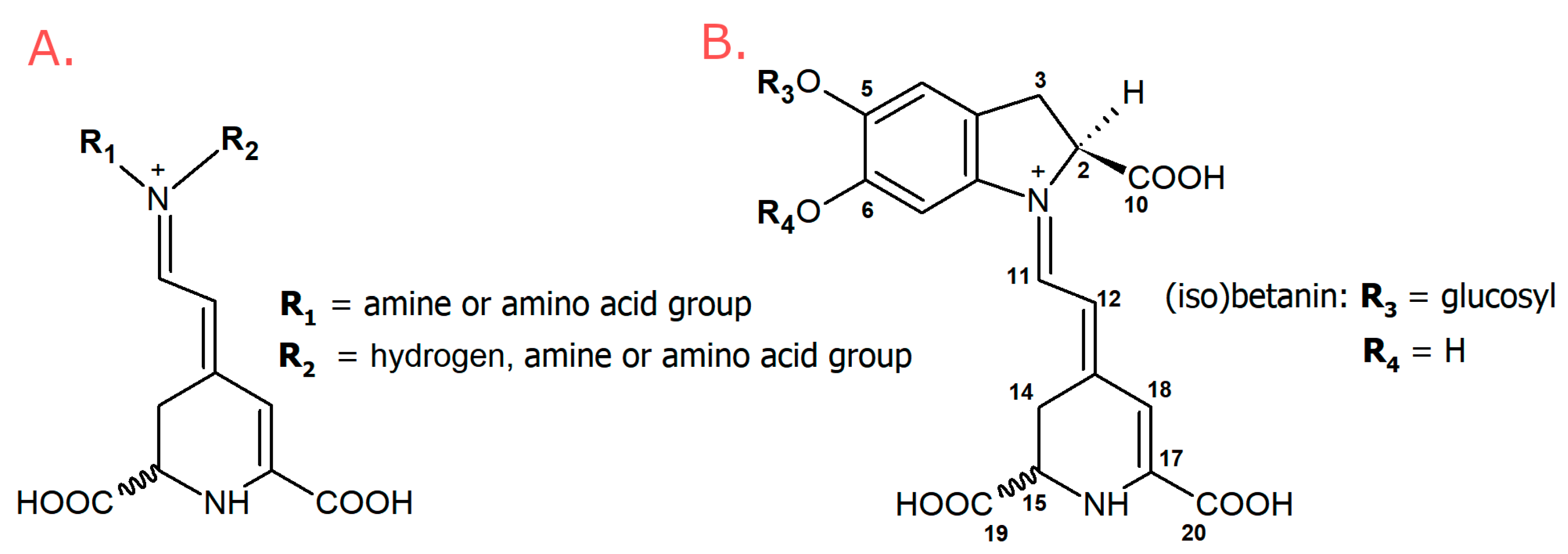
Figure 1. General structures: (A) betaxanthins, (B) betacyanins.
Metabolites 13 00408 g001
Figure 2. Selected-ion monitoring chromatogram (ESI-MS) in positive ion mode for betaxanthins of yellow Beta vulgaris L.: (A) peel, and (B) flesh. Numbers and names are available in Table 1.
Metabolites 13 00408 g002
Figure 3. Selected-ion monitoring chromatogram (ESI-MS) in positive ion mode for betaxanthins of red Beta vulgaris L. peels: (A) Ceryl, (B) Chrobry, (C) Forono, and (D) Tytus. Numbers and names are available in Table 1.
Metabolites 13 00408 g003
Figure 4. Selected-ion monitoring chromatogram (ESI-MS) in positive ion mode for betaxanthins of red Beta vulgaris L. flesh: (A) Ceryl, (B) Chrobry, (C) Forono, and (D) Tytus. Numbers and names are available in Table 1.
Metabolites 13 00408 g004
Figure 5. Predominant betaxanthins detected in Beta vulgaris, L. cultivars: (A) glutamine-Bx (5), (B) leucine-Bx (21), (C) isoleucine-Bx (21′), (D) γ-aminobutyric acid-Bx (13), (E) proline-Bx (14), (F) valine-Bx (19).
Metabolites 13 00408 g005
Figure 6. The dose-response impact of extracts from red and yellow B. vulgaris cultivars on non-cancerous and cancer cell lines.
Metabolites 13 00408 g006
Figure 7. The influence of serial dilutions of extracts from the peel of B. vulgaris Ceryl cultivar on the cell line derived from cervical adenocarcinoma.
Metabolites 13 00408 g007
Table 1. Chromatographic, spectrophotometric, and mass-spectrometric data of analyzed betaxanthins in fresh Beta vulgaris L. var. Ceryl, Chrobry, Forono, Tytus, Boldor.
No.BetaxanthinsTrivial NametR
(min)
λmax
(nm)
m/z
(M + H)+
1Histidine-Bxmuscaaurin VII4.4470349
2Asparagine-Bxvulgaxanthin III5.1468326
3Serine-Bx 5.7469299
4Arginine-Bx 6.3468368
5Glutamine-Bxvulgaxanthin I6.6467340
6Ornithine-Bx 7.1465326
7Glycine-Bx 7.2470269
8Ethanolamine-Bx 7.5454255
9Lysine-Bx 7.6458340
10Threonine-Bx 8.7469313
11Glutamic acid-Bxvulgaxanthin II9.2469341
12Alanine-Bx 11.5466283
13γ-Aminobutyric acid-Bx 11.6454297
14Proline-Bxindicaxanthin14.6477309
15Dopa-Bxdopaxanthin16.1470391
16Dopamina-Bxmiraxanthin V18.2460347
17Tyrosine-Bxportulacaxanthin II18.4471375
18Methionine-Bx 19.2468343
19Valine-Bx 19.6469311
203-methoxytyramine-Bx 20.0471361
21Isoleucine-Bx 22.0469325
21Leucine-Bxvulgaxanthin IV22.2469325
22Phenylalanine-Bx 22.3469359
23Tryptophan-Bxvulgaxanthin IV22.3473398
Table 2. Content of individual betaxanthins and total betaxanthins in the peel of red and yellow Beta vulgaris L. cultivars (mg/100 g of Dry Extract) analyzed by LC-DAD-ESI-MS.
Mass of Betaxanthins (mg) in 100 g of Dry Extract (DE) Beta vulgaris L.
Red CultivarYellow Cultivar
CerylChrobryForonoTytusBoldor
No.Betaxanthins
1Histidine-Bx0.15 ± 0.01 e1.06 ± 0.08 c0.88 ± 0.05 d1.87 ± 0.14 b1.93 ± 0.14 a
2Asparagine-Bx42.52 ± 2.6 a26.91 ± 1.8 b24.40 ± 1.5 c42.52 ± 2.6 a11.12 ± 0.80 d
3Serine-Bx23.81 ± 1.8 a16.28 ± 1.3 c9.72 ± 0.74 d19.64 ± 1.7 b5.94 ± 0.47 e
4Arginine-Bx10.35 ± 0.68 d5.64 ± 0.37 e15.61 ± 1.0 c19.50 ± 1.5 b23.71 ± 1.5 a
5Glutamine-Bx264.95 ± 0.88 b257.82 ± 15 c208.23 ± 16 d350.82 ± 21 a93.35 ± 6.1 e
6Ornithine-Bx8.68 ± 0.57 e11.99 ± 0.79 d27.87 ± 1.7 c39.69 ± 2.7 a36.31 ± 2.4 b
7Glycine-Bx3.28 ± 0.24 b1.91 ± 0.12 d2.51 ± 0.17 c7.72 ± 0.5 a1.64 ± 0.14 e
8Ethanolamine-Bx10.07 ± 0.68 b5.26 ± 0.34 e6.55 ± 1.1 d10.83 ± 0.8 a8.53 ± 0.60 c
9Lysine-Bx1.26 ± 0.11 c0.80 ± 0.061 e0.96 ± 0.06 d12.41 ± 0.87 a3.10 ± 0.21 b
10Threonine-Bx12.17 ± 0.81 e20.58 ± 1.4 c15.88 ± 1.0 d35.43 ± 2.4 a20.70 ± 1.5 b
11Glutamic acid-Bx56.49 ± 4.8 a25.46 ± 1.7 c18.25 ± 1.3 d56.54 ± 3.5 a28.09 ± 1.8 b
12Alanine-Bx14.23 ± 0.91 b10.87 ± 0.82 c9.40 ± 0.68 e21.59 ± 1.8 a10.57 ± 0.8 d
13γ-Aminobutyric acid-Bx66.03 ± 5.1 b35.14 ± 2.1 e36.88 ± 2.8 d70.06 ± 4.8 a50.32 ± 3.1 c
14Proline-Bx65.79 ± 5.4 c35.70 ± 2.3 e52.97 ± 4.1 d82.53 ± 5.2 b140.95 ± 10a
15Dopa-Bx0.21 ± 0.01 a0.11 ± 0.008 b0.05 ± 0.004 c0.30 ± 0.02 a0.03 ± 0.002 c
16Dopamine-Bx0.13 ± 0.009 d0.21 ± 0.014 c0.04 ± 0.003 e2.36 ± 0.17 a0.39 ± 0.03 b
17Tyrosine-Bx0.10 ± 0.006 e1.01 ± 0.074 c1.08 ± 0.07 b1.65 ± 0.12 a0.25 ± 0.02 d
18Methionine-Bx7.81 ± 0.63 a3.85 ± 0.25 c3.05 ± 0.23 d4.57 ± 0.32 b0.99 ± 0.06 e
19Valine-Bx55.61 ± 4.6 b45.22 ± 2.8 d45.52 ± 2.8 c74.29 ± 4.5 a40.69 ± 2.8 e
203-methoxytyramine-Bx27.82 ± 1.8 b14.52 ± 1.3 d15.47 ± 1.3 c34.14 ± 2.6 a3.40 ± 0.26 e
21′Isoleucine-Bx115.09 ± 7.1 b102.17 ± 6.4 c74.83 ± 5.4 d159.96 ± 10 a34.14 ± 2.3 e
21Leucine-Bx100.32 ± 6.8 b74.91 ± 5.2 c74.38 ± 4.8 d150.47 ± 9.7 a32.62 ± 2.1 e
22Phenylalanine-Bx20.58 ± 1.5 a13.86 ± 0.8 c15.00 ± 1.0 b20.75 ± 1.5 a4.16 ± 0.35 d
23Tryptophan-Bx12.05 ± 0.83 c7.28 ± 0.57 e9.75 ± 0.78 d12.23 ± 0.78 b21.37 ± 1.4 a
Total concentration919 ± 52 b718 ± 36 c669 ± 28 d1231 ± 64 a574 ± 26 e
The superscript letters within each row (a–e) mean significant differences between results (p < 0.05).
Table 3. Content of individual betaxanthins and total betaxanthins in the flesh of red and yellow Beta vulgaris L. cultivars (mg/100 g of Dry Extract) analyzed by LC-DAD-ESI-MS.
Mass of Betaxanthins (mg) in 100 g of Dry Extract (DE) Beta vulgaris L.
Red CultivarYellow Cultivar
CerylChrobryForonoTytusBoldor
No. Betaxanthins
1Histidine-Bx1.56 ± 0.09 c2.84 ± 0.21 a1.85 ± 0.16 b0.07 ± 0.005 e0.38 ± 0.02 d
2Asparagine-Bx24.64 ± 1.8 a2.84 ± 0.18 e21.33 ± 1.4 c21.37 ± 1.5 b13.84 ± 0.90 d
3Serine-Bx16.92 ± 1.1 c22.05 ± 1.7 a12.90 ± 0.84 d18.19 ± 1.2 b7.53 ± 0.55 e
4Arginine-Bx2.11 ± 0.16 c2.80 ± 0.22 b3.71 ± 0.25 a2.31 ± 0.20 c0.51 ± 0.03 d
5Glutamine-Bx179.79 ± 11 c219.64 ± 15 a172.67 ± 12 d190.90 ± 12 b80.10 ± 6.1 e
6Ornithine-Bx3.84 ± 0.34 c3.24 ± 0.24 d6.81 ± 0.44 b3.91 ± 0.30 c11.37 ± 0.80 a
7Glycine-Bx7.47 ± 0.56 c7.23 ± 0.44 d8.37 ± 0.68 b13.59 ± 0.95 a1.25 ± 0.08 e
8Ethanolamine-Bx4.46 ± 0.27 b 4.80 ± 0.29 a3.28 ± 0.26 c2.81 ± 0.21 d1.77 ± 0.12 e
9Lysine-Bx0.61 ± 0.05 c0.35 ± 0.02 e0.49 ± 0.04 d4.91 ± 0.32 a4.25 ± 0.32 b
10Threonine-Bx15.55 ± 1.1 d17.62 ± 1.2 a16.12 ± 1.4 c16.29 ± 1.2 b8.41 ± 0.65 e
11Glutamic acid-Bx21.53 ± 1.4 a14.49 ± 0.98 d14.95 ± 1.2 c15.32 ± 0.97 b8.27 ± 0.58 e
12Alanine-Bx15.90 ± 0.98 d24.96 ± 1.6 a16.65 ± 1.3 c23.46 ± 1.6 b11.34 ± 0.76 e
13γ-Aminobutyric acid-Bx32.88 ± 2.3 a29.47 ± 2.0 b22.31 ± 1.6 d23.93 ± 1.7 c22.09 ± 0.22 e
14Proline -Bx40.13 ± 2.6 c29.63 ± 2.0 d41.73 ± 2.6 b26.82 ± 1.9 e62.47 ± 4.2 a
15Dopa-Bx0.08 ± 0.006 d0.21 ± 0.02 c0.34 ± 0.03 b0.45 ± 0.03 a0.20 ± 0.02 c
16Dopamine-Bx0.12 ± 0.008 c0.24 ± 0.02 b0.10 ± 0.006 c0.12 ± 0.008 c0.65 ± 0.04 a
17Tyrosine-Bx1.87 ± 0.15 d3.04 ± 0.24 c6.02 ± 0.43 b1.71 ± 0.14 d7.71 ± 0.52 a
18Methionine-Bx8.66 ± 0.59 b11.50 ± 0.7 a5.08 ± 0.37 d6.17 ± 0.50 c0.14 ± 0.009 e
19Valine-Bx42.70 ± 2.7 c48.81 ± 3.5 b50.66 ± 3.7 a39.35 ± 2.8 d30.69 ± 2.2 e
203-methoxytyramine-Bx16.00 ± 0.98 a9.61 ± 0.74 b6.70 ± 0.5 d9.42 ± 0.57 c6.66 ± 0.44 d
21′Isoleucine-Bx83.54 ± 5.8 a80.24 ± 5.4 b68.30 ± 4.1 c55.96 ± 3.6 d17.49 ± 1.3 e
21Leucine-Bx55.17 ± 3.7 b59.86 ± 3.7 a55.08 ± 3.5 c41.21 ± 3.2 d13.17 ± 0.87 e
22Phenylalanine-Bx8.95 ± 0.77 b6.80 ± 0.54 c9.30 ± 0.61 a6.21 ± 0.43 d1.23 ± 0.09 e
23Tryptophan-Bx6.43 ± 0.55 b7.33 ± 0.61 a3.95 ± 0.30 e4.46 ± 0.31 d5.89 ± 0.47 c
Total concentration590 ± 32 b609 ± 40 a548 ± 28 c528 ± 34 d317 ± 22 e
The superscript letters within each row (a–e) mean significant differences between results (p < 0.05).
Table 4. Antimicrobial activity of extracts obtained from red and yellow B. vulgaris cultivars assessed as MIC (minimum inhibitory concentration) and MBC (minimum bactericidal concentration) against strains of Gram-positive bacteria and correlation coefficients between identified betaxanthins (absolute peak areas) and microbial activity (MIC values). Statistical significance is marked by font: boldface means 95% significance, normal font lack of significance at 95%.
Gram-Positive Bacteria
Microorganism/
Extract
Staphylococcus aureus
ATCC 29213
Staphylococcus aureus
ATCC 25923
Staphylococcus aureus
ATCC 43300
Staphylococcus aureus
ATCC BAA-1707
Staphylococcus epidermidis
ATCC 12228
Entarococcus faecalis
ATCC 29212
Micrococcus luteus
ATCC 10240
Bacillus subtilis
ATCC 6633
Bacillus cereus
ATCC 10876
MIC;MBC
CerylPeel1;40.06;0.54;44;44;41;80.06;0.060.125;816;16
Flesh16;1616;1616;1616;164;44;84;816;1616;16
ChrobryPeel0.125;80.125;40.25;40.25;80.25;10.5;80.125;0.250.125;81;8
Flesh16;1616;1616;1616;164;416;162;816;1616;16
ForonoPeel16;164;44;84;42;416;160.06;0.060.06;84;8
Flesh16;164;416;1616;162;416;161;20.125;88;8
TytusPeel0.25;80.25;80.25;40.5;80.25;80.5;80.125;0.2516;1616;16
Flesh16;1616;1616;1616;164;44;80.5;0.516;1616;16
BoldorPeel16;1616;1616;1616;1616;164;84;816;164;8
Flesh16;1616;1616;1616;1616;164;82;416;1616;16
Correlation
No.Betaxanthins
1Histidine-Bx 0.274 0.274 0.307 0.308−0.306 0.597 0.3280.127 0.223
2Asparagine-Bx−0.453−0.543−0.413−0.414−0.629−0.287−0.398−0.451−0.049
3Serine-Bx−0.0860.0090.0780.077−0.5920.109−0.131−0.029 0.362
4Arginine-Bx−0.368−0.587−0.646−0.645−0.2590.026−0.349−0.363−0.459
5Glutamine-Bx−0.307−0.346−0.276−0.276−0.8700.134−0.407−0.2750.159
6Ornithine-Bx−0.307−0.547−0.651−0.648−0.2810.046−0.344−0.236−0.376
7Glycine-Bx 0.343 0.326 0.453 0.455−0.383 0.206−0.001 0.253 0.527
8Ethanolamine-Bx−0.513−0.524−0.510−0.511−0.574−0.075−0.300−0.352−0.091
9Lysine-Bx−0.304−0.108−0.240−0.233−0.236−0.416−0.347 0.411 0.450
10Threonine-Bx−0.124−0.166−0.108−0.105−0.818 0.197−0.1840.015 0.253
11Glutamic acid-Bx−0.655−0.554−0.504−0.504−0.508−0.368−0.340−0.3080.002
12Alanine-Bx 0.234 0.268 0.363 0.364−0.479 0.315−0.023 0.215 0.526
13γ-Aminobutyric acid-Bx−0.507−0.459−0.437−0.438−0.586−0.177−0.274−0.2830.026
14Proline-Bx−0.178−0.314−0.172−0.171−0.223−0.0190.113−0.240−0.235
15Dopa-Bx0.1350.065 0.296 0.297−0.380 0.182−0.3290.028 0.373
16Dopamine-Bx−0.515−0.318−0.433−0.424−0.279−0.327−0.222 0.356 0.384
17Tyrosine-Bx 0.4510.123 0.518 0.519−0.219 0.643 0.067−0.091 0.247
18Methionine-Bx0.171 0.248 0.306 0.305−0.426 0.2880.1530.130 0.443
19Valine-Bx−0.097−0.256−0.052−0.052−0.789 0.321−0.238−0.259 0.193
203-methoxytyramine-Bx−0.552−0.483−0.515−0.514−0.652−0.265−0.338−0.2330.152
21′Isoleucine-Bx−0.426−0.435−0.359−0.359−0.8400.015−0.306−0.2800.141
21Leucine-Bx−0.527−0.608−0.528−0.527−0.8570.032−0.474−0.3730.060
22Phenylalanine-Bx−0.555−0.715−0.607−0.609−0.774−0.029−0.541−0.614−0.181
23Tryptophan-Bx−0.273−0.241−0.312−0.313−0.307−0.0170.015−0.192−0.212
MIC and MBC were expressed as mg/mL. The representative data (mode) are presented.
Table 5. Antimicrobial activity of extracts obtained from red and yellow B. vulgaris cultivars assessed as MIC (minimum inhibitory concentration) and MBC (minimum bactericidal concentration) against strains of Gram-negative bacteria and correlation coefficients between identified betaxanthins (absolute peak areas) and microbial activity (MIC values). Statistical significance is marked by font: boldface means 95% significance, and normal font lack of significance at 95%.
Gram-Negative Bacteria
Microorganism/
Extract
Salmonella Typhimurium ATCC 14028Proteus mirabilis ATCC 12453Bordetella bronchiseptica ATCC 4617Escherichia coli ATCC 25922Klebsiella pneumoniae
ATCC 13883
Pseudomonas aeruginosa
ATCC 27853
MIC;MBC
CerylPeel16;1616;162;84;816;164;8
Flesh16;1616;1616;1616;1616;164;8
ChrobryPeel4;88;82;44;416;162;8
Flesh16;1616;164;816;1616;164;8
ForonoPeel16;1616;162;44;416;164;4
Flesh16;1616;168;816;1616;164;8
TytusPeel16;1616;164;44;816;164;8
Flesh16;1616;164;84;816;164;8
BoldorPeel16;1616;168;816;1616;164;8
Flesh16;1632;3216;1616;1632;324;8
Correlation
No.Betaxanthins
1Histidine-Bx0.096−0.340.092 0.540−0.3430.096
2Asparagine-Bx−0.025−0.46−0.228−0.538−0.462−0.025
3Serine-Bx0.068−0.51−0.246−0.129−0.5150.068
4Arginine-Bx0.137−0.48−0.585−0.531−0.4770.137
5Glutamine-Bx−0.084−0.67−0.474−0.403−0.673−0.084
6Ornithine-Bx0.041−0.34−0.452−0.495−0.3440.041
7Glycine-Bx 0.279−0.360.0120.023−0.359 0.279
8Ethanolamine-Bx0.099−0.63−0.501−0.442−0.6320.099
9Lysine-Bx 0.194−0.14−0.233−0.508−0.138 0.194
10Threonine-Bx−0.055−0.73−0.320−0.205−0.731−0.055
11Glutamic acid-Bx0.042−0.48−0.380−0.488−0.4850.042
12Alanine-Bx 0.232−0.45−0.1400.053−0.450 0.232
13γ-Aminobutyric acid -Bx0.102−0.61−0.414−0.440−0.6090.102
14Proline-Bx 0.345−0.66−0.202−0.091−0.660 0.345
15Dopa-Bx 0.205−0.28−0.240−0.146−0.281 0.205
16Dopamine-Bx0.094−0.10−0.145−0.263−0.1030.094
17Tyrosine-Bx 0.202−0.02 0.252 0.516−0.021 0.202
18Methionine-Bx 0.188−0.42−0.019 0.182−0.421 0.188
19Valine-Bx0.117−0.68−0.310−0.147−0.6780.117
203-methoxytyramine-Bx0.051−0.47−0.299−0.511−0.4700.051
21′Isoleucine-Bx−0.082−0.65−0.348−0.335−0.645−0.082
21Leucine-Bx−0.011−0.65−0.499−0.481−0.647−0.011
22Phenylalanine-Bx−0.082−0.58−0.509−0.576−0.576−0.082
23Tryptophan-Bx0.155−0.69−0.449−0.264−0.6940.155
MIC and MBC were expressed as mg/mL. The representative data (mode) are presented.
Table 6. Antimicrobial activity of extracts obtained from red and yellow B. vulgaris cultivars assessed as MIC (minimum inhibitory concentration) and MFC (minimum fungicidal concentration) against Candida species and correlation coefficients between identified betaxanthins (absolute peak areas) and microbial activity (MIC values). Statistical significance is marked by font: boldface means 95% significance, and normal font lack of significance at 95%.
Fungal (Yeasts) Strains
Microorganism/
Extract
Candida parapsilosis
ATCC 22019
Candida albicans
ATCC 10231
Candida albicans
ATCC 2091
Candida glabrata
ATCC 90030
Candida krusei
ATCC 14243
Candida auris
CDC B11903
Candida lusitaniae
ATCC 3449
Candida tropicalis
ATCC 1369
MIC;MFC
CerylPeel0.25;10.25;10.25;10.5;20.5;40.5;11;41;4
Flesh4;84;84;84;84;84;84;44;4
ChrobryPeel2;81;81;84;82;81;82;82;8
Flesh4;44;84;84;84;84;84;44;4
ForonoPeel0.5;21;41;44;41;42;42;44;4
Flesh4;84;84;88;84;48;88;48;4
TytusPeel0.25;20.25;20.25;21;40.25;0.50.5;20.125;40.25;4
Flesh8;84;44;48;84;48;84;44;4
BoldorPeel4;84;84;84;84;84;84;82;4
Flesh4;44;44;44;84;44;44;44;4
Correlation
No.Betaxanthins
1Histidine-Bx0.0660.066 0.3440.149 0.335 0.194 0.405 0.410
2Asparagine-Bx−0.279−0.494−0.494−0.165−0.497−0.168−0.260−0.126
3Serine-Bx 0.1810.0190.0190.0590.0240.1400.0200.091
4Arginine-Bx−0.719−0.717−0.717−0.513−0.780−0.563−0.572−0.470
5Glutamine-Bx−0.109−0.308−0.308−0.033−0.299−0.062−0.1640.010
6Ornithine-Bx−0.653−0.648−0.648−0.378−0.706−0.497−0.520−0.381
7Glycine-Bx 0.678 0.441 0.441 0.621 0.411 0.703 0.418 0.451
8Ethanolamine-Bx−0.590−0.631−0.631−0.588−0.661−0.526−0.517−0.418
9Lysine-Bx0.105−0.240−0.240−0.056−0.2990.012−0.386−0.391
10Threonine-Bx0.096−0.065−0.065 0.198−0.0690.1560.0290.128
11Glutamic acid-Bx−0.495−0.627−0.627−0.573−0.641−0.474−0.511−0.475
12Alanine-Bx 0.500 0.338 0.338 0.402 0.316 0.497 0.297 0.343
13γ-Aminobutyric acid-Bx−0.460−0.558−0.558−0.517−0.583−0.432−0.473−0.391
14Proline-Bx−0.395−0.299−0.299−0.338−0.362−0.208−0.143−0.201
15Dopa-Bx 0.575 0.267 0.267 0.604 0.248 0.675 0.393 0.413
16Dopamine-Bx−0.358−0.412−0.412−0.459−0.465−0.381−0.489−0.506
17Tyrosine-Bx 0.387 0.538 0.538 0.666 0.522 0.709 0.836 0.883
18Methionine-Bx 0.258 0.255 0.2550.110 0.245 0.224 0.187 0.236
19Valine-Bx−0.025−0.104−0.1040.116−0.1230.1490.116 0.255
203-methoxytyramine-Bx−0.483−0.611−0.611−0.535−0.636−0.481−0.553−0.402
21′Isoleucine-Bx−0.298−0.400−0.400−0.236−0.395−0.234−0.243−0.097
21Leucine-Bx−0.488−0.593−0.593−0.362−0.611−0.362−0.382−0.200
22Phenylalanine-Bx−0.577−0.693−0.693−0.381−0.697−0.423−0.406−0.201
23Tryptophan-Bx−0.447−0.426−0.426−0.534−0.458−0.463−0.447−0.444
MIC and MFC were expressed as mg/mL. The representative data (mode) are presented.
Table 7. Cytotoxicity and anticancer selectivity of B. vulgaris extracts on different cell lines after 24 h incubation.
ExtractVERORKOHeLa
CC50CC50SICC50SI
CerylPeel1992.00 ± 249.10717.20 ± 79.322.78225.30 ± 21.288.84
Flesh3432.04 ± 631.002100.23 ± 352.701.63367.92 ± 33.309.33
ChrobryPeel3330.00 ± 865.151275.75 ± 124.202.61354.80 ± 22.649.39
Flesh2896.15 ± 533.501470.35 ± 139.701.97312.21 ± 19.809.28
ForonoPeel361.10 ± 42.17674.20 ± 51.810.54290.23 ± 20.581.24
Flesh1754.31 ± 253.72993.10 ± 76.911.77328.10 ± 18.565.35
TytusPeel405.40 ± 36.471259.00 ± 101.900.32420.90 ± 29.370.96
Flesh908.45 ± 110.901479.66 ± 155.600.61282.38 ± 25.243.22
BoldorPeel527.70 ± 35.85413.80 ± 26.251.28274.74 ± 22.141.92
Flesh4948.60 ± 1440.002090.00 ± 98.992.37980.70 ± 74.375.05
CC50—50% cytotoxic concentration (mean ± SD) μg/mL; VERO—non-cancerous cells; RKO—colon cancer cell line; HeLa—cervical adenocarcinoma; SI—selectivity index (CC50VERO/CC50CancerCells).
Table 8. Correlation coefficients between identified betaxanthins (absolute peak areas) and microbial activity (CC50). Statistical significance is marked by font: boldface means 95% significance, and normal font lack of significance at 95%.
RKOHeLa
No. Betaxanthins
1Histidine-Bx 0.177−0.242
2Asparagine-Bx−0.080−0.453
3Serine-Bx 0.211−0.520
4Arginine-Bx−0.771−0.490
5Glutamine-Bx0.044−0.628
6Ornithine-Bx−0.575−0.274
7Glycine-Bx 0.320−0.334
8Ethanolamine-Bx−0.357−0.650
9Lysine-Bx0.068−0.044
10Threonine-Bx0.113−0.593
11Glutamic acid-Bx−0.221−0.499
12Alanine-Bx 0.282−0.430
13γ-Aminobutyric acid-Bx−0.220−0.622
14Proline-Bx−0.574−0.689
15Dopa-Bx0.089−0.300
16Dopamine-Bx0.0750.074
17Tyrosine-Bx 0.2420.030
18Methionine-Bx 0.318−0.416
19Valine-Bx0.010−0.636
21′Isoleucine-Bx0.059−0.571
21Leucine-Bx−0.130−0.585
22Phenylalanine-Bx−0.265−0.573
23Tryptophan-Bx−0.498−0.753
CC50—50% cytotoxic concentration μg/mL; RKO—colon cancer cell line; HeLa—cervical adenocarcinoma.
Disclaimer/Publisher’s Note: The statements, opinions and data contained in all publications are solely those of the individual author(s) and contributor(s) and not of MDPI and/or the editor(s). MDPI and/or the editor(s) disclaim responsibility for any injury to people or property resulting from any ideas, methods, instructions or products referred to in the content.