Article

Targeted Peptide-Mediated Delivery of Antisense Oligonucleotides to SMA Cells for SMN2 Gene Splicing Correction

1
Department of Genomic Medicine named after V.S. Baranov, D.O. Ott Research Institute of Obstetrics, Gynecology and Reproductology, Mendeleevskaya Line 3, 199034 Saint-Petersburg, Russia
2
Department of Children’s Diseases, Medical Institute of Surgut State University, 628400 Surgut, Russia
*
Author to whom correspondence should be addressed.
Sci. Pharm. 2025, 93(3), 38; https://doi.org/10.3390/scipharm93030038 (registering DOI)
Submission received: 16 April 2025 / Revised: 11 July 2025 / Accepted: 12 August 2025 / Published: 14 August 2025

Abstract

Spinal muscular atrophy (SMA) is a severe neurodegenerative disorder that has an approved treatment that can still be improved. Antisense oligonucleotides (AONs) are currently delivered intrathecally for SMA therapy based on SMN2 gene splicing correction, and high concentrations are required to achieve an improvement of the disease symptoms. In this study, AONs were introduced into SMA fibroblast cell cultures by means of an arginine–histidine-rich peptide carrier that had been decorated with iRGD ligands. Due to the protected and receptor-mediated nature of AON delivery within these complexes, low concentrations can be used. We assessed the RNA-binding characteristics, cytotoxicity, size, and zeta potential of AON/carrier complexes as well as the efficiency of SMN2 gene splicing correction following transfections. After testing a variety of AON/carrier formulations, we selected those that produced the best outcomes. The AON/carrier complexes that were found to be the most effective significantly increased the proportion of full-length SMN transcripts and the quantity of nuclear gems. Thus, we demonstrated the potential of delivering therapeutic AONs into SMA cells using a ligand-modified peptide carrier.

1. Introduction

Spinal muscular atrophy (SMA) is a devastating disorder that manifests as generalized progressive muscle weakness and atrophy in the absence of proper treatment. SMA is caused by homozygous mutations in the survival motor neuron 1 (SMN1) gene [1]. The majority of these mutations, approximately 95%, are deletions, which lead to a deficiency of the SMN protein crucial for the proper functioning of motor neurons.
Another (but less efficient) source of the SMN protein is the SMN2 gene, which is a nearly identical copy of the SMN1 gene. Due to a C-to-T substitution in exon 7 of the SMN2 gene, most transcripts produced by this gene undergo aberrant splicing and lack exon 7 [2]. The protein translated from such RNA is unstable and cannot perform its functions properly [3]. The amount of the full-length SMN protein originating from the SMN2 gene is not enough to compensate for the absence of the SMN1 gene product. Only in rare cases when the SMN2 copy number achieves more than four copies or when some additional positive modifiers act may the SMA phenotype be drastically ameliorated [4,5,6]. The SMN2 gene copy number is the main disease modifier and is inversely correlated with SMA severity [4]. Thus, patients with the most severe SMA forms 0 and I usually possess one and two SMN2 copies, respectively. Such patients commonly demonstrate first symptoms in the prenatal or neonatal period. Three SMN2 copies are characteristic of intermediate and lighter forms II and IIIa [7]. Four SMN2 copies are usually found in patients with SMA types III and IV. Such patients are able to walk and have near-normal life expectancy.
Correction of the SMN2 splicing is one of the most attractive strategies for SMA therapy. This strategy underlies the working principle of two drugs approved for the treatment of SMA. The first is nusinersen—an antisense oligonucleotide that blocks the silencer of the exon 7 splicing [8]. The second is risdiplam—a small molecule that was also shown to positively modify SMN2 exon 7 splicing [9]. The third drug approved for the treatment of SMA is onasemnogene abeparvovec that provides cells with a functional copy of the SMN1 gene [10]. Thus, stimulating the production of a more functional SMN protein is the basic aim of SMA therapeutic approaches.
The SMN protein is an ubiquitously expressed protein that fulfills vital functions in cells. Its primarily identified function is the participation in the assembly of small nuclear ribonucleoprotein particles (snRNPs), while its role in the axonal mRNA trafficking, endocytosis, transcription, translation, cytoskeleton maintenance as well as in other processes has also been described [11,12]. The SMN protein is localized in both the cytoplasm and the nucleus, where it accumulates in structures known as gems [13]. The number of gems was shown to inversely correlate with SMA severity [14,15]. Meanwhile, therapeutic agents who aimed to restore SMN protein levels increased the number of gems as well [16,17,18].
Existing drugs proved their efficacy for patients with SMA. However, these approaches exhibit certain limitations, including adverse effects, and do not provide uniform efficacy across all SMA patients [19,20]. Also combined therapy with different drugs is sometimes required [21]. Antisense oligonucleotides (AONs), which became the basis for the development of the first drug for SMA, seem to be favorable therapeutic agents that can be flexibly improved [8]. Thus, the AON sequence, length, and modification can be modified smoothly depending on the demand.
In addition to the ISS-N1 splicing silencer in SMN2 intron 7, which is blocked by nusinersen, there are other negative motifs that weaken exonic 7 splice sites [22,23]. Targeting both of them may result in a cumulative positive effect. In the study by Pao et al., the concurrent inhibition of two negative splicing regulatory elements caused a more efficient restoration of SMN transcripts and protein levels than separate targeting [24].
In our previous study, we described several AONs with various chemical modifications and lengths that were designed to mask different splicing silencers in the SMN2 gene [25]. We delivered them in SMA fibroblast cell cultures and observed a significant increase in the percentage of full-length (FL) SMN transcripts and the number of gems. Previously, we used the transfection reagent x-tremeGENE (Roche), which has been shown to be effective in vitro but is not recommended for use in the human body. Therefore, the development of a biocompatible system for delivering AONs is of interest in order to address this challenge.
Previously, we developed a nucleic acid (NA) delivery system RGD1-R6 as a combination of the iRGD (internalizing RGD) ligand-modified carrier RGD1 and the cross-linking peptide carrier R6 [26,27]. R6 is a peptide comprising arginine and cysteine for the efficient condensing and protection of nucleic acids, while histidine residues enable the endosomal escape of NA–peptide complexes. The addition of the iRGD ligand promotes specific cellular uptake via binding to αvβ3 integrins. In our previous study, we demonstrated the safety and efficiency of RGD1-R6 in DNA delivery to PANC-1 carcinoma and primary uterine leiomyoma cells as well as in siRNA delivery to endometrial heterotopias [26,27]. αvβ3 integrins were shown to be expressed on the neuronal cell surface as well; therefore, the RGD ligand can facilitate the neuronal uptake of complexes [28,29,30]. There have also been reports on the ability of RGD-functionalized nanoparticles to cross the blood–brain barrier [31,32].
In this study, we investigated the complexes of splicing-correcting AONs with the RGD1-R6 peptide carrier in order to target the SMN2 gene in an SMA cellular model.

2. Materials and Methods

2.1. Cell Cultures

The study was carried out on the basis of large-scale research facility #3076082 “Human Reproductive Health” in the D.O. Ott Research Institute of Obstetrics, Gynecology and Reproductology. Primary fibroblast cultures were derived from a skin biopsy of an SMA type II patient, providing an SMA cell model. Informed consent has been obtained from the patient’s representative. Fibroblasts were cultured in DMEM with L-glutamine and 4.5 g/L glucose (Biolot, Saint-Petersburg, Russia) with the addition of 10% fetal bovine serum (FBS) (Gibco, Grand Island, NY, USA) and antibiotics (penicillin 100 U/mL, streptomycin 100 µg/mL) (Biolot, Saint-Petersburg, Russia) at 37 °C in 5% CO2. The SMA fibroblast cell culture was confirmed for the presence of a homozygous exon 7 deletion in the SMN1 gene and for three copies of the SMN2 gene as previously described [33].

2.2. Antisense Oligonucleotides

The sequences of AONs are indicated in Table 1.
All AONs were modified with phosphorothioate and 2′-O-methyl in each nucleotide and synthesized by Syntol JSC (Moscow, Russia).

2.3. Peptide Carrier

RGD1 (R9H4CRGDRGPDC) and R6 (CHR6HC) peptide carriers were synthesized by NPF Verta, LLC (Saint-Petersburg, Russia) and stored lyophilized at −20 °C as previously described [35]. R6 was then dissolved in dH2O at 2 mg/mL and stored at −20 °C. The RGD1 carrier was cyclized, concentrated to 2 mg/mL as described earlier and stored at −70 °C [35].

2.4. RNA Binding Assay

An RNA binding assay was performed in accordance with previously described protocol [36]. AON/carrier complexes with charge ratios ranging from 1/0 to 1/24 were prepared in the wells of a 96-well plate. The plates were then left at RT for 2 h for final complexes formation. Then, a solution of 1× SybrGreen I dye (Amresco, Solon, OH, USA) was added to each well. The fluorescence intensity was measured using a Wallac 1420D fluorometer (Wallac Oy, Turku, Finland) at 495/600 nm. For each AON/carrier ratio, the fluorescence intensity (F) was measured in 2 repetitions. The efficiency of SybrGreen dye displacement was calculated using the formula (F − Ff)/(Fb − Ff), where Ff and Fb are the fluorescence intensities of SybrGreen in the absence and presence of AONs, respectively. Three replicates of the experiment were conducted.

2.5. Flow Cytometry

Fibroblast cell cultures were removed from the flask with 5 mM EDTA and centrifuged; then, the supernatant was discarded, and the cells were resuspended in 1× PBS. FITC-conjugated mouse anti-human CD51/CD61 antibodies (BD Pharmingen, San Jose, CA, USA) were added, and the cells were incubated with antibodies for 20 min at room temperature (RT). Flow cytometry was performed using a BD FACS-Canto II cytofluorimeter (Becton-Dickinson Biosciences, Franklin Lakes, NJ, USA). A total of 10,000 fibroblasts were estimated for the presence of αvβ3 integrins.

2.6. Transfections

SMA fibroblasts were detached from the flask and counted using a hemocytometer 1 day before transfection. Cells were seeded in full DMEM (with L-glutamine and 10% FBS) in a 24-well plate for transcripts analysis or in 8-well chamber slides for gems counting. The percentage of seeded cells was calculated to reach 85% of the monolayer by the time of transfection.
On the day of transfection, the RGD1-R6 carrier was prepared as previously described by mixing the RGD1 peptide with the R6 peptide at equimolar concentrations [35]. The AON/carrier charge ratios tested were 1/8 and 1/16. The complexes were prepared in Hepes-buffered mannitol (HBM) (5% w/v mannitol, 5 mM Hepes, pH 7.5). After vortexing, the complexes were left at RT for 2 h for disulfide bonds formation [35].
Before adding the complexes, the medium in the wells was replaced with clean DMEM without FBS or antibiotics. Each RNA-carrier complex was added to fibroblasts in duplicate. The transfected cell cultures were then incubated for 4 h at 37 °C in 5% CO2. Thereafter, the medium was replaced with a full medium containing FBS and antibiotics, and the cells were maintained in a CO2 incubator for 48 h.
Then, the fibroblasts were removed from the culture plate, and the RNA was isolated. All transfection experiments were repeated in triplicates.

2.7. Toxicity Assay

The cytotoxicity of AON/carrier complexes was evaluated using the Alamar Blue (resazurin) assay. SMA fibroblasts were plated in a 96-well plate and transfected the next day as described in Section 2.6. Four hours after the addition of the complexes, the medium was replaced with a full medium containing a 10% solution of Alamar Blue (BioSource, Camarillo, CA, USA). The cells were incubated in CO2 for 16 h. The fluorescence intensity was measured using a Wallac 1420D fluorometer at 530/590 nm. The relative number of cells after complex addition was calculated using the formula (F − Ff)/(Fb − Ff) × 100%, where Fb is the fluorescence intensity of Alamar Blue in the absence of AON/carrier complexes and Ff is the fluorescence of the dye in the absence of cells.

2.8. Size and Zeta Potential Measurements of AON/Carrier Complexes

The size of the complexes was determined by means of dynamic light scattering, while the zeta potential was measured using the microelectrophoresis technique. First, 5 μg of oligonucleotide was used to preparate the AON/carrier complexes at a charge ratio of 1/16. The measurements were performed three times independently for each complex on a zetasizer Nano ZS (Malvern Instruments, Malvern, UK).

2.9. RNA Isolation and cDNA Synthesis

Fibroblast cells in a 24-well plate were relieved from the medium and washed twice with 200 μL of PBS. Cells were then removed from the culture surface after the incubation with 200 µL of the Trypsin–Versen mixture (1:3) (Biolot LLC, Saint-Petersburg, Russia) for 10 min at 37 °C. After that, 300 μL of PBS was added to the wells. The contents of the wells were mixed, transferred to tubes, and centrifuged at 2200 rpm for 10 min (+4 °C). The supernatant was removed, and 125 μL of TRIzol (Invitrogen, Burlington, ON, Canada) was added to the sediments and resuspended. After 5 min of incubation at RT, 25 μL chloroform was added, and the mixture was shaken and incubated for another 3 min. The mixture was then centrifuged at 12,000 rpm (+4 °C) for 15 min. The upper phase was transferred into a new tube. Isopropanol was added and RNA was precipitated. The tubes were frozen at −70 °C overnight. The next day, the tubes were centrifuged again at 10,000 rpm for 20 min (+4 °C), and the supernatant was discarded. The residue was rinsed with 125 μL of cold 70% ethanol and then centrifuged for 3 min at 14,000 rpm (+4 °C). The supernatant was removed, and the RNA precipitate was dried out at RT for an hour. The RNA pellet was then dissolved in 20 μL DEPC-treated water and incubated at RT for 40 min.
Approximately 500 ng of total RNA was used for reverse transcription using the first-strand cDNA synthesis kit with random hexaprimers (Sileks, Moscow, Russia), following the manufacturer’s protocol.

2.10. Semiquantitative and Quantitative Fluorescence (QF) RT-PCR

FL-SMN and exon 7-deleted (∆7-SMN) transcripts amplification was performed as previously described with SMN F (5′-GTCCAGATTCTCTTGATGAT-3′) and SMN R (5′-CTATAACGCTTCACATTCCA-3′) primers [37]. For QF-PCR analysis, the forward primer was labeled with the FAM dye.
The amplification reaction was carried out at the following conditions: initial denaturation at 94 °C for 4 min, which was followed by repeated cycles (the number of cycles was chosen to analyze PCR products within the exponential phase and did not exceed 29): denaturation at 94 °C for 45 s, annealing at 50 °C for 45 s, and extension at 72 °C for 45 s. The final synthesis was performed at 72 °C for 8 min. Each cDNA sample was amplified at least twice.
PCR products obtained after semiquantitative RT-PCR were separated on a 6% polyacrylamide gel and stained with an ethidium bromide solution (0.5 μg/mL). The gel was then photographed under transmitted UV light (380 nm wavelength) using a transilluminator. The intensity of the PCR product luminescence was assessed using ImageJ version 1.54d (National Institutes of Health, Bethesda, Rockville, MD, USA).
To analyze the results of QF RT-PCR, 1 μL of the amplified product was mixed with 12 μL formamide (MCLAB, San Francisco, CA, USA) and 0.25 μL molecular weight marker LIZ 500 (Applied Biosystems, Foster City, CA, USA), and the fragments were then separated in an ABI 3130xl capillary electrophoresis instrument at 60 °C. The results were visualized using GeneMapper software version 3.7 (Applied Biosystems, Foster City, CA, USA).

2.11. Immunocytochemistry

A detailed protocol for immunocytostaining is described in the article by Al-Hilal et al. [18]. In brief, cells in 8-well chambers were washed with PBS and fixed with 4% paraformaldehyde for 10 min at RT. After that, the wells were washed twice with PBS. The cells were then incubated in 0.1% Triton X-100 in PBS for 5 min at RT, which was followed by another PBS wash. To reduce background fluorescence, the cells were treated with 1% bovine serum albumin (BSA) in PBS for one hour at RT. SMN antibody (Novus Biologicals, Littleton, CO, USA) diluted to 5 μg/mL in 1% BSA was added to wells and stored overnight at 4 °C. The next day, cells were washed three times in PBS for 5 min each and then incubated for one hour in the dark at RT with secondary IgG NL493-conjugated antibody (R&D Systems, Minneapolis, MN, USA) diluted 1:200 in 1% BSA. The cells were rinsed three more times with PBS, dried, and mounted with DAPI mounting medium (Vector Laboratories, Burlingame, CA, USA). Then, another glass slide was placed on top of the coverslip and sealed with nail polish. The analysis was performed using a Leica DM 2500 fluorescent microscope (Leica Microsystems, Wetzlar, Germany) at 1000× magnification. The number of gems was counted per 100 nuclei.

2.12. Statistical Methods

The diagrams were created using GraphPad PRISM 8.0.2 software (GraphPad Software Inc., San Diego, CA, USA). For statistical comparisons, a Kruskal–Wallis test was employed for the transcripts analysis, and a Mann–Whitney U-test was employed for the gems number analysis.

3. Results

3.1. Physicochemical Characterization of AON/Carrier Complexes

The ability of the RGD1-R6 carrier to bind oligoribonucleotides was assessed using the Sybr Green dye exclusion assay with the longest ASO VII (20-mer) among those tested. The Sybr Green fluorescence intensity was set at 100% in the absence of any carrier. As the concentration of the RGD1-R6 complex increased, the amount of unbound RNA decreased, along with a corresponding decrease in the intensity of Sybr Green fluorescence, suggesting that the RNA was bound by the carrier (Figure 1).
It can be seen in Figure 1 that at charge ratios of AON to carrier of 1/16 and 1/24, the fluorescence was virtually absent, indicating the formation of dense nucleopeptide complexes.
Then, we assessed the size and zeta potential of AON/RGD1-R6 complexes (Table 2). Due to the fact that the studied AONs have different lengths, we tested complexes formed with all oligonucleotides. The sizes of the complexes ranged from 114 to 234 nm with the smallest complex size observed for the ASOIV/RGD1-R6 complexes. All complexes were shown to be positively charged with a zeta potential ranging from +17.8 to +22.9 mV.

3.2. Assessment of Cytotoxic Properties of AON/Carrier Complexes

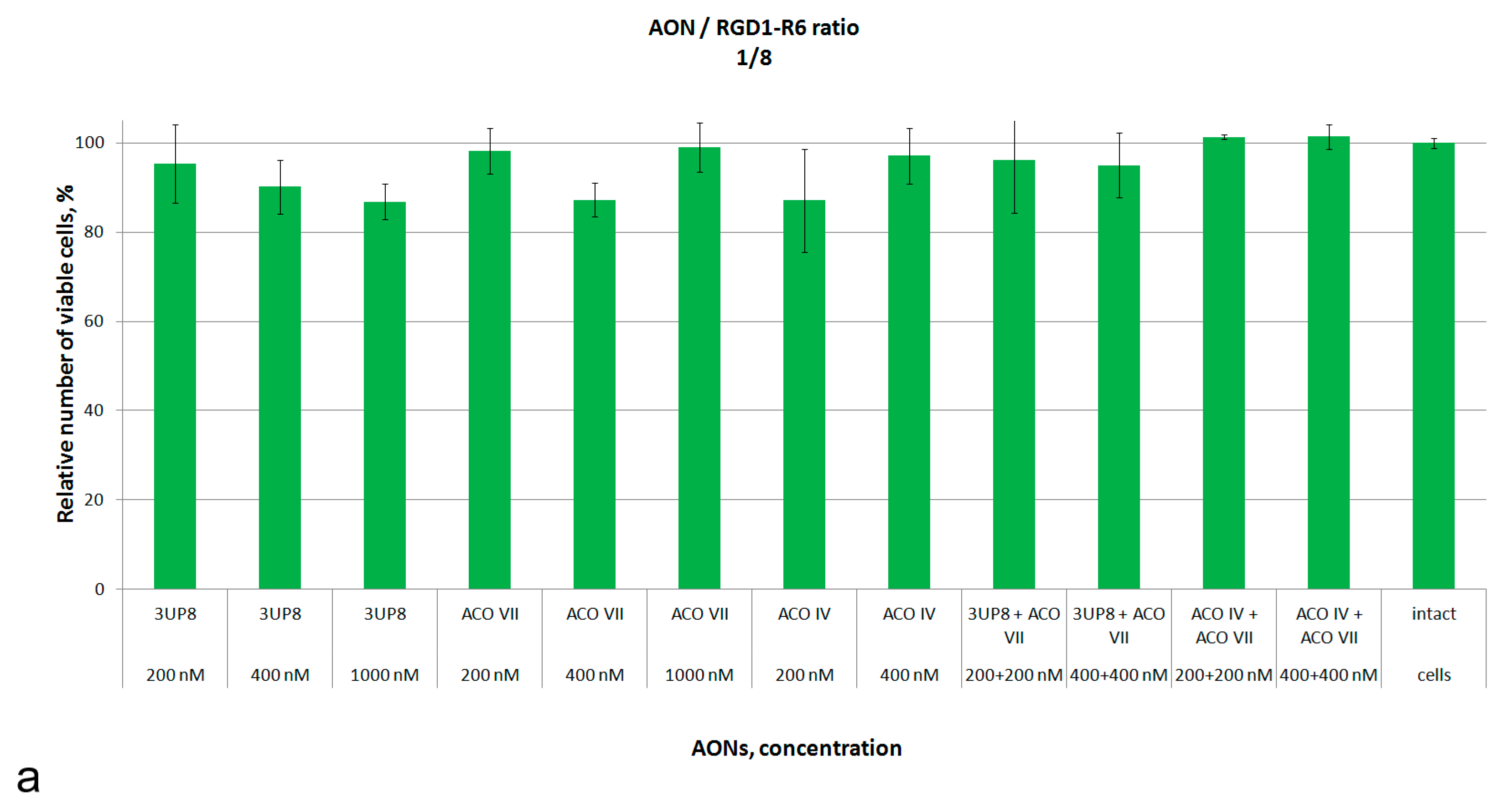
The cytotoxic properties were studied by means of resazurin viability assay using Alamar Blue reagent. Primary fibroblasts with mutation in the SMN1 gene were used as a cellular model for spinal muscular atrophy. Transfection was performed with AON concentrations ranging from 200 to 1000 nM, and two different charge ratios were used to form AON/carrier complexes. Additionally, combinations of two AONs were evaluated in the experiment (Figure 2).
It can be seen in Figure 2 that AON/RGD1-R6 complexes at the all tested charge ratios do not significantly increase the cytotoxicity level. In fact, more than 85% of cells survive after transfection with the complexes formed at 1/8 and 1/16 ratios (Figure 2). Therefore, it can be assumed that all formulations tested do not exhibit cytotoxicity toward SMA fibroblasts.

3.3. SMN2 Splicing Correction Efficiency of AON/RGD1-R6 Complexes

The complexes containing AONs, either individually or in combinations, with different concentrations, were evaluated for their ability to stimulate exon 7 inclusion into SMN2 mRNA. Before transfections the flow cytometry analysis demonstrated the presence of αvβ3 integrins, which are the targets of the iRGD ligand on the surface of fibroblast cells. Approximately 30% of cells were found to be positive for these receptors. The efficacy of SMN2 splicing correction was analyzed using a method based on the evaluation of full-length SMN transcript percentage by means of quantitative fluorescent RT-PCR and semiquantitative RT-PCR gel densitometric assays [37]. In the current study, we tested the AONs that were previously demonstrated to be efficient splicing modulators of the SMN2 exon 7 when delivered using commercially available liposomes [25].
We investigated the SMN2 splicing correction efficiency of AON/RGD1-R6 complexes at charge ratios of 1/8 (Figure 3a) and 1/16 (Figure 3b). On average, the enhancement in the proportion of full-length SMN transcripts was observed to be 10% greater at the 1/16 ratio compared to the 1/8 ratio. AONs were administered at concentrations of 200, 400, and 1000 nM. The extent of splicing correction observed was dose-dependent, aligning with a prior study utilizing the x-tremeGENE siRNA transfection reagent [25]. Additionally, ASOVII, which targets the ISS+100 negative splicing element, was evaluated in combination with 3UP8 or ASOIV that inhibits the ISS-N1 element. The strategy of dual masking exhibited significant efficacy in enhancing the percentage of full-length SMN transcripts, particularly when administered at an AON/RGD1-R6 charge ratio of 1/16.
To evaluate the degree of SMN protein restoration and its appropriate localization, we performed the staining of SMN-abundant nuclear gems in SMA fibroblast cell cultures after the administration of the most effective AONs/RGD1-R6 complexes at a charge ratio of 1/16 in addition to performing this staining in intact SMA and healthy fibroblast cultures. Based on a protocol earlier devised by our group, the gems count was performed in live mode [18]. This approach is effective because it eliminates the need to individually adjust the exposure for the blue and green channels. Additionally, the live mode enabled us to eliminate any potential misinterpretation of the results. Through meticulous attention to each gem, we were able to accurately estimate their quantity with each gem requiring individual focus for optimal visibility.
The standard representation of SMN-specific staining within fibroblast cultures is illustrated in Figure 4a–c. The median count of gems per 100 nuclei in SMA-affected cells was 6, indicating that a significant majority of these cells exhibit no specific staining, in contrast to the healthy and treated cells.
An observable trend indicating an increase in the number of gems was noted across all treated cell populations (Figure 4d). The most significant effect was observed with ASOVII at a concentration of 400 nM, particularly when this AON was co-delivered with either 3UP8 at 400 nM or ASOIV at 200 nM concentrations.

4. Discussion

The efficacy of the use of the peptide carrier RGD1-R6 for the delivery of antisense oligonucleotides to fibroblast cell cultures derived from individuals with spinal muscular atrophy was evaluated in this study. The use of carriers to facilitate the introduction of therapeutic AONs into cells enables a reduction in AON concentrations, thereby decreasing both the associated costs and potential toxicity [38]. This is ensured by active receptor-mediated endocytosis and the protection of RNA from degradation with RNases. In the case of SMA, the delivery of AONs in complex with the carrier may enable the use of less complicated and painful intravenous or intramuscular injections [39,40]. The presence of the ligand as a part of the carrier that will interact with motor neurons receptors should stimulate the targeted cellular uptake of the complexes. The intravenous and intramuscular delivery of therapeutic nucleic acids for targeting neuronal cells is currently performed mostly using viral vectors [39,40,41]. However, there are risks of immunogenicity, toxicity and potential oncogenicity. Due to the immune response, repeated injections of viral vectors are problematic. In this regard, significant progress is achieved in the development of non-viral delivery systems to target nerve cells [42,43,44,45].
Arginine-rich peptides were shown to be efficient NA delivery agents into different types of cells, including muscle and CNS cells [46]. Due to their low toxicity, BBB permeability, relatively small size, and ability to accommodate a wide range of cargo sizes, these carriers represent a promising tool for targeting CNS cells [42,47,48]. The efficacy of using arginine-rich peptides was demonstrated across a variety of muscular and neurodegenerative diseases models including DMD, ALS, and SMA [48,49,50,51].
Previously, we demonstrated that when linked to the ligand iRGD, arginine-rich peptides exhibited enhanced targeting specificity [26,35]. iRGD modified polymeric micelles showed increased efficacy in crossing the BBB and targeting cells via αvβ3 integrins [32]. RGD1-R6 is a peptide carrier that has previously shown great potential for entering cells that express αvβ3 integrin [35]. SMA fibroblast cell culture, which is an appropriate model to examine the disease therapeutic approaches, was demonstrated to have αvβ3 integrins on their surface [52]. More importantly, neurons, which are target cells for SMA therapy, also express these receptors [28,29,30].
According to the results of the current study, the arginine–histidine-rich peptide carrier RGD1-R6 modified with the iRGD ligand meets the requirements for AONs delivery in SMA cell cultures. This carrier exhibited high efficacy in RNA binding and demonstrated an absence of cytotoxicity. A pronounced increase in the levels of FL-SMN transcripts was observed in SMA fibroblast cell cultures following the delivery of AONs/RGD1-R6 complexes. The effect was found to be dose-dependent and particularly pronounced for ASOIV, especially when using a combination of AONs to target non-adjacent regulatory sites. The delivery of AONs via the RGD1-R6 carrier resulted in the restoration of SMN protein synthesis and its proper localization, which was evidenced by the increased number of gems in the treated cells. Notably, complexes containing two distinct AONs were shown to be highly effective, confirming our earlier findings [25].

5. Conclusions

In this study, we demonstrated the effectiveness of using an iRGD ligand-modified arginine–histidine-rich peptide carrier for the targeted delivery of splicing correcting AONs into SMA fibroblast cell cultures. Neuronal cells, which also express αvβ3 integrins on their surface, may serve as potential targets for the developed receptor-mediated delivery system. The protected delivery of AONs as part of the peptide carrier complex resulted in a significant increase in the SMN transcript level and the number of gems even at low concentrations of AONs.

Author Contributions

Conceptualization, O.G. and A.K.; methodology, M.M. and A.E.; formal analysis, M.M. and A.K.; investigation, M.M., A.E., A.I. and N.K.; visualization, M.M., resources, M.D.; writing—original draft preparation, M.M.; writing—review and editing, A.K.; supervision, A.K.; project administration, O.G. and A.K.; funding acquisition, O.G. All authors have read and agreed to the published version of the manuscript.

Funding

This research was funded by Foundation for Scientific and Technological Development of Yugra, grant number 2023-561-05/2023-1058.

Institutional Review Board Statement

The study was conducted according to the guidelines of the Declaration of Helsinki, and it was approved by the Ethics Committee of D.O. Ott Research Institute of Obstetrics, Gynecology and Reproductology (protocol 117 from 19 April 2022).

Informed Consent Statement

Informed consent was obtained from all subjects involved in the study.

Data Availability Statement

Data available on request from authors.

Conflicts of Interest

The authors declare no conflicts of interest.

References

  1. Lefebvre, S.; Bürglen, L.; Reboullet, S.; Clermont, O.; Burlet, P.; Viollet, L.; Benichou, B.; Cruaud, C.; Millasseau, P.; Zeviani, M.; et al. Identification and characterization of a spinal muscular atrophy-determining gene. Cell 1995, 80, 155–165. [Google Scholar] [CrossRef]
  2. Monani, U.R.; Lorson, C.L.; Parsons, D.W.; Prior, T.W.; Androphy, E.J.; Burghes, A.H.M.; McPherson, J.D. A single nucleotide difference that alters splicing patterns distinguishes the SMA gene SMN1 from the copy gene SMN2. Hum. Mol. Genet. 1999, 8, 1177–1183. [Google Scholar] [CrossRef]
  3. Burnett, B.G.; Muñoz, E.; Tandon, A.; Kwon, D.Y.; Sumner, C.J.; Fischbeck, K.H. Regulation of SMN Protein Stability. Mol. Cell. Biol. 2009, 29, 1107–1115. [Google Scholar] [CrossRef]
  4. Wirth, B.; Brichta, L.; Hahnen, E. Spinal Muscular Atrophy: From Gene to Therapy. Semin. Pediatr. Neurol. 2006, 13, 121–131. [Google Scholar] [CrossRef]
  5. Prior, T.W.; Krainer, A.R.; Hua, Y.; Swoboda, K.J.; Snyder, P.C.; Bridgeman, S.J.; Burghes, A.H.M.; Kissel, J.T. A Positive Modifier of Spinal Muscular Atrophy in the SMN2 Gene. Am. J. Hum. Genet. 2009, 85, 408–413. [Google Scholar] [CrossRef] [PubMed]
  6. Maretina, M.A.; Zheleznyakova, G.Y.; Lanko, K.M.; Egorova, A.A.; Baranov, V.S.; Kiselev, A.V. Molecular Factors Involved in Spinal Muscular Atrophy Pathways as Possible Disease-modifying Candidates. Curr. Genom. 2018, 19, 339–355. [Google Scholar] [CrossRef] [PubMed]
  7. Wadman, R.I.; Stam, M.; Gijzen, M.; Lemmink, H.H.; Snoeck, I.N.; Wijngaarde, C.A.; Braun, K.P.J.; Schoenmakers, M.A.G.C.; Van Den Berg, L.H.; Dooijes, D.; et al. Association of motor milestones, SMN2 copy and outcome in spinal muscular atrophy types 0–4. J. Neurol. Neurosurg. Psychiatry 2017, 88, 364–367. [Google Scholar] [CrossRef] [PubMed]
  8. Aartsma-Rus, A. FDA Approval of Nusinersen for Spinal Muscular Atrophy Makes 2016 the Year of Splice Modulating Oligonucleotides. Nucleic Acid. Ther. 2017, 27, 67–69. [Google Scholar] [CrossRef]
  9. Singh, R.N.; Ottesen, E.W.; Singh, N.N. The First Orally Deliverable Small Molecule for the Treatment of Spinal Muscular Atrophy. Neurosci. Insights 2020, 15, 263310552097398. [Google Scholar] [CrossRef]
  10. Hoy, S.M. Onasemnogene Abeparvovec: First Global Approval. Drugs 2019, 79, 1255–1262. [Google Scholar] [CrossRef]
  11. Singh, R.N.; Howell, M.D.; Ottesen, E.W.; Singh, N.N. Diverse role of survival motor neuron protein. Biochim. Biophys. Acta-Gene Regul. Mech. 2017, 1860, 299–315. [Google Scholar] [CrossRef]
  12. Chaytow, H.; Huang, Y.-T.; Gillingwater, T.H.; Faller, K.M.E. The role of survival motor neuron protein (SMN) in protein homeostasis. Cell. Mol. Life Sci. 2018, 75, 3877–3894. [Google Scholar] [CrossRef]
  13. Liu, Q.; Dreyfuss, G. A novel nuclear structure containing the survival of motor neurons protein. EMBO J. 1996, 15, 3555–3565. [Google Scholar] [CrossRef]
  14. Lefebvre, S.; Burlet, P.; Liu, Q.; Bertrandy, S.; Clermont, O.; Munnich, A.; Dreyfuss, G.; Melki, J. Correlation between severity and SMN protein level in spinal muscular atrophy. Nat. Genet. 1997, 16, 265–269. [Google Scholar] [CrossRef]
  15. Coovert, D.D.; Le, T.T.; McAndrew, P.E.; Strasswimmer, J.; Crawford, T.O.; Mendell, J.R.; Coulson, S.E.; Androphy, E.J.; Prior, T.W.; Burghes, A.H.M. The survival motor neuron protein in spinal muscular atrophy. Hum. Mol. Genet. 1997, 6, 1205–1214. [Google Scholar] [CrossRef] [PubMed]
  16. Mattis, V.B.; Rai, R.; Wang, J.; Chang, C.W.T.; Coady, T.; Lorson, C.L. Novel aminoglycosides increase SMN levels in spinal muscular atrophy fibroblasts. Hum. Genet. 2006, 120, 589–601. [Google Scholar] [CrossRef] [PubMed]
  17. Ebert, A.D.; Yu, J.; Rose, F.F.; Mattis, V.B.; Lorson, C.L.; Thomson, J.A.; Svendsen, C.N. Induced pluripotent stem cells from a spinal muscular atrophy patient. Nature 2009, 457, 277–280. [Google Scholar] [CrossRef] [PubMed]
  18. Al-Hilal, H.; Maretina, M.; Egorova, A.; Glotov, A.; Kiselev, A. Assessment of Nuclear Gem Quantity for Evaluating the Efficacy of Antisense Oligonucleotides in Spinal Muscular Atrophy Cells. Methods Protoc. 2024, 7, 9. [Google Scholar] [CrossRef]
  19. Haché, M.; Swoboda, K.J.; Sethna, N.; Farrow-Gillespie, A.; Khandji, A.; Xia, S.; Bishop, K.M. Intrathecal Injections in Children with Spinal Muscular Atrophy: Nusinersen Clinical Trial Experience. J. Child Neurol. 2016, 31, 899–906. [Google Scholar] [CrossRef] [PubMed]
  20. Chand, D.; Mohr, F.; McMillan, H.; Tukov, F.F.; Montgomery, K.; Kleyn, A.; Sun, R.; Tauscher-Wisniewski, S.; Kaufmann, P.; Kullak-Ublick, G. Hepatotoxicity following administration of onasemnogene abeparvovec (AVXS-101) for the treatment of spinal muscular atrophy. J. Hepatol. 2021, 74, 560–566. [Google Scholar] [CrossRef]
  21. Mirea, A.; Shelby, E.-S.; Axente, M.; Badina, M.; Padure, L.; Leanca, M.; Dima, V.; Sporea, C. Combination Therapy with Nusinersen and Onasemnogene Abeparvovec-xioi in Spinal Muscular Atrophy Type I. J. Clin. Med. 2021, 10, 5540. [Google Scholar] [CrossRef]
  22. Osman, E.Y.; Washington, C.W.; Kaifer, K.A.; Mazzasette, C.; Patitucci, T.N.; Florea, K.M.; Simon, M.E.; Ko, C.P.; Ebert, A.D.; Lorson, C.L. Optimization of morpholino antisense oligonucleotides targeting the intronic repressor element1 in spinal muscular atrophy. Mol. Ther. 2016, 24, 1592–1601. [Google Scholar] [CrossRef] [PubMed]
  23. Wu, X.; Wang, S.H.; Sun, J.; Krainer, A.R.; Hua, Y.; Prior, T.W. A-44G transition in SMN2 intron 6 protects patients with spinal muscular atrophy. Hum. Mol. Genet. 2017, 26, 2768–2780. [Google Scholar] [CrossRef]
  24. Pao, P.W.; Wee, K.B.; Yee, W.C.; Dwipramono, Z.A. Dual masking of specific negative splicing regulatory elements resulted in maximal exon 7 inclusion of SMN2 gene. Mol. Ther. 2014, 22, 854–861. [Google Scholar] [CrossRef]
  25. Maretina, M.; Il’ina, A.; Egorova, A.; Glotov, A.; Kiselev, A. Development of 2′-O-Methyl and LNA Antisense Oligonucleotides for SMN2 Splicing Correction in SMA Cells. Biomedicines 2023, 11, 3071. [Google Scholar] [CrossRef]
  26. Egorova, A.; Shtykalova, S.; Selutin, A.; Shved, N.; Maretina, M.; Selkov, S.; Baranov, V.; Kiselev, A. Development of iRGD-Modified Peptide Carriers for Suicide Gene Therapy of Uterine Leiomyoma. Pharmaceutics 2021, 13, 202. [Google Scholar] [CrossRef]
  27. Egorova, A.; Petrosyan, M.; Maretina, M.; Bazian, E.; Krylova, I.; Baranov, V.; Kiselev, A. iRGD-Targeted Peptide Nanoparticles for Anti-Angiogenic RNAi-Based Therapy of Endometriosis. Pharmaceutics 2023, 15, 2108. [Google Scholar] [CrossRef] [PubMed]
  28. Gardiner, N.J.; Moffatt, S.; Fernyhough, P.; Humphries, M.J.; Streuli, C.H.; Tomlinson, D.R. Preconditioning injury-induced neurite outgrowth of adult rat sensory neurons on fibronectin is mediated by mobilisation of axonal α5 integrin. Mol. Cell. Neurosci. 2007, 35, 249–260. [Google Scholar] [CrossRef]
  29. Shida, M.; Mikami, T.; Tamura, J.; Kitagawa, H. Chondroitin sulfate-D promotes neurite outgrowth by acting as an extracellular ligand for neuronal integrin αVβ3. Biochim. Biophys. Acta-Gen. Subj. 2019, 1863, 1319–1331. [Google Scholar] [CrossRef] [PubMed]
  30. Odnoshivkina, U.G.; Kuznetsova, E.A.; Petrov, A.M. 25-Hydroxycholesterol as a Signaling Molecule of the Nervous System. Biochemistry 2022, 87, 524–537. [Google Scholar] [CrossRef] [PubMed]
  31. Shi, K.; Long, Y.; Xu, C.; Wang, Y.; Qiu, Y.; Yu, Q.; Liu, Y.; Zhang, Q.; Gao, H.; Zhang, Z.; et al. Liposomes Combined an Integrin α v β 3 -Specific Vector with pH-Responsible Cell-Penetrating Property for Highly Effective Antiglioma Therapy through the Blood–Brain Barrier. ACS Appl. Mater. Interfaces 2015, 7, 21442–21454. [Google Scholar] [CrossRef]
  32. Lu, L.; Zhao, X.; Fu, T.; Li, K.; He, Y.; Luo, Z.; Dai, L.; Zeng, R.; Cai, K. An iRGD-conjugated prodrug micelle with blood-brain-barrier penetrability for anti-glioma therapy. Biomaterials 2020, 230, 119666. [Google Scholar] [CrossRef]
  33. Zheleznyakova, G.Y.; Kiselev, A.V.; Vakharlovsky, V.G.; Rask-Andersen, M.; Chavan, R.; Egorova, A.A.; Schiöth, H.B.; Baranov, V.S. Genetic and expression studies of SMN2 gene in Russian patients with spinal muscular atrophy type II and III. BMC Med. Genet. 2011, 12, 96. [Google Scholar] [CrossRef]
  34. Singh, N.N.; Shishimorova, M.; Cao, L.C.; Gangwani, L.; Singh, R.N. A short antisense oligonucleotide masking a unique intronic motif prevents skipping of a critical exon in spinal muscular atrophy. RNA Biol. 2009, 6, 341–350. [Google Scholar] [CrossRef]
  35. Egorova, A.; Selutin, A.; Maretina, M.; Selkov, S.; Baranov, V.; Kiselev, A. Characterization of iRGD-Ligand Modified Arginine-Histidine-Rich Peptides for Nucleic Acid Therapeutics Delivery to αvβ3 Integrin-Expressing Cancer Cells. Pharmaceuticals 2020, 13, 300. [Google Scholar] [CrossRef] [PubMed]
  36. Egorova, A.; Shubina, A.; Sokolov, D.; Selkov, S.; Baranov, V.; Kiselev, A. CXCR4-targeted modular peptide carriers for efficient anti-VEGF siRNA delivery. Int. J. Pharm. 2016, 515, 431–440. [Google Scholar] [CrossRef] [PubMed]
  37. Maretina, M.; Egorova, A.; Lanko, K.; Baranov, V.; Kiselev, A. Evaluation of Mean Percentage of Full-Length SMN Transcripts as a Molecular Biomarker of Spinal Muscular Atrophy. Genes 2022, 13, 1911. [Google Scholar] [CrossRef]
  38. Goyal, N.; Narayanaswami, P. Making sense of antisense oligonucleotides: A narrative review. Muscle Nerve 2018, 57, 356–370. [Google Scholar] [CrossRef]
  39. Federici, T.; Boulis, N.M. Gene-based treatment of motor neuron diseases. Muscle Nerve 2006, 33, 302–323. [Google Scholar] [CrossRef] [PubMed]
  40. Tosolini, A.P.; Sleigh, J.N. Intramuscular Delivery of Gene Therapy for Targeting the Nervous System. Front. Mol. Neurosci. 2020, 13, 129. [Google Scholar] [CrossRef]
  41. Notarte, K.I.; Catahay, J.A.; Macasaet, R.; Liu, J.; Velasco, J.V.; Peligro, P.J.; Vallo, J.; Goldrich, N.; Lahoti, L.; Zhou, J.; et al. Infusion reactions to adeno-associated virus (AAV)-based gene therapy: Mechanisms, diagnostics, treatment and review of the literature. J. Med. Virol. 2023, 95, e29305. [Google Scholar] [CrossRef] [PubMed]
  42. Kang, T.; Gao, X.; Chen, J. Harnessing the Capacity of Cell-Penetrating Peptides for Drug Delivery to the Central Nervous System. Curr. Pharm. Biotechnol. 2014, 15, 220–230. [Google Scholar] [CrossRef]
  43. Yao, H.; Wang, K.; Wang, Y.; Wang, S.; Li, J.; Lou, J.; Ye, L.; Yan, X.; Lu, W.; Huang, R. Enhanced blood–brain barrier penetration and glioma therapy mediated by a new peptide modified gene delivery system. Biomaterials 2015, 37, 345–352. [Google Scholar] [CrossRef]
  44. Shtykalova, S.; Deviatkin, D.; Freund, S.; Egorova, A.; Kiselev, A. Non-Viral Carriers for Nucleic Acids Delivery: Fundamentals and Current Applications. Life 2023, 13, 903. [Google Scholar] [CrossRef]
  45. Wei, X.; Zhan, C.; Shen, Q.; Fu, W.; Xie, C.; Gao, J.; Peng, C.; Zheng, P.; Lu, W. A D-Peptide Ligand of Nicotine Acetylcholine Receptors for Brain-Targeted Drug Delivery. Angew. Chem. 2015, 127, 3066–3070. [Google Scholar] [CrossRef]
  46. Eskandari, S.; Rezayof, A.; Asghari, S.M.; Hashemizadeh, S. Neurobiochemical characteristics of arginine-rich peptides explain their potential therapeutic efficacy in neurodegenerative diseases. Neuropeptides 2023, 101, 102356. [Google Scholar] [CrossRef]
  47. Du, L.; Kayali, R.; Bertoni, C.; Fike, F.; Hu, H.; Iversen, P.L.; Gatti, R.A. Arginine-rich cell-penetrating peptide dramatically enhances AMO-mediated ATM aberrant splicing correction and enables delivery to brain and cerebellum. Hum. Mol. Genet. 2011, 20, 3151–3160. [Google Scholar] [CrossRef]
  48. Shabanpoor, F.; Hammond, S.M.; Abendroth, F.; Hazell, G.; Wood, M.J.A.; Gait, M.J. Identification of a Peptide for Systemic Brain Delivery of a Morpholino Oligonucleotide in Mouse Models of Spinal Muscular Atrophy. Nucleic Acid Ther. 2017, 27, 130–143. [Google Scholar] [CrossRef] [PubMed]
  49. Hammond, S.M.; Hazell, G.; Shabanpoor, F.; Saleh, A.F.; Bowerman, M.; Sleigh, J.N.; Meijboom, K.E.; Zhou, H.; Muntoni, F.; Talbot, K.; et al. Systemic peptide-mediated oligonucleotide therapy improves long-term survival in spinal muscular atrophy. Proc. Natl. Acad. Sci. USA 2016, 113, 10962–10967. [Google Scholar] [CrossRef]
  50. Nguyen, Q.; Yokota, T. Antisense oligonucleotides for the treatment of cardiomyopathy in Duchenne muscular dystrophy. Am. J. Transl. Res. 2019, 11, 1202–1218. [Google Scholar] [PubMed]
  51. Kurano, T.; Kanazawa, T.; Iioka, S.; Kondo, H.; Kosuge, Y.; Suzuki, T. Intranasal Administration of N-acetyl-L-cysteine Combined with Cell-Penetrating Peptide-Modified Polymer Nanomicelles as a Potential Therapeutic Approach for Amyotrophic Lateral Sclerosis. Pharmaceutics 2022, 14, 2590. [Google Scholar] [CrossRef] [PubMed]
  52. Lygoe, K.A.; Norman, J.T.; Marshall, J.F.; Lewis, M.P. αv integrins play an important role in myofibroblast differentiation. Wound Repair Regen. 2004, 12, 461–470. [Google Scholar] [CrossRef] [PubMed]
Figure 1. The relative intensity of SybrGreen fluorescence at different AON/RGD1-R6 charge ratios. The mean and SD are given.
Scipharm 93 00038 g001
Figure 2. The relative number of viable SMA fibroblasts (in percent) after the delivery of AON/RGD1-R6 complexes formed at 1/8 (a) and 1/16 (b) charge ratios with different concentrations of AONs.
Scipharm 93 00038 g002aScipharm 93 00038 g002b
Figure 3. Proportion of full-length SMN transcripts in SMA fibroblast cell culture after the delivery of AON/RGD1-R6 complexes in a 1/8 (a) and 1/16 (b) charge ratio. 3UP8 delivered with x-tremeGENE siRNA transfection reagent was used as a positive control. * p < 0.05, ** p < 0.01, *** p < 0.001, **** p < 0.0001. The comparison was performed by means of Kruskal–Wallis test relative to intact SMA cells. Medians with interquartile range are given.
Scipharm 93 00038 g003
Figure 4. Typical appearance of SMN-specific staining in the fibroblast cultures (a) derived from SMA patient, (b) healthy individual, and (c) treated by AON/RGD1-R6 complexes, arrows indicate nuclear gems; (d) the number of gems per 100 nuclei in SMA fibroblasts after the delivery of AON/RGD1-R6 complexes at 1/16 charge ratio vs. intact as well as in cultured healthy fibroblasts. * p < 0.05, ** p < 0.01. The comparison was performed by means of Mann–Whitney U-test relative to intact SMA cells. Medians with interquartile range are given.
Scipharm 93 00038 g004
Table 1. The sequences of phosphorothioate RNA AONs modified at each nucleotide by 2′-O-methyl moiety.
NameSequenceTarget SiteReference
3UP85′-GCU GGC AG-3′ISS-N1[34]
ASO IV5′-UCA CUU UCA UAA UGC UGG-3′ISS-N1[25]
ASO VII5′-UUC AAC UUU CUA ACA UCU GA-3′ISS+100[25]
Table 2. Size and zeta potential of AON/RGD1-R6 complexes in 1/16 charge ratio.
AONSize (nm) ± S.D.PdI ± S.D.Zeta Potential (mV) ± S.D.
3UP8233.73 ± 1.680.135 ± 0.0517.8 ± 0.82
ASOIV113.83 ± 0.640.179 ± 0.01 20.33 ± 0.98
ASOVII151.73 ± 8.560.379 ± 0.0122.93 ± 0.61
Disclaimer/Publisher’s Note: The statements, opinions and data contained in all publications are solely those of the individual author(s) and contributor(s) and not of MDPI and/or the editor(s). MDPI and/or the editor(s) disclaim responsibility for any injury to people or property resulting from any ideas, methods, instructions or products referred to in the content.