Article

Comparative Evaluation of Metformin and Metronidazole Release from Oral Lyophilisates with Different Methods

by
Venera R. Timergalieva
1,
Chiara G. M. Gennari
2,
Francesco Cilurzo
2,
Francesca Selmin
2 and
Rouslan I. Moustafine
1,*
1
Institute of Pharmacy, Kazan State Medical University, 16 Fatykh Amirkhan Street, 420126 Kazan, Russia
2
Department of Pharmaceutical Sciences, Università degli Studi di Milano, Via G. Colombo 71, 20133 Milan, Italy
*
Author to whom correspondence should be addressed.
Sci. Pharm. 2023, 91(2), 23; https://doi.org/10.3390/scipharm91020023
Submission received: 18 January 2023 / Revised: 29 March 2023 / Accepted: 12 April 2023 / Published: 25 April 2023

Abstract

:
The aim of this study is to compare three different dissolution methods to assess the drug release from oral lyophilisates, based on interpolyelectrolyte complexes (IPECs). IPECs were prepared by mixing solutions of a linear polymer, Eudragit® EPO, with a polymer with a cross-linked structure, Noveon® AA-1 or Carbopol® 10 Ultrez (in ratios of 1:2 and 1:1, respectively). Metformin or metronidazole were used as model drugs to achieve a systemic or local effect. A comparative assessment of the drug release kinetics was carried out using artificial saliva and three different set-ups: a paddle stirrer (USP apparatus 2), a flow cell (USP apparatus 4) and a Franz diffusion cell. The results demonstrated that oral lyophilisates disintegrated within 1 min. In the case of metformin, the drug release was completed in about 90 min independently of the set-up. The static conditions in the Franz diffusion cell and USP apparatus 2 permitted the aggregation of the IPEC; therefore, the release profiles show a significant difference compared to the USP apparatus 4.

1. Introduction

Recently, interest in the development of “orodispersible dosage forms” has been increasing exponentially, and many drug products have been already approved, such as oral lyophilisates, orodispersible tablets, orodispersible granules and orodispersible films [1,2,3,4,5,6,7,8,9,10]. The main advantage of these dosage forms is an ability to quickly and easily disintegrate in the oral cavity from a solid state to a solution or suspension; therefore, they can facilitate the oral administration of a drug to a specific population of patients [11,12,13,14,15,16,17]. Orodispersible tablets (ODT) can be prepared by freeze-drying and by compressing preformed granules [18,19]. The active ingredient can be released locally in the oral cavity or absorbed, either directly via the oral mucosa (transmucosal absorption) or via the intestinal barrier (intestinal absorption) after swallowing. The different absorption routes and rates may have an important impact on the pharmacokinetic properties of the product, as has become known from the selegiline study [20]. Generally, drug oral mucosal (e.g., sub- or supralingual or buccal) administration is often the route of administration of choice when the drug shows a large first-pass effect after oral delivery. Moreover, due to a non-keratinized epithelium, fast onset of action is expected. However, rapid clearance of a drug from the oral cavity does not necessarily lead to rapid systemic exposure [21]. A similar issue is also described in the case of topical administration. Therefore, it is inevitable that mucoadhesive nanoparticles are relevant in the development of sublingual and buccal drug delivery systems since they promote interactions with mucus for immobilization and sustained drug release [22].
Regarding mucoadhesive nanoparticles, two main factors should be considered. Firstly, the physicochemical properties of the nanoparticles themselves should be considered (e.g., size, charge, composition and surface properties) for an optimal interaction with the mucosa [21,23]. Secondly, nanoparticles should be stable when incorporated into the orodispersible dosage form, especially during manufacturing and storage. For example, a mucoadhesive interpolyelectrolyte complex (IPEC) loaded with clobetasol propionate was incorporated into oral lyophilisates (i.e., highly porous tablets obtained by freeze-drying which dissolve in few seconds in the mouth) made of maltodextrins, improving the colloidal stability and drug solubility [24]. Mucoadhesion is a necessary condition for sub-, supralingual or buccal dosage forms and is achieved due to the interaction of mucin with mucoadhesive polymers [25,26,27].
Considering the complexity of this drug delivery system, the definition of a suitable dissolution method is mandatory for both drug development and quality control purposes. It is very important to create a standardised method for assessing disintegration time and dissolution behaviour. In the European Pharmacopoeia, dosage forms are defined as “orodispersible” if their disintegration time is faster than 3 min; however, this assay provides only a single time point measure. Thus, disintegration tests for orodispersible dosage forms were presented [28]. Desai et al. studied tablet disintegration using an oral cavity model [29]. A number of other methods have been proposed to determine drug release based on the use of a compendial apparatus, USP 2 (paddle method) and USP 4 (flow cell), or the “sample and separate” method [24,30,31,32,33]. However, only a few studies have performed a comparative assessment of drug release by USP apparatus 2 and USP apparatus 4 [34]. In other papers, the attention was focused on the biorelevance of the dissolution media because of many variations of pH and osmolarity occur in the gastro-intestinal tract [31,32,35].
In this study, a comparative evaluation of drug release from oral lyophilisates containing a mucoadhesive IPEC loaded with metformin or metronidazole was carried out using three different types of dissolution apparatus (USP 2, USP 4 and vertical Franz cell). Metronidazole was selected because it is commonly used for the local treatment of periodontal diseases [36]; metformin can take advantage of sublingual administration using mucoadhesive nanocarriers since the intestinal absorption is erratic and incomplete [37,38,39,40], and the permeation through intestinal mucosa is limited [38].
Thus, in this work, oral lyophilisates containing IPECs with mucoadhesive properties that were capable of forming nanosized particles upon disintegration were. The novelty of this work is a comparative study of the release of drugs using three different dissolution testers, which was insufficiently studied previously.

2. Materials and Methods

2.1. Materials

Eudragit® E PO (EPO), a terpolymer of N,N-dimethylaminoethyl methacrylate (DMAEMA) with methylmethacrylate (MMA) and butylmethacrylate (BuMA) (PDMAEMA–co–MMA–co–BuMA) (molar ratio 2:1:1, MW 150 kDa), was used in this study as a cationic copolymer (Evonik Industries AG, Germany). The carbomer derivative Carbopol® Ultrez 10 NF polymer (C10) and Noveon® AA-1 Polycarbophil USP (NAA-1)) were used as polyanions. EPO, Carbopol® (C10) and Noveon® AA-1 (NAA-1) were generously donated by Evonik Industries AG (Darmstadt, Germany) and Lubrizol Advanced Materials (Wickliffe, OH, USA), respectively. The polymers were used after drying at 40 °C under a vacuum over a 2-day period. Maltodextrin—MDX (DE 16.5–19.5), Span®80 (TM 80, 1000–2000 Mpa), metformin hydrochloride and metronidazole were purchased from Merck group (Sigma Aldrich, St. Louis, MO, USA). Polyethylene (low density, Sigma Aldrich, USA) and chitosan (medium molecular weight, Sigma Aldrich, USA) compacts were used as negative and positive controls in the mucoadhesive test. The composition of the artificial saliva included deionised water, up to 1 L; calcium chloride (chemically pure grade), 0.444 g, potassium chloride (chemically pure grade), 0.745 g; sodium chloride (chemically pure grade), 0.4096 g; sodium bicarbonate (chemically pure grade), 0.168 g; potassium dihydrogen phosphate (chemically pure grade), 0.9112 g (all salts were purchased from the manufacturer Tatkhimprodukt, Kazan, Russia).

2.2. Preparation of IPEC

Aqueous dispersions of C10 and NAA-1 (0.005 M) were prepared by dispersing these polymers for 24 h in purified water and then adjusting the pH to a value of 7.0. EPO solutions (0.005 M) were prepared by dissolving the polymer powder in purified water with the addition of 1M acetic acid solution, followed by an adjustment to pH = 7.0. Then, EPO solutions were added to hydrated C10 or NAA-1 microgels by constant stirring at 250 rpm at 20 °C. After the precipitation of IPEC, these samples were washed with deionised water 3 times. Table 1 summarizes the IPEC composition. The yield of the IPEC was 95%. The A-MZ and B-MT orodispersible tablets contained 100 mg of drug substance.

2.3. IPEC Characterization

2.3.1. Dynamic Light Scattering

To determine the hydrodynamic diameter (Dh) of the IPECs, laser diffraction analysis was carried out using a Zetasizer Nano ZS (Malvern Instruments, Worcestershire, UK). The analysis was conducted using 0.05% (w/v) sample in deionised water at a scattering angle of 173° and a temperature of 25 °C after the preparation of the IPEC and the disintegration of the oral lyophilisates. The results are reported as the mean ± standard deviation (n = 3).

2.3.2. Zeta Potential Measurements

The Zeta potential (ζ) of the IPEC was determined using a Zetasizer Nano ZS (Malvern Instruments, Worcestershire, UK) in a folded capillary cell at 25 °C in deionised water. The concentration of the IPEC in dispersions was 0.05% (w/v). The analysis was carried out after IPEC preparation and dosage form disintegration. The results are reported as mean ± standard deviation (n = 3).

2.4. In Vitro Mucoadhesive Properties of IPEC

The tensile test was performed as previously described [29], using compressed mucin as a model substrate. Mucoadhesive properties were determined by using a software-controlled mechanical instrument (Instron 5965, Instron, Pianezza, Italy) equipped with a 50 N force cell in adhesion mode. A flat-faced compact of testing materials (weight: 170 mg; diameter: 11.28 mm) was prepared by applying a compression force of 10 tons for 30 s by means of a hydraulic press (Glenrothes, UK). Compacts were glued to the mobile steel punch. A mucin compact (weight: 130 mg; diameter: 11.28 mm), prepared by applying a compression force of 10 tons for 60 s, was glued to a steel plate fixed at the bottom of the tensile apparatus. Both compacts were hydrated with 50 µL deionized water for 5 min to form a gel layer. Upon making contact between the two hydrated compacts, a constant force of 1.3 N was applied over 360 s. The mucoadhesive properties were expressed as the maximum detachment force (MDF), namely, the force required to separate the IPEC compact from the mucin compact upon an elongation of 25 mm at the rate of 0.1 mm/s; the work of adhesion (WA), namely, the area under the curve of the detachment force versus the elongation, represents the energy necessary to detach two compacts. Polyethylene plates and chitosan compacts were used as negative and positive controls, respectively. The results are expressed as the mean ± standard deviation (n = 4) [24].

2.5. Preparation of Oral Lyophilisates

For the preparation of oral lyophilisates, the IPECs were dispersed in a maltodextrin solution, according to the composition reported in Table 2. The qualitative and quantitative compositions were selected based on previous experience [24]. Then, the mixture was poured into blisters for tablets and frozen directly on the plate of a Freezone 1L laboratory freeze dryer (“Labconco”, Kansas City, MO, USA) for 24 h at −49 °C. The main drying was carried out at a pressure of 0.350 mbar. The yield of orodispersible lyophilisates was 95%.

2.6. Characterization of Oral Lyophilisates

The disintegration time of the oral lyophilisates was evaluated in 200 mL of artificial saliva, according to the requirements of European Pharmacopoeia (10th edition). After disintegrating the oral lyophilisates, the volume of the resulting dispersion (consisting of artificial saliva and the disintegrated lyophilisate particles) was placed in a cell (cuvette) to measure the size or in a folded capillary cell to measure the particle charge. Due to the fact that all components except for the IPEC particles are soluble in water, the charge and size of the particles should be due to the presence of the IPEC particles formed after the disintegration of the lyophilisates.

2.7. In Vitro Drug Release Test

2.7.1. USP 2 Apparatus (Paddle Method)

The release test was carried out in a dissolution tester, the DT-828 (Erweka, Langen, Germany), at 37 ± 0.5 °C, by applying a paddle rotation speed of 50 rpm to 6 analytical samples of lyophilisates, using filtration filters with a pore size of 0.2 µm were (PTFE, Millex-LG). Volumes of 1000 mL and 500 mL of artificial saliva were used as a dissolution medium for metronidazole and metformin, respectively.
The oral lyophilisates were placed at the bottom of the beaker and covered with a perforated disk, intended for analysis using the USP 5 method—paddle over disk—a highly specialized method that allows for the assessment of the solubility of transdermal and sublingual pharmaceuticals [41]. It was recently proposed by our group for the release-testing of ODT [42]. Every 30 min, 5 mL aliquots were withdrawn for UV analysis and replaced with fresh artificial saliva. The release test was carried out for 5 h.
The amounts of metformin and metronidazole released were quantified spectrophotometrically (Lambda 25 spectrophotometer, PerkinElmer, Waltham, MA, USA) at 233 nm and 319 nm, respectively.

2.7.2. USP 4 Apparatus (Flow-through Cell Method)

The release test was carried out at 37 ± 0.5 °C on the CE 7Smart “Flow-Through Cell” apparatus (Sotax, Aesch, Switzerland) in which the medium flow rate was chosen to be minimal (2 mL/min) in the open cycle mode to mimic the conditions in the buccal cavity [43]. Artificial saliva was used as a dissolution medium. The release test was carried out for 5 h.

2.7.3. Vertical Franz Cell

The release test was carried out at 37 ± 0.5 °C on a “Vertical Franz Cell” (PHOENIX ™ Dry Heat Manual Diffusion System DB-6 (Telodyne Hanson Research, Chatsworth, GA, USA). The oral lyophilisates were placed on the surface of a dialysis membrane (MWCO = 12–14 kDa, Medicell International Ltd., London, UK), and the speed of rotation of the mixer was chosen as 200 rpm. In all studies, artificial saliva was used as the medium. The release test was carried out for 5 h.

2.7.4. Calibration Curves for Drug Content Analysis

Calibration curves were prepared for metformin and metronidazole in the range from 1 to 30 µg/mL, with R2 = 0.9998 and R2 = 0.9996 for metformin and metronidazole, respectively.

2.8. Statistical Analysis

GraphPad Prism 8.0.1 software (GraphPad, San Diego, CA, USA) was used for statistical analysis. The mean values ± standard deviations were calculated and assessed for significance using a two-way analysis of variance (ANOVA). p < 0.05 was fixed as the statistical significance criterion.

2.9. Mathematical Modelling

The mathematical modelling of the release curves was carried out using the Origin software (Scientific Graphing & Analysis software, Version 7.5, Origin Lab Corp., Northampton, MA, USA). The Korsemeyer–Peppas and Gompertz models were chosen for this analysis. A nonlinear curve fit was chosen as a fitting model.

3. Results and Discussion

3.1. Characterization of Drug-Free IPEC

All IPECs were prepared by mixing the linear copolymer EPO and two types of carbomers that differed in chemical composition, molecular weight or cross-linking (Carbopol 10 Ultrez or Noveon AA-1) and were insoluble in water. The IPEC yields were about 95%. To evaluate the possible interactions between components in our previous work, a physicochemical study was carried out using MDSC and FTIR spectroscopy. The results of the FTIR spectroscopy and MDSC confirmed the formation of IPECs as chemically new, individual compounds. This absorption band is diagnostic of the formation of ionic bonds between the carboxyl groups of the Carbopol® and the dimethylamino groups of the Eudragit® EPO in the complex [24]. The IPECs’ weight loss upon drying was 2%.
As expected, the drug-free IPEC C10/EPO and NAA-1/EPO (A-IPEC and B-IPEC, respectively) had particle sizes of approximately 300 nm (Table 1).
In addition, the Zeta potential of the IPEC particles was studied, with −14.40 mV recorded for A-IPEC and +15.00 mV for B-IPEC. The reason for the negative charge of the A-IPEC particles may be related to the presence of negatively charged free carboxyl groups from the Carbopol® 10 Ultrez on the surface of the IPEC particles that were not associated with the positively charged dimethylamino groups of Eudragit® EPO [24]. The positive charge in the case of B-IPEC is due to the fact that a twofold excess of EPO was used for the preparation of this IPEC; the positive charge of the particles is due to the presence of the positively charged dimethylamino groups of Eudragit® EPO on the IPEC surface. The values of the Zeta potentials are summarized in Table 1.
Regarding mucoadhesion, due to the hydration of the mucin and the IPEC sample attached to it, a hydrogel layer was formed, which contributed to the interactions between the two substrates. Both MDF and WA presented values higher than those observed when using chitosan, which was selected as a positive control, confirming the good mucoadhesive properties of these IPECs (Table 1). Furthermore, these data demonstrate a possible relationship between the adhesion strength and Zeta potential values, confirming that both the hydrogen bonds and surface charge have a role in the interactions between the dosage form and the substrate. Regarding the proposed IPEC, mucoadhesion could be attributed to the interaction of the carboxyl groups of a lightly crosslinked polymer, namely, C10 and NAA-1, with the oligosaccharide chains of the mucin and the interaction of the positively charged dimethylamino groups of the EPO with the negatively charged residues of sialic acid and sulfoester sugar [24]. In particular, the positive surface charge of B-IPEC favoured the interactions with mucin, as demonstrated by an increase in the work of adhesion.

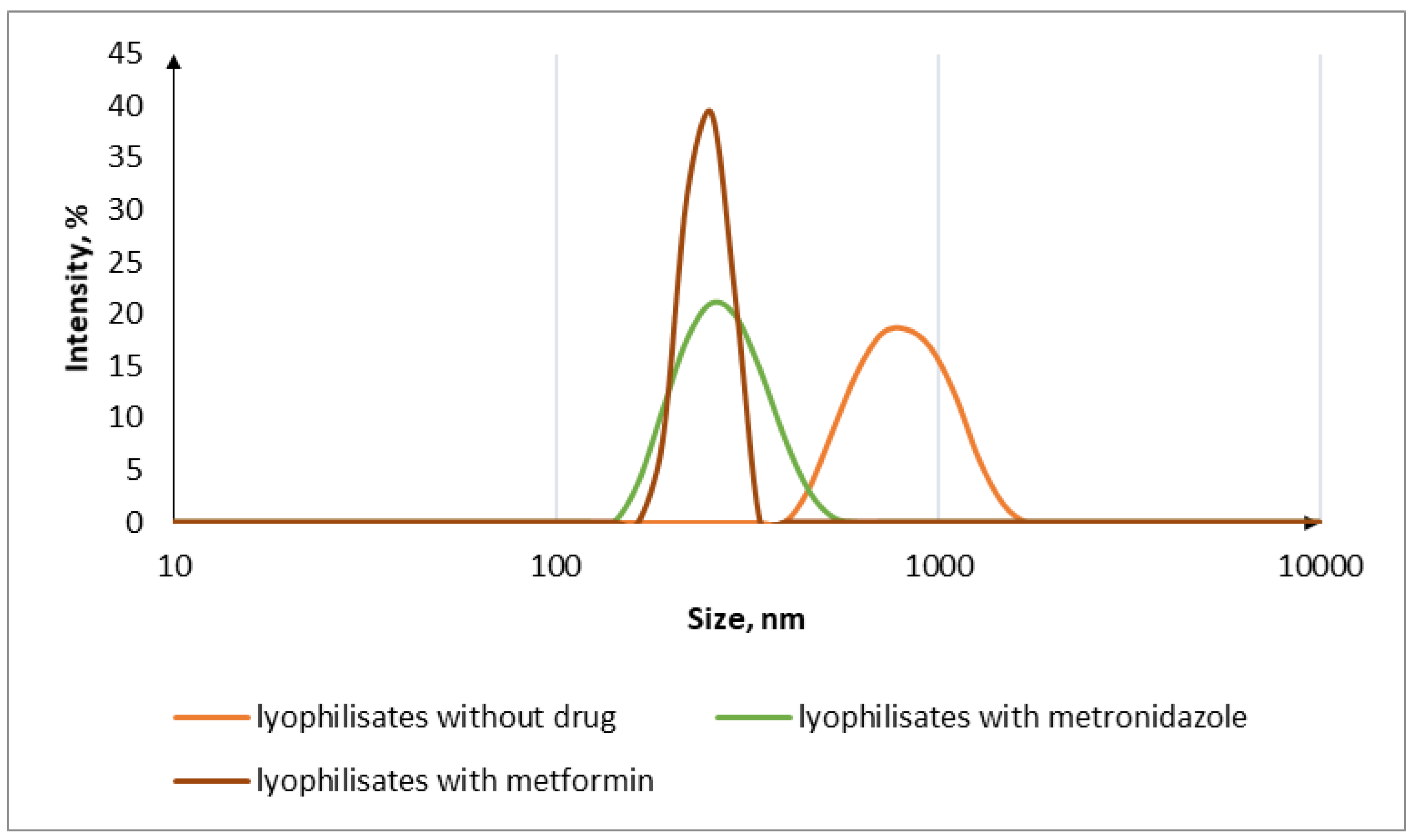
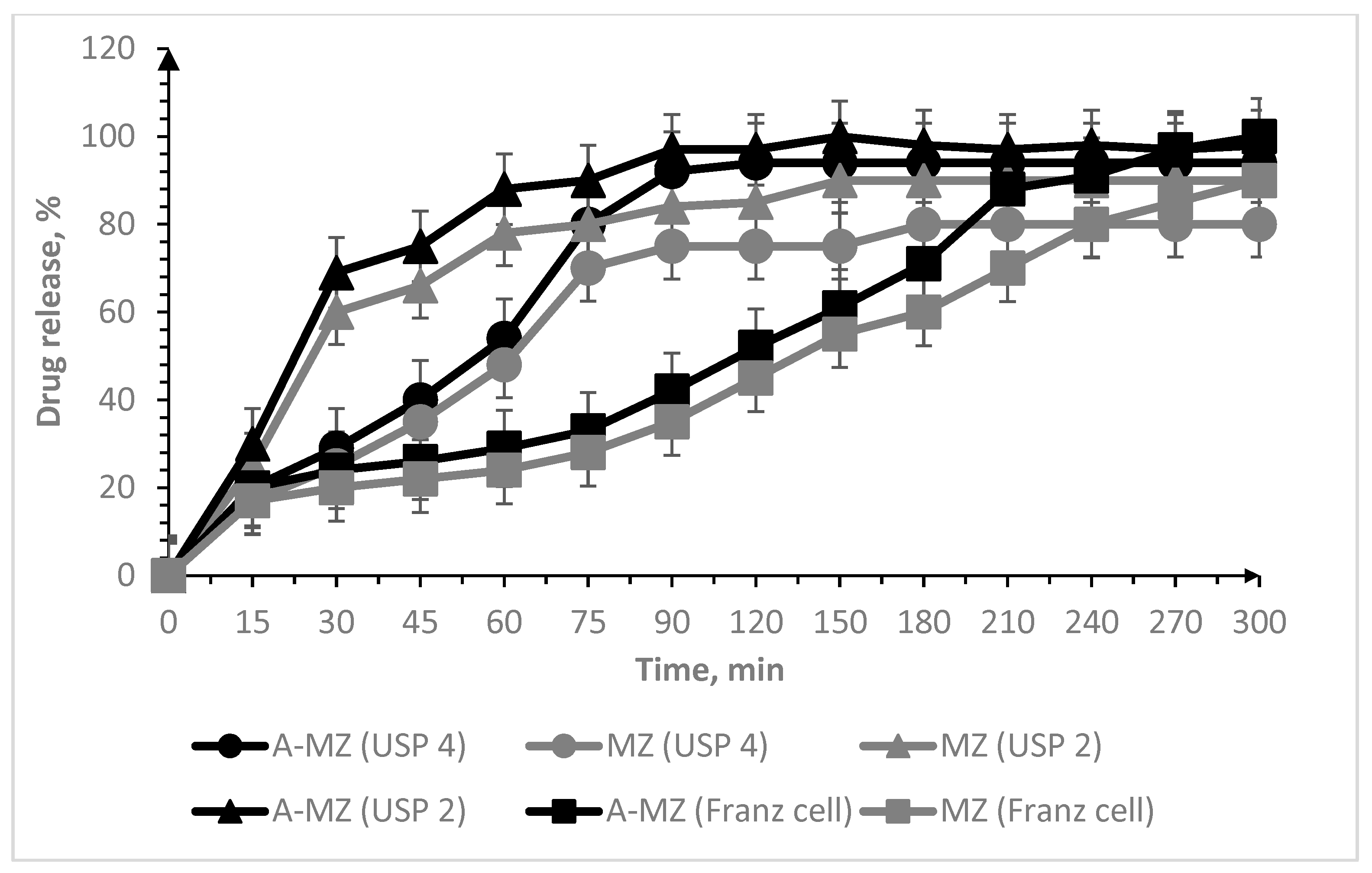
3.2. Oral Lyophilisates

Three types of oral lyophilisates were prepared on the basis of maltodextrin and Span-80, including IPEC without the drugs (A-lyo and B-lyo), IPEC and the drugs (A-MZ and B-MT) and the drugs without IPEC (MZ and MT). First the disintegration time of the lyophilisates without the drugs was investigated. After this evaluation, the particle size and surface charge were measured. It should be noted that the particle size measured after evaluating the disintegration of the obtained lyophilisates increased from 294 ± 20 nm for pure A-IPEC and from 298 ± 30 nm to 921 ± 34 nm for pure B-IPEC (Table 1) in the case of lyophilisates prepared with these IPECs. This may be due to the fact that the particles prepared after the disintegration of lyophilisates aggregate into larger conglomerates compared to the initial IPEC formed immediately after mixing the polymer ingredients. The charge of the IPEC particles after their preparation and the particles formed after the evaluation of the disintegration decrease, which may be due to the presence of the carboxyl groups of Carbopol® (Noveon®) on the surface of the particles.
Independent of the drug load, after freeze-drying, all tablets appeared as elegant solids without defects or signs of collapse, and they were easy to remove from blister and to handle. The disintegration time in artificial saliva of all oral lyophilisates was equal to or less than 60 s (Table 2).
The particle size decreased with the introduction of a drug into the composition of the oral lyophilisates (Figure 1). From an average particle size without drugs of 921 ± 34 nm with the introduction of metformin, the size became almost two times smaller, 465 ± 50 nm, and in the case of metronidazole, 272 ± 35 nm (Table 1). The reason for the decrease in size may be the compaction of the IPEC particles after the introduction of the drug into the lyophilisates. In addition, the charge of the particles increased, which may be due to the presence of a smaller number of free carboxyl groups of Carbopol® compared to the particles obtained after the disintegration of lyophilisates without the drug (Table 1).
The next stage of the research was the assessment of the drug release kinetics of the oral lyophilisates using three methods: a paddle stirrer (USP 2), a flow cell (USP 4) and a Franz cell. The novelty of our work lies in the comparative study of three methods of drug release: USP 2, USP 4 and Franz cells, from our oral lyophilisates containing IPEC particles. Each of the methods used made it possible to evaluate the release of the active ingredients; however, it was interesting to consider the above methods from the perspective of release from the same oral lyophilisates. From the point of view of proximity to the conditions in the oral cavity, the USP 4 method and the Franz cell are the most suitable methods for simulating drug release from oral lyophilisates; however, the USP 2 method can also be applied, since a modification of this method using a perforated disk designed for USP 5 was used.
Studies on the release of active ingredients from orodispersible dosage forms have been reflected in previous research studies [6,9,10,11,24,30,31,32,33]. The USP 2 method was used with a medium volume of 900 mL, a paddle speed of 50 rpm and a pH value of the medium of 6.8 or 7.2 [6,10,11]. The USP 1 method was used with a medium volume of 900 mL, a paddle speed of 50 rpm and a pH value of 6.8 [9]. Another team of authors has conducted a comparative study of the release of carbamazepine from systems with immediate release using the USP 2 and USP 4 apparatus [34]. Comparative release from transdermal systems was previously performed using the USP 5 and USP 7 methods [41]. Therefore, our goal was to compare these methods of drug release assessment with respect to our developed oral lyophilisates, using artificial saliva as the most appropriate dissolution medium.
All experimental conditions were intended to simulate the residence of oral lyophilisates in the oral cavity. In the case of method 2, a tablet was placed at the bottom of a beaker and covered with a disc with a mesh surface to prevent its floating. In other words, we proposed a modification of the USP 5 method, which is the compendial assay to determine the drug release from transdermal dosage forms. In the flow cells, the saliva flow rate was set at 2 mL/min to mimic the conditions in the oral cavity. In the case of the Franz cell, the dialysis membrane simulated the mucosal surface, and the test was intended to measure the drug diffusion through it.
Figure 2 and Figure 3 report the release profiles in the different apparatus, and Table 3 and Table 4 present the kinetic constants.
In the case of oral lyophilisates containing metformin, the plateau in the release pattern was achieved after 90 min, independent of the set-up (Figure 2), and the release rates were comparable (Table 3).
In the case of metronidazole (Figure 3), the drug release was also completed in 90 min using both the USP 2 apparatus and the “flow cell” method. On the other hand, the membrane present between the two compartments of the Franz diffusion cell limited the drug diffusion, and the plateau was reached in about 270 min. The IPEC-free samples had similar release profiles; however, the IPEC-based lyophilisates are more suitable for immediate release systems.
The results of the kinetics of drug release from oral lyophilisates (Table 3) evidence that the drug release occurred nonlinearly. It can be assumed that aside from the control of the drug release kinetics exerted by the IPEC, the drug release appeared slower when using the Franz cell and the USP 2 apparatus (p < 0.05) since the static environment of the Franz cell and the pseudo-laminar movement of the dissolution medium in apparatus 2 could favour the aggregation of IPECs.
According to statistical analysis significantly different results were observed in the cases of USP 2 and USP 4 (p = 0.0054) and USP 2 and Franz cell (p = 0.0054) in the case of metformin release. The Franz cell and USP 4 showed similar release results; therefore, there is no significant difference between these data (p > 0.05). However, for metronidazole lyophilisates, only method 2 and the Franz cell showed statistically significant results (p = 0.0251). This may be due to the peculiarities of the release of metronidazole and the influence of the static environment of the Franz cell. Thus, when evaluating the release from orodispersible tablets, both USP 4 and the Franz cell can be used (pmetronidazole = 0.1272 and pmetformin = 0.1272).
The tablets containing the IPEC in their composition achieved the maximum release parameters in a faster time and maintained the release for 5 h of the experiment.
When carrying out the mathematical modelling (Table 3), the Korsemeyer–Peppas model was chosen to analyse the release of drugs from the polymer systems because the tablets included IPECs (ft = K · tn).
For lyophilisates based on B-MT, the release exponent (n) for all the methods used was less than 0.45, which indicates that the drug transport mechanism occurred according to Fickian diffusion. In the case of lyophilisates based on A-MZ, the release exponent (n) for the USP 2 and USP 4 methods was less than 0.45, which indicates the drug transport mechanism is a Fickian diffusion; however, in the case of the Franz cell, the release exponent (n) was in the range of 0.45 to 0.89, that is, the release occurred according to non-Fickian transport.
Due to the fact that the correlation coefficient obtained when performing calculations according to the Korsemeyer–Peppas equation turned out to be low in the case of some lyophilisates, we also chose the Gompertz model to describe the release profiles (Xt = Xmax·exp[−α·eβ·logt] or y = a·exp(−exp(−k·(x − xc)))) (Table 4). This model has a steep rise at the beginning and gradually decreases to maximum. This model is used when active ingredients are released from immediate release systems [44]. It should be noted that this model is more suitable for the systems we are studying, and it has large correlation indicators.
Thus, the release results in all three release devices are comparable, and any of the compared methods used to evaluate the release can be applied in the case of oral lyophilisates. The drug release studies that used the Franz cell and USP 4 achieved the most similar results.
Thus, in our work, various methods for evaluating the release of active ingredients from oral lyophilisates (USP 2, USP 4 and Franz cell) were investigated. Since the oral cavity contains a small amount of salivary fluid, USP 4 and the Franz cell were the closest, however, USP 2 can also be used in the case of a covered tablet with a disc with a mesh surface to prevent its floating. Thus, any of these methods can be applied to evaluate the release of active ingredients from oral lyophilisates.

4. Conclusions

The IPEC samples prepared in this study have mucoadhesive ability (A-IPEC: MDF = 103.5 ± 0.7 kPa; WA = 2154 ± 66 mJ; B-IPEC: MDF = 111.0 ± 9.5 kPa; WA = 2611 ± 413 mJ). The orodispersible tablets prepared met the requirements for disintegration (the disintegration time in artificial saliva of all oral lyophilisates was equal or less than 60 s). The analysis of the particle size showed that the IPECs obtained after disintegration had nano-dimensions (A-MZ: Dh = 272 ± 35 nm; B-MT: Dh = 465 ± 50 nm) which, in combination with their mucoadhesive ability, is promising for the transmucosal delivery of drugs. The evaluation of the drug release using different dissolution methods showed that these approaches can all be used to evaluate drug release from orodispersible tablets; the results obtained are comparable. Thus, when evaluating release from orodispersible tablets, both USP 4 and the Franz cell can be used.
Drug release from lyophilisates begins when they are placed in a liquid medium. Due to the presence of an IPEC in the tablets, when the tablets are dispersed into small particles under normal conditions, which have mucoadhesive properties, it becomes possible to release drugs for a long time.

Author Contributions

V.R.T. was carry out synthesis and characterization of interpolyelectrolite complexes, preparation and characterization of oral lyophilisates, wrote and corrected the article, C.G.M.G. was investigated in vitro mucoadhesive properties of IPEC, F.C. and F.S. reviewed and corrected the article R.I.M. was responsible for conceptualization and research methodology, as well as reviewed and corrected the article. The article was written with the participation of all co-authors. All authors have read and agreed to the published version of the manuscript.

Funding

This research was funded by the Russian Science Foundation via grant 20-75-00051 (for V.R.T.). The metformin part was conducted with the financial support of Kazan State Medical University via grant N 1/8/22 (for R.I.M.).

Institutional Review Board Statement

Not applicable.

Informed Consent Statement

Not applicable.

Data Availability Statement

Data is contained within the article.

Conflicts of Interest

The authors declare no conflict of interest.

References

  1. Wiedey, R.; Kokott, M.; Breitkreutz, J. Orodispersible Tablets for Pediatric Drug Delivery: Current Challenges and Recent Advances. Expert Opin. Drug Deliv. 2021, 18, 1873–1890. [Google Scholar] [CrossRef] [PubMed]
  2. Stegemann, S.; Gosch, M.; Breitkreutz, J. Swallowing Dysfunction and Dysphagia Is an Unrecognized Challenge for Oral Drug Therapy. Int. J. Pharm. 2012, 430, 197–206. [Google Scholar] [CrossRef] [PubMed]
  3. Musazzi, U.M.; Khalid, G.M.; Selmin, F.; Minghetti, P.; Cilurzo, F. Trends in the Production Methods of Orodispersible Films. Int. J. Pharm. 2019, 576, 118963. [Google Scholar] [CrossRef] [PubMed]
  4. Sharma, S.; Singh, K. Oral Disintegrating Tablets—An Updated Patent Perspective. Recent Pat. Drug Deliv. Formul. 2020, 14, 166–190. [Google Scholar] [CrossRef] [PubMed]
  5. Sachan, A.K.; Singh, S.; Kumar, V.; Kumari. Comparative Study of Natural and Synthetic Superdisintegrants In Orodispersible Metformin Tablet. Asian J. Pharm. Res. Dev. 2019, 7, 46–53. [Google Scholar] [CrossRef]
  6. Bhavna, B.G.; Bhushan, R.R.; Jain, A.S. Formulation and Evaluation of Orodispersible Tablet of Sulindac. Eur. J. Pharm. Res. 2022, 2, 11–19. [Google Scholar] [CrossRef]
  7. Khalid, G.M.; Selmin, F. Applications of Alginates in the Design and Preparation of Orodispersible Dosage Forms. In Properties and Applications of Alginates; IntechOpen: London, UK, 2021. [Google Scholar] [CrossRef]
  8. Kumar, R.; Sheela, M.A.; Sachdeva, M. Formulation and Evaluation of Orodispersible Tablets Containing CoCrystals of Modafinil. J. Drug Deliv. Ther. 2022, 12, 82–89. [Google Scholar] [CrossRef]
  9. Mohana, M.; Vijayalakshmi, S. Development and characterization of solid dispersion-based orodispersible tablets of cilnidipine. Beni Suef Univ. J. Basic Appl. Sci. 2022, 11, 83. [Google Scholar] [CrossRef]
  10. Kumari, P.V.K.; Srinivasa, Y.R. Formulation and evaluation of orodispersible tablets of donepezil hydrochloride. Int. J. Curr. Pharm. Res. 2020, 12, 45–51. [Google Scholar] [CrossRef]
  11. Bhattacharya, S.; Mohanta, T.D.S.; Basak, R. Orodispersible Tablet in Treatment of Migraine: Opportunities, Challenges and Recent Advancements. J. Drug Deliv. Ther. 2021, 11, 149–156. [Google Scholar] [CrossRef]
  12. Manda, P.; Popescu, C.; Juluri, A.; Janga, K.; Kakulamarri, P.R.; Narishetty, S.; Narasimha Murthy, S.; Repka, M.A. Micronized Zaleplon Delivery via Orodispersible Film and Orodispersible Tablets. AAPS PharmSciTech 2018, 19, 1358–1366. [Google Scholar] [CrossRef] [PubMed]
  13. Kuck, J.; Breitkreutz, J. Impact of lubrication on key properties of orodispersible minitablets in comparison to conventionally sized orodispersible tablets. Eur. J. Pharm. Biopharm. 2022, 180, 71–80. [Google Scholar] [CrossRef] [PubMed]
  14. Mhetre, L.R.; Kadam, P.S.; Gadhire, P.H. Formulation and Evaluation of Naproxen Orodispersible Tablets. Int. J. Pharm. Sci. Nanotechnol. 2022, 15, 6055–6060. [Google Scholar] [CrossRef]
  15. Van Nguyen, K.; Dang, T.K.; Vu, L.T.; Ha, N.T.; Truong, H.D.; Tran, T.H. Orodispersible film incorporating nanoparticulate loratadine for an enhanced oral bioavailability. J. Pharm. Investig. 2023. [Google Scholar] [CrossRef]
  16. Jassem, N.A. Orodispersible Tablets: A Review on Recent Trends in Drug Delivery. Int. J. Drug Deliv. Technol. 2022, 12, 432–436. [Google Scholar]
  17. Sinha, S.; Sonali; Garg, V.; Thapa, S.; Singh, S.; Chauhan, M. Empagliflozin containing chitosan-alginate nanoparticles in orodispersible film: Preparation, characterization, pharmacokinetic evaluation and its in-vitro anticancer activity. Drug Dev. Ind. Pharm. 2022, 48, 279–291. [Google Scholar] [CrossRef]
  18. Petrovick, G.F.; Kleinebudde, P.; Breitkreutz, J. Orodispersible tablets containing taste-masked solid lipid pellets with metformin hydrochloride: Influence of process parameters on tablet properties. Eur. J. Pharm. Biopharm. 2018, 122, 137–145. [Google Scholar] [CrossRef]
  19. Oliveira, L.J.; Veiga, A.; Stofella, N.C.F.; Cunha, A.C.; Graça, M.; Toledo, T.; Andreazza, I.F.; Murakami, F.S. Development and Evaluation of Orodispersible Tablets Containing Ketoprofen. Curr. Drug Deliv. 2020, 17, 348–360. [Google Scholar] [CrossRef]
  20. Lew, M.F. Selegiline Orally Disintegrating Tablets for the Treatment of Parkinson’s Disease. Expert Rev. Neurother. 2005, 5, 705–712. [Google Scholar] [CrossRef]
  21. Hua, S. Advances in Nanoparticulate Drug Delivery Approaches for Sublingual and Buccal Administration (mini-review). Front. Pharmacol. 2019, 10, 1328. [Google Scholar] [CrossRef] [Green Version]
  22. Watchorn, J.; Clasky, A.J.; Prakash, G.; Johnston, I.A.E.; Chen, P.Z.; Gu, F.X. Untangling Mucosal Drug Delivery: Engineering, Designing, and Testing Nanoparticles to Overcome the Mucus Barrier. ACS Biomater. Sci. Eng. 2022, 8, 1396–1426. [Google Scholar] [CrossRef] [PubMed]
  23. Mehanna, M.M.; Mneimneh, A.T.; Domiati, S.; Allam, A.N. Tadalafil-Loaded Limonene-Based Orodispersible Tablets: Formulation, in vitro Characterization and in vivo Appraisal of Gastroprotective Activity. Int. J. Nanomed. 2020, 15, 10099–10112. [Google Scholar] [CrossRef] [PubMed]
  24. Garipova, V.R.; Gennari, C.G.M.; Selmin, F.; Cilurzo, F.; Moustafine, R.I. Mucoadhesive Interpolyelectrolyte Complexes for the Buccal Delivery of Clobetasol. Polymers 2018, 10, 85. [Google Scholar] [CrossRef] [Green Version]
  25. Russo, E.; Selmin, F.; Baldassari, S.; Gennari, C.G.M.; Caviglioli, G.; Cilurzo, F.; Minghetti, P.; Parodi, B. A focus on mucoadhesive polymers and their application in buccal dosage forms. J. Drug Deliv. Sci. Technol. 2016, 32, 113–125. [Google Scholar] [CrossRef]
  26. Ruiz-Rubio, L.; Alonso, M.L.; Pérez-Álvarez, L.; Alonso, R.M.; Vilas, J.L.; Khutoryanskiy, V.V. Formulation of Carbopol®/poly(2-ethyl-2-oxazoline)s mucoadhesive tablets for buccal delivery of hydrocortisone. Polymers 2018, 10, 175. [Google Scholar] [CrossRef] [Green Version]
  27. Khutoryanskiy, V.V. Advances in mucoadhesion and mucoadhesive polymers. Macromol. Biosci. 2011, 11, 748–764. [Google Scholar] [CrossRef]
  28. Gupta, M.S.; Kumar, T.P. Characterization of Orodispersible Films: An Overview of Methods and Introduction to a New Disintegration Test Apparatus Using LDR-LED Sensors. J. Pharm. Sci. 2020, 109, 2925–2942. [Google Scholar] [CrossRef]
  29. Desai, N.; Redfearn, A.; MacLeod, G.; Tuleu, C.; Hanson, B.; Orlu, M. How Do Orodispersible Tablets Behave in an In Vitro Oral Cavity Model: A Pilot Study. Pharmaceutics 2020, 12, 651. [Google Scholar] [CrossRef]
  30. Khalid, G.M.; Selmin, F.; Musazzi, U.M.; Gennari, C.G.M.; Minghetti, P.; Cilurzo, F. Trends in the Characterization Methods of Orodispersible Films. Curr. Drug Deliv. 2021, 18, 935–946. [Google Scholar] [CrossRef]
  31. Guhmann, M.; Preis, M.; Gerber, F.; Pöllinger, N.; Breitkreutz, J.; Weitschies, W. Design, Development and in-Vitro Evaluation of Diclofenac Taste-Masked Orodispersible Tablet Formulations. Drug Dev. Ind. Pharm. 2015, 41, 540–551. [Google Scholar] [CrossRef]
  32. Wasilewska, K.; Winnicka, K. How to Assess Orodispersible Film Quality? A Review of Applied Methods and Their Modifications. Acta Pharm. 2019, 69, 155–176. [Google Scholar] [CrossRef] [PubMed] [Green Version]
  33. Cilurzo, F.; Musazzi, U.M.; Franzé, S.; Selmin, F.; Minghetti, P. Orodispersible dosage forms: Biopharmaceutical improvements and regulatory requirements. Drug Discov. Today 2018, 23, 251–259. [Google Scholar] [CrossRef] [PubMed] [Green Version]
  34. Medina, J.R.; Salazar, D.K.; Hurtado, M.; Cortés, A.R.; Domínguez-Ramírez, A.M. Comparative in Vitro Dissolution Study of Carbamazepine Immediate-Release Products Using the USP Paddles Method and the Flow-through Cell System. Saudi Pharm. J. 2014, 22, 141–147. [Google Scholar] [CrossRef] [PubMed] [Green Version]
  35. Selmin, F.; Khalid, G.M.; Musazzi, U.M.; Demartin, F.; Minghetti, P.; Cilurzo, F. Relevance of Production Method on the Physical Stability and in Vitro Biopharmaceutical Performances of Olanzapine Orodispersible Film. Int. J. Pharm. 2021, 603, 120697. [Google Scholar] [CrossRef]
  36. Perioli, L.; Ambrogi, V.; Rubini, D.; Giovagnoli, S.; Ricci, M.; Blasi, P.; Rossi, C. Novel Mucoadhesive Buccal Formulation Containing Metronidazole for the Treatment of Periodontal Disease. J. Control. Release 2004, 95, 521–533. [Google Scholar] [CrossRef]
  37. Proctor, W.R.; Bourdet, D.L.; Thakker, D.R. Mechanisms Underlying Saturable Intestinal Absorption of Metformin. Drug Metab. Dispos. 2008, 36, 1650–1658. [Google Scholar] [CrossRef]
  38. Sander, C.; Nielsen, H.M.; Jacobsen, J. Buccal Delivery of Metformin: TR146 Cell Culture Model Evaluating the Use of Bioadhesive Chitosan Discs for Drug Permeability Enhancement. Int. J. Pharm. 2013, 458, 254–261. [Google Scholar] [CrossRef]
  39. Sander, C.; Madsen, K.D.; Hyrup, B.; Nielsen, H.M.; Rantanen, J.; Jacobsen, J. Characterization of spray dried bioadhesive metformin microparticles for oromucosal administration. Eur. J. Pharm. Biopharm. 2013, 85, 682–688. [Google Scholar] [CrossRef]
  40. Belayneh, A.; Molla, F.; Kahsay, G. Formulation and Optimization of Monolithic Fixed-Dose Combination of Metformin HCl and Glibenclamide Orodispersible Tablets. Adv. Pharmacol. Pharm. Sci. 2020, 2020, 3546597. [Google Scholar] [CrossRef] [Green Version]
  41. Li, M.; Tan, H. Technical Note: Comparison of USP Apparatus 5 and 7 for In Vitro Drug Release from Nicotine Transdermal Systems. Dissolut. Technol. 2019, 26, 68–71. [Google Scholar] [CrossRef]
  42. Timergalieva, V.R.; Khusnutdinov, R.R.; Musina, R.R.; Elizarova, E.S.; Alsynbaev, R.R.; Nasibullin, S.F.; Moustafine, R.I. Development of orodispersible ibuprofen tablets based on a polymer-drug complex. Drug Dev. Regist. 2022, 11, 113–120. [Google Scholar] [CrossRef]
  43. Viktorova, A.S.; Elizarova, E.S.; Romanova, R.S.; Timergalieva, V.R.; Khutoryanskiy, V.V.; Moustafine, R.I. Interpolymer complexes based on Carbopol® and poly(2-ethyl-2-oxazoline) as carriers for buccal delivery of metformin. Drug Dev. Regist. 2021, 10, 48–55. [Google Scholar] [CrossRef]
  44. Dash, S.; Murthy, P.N.; Nath, L.; Chowdhury, P. Kinetic modeling on drug release from controlled drug delivery systems. Acta Pol. Pharm. Drug Res. 2010, 67, 217–223. [Google Scholar]
Figure 1. Particle size distribution after disintegrating test of lyophilisates.
Scipharm 91 00023 g001
Figure 2. Release of metformin from lyophilisates by methods: paddle stirrer (USP 2); “flow cell” (USP 4); Franz cell.
Scipharm 91 00023 g002
Figure 3. Release of metronidazole from lyophilisates using paddle stirrer (USP 2); “flow cell” (USP 4); and Franz cell methods.
Scipharm 91 00023 g003
Table 1. Main physical properties such as hydrodynamic diameter (Dh), zeta potential (ζ), and mucoadhesive characteristics of drug-free IPEC, oral lyophilisates with and without active ingredient.
Sample CodeIPEC (mol/mol)Oral LyophilisatesMTMZDh
(nm)
ζ
(mV)
MDF
(kPa)
WA
(mJ)
EPOC10NAA-1
A-IPEC11--------294 ± 20−14.40103.5 ± 0.72154 ± 66
B-IPEC2--1------298 ± 3015.00111.0 ± 9.52611 ± 413
A-lyo11--+----921 ± 34−48.86----
B-lyo2--1+----921 ± 34−20.00----
A-MZ11--+-+272 ± 35−10.10----
B-MT2--1++-465 ± 50−19.00----
PE--- ----13.4 ± 0.7407 ± 175
Chitosan--- ----62.0 ± 10.01682 ± 162
Sample codes: A-IPEC, B-IPEC—IPEC samples; A-lyo, B-lyo—oral lyophilisates, including IPEC; A-MZ, B-MT—oral lyophilisates, including IPEC and metronidazole or metformin, respectively.
Table 2. Oral lyophilized composition and disintegration time.
Oral Lyophilisates CodeOral Lyophilisates Composition (%, w/w)Disint. Time (s)
IPEC AIPEC BDrug MDXS80
A-lyo9.95--89.560.4923
B-lyo-9.95-89.560.4923
A-MZ2.76-46.1049.761.3860
B-MT-2.7646.1049.761.3850
MZ--46.1052.521.3850
MT--46.1052.521.3850
Table 3. Results of mathematical modelling of drug release from oral lyophilisates, according to the Korsemeyer–Peppas model.
Parameters
B-MT USP 2B-MT USP 4B-MT Franz Cell
Exponential release (n)0.157 ± 0.0230.124 ± 0.0360.106 ± 0.069
Constant release (k)31.094 ± 3.69151.832 ± 9.46745.605 ± 15.972
Correlation coefficient (R2)0.852280.493910.16469
A-MZ USP 2A-MZ USP 4A-MZ Franz cell
Exponential release (n)0.113 ± 0.0290.390 ± 0.1160.713 ± 0.056
Constant release (k)53.672 ± 8.02012.448 ± 7.3031.787 ± 0.540
Correlation coefficient (R2)0.638390.689020.96691
Table 4. Results of mathematical modelling of drug release from oral lyophilisates, according to the Gompertz model.
Parameters
B-MT USP 2B-MT USP 4B-MT Franz cell
Maximum dissolution (a)70.275 ± 1.79298.931 ± 1.74898.732 ± 2.235
Dissolution rate (xc)12.820 ± 2.68911.104 ± 1.61021.335 ± 2.591
Undissolved proportion (k)0.052 ± 0.0090.082 ± 0.0130.037 ± 0.004
Correlation coefficient (R2)0.945730.968120.97017
A-MZ USP 2A-MZ USP 4A-MZ Franz cell
Maximum dissolution (a)96.894 ± 1.29695.704 ± 2.249119.621 ± 10.359
Dissolution rate (xc)17.357 ± 1.29435.900 ± 2.60498.052 ± 12.697
Undissolved proportion (k)0.063 ± 0.0060.035 ± 0.0040.008 ± 0.001
Correlation coefficient (R2)0.986600.977840.98352
Disclaimer/Publisher’s Note: The statements, opinions and data contained in all publications are solely those of the individual author(s) and contributor(s) and not of MDPI and/or the editor(s). MDPI and/or the editor(s) disclaim responsibility for any injury to people or property resulting from any ideas, methods, instructions or products referred to in the content.

