Article

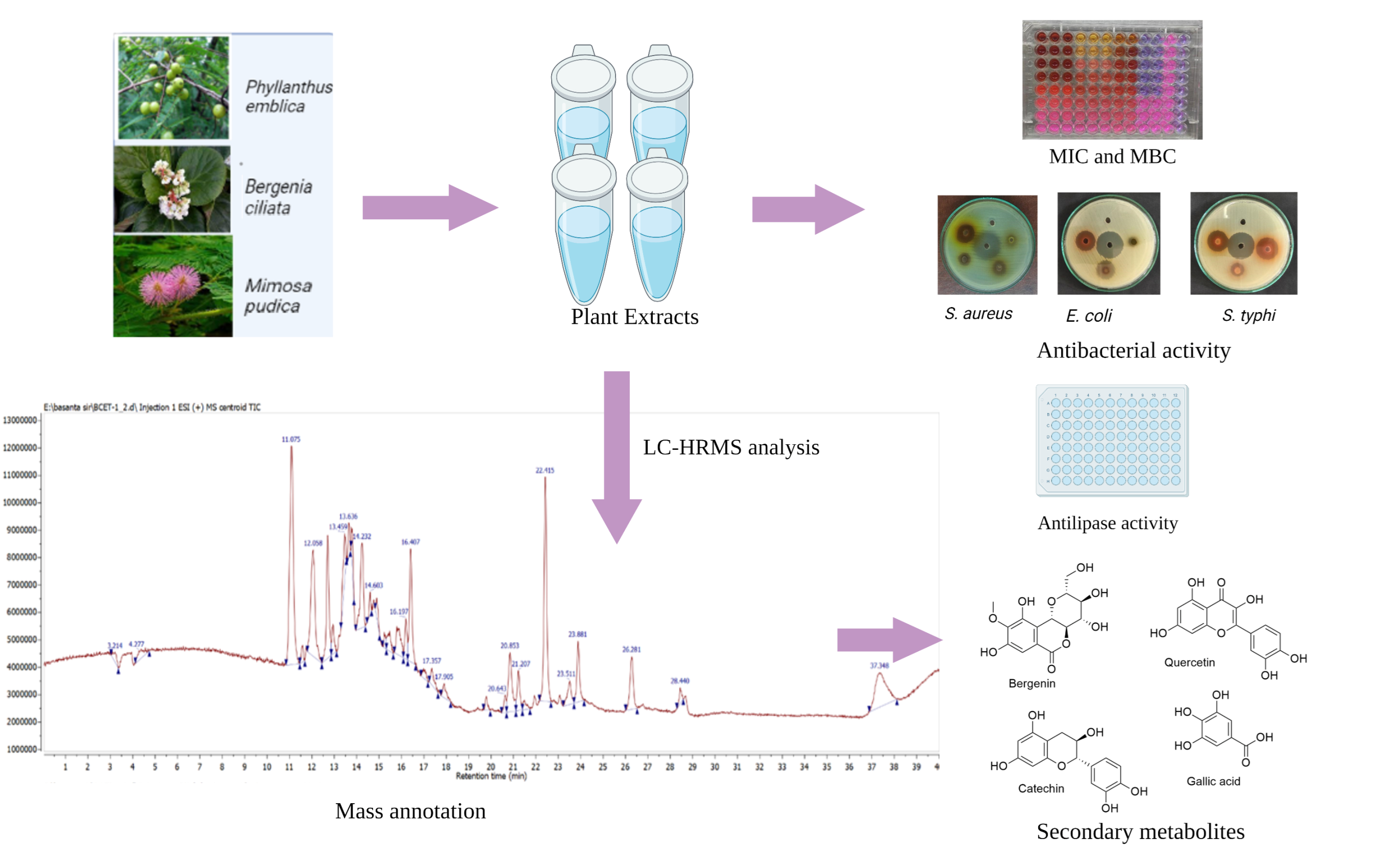
LC-HRMS-Based Profiling: Antibacterial and Lipase Inhibitory Activities of Some Medicinal Plants for the Remedy of Obesity

Biological Chemistry Laboratory, Central Department of Chemistry, Tribhuvan University, Kirtipur 44618, Nepal
*
Author to whom correspondence should be addressed.
Sci. Pharm. 2022, 90(3), 55; https://doi.org/10.3390/scipharm90030055
Submission received: 14 August 2022 / Revised: 3 September 2022 / Accepted: 5 September 2022 / Published: 8 September 2022

Abstract

:
Globally, obesity is a serious health concern that causes numerous diseases, including type 2 diabetes, hypertension, cardiovascular diseases, etc. Medicinal plants have been used to aid in weight loss since ancient times. Thus, this research is focused on the exploration of pancreatic lipase inhibitory activity and secondary metabolite profiling of Bergenia ciliata, Mimosa pudica, and Phyllanthus emblica, selected based on an ethnobotanical survey. The lipase inhibition was investigated using 4-nitrophenyl butyrate (p-NPB) as a substrate. To uncover further therapeutic potentials of these medicinal plants, antimicrobial activity and minimum inhibitory concentration (MIC) of the extracts were also determined. The ethyl acetate plant extracts showed higher antimicrobial activity against Staphylococcus aureus, Escherichia coli, Salmonella typhi, and Shigella sonnei. The MIC of ethyl acetate extracts of medicinal plants considered in this study ranges from 1.56 to 6.25 mg/mL. The hexane fraction of Mimosa pudica and Phyllanthus emblica showed a higher lipase inhibitory activity as compared to others, with IC50 values of 0.49 ± 0.02 and 2.45 ± 0.003 mg/mL, respectively. In the case of Bergenia ciliata, the methanolic extract inhibited lipase more effectively than others, with an IC50 value of 1.55 ± 0.02 mg/mL (IC50 value of orlistat was 179.70 ± 3.60 µg/mL). A mass spectrometry analysis of various solvent/solvent partition fractions (extracts) revealed 29 major secondary metabolites. The research offers a multitude of evidence for using medicinal plants as antiobesity and antimicrobial agents.

1. Introduction

A metabolic disorder is one of the known underlying reasons for the rise in obesity, and abdominal obesity is a direct or indirect consequence of a group of metabolic risk factors that also cause type II diabetes, cardiovascular disease, and non-alcoholic fatty liver disease [1]. The World Obesity Atlas 2022 predicted that approximately one billion people will be living with obesity globally by 2030 [2]. Prolonged maintenance of significant weight loss persists as a challenging problem in obesity treatment.
Pancreatic lipase inhibition is a considerable approach to treating metabolic syndrome since it is liable for 50–70% of all-out dietary fat hydrolysis. [3]. Alternative lipase inhibitors have piqued the interest of researchers because some lipase inhibitors have been suggested as effective weight-controlling medications. Additionally, the inordinate buildup of lipids in the pancreas is a leading cause of type II diabetes, which incites the dysfunction of insulin-producing pancreatic β-cells [4]. Intestinal lipase catalyzes the breakdown of triacylglycerols into fatty acids and glycerol in the intestinal lumen [5]. Slackening the lipolytic process can protect the pancreas by minimizing lipid absorption and eventually restoring regular insulin production [6]. Several FDA-approved antiobesity medications, such as orlistat, lorcaserin, topiramate extended-release, phentermine, naltrexone sustained-release, and liraglutide (injectable formulation), are available on the market [7]. Each drug fluctuates in its after-effect profiles and efficacy. Orlistat covalently bonds to serine at the active site of lipase but is also associated with several gastrointestinal adverse effects [8].
Numerous studies on natural products for obesity management also yielded positive results in terms of long-term safety, mode of action, and metabolic activity [9]. The long history of using natural products for weight loss demonstrates a preference for investigation over synthetic drugs [10]. Vitis vinifera, Rhus coriaria, Origanum dayi, Averrhoa carambola, Archidendron jiringa, Cynometra cauliflora, and Aleurites moluccana imply their potential for obesity treatment by antilipase activity [11]. Phyllanthus emblica widely known as Indian gooseberry or amla, and belonging to the Euphorbiaceae family, is a significant herbal remedy utilized in both the Unani (Graceo—Arab) and Ayurveda traditions of medicine. The fruit, which has been used in traditional medicine and Ayurveda as a robust Rasayana to cure diarrhea, jaundice, and inflammation, is the most commonly utilized portion of the plant for medicinal purposes out of all its parts [12]. Similarly, the pharmacological profile of Mimosa pudica L. (Mimosaceae), commonly known as the touch-me-not, live-and-die, and shame plant, suggests that it is a good herbal candidate for further investigation. The plant has a long history of usage in traditional medicine, having been applied to wounds and used to treat piles, dysentery, sinuses, and urogenital diseases [13]. Additionally, B. ciliata has indeed been reported as a remedy for over 100 ailments, with the greatest potential in the solution of gastrointestinal problems. Hence, the investigation of the unexplored potential of medicinal plants could result in alternative lipase inhibitors with minimal side effects.
Bacterial infections are viewed as a global concern and are thus acknowledged as a threat to human life. Resistance to antibacterial and antifungal medications has intensified in recent years, amplifying serious concerns for global health. As a consequence, infectious diseases are now more challenging to treat in the healthcare system. This resistance is due to the misuse of antibiotics [14]. The prime factor for antibiotic resistance or drug failure is due to formation of biofilm by microorganisms, which can be overcome by using alizarin as a natural antibiofilm agent [15]. Additionally, different natural products, such as flavonoids, alkaloids, polyphenols, and many other phytochemicals, have been evidenced to display antimicrobial activity [16]. Plants with a diverse range of secondary metabolites, such as P. emblica [17] and B. ciliata [18], provide an appealing conclusion of potential phytochemicals to control microbial diseases. Similarly, a recent study also revealed that the modified polymeric form of Gum kayara polysaccharides exhibits compelling antibacterial activity against various bacteria [19].
Identification of secondary metabolites is an important prerequisite in validating and acquiring a decisive result in the analysis of plant bioactivities. Metabolic profiling allows for comprehensive analyses of a wide range of metabolites, which greatly increases the value of common findings of plant bioactivities [20]. The complementary analytical platform of liquid chromatography-mass spectrometry (LC-MS) is used to identify a wide range of primary and secondary metabolites [21]. Recent advancements in mass spectrometry with advanced data processing technology allow for the simultaneous measurement of hundreds of chemically different metabolites and investigate more thoroughly the regulation of metabolic networks to study their influence on complex traits as well [22].
The current study is focused on the identification of the lipase inhibitory activity of B. ciliata, M. pudica, and P. emblica, followed by their antimicrobial studies against four microbial strains. Moreover, this study is also aimed at secondary metabolite profiling, using LC-HRMS to precisely measure the mass of unknown molecules, parent ions, and fragment ions of the plant extracts. The overview of this study is represented in Figure 1.

2. Materials and Methods

2.1. Chemicals and Reagents

Methanol, ethanol, ethyl acetate, dichloromethane, and hexane were purchased from Thermo Fisher Scientific (Powai, Mumbai, India). Resazurin was purchased from HiMedia (Thane West, Maharashtra, India). The lipase from the porcine pancreas (Type II), 4-nitrophenyl butyrate (p-NPB), orlistat, and neomycin were obtained from Sigma-Aldrich (St. Louis, MO, USA).

2.2. Plant Collection and Extract Preparation

M. pudica and P. emblica were collected from Shankar Nagar (27°39′48.3″ N; 83°28′52.3″ E), Rupandehi, Nepal, and B. ciliata were collected from Shantipur (28°11′24″ N; 82°13′48″ E), Gulmi, Nepal. Their taxonomy was authenticated and verified by the National Herbarium and Plant Laboratory (KATH), Godavari, Nepal. Plant materials harvested in the same climatic session were dried in the shade at room temperature. They were then pulverized by a grinder and drenched in methanol for 1 day. Subsequently, a cold percolation method was carried out and incubated for the next day, followed by filtration (Whatman filter paper 1). The procedure was done recurrently for 3 days in a row. The rotary evaporator was used under reduced pressure at 40 °C to evaporate the collected methanol from primary extracts, whereas the secondary extracts were prepared after the solvation of the primary extract in water followed by fractionation processes with different solvents, such as hexane, dichloromethane, and ethyl acetate-based on polarity.
The ethnobotanical uses and the pharmacological studies of the selected plant with the voucher specimen are listed in Table 1.

2.3. Lipase Assay

The porcine pancreas lipase inhibition assay was performed by modifying the method previously reported [25]. The 20 µL of plant extracts, 40 µL of lipase, and 100 µL of 0.1 mM PBS were taken at pH 8.0 and incubated at 37 °C for 15 min. The initial absorbance was observed at 405 nM.
Henceforth, the 40 µL of 3 mM substrate and p-NPB prepared in ethanol was added to each well and incubated at 37 °C for 30 min. The final absorbance was noted at 405 nm (SynergyLX, BioTek, Winooski, VT, USA). Orlistat and 30% DMSO were used as positive and negative controls, respectively. The lipase inhibitory activity was calculated using the given formula:
Inhibition   % = ( ODcontrol ODtest   sample ODcontrol   ) × 100

2.4. Antibacterial Assays

The antibacterial assay was performed using the agar well diffusion method [26]. The test microorganisms were inoculated in Mueller Hinton Broth and incubated at 37 °C until the turbidity matched 0.5 McFarland. Then, the lawn culture of test microorganisms was performed in Mueller Hinton Agar (MHA), with 1.5 × 108 CFU/mL microbial inoculum. Five wells were made on the lawn cultured MHA plate with the help of a sterile cork borer. A total of 1 mg/mL neomycin was used as a positive control, and 50% DMSO was used as a negative control. The plates were then incubated at 37 °C for 18–24 h, and the zone of inhibition was measured.

2.5. Minimum Inhibitory Concentration (MIC) and Minimum Bactericidal Concentration (MBC)

The minimum inhibitory concentration was done according to the Clinical and Laboratory Standards Institute (CLSI) [27]. The sterile 96-plate with a flat bottom was used for 2-fold serial dilution of the extracts in MHB. Then, a bacterial concentration of 106 CFU/mL was used in each well except for the negative control. The plate was then covered with a lid and incubated at 37 °C for 18–24 h. After incubation, resazurin was added to each well at a 0.003% concentration and left for 3–4 h of incubation at the same temperature. The lowest concentration with a blue color was considered MIC and for the determination of MBC, the concentration with MIC and above were streaked in nutrient agar plates and incubated for 18–24 h at 37 °C. The resazurin is converted to pink by the reductase enzyme of bacteria, so it is considered bacterial growth, whereas the blue color showed no bacterial growth. The MIC was done in duplicate and triplicate.

2.6. Statistical Analysis

The Gen5 Microplate Data Collection and Analysis Software was used for the processing of results, followed by MS Excel. The data were expressed as mean ± standard error of the mean. The IC50 values were determined using GraphPad Prism version 8 (San Diego, CA, USA).

2.7. LC-HRMS Analysis

The LC-HRMS analyses of ethyl acetate and the hexane fraction were carried out using an Agilent 6520, Accurate-Mass Q-TOF Mass Spectrometer outfitted with a G1311A quaternary pump, a G1329A autosampler, and a G1315D diode array detector (DAD). The aforementioned parameters were set for the source and scan: gas temp: 30 °C, gas flow: 11.01/min, nebulizer: 40 psi, VCap: 3500, fragmentor: 175, skimmer 1: 65.0, and octopole RF Peak: 750. The components of the solvent elution included acetonitrile (ACN), a 5 mM acetate buffer, and water, which was carried out at a flow rate of 1.5 mL/min. The elution gradient was initiated with 5% acetonitrile for 0.1 min, followed by 30% acetonitrile for 10 min, 80% acetonitrile for 32 min, and eventually back to the initial conditions. Throughout the procedure, the column temperature was kept consistent at 30 °C. The column elute was channeled to Q-TOF HRMS fitted with an electrospray interface after passing through the flow cell of the diode array detector. Positive electrospray ionization (ESI-positive mode) was used to analyze the mass spectrum in the mass range of 100–2000 Daltons at a scan rate of 1.03 [28].
The collected data was analyzed using Gen5 Microplate Data Collection and Analysis Software, followed by MS Excel. Using GraphPad, the 50% inhibition of enzymatic hydrolysis of the substrate (IC50) was determined. Each experiment was performed in triplicate, and the data were shown as mean ± standard deviation. Mestre Nova 12.0 was used to process and analyze data files from the LC-HRMS for compound annotation using PubChem, Dictionary of Natural Products 2, ChemSpider, and the METLIN database.

3. Results

3.1. Lipase Inhibition

At different concentrations, the ability of particular medicinal plants to inhibit lipase was tested. Lipase inhibition was tested at 5 mg/mL, and further dilution of different concentrations was performed based on the screening results. When compared to the IC50 value of orlistat, a positive control (179.70 ± 3.60 µg/mL), the results showed moderate to poor activity (IC50 values: 0.82 ± 0.05 to 5.37 ± 0.07 mg/mL). Among all fractions, crude, hexane, and EA showed higher activity than DCM and aqueous fractions. Table 2 shows the results for lipase inhibition.

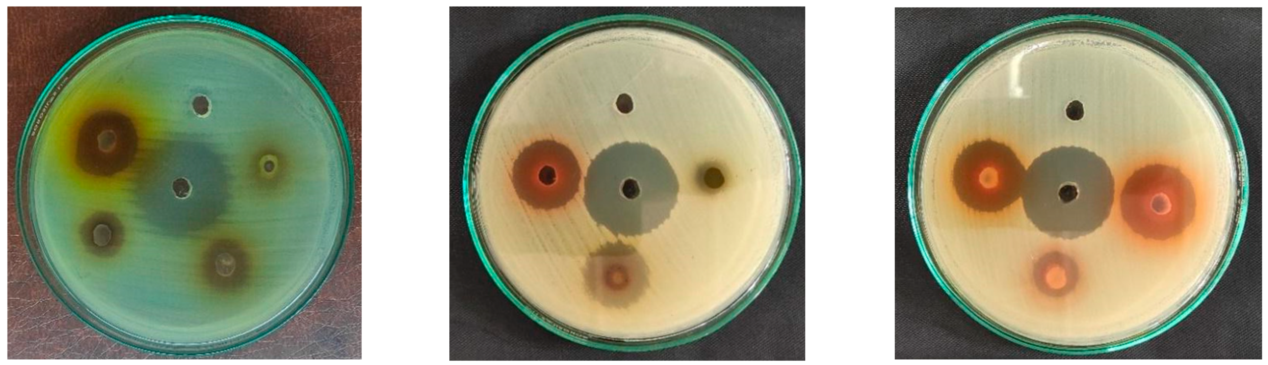
3.2. Analysis of Antimicrobial Activity

Different fractions of plant extracts were tested for antibacterial activity against ATCC strains of bacteria (Figure 2). The tested strains were: Staphylococcus aureus ATCC 25923, Escherichia coli ATCC 25923, Salmonella typhi ATCC 14028, and Shigella sonnei ATCC 25931. The details of antibacterial activity with ZoI are displayed in Table 3.

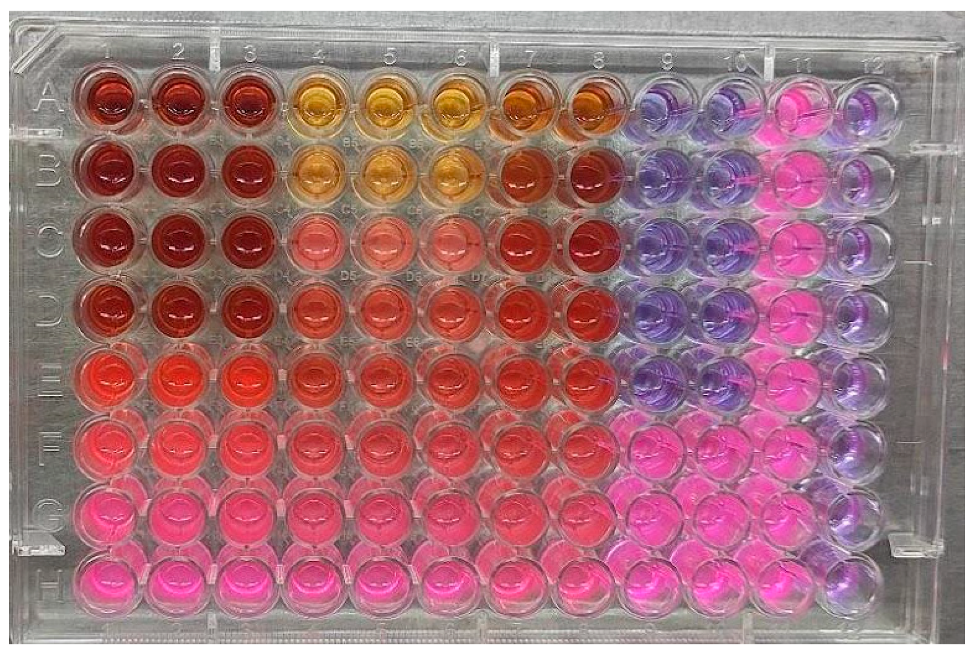
3.3. Determination of MIC and MBC

The highest zone of inhibition against the tested microorganisms was seen in the ethyl acetate fraction from all plants which was then subjected to the determination of MIC/MBC (Figure 3 and Figure S1). The MIC value ranges from 1562.5 to 6250 µg/mL, while the MBC ranges from 6250–12,500 µg/mL. Neomycin, the positive control, had demonstrated strong activity against the test microorganisms. Table 4 shows the details of MIC and MBC.

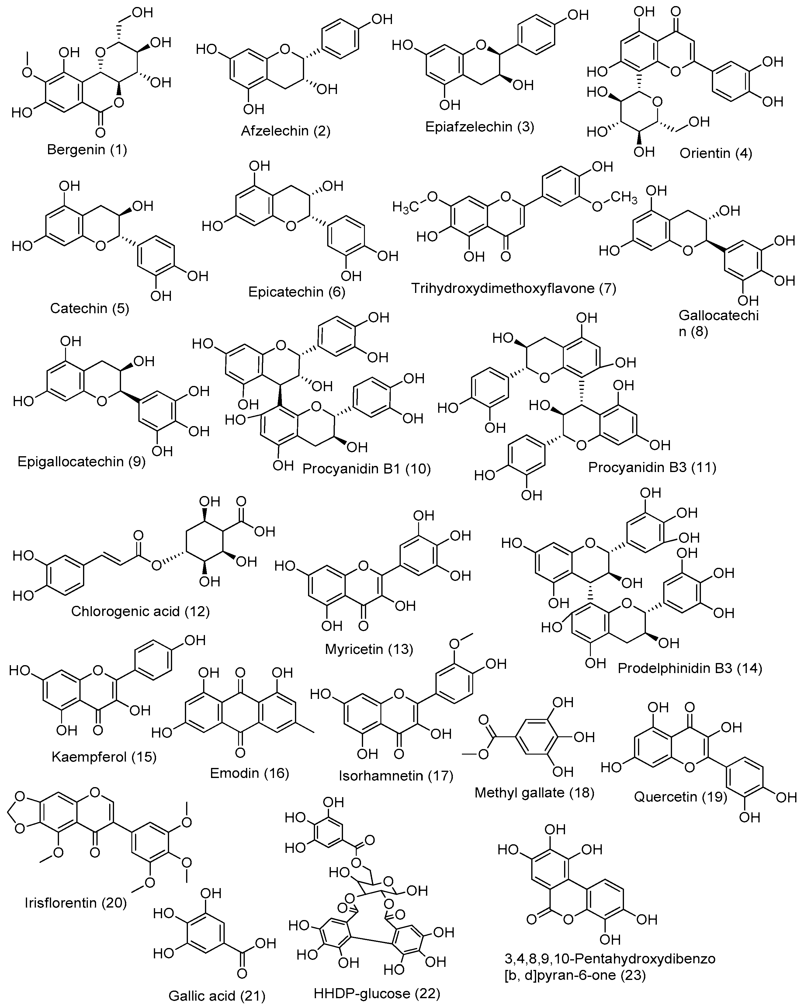
3.4. LC-HRMS-Based Molecular Annotation

The raw LC-HRMS data were processed, and the fraction with the best total ion chromatogram (TIC) was considered for the study with the MestreNova 12.0 software (Mestrelab Research, Santiago de Compostela, Spain). Table 5 shows the details of the identified compounds, along with their theoretical and observed mass-to-charge ratio, double bond equivalence (DBE), molecular formula, and absolute errors in parts per million (ppm) and retention time (Rt) in the positive ion mode in ESI. The mass spectra of B. ciliata, M. pudica, and P. emblica are shown in Figures S2–S4.
Based on the observed mass spectra, the compounds were identified, and the results were evaluated by comparing them to literature data. Structures of secondary metabolites identified based on the mass spectra of B. ciliata, M. pudica, and P. emblica were drawn using Chemdraw (Figure 4). In the ethyl extract of P. emblica, we observed the presence of phenolic compounds methyl gallate (m/z = 185.05), gallic acid (m/z = 171.02), flavonoids quercetin (m/z = 303.05), an isoflavone irisflorentin (m/z = 387.1), galloyl-hexahydroxydiphenoyl (HHDP)-glucose—a hydrolysable tannin (m/z = 483.07), derivatives of hydroxycinnamic acid 2-O-Caffeoylhydroxycitric acid (m/z = 371.06), 3,4,8,9,10-pentahydroxydibenzo [b, d]pyran-6-one (m/z = 277.06), isoquercetin (m/z = 465.1), prodelphinidin B3 (m/z = 595.14), cassiaoccidentalin B (m/z = 576.15), trihydroxydimethoxyflavone (m/z = 331.08), aflotaxin B1 (m/z = 329.06), kaemferol (m/z = 287.05), emodin (m/z = 271.06), isorhamnetin (m/z = 317.06), and trigalloyllevoglucosan IX (m/z = 619.09). The phytochemicals annotated in the ethyl extract of M. pudica are catechin/epicatechin (m/z = 291.08), gallocatechin/epigallocatechin (m/z = 307.8), procyanidin B1/procyanidin B3 (m/z =578.15), chlorogenic acid (m/z = 355.10), vitexin (m/z = 433.11), and myricetin (m/z = 319.04), In addition, important phytochemicals observed in the fraction of B. ciliata are bergenin (m/z = 329.08), afzelechin/epiafzelechin (m/z = 275.08), orientin (m/z = 449.10), and diosmetin (m/z = 301.07).

4. Discussion

Obesity is due to the unusual deposition of fat in the body and leads to different types of health problems, such as cardiovascular cancers, diabetes, hypertension, stroke, dyslipidemia, and osteoarthritis. Previous findings suggest that obese and diabetic patients are vulnerable to cardiovascular diseases [62]. These dietary fats are hydrolyzed by different types of lipases, such as tongue, gastric, and pancreatic lipases. Approximately 90% of dietary fats are composed of mixed triglycerides, and pancreatic lipase is responsible for the digestion of 50–70% of dietary fats into fatty acids and monoglycerides. Then, mixed micelles are formed with bile salts, cholesterol, and lysophosphatidic acid to produce triglycerides that are absorbed into enterocytes. The adipocytes present in the body are the main site for the storage of triglycerides and act as the major source of energy [63]. One of the strategies to combat obesity is to inhibit the lipase enzyme. Plant-based inhibitors are gaining popularity as safer, more affordable, and readily available alternatives to synthetic drugs due to their side effects, cost, and availability [64]. The plant contains different constituents, such as polyphenols, saponins, terpenes, flavonoids, and tannins, that are responsible for the inhibition of lipase enzymes [65]. The antilipase activity of our extracts also may be explained by the presence of these compounds, which were reported in our earlier study [66].
In contrast to DCM and aqueous fractions, hexane, methanol, and ethyl acetate fractions demonstrated higher inhibitory activity, according to our research. Methanolic extracts contain a mixture of compounds that might act synergistically to inhibit lipase enzymes. Drug combinations that work well together therapeutically are more prominent and highly effective. By preventing biological compensation, allowing lower dosages of each compound, or gaining access to context-specific multitarget mechanisms, synergistic combinations of two or more agents can overcome the toxicity and other side effects connected with high doses of single drugs [67]. At relatively low concentrations, the combination of kaempferol and orlistat demonstrated the synergistic inhibition of pancreatic lipase. When the combined concentrations of kaempferol and orlistat were less than 114.60 µM and 30.24 µM, the results showed the activity synergistically, however, kaempferol could partially replace orlistat to produce the same antiobesity results. [68]. The combination of the three drugs (ECG-EGCG-orlistat) exhibited potent synergy in inhibiting pancreatic lipase [69].
A previous study done on the hexane fraction of M. flagellipes and P. mildbraedii had shown significant activity against lipase enzyme. Besides that, hexane fraction significantly decreased total glycerides, total cholesterol, and low-density lipoprotein cholesterol as compared to hyperlipidemic control rats from both extracts. The GCMS analysis revealed two major compounds, 9-octadecenoic acid, and hexadecanoic acid, in P. mildbraedii, with hexadecanoic acid and 9,12-octadecadienoic acid in M. flagellipes [70]. So, the presence of these compounds in our study might be responsible for the inhibition of lipase enzyme from hexane fraction. The antiobesity activity of major constituent bergenin is due to increased norepinephrine-induced lipolysis in endogenous lipid droplets, slightly stimulated adrenocorticotropic hormone-induced lipolysis, and inhibited insulin-induced lipogenesis from glucose in fat cells obtained from rat epididymal adipose tissues [71]. The in vivo experiment in rats significantly reduced serum, cholesterol, triglycerides, and low-density lipoprotein-cholesterol levels after 21 days of oral administration [72].
The ethyl acetate fraction of M. pudica contains stigmasterol, quercetin, and avicularin. A previous study showed competitive inhibition by quercetin with an IC50 value of 53.05 µM, while non-competitive or mixed inhibition by avicularin with an IC50 value of 141.84 µM [73]. Stigmasterol showed weak porcine pancreatic lipase inhibition of 2.7 ± 0.4% at 100 μg/mL as compared to 34.5 ± 5.4% of orlistat at the same concentration [74]. Antidiabetic constituents, such as gallic acid, ellagic acid, chebulagic acid, and quercetin, along with other natural compounds, were reported earlier from P. emblica [75]. A previous study showed that chebulagic acid, ellagic acid, and gallic acid showed an IC50 value of 57.4 µg/mL, 90 µg/mL, and 5192 µg/mL, respectively, for pancreatic lipase inhibition [76]. The ethanolic fruit extract of P. emblica showed antilipase activity due to decreased triglyceride accumulation and downregulating adiponectin, FABP4, PPARγ, and cEBPα, respectively [77]. There is a rise in the number of multidrug-resistant pathogens. So, to cope with this situation, antimicrobial drugs are in high demand globally, but their production is delayed. For this reason, scientists are now attracted to natural resources. Plants are an easily available option and have been in use since ancient times as an ethnobotanical remedy. A previous study showed that the highest zone of inhibition in EA extracts of B. ciliata with ZoI, was 21 mm for S. aureus and 11 mm for E. coli. Similarly, another study from ethanolic extracts showed ZoI (24.0 ± 0.10) mm against S.aureus, (23.7 ± 0.25) mm against E. coli, and (22.8 ± 0.15) mm against S. typhi at 50 mg/mL [78]. The MIC was reported as 2500 µg/mL [79]. We found the highest zone of inhibition in the EA fraction in all tested bacteria (Table 3), and the MIC value was 1250 µg/mL (Table 4). The antibacterial activity of methanolic extract of M. pudica was reported with a value of 15 mm for S. aureus, 20 mm for K. pneumoniae, 12 mm for E. coli, and 14.5 mm for S. typhi, using 5 mg/mL disc [80]. In our study, the following was observed: 19 mm for S. aureus, 8 mm for E. coli, 12 mm for S. typhi, and 23 mm for S. sonnei using 50 mg/mL methanolic extracts. In the previous study, the ZoI of S. aureus and S. typhi was reported as 9 and 8 mm, respectively, from P. emblica extract. The MIC and MBC were observed at 50 mcg/mL and 65 mcg/mL, respectively, for S. aureus (methanol extract), in contrast to our observed values of 6250 µg/mL and 12,500 µg/mL (ethyl acetate fraction). The MIC and MBC for S. typhi were reported as 35 and 45 mcg/mL as compared to 3125 and 6250 µg/mL in our study [81]. Our study revealed that the ethyl acetate fraction is the more potent fraction, followed by the methanolic (crude) extract for antimicrobial activity. The methanolic extracts contain several metabolites, such as phenols, flavonoids, tannins, terpenoids, alkaloids, saponins, glycosides, and steroids. The antimicrobial activity of these phenolic and flavonoid compounds is mediated by different mechanisms. These compounds may have a synergistic effect on antimicrobial activity, whereas the ethyl acetate fractions contain high phenolic and flavonoid content, as previously reported in our study [66]. The antimicrobial activity of these phenolic and flavonoid compounds is mediated by different mechanisms. These include inhibition of nucleic acid synthesis, inhibition of attachment and biofilm formation, inhibition of cytoplasmic membrane function, and alteration of membrane permeability, leading to cell destruction as well as attenuation of pathogenicity [82]. To the best of our knowledge, the lipase inhibitory activity of B. ciliata, M. pudica, and P. embilica was performed for the first time in Nepal. The study was limited to in vitro testing. However, additional research on the isolation of potent compounds, enzyme kinetics, in silico, and in vivo testing can be conducted.
The isolation and characterization of plant metabolites is still a significant challenge due to the lack of multivariate analyses, funding sources, and laboratory accessibility in our perspective. In this study, LC-HRMS was performed for the identification of bioactive metabolites in the fractions of B. ciliata, M. pudica, and P. emblica. Mestrenova 12.0 software, USA was used to annotate the metabolites based on m/z, retention time, and molecular formula, and other databases were used to search, assign formulas, and compound structures.
The compounds annotated from the ethyl extracts of B. ciliata with the base peak at m/z 329.08, molecular formula C14H16O9, DBE 7, and fragment peaks at 314.78, 251.05, 237.07, and 194.40 are stated as bergenin [29]. The fragmentation pattern of bergenin is shown in Figure S5. Likewise, the base peak at m/z 275.08, molecular formula C15H14O5, DBE 9, along with fragment peaks at 257.17 (loss of water) and 233.08 is considered afzelechin/epiafzelechin [32]. The compound with a base peak at m/z 449.10, molecular formula C21H20O11, DBE 12, and the characteristic fragment ions at 431.12 [M + H-H20]+ corresponding to the losses of the molecule H2O (18 Da) between the 2″-hydroxyl group of the sugar and the 5 or 7-hydroxyl group of the aglycone, 329.36 and 299.30 fragment peaks indicate the losses of C4H8O (120 Da) and 150 Da from the [M + H]+ molecule is annotated as orientin [34]. In addition, the base peak at m/z 301.07, molecular formula C16H12O6, and DBE 11 with characteristic fragment ions 258.12, 153.31, and 149.09 are explicated as diosmetin. The fragmentation pattern of diosmetin is shown in Figure S6. Additionally, a base peak at m/z 331.08, molecular formula C17H1407, DBE 11, and fragment ions at 301.08 and 315.09 are considered trihydroxy methoxy flavone [38]. The fragmentation pattern of trihydroxy methoxyflavone is shown in Figure S7.
The compounds in the extract of M. pudica with the base peak at m/z 291.08, molecular formula C15H14O6, DBE 9, and fragment peaks at 313.07 [M + Na] + and 139.03 are speculated to be catechin/epicatechin [35,36,37]. The fragmentation pattern of catechin and epicatechin is shown in Figure S8. Likewise, [M + H] + at m/z 307.08, molecular formula C15H14O7, DBE 9 and along with fragment peak at 329.07 [M + Na] +, 289.07 and 139.03 is considered as gallocatechin/ epigallocatechin [36,37,38]. The fragmentation pattern of gallocatechin/epigallocatechin is shown in Figure S9. Compounds with characteristic fragment ions 427.10 [M + H − 152], and 289.07 (kaempferol) and base peak [M + H] + at m/z 579.15, molecular formula C30H26O12, and DBE 18 are annotated as procyanidin B1/procyanidin B3 [36,40]. The molecular ion at m/z 355.10, molecular formula C16H18O9, DBE 8, and fragment ions at 193.02 is characterized as chlorogenic acid [41,42]. The fragmentation pattern of chlorogenic acid is shown in Figure S10. The base peak at m/z 433.11, molecular formula C21H20O10, and DBE 12 with fragment peaks of 343.04; 313.07; and 285.14 manifested could be vitexin. The fragmentation peaks at m/z 343.04 and 313.07 were formed by the crisscross cleavage of the hexose unit and were formed due to the loss of C3H6O3 (90 Da) and C4H8O4 (120 Da) from [M + H] + ion, respectively. Due to the loss of CO (28 Da) from the m/z 313.07 ion, the product ions at m/z 285.14 were produced [44]. The base peak at m/z 319.04 with molecular formula C15H10O8 and DBE 11 with fragments peaking at 181.05 and 153.01 is assigned as myricetin. The fragmentation pattern of myricetin is shown in Figure S11 [31,79]. Likewise, the compound in the extract of P. emblica base peak with m/z 465.1, molecular formula C21H20O12, and the fragment ion at 303.05 (quercetin) and 289.07 (kaempferol) are interpreted as isoquercetin [60]. The fragmentation pattern of isoquercetin is shown in Figure S12. The mass spectrum with a base peak at m/z 594.13, molecular formula C30H26O13, DBE 18, and fragment peaks at 427.08, 169.07, 291.09, and 305.07 is predicted to be prodelphinidin B3 [40,47]. The fragmentation pattern of prodelphinidin B3 is shown in Figure S13. Moreover, in our spectra base peak with m/z 577.15, molecular formula C27H28O14, DBE 14 is annotated as cassiaoccidentalin [83]. The molecular ion peak at m/z 329.06, molecular formula C17H12O7, and DBE 12, is annotated as aflatoxin B1 [49]. Likewise, the base peak with m/z 287.05, molecular formula C15H10O6, DBE 11, and fragment peak at m/z 259.13, 165.09, and 153.12 are predicted to be kaempferol [50]. The fragmentation pattern of kaempferol is shown in Figure S14. Additionally, a base peak at m/z 271.06, molecular formula C15H10O5, DBE 11, and fragment ions at 253.16, 243.17, 229.14, 225.13, and 197.08 is considered emodin [51]. The fragmentation pattern of emodin is shown in Figure S15. The molecular ion peak at m/z 317.06, molecular formula C16H12O7, and DBE 11, and fragment ions at 303.21, 274.20, and 153.12 are annotated as isorhamnetin [52]. The fragmentation pattern of isorhamnetin is shown in Figure S16.
Additionally, the ethyl extract of P. emblica with [M + H] + at m/z 185.05, molecular formula C8H8O5, DBE 5, and fragment ions at 170.97 and 127.03 is considered methyl gallate [61,84]. The fragmentation pattern of methyl gallate is shown in Figure S17. Base peak m/z 303.05, molecular formula C15H10O7, DBE 11, and fragment ions at 273.12 and 257.13 due to the loss of [Y-CHO] + and [CO + H2O] + is annotated as quercetin [53]. The base peak at m/z 387 molecular formula C20H18O8, and DBE 12 with the fragment ions at 357.09 [M + H − CH3 × 2] +, 372.07 [M + H − CH3]+ manifested it could be irisflorentin [54,55]. Moreover, another annotated compound is gallic acid with a base peak at m/z 171.02, molecular formula C7H6O5 and DBE 5, with the fragment ions peak at 127.03 [M + H-CO2] + [57]. Likewise, [M + H] + at 483.07, molecular formula C20H18O14. DBE 12 along with a fragment peak at m/z 423.04 with glucose ring cleavage m/z 303.20 equivalent to an HHDP residue, and m/z 277.03 by the decarboxylation of the HDDP moiety is considered as HHDP-glucose [57]. Additionally, [M + H] + at m/z 371.06, molecular formula C15H14O11, and DBE 9 are considered as 2-O-Caffeoylhydroxycitric acid [59]. Base peak m/z 277.06, molecular formula C13H8O7, and DBE 10 annotated as 3,4,8,9,10-pentahydroxydibenzo [b, d] pyran-6-one [84]. Likewise, the base peak at m/z 619.09, molecular formula C20H26O22, and DBE 8 is annotated as trigalloyllevoglucosan IX [61]. However, to confirm their various pharmacological significance, additional research on the isolation and characterization of plant extract-derived compounds is required.

5. Conclusions

The current research is centered on the observable evidence of B. ciliata, M. pudica, and P. emblica’s lipase inhibitory action, preceded by their antimicrobial examinations. One of the strategies to combat obesity is to inhibit the lipase enzyme. The antilipase activities of different extracts were evaluated by pancreatic lipase inhibition assay. Among all fractions of the plants, crude, hexane, and EA showed higher activity for lipase inhibition than DCM and aqueous fractions. From the study, it was found that the highest zone of inhibition against the tested micro-organism was in EA fractions of all the plant extracts depicting higher antimicrobial activity than other fractions.
The antilipase activity of a plant is explained by the presence of these compounds, which were reported in our earlier study as well as analyzed through LC-HRMS. A further experiment on the isolation of potent inhibitory compounds and their mechanism of action in animal models is required to favor the drug discovery program.

Supplementary Materials

The following supporting information can be downloaded at: https://www.mdpi.com/article/10.3390/scipharm90030055/s1, Figure S1: MBC of antibiotic and P. emblica against E. coli; Figure S2: Mass spectrum of Bergenia ciliata; Figure S3: Mass spectrum of Mimosa pudica; Figure S4: Mass spectrum of Phyllanthus emblica; Figure S5: Fragmentation pattern of bergenin; Figure S6: Fragmentation pattern of diosmetin; Figure S7: Fragmentation pattern of trihydroxy-dimethoxyflavone; Figure S8: Fragmentation pattern of catechin/epicatechin; Figure S9: Fragmentation pattern of gallocatechin/epigallocatechin; Figure S10: Fragmentation pattern of chlorogenic acid; Figure S11: Fragmentation pattern of myricetin; Figure S12: Fragmentation pattern of isoquercetin; Figure S13: Fragmentation pattern of prodelphinidin B3; Figure S14: Fragmentation pattern of kaempferol; Figure S15: Fragmentation pattern of emodin; Figure S16: Fragmentation pattern of isorhamnetin; Figure S17: Fragmentation pattern of methyl gallate.

Author Contributions

N.P. designed and supervised the research; B.K.S. and K.K. performed research; B.A. analyzed mass spectrometry data; K.K., B.A., J.B. and N.P. wrote the manuscript; and S.J., reviewed the literature and edited the manuscript. All authors have read and agreed to the published version of the manuscript.

Funding

The funding for this work was provided by University Grants Commission, Nepal to Basanta Kumar Sapkota (UGC Award No: PhD-76/77-S&T-12). We are also thankful to Ashis Acharya (Shimane University, Japan) for providing high resolution images.

Institutional Review Board Statement

Not applicable.

Informed Consent Statement

Not applicable.

Data Availability Statement

The datasets for this study are available upon reasonable request to the corresponding author.

Acknowledgments

The authors are grateful to the National Herbarium and Plant Laboratories (NHPL)/KATH, Lalitpur, Nepal for their assistance in the identification of plants and herbarium deposition and for providing voucher specimens.

Conflicts of Interest

The authors declare no conflict of interest.

References

  1. Brahe, L.K.; Astrup, A.; Larsen, L.H. Can We Prevent Obesity-Related Metabolic Diseases by Dietary Modulation of the Gut Microbiota? Adv. Nutr. 2016, 7, 90–101. [Google Scholar] [CrossRef] [PubMed]
  2. World Obesity Federation. Global Obesity Observatory. Available online: https://data.worldobesity.org/?_ga=2.125462479.490317358.1656440210-1950410177.1656440210 (accessed on 29 June 2022).
  3. Liu, T.-T.; Liu, X.-T.; Chen, Q.-X.; Shi, Y. Lipase Inhibitors for Obesity: A Review. Biomed. Pharmacother. 2020, 128, 110314. [Google Scholar] [CrossRef] [PubMed]
  4. Galicia-Garcia, U.; Benito-Vicente, A.; Jebari, S.; Larrea-Sebal, A.; Siddiqi, H.; Uribe, K.B.; Ostolaza, H.; Martín, C. Pathophysiology of Type 2 Diabetes Mellitus. Int. J. Mol. Sci. 2020, 21, 6275. [Google Scholar] [CrossRef] [PubMed]
  5. Hou, C.T.; Shimada, Y. Lipases. In Encyclopedia of Microbiology, 3rd ed.; Schaechter, M., Ed.; Academic Press: Oxford, UK, 2009; pp. 385–392. ISBN 978-0-12-373944-5. [Google Scholar]
  6. Inthongkaew, P.; Chatsumpun, N.; Supasuteekul, C.; Kitisripanya, T.; Putalun, W.; Likhitwitayawuid, K.; Sritularak, B. α-Glucosidase and Pancreatic Lipase Inhibitory Activities and Glucose Uptake Stimulatory Effect of Phenolic Compounds from Dendrobium formosum. Rev. Bras. Farmacogn. 2017, 27, 480–487. [Google Scholar] [CrossRef]
  7. Srivastava, G.; Apovian, C.M. Current Pharmacotherapy for Obesity. Nat. Rev. Endocrinol. 2018, 14, 12–24. [Google Scholar] [CrossRef] [PubMed]
  8. Heck, A.M.; Yanovski, J.A.; Calis, K.A. Orlistat, a New Lipase Inhibitor for the Management of Obesity. Pharmacother. J. Hum. Pharmacol. Drug Ther. 2000, 20, 270–279. [Google Scholar] [CrossRef]
  9. Mohamed, G.A.; Ibrahim, S.R.M.; Elkhayat, E.S.; El Dine, R.S. Natural Anti-Obesity Agents. Bull. Fac. Pharm. Cairo Univ. 2014, 52, 269–284. [Google Scholar] [CrossRef]
  10. Sun, N.-N.; Wu, T.-Y.; Chau, C.-F. Natural Dietary and Herbal Products in Anti-Obesity Treatment. Molecules 2016, 21, 1351. [Google Scholar] [CrossRef] [PubMed]
  11. Jaradat, N.; Zaid, A.N.; Hussein, F.; Zaqzouq, M.; Aljammal, H.; Ayesh, O. Anti-Lipase Potential of the Organic and Aqueous Extracts of Ten Traditional Edible and Medicinal Plants in Palestine; a Comparison Study with Orlistat. Medicines 2017, 4, 89. [Google Scholar] [CrossRef]
  12. Gaire, B.P.; Subedi, L. Phytochemistry, Pharmacology and Medicinal Properties of Phyllanthus emblica Linn. Chin. J. Integr. Med. 2014. [Google Scholar] [CrossRef]
  13. Ahmad, H.; Sehgal, S.; Mishra, A.; Gupta, R. Mimosa pudica L. (Laajvanti): An Overview. Pharmacogn. Rev. 2012, 6, 115–124. [Google Scholar] [CrossRef] [PubMed]
  14. Ventola, C.L. The Antibiotic Resistance Crisis: Part 1: Causes and Threats. Pharm. Ther. 2015, 40, 277–283. [Google Scholar]
  15. Raj, V.; Kim, Y.; Kim, Y.-G.; Lee, J.-H.; Lee, J. Chitosan-Gum Arabic Embedded Alizarin Nanocarriers Inhibit Biofilm Formation of Multispecies Microorganisms. Carbohydr. Polym. 2022, 284, 118959. [Google Scholar] [CrossRef]
  16. Patra, A.K. An Overview of Antimicrobial Properties of Different Classes of Phytochemicals. In Dietary Phytochemicals and Microbes; Patra, A.K., Ed.; Springer: Dordrecht, The Netherlands, 2012; pp. 1–32. ISBN 978-94-007-3926-0. [Google Scholar]
  17. Nair, A.; Balasaravanan, T.; Jadhav, S.; Mohan, V.; Kumar, C. Harnessing the Antibacterial Activity of Quercus infectoria and Phyllanthus emblica against Antibiotic-Resistant Salmonella typhi and Salmonella Enteritidis of Poultry Origin. Vet. World 2020, 13, 1388–1396. [Google Scholar] [CrossRef] [PubMed]
  18. Srivastava, N.; Tiwari, S.; Bhandari, K.; Biswal, A.K.; Rawat, A.K.S. Novel Derivatives of Plant Monomeric Phenolics: Act as Inhibitors of Bacterial Cell-to-Cell Communication. Microb. Pathog. 2020, 141, 103856. [Google Scholar] [CrossRef] [PubMed]
  19. Raj, V.; Lee, J.-H.; Shim, J.-J.; Lee, J. Recent Findings and Future Directions of Grafted Gum Karaya Polysaccharides and Their Various Applications: A Review. Carbohydr. Polym. 2021, 258, 117687. [Google Scholar] [CrossRef]
  20. Méndez-López, L.F.; Garza-González, E.; Ríos, M.Y.; Ramírez-Cisneros, M.Á.; Alvarez, L.; González-Maya, L.; Sánchez-Carranza, J.N.; Camacho-Corona, M.d.R. Metabolic Profile and Evaluation of Biological Activities of Extracts from the Stems of Cissus trifoliata. Int. J. Mol. Sci. 2020, 21, 930. [Google Scholar] [CrossRef] [PubMed]
  21. Dias, D.A.; Jones, O.A.H.; Beale, D.J.; Boughton, B.A.; Benheim, D.; Kouremenos, K.A.; Wolfender, J.-L.; Wishart, D.S. Current and Future Perspectives on the Structural Identification of Small Molecules in Biological Systems. Metabolites 2016, 6, 46. [Google Scholar] [CrossRef]
  22. Strano-Rossi, S.; Odoardi, S.; Castrignanò, E.; Serpelloni, G.; Chiarotti, M. Liquid Chromatography–High Resolution Mass Spectrometry (LC–HRMS) Determination of Stimulants, Anorectic Drugs and Phosphodiesterase 5 Inhibitors (PDE5I) in Food Supplements. J. Pharm. Biomed. Anal. 2015, 106, 144–152. [Google Scholar] [CrossRef] [PubMed]
  23. Zafar, R.; Ullah, H.; Zahoor, M.; Sadiq, A. Isolation of Bioactive Compounds from Bergenia ciliata (Haw.) Sternb Rhizome and Their Antioxidant and Anticholinesterase Activities. BMC Complement. Altern. Med. 2019, 19, 296. [Google Scholar] [CrossRef]
  24. Saini, R.; Sharma, N.; Oladeji, O.S.; Sourirajan, A.; Dev, K.; Zengin, G.; El-Shazly, M.; Kumar, V. Traditional Uses, Bioactive Composition, Pharmacology, and Toxicology of Phyllanthus emblica Fruits: A Comprehensive Review. J. Ethnopharmacol. 2022, 282, 114570. [Google Scholar] [CrossRef] [PubMed]
  25. Spínola, V.; Pinto, J.; Castilho, P.C. Hypoglycemic, Anti-Glycation and Antioxidant In Vitro Properties of Two Vaccinium Species from Macaronesia: A Relation to Their Phenolic Composition. J. Funct. Foods 2018, 40, 595–605. [Google Scholar] [CrossRef]
  26. Daoud, A.; Malika, D.; Bakari, S.; Hfaiedh, N.; Mnafgui, K.; Kadri, A.; Gharsallah, N. Assessment of Polyphenol Composition, Antioxidant and Antimicrobial Properties of Various Extracts of Date Palm Pollen (DPP) from Two Tunisian Cultivars. Arab. J. Chem. 2019, 12, 3075–3086. [Google Scholar] [CrossRef]
  27. Kowalska-Krochmal, B.; Dudek-Wicher, R. The Minimum Inhibitory Concentration of Antibiotics: Methods, Interpretation, Clinical Relevance. Pathogens 2021, 10, 165. [Google Scholar] [CrossRef]
  28. Willcott, M.R. MestRe Nova. J. Am. Chem. Soc. 2009, 131, 13180. [Google Scholar] [CrossRef]
  29. Li, B.-H.; Wu, J.-D.; Li, X.-L. LC–MS/MS Determination and Pharmacokinetic Study of Bergenin, the Main Bioactive Component of Bergenia Purpurascens after Oral Administration in Rats. J. Pharm. Anal. 2013, 3, 229–234. [Google Scholar] [CrossRef] [PubMed]
  30. Zhang, D.; Ma, X.; Gu, Y.; Huang, H.; Zhang, G. Green Synthesis of Metallic Nanoparticles and Their Potential Applications to Treat Cancer. Front. Chem. 2020, 8, 799. [Google Scholar] [CrossRef]
  31. Lin, Y.; Wu, B.; Li, Z.; Hong, T.; Chen, M.; Tan, Y.; Jiang, J.; Huang, C. Metabolite Identification of Myricetin in Rats Using HPLC Coupled with ESI-MS. Chromatographia 2012, 75, 655–660. [Google Scholar] [CrossRef]
  32. Mittal, A.; Kadyan, P.; Gahlaut, A.; Dabur, R. Nontargeted Identification of the Phenolic and Other Compounds of Saraca asoca by High Performance Liquid Chromatography-Positive Electrospray Ionization and Quadrupole Time-of-Flight Mass Spectrometry. ISRN Pharm. 2013, 2013, e293935. [Google Scholar] [CrossRef]
  33. Brito, A.; Ramirez, J.E.; Areche, C.; Sepúlveda, B.; Simirgiotis, M.J. HPLC-UV-MS Profiles of Phenolic Compounds and Antioxidant Activity of Fruits from Three Citrus Species Consumed in Northern Chile. Molecules 2014, 19, 17400–17421. [Google Scholar] [CrossRef]
  34. Wang, J.; Yue, Y.; Jiang, H.; Tang, F. Rapid Screening for Flavone C-Glycosides in the Leaves of Different Species of Bamboo and Simultaneous Quantitation of Four Marker Compounds by HPLC-UV/DAD. Int. J. Anal. Chem. 2012, 2012, e205101. [Google Scholar] [CrossRef] [PubMed] [Green Version]
  35. Ibrahim, T.A.; El Dib, R.A.; Al-Youssef, H.M.; Amina, M. Chemical Composition and Antimicrobial and Cytotoxic Activities of Antidesm abunius L. Pak. J. Pharm. Sci. 2019, 32, 153–163. [Google Scholar]
  36. Shen, D.; Wu, Q.; Wang, M.; Yang, Y.; Lavoie, E.J.; Simon, J.E. Determination of the Predominant Catechins in Acacia catechu by Liquid Chromatography/Electrospray Ionization-Mass Spectrometry. J. Agric. Food Chem. 2006, 54, 3219–3224. [Google Scholar] [CrossRef] [PubMed]
  37. Shi, M.; Nie, Y.; Zheng, X.-Q.; Lu, J.-L.; Liang, Y.-R.; Ye, J.-H. Ultraviolet B (UVB) Photosensitivities of Tea Catechins and the Relevant Chemical Conversions. Molecules 2016, 21, 1345. [Google Scholar] [CrossRef] [PubMed]
  38. Zhang, F.; Li, Z.; Li, M.; Yuan, Y.; Cui, S.; Chen, J.; Li, R. An Integrated Strategy for Profiling the Chemical Components of Scutellariae Radix and Their Exogenous Substances in Rats by Ultra-High-Performance Liquid Chromatography/Quadrupole Time-of-Flight Mass Spectrometry. Rapid Commun. Mass Spectrom. RCM 2020, 34, e8823. [Google Scholar] [CrossRef] [PubMed]
  39. Yuan, Y.; Song, Y.; Jing, W.; Wang, Y.; Yang, X.; Liu, D. Simultaneous Determination of Caffeine, Gallic Acid, Theanine, (−)-Epigallocatechin and (−)-Epigallocatechin-3-Gallate in Green Tea Using Quantitative 1H-NMR Spectroscopy. Anal. Methods 2014, 6, 907–914. [Google Scholar] [CrossRef]
  40. Friedrich, W.; Eberhardt, A.; Galensa, R. Investigation of Proanthocyanidins by HPLC with Electrospray Ionization Mass Spectrometry. Eur. Food Res. Technol. 2000, 211, 56–64. [Google Scholar] [CrossRef]
  41. Klausen, K.; Mortensen, A.G.; Laursen, B.; Haselmann, K.F.; Jespersen, B.M.; Fomsgaard, I.S. Phenolic Compounds in Different Barley Varieties: Identification by Tandem Mass Spectrometry (QStar) and NMR; Quantification by Liquid Chromatography Triple Quadrupole-Linear Ion Trap Mass Spectrometry (Q-Trap). Nat. Prod. Commun. 2010, 5, 407–414. [Google Scholar] [CrossRef]
  42. Ijaz, S.; Shoaib Khan, H.M.; Anwar, Z.; Talbot, B.; Walsh, J.J. HPLC Profiling of Mimosa pudica Polyphenols and Their Non-Invasive Biophysical Investigations for Anti-Dermatoheliotic and Skin Reinstating Potential. Biomed. Pharmacother. 2019, 109, 865–875. [Google Scholar] [CrossRef]
  43. Ncube, E.N.; Mhlongo, M.I.; Piater, L.A.; Steenkamp, P.A.; Dubery, I.A.; Madala, N.E. Analyses of Chlorogenic Acids and Related Cinnamic Acid Derivatives from Nicotiana tabacum Tissues with the Aid of UPLC-QTOF-MS/MS Based on the in-Source Collision-Induced Dissociation Method. Chem. Cent. J. 2014, 8, 66. [Google Scholar] [CrossRef]
  44. Zhang, J.; Yuan, K.; Zhou, W.; Zhou, J.; Yang, P. Studies on the Active Components and Antioxidant Activities of the Extracts of Mimosa pudica Linn. from Southern China. Pharmacogn. Mag. 2011, 7, 35. [Google Scholar] [CrossRef] [PubMed]
  45. Saldanha, L.L.; Vilegas, W.; Dokkedal, A.L. Characterization of Flavonoids and Phenolic Acids in Myrcia bella Cambess. Using FIA-ESI-IT-MSn and HPLC-PAD-ESI-IT-MS Combined with NMR. Molecules 2013, 18, 8402–8416. [Google Scholar] [CrossRef] [PubMed] [Green Version]
  46. Cai, W.; Guan, Y.; Zhou, Y.; Wang, Y.; Ji, H.; Liu, Z. Detection and Characterization of the Metabolites of Rutaecarpine in Rats Based on Ultra-High-Performance Liquid Chromatography with Linear Ion Trap-Orbitrap Mass Spectrometer. Pharm. Biol. 2016, 55, 294–298. [Google Scholar] [CrossRef] [PubMed]
  47. Pinto, G.; De Pascale, S.; Aponte, M.; Scaloni, A.; Addeo, F.; Caira, S. Polyphenol Profiling of Chestnut Pericarp, Integument and Curing Water Extracts to Qualify These Food By-Products as a Source of Antioxidants. Molecules 2021, 26, 2335. [Google Scholar] [CrossRef]
  48. Lobstein, A.; Weniger, B.; Um, B.H.; Steinmetz, M.; Declercq, L.; Anton, R. 4″-Hydroxymaysin and Cassiaoccidentalin B, Two Unusual C-Glycosylflavones from Mimosa pudica (Mimosaceae). Biochem. Syst. Ecol. 2002, 30, 375–377. [Google Scholar] [CrossRef]
  49. Hernandez, C.; Cadenillas, L.; Maghubi, A.E.; Caceres, I.; Durrieu, V.; Mathieu, C.; Bailly, J.-D. Mimosa tenuiflora Aqueous Extract: Role of Condensed Tannins in Anti-Aflatoxin B1 Activity in Aspergillus flavus. Toxins 2021, 13, 391. [Google Scholar] [CrossRef]
  50. March, R.E.; Miao, X.-S. A Fragmentation Study of Kaempferol Using Electrospray Quadrupole Time-of-Flight Mass Spectrometry at High Mass Resolution. Int. J. Mass Spectrom. 2004, 231, 157–167. [Google Scholar] [CrossRef]
  51. Zhan, C.; Xiong, A.; Shen, D.; Yang, L.; Wang, Z. Characterization of the Principal Constituents of Danning Tablets, a Chinese Formula Consisting of Seven Herbs, by an UPLC-DAD-MS/MS Approach. Molecules 2016, 21, 631. [Google Scholar] [CrossRef]
  52. Chen, Y.; Yu, H.; Wu, H.; Pan, Y.; Wang, K.; Jin, Y.; Zhang, C. Characterization and Quantification by LC-MS/MS of the Chemical Components of the Heating Products of the Flavonoids Extract in Pollen Typhae for Transformation Rule Exploration. Molecules 2015, 20, 18352–18366. [Google Scholar] [CrossRef]
  53. Scigelova, M.; Hornshaw, M.; Giannakopulos, A.; Makarov, A. Fourier Transform Mass Spectrometry. Mol. Cell. Proteomics MCP 2011, 10, M111.009431. [Google Scholar] [CrossRef]
  54. Roger, B.; Jeannot, V.; Fernandez, X.; Cerantola, S.; Chahboun, J. Characterisation and Quantification of Flavonoids in Iris germanica L. and Iris pallida Lam. Resinoids from Morocco. Phytochem. Anal. PCA 2012, 23, 450–455. [Google Scholar] [CrossRef] [PubMed]
  55. Zhang, Y.-Y.; Wang, Q.; Qi, L.-W.; Qin, X.-Y.; Qin, M.-J. Characterization and Determination of the Major Constituents in Belamcandae Rhizoma by HPLC–DAD–ESI-MSn. J. Pharm. Biomed. Anal. 2011, 56, 304–314. [Google Scholar] [CrossRef] [PubMed]
  56. Tamfu, A.N.; Kucukaydin, S.; Yeskaliyeva, B.; Ozturk, M.; Dinica, R.M. Non-Alkaloid Cholinesterase Inhibitory Compounds from Natural Sources. Molecules 2021, 26, 5582. [Google Scholar] [CrossRef] [PubMed]
  57. Singh, M.; Pandey, N.; Agnihotri, V.; Singh, K.K.; Pandey, A. Antioxidant, Antimicrobial Activity and Bioactive Compounds of Bergenia ciliata Sternb.: A Valuable Medicinal Herb of Sikkim Himalaya. J. Tradit. Complement. Med. 2017, 7, 152–157. [Google Scholar] [CrossRef]
  58. Sawant, L.; Pandita, N.; Prabhakar, B. Determination of Gallic Acid in Phyllanthus emblica Linn. Dried Fruit Powder by HPTLC. J. Pharm. Bioallied Sci. 2010, 2, 105. [Google Scholar] [CrossRef]
  59. Wu, M.; Cai, J.; Fang, Z.; Li, S.; Huang, Z.; Tang, Z.; Luo, Q.; Chen, H. The Composition and Anti-Aging Activities of Polyphenol Extract from Phyllanthus emblica L. Fruit. Nutrients 2022, 14, 857. [Google Scholar] [CrossRef]
  60. Liu, W.; Huang, J.; Zhang, F.; Zhang, C.-C.; Li, R.-S.; Wang, Y.-L.; Wang, C.-R.; Liang, X.-M.; Zhang, W.-D.; Yang, L.; et al. Comprehensive Profiling and Characterization of the Absorbed Components and Metabolites in Mice Serum and Tissues following Oral Administration of Qing-Fei-Pai-Du Decoction by UHPLC-Q-Exactive-Orbitrap HRMS. Chin. J. Nat. Med. 2021, 19, 305–320. [Google Scholar] [CrossRef]
  61. Abu-Reidah, I.M.; Ali-Shtayeh, M.S.; Jamous, R.M.; Arráez-Román, D.; Segura-Carretero, A. HPLC–DAD–ESI-MS/MS Screening of Bioactive Components from Rhus coriaria L. (Sumac) Fruits. Food Chem. 2015, 166, 179–191. [Google Scholar] [CrossRef]
  62. Pi-Sunyer, F.X. The Obesity Epidemic: Pathophysiology and Consequences of Obesity. Obes. Res. 2002, 10, 97S–104S. [Google Scholar] [CrossRef]
  63. Hajer, G.R.; van Haeften, T.W.; Visseren, F.L.J. Adipose Tissue Dysfunction in Obesity, Diabetes, and Vascular Diseases. Eur. Heart J. 2008, 29, 2959–2971. [Google Scholar] [CrossRef]
  64. Bhutani, K.K.; Birari, R.; Kapat, K. Potential Anti-Obesity and Lipid Lowering Natural Products: A Review. Nat. Prod. Commun. 2007, 2, 1934578X0700200316. [Google Scholar] [CrossRef]
  65. De la Garza, A.L.; Milagro, F.I.; Boque, N.; Campión, J.; Martínez, J.A. Natural Inhibitors of Pancreatic Lipase as New Players in Obesity Treatment. Planta Med. 2011, 77, 773–785. [Google Scholar] [CrossRef] [PubMed] [Green Version]
  66. Sapkota, B.K.; Khadayat, K.; Sharma, K.; Raut, B.K.; Aryal, D.; Thapa, B.B.; Parajuli, N. Phytochemical Analysis and Antioxidant and Antidiabetic Activities of Extracts from Bergenia ciliata, Mimosa pudica, and Phyllanthus emblica. Adv. Pharmacol. Pharm. Sci. 2022, 2022, e4929824. [Google Scholar] [CrossRef] [PubMed]
  67. Lehár, J.; Krueger, A.S.; Avery, W.; Heilbut, A.M.; Johansen, L.M.; Price, E.R.; Rickles, R.J.; Short, G.F., III; Staunton, J.E.; Jin, X.; et al. Synergistic Drug Combinations Tend to Improve Therapeutically Relevant Selectivity. Nat. Biotechnol. 2009, 27, 659–666. [Google Scholar] [CrossRef]
  68. Li, S.; Pan, J.; Hu, X.; Zhang, Y.; Gong, D.; Zhang, G. Kaempferol Inhibits the Activity of Pancreatic Lipase and Its Synergistic Effect with Orlistat. J. Funct. Foods 2020, 72, 104041. [Google Scholar] [CrossRef]
  69. George, G.; Paul, A.T. Investigation of Synergistic Potential of Green Tea Polyphenols and Orlistat Combinations Using Pancreatic Lipase Assay-Based Synergy Directed Fractionation Strategy. S. Afr. J. Bot. 2020, 135, 50–57. [Google Scholar] [CrossRef]
  70. Anyanwu, G.O.; Anzaku, D.; Donwell, C.C.; Usunobun, U.; Adegbegi, A.J.; Ofoha, P.C.; Rauf, K. Chemical Composition and in Vitro Antiobesity and in Vivo Anti-Hyperlipidemic Effects of Ceratotheca Sesamoides, Jatropha tanjorensis, Mucuna flagellipes, Pterocarpus mildbraedii and Piper guineense. Phytomed. Plus 2021, 1, 100042. [Google Scholar] [CrossRef]
  71. Han, L.-K.; Ninomiya, H.; Taniguchi, M.; Baba, K.; Kimura, Y.; Okuda, H. Norepinephrine-Augmenting Lipolytic Effectors from Astilbe thunbergii Rhizomes. J. Nat. Prod. 1998, 61, 1006–1011. [Google Scholar] [CrossRef]
  72. Jahromi, M.A.F.; Chansouria, J.P.N.; Ray, A.B. Hypolipidaemic Activity in Rats of Bergenin, the Major Constituent of Flueggea microcarpa. Phytother. Res. 1992, 6, 180–183. [Google Scholar] [CrossRef]
  73. Park, J.-Y.; Kim, C.S.; Park, K.-M.; Chang, P.-S. Inhibitory Characteristics of Flavonol-3-O-Glycosides from Polygonum aviculare L. (Common Knotgrass) against Porcine Pancreatic Lipase. Sci. Rep. 2019, 9, 18080. [Google Scholar] [CrossRef]
  74. Ong, S.L.; Mah, S.H.; Lai, H.Y. Porcine Pancreatic Lipase Inhibitory Agent Isolated from Medicinal Herb and Inhibition Kinetics of Extracts from Eleusine indica (L.) Gaertner. J. Pharm. 2016, 2016, e8764274. [Google Scholar] [CrossRef] [PubMed]
  75. Huang, H.; Qiu, M.; Lin, J.; Li, M.; Ma, X.; Ran, F.; Luo, C.; Wei, X.; Xu, R.; Tan, P.; et al. Potential Effect of Tropical Fruits Phyllanthus emblica L. for the Prevention and Management of Type 2 Diabetic Complications: A Systematic Review of Recent Advances. Eur. J. Nutr. 2021, 60, 3525–3542. [Google Scholar] [CrossRef] [PubMed]
  76. Patil, V.; Bandivadekar, A.; Debjani, D. Inhibition of Propionibacterium acnes Lipase by Extracts of Indian Medicinal Plants. Int. J. Cosmet. Sci. 2012, 34, 234–239. [Google Scholar] [CrossRef] [PubMed]
  77. Balusamy, S.R.; Veerappan, K.; Ranjan, A.; Kim, Y.-J.; Chellappan, D.K.; Dua, K.; Lee, J.; Perumalsamy, H. Phyllanthus emblica Fruit Extract Attenuates Lipid Metabolism in 3T3-L1 Adipocytes via Activating Apoptosis Mediated Cell Death. Phytomedicine 2020, 66, 153129. [Google Scholar] [CrossRef]
  78. Khan, N.; Abbasi, A.M.; Dastagir, G.; Nazir, A.; Shah, G.M.; Shah, M.M.; Shah, M.H. Ethnobotanical and Antimicrobial Study of Some Selected Medicinal Plants Used in Khyber Pakhtunkhwa (KPK) as a Potential Source to Cure Infectious Diseases. BMC Complement. Altern. Med. 2014, 14, 122. [Google Scholar] [CrossRef]
  79. Shan, B.; Cai, Y.-Z.; Brooks, J.D.; Corke, H. The in Vitro Antibacterial Activity of Dietary Spice and Medicinal Herb Extracts. Int. J. Food Microbiol. 2007, 117, 112–119. [Google Scholar] [CrossRef]
  80. Arokiyaraj, S.; Sripriya, N.; Bhagya, R.; Radhika, B.; Prameela, L.; Udayaprakash, N. Phytochemical Screening, Antibacterial and Free Radical Scavenging Effects of Artemisia nilagirica, Mimosa pudica and Clerodendrum siphonanthus—An in–Vitro Study. Asian Pac. J. Trop. Biomed. 2012, 2, S601–S604. [Google Scholar] [CrossRef]
  81. Goud, M.J.P.; Komraiah, A.; Rao, K.; Ragan, A.; Raju, V.S.; Charya, M.S. Antibacterial Activity of Some Folklore Medicinal Plants from South India. Afr. J. Tradit. Complement. Altern. Med. 2008, 5, 421–426. [Google Scholar] [CrossRef]
  82. Cowan, M.M. Plant Products as Antimicrobial Agents. Clin. Microbiol. Rev. 1999, 12, 564–582. [Google Scholar] [CrossRef]
  83. Snook, M.E.; Widstrom, N.W.; Wiseman, B.R.; Byrne, P.F.; Harwood, J.S.; Costello, C.E. New C-4″-Hydroxy Derivatives of Maysin and 3′-Methoxymaysin Isolated from Corn Silks (Zea Mays). J. Agric. Food Chem. 1995, 43, 2740–2745. [Google Scholar] [CrossRef]
  84. Singh, A.; Bajpai, V.; Kumar, S.; Sharma, K.R.; Kumar, B. Profiling of Gallic and Ellagic Acid Derivatives in Different Plant Parts of Terminalia arjuna by HPLC-ESI-QTOF-MS/MS. Nat. Prod. Commun. 2016, 11, 1934578X1601100227. [Google Scholar] [CrossRef] [Green Version]
Figure 1. Overview of the study.
Scipharm 90 00055 g001
Figure 2. Antibacterial assays of plant extracts against Staphylococcus aureus ATCC 25923, Escherichia coli ATCC 25923, Salmonella typhi ATCC 14028, and Shigella sonnei ATCC 25931.
Scipharm 90 00055 g002
Figure 3. MIC of different extracts and antibiotics against E. coli: 1–3: B. ciliata (A–H: 12.5–0.098 mg/mL), 4–6: P. emblica (A–H: 12.5–0.098 mg/mL), 7–8: M. pudica (A–H: 12.5–0.098 mg/mL), 9–10: Antibiotic (Neomycin A–H: 250–1.95 µg/mL), 11: Positive control (A–H: Media + bacteria), 12: Negative control (A–H: Media only).
Scipharm 90 00055 g003
Figure 4. Chemdraw structures of secondary metabolites identified from B. ciliata, M. pudica, and P. emblica through mass spectrometry.
Scipharm 90 00055 g004
Table 1. List of selected medicinal plants for the study with their reported traditional uses.
Medicinal PlantFamilyVoucher SpecimenIndigenous UsesPharmacological Studies
Bergenia ciliataSaxifragaceaeBS-02Treatment of diarrhea, vomiting, fever, cough, diabetes, cancer, pulmonary disorders, and wound healing [23].B. ciliata has antibacterial, anti-inflammatory, anticancer, antitussive, antidiabetic, antilithotriptic, antidiabetic, and antimicrobial properties [23].
Mimosa pudicaFabaceaeBS-04Treatment of urogenital disorders, piles, dysentery, sinusitis, and wounds [13].Pharmacological activity as an antidiabetic, antitoxin, antihepatotoxic, antioxidant, and wound healer [13].
Phyllanthus emblicaPhyllanthaceaeBS-05It is used to treat diarrhea, jaundice, and inflammation, and as a powerful Rasayana (life-extension technique) [24].P. emblica has previously been reported to have antimicrobial, antioxidant, anti-inflammatory, analgesic, antipyretic, adaptogenic, hepatoprotective, antitumor, and antiulcerogenic potential [24]
Table 2. Lipase inhibition at different concentrations of medicinal plants and their IC50 values.
Standard/PlantsFractionsConcentration% InhibitionIC50 Value
Orlistat (µg/mL)-50065.66 ± 0.40179.70 ± 3.60
25054.76 ± 1.38
12544.61 ± 1.73
62.533.64 ± 3.81
Bergenia ciliata
(mg/mL)
Crude2.579.05 ± 1.181.07 ± 0.03
1.2557.42 ± 1.21
0.62528.60 ± 2.26
Hexane562.49 ± 0.631.55 ± 0.02
2.554.54 ± 1.41
1.2548.00 ± 0.44
DCM1093.33 ± 3.883.11 ± 0.10
557.39 ± 1.49
2.546.00 ± 1.54
EA2.554.90 ± 0.392.01 ± 0.08
1.2538.74 ± 2.06
0.62522.23 ± 3.90
Aqueous559.37 ± 1.421.99 ± 0.17
2.552.83 ± 1.56
1.2545.26 ± 0.61
Mimosa pudica (mg/mL)Crude2.579.35 ± 1.701.33 ± 0.05
1.2544.86 ± 2.81
0.62519.94 ± 3.76
Hexane173.68 ± 1.490.49 ± 0.02
0.549.42 ± 0.75
0.2526.18 ± 3.34
DCM1077.32 ± 1.065.37 ± 0.07
545.02 ± 1.16
2.520.49 ± 0.96
EA1.2571.51 ± 4.710.82 ± 0.05
0.62534.17 ± 0.22
0.312518.78 ± 1.82
Aqueous568.85 ± 1.731.84 ± 0.09
2.555.72 ± 0.97
1.2542.68 ± 1.74
Phyllanthus emblica (mg/mL)Crude1034.68 ± 0.14-
522.15 ± 1.48
2.517.66 ± 1.79
Hexane573.02 ± 1.092.45 ± 0.03
2.545.90 ± 0.97
1.2534.82 ± 1.35
DCM553.88 ± 0.854.19 ± 0.09
2.537.56 ± 2.39
1.2514.39 ± 3.67
EA1082.87 ± 1.223.64 ± 0.12
560.86 ± 3.92
2.536.80 ± 1.49
Aqueous1032.10 ± 1.63-
519.67 ± 0.47
2.510.38 ± 1.15
Table 3. Zone of inhibition of different solvent fractions of plant extracts against B. ciliata, M. pudica, and P. emblica.
MicroorganismZone of Inhibition (mm)
B. ciliataM. pudicaP. emblicaNeomycin50% DMSO
CHDEACHDEACHDEA
S. aureus2013132120198-2712181519281727-
E. coli1813921188--12--9-11-17-
S. typhi13109141112--17121171414823-
S. sonnei23151025222312-3021231721282130-
Note: C = crude, H = hexane, D = DCM, E = ethyl acetate, and A = aqueous.
Table 4. Minimum inhibitory and minimum bactericidal concentration of plant extract ethyl acetate fraction.
MicroorganismConcentration (µg/mL)
B. ciliataM. pudicaP. emblicaNeomycin
MICMBCMICMBCMICMBCMICMBC
S. aureus1562.512,500312512,500625012,5001.5612.5
E. coli1562.562501562.512,500625012,50015.6362.5
S. typhi312562501562.512,500312562501.5612.5
S. sonnei1562.512,500312512,500312512,5001.566.25
Table 5. Secondary metabolites identified from B. ciliata, M. pudica, and P. emblica through mass spectrometry.
Annotated CompoundsCalculated MassObserved Mass (m/z)FormulaDBEAbsolute Error (ppm)Rt MinuteFragment PeakSourceReferences
Bergenin328.08329.08C14H16O97.02.8411.20314.78; 251.05; 237.07; 194.40B. ciliata[29]
Afzelechin274.08275.08C15H14O59.00.2913.28257.17, 233.08 B. ciliata[30]
Epiafzelechin274.08275.08C15H14O59.00.2913.28257.17, 233.08B. ciliata[31,32,33]
Orientin448.10449.10C21H20O1112.03.1816.34329.36; 299.30B. ciliata[34]
Catechin290.07291.08C15H14O691.2512.22313.07 [M + Na] +, and 139.03M. pudica[35,36]
Epicatechin290.07291.08C15H14O691.2512.22313.07 [M + Na] +, and 139.03M. pudica[35,36,37]
Trihydroxydimethoxyflavone330.07331.08C17H14O7110.9015.83301.08, and 315.09B. ciliata[38]
Gallocatechin306.07307.08C15H14O790.7710.16329.07 [M + Na] +, 289.07, 139.03M. pudica[36]
Epigallocatechin306.07307.08C15H14O792.267.15329.07 [M + Na] +, 289.07, 139.03M. pudica[36,37,39]
Procyanidin B1578.15579.15C30H26O12180.0111.82427.10 [M + H − 152] +, 289.07 (kaempferol)M. pudica[36,40]
Procyanidin B3578.15579.15C30H26O12180.0111.82427.10 [M + H − 152] +, 289.07 (kaempferol)M. pudica[36,41]
Chlorogenic acid354.09355.10C16H18O98.00.6811.97193.02M. pudica[42,43]
Vitexin432.11433.11C21H20O1012.01.8114.30343.04; 313.07; 285.14M. pudica[44]
Myricetin318.03319.04C15H10O8114.5814.51181.05; 153.01M. pudica[45,46]
Isoquercetin464.09465.1C21H20O12123.5914.72303.05 (Quercetin), 289.07 (Kaempferol)P. emblica[46]
Prodelphinidin B3594.13595.14C30H26O13183.2314.79427.08, 169.07, 291.09, 305.07P. emblica[40,46,47]
Cassiaoccidentalin B576.15577.15C27H28O1414.03.6615.33-P. emblica[48]
Aflotaxin B1328.06329.06C17H12O712.00.1816.20-P. emblica[49]
Kaempferol286.04287.05C15H10O6110.9018.33259.13, 165.09, 153.12P. emblica[50]
Emodin270.05271.06C15H10O5111.7319.28253.16, 243.17, 229.14, 225.13 and 197.08P. emblica[51]
Isorhamnetin316.05317.06C16H12O7113.8618.72303.21, 274.20, 153.12P. emblica[52]
Methyl gallate184.04185.05C8H8O55.02.3812.43170.97; 127.03P. emblica[44]
Quercetin302.04303.05C15H10O711.04.8215.28273.12, 257.13 P. emblica[53]
Irisflorentin386.09387.1C20H18O8121.5911.09357.09 [M + H − CH3 × 2] +, 372.07 [M + H − CH3] +P. emblica[54,55,56]
Gallic acid170.02171.02C7H6O55.00.627.30127.03 [M + H − CO2] +P. emblica[57]
HHDP-glglucose482.07483.07C20H18O1412.02.1212.07251.21; 277.03; 303.20;P. emblica[58]
2-O-Caffeoylhydroxycitric acid370.05371.06C15H14O119.03.239.30-P. emblica[59]
3,4,8,9,10-Pentahydroxydibenzo [b, d]pyran-6-one276.04277.06C13H8O710.01.0313.54-P. emblica[60]
Trigalloyllevoglucosan IX618.09619.09C20H26O228.04.3313.76 -P. emblica[61]
Publisher’s Note: MDPI stays neutral with regard to jurisdictional claims in published maps and institutional affiliations.

