Communication

A New DNA Repair-Related Platform for Pharmaceutical Outlook in Cancer Therapies: Ultrashort Single-Stranded Polynucleotides

by
Sergey Stovbun
1,
Kirill Ermakov
2,*,
Alexander Bukhvostov
2,
Alexander Vedenkin
1 and
Dmitry Kuznetsov
1,2,*
1
N.N. Semenov Institute of Chemical Physics, Russian Academy of Sciences, Kosygin St., 4, Moscow 119991, Russia
2
School of Biomedicine, N.I. Pirogov Russian National Research Medical University, Ostrovityanov St., 1, Moscow 117997, Russia
*
Authors to whom correspondence should be addressed.
Sci. Pharm. 2019, 87(4), 25; https://doi.org/10.3390/scipharm87040025
Submission received: 6 August 2019 / Revised: 30 September 2019 / Accepted: 3 October 2019 / Published: 5 October 2019

Abstract

:
Thio- and cyano- modified single-stranded poly(dNTP) sequences of different molecular sizes (20–200 n) and the same lengths routine poly(dNTP) and poly(NTP) species were tested for their impact on catalytic activities of β-like DNA polymerases from chromatin of HL-60, WERI-1A and Y-79 cells as well as for the affinity patterns in DNApolβ-poly(dNTP)/(NTP) pairs, respectively. An essential link between the lengths of ultrashort (50–100 n) single-stranded poly(dNTP) sequences of different structures and their inhibitory effects towards the cancer-specific DNA polymerases β was found. A possible significance of this phenomenon for both DNA repair suppression in tumors and a consequent anti-cancer activity of the DNA repair related short poly(dNTP) fragments is under discussion.

Graphical Abstract

1. Introduction

A background-lying platform of this work derives from experimental data on magnetic isotope effects (MIE) towards both DNApolβ catalytic activity [1,2,3] and the viability of cancer cells [1,2]. Thus, a sharp decrease of the survival ability patterns of 25Mg2+-treated AML/HL-60 cells as compared to abundant spineless, non-magnetic magnesium ions impact was found [1]. Furthermore, a similar result was then obtained from the same leukemia cells treated with magnetic, nuclear spin possessing, 43Ca2+ and 67Zn2+ ions [1,3] as well as on human retinoblastoma cells subjected to these metal isotopes [2,3].
It was also shown that a processivity of beta-like DNA polymerases isolated from the all abovementioned malignant cells depends on MIE, so the resulted DNA fragment sizes becomes shorter within a 40–250 n range following the increase of magnetic isotope content in a total metal pool [2,3]. For instance, an up to 58% elevation of 43Ca2+ content leads to a monotonic decrease of sizes of polynucleotides processed from average 230–250 n to an abnormal (DNA repair invalid) 36–40 n as directed by beta-like DNA polymerases from Y-79 and WERI-1A retinoblastoma cells [1].
This nuclear–magnetic control over the DNA synthesis, therefore, allows a firm statement that a replication mechanism involves some ion–radical steps consisting of the ion–radical pairs formation [1]. A key element of this mechanism deals with an electron transfer from the nascent DNA deoxyribose anion to a bivalent ion coordinated inside the DNA polymerase catalytic site [1,4]—Figure 1.
However, from the chemophysical point of view, it is still obscure what makes the difference between molecular dynamics designs of these ion–radical DNA synthesis paths processed in healthy and malignant cells. It is worth noting that within the DNApolβ catalytic site nanotopology landscape which provides a «compartment» for this reaction (10–15 nm or less), all physical parameters describing a molecular machinery in normal and cancer cells at proliferation rates observed are identical or pretty close to each other [5,6,7,8,9,10].
Obviously, the DNA synthesis reaction [1,2,3] is unlikely a source for apoptosis of tumor cells as relates to dependence of their viability on MIE [1]. Nonetheless, this MIE-viability dependence might have a link to DNA repair activity expressed by beta-like DNA polymerases in neoplasma [2,3,11].
According to our assumption, these enzymes are to produce the ultrashort PDRN sequences (≈40n) playing a role of DNApolβ inhibitors. Acting as the DNA repair limiting/damaging factors, these endogenous inhibitors may promote an essential anti-cancer effect. In case of 25Mg/43Ca/67Zn-induced impacts on enzymatic activity in situ, these inhibitors might be originated as a direct response to MIE.
Moreover, due to a random mode of the cancer-specific repair-required local DNA damages, the DNA repair itself means a wide variability in primary structures of the repair-related PDRN species. This also means that the PDRN inhibitory function should be determined predominantly, if not exclusively, by the lengths of these sequences. These lengths alone might determine not only the molecular dynamics in DNApolβ–PDRN docking pairs but, simultaneously, they might make an impact on specific Coulomb and dispersion couplings and, hence, on the mean amounts of hydrogen bonds per one nucleotide of the DNA primer inside the enzyme catalytic site.
Thus, if this assumption is true, the DNApolβ activity must be “suppressible” by ≈40n-long polynucleotides regardless of their primary structures. This would make the MIE-promoting treatment a legitimate way to gain the in situ anti-cancer molecular events.
The aim of a present study is to test this hypothesis. For this purpose, several types of synthetic single-stranded polynucleotides (both DNA- and RNA-like ones) with molecular sizes ranging from 20 n to 200 n were investigated to reveal (a) their effects on catalytic activities of several beta-like DNA polymerases from HL-60, Y-79 and WERI-1A cancer cells, and (b) their enzyme-binding parameters.

2. Materials and Methods

2.1. Reagents and Disposal Materials

Aphidicolin (Fluka, Inc., Charlotte, NC, USA); ddTTP (Boehringer-Mannheim GmbH, Boehringer-Mannheim, Germany); Proteinase K (Serva GmbH, Darmstadt, Germany); HSA (Sigma Corp., Durham, NC, USA); yeast tRNA (Serva GmbH, Darmstadt, Germany); calf thymus DNA primer (Merk KGaA, Darmstadt, Germany); dATP, dTTP, dCTP, dGTP (Serva GmbH, Darmstadt, Germany); dioxane- and toluene-based media for liquid beta-scintillation count MS460 and MS710 (Merk KGaA, Darmstadt, Germany); CS20 25mm fiberglass filters (Merk KGaA, Darmstadt, Germany); AccuPrep DNA extraction K2600 kit (Bioneer Inc., Taijon, Rep. Korea); [Methyl-1,2-3H]dTTP, 120-160 Ci/mmol (Amersham Ltd., Amersham, UK).

2.2. Ligands

Poly(dT)25, Poly(dT)50, Poly(dT)100, Poly(dT)200 (ThermoFisher, Walton, MA, USA); Poly(A)49, Poly(2-thio-dC)49, Poly(dT)40 (IBC RAS, Moscow, Russia); Poly(dT)40, Poly(dT)80 (ICBFM RAS, Novosibirsk, Russia); Poly([2-14C]CN-dT)40, 20–38 Ci/mmol; Poly([2-14C]CN-dT)80, 20–32 Ci/mmol; Poly([5-14C]CN-dT)40, 18-24 Ci/mmol; Poly([5-14C]CN-dT)80, 16-22 Ci/mmol (Perkin Elmer, Walton, MA, USA).

2.3. Enzymes

PAGE-homogenous beta-like monomeric, 66.5kDa and 23.5 kDa, DNA polymerases (EC 2.7.7.7) purified from chromatine of HL-60 acute myeloid leukemia cells [11] and WERI-1A, Y-79 retinoblastoma cells [2] were employed.

2.4. Enzyme Activity Estimation

Beta-like catalytic activity values were expressed in amounts of [3H]dTTP incorporated into nascent DNA chains in 1 min of incubation at optimal conditions corrected per 1.0 mg of pure enzyme protein ([3H]DNAcpm/mg protein) as described in [2,11].

2.5. DNA and Protein Measurements

The DNA ultramirco amounts measurements were performed in diluted water solutions according to [12,13]. The protein ultramicro amounts were estimated by method [14] modified in [12].

2.6. Radioactivity Measurements

For [3H] and [14C] radioactivity quantitative detection, the cpm values were determined using the DNA-retaining fiberglass filters (DNApolβ post-incubation ethanol-precipitation pellets) [11] placed into dioxan or tolyene media [2,3,11] with a following processing in Wallac 2200LX liquid scintillation counter (Perkin Elmer, Walton, MA, USA).

2.7. Ligand–Enzyme Binding Measurements

To quantify a ligand–enzyme binding patterns, a conventional criteria such as Kdis (Kd) and Ac were taken into account.
A Baileyan model was applied for an algorithm leading to a ligand–enzyme (ligand–receptor) dissociation constant estimate [7,15].
In this approach, a ligand–receptor interaction R + L α β RL , where R is the receptor, L is the ligand, RL is the ligand–receptor complex, α is the probability of formation of a complex molecule, and β is the probability of its dissociation. If the random number of ligand–receptor complex molecules is x , and the initial number of receptors is R 0 , the number of free receptors makes R 0 x . Assume that the process unfolds under the condition of large ligand surplus, so that the number of ligand molecules stays equal to its initial value L 0 . The formation of ligand–receptor complexes is described by the function f 1 = α L 0 ( R 0 x ) , and their decomposition—by f 1 = β x . Bailey’s equation system is:
M ( θ , t ) t = L 0 R 0 α ( e θ 1 ) M ( θ , t ) L 0 α ( e θ 1 ) M ( θ , t ) θ + β ( e θ 1 ) M ( θ , t ) θ
d k 1 ( t ) d t = L 0 R 0 α L 0 α k 1 ( t ) β k 1 ( t ) ,
d k 2 ( t ) d t = L 0 R 0 α L 0 α k 1 ( t ) 2 L 0 α k 2 ( t ) β k 1 ( t ) + 2 β k 2 ( t ) ,
k 1 ( t ) = m ( t ) = β l r β l + α ( 1 exp [ ( β l + α ) t ] ) ,
k 2 ( t ) = σ 2 ( t ) = α β l r ( β l + α ) 2 ( 1 exp [ ( β l + α ) t ] ) + β 2 l 2 r ( β l + α ) 2 exp [ ( β l + α ) t ] ( 1 exp [ ( β l + α ) t ] )
Following this algorithm, an experimental data on the ligand–enzyme complexes stability in water solutions obtained by techniques described in [7,15] and modified in [16], and affected by temperature/ultrasound/ionic strength were processed using a LQ170 SigmaLab software in HP9000 analytical system (Hewlett Packard, Palo Alto, CA, USA).
For a non-specific binding control, HSA and denatured–renatured misfolded yeast tRNA were employed as the pseudoligands [17,18].
The UV-spectrophotometry (Lambda 1050 Scanning Spectrophotometer, Perkin Elmer, Walton, MA, USA) was employed using the Spectra Manager II cross-platform software (JASCO Inc., Easton, MD, USA) for automated data treatment with an aim to gain the Ac index indicating an extent of polynucleotide release from ligand–enzyme in water solution owing to a certain consent of A210, A254, A280 values, Ac=[A254/(A280-A254)]/A210 [16].

2.8. Statistics

A Dunnett’s non-parametric (n ≤ 6) technique was used to elucidate reproductability of the data along with a significance of differences in control–experiment comparisons [19].

3. Results

A variable set of polynucleotide ligands (see Methods) was tested for both inhibitory effects and the ligand–enzyme-binding properties using beta-like DNA polymerase species from acute myeloid leukemia (HL-60) and retinoblastoma (WERI-1A, Y-79) cells.
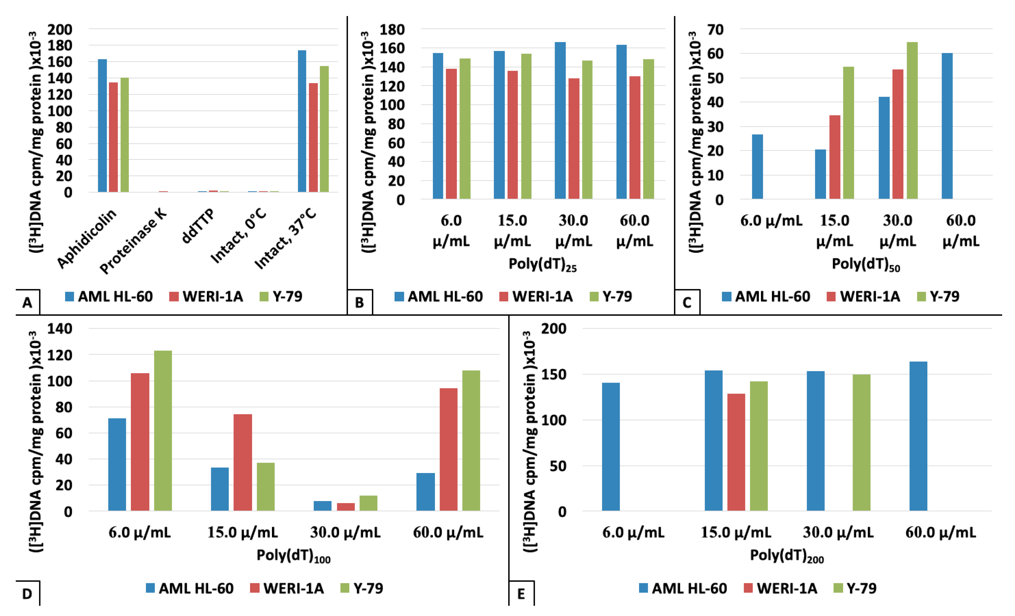
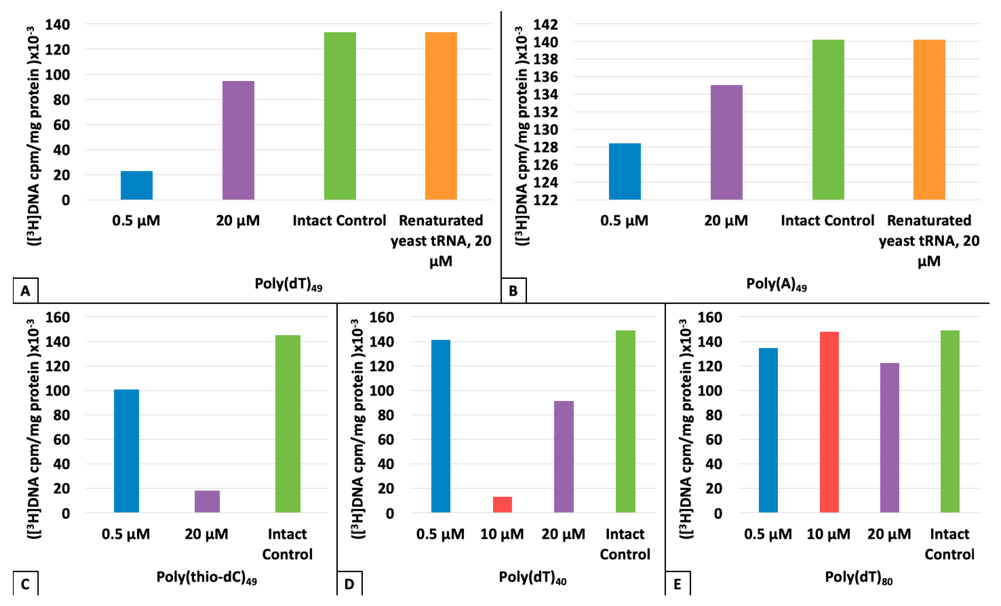
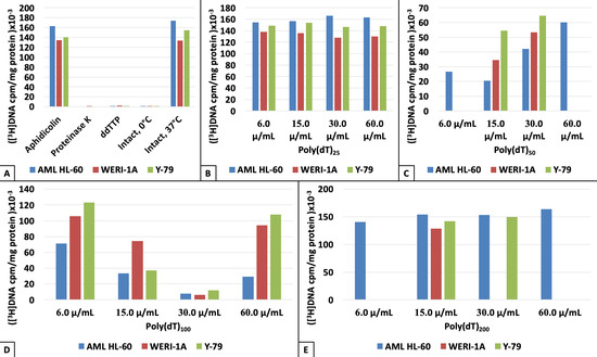
As seen from the data presented in Figure 2; Figure 3, no matter what ligand concentration tested and whatever enzyme sample coupled, a molecular size of synthetic polynucleotide is the only, crucial, impact-making factor. It should be emphasized that a maximal inhibition effects were observed in cases of 50 n–100 n-long polynucleotides (Figure 2C,D and Figure 3A–E), while a post-denaturation misfolded tRNA test show no inhibition effect at all (Figure 3A,B). As per Aphidicolin and ddTTP controls, they just prove a true beta-specific nature of enzymes tested [2,3,11] (Figure 2A).
Unlike polydeoxyribonucleotides (PDRN), polyribonucleotides were found to be inert towards the DNApolβ processivity regardless on lengths of these ligand molecules (Figure 3A,B).
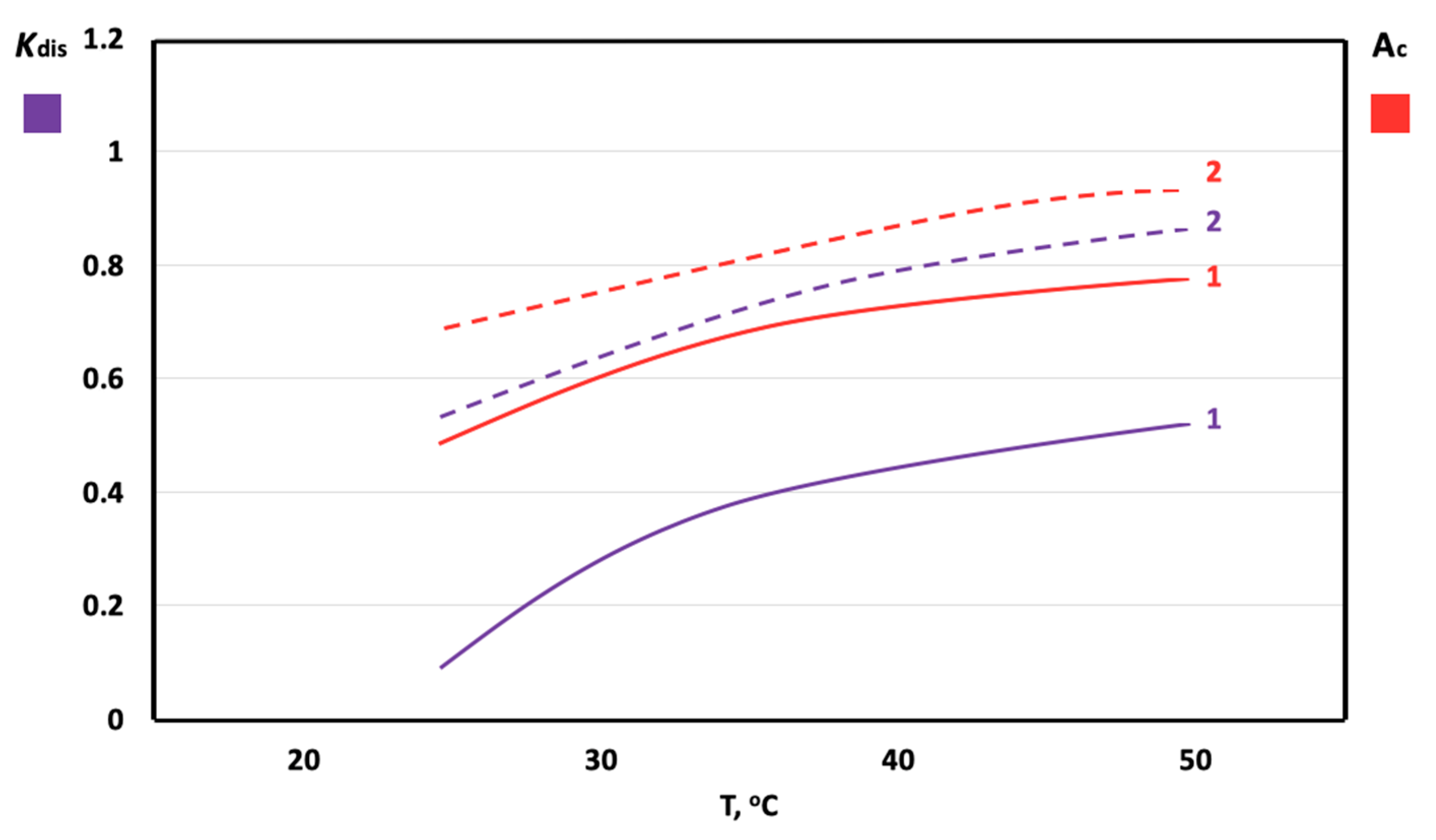
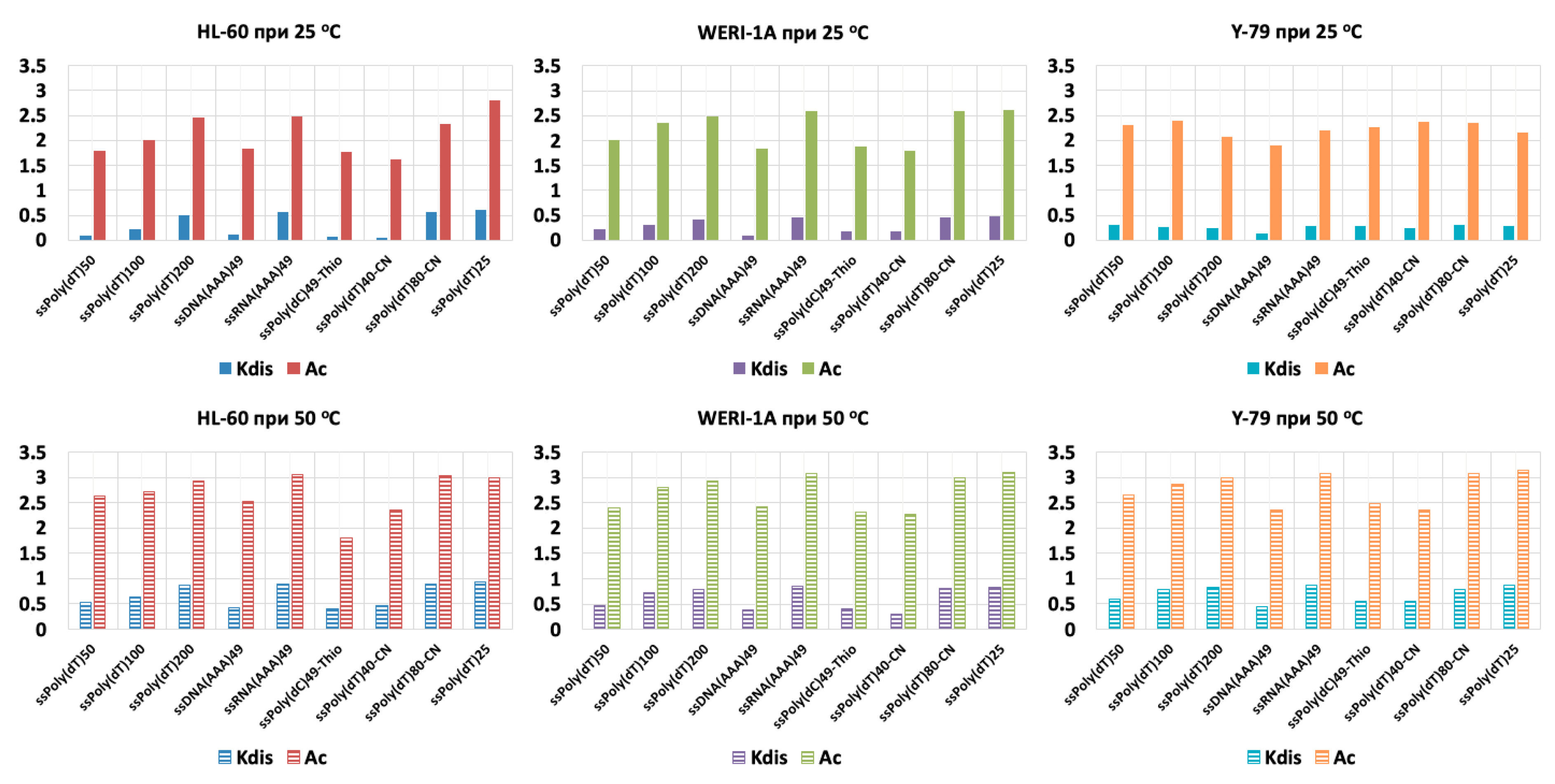
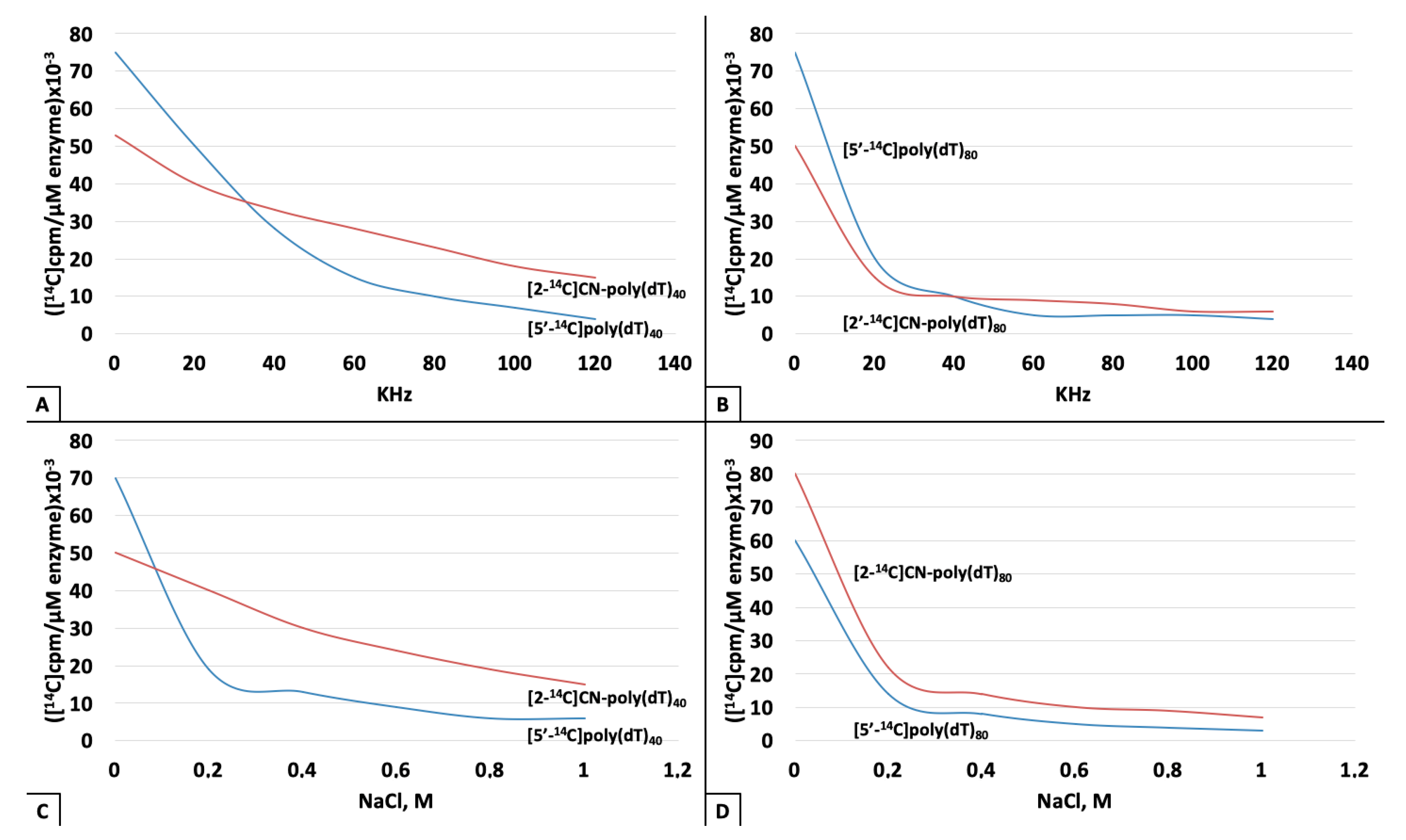
The affinity isotherms clearly show an essential difference between the ligand–enzyme complex stability patterns, Kd and Ac, estimated for numerous compositions of these couples (Figure 4 and Figure 5). As seen from Methods, both the dissociation constant (Kdis or Kd) and the dissociation spectrophotometric index (Ac) were estimated according to a routine procedure presented in [7,15] and then specified in Discussion by Equations (1) and (2). Thus, dissociation constant Kd for Poly(dT)50-DNApolβ was found by 2.5-fold smaller as compared to Poly(dT)200-DNApolβ pair (Figure 5).
In all cases studied, except for the RNA-like Poly(A)49-containing pairs (Figure 5), Kd and Ac values are minimal when the ligand length is close to 50n (Figure 6). For all RNA-like ligands, the high values of Kd and Ac were found practically equal to the ones estimated for Poly(dT)200 (Figure 5).

4. Discussion

A wide diversity of investigated synthetic homopolynucleotides and their monotonic thio- and cyano-derivatives is itself evidence for the universal, indiscriminate mode of the DNApolβ inhibition effect we described. This is in a favor with our assumption stated that the key role in this phenomenon belongs exclusively to molecular sizes of small DNA fragments regardless of their primary structure peculiarities.
The ligand diversity mentioned does not explain a formation of the ligand–enzyme complexes just by a routine docking (“complimentary”) paradigm. It is worth noting that even though this statement sounds «too firm» or ambiguous, it does not cover a variable data on nucleotide and nucleoside ligands but, instead, this conclusion-forming point relates exclusively to the primer-like polynucleotide ligands and their semi-synthetic (chemically modified) versions [7,9,10,15,18,20]. This DNA-protein binding act may involve some random appearing hydrogen bonds, Coulomb hyperfine coupling along with dispersion interaction and, last not least, the hydrophobic connections, might also be a case in this scenario. A key point of this model derives from the homopolynucleotides related data and reveals that the mean energy of these “weak” interaction depends predominantly on the length of a ligand. Obviously, this does not exclude the known structure–function mutual dependence paradigm as long as the holistic enzyme functioning model is the case. On the other hand, however, all different nucleotide composition ligands of one and the same size show merely identical inhibitory effects being incorporated into the enzyme–ligand complexes of all sorts tested (Figure 2 and Figure 3). It is hardly possible to explain this easily but this in itself deserves a special attention and, no doubt, requires some further extensive studies.
A similar event occurs in the DNA repair process as long as a randomly appeared local DNA damage comes first. In this case, a clearly indiscriminate mode of the ligand–enzyme coupling is a consequence of unpredictable variability in primary structures of DNA fragments suitable for the repair purposes.
Therefore, a polynucleotide length alone is an impact-making factor towards the DNApolβ catalytic function. This factor might determine a formation of the above listed «weak» non-covalent interactions and, hence, to control the mean amount of hydrogen bonds per one nucleotide of DNA-primer (ligand) inside the DNA polymerase catalytic site. That is indeed critical for the enzyme suppression in situ.
All the affinity isotherms presented and a variable ligand–enzyme binding data (Figure 4, Figure 5 and Figure 6) reveal a maximal strength of this peculiar intermolecular interaction within the short interval of ligand sizes, 50–100 n. This is to confirm that the enzyme inhibition we observed (Figure 2B–E and Figure 3) is indeed a consequence of the stable ligand–enzyme pair formation which has nothing to do with either the solution properties changes or the enzyme surrounding disperse phase modulations.
A comparison between the ligand amounts bound with (a) DNApolβ and (b) HSA show that the ligand–enzyme complex includes two molecules of ligand per one molecule of enzyme, while the ligand–HSA (control) pair contains just one ligand per HSA molecule (Figure 4, Figure 7 and Figure 8). This is in favor to a mere fact of existence of two separate Mg2+-coordinating catalytic sites in β- and β-like DNA polymerases [8,10,21,22,23].
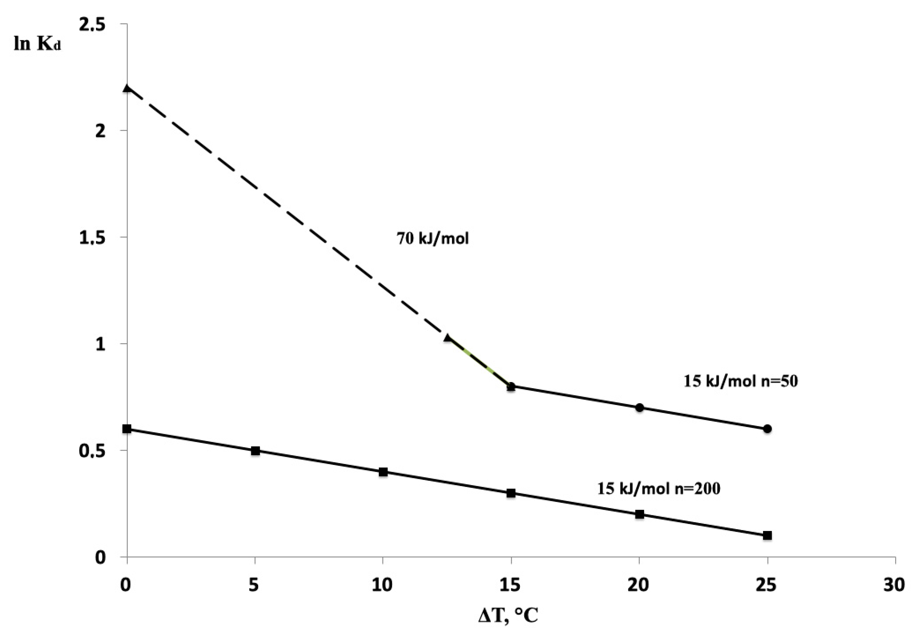
Now, we evaluate the activation energy value for a ligand–enzyme complex dissociation following the data listed in Figure 4; Figure 8. Obviously, a dependence of Kd on temperature has a clear Ahrrenius mode:
K d = K 0 exp ( E / RT )
where R—a universal gas constant and T-enzyme functioning temperature.
Taking into account a low level of the temperature interval tested as compared to an initial T0 = 298 K, we will use further a linear decay as T in (1). To estimate E, a linear regularity must be treated as:
ln K d ln K 0 E / ( RT 0 ) + E Δ T / RT 0 2
where Δ T is a temperature change in experiment (Figure 7; Figure 8).
Equation (2) revealed activation energy value E was found 15 kJ for Poly(dT)200 ligand within a whole range of temperatures tested, while the very same dependence for Poly(dT)50 include two linear branches crossing each other nearby the 40 °C point where, most likely, a slight protein denaturation starts (Figure 7). Once the temperature is close to an enzyme function required optimal level (25–35 °C), Equation (2) revealed E50 = 70 kJ/mole whereas this value estimated at 40 °C is as low as 15 kJ/mole (Figure 7). The attention catching point here is that the E value measured at the enzyme denaturation launch-point (40 °C) is exactly the same as it was found in case of the «inert», non-inhibiting, polydeoxyribonucleotide ligands. This supports a statement on the enzyme inactivating protein structure dynamics initiated starting with a 40 °C heating point. A lack of two-compartment, «angle-breaking», Kd = f(T) dependence for long ligands and their E values low level are in a good accordance with a high probability of 3D-compactization of such sequences at our test conditions [7,16,17,24].
It’s easy to find out that the inhibition-promoting Poly(dNTP) species provide a pretty low value of activation energy for dissociation with DNApolβ-HL60, ε = 1.40 kJ/mole. That means, the complimentary hydrogen bonds do not contribute to formation of these particular Poly(dNTP)-enzyme complexes which is evidence of the absence of the affinity-specific, true docking links in these pairs. Most probably, this remarkable 1.40 kJ/mole value is determined by the Ud-dispersion related weak Van der Waals interactions along with a Uq Coulomb hyperfine coupling. These kinds of the ligand–enzyme complex stabilizing links are rather superficial ones due to both a Ud~1/r6 potential and the efficient screen-like separation of Coulomb forces in water occurred within a distance smaller than 0.4 nm [15,17,24,25]. This certainly takes into account the superficial hydrophobic bounds as well which are nothing but the result of a cooperative (cumulative) act of all the above specified forces [24,25].
Thus, the key role in formation of Poly(dNTP)/DNApolβ complexes belongs to Van der Waals bounds which energy corresponds directly to either (a) the efficient surface of ligand–enzyme pair or (b) a length of an enzyme-bound ligand.
Furthermore, since the mean value of the Ud dispersion patterns is hardly dependent on the element composition, and as long as the amounts of charged groups in Uq-determining Poly(dNTP) ligands tested are about the same, a very similar parameters of their inhibitory activity should be expected then. This explains well a lack of dependence of inhibitory capabilities of these ligands on their nucleotide composition (Figure 2B–E and Figure 3).
Once ε ~ RT , we might suppose that the short ligands (50–100n, or shorter) are to be bound with the enzyme molecule throughout a whole linear chain, that is, through nearly every nucleotide of Poly(dNTP) sequence.
Using the Ahrrenius equation, we may evaluate a ratio between the ligand–enzyme binding time values τ for Poly(dNTP)20 and Poly(dNTP)50, respectively:
τ 20 / τ 50 = exp ( Δ n ε / RT ) 10 7
where Δ = 30 (difference in lengths between 50n and 20n). It shows that a short time of binding τ 20 does not allow n20-ligand to promote a sharp suppression of the enzyme activity. A shorter time for the ligand «neighboring» around the enzyme catalytic site, the lesser inhibitory effect to be expressed.
On other hand, the longest ligands tested were found to be a rather poor inhibitors as well (Figure 2E and Figure 4) due to their compactization-caused small square of the enzyme–ligand efficient interaction [24,26] as also seen from Equation (3).
As per a zero inhibition affect shown by RNA-like ligands tested (Figure 3A,B), their remarkable refolding capabilities [18,26] and [–O–Mg2+] coordination input [20,27] are presumably beyond this phenomenon.
A clear pharmacological potential of oligo- and polydeoxyribinucleotides is a subject for numerous experimental and clinical studies performed since early 1990s [18,20,28,29,30]. Their applied medicinal significance, however, were treated predominantly by focusing on the aptomer properties, that is, on capabilities to find the specific target molecules such as signaling proteins, membrane receptors, chromatin structure elements, etc [17,27,28,30,31]. Unlike our present work, therefore, these studies are all about the true docking participants. The inhibitory power of Poly(dNTP) species we found has nothing to do with their structures and composition being entirely dependent on molecular size (Figure 2B–E and Figure 3A,C–E). This allows to “turn the page” in a long record of polynucleotide medicinal applications by just paying attention to the Poly(dNTP) motion on how to shut down a DNA repair in malignant cells. It makes sense for DNApolβ, key DNA repair enzymes, are found hyperexpressed in response to a high frequency of DNA damages followed by an increase of the cancer related cell proliferation rate [2,3,10,11,21]. With a respect to this statement, it has been already emphasized that the members of this enzyme family are the «legitimate targets» for cytostatic inhibitors like dNTP antimetabolites [8,9,10,28,32] and magnetic metal isotopes [1,2,3,25].
There is a good enough understanding on molecular mechanisms beyond the inhibition of the cancer-hyperexpressed DNApolβ species provided by magnetic isotopes of bivalent metals, –25Mg, 43Ca and 67Zn [1,2,3,9,25]. In particular, these mechanisms are dealing with the origin of «too short», DNA repair insufficient, DNA fragments [1,2,3]. This effect leads to a massive release of the DNA repair related 40–100n DNA sequences with the unpredictable, unique structures [1,2], that is, this provokes a magnetic isotope induced formation of the DNApolβ endogenous inhibitors. It looks like a good explanation for cytostatic activity shown by magnetic metal isotopes in several cancer cells [1,25].
Everything we know about pharmacokinetics of various DNA-based drugs and pharmacophores indicates to the fact that their bioavailability is far of being perfect mostly because of inevitable nucleases attack [18,20,28,29,30]. Thus, this is the hardest task to manage a targeted delivery of Poly(dNTP) sequences to the DNApolβ-operating in situ compartments, for which some certain nanocarriers required [7,18,30,33]. As for the magnetic metal ions, they can be delivered to the DNApolβ in situ targets with [1,25] or even without [2,3] nanocationite administration which would make the magnetic isotope effect a promising tool to gain the DNA repair machinery breakdown in neoplasma.
Knowing that the abundance of 25Mg is relatively high (≈10%) and considering a remarkable role of magnesium in enzymatic phosphorylation processes [1,25,34], it would be logical to suppose the existence of so called «hidden» effects of Magnesium-25 on numerous metabolic pathways. In this case, a high content of endogenous Fe2+ in cells and tissues of a living organism becomes a natural limiting factor to prohibit any expression of magnetic isotope effects in vivo. This is in accordance with the data on correlations between the 25Mg2+-affected ATP synthesis levels in mitochondria and the Fe2+ contents in these mitochondria isolated from different rat tissues [34] as well as with the results stated an increase of Fe/Mg ratios in several types of cancer (ovarian cancer, renal adenocarcinoma, fibroblast lung cancer, hepatocellular cancer, osteosarcoma, thyroid follicular adenocarcinoma) compared to corresponding normal, non-malignant, tissues [9,21,27,32].
A variety of clinical case reports showing an appearance of short (100–300n) single-stranded DNA fragments in a blood plasma of oncology patients [27,35] may now also be treated under a point of view of the probable DNA repair related origin of these tumor-released Poly(dNTP) sequences.
Last but not least, the infamous quorum sensing phenomena [36] could be engaged with an effort to uncover a biological meaning of what we’ve found testing the capabilities of different Poly(dNTP) species to affect some tumor-specific DNA repair key enzymes.

5. Conclusions

Ultrashort (40–100n) polydeoxyribonucleotides are found to be the efficient non-specific inhibitors of DNA polymerases β from chromatin of several cancer cells: HL-60, WERI-1A, Y-79.
Manifesting a positive correlation between these inhibitory capabilities and the strength of affinity in enzyme-ligand pairs, such poly(dNTP) species promote a sharp suppression of enzymatic catalysis regardless on their primary structures. Only molecular size matters.
The data presented show a clear regulatory potential of some short ssDNA entities once they are about to interact with the key DNA repair enzymes.
This may provide a promising platform for further research aiming to shut down the DNA repair machinery in malignancies.

Author Contributions

Conceptualization, S.S.; Data curation, A.V.; Formal analysis, A.V.; Funding acquisition, S.S.; Investigation, K.E., A.V. and A.B.; Methodology, D.K.; Project administration, S.S. and D.K.; Resources, A.V. and D.K.; Software, A.V.; Supervision, S.S. and D.K.; Validation, K.E. and A.B.; Visualization, K.E. and A.B.; Writing—original draft, D.K.; Writing—review & editing, S.S. and D.K.

Acknowledgments

This work was performed within the aims and scope of the Russian Federal Spe- cialized Program on priorities in science and technologies trends for 2014-2020 supported by Russian Federal Ministry of Science and University Education. The RF Government budget beyond.

Conflicts of Interest

The authors declare no conflict of interest. The funders had no role in the design of the study; in the collection, analyses, or interpretation of data; in the writing of the manuscript, or in the decision to publish the results.

Abbreviations

ssDNAsingle-stranded DNA
DNApolβDNA polymerase beta
PDRNpolydeoxyribonucleotides
MIEmagnetic isotope effects
AMLacute mueloid leukemia
RBretinoblastoma

References

  1. Buchachenko, A.L.; Bukhvostov, A.A.; Ermakov, K.V.; Kuznetsov, D.A. Nuclear spin selectivity in enzymatic catalysis. A caution for applied biophysics. Arch. Biochem. Biophys. 2019, 667, 30–35. [Google Scholar] [CrossRef] [PubMed]
  2. Bukhvostov, A.A.; Dvornikov, A.S.; Ermakov, K.V.; Kurapov, P.B.; Kyznetsov, D.A. Retinoblastoma: Magnetic isotope effects might make a difference in the current anti-cancer research strategy. Acta Medica (Hradec Kralove) 2017, 60, 93–96. [Google Scholar] [CrossRef] [PubMed]
  3. Bukhvostov, A.A.; Dvornikov, A.S.; Ermakov, K.V.; Kuznetsov, D.A. Retinoblastoma case: Shall we get a paramagnetic trend in chemotherapy? Arch. Cancer Res. 2017, 5, 158–162. [Google Scholar] [CrossRef]
  4. Chen, S.J. Site-specific binding of non-site-specific ions. Biophys. J. 2019, 116, 38–46. [Google Scholar] [CrossRef]
  5. Muller, W.E.; Obermeyer, J.; Totsuka, A. Influence of template inactivators on the binding of DNA polymerases to DNA. Nucl. Acids Res. 1974, 1, 63–69. [Google Scholar] [CrossRef]
  6. Corces, M.R.; Granja, J.M.; Shams, S.; Louie, B.H.; Seoane, J.A.; Zhou, W.; Silva, T.C.; Chang, H.V. The chromatin accessibility landscape of primary human cancers. Science 2018, 362, 16–28. [Google Scholar] [CrossRef]
  7. Ashley, J.S.; Kronenberg, G.H.; Lipman, K.R.; Bielka, H. Coupling the DNA turnover enzymes. Poly(dNMP) ligands. In Methods in Macromolecular Docking Research; Pitot, H., Van Nijemen, J.T., Eds.; University of Ghent Press: Ghent-Antwerp, Belgium, 2019; pp. 216–229. [Google Scholar]
  8. Turgeon, M.O.; Perry, N.J.S.; Poulogiannis, G. DNA damage, repair and cancer metabolism. Front. Oncol. 2018, 8, 15–22. [Google Scholar] [CrossRef]
  9. Breitenbach, M.; Hoffmann, J. Cancer Models. Front. Oncol. 2018, 8, 401–419. [Google Scholar] [CrossRef]
  10. Jain, R.; Aggarwal, A.K.; Rechkoblit, O. Eukaryotic DNA polymerases. Curr. Opinion Struct. Biol. 2018, 53, 77–87. [Google Scholar] [CrossRef]
  11. Bukhvostov, A.A.; Shatalov, O.A.; Orlov, A.P.; Kuznetsov, D.A. An atypical DNA polymerase beta overexpressed in human AML/HL-60 malignant cells. J. Cancer Sci. Therap. 2013, 5, 94–99. [Google Scholar] [CrossRef]
  12. Katoh, R. Analytical Techniques in Biochemistry and Molecular Biology; Springer Publ GmbH: Berlin/Heidelberg, Germany, 2011. [Google Scholar]
  13. Dewar, J.M.; Lyndall, D. Simple non-radioactive measurement of single-stranded DNA. In DNA Repair Protocols (Methods in Molecular Biology); Bjergbaek, L., Ed.; Humana Press, Inc.: Totowa, NJ, USA, 2012; Volume 920, pp. 341–349. [Google Scholar]
  14. Fukami, T.; Uchiyama, K.; Yoshimura, Y. Ultramicroanalysis by use of light-scanning photoacoustic densitometry of electrophoresed protein. Analyt. Biochem. 1996, 238, 60–67. [Google Scholar] [CrossRef] [PubMed]
  15. Rastogi, C.; Rube, H.T.; Kribelbauer, J.F.; Crocker, J.; Locker, R.E.; Martini, G.D.; Laptenko, O. Accurate and sensitive quantification of protein-DNA binding affinity. Proc. Nat. Acad. Sci. USA 2018, 115, E3692–E3701. [Google Scholar] [CrossRef] [PubMed] [Green Version]
  16. Laurent, C.; Neurath, K.J.; Baglioni, G.; Sieliwanowicz, B. Advances in multiwave UV-spectrophotometry: RNP/DNP integrity studies. In Physics of Biopolymers; Lindsey, A.L., Cohen, K., Eds.; Adler & Adler Publisher Inc.: Perth, Australia, 2019; pp. 41–56. [Google Scholar]
  17. Chowdhury, N.; Bagchi, A. An overview of DNA-protein Interactions. Curr. Chem. Biol. 2015, 9, 71–83. [Google Scholar] [CrossRef]
  18. Telashima, Y. Revival of Oligonucleotide Therapy Development; KOKEN Publ.: Tokyo-Nagoya, Japan, 2019. [Google Scholar]
  19. Byron, W.M.; Hollander, B.; Hollander, M. Statistics. A Biomedical Introduction; Wiley-Blackwell Publishers Inc.: Oxford, UK; Weinheim, Germany; Hoboken, NJ, USA, 2007. [Google Scholar]
  20. Ueda, S.; Watanabe, I.; Taichi, K. Nucleic Acid-Based Drugs; Sumitomo Chemical Co. Ltd.: Utajima-Osaka, Japan, 2019. [Google Scholar]
  21. Lavrik, I.N. Systems biology of death receptor networks: Live and let die. Cell Death Dis. 2014, 5, e1259–e1266. [Google Scholar] [CrossRef] [PubMed]
  22. Peterson, L.E. Small molecule docking of DNA repair proteins associated with cancer survival following PCNA metagene adjustment. A potential novel class of repair inhibitors. Molecules 2019, 24, 645. [Google Scholar] [CrossRef] [PubMed]
  23. Wu, Y.; Lu, J.; Kang, T. Human single-stranded DNA binding proteins: Guardians of genome stability. Acta Biochimica et Biophysica Sinica. 2016, 48, 671–677. [Google Scholar] [CrossRef]
  24. Finkelsten, A.V.; Ptitsyn, O.V. Physics of Proteins; Nova Science Publishers Inc.: Princeton, NJ, USA, 2012. [Google Scholar]
  25. Buchachenko, A.L. Magneto-Biology and Medicine; Nova Science Publishers Inc.: Princeton, NY, USA, 2015. [Google Scholar]
  26. Morten, M.J.; Gamsjaejer, R.; Cubeddu, L.; Kariawasam, R.; Peregrina, J.; Penedo, J.C.; White, M.F. High affinity RNA binding by a hyperthermophilic single-stranded DNA-binding protein. Extremophiles 2017, 21, 369–379. [Google Scholar] [CrossRef]
  27. Lyu, J.; Wang, S.; Balius, T.E.; Singh, I.; Levit, A.; Moroz, Y.S.; O’Meara, M.J. Ultra-large library docking for discovering new chemotypes. Nature 2019, 566, 224–229. [Google Scholar] [CrossRef]
  28. Udvardi, L.; Lakatos, S. Aptamers. Towards the Pharmacological Breakthrough; Alba Regia: Szegen-Budapest, Hungary, 2019. [Google Scholar]
  29. Zavyalova, E.; Samoylenkova, N.; Revishchin, A.; Turashev, A.; Gordeychuk, I.; Golovin, A.; Kopylov, A.; Pavlova, G. The evaluation of pharmacodynamics and pharmacokinetics of anti-thrombin DNA aptamer RA-36. Front Pharmacol. 2017, 8, 922–930. [Google Scholar] [CrossRef]
  30. Squadrito, F.; Bitto, A.; Irrera, N.; Pizzino, G.; Pallio, G.; Minutoli, L.; Altavilla, D. Pharmacological activity and clinical use of PDRN (polydeoxyribonucleotides). Front. Pharmacol. 2017, 8, 224–233. [Google Scholar] [CrossRef]
  31. Ashton, N.W.; Bolderson, E.; Cubeddu, L.; O’Byrne, K.J.; Richard, D.J. Human single-stranded DNA binding proteins are essential for maintaining genomic stability. BMC Mol. Biol. 2013, 14, 1–20. [Google Scholar] [CrossRef] [PubMed] [Green Version]
  32. Bozic, I.; Nowak, M.A. Resisting resistance. Ann. Rev. Cancer Biol. 2017, 1, 203–221. [Google Scholar] [CrossRef]
  33. Ansari, A.S.; Santerre, P.J.; Uludag, H. Biomaterials for polynucleotide delivery to anchorage-independent cells. J. Mater. Chem. B. 2017, 5, 7238–7261. [Google Scholar] [CrossRef]
  34. Svistunov, A.A.; Napolov, Y.K.; Bukhvostov, A.A.; Shatalov, O.A.; Alayutdin, R.N.; Kuznetsov, D.A. The mitochondria free iron content to limit an isotope effect of 25Mg2+ in ATP synthesis: A caution. Cell Biochem. Biophys. 2013, 66, 417–419. [Google Scholar] [CrossRef]
  35. Mouliere, F.; Chandrananda, D.; Piskorz, A.M.; Moore, E.K.; Morris, J.; Ahlborn, L.B.; Rosenfeld, N. Enhanced detection of circulating tumor DNA by fragment size analysis. Science Transl. Med. 2018, 10, 117–129. [Google Scholar] [CrossRef]
  36. Catachura, S.C.; Leys, N.; Mastroleo, J. Quorum sensing in life support systems. In Quorum Sensing; Kalia, V.C., Ed.; Springer Nature, Ltd.: Singapore, 2018; pp. 249–260. [Google Scholar]
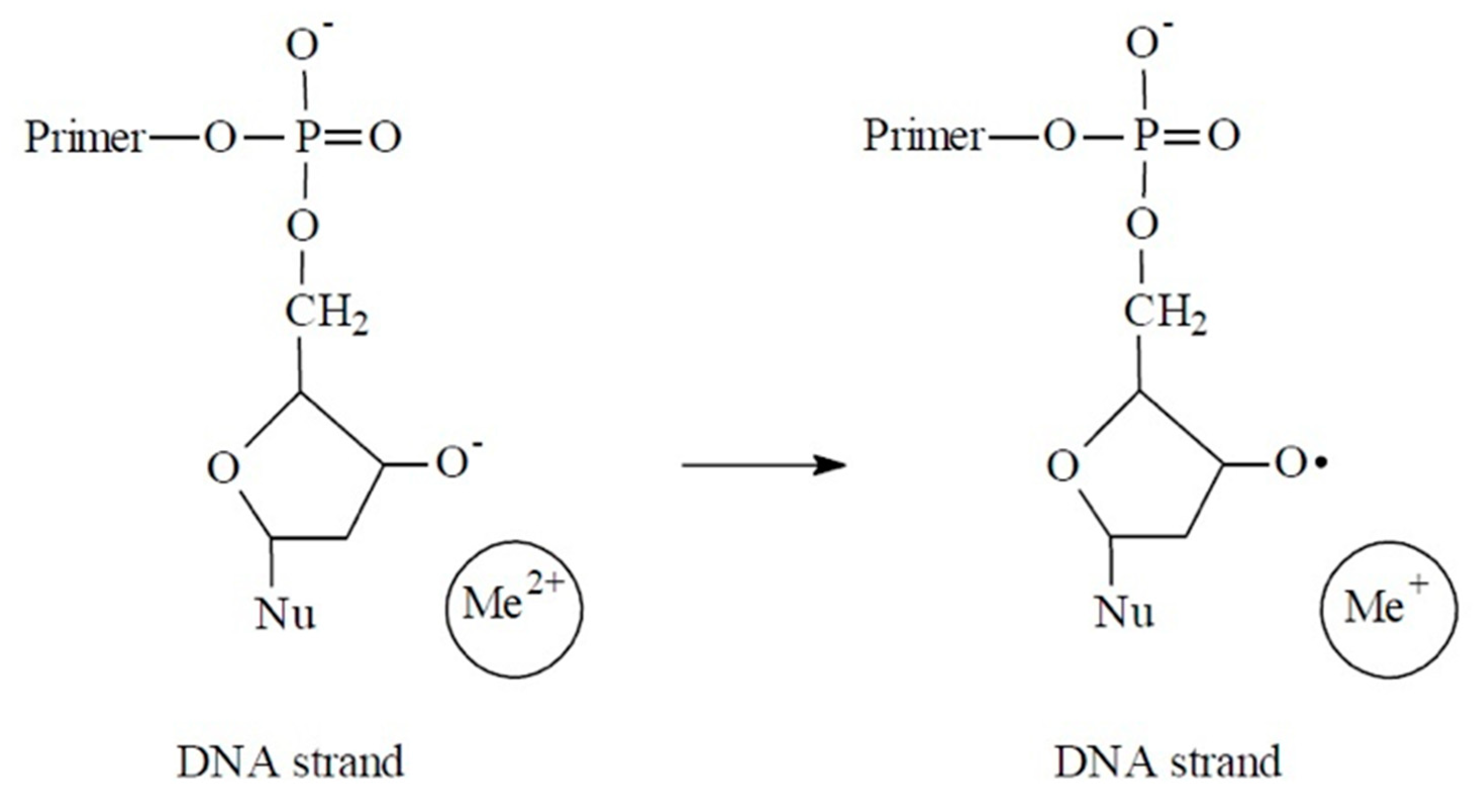
Figure 1. A reaction of electron transfer from the nascent DNA deoxyribose anion to a bivalent metal ion.
Scipharm 87 00025 g001
Figure 2. (A) Catalytic activities of beta-like DNA polymerases from HL-60, WERI-1A and Y-79 cancer cells under optimal and modified incubation conditions. (B) Catalytic activities of beta-like DNA polymerases from HL-60, WERI-1A and Y-79 cancer cells in the presence of Poly(dT)25. (C) Catalytic activities of beta-like DNA polymerases from HL-60, WERI-1A and Y-79 cancer cells in the presence of Poly(dT)50. (D) Catalytic activities of beta-like DNA polymerases from HL-60, WERI-1A and Y-79 cancer cells in the presence of Poly(dT)100. (E) Catalytic activities of beta-like DNA polymerases from HL-60, WERI-1A and Y-79 cancer cells in the presence of Poly(dT)200.
Scipharm 87 00025 g002
Figure 3. (A) Catalytic activity of AML/HL-60 beta-like DNA polymerase in the presence of Poly(dT)49 and renatured yeast tRNA. (B) Catalytic activity of AML/HL-60 beta-like DNA polymerase in the presence of Poly(A)49 and renatured yeast tRNA. (C) Catalytic activity of AML/HL-60 beta-like DNA polymerase in the presence of Poly(thio-dC)49 and renatured yeast tRNA. (D) Catalytic activity of AML/HL-60 beta-like DNA polymerase in the presence of Poly(dT)40. (E) Catalytic activity of AML/HL-60 beta-like DNA polymerase in the presence of Poly(dT)80.
Scipharm 87 00025 g003
Figure 4. Affinity isotherms for HL-60-DNApolβ/Poly(dT)50 (1) and HL-60-DNApolβ/Poly(dT)200 (2) ligand–acceptor complexes. (______ Kdis, - - - - Ac) For technical details, see Methods.
Scipharm 87 00025 g004
Figure 5. Polynucleotide–enzyme binding patterns (Kdis, Ac) estimated for beta-like DNA polymerase species from three cancer cell lines (HL-60, WERI-1A, Y-79). For technical details, see Methods.
Scipharm 87 00025 g005
Figure 6. (A) A sonication affected stability of the couples of HL-60 beta-like DNA polymerase and 14C-labeled Poly(CN-dT)40 evaluated by detection of free ligand release. For technical details, see Methods. (B) A sonication affected stability of the couples of HL-60 beta-like DNA polymerase and 14C-labeled Poly(CN-dT)80 evaluated by detection of free ligand release. For technical details, see Methods. (C) A salt-caused decay of the unstable complexes between HL-60 beta-like DNA polymerase and 14C-labeled Poly(CN-dT)40 evaluated by detection of free ligand release. For technical details, see Methods. (D) A salt-caused decay of the unstable complexes between HL-60 beta-like DNA polymerase and 14C-labeled Poly(CN-dT)80 evaluated by detection of free ligand release. For technical details, see Methods.
Scipharm 87 00025 g006
Figure 7. An activation energy of the ligand–enzyme binding as affected by temperature in DNApolβ-HL60/Poly(dT)50 pair formation. ln K d ln K 0 E / RT 0 + E Δ T / RT 0 2 , (______ monocompartment mode, - - - - - bicompartment, “angle-breaking”, mode) see Discussion.
Scipharm 87 00025 g007
Figure 8. Temperature-dependent ligand binding in DNApolβ-HL60/Poly(dT)50 pair. L’—DNApolβ, receptor; L*—HAS, receptor (control). For technical details, see Methods.
Scipharm 87 00025 g008