Article

Modulatory Effect of Lippia alba Essential Oil on the Activity of Clinically Used Antimicrobial Agents on Salmonella typhi and Shigella dysenteriae Biofilm

Clinical Department of Toxicological Analysis, Faculty of Pharmacy, Federal University of Ceará, Fortaleza 60356000, Brazil
*
Author to whom correspondence should be addressed.
Sci. Pharm. 2018, 86(4), 52; https://doi.org/10.3390/scipharm86040052
Submission received: 7 November 2018 / Revised: 22 November 2018 / Accepted: 26 November 2018 / Published: 4 December 2018

Abstract

:
The essential oil obtained from the leaves of Lippia alba (Mill.) N.E. Brown (Verbenaceae) has shown great pharmacological potential as an analgesic, antispasmodic, and antimicrobial agent. The aim of this study was to evaluate the modulatory effect of Lippia alba essential oil (LaEO I) on the activity of clinically used antimicrobial agents on Salmonella enterica serovar Typhi (Salmonella typhi) and Shigella dysenteriae biofilms. The Minimum Inhibitory Concentration of LaEO I (MICLaEO I) was determined by the microdilution method, and the effect of LaEO I on the activity of clinically used antimicrobials was assessed by the Checkboard method. The values obtained from MICLaEO I and ciprofloxacin were used to evaluate the effect of time of exposure on cell viability. LaEO I main components were geranial (34.2%), neral (25.9%), and myrcene (12.5%). The MICLaEO I was 1 mg/mL for both strains. LaEO I positively modulated the action of ciprofloxacin, cefepime, and ceftriaxone. After the first hour of treatment with MICLaEO I, the cell viability of the strains showed a 5 log10 CFU/mL reduction, and the LaEO I-CIP association was able to inhibit growth during the first 6 h of the test. Regarding the anti-biofilm activity, MICLaEO I was able to reduce the biofilm mass of Salmonella typhi by 61.2% and of Shigella dysenteriae by 38.9%. MICLaEO I was not able to eradicate the preformed biofilm; however, there was a reduction in the biofilm microbial viability. LaEO I has the potential to be used as an antimicrobial agent and interferes with biofilm formation; also, it is able to reduce cell viability in preformed biofilm and synergistically modulate the activity of ciprofloxacin.

1. Introduction

Reports from the World Health Organization (WHO) estimate that every year more than 600 million individuals become sick after consuming contaminated food, resulting in almost 420,000 deaths. Diarrhea remains a main cause of mortality among young children in low-income countries [1].
Bacteria involved with gastrointestinal infections can cause digestive tract inflammation, as well as symptoms such as diarrhea, stomach cramps, and fever. The bacteria most often involved in gastroenteritis are Campylobacter spp., Escherichia coli, Salmonella spp., Shigella spp., and Staphylococcus aureus [2].
Salmonella spp. infections accounted for more than 25% of all foodborne disease outbreaks in Brazil from 2007 to 2016 [3]. Among the more than 2500 serotypes of Salmonella that have been described, S. enteritidis and S. typhimurium are two of the most prevalent isolated serotypes [4].
The genus Shigella, in turn, consists of four different species, each comprising several serogroups, all showing phenotypic similarity, including invasive pathogenicity [5].
Adhesion to surfaces, and biofilm formation, are two of the important mechanisms that Salmonella and Shigella strains use to survive outside host cells. Biofilm production may play a key role in supporting colonization and the chronic persistence of S. typhi due to the bile induces exopolysaccharide matrix production, which facilitates biofilm formation in human gallstones [6]. Biofilm production increases microbial resistance to physical forces, to antimicrobial agents, and to the host’s immune system [7,8].
Ciprofloxacin and ceftriaxone are recommended by the WHO for the treatment of infections caused by Salmonella spp. and Shigella spp. strains; however, the emergence of multiresistant strains has been reported [9,10], showing the need for therapeutic alternatives.
Lippia alba (Verbenaceae) is a plant native to South America. Its pharmacological potential is associated with the variation in its essential oil chemical composition. This variation leads to the classification of this species into chemotypes, named according to the main chemical constituent present in the essential oils [11,12].
This plant is widely used in folk medicine for the treatment of gastrointestinal and respiratory diseases, in addition to being used as an antispasmodic, antipyretic, anti-inflammatory, antidiarrheal, analgesic, and sedative agent [13,14,15,16]. The Lippia alba essential oil (LaEO) shows antimicrobial potential and interaction with food ingredients [17]. According to the study by Porfirio et al. [18], the LaEO shows activity on planktonic cells and Staphylococcus aureus biofilm; these properties are due to its active constituents, such as monoterpenes and phenolic compounds [19].
This study aims to evaluate the modulatory effect of Lippia alba essential oil on the activity of clinically used antimicrobial agents on Salmonella typhi and Shigella dysenteriae biofilms.

2. Materials and Methods

2.1. Characterization of Lippia alba Essential Oil

Lippia alba (Mill.) N.E. Brown leaves were collected from the Francisco José de Abreu Matos Garden of Medicinal Plants of the Federal University of Ceará. Exsiccates of the species were placed in the Prisco Bezerra Herbarium of the Biology Department, under number 24,150. The access to the Genetic Patrimony was registered in the National Genetic Patrimony and Associated Traditional Knowledge Management System (SisGen) under registration number A5E434F, in compliance with the provisions of Lawn. 13,123/2015.
The essential oil was extracted in the Laboratory of Natural Products, using the water vapor distillation technique [16]. Its yield was calculated and expressed in g/g% of fresh weight. Its composition was determined by gas chromatography-mass spectrometry (GC/MS), using the CG/EM-QP 2010 equipment (SHIMADZU, Kioto, Japan), under the following conditions: OV-5 capillary column (5% Phenyl, 95% dimethylpolysiloxane) measuring 30 m long × 0.25 mm of internal diameter × 0.25 μm film thickness, with a total flow of 50mL/min of Helium as carrier gas, and temperature gradient of 25 °C/min (50–180 °C) and 50 °C/min (180–300 °C), with an injector temperature of 230 °C [20]. For the experiments, LaEO was diluted in ultra-pure water containing 1% (v/v) Tween 80.

2.2. Bacterial Strains and Growth Conditions

The bacterial strains used in the study were Salmonella enterica subsp. enterica serovar typhi ATCC 10,749 and Shigella dysenteriae ATCC 13,313. Brain Heart Infusion (BHI), Plate count gar (PCA), tryptic soy agar (TSA) and tryptic soy broth (TSB) were obtained from Himedia (Mumbay, India).
The bacteria were seeded in TSA and the plates were incubated at 37 °C for 24 h. After solid medium growth, some isolated colonies were inoculated in BHI broth for experiments with planktonic cells and TSB for biofilm experiments. The inoculum was incubated at 37 °C for 24 h under constant agitation in a shaker. For the experiments, the cell density of each inoculum was adjusted to 106 CFU/mL. Regarding bacterial adhesion and biofilm formation after 24 h of incubation, it was possible to observe that the tested strains show moderate-to-strong adhesion capability.

2.3. Determination of the Minimum Inhibitory Concentration (MIC) and Minimum Bactericidal Concentration (MBC)

The MIC of LaEO and the antimicrobials ciprofloxacin (CIP), amikacin (AMI), ceftazidime (CAZ), cefepime (CPM), and ceftriaxone (CRO) for the Salmonella typhi and Shigella dysenteriae strains were determined by the microdilution method in culture broth [21], and 80 μL of the microbial suspension (106 CFU/mL), 100 μL of BHI broth, and 20 μL of the different concentrations of LaEO (0.125 to 4 mg/mL) and the antimicrobials (ATM) (0.048 to 100 μg/mL) were added to each well of the 96-well polystyrene microplate were from obtained Kasvi (Curitiba, Brazil). The negative control (non-inhibition of microbial growth) used culture medium, diluent (1% Tween 80 in aqueous solution), and microbial inoculum. The positive control (inhibition of microbial growth) used culture medium, microbial inoculum, and ATM. The microplates were incubated at 37 °C/24 h.
The MIC was considered the lowest concentration of LaEO and ATM capable of completely inhibiting microbial growth, evidenced by the absence of visible turbidity [21] and the absorbance at 620 nm was measured using a microplate reader Bio-Tek (Winooski, VT, USA). The MBC was determined from the microplate wells used for MIC determination that did not show any visible microbial growth (no turbidity), and 5 μL-aliquots were collected and placed on the surface of PCA using the microdrop technique [22]. The plates were incubated at 37 °C for 24 h, and then the colonies grown on the agar surface were counted. The concentration of LaEO able to determine a reduction of microbial growth ≥ 99.9% of the initial inoculum was considered the MBC [23]. Two experiments were carried out in triplicate.

2.4. Determination of the Modulatory Effect of LaEO on the Activity of Clinically Used Antimicrobial Agents

The effect of LaEO on the activity of clinically used antimicrobial agents was evaluated by the Checkboard method [24]. 80 μL aliquots of the microbial cultures (106 CFU/mL), 80 μL of BHI broth, 20 μL of LaEO, and 20 μL of the ATM were added to the microplate wells. The final concentrations of LaEO and ATM were equal to 1/2, 1/4, 1/8, and 1/16 × MIC. After microplate incubation at 37 °C for 24 h, a visual inspection of microbial growth was performed. To evaluate the effect of the associations, the Fractional Inhibitory Concentration Index (FICI) was calculated according to Shin and Lin [25]:
FICI = FICLaEO I + FICATM = [LaEO]/MICLaEO + [ATM]/MICATM
FICLaEO I and FICATM are the Fractional Inhibitory Concentrations of LaEO and ATM, respectively; [LaEO] and [ATM] are the lowest concentrations of LaEO and ATM in the LaEO-ATM association, with antimicrobial activity, respectively; MICLaEO and MICATM are the MICs of LaEO and ATM, respectively. The FIC index (FICI) was interpreted as synergistic effect for FICI ≤ 0.5, additive, or indifferent effect for FICI > 0.5 and < 1.0, and antagonistic effect for FICI ≥ 1.0 [26]. Two experiments were carried out in triplicate.

2.5. Determination of the Effect of Time of Exposure to LaEO I and LaEO-ATM Association on Microbial Viability (Time Kill)

20 μL aliquots of different concentrations of LaEO (MIC, 1/16 MIC) and ATM (MIC, 1/16 MIC) were added to the microplate wells containing 100 μL of BHI broth and 80 μL of microbial suspension (106 CFU/mL). The best combined concentrations, obtained with the Checkboard assay, were added to the microplate wells containing 20 μL of LaEO, 20 μL of ATM, 80 μL of BHI broth, and 80 μL of microbial suspension [27]. The microplates were incubated at 37 °C, and 5 μL aliquots were removed, diluted in 0.85% sterile saline solution, and seeded on Plate-Count agar at times 0, 1, 2, 3, 4, 5, and 6 h. The colonies were counted after 24 h of incubation, and the results were expressed in CFU/mL [28]. Two experiments were carried out in triplicate.

2.6. Determination of Biofilm Formation Inhibition by LaEO and LaEO-ATM Association

The determination of biofilm formation inhibition was performed according to the methodology described by NOSTRO et al. [29]. The assessed strains were cultivated in TSB, supplemented with 1% (w/v) glucose and incubated at 37 °C for 24 h. Subsequently, 100 μL aliquots of TSB medium, 80 μL of the microbial culture (106 CFU/mL), and 20 μL of each LaEO concentrations (0.125 to 4 mg/mL) were added to the microplate wells. After 24 h of incubation at 37 °C, the biofilm mass was quantified using the crystal violet technique [30].
The wells were washed with 0.85% sterile saline solution. Methanol (99%) was used for cell fixation, and after 15 min the methanol was removed. 200 μL aliquots of 1% crystal violet (CV) solution (v/v) were added, and after 15 min the excess was removed. The CV was released by adding 160 μL of 33% acetic acid. After 15 min, the absorbance at 570 nm was measured using a microplate reader Bio-Tek (Winooski, VT, USA) [30]. The best association concentrations obtained with the Checkboard assay were added to the microplate wells [27]. The negative control of the experiment consisted of wells containing only TSB, and the positive control consisted of wells containing TSB and inoculum, without LaEO. Two experiments were carried out in triplicate.

2.7. Determination of Preformed Biofilm Eradication by LaEO and the LaEO-ATM Association

The determination of biofilm eradication was performed through cell viability in the preformed biofilm [31], and 100 μL aliquots of microbial culture (106 CFU/mL) were transferred to microplates and incubated at 37 °C for 24 h. After it had formed, the biofilm was washed with 0.85% sterile saline solution and the culture medium was discarded. For each well containing biofilm, 80 μL of TSB and 20 μL of LaEO were added at different concentrations (0.125 to 4 mg/mL). To determine cell viability, the biofilm from each well was resuspended in 0.85% saline solution and 5 μL aliquots of the serial dilutions of the biofilm suspensions were seeded on PCA. The best association concentrations obtained with the Checkboard assay were added to the microplate wells [27]. Two experiments were carried out in triplicate.

2.8. Statistical Analysis

All tests were considered significant at p < 0.05 using the software GraphPad Prism 7. Results are presented as means ± standard deviation from three replicates of each experiment. Differences between mean values were determined by the analysis of variance (ANOVA). The post hoc analysis was performed by the Dunnett test.

3. Results

3.1. Chemical Composition of the Essential Oil Extracted from the Lippia Alba Chemotype I Leaves

The chromatography analysis performed for the characterization of LaEO disclosed the presence of monoterpenes and sesquiterpenes, totaling 20 components. The major constituents were geranial (34.16%), followed by neral (25.90%) and myrcene (12.56%). The other components of the oil showed concentrations ranging from 0.38% to 4.06%. According to Matos [16], the major presence of citral (cis-citral and trans-citral) and myrcene in LaEO allows its classification as L. alba chemotype I (LaEO I) (Table 1).

3.2. Antimicrobial Effect of LaEO I on Planktonic Cells

LaEO I was tested at concentrations of 0.125 to 4 mg/mL with the strains of Salmonella typhi ATCC 10749 and Shigella dysenteriae ATCC 13313. MIC and MBC concentrations of LaEO I were 1 mg/mL (Table 2).

3.3. Modulatory Effect of LaEO on the Activity of Clinically used Antimicrobial Agents

LaEO I positively modulated the action of ciprofloxacin, cefepime, and ceftriaxone, resulting in a 16-fold decrease in the MIC of the drugs used in the associations. The most significant synergism was found in the LaEO I-CIP association (1/16 × MICLaEO I—1/16 MICCIP) on both strains, and this association was selected for subsequent studies (Table 3).

3.4. Effect of Time of Exposure to LaEO I and LaEO I-CIP Association on Microbial Viability (Time kill)

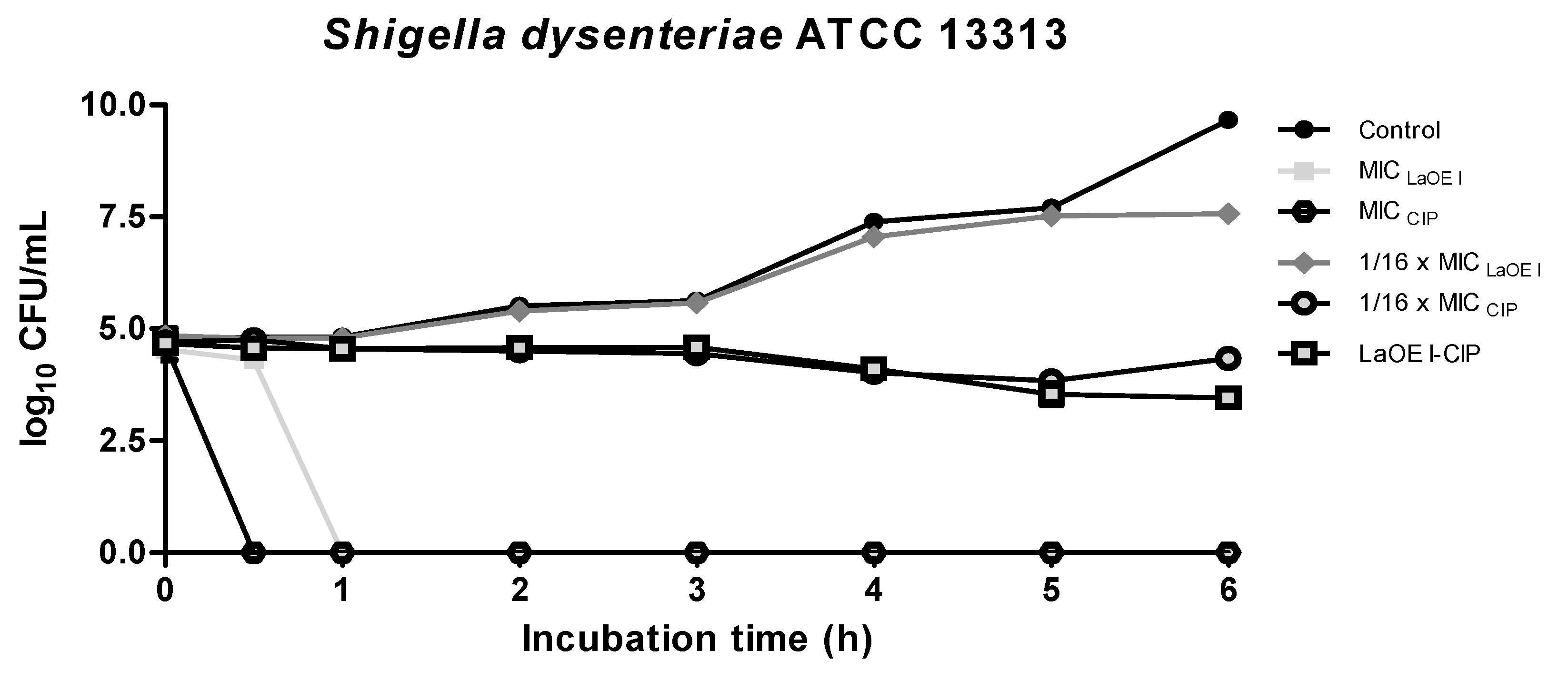
After the first hour of treatment with MICLaEO I, cell viability of Salmonella typhi and Shigella dysenteriae was reduced by 5-log10 CFU/mL. The LaEO I-CIP association (1/16 × MICLaEO I—1/16 MICCIP) was able to inhibit growth during the first 6 h of the test for both strains (Figure 1 and Figure 2).

3.5. Anti-Biofilm Effect of LaEO I-CIP Association on Biofilm Formation Inhibition

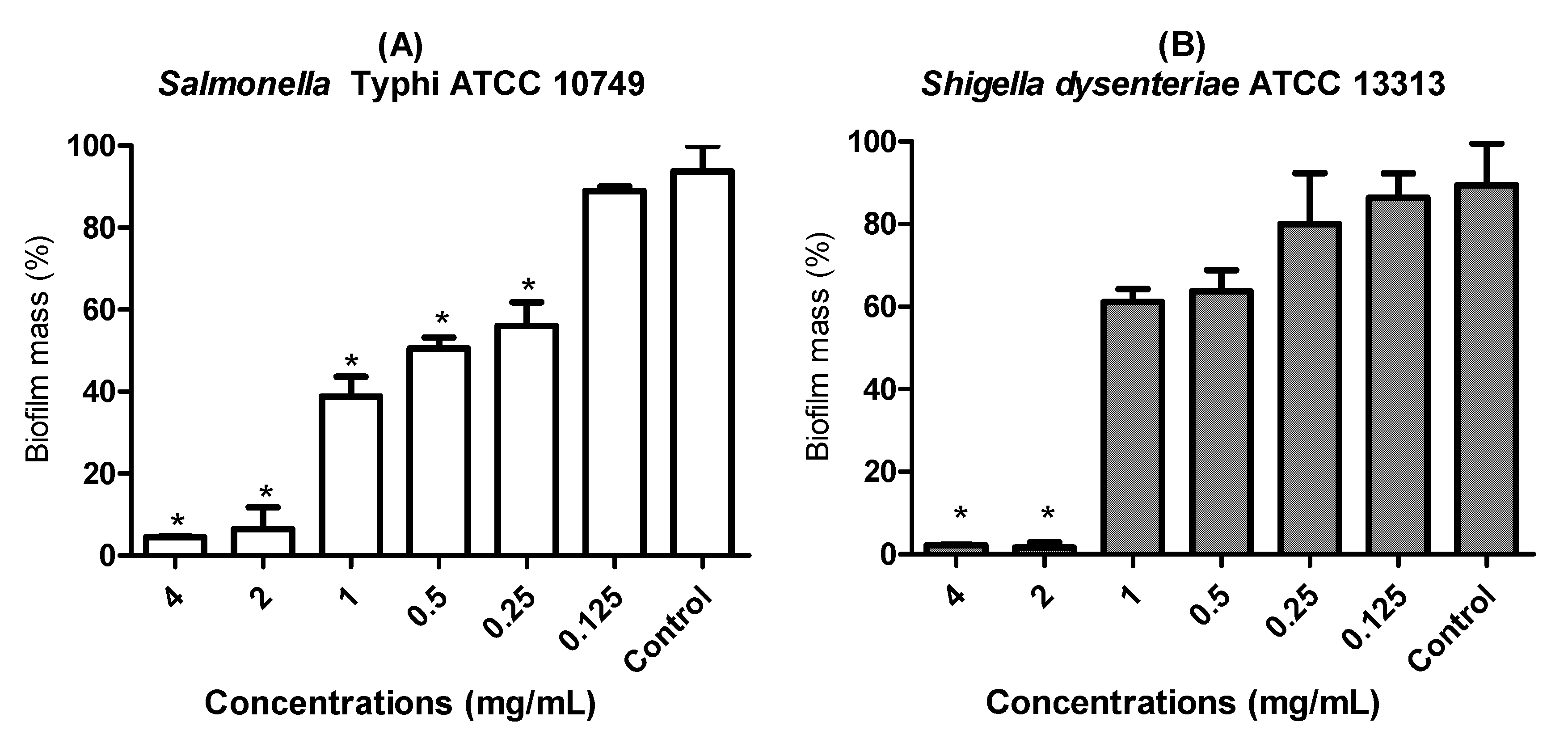
The biofilm mass was reduced when the cultures were exposed to different concentrations of LaEO I. MICLaEO I was able to reduce by 61.2% the biofilm mass of Salmonella typhi and 38.9% of Shigella dysenteriae. At higher concentrations (2 and 4 mg/mL) biofilm mass reduction occurred in a dose-dependent manner (Figure 3).
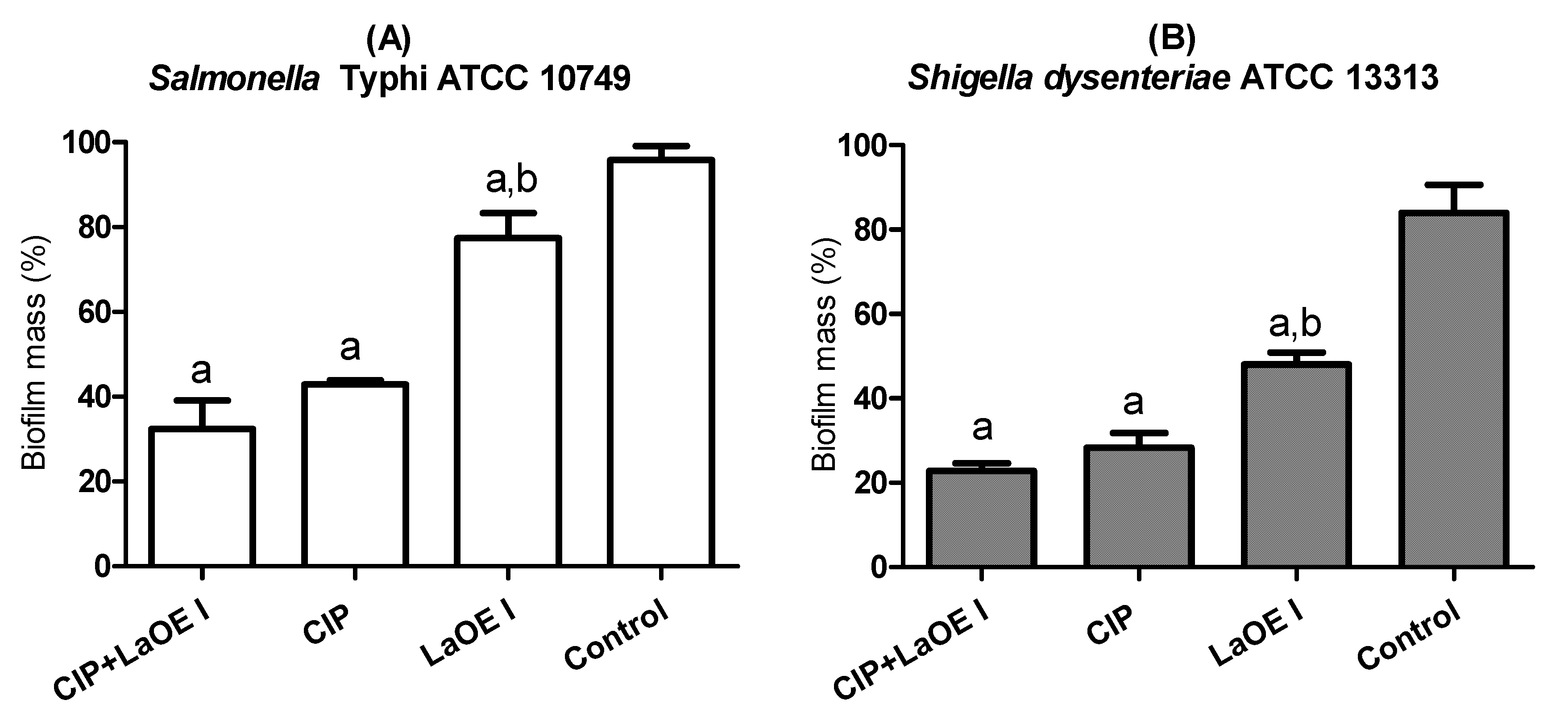
The association of LaEO I and CIP that inhibited planktonic cell growth was tested in biofilm inhibition evaluation. The association 1/16 × MICLaEO I—1/16 MICCIP inhibited biofilm formation by 67.6% and 77.2% for Salmonella typhi and Shigella dysenteriae, respectively (Figure 4).

3.6. Anti-Biofilm Effect of the LaEO I-CIP Association on Biofilm Eradication

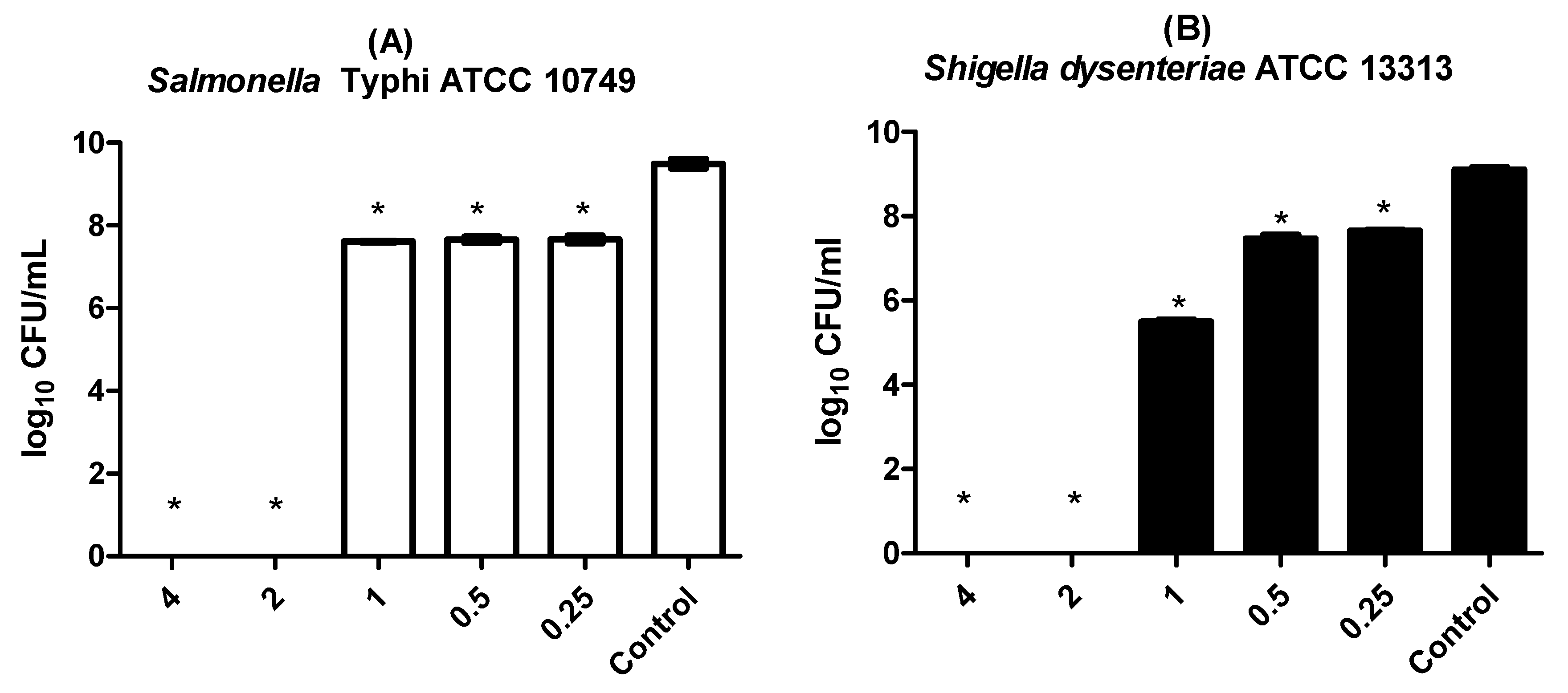
When the 48 h preformed biofilms were exposed to a concentration equal to 2 × MICLaEO I (2 mg/mL), loss of cell viability occurred 24 h after exposure to LaEO I. MICLaEO I (1 mg/mL) was not able to eradicate the preformed biofilm; however, there was a significant reduction in microbial viability in the biofilm when compared to the control (Figure 5).
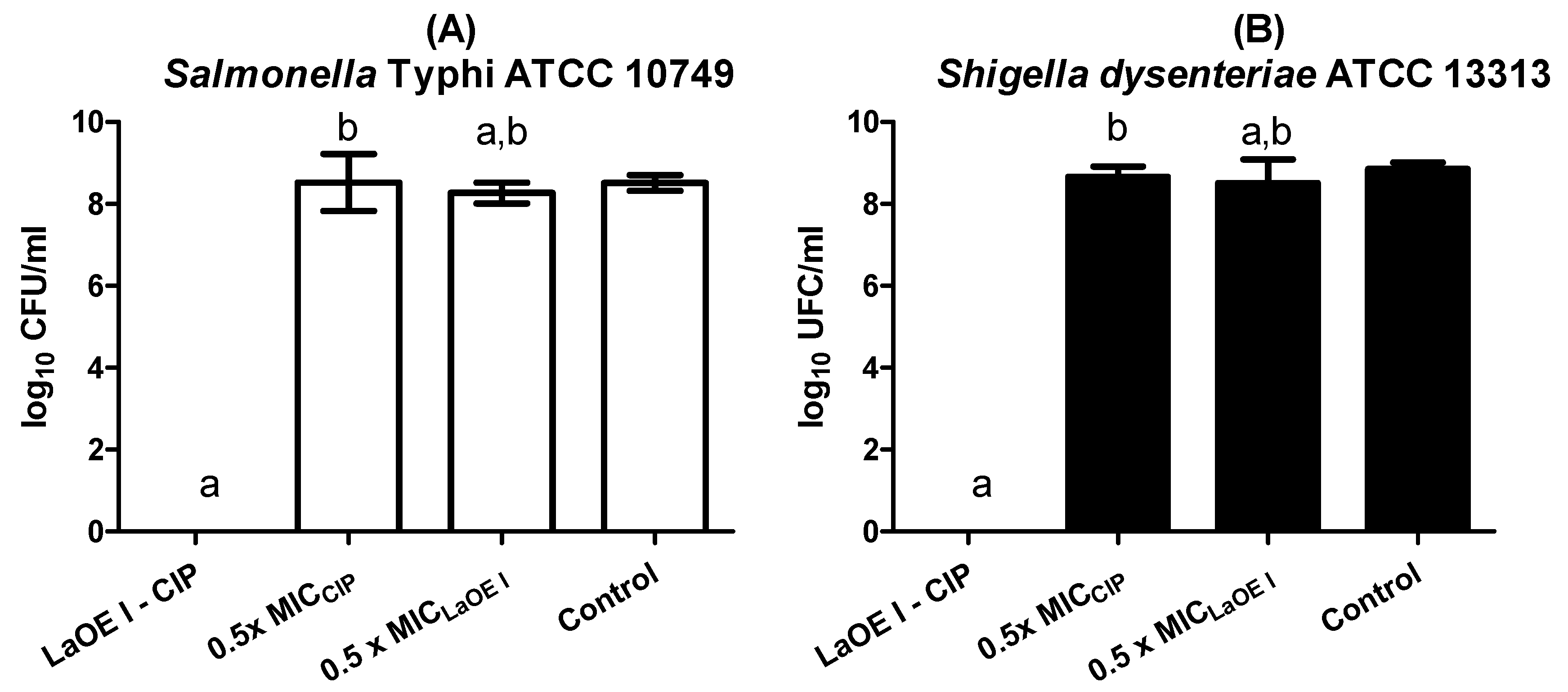
The LaEO I-CIP association promoted loss of cell viability 24 h after exposure. The concentrations 0.5 × MICLaEO I and 0.5 × MICCIP alone were not able to eradicate the preformed biofilm. However, when they were associated, there was a significant reduction in microbial viability in the biofilm compared to the control for both strains (Figure 6).

4. Discussion

In the present study, the phytochemical analysis obtained for LaEO I identified 20 constituents, among them ketones (5%), monoterpenes (40%), and sesquiterpenes (55%). The main components were citral and myrcene, which characterized it as a chemotype I essential oil (Table 1) [16].
Machado et al. [17] demonstrated LaEO activity over S. choleraesuis ATCC 10708, E. coli ATCC 10536, L. innocua ATCC 19115, L. monocytogenes ATCC 33090, P. aeruginosa ATCC 9027, and S. aureus ATCC 6538P strains, and obtained as the main constituent e-citral (31.57%), neral (25.50%), d-limonene (14.07%), germacrene D (5.47%), b-elemol (5.37%), g-terpinen (4.09%), and p-cymene (1.56%).
The variability of the chemical composition of essential oils can be influenced by the cultivation conditions and by the lack of production models that define the specific practices and conditions that can maximize production efficiency [32]. Factors such as climate, soil, geographic region, and length of day and night contribute to the variation in LaEO constituents [33].
The pharmacological potential of Lippia alba essential oils has been widely described in the literature [13,17,32]; however, its action on Gram-negative bacteria of medical importance has been little explored. The present study demonstrated the excellent antimicrobial potential of this oil on strains causing gastrointestinal infections. The LaEO I showed rapid effect, and bacteriostatic and bactericidal action on Salmonella typhi and Shigella dysenteriae (Figure 1 and Figure 2).
According to GU et al. [10], the antimicrobial resistance of Shigella spp. to quinolones in Asia and Africa has increased at an extremely rapid pace, and the proportion of resistant strains is ten-fold higher than in Europe and America. The antimicrobial resistance of Salmonella spp., to quinolones has been reported to the Italian Surveillance System for food and waterborne diseases [9].
Considering the increasing bacterial resistance to multiple drugs, synergistic combinations between new molecules and commercial drugs could be good alternatives for the control of such microorganisms [27]. The advantages of combined therapies are the increase in antimicrobial activity, drug resistance prevention, and decrease in required doses, which can subsequently reduce costs, adverse effects, and drug toxicity [34].
The assays on the modulatory activity on planktonic cells showed that LaEO I was able to synergistically modulate the action of clinically used antimicrobial agents, such as ciprofloxacin for Salmonella typhi and Shigella dysenteriae, reducing the MIC values for these microorganisms by up to 16 times (Table 3).
The time kill assay allowed identifying the bacteriostatic action time of the best association found in the Checkboard test (LaEO I-CIP). The tested association of LaEO I-CIP (1/16 MICLaEO I-1/16 MICCIP) failed to cause cell death, as these associations included the lowest tested concentrations of LaEO I and CIP.
However, the LaEO I-CIP association resulted in a 2-log10 CFU/mL cell viability reduction after 4 h (Figure 1 and Figure 2). The bacterial mortality rate is considered to be a synergistic effect when a reduction ≥ 2 log10 CFU/mL occurs [35].
Both Salmonella spp. and Shigella spp. have the biofilm-forming capacity to be important virulence factor [36,37]. Biofilms are bacterial agglomerations attached to a surface, and embedded in an extracellular matrix, which provides protection against both antimicrobial substances and host defense mechanisms. This structure allows these bacteria to survive in hostile conditions such as exposure to UV light, metals, and acids, as well as dehydration and salinity [38].
Some mechanisms that provide these protective characteristics to biofilms include the fact that the matrix can represent a mechanical barrier difficult to be penetrated by the immune system cells, and the matrix also limits diffusion for some antibiotic agents. Additionally, biofilm cells show physiological differences in relation to planktonic cells; for instance, bacteria in biofilm have a reduced activity of some cell processes, such as DNA replication, cell division, and protein synthesis [39].
As they are resistant structures, biofilms need higher concentrations for their eradication, approximately 10 to 1000 times higher, when compared to planktonic cells [39,40,41]. However, in this study, LaEO was able to inhibit biofilm formation at the same concentration that inhibited planktonic cell growth; it also promoted loss of viability, de-structuring the already mature biofilm at a concentration of only 2xMICLaEO I, showing the excellent antibiofilm action of the essential oil.
The management and prevention of Salmonella and Shigella infections represent a major public health challenge [9,42], and the present study demonstrated the great potential of LaEO I in the treatment of these infections.

5. Conclusions

The present study demonstrated that LaEO I demonstrated antibacterial activity on planktonic cell growth, biofilm formation, and rupture of Salmonella typhi and Shigella dysenteriae mature biofilm. LaEO I was able to modulate the antimicrobial activity of ciprofloxacin to reduce the concentration used, with a synergistic effect. LaEO I have shown promising activity against infections caused by Salmonella typhi and Shigella dysenteriae.

Author Contributions

M.R. and G.S. assisted in the characterization of LaOE. A.B., H.D., P.P., and S.M. assisted in the implementation of microbiological experiments. N.N. contributes to the coordination of the study.

Funding

This research received no external funding.

Acknowledgments

We thank Coordination for Enhancement of Higher Education Personnel (CAPES) and Federal University of Ceará for their financial support.

Conflicts of Interest

The authors declare no conflict of interest.

References

  1. WHO. World Health Organization. WHO’s First Ever Global Estimates of Foodborne Diseases Find Children under 5 Account for almost One Third of Deaths—2015. Available online: http://www.who.int/mediacentre/news/releases/2015/foodborne-diseaseestimates/en/ (accessed on 28 January 2017).
  2. Fhogartaigh, C.N.; Edgeworth, J.D. Bacterial gastroenteritis. Medicine 2009, 37, 586–593. [Google Scholar]
  3. BRASIL. Ministério da Saúde. Surtos de Doenças Transmitidas por Alimentos no Brasil. 2016. Available online: http://portalarquivos.saude.gov.br/images/pdf/2016/junho/08/Apresenta----o-Surtos-DTA-2016.pdf (accessed on 15 December 2016).
  4. Capalonga, R.; Ramos, R.C.; Both, J.M.; Soeiro, M.L.; Longaray, S.M.; Haas, S.; Tondo, E.C. Salmonella serotypes, resistance patterns, and food vehicles of salmonellosis in southern Brazil between 2007 and 2012. J. Infect. Dev. Ctries. 2014, 8, 811–817. [Google Scholar] [CrossRef] [PubMed]
  5. The, H.C.; Thanh, D.P.; Holt, K.E.; Thomson, N.R.; Baker, S. The genomic signatures of Shigella evolution, adaptation and geographical spread. Nat. Rev. Microbiol. 2016, 14, 235. [Google Scholar] [CrossRef] [PubMed]
  6. Crawford, R.W.; Gibson, D.L.; Kay, W.W.; Gunn, J.S. Identification of a bile-induced exopolysaccharide required for Salmonella biofilm formation on gallstone surfaces. Infect. Immun. 2008, 76, 5341–5349. [Google Scholar] [CrossRef] [PubMed]
  7. Abdullahi, U.F.; Igwenagu, E.; Mu’azu, A.; Aliyu, S.; Umar, M.I. Intrigues of biofilm: A perspective in veterinary medicine. Vet. World 2016, 9, 12. [Google Scholar] [CrossRef] [PubMed]
  8. Satpathy, S.; Sen, S.K.; Pattanaik, S.; Raut, S. Review on bacterial biofilm: An universal cause of contamination. Biocatal. Agric. Biotechnol. 2016, 7, 56–66. [Google Scholar] [CrossRef]
  9. García-Fernández, A.; Gallina, S.; Owczarek, S.; Dionisi, A.M.; Benedetti, I.; Decastelli, L.; Luzzi, I. Emergence of ciprofloxacin-resistant Salmonella enterica serovar Typhi in Italy. PLoS ONE 2015, 10, e0132065. [Google Scholar] [CrossRef]
  10. Gu, B.; Cao, Y.; Pan, S.; Zhuang, L.; Yu, R.; Peng, Z.; Tong, M. Comparison of the prevalence and changing resistance to nalidixic acid and ciprofloxacin of Shigella between Europe–America and Asia–Africa from 1998 to 2009. Int. J. Antimicrob. Agents 2012, 40, 9–17. [Google Scholar] [CrossRef]
  11. Braga, M.E.; Ehlert, P.A.; Ming, L.C.; Meireles, M.A.A. Supercritical fluid extraction from Lippia alba: Global yields, kinetic data, and extract chemical composition. J. Supercrit. Fluid 2005, 34, 149–156. [Google Scholar] [CrossRef]
  12. Julião, L.S.; Tavares, E.S.; Lage, C.L.S.; Leitão, S.G. Cromatografia em camada fina de extratos de três quimiotipos de Lippia alba (Mill) NE Br.(erva-cidreira). Rev. Bras. Farmacogn 2003, 13, 36–38. [Google Scholar] [CrossRef]
  13. Carvalho, P.M.; Macêdo, C.A.; Ribeiro, T.F.; Silva, A.A.; Da Silva, R.E.; de Morais, L.P.; Barbosa, R. Effect of the Lippia alba (Mill.) NE Brown essential oil and its main constituents, citral and limonene, on the tracheal smooth muscle of rats. Biotechnol. Rep. 2018, 17, 31–34. [Google Scholar] [CrossRef] [PubMed]
  14. Peixoto, M.G.; Costa-Júnior, L.M.; Blank, A.F.; da Silva Lima, A.; Menezes, T.S.A.; de Alexandria Santos, D.; de Fátima Arrigoni-Blank, M. Acaricidal activity of essential oils from Lippia alba genotypes and its major components carvone, limonene, and citral against Rhipicephalus microplus. Vet. Parasitol. 2015, 210, 118–122. [Google Scholar] [CrossRef] [PubMed]
  15. Morais, L.A.S. de. Influência dos fatores abióticos na composição química dos óleos essenciais. Hortic. Bras. 2009, 27, 4050–4063. [Google Scholar]
  16. Matos, F.J.A. As ervas cidreiras do Nordeste do Brasil. Estudo de três quimiotipos de Lippia alba (Mill) N.E. Brown (Verbenaceae). Parte I Farmacognosia. Rev. Bras. Farmacogn 1996, 72, 65–67. [Google Scholar]
  17. Machado, T.F.; Nogueira, N.A.P.; Pereira, R.D.C.A.; Sousa, C.T.D.; Batista, V.V. The antimicrobial efficacy of Lippia alba essential oil and its interaction with food ingredients. Braz. J. Microbiol. 2014, 45, 699–705. [Google Scholar] [CrossRef] [PubMed]
  18. Porfírio, E.M.; Melo, H.M.; Pereira, A.M.G.; Cavalcante, T.T.A.; Gomes, G.A.; Carvalho, M.G.D.; Júnior, F.E.A.C. In vitro antibacterial and antibiofilm activity of Lippia alba essential oil, citral, and carvone against Staphylococcus aureus. Sci. World J. 2017, 2017, 7. [Google Scholar] [CrossRef] [PubMed]
  19. Trevisan, M.T.; Marques, R.A.; Silva, M.G.; Scherer, D.; Haubner, R.; Ulrich, C.M.; Owen, R.W. Composition of essential oils and ethanol extracts of the leaves of Lippia species: Identification, quantitation and antioxidant capacity. Rec. Nat. Prod. 2016, 10, 485. [Google Scholar]
  20. Adams, R.P. Review of Identification of Essential Oil Components by Gas Chromatography/Mass Spectrometry. J. Am. Soc. Mass. Spectrom. 2007, 18, 803–806. [Google Scholar]
  21. CLSI. Methods for Dilution Antimicrobial Susceptibility Tests for Bacteria That Grow Aerobically: Approved Standard—Tenth Edition; CLSI document M07-A10; Clinical and Laboratory Standards Institute: Wayne, PA, USA, 2015. [Google Scholar]
  22. Romeiro, R. Técnica de Microgota Para Contagem de Células Bacterianas Viáveis em uma Suspensão; UFV Viçosa: Viçosa, Brazil, 2007. [Google Scholar]
  23. Baron, E.J.; Peterson, L.R.; Finegold, S.M. Bailey & Scott’s Diagnostic Microbiology, 9th ed.; Mosby: St. Louis, MO, USA, 1994. [Google Scholar]
  24. Cleeland, R.; Squires, E. Evaluation of new antimicrobials in vitro and in experimental animal infections. Antibiot. Lab. Med. 1991, 3, 739–787. [Google Scholar]
  25. Shin, S.; Lim, S. Antifungal effects of herbal essential oils alone and in combination with ketoconazole against Trichophyton spp. J. Appl. Microbiol. 2004, 97, 1289–1296. [Google Scholar] [CrossRef] [Green Version]
  26. EUCAST. Determination of minimum inhibitory concentrations (MICs) of antibacterial agentes by broth diluition. Clin. Microbiol. J. 2003, 9, 1–7. [Google Scholar]
  27. Dodou, H.V.; de Morais Batista, A.H.; Sales, G.W.P.; de Medeiros, S.C.; Rodrigues, M.L.; Nogueira, P.C.N.; Nogueira, N.A.P. Violacein antimicrobial activity on Staphylococcus epidermidis and synergistic effect on commercially available antibiotics. J. Appl. Microbiol. 2017, 123, 853–860. [Google Scholar] [CrossRef]
  28. Olajuyigbe, O.O.; Anthony, J.A. In vitro antibacterial and time-kill assessment of crude methanolic stem bark extract of Acacia mearnsii De Wild against bacteria in shigellosis. Molecules 2012, 17, 2103–2118. [Google Scholar] [CrossRef]
  29. Nostro, A.; Blanco, A.R.; Cannatelli, M.A.; Enea, V.; Flamini, G.; Morelli, I.; Alonzo, V. Susceptibility of methicillin-resistant staphylococci to oregano essential oil, carvacrol and thymol. FEMS Microbiol. Lett. 2004, 230, 191–195. [Google Scholar] [CrossRef]
  30. Stepanović, S.; Vuković, D.; Dakić, I.; Savić, B.; Švabić-Vlahović, M. A modified microtiter-plate test for quantification of staphylococcal biofilm formation. J. Microbiol. Methods 2000, 40, 175–179. [Google Scholar] [CrossRef]
  31. Mah, T.-F. Establishing the minimal bactericidal concentration of an antimicrobial agent for planktonic cells (MBC-P) and biofilm cells (MBC-B). J. Vis. Exp. 2014, 83, 50854. [Google Scholar] [CrossRef] [PubMed]
  32. Tofiño-Rivera, A.; Ortega-Cuadros, M.; Galvis-Pareja, D.; Jiménez-Rios, H.; Merini, L.J.; Martínez-Pabón, M.C. Effect of Lippia alba and Cymbopogon citratus essential oils on biofilms of Streptococcus mutans and cytotoxicity in CHO cells. J. Ethnopharmacol. 2016, 194, 749–754. [Google Scholar]
  33. Tavares, I.B.; Momenté, V.G.; do Nascimento, I.R. Lippia alba: Estudos químicos, etnofarmacológicos e agronômicos. Pesqui. Apl. Agrotecnol. 2011, 4, 204–220. [Google Scholar] [CrossRef]
  34. Szabo, M.R.; Radu, D.; Gavrilas, S.; Chambre, D.; Iditoiu, C. Antioxidant and antimicrobial properties of selected spice extracts. Int. J. Food Prop. 2010, 13, 535–545. [Google Scholar] [CrossRef]
  35. Jayaraman, P.; Sakharkar, M.K.; Lim, C.S.; Tang, T.H.; Sakharkar, K.R. Activity and interactions of antibiotic and phytochemical combinations against Pseudomonas aeruginosa in vitro. Int. J. Biol. Sci. 2010, 6, 556. [Google Scholar] [CrossRef]
  36. Di Domenico, E.G.; Cavallo, I.; Pontone, M.; Toma, L.; Ensoli, F. Biofilm producing Salmonella typhi: Chronic colonization and development of gallbladder cancer. Int. J. Mol. Sci. 2017, 18, 1887. [Google Scholar] [CrossRef] [PubMed]
  37. Kang, J.; Liu, L.; Liu, M.; Wu, X.; Li, J. Antibacterial activity of gallic acid against Shigella flexneri and its effect on biofilm formation by repressing mdoH gene expression. Food Control 2018, 94, 147–154. [Google Scholar] [CrossRef]
  38. Hall-Stoodley, L.; Costerton, J.; Stoodley, P. Bacterial biofilms: From the natural environment to infectious diseases. Nat. Rev. Microbiol. 2004, 2, 95. [Google Scholar] [CrossRef] [PubMed]
  39. Otto, M. Molecular basis of Staphylococcus epidermidis infections. Semin. Immunopathol. 2012, 34, 2. [Google Scholar] [CrossRef]
  40. Aguila-Arcos, S.; Ding, S.; Aloria, K.; Arizmendi, J.M.; Fearnley, I.M.; Walker, J.E.; Alkorta, I. A commensal strain of Staphylococcus epidermidis overexpresses membrane proteins associated with pathogenesis when grown in biofilms. J. Membr. Biol. 2015, 248, 431–442. [Google Scholar] [CrossRef] [PubMed]
  41. Sahal, G.; Bilkay, I.S. Multi drug resistance in strong biofilm forming clinical isolates of Staphylococcus epidermidis. Braz. J. Microbiol. 2014, 45, 539–544. [Google Scholar] [CrossRef]
  42. Zaidi, M.B.; Estrada-García, T. Shigella: A highly virulent and elusive pathogen. Curr. Trop. Med. Rep. 2014, 1, 81–87. [Google Scholar] [CrossRef]
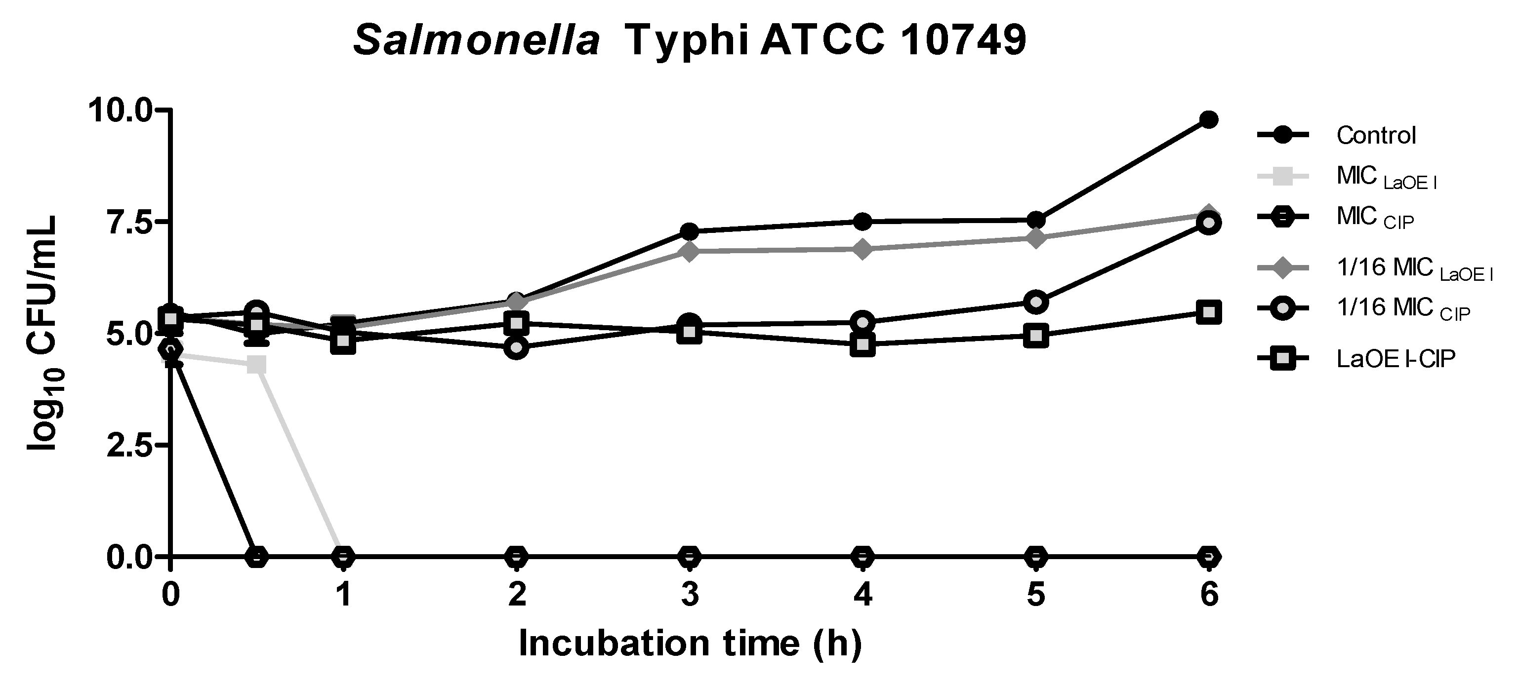
Figure 1. Effect of time of exposure to LaEO I and the LaEO I-CIP association on the viability of Salmonella typhi ATCC 10749. Values are expressed as mean ± SEM of two assays. The analysis was performed by Analysis of Variance (ANOVA), with Dunnett’s post-test and p value < 0.05 when compared to the control. Control: culture medium + microorganism; MICLaEO I = 1 mg/mL; MICCIP = 0.25 μg/mL; FICI LaEO I-CIP = 0.125 (lower concentrations of LaEO I and CIP in the association, with antimicrobial activity = 1/16 × MIC).
Scipharm 86 00052 g001
Figure 2. Effect of time of exposure to LaEO I and LaEO I-CIP association on the viability of Shigella dysenteriae ATCC 13313. Values are expressed as mean ± SEM of two assays. The analysis was performed by ANOVA, with Dunnett’s post-test and p value < 0.05 compared to the control. Control: culture medium + microorganism; MICLaEO I = 1 mg/mL; MICCIP = 0.25 μg/mL; FICILaEO I-CIP = 0.125 (lowest concentrations of the LaEO I and CIP association, with antimicrobial activity = 1/16 × MIC).
Scipharm 86 00052 g002
Figure 3. Inhibition of Salmonella typhi (A) and Shigella dysenteriae (B) biofilm formation by LaEO I. Values are expressed as mean ± SEM of three assays, each in triplicate. Control: culture medium + microorganism. The analysis was performed by ANOVA, with Dunnett’s post-test and p value < 0.05 (*) when compared to the control.
Scipharm 86 00052 g003
Figure 4. Inhibition of Salmonella typhi (A) and Shigella dysenteriae (B) biofilm formation by the association LaEO I-CIP. Values is expressed as mean ± SEM of two assays. The analysis was performed by ANOVA, with Dunnett’s post-test and a p value < 0.05 when compared to the control (a) and a p value < 0.05 when compared to the CIP+LaEO association (b). Control: culture medium + microorganism; MICLaEO I = 1 mg/mL; MICCIP = 0.25 μg/mL; and FICILaEO I-CIP = 0.125 (lowest concentrations of the LaEO I and CIP association, with antimicrobial activity = 1/16 × MIC).
Scipharm 86 00052 g004
Figure 5. Effect of LaEO I on the eradication of Salmonella typhi (A) and Shigella dysenteriae (B) mature biofilm. Values are expressed as the mean ± SEM of two assays, in triplicate. The analysis was performed by ANOVA, with Dunnett’s post-test and p value < 0.05 (*) when compared to the control. Control: culture medium + microorganism.
Scipharm 86 00052 g005
Figure 6. Modulatory effect of LaEO I on the antimicrobial activity of ciprofloxacin on Salmonella typhi (A) and Shigella dysenteriae (B) mature biofilm. Values are expressed as the mean ± SEM of two assays, in triplicate. The analysis was performed by ANOVA, with Dunnett’s post-test and a p value < 0.05 when compared to the control (a) and a p value < 0.05 when compared to the CIP+LaEO association (b). Control: culture medium + microorganism.
Scipharm 86 00052 g006
Table 1. Chemical components of LaEO I identified by gas chromatography-mass spectrometry (GC/MS).
Number of PeakRetention Time (min)FormulaComponent% Area
115.154C8H14OSulcatone0.95
215.416C10H16Myrcene12.56
317.480C10H14p-cymene4.06
419.527C10H16γ-Terpinene0.97
521.883C10H18OLinalool1.45
626.890C10H16OCamphor0.74
730.459C10H16ONeral (cis-citral)25.90
830.762C10H14OCarvone2.06
931.138C10H18ONerol0.74
1032.160C10H16OGeranial (trans-citral)34.16
1138.387C15H24α-copaene3.13
1240.927C15H24trans-Caryophyllene4.03
1344.867C15H24Germacrene1.08
1446.559C15H24γ-muurolene0.41
1547.176C15H26OElemol1.88
1647.316C15H26ONerolidol0.70
1747.924C15H24OCaryophyllene oxide3.47
1848.062C15H26OGuaiol0.58
1948.857C15H26Oβ-Eudesmol0.75
2050.992C15H26OHedycaryol0.38
Monoterpene17.59
Oxygenated monoterpene39.15
Sesquiterpene34.55
Oxygenated sesquiterpene7.76
Ketone0.95
Table 2. MIC and MBC concentrations of LaEO I and ciprofloxacin on Salmonella typhi and Shigella dysenteriae strains.
StrainLaEO I 1 (mg/mL)CIP 2 (μg/mL)
MIC 3MBC 4MIC
Salmonella typhi ATCC 10749110.25
Shigella dysenteriae ATCC 13313110.25
1 LaEO I: essential oil of chemotype I leaves; 2 CIP: ciprofloxacin.3 MIC: Minimum Inhibitory Concentration; 4 MBC: Minimum Bactericidal Concentration.
Table 3. Modulatory effect of LaEO I on the activity of clinically used antimicrobial agents on Salmonella typhi and Shigella dysenteriae strains.
Salmonella typhi ATCC 10749Shigella dysenteriae ATCC 13313
LaEO I-ATMFICLaEO I 1FICATM 2FICI 3ME 4FICLaEO I 1FICATM 2FICI 3ME 4
LaEO I-CIP 50.06250.06240.1249S0.06250.06240.1249S
LaEO I-AMI 6000ND000ND
LaEO I-CAZ 7000ND000ND
LaEO I-CPM 80.06250.1250.1875S0.06250.06250.1249S
LaEO I-CRO 90.06250.250.3125S0.06250.06250.1249S
1 FICOELaI: Fractional Inhibitory Concentration of LaEO I; and 2 FICATM: Fractional Inhibitory Concentration of LaEO I antimicrobials; 3 FICI: Fractional Inhibitory Concentration Index; 4 ME: Modulatory Effect. 5 CIP: ciprofloxacin, 6 AMI: amikacin, 7 CAZ: ceftazidime, 8 CPM: cefepime, 9 CRO: ceftriaxone; FICI was interpreted as: synergistic effect for FICI ≤ 0.5 (S), additive or indifferent for FICI > 0.5 (I) and < 1.0, antagonist for FICI ≥ 1.0 (A) (EUCAST, 2003), and not determined (ND).