Article

A Personalized 3D-Printed Smart Splint with Integrated Sensors and IoT-Based Control: A Proof-of-Concept Study for Distal Radius Fracture Management

1
Department of Orthopedics and Traumatology, Third Affiliated Hospital of Beijing University of Chinese Medicine, Beijing 100029, China
2
College of Computer Science and Technology, Beijing University of Technology, Beijing 100124, China
*
Authors to whom correspondence should be addressed.
Electronics 2025, 14(17), 3542; https://doi.org/10.3390/electronics14173542
Submission received: 31 July 2025 / Revised: 1 September 2025 / Accepted: 2 September 2025 / Published: 5 September 2025

Abstract

Conventional static fixation for distal radius fractures (DRF) is clinically challenging, with methods often leading to complications such as malunion and pressure-related injuries. These issues stem from uncontrolled pressure and a lack of real-time biomechanical feedback, resulting in suboptimal functional recovery. To overcome these limitations, we engineered an intelligent, adaptive orthopedic device. The system is built on a patient-specific, 3D-printed architecture for a lightweight, personalized fit. It embeds an array of thin-film pressure sensors at critical anatomical sites to continuously quantify biomechanical forces. This data is transmitted via an Internet of Things (IoT) module to a cloud platform, enabling real-time remote monitoring by clinicians. The core innovation is a closed-loop feedback controller governed by a robust Interval Type-2 Fuzzy Logic (IT2-FLC) algorithm. This system autonomously adjusts servo-driven straps to dynamically regulate fixation pressure, adapting to changes in limb swelling. In a preliminary clinical evaluation, the group receiving the integrated treatment protocol, which included the smart splint and TCM herbal therapy, demonstrated superior anatomical restoration and functional recovery, evidenced by higher Cooney scores (91.65 vs. 83.15) and lower VAS pain scores. This proof-of-concept study validates a new paradigm for adaptive orthopedic devices, showing high potential for clinical translation.

1. Introduction

Distal radius fractures (DRF) are a prevalent clinical condition with a high incidence rate, defined as a fracture of the cancellous bone within 3 cm of the radiocarpal joint surface [1]. It accounts for 17% of emergency fractures [2] and one-sixth of all fracture cases [3]. Notably, the demographic characteristics of DRF patients are age-dependent. In elderly patients, DRF is frequently the result of low-energy trauma, such as falls from a standing height, and is linked to osteoporosis [4,5,6] and estradiol level [7,8,9,10]. This condition accounts for 18% of fractures in this demographic [11]. In China, DRF is typically the consequence of high-energy trauma in younger patients, accounting for 25% of pediatric fractures and 74.58% of adult ulnar and radial fractures [12]. Colles’ fracture, an extension-type fracture of the distal radius, demonstrates a particularly high prevalence within this population [11]. The prevalence of DRF in elderly patients is increasing as a result of the global geriatric population [11,12].
Although the primary objective of distal radius fracture (DRF) treatment is to restore anatomical structure and function, achieving optimal outcomes remains clinically challenging. Current evidence indicates that approximately 30% of patients experience chronic wrist/hand pain one year after a fracture (≥35/50 on the PRWE pain subscale) [13]. These persistent symptoms, alongside complications such as chronic stiffness, complex regional pain syndrome (CRPS), malunion, and delayed return to work, significantly impair patients’ quality of life and occupational capacity [13,14,15,16]. Thus, timely and effective intervention for DRF is critical to optimize functional recovery, minimize complications, and improve long-term prognosis.
However, conventional static fixation methods are unable to adapt to the dynamic post-traumatic swelling of the limb. In the acute phase, as swelling progresses, excessive pressure within a rigid cast can lead to severe complications, including pressure sores and compartment syndrome [17]. Conversely, as swelling subsides in the subacute phase, the resulting loose fit often leads to a loss of reduction and eventual fracture malunion [18]. These challenges stem from the lack of real-time biomechanical feedback, motivating the development of an adaptive therapeutic device.

2. Related Work

2.1. Treatment Options for Distal Radius Fractures

The main objective in managing distal radius fractures (DRF) is to reestablish the normal anatomical configuration of the distal radius, which is essential for restoring wrist joint function and hand grip strength. Conservative treatments for DRF vary by type and include closed manual reduction with plaster, splint, or brace fixation. Surgical options include percutaneous closed reduction and internal fixation, external fixation, open reduction and internal fixation, and arthroscopically assisted fixation. There remains a debate concerning the optimal treatment strategy for elderly patients with DRF, with no consensus achieved [19]. Long-term follow-up studies demonstrate that elderly patients prioritize pain management, restoring self-care capabilities, and prompt reintegration into society [20,21,22]. The optimal treatment for DRF should be minimally invasive, tailored to the individual, associated with reduced complications, and characterized by a shorter duration, a notion that has received considerable acknowledgment.
Minimally invasive closed manual reduction, along with plaster, splint, or brace fixation, is suitable for non-displaced or minimally displaced extra-articular DRF and certain stable intra-articular DRF cases. In stable distal radius fractures, most patients attain favorable results through closed manual reduction and external fixation [23]. While the reduction and stability of this fixation method are less effective than plate internal fixation, research indicates that despite statistical differences in postoperative imaging scores, there is no significant difference in wrist function scores one year after surgery [24,25]. Excessive pursuit of anatomical reduction is unnecessary, as favorable outcomes can still be attained through postoperative joint micromotion and appropriate rehabilitation exercises, even when the joint surface is not perfectly restored. Intraoperative precision in restoring volar tilt, ulnar inclination, and radial height is essential for minimizing postoperative complications. Internal fixation using plates is associated with increased surgical trauma, potential damage to adjacent muscles and periosteal tissues, and the risk of injury to the median nerve and radial artery [26,27]. The procedure necessitates a subsequent surgery for implant removal, potentially resulting in irritation or rupture of the extensor and flexor tendons, increased surgical expenses, extended hospital stays, and complications including disuse osteoporosis and Sudeck’s atrophy [28]. Surgical treatment may more effectively restore the anatomical relationship of the wrist joint than closed manual reduction and external fixation; however, it does not significantly influence long-term functional recovery. Conversely, conservative treatment may decrease fracture healing duration, minimize complication rates, lower treatment expenses, mitigate psychological stress and pain related to surgery, and lessen the financial burden on patients [27].

2.2. Analysis of Traditional TCM Small Splint Technique

The TCM small splint technique has shown notable clinical benefits in managing DRF. Zhang et al. [3] demonstrated that TCM small splints surpass traditional plaster fixation regarding treatment success rates, Cooney scores, and fracture healing times, underscoring their distinct advantages. The small splint utilizes a closed reduction technique that maintains intra-articular blood supply and ensures elastic fixation, safeguarding the radial, intermediate, and ulnar columns while providing elastic support during functional exercises [29]. The mechanical compression applied by the splint to the wrist joint ensures sustained pressure, which can be modified as swelling decreases to avert complications like pressure sores and compartment syndrome. Early functional exercises reduce joint stiffness resulting from extended immobilization and facilitate the recovery of wrist function [30].
Research indicates that small splints are more effective than plaster fixation in enhancing wrist function recovery, alleviating pain, and improving imaging indicators. Their advantages include enhanced mobility and elasticity, reduced compression sensations from swelling, improved blood circulation and patient comfort, and prevention of fracture re-displacement resulting from frequent plaster changes. The small splint treatment is non-invasive, promotes rapid recovery, is cost-effective, and enables straightforward observation and adjustment by physicians [31]. The combination of splints and pressure pads at various positions allows the small splint to effectively manage fracture alignment [24], enhance limb blood circulation, and expedite the healing process of fractures [31]. The integration of 3D printing technology allows for the customization of small splints for individual patients, resulting in lighter and more breathable designs than traditional splints [31,32,33,34], which enhances treatment efficacy and patient experience. Modified splints, including Topology-optimized splints [35], Dynamic splints [36], and Cedar skin splints [3], have notably improved clinical outcomes for DRF.
Furthermore, TCM small splints may be used with traditional Chinese herbal decoctions to enhance fracture healing. Research indicates that applying herbal decoctions, including Yiqi Bushen Tongluo Decoction [37] and Zhenggu Zijin Pill [38], alongside small splint fixation, can significantly reduce the duration of the fracture healing process. The development of intelligent splints that address clinical needs is consistent with global trends in the treatment of distal radius fractures and signifies a crucial direction for the advancement of traditional Chinese medicine orthopedics.
While the TCM small splint technique offers notable benefits in treating DRF, including simplicity, cost-effectiveness, and enhancement of fracture healing and functional recovery, it has limitations. Although the TCM small splint technique demonstrates significant advantages in treating DRF, including operational simplicity, cost-effectiveness, and its ability to promote fracture healing and functional recovery, it still presents certain limitations. The conventional splint treatment protocol requires patients to undergo frequent follow-up visits for splint tightness adjustment, elevating infection risks and substantially increasing clinicians’ workload. Current research primarily emphasizes the combination of small splint fixation with oral herbal decoctions, while insufficient attention is directed toward integrating drug release functionality into the splint surface. This oversight may impede fracture healing and diminish treatment efficacy. The lack of real-time monitoring of limb blood flow and nerve compression heightens the risk of postoperative complications [13,14,15,16,39,40]. The limitations have impeded the broad implementation of small splint technology in primary healthcare environments. While 3D-printed lightweight and personalized splints have enhanced patient comfort and treatment outcomes to a degree, these innovations are still primarily experimental and have not yet realized real-time monitoring of radial artery pressure or extensive clinical application. Future research should prioritize the development of scientifically advanced, convenient, and comfortable splints that incorporate intelligent monitoring technology to facilitate real-time feedback and personalized treatment. This approach will enhance treatment efficacy and patient experience while driving the modernization of TCM small splint technology.

2.3. Advances in Smart Splints for Wearable Monitoring

Parallel to advancements in 3D printing, the field of “smart splints” has emerged, focusing on integrating sensing technologies to provide objective, data-driven feedback. Work by De Agustín Del Burgo et al. on the development of a smart splint to monitor different parameters during the treatment process highlights this trend by showcasing how smart technologies can be embedded in orthoses to monitor physiological parameters [41]. In the context of rehabilitation, a scoping review by Kim et al. on the use of wearable sensors to assess and treat the upper extremity after stroke details how various sensors can track motor function and activity levels to offer quantitative biofeedback [42]. Specific implementations further demonstrate the state of the art; for example, research by Borchani et al. on a smart trauma-fixation device with integrated self-powered piezo-floating-gate sensors shows a system capable of monitoring postoperative bone healing [43]. However, existing smart devices are typically limited to passive monitoring, leaving a research gap for active, closed-loop systems that can autonomously regulate therapeutic parameters like fixation pressure.

3. Methods

This research introduces an innovative, intelligent external fixation apparatus that combines Internet of Things (IoT) technology with the conventional TCM tiny splint method for the accurate treatment and expedited rehabilitation of DRF. The apparatus integrates 3D printing technology, an intelligent control system, a remote monitoring and adjustment mechanism, and TCM herbal medicine pads to create an efficient, tailored, and intelligent therapeutic solution. Initially, accurate measurements of the damaged limb are acquired by 3D scanning, followed by the creation of customized designs with 3D modeling software. The external fixation device is then produced utilizing a 3D printer. Secondly, the apparatus features an intelligent control system comprising pressure sensors, microprocessors, and servo drivers, facilitating real-time monitoring of pressure fluctuations at the fracture site and automatic modulation of clamping force according to predefined logic. Furthermore, an IoT-based remote monitoring and adjustment system enables physicians to oversee patients’ recovery, acquire real-time pressure data, and implement requisite treatment modifications through a mobile application or PC interface. Ultimately, following the TCM idea of three-stage fracture healing, herbal medicine pads specifically formulated for the initial, intermediate, and late stages of fracture healing are developed to enhance healing and functional recovery. This technology seeks to enhance fracture reduction and functional recovery, increase treatment efficacy, and decrease complication rates by meticulously regulating treatment pressure and monitoring patient recovery data in real time. Figure 1 depicts the comprehensive research design.
Therefore, the objective of this study is to present the design, development, and proof-of-concept of a novel intelligent external fixation apparatus. We detail its personalized fabrication, closed-loop control system, and IoT-based monitoring platform. Furthermore, we report the results of a preliminary clinical trial designed to assess the initial feasibility, safety, and potential efficacy of this integrated system.

3.1. Personalized External Fixation Device Utilizing 3D Printing

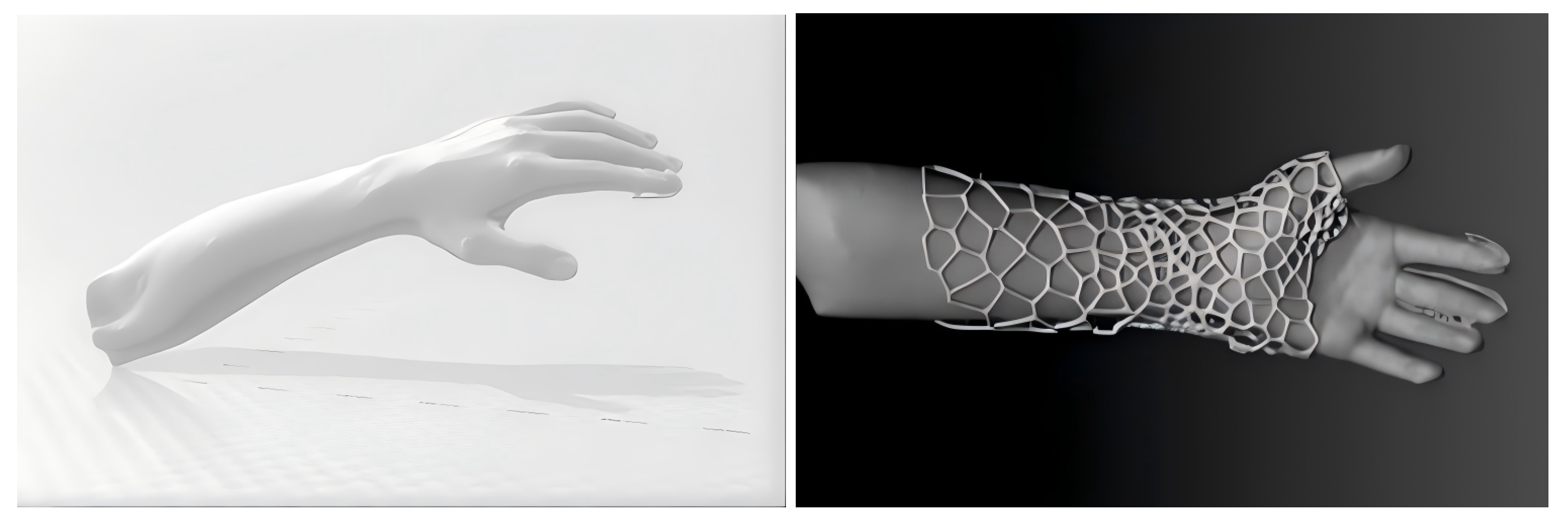
Conventional 3D printing technology has become an invaluable tool in enhancing preoperative planning for orthopedic procedures, where CT-derived 3D reconstructions of the fracture site are used to print physical bone models for surgical simulation. This study leverages and expands upon this principle by introducing a systematic design and manufacturing workflow for the individualized smart splint. This workflow is composed of several key stages: (1) The process begins with the construction of a forearm model. To ensure the optimal therapeutic effect, the patient’s wrist is held in a relaxed state of dorsal flexion while a high-precision 3D scanner (Shining3D) with a point accuracy of up to 0.1 mm captures the external morphology, and the acquired point cloud data is processed to construct a high-fidelity model of the forearm in Stereolithographic (STL) format (Figure 2). (2) Based on this anatomical model, the initial design space is defined by applying a uniform 3 mm wall thickness to the external contour, and within this domain, critical functional features are integrated, including hook-like structures for the tensioning ropes and designated housings for all electronic components. (3) The solid-body splint is subsequently optimized for lightweighting and comfort by computationally applying a hollow, web-like pattern to the design domain, which removes redundant material while maintaining structural integrity. (4) The final optimized structure is then fabricated by additive manufacturing using an HP Multi Jet Fusion (MJF) 3D printer with medical-grade Polyamide 12 (PA12), which is certified for biocompatibility according to ISO 10993 standards [44]. Key printing parameters, such as a layer height of 0.08 mm, are set to ensure dimensional accuracy, and the device undergoes a bead-blasting post-processing step to achieve a smooth surface finish. (5) Finally, the electronic components are installed, and the two halves of the smart splint are secured to the limb using the integrated servo-controlled rope lock system, resulting in a precise, functional, and personalized solution for the treatment of DRF.

3.2. Advanced Closed-Loop Control System for External Fixation Devices

The intelligent closed-loop control system is the fundamental component of the smart external fixation device. It comprises a pressure sensing mechanism, a microprocessor, and servo drivers, facilitating real-time monitoring of pressure variations at the fracture site and automatic modulation of the external fixation device’s clamping force according to established logic.

3.2.1. Pressure Sensing System

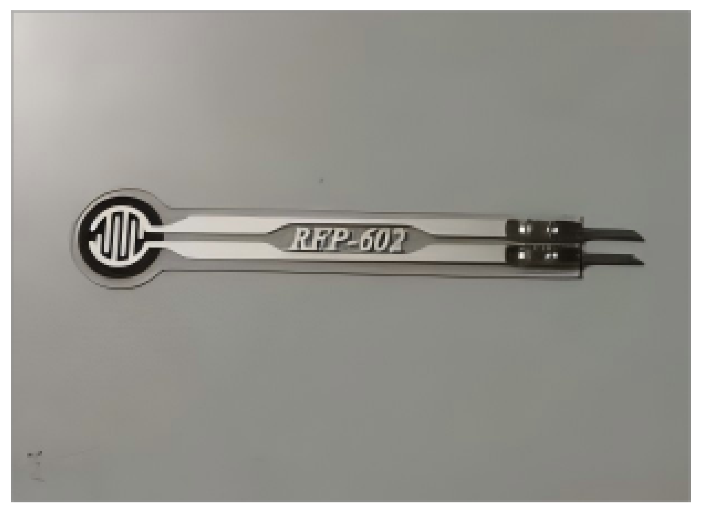
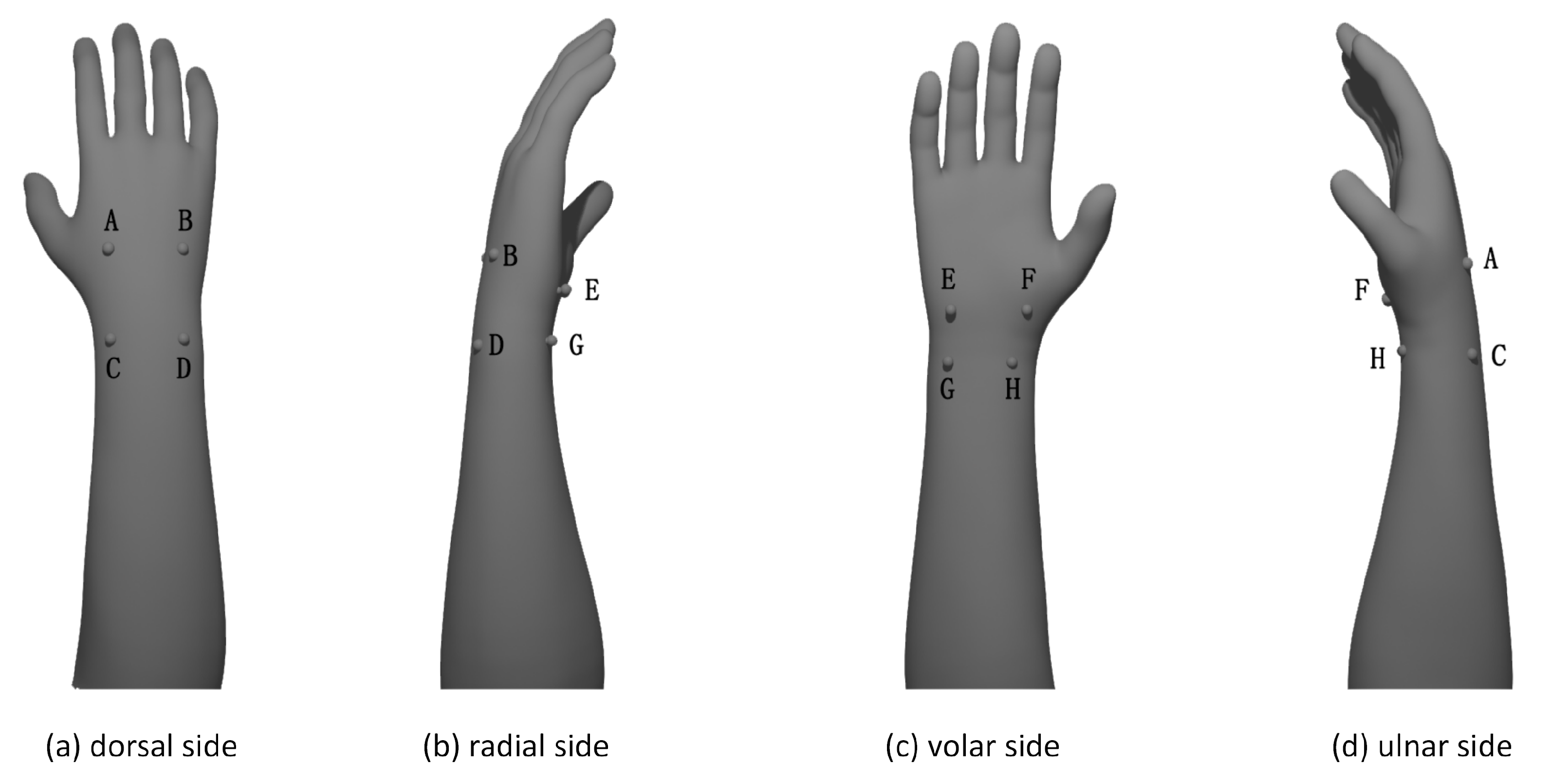
In this study, a total of eight thin-film pressure sensors (Figure 3) are strategically placed at multiple key sites to achieve comprehensive biomechanical monitoring, as illustrated in Figure 4. The selection of these locations is determined entirely by expert clinical experience to ensure both therapeutic efficacy and safety. The sites include key anatomical landmarks, positions proximal and distal to the fracture line to monitor the effectiveness of three-point fixation, and areas near superficial nerves to prevent compression. This layout allows for the real-time monitoring of both the corrective forces at the fracture end and the safety-critical pressure over the radial artery. Compared to other pressure sensors, thin-film pressure sensors offer advantages such as compact size, high sensitivity, and better skin conformity. Suppose the pressure sensor on the radial artery detects abnormally high pressure or significant swelling in the affected limb. In that case, the smart external fixation device will immediately alert the user through visual and auditory signals. Simultaneously, the IoT system and management platform will notify the physician. The microprocessor in the device will then adjust the tension of the servo-controlled ropes based on the physician’s predefined logic, alleviating the pressure on the radial artery. Conversely, if the pressure is too low, the device will prompt the user and increase the tension to maintain continuous fixation. Prior to clinical use, all thin-film pressure sensors underwent benchtop calibration against a standardized pressure gauge to ensure a linear and repeatable response within the target therapeutic pressure range. This step confirmed their suitability for the intended application, although a formal long-term reliability study was beyond the scope of this proof-of-concept work.

3.2.2. Automatic Pressure Adjustment System

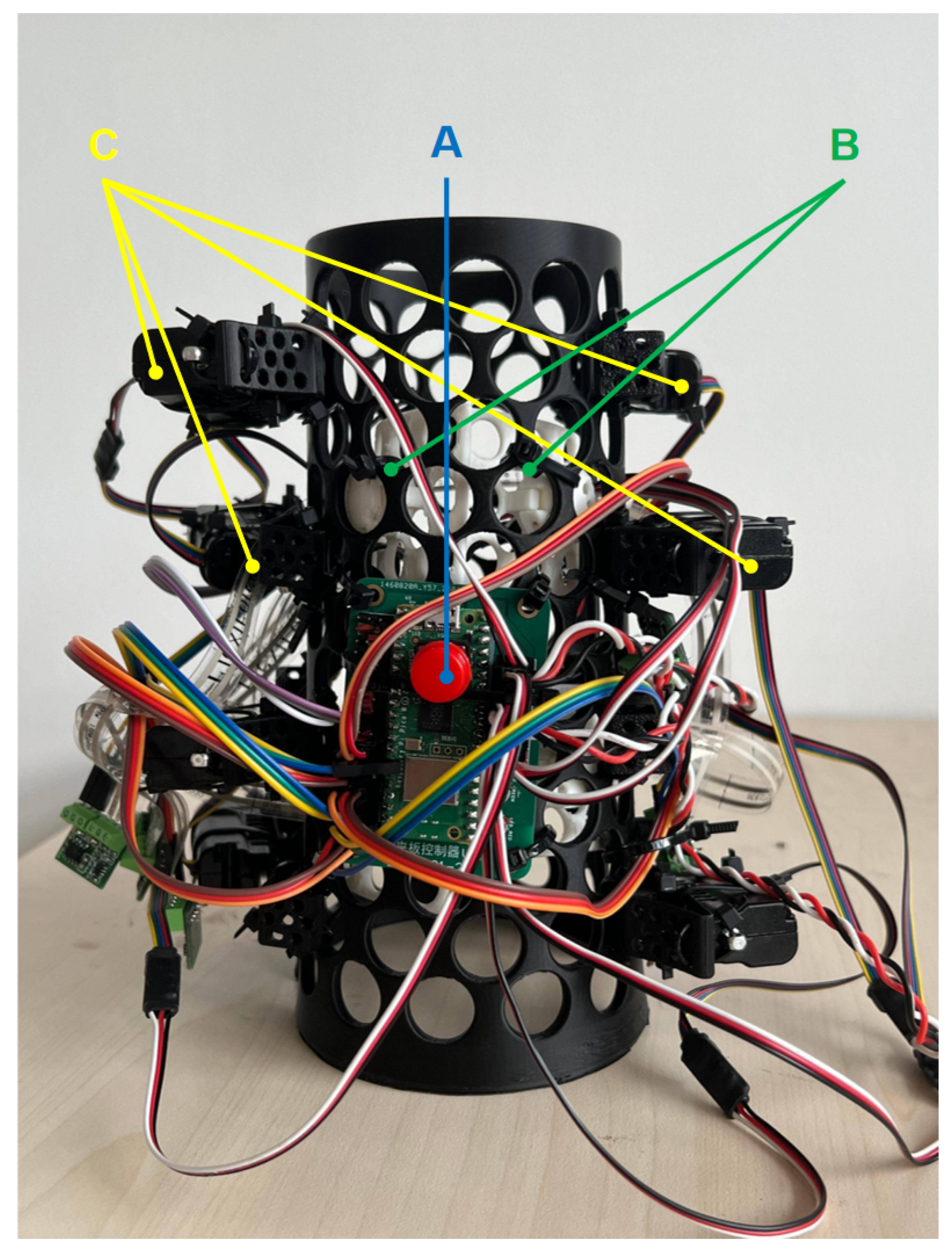
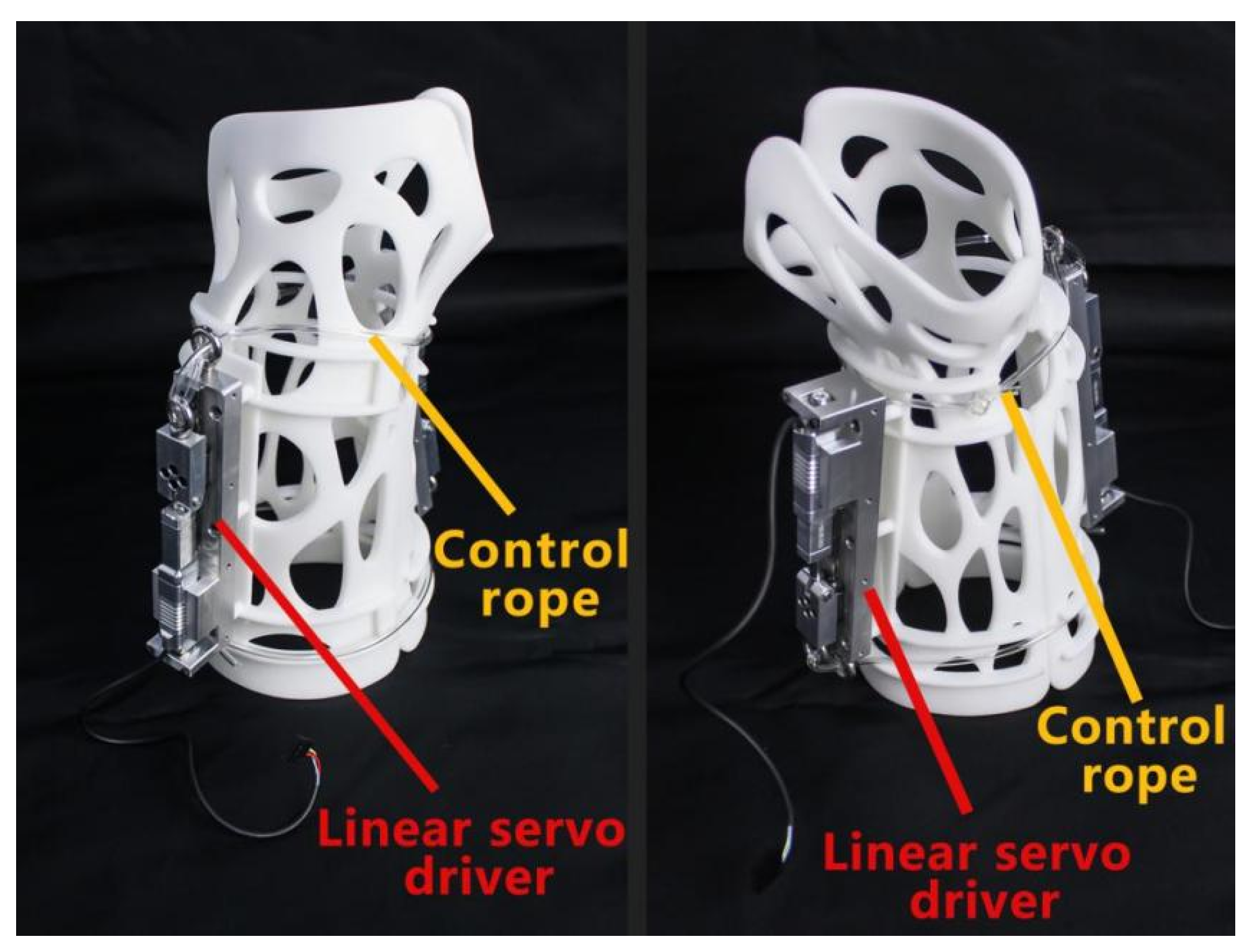
A photograph of the complete smart splint system is shown in Figure 5. The pressure automatic adjustment system employs a 3D-printed nylon external fixation device to stabilize the affected limb. The device features hook-like structures along its edges and is initially secured with a rope lock arranged in a figure-eight configuration. A linear servo driver is positioned on the dorsal aspect of the external fixation device (refer to Figure 6). The ends of the rope are linked to the push rod of the linear servo driver through a pulley or sheave mechanism. The extension or retraction of the linear driver’s push rod results in the tightening or loosening of the rope, thereby adjusting the clamping force of the external fixation device. Cable mechanisms are installed at the device’s proximal palm and proximal finger ends to regulate the tightening force at both locations.
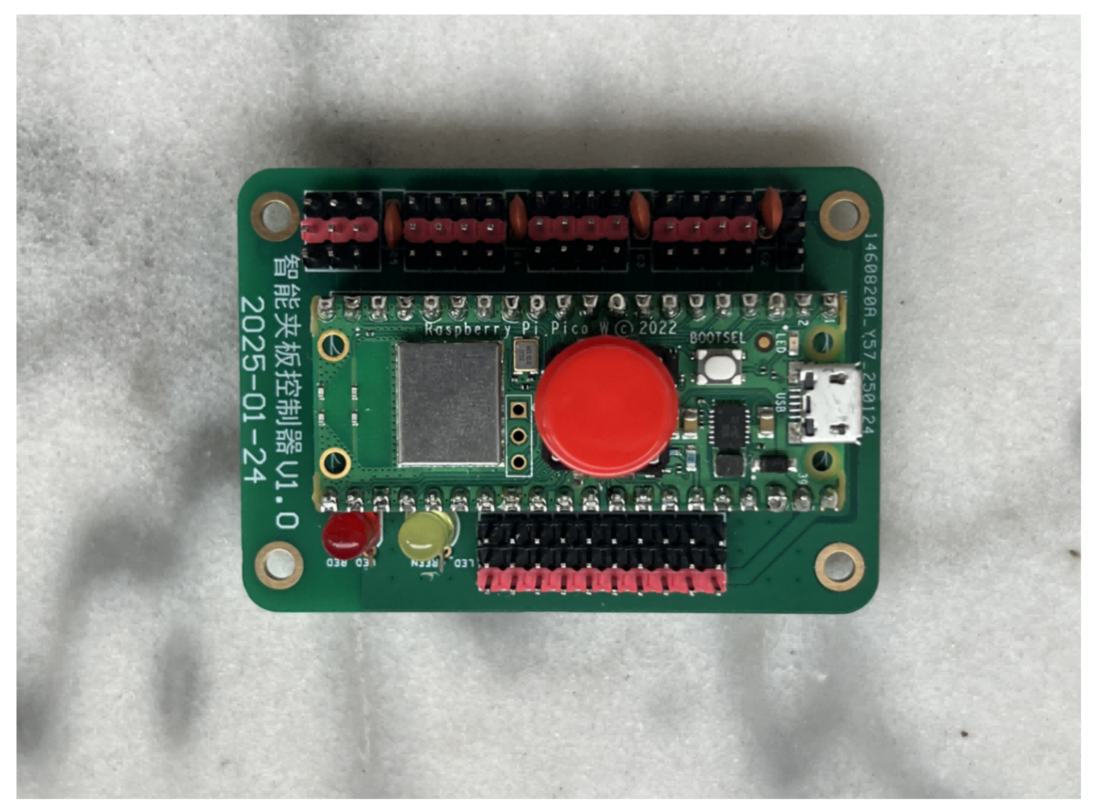
A force sensor is positioned at the linear driver’s fixed end to quantify the rope’s tension. The external fixation device features a waterproof battery compartment and designated space for a PCB control board (Figure 7), which supplies power to the driver and transmits signals for clamping force adjustments. The system is designed with a multi-level fail-safe architecture to ensure patient safety. The primary safety feature is a one-touch emergency power-off button (Figure 8), which is conveniently located on the exterior of the splint for easy one-handed operation by the patient. Critically, the linear motors are custom-designed with a fail-safe mechanism: upon any loss of power, the motor shafts immediately and rapidly retract to their maximum extension, ensuring a complete and instantaneous release of all tensioning force. This, combined with the flexible nature of all connecting materials, ensures the patient’s limb can be freed in any emergency. Furthermore, the control algorithm includes software-based safety protocols. The system continuously monitors sensor data for anomalies (e.g., readings outside a plausible physiological range); if a sensor malfunction is detected, automatic adjustments are immediately halted, and an audiovisual alert is triggered. To prevent servo jamming or incorrect pressure adjustments, the software enforces a hard-coded pressure safety limit, rejecting any control command that would result in a pressure value outside of this predefined safe therapeutic window.
The electronic control system is integrated onto a custom-designed Printed Circuit Board (PCB) housed within the splint. The central processing unit is a Raspberry Pi Pico W, chosen for its powerful dual-core processor and integrated Wi-Fi module. Data is acquired from eight thin-film pressure sensors; to interface these with the microcontroller’s limited Analog-to-Digital Converter (ADC) inputs, a CD4051BE 8-channel analog multiplexer is utilized. The Pico W generates precise Pulse-Width Modulation (PWM) signals to directly control the eight linear servo motors, with the main power supply to all servos being managed by a power MOSFET for safety and power management. The entire system is powered by a 3.7 V 800 mAh rechargeable Lithium Polymer (LiPo) battery with integrated protection circuits. Onboard user feedback is provided by a piezo buzzer and status LEDs for auditory and visual alerts.
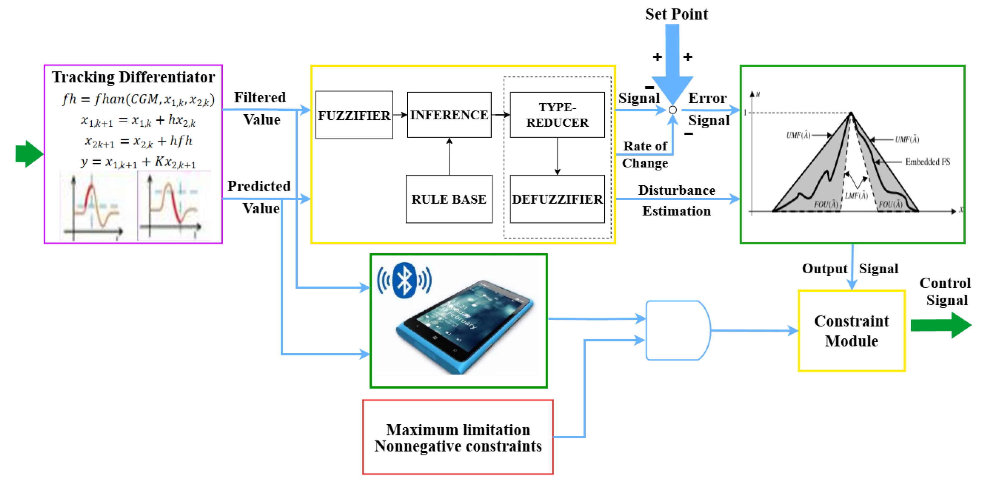
An event-triggered control mechanism is implemented, considering the time-varying characteristics of treatment pressure. A tracking differentiator calculates and predicts pressure changes, and based on these predicted values, a predefined triggering strategy is employed to initiate an adjustment cycle. A type-2 fuzzy logic-based nonlinear feedback control law is employed to determine the required displacement of the linear motor, which transmits driving instructions to the servo driver tasked with adjusting the rope tension. The process is executed sequentially until the tension aligns with the system’s established error limits, as shown in Figure 9. Throughout each adjustment cycle, the device’s intelligent control system continuously gathers pressure data, conducts nonlinear correction and anti-interference filtering, and employs the tracking differentiator to compute and predict the control cycle’s current and future force values. Based on the force error level, one of two control logics is implemented: If the measured force is below the target value, a tightening control is implemented to elevate the pressure within the permissible error margin. When the measured force surpasses the target value, a release control mechanism is activated to decrease the pressure within the permissible range. The system subsequently transitions to a low-power mode, continuously monitoring pressure and awaiting the next triggering event. The system can continuously adjust the pressure of the external fixation device in response to changes in the hand’s morphology during the patient’s hand swelling period, thereby maintaining the clamping force within the optimal range. This approach not only corrects the bone position effectively but also mitigates risks such as ischemia in the extremities of the hand, as shown in Figure 10.

3.3. Optimization of Type-2 Fuzzy Control Algorithm

This system employs an Interval Type-2 Fuzzy Logic Controller (IT2-FLC) to construct the nonlinear feedback control law, with its optimization process comprising the following core aspects:

3.3.1. Determination of the Therapeutic Pressure Range

The determination and adjustment of the optimal therapeutic pressure range in this study were guided entirely by expert clinical experience to ensure both safety and efficacy. The initial target pressure for the smart splint system was set by a senior orthopedic surgeon based on established clinical protocols for distal radius fracture fixation. To provide a quantitative reference for this decision-making process, this clinical judgment was supplemented by data from preliminary measurements, where the initial fixation pressure of newly applied traditional splints was monitored.
Throughout the treatment period, the system’s key advantage is its dynamic adjustability. Physicians utilized the remote platform to continuously monitor the real-time pressure data. Based on their clinical experience and the patient’s specific condition (e.g., degree of detumescence, subjective comfort), physicians could make dynamic adjustments to the pressure at any time, ensuring the fixation force remained within the optimal therapeutic window for the entirety of the healing process.

3.3.2. Membership Function Parameter Optimization

An improved Quantum Particle Swarm Optimization (QPSO) algorithm dynamically tunes the membership function parameters for input and output variables. In the pressure regulation scenario, pressure error (e) and its rate of change ( Δ e ) are designated as input variables, while the linear motor displacement serves as the output variable. Through 300 generations of iterative optimization, the optimal combination of membership function parameters was determined: Gaussian membership functions for input variables (uncertainty domain covering ±0.15 MPa) to better handle uncertainty, and triangular membership functions for the output variable (covering a ±2 mm displacement range) for computational efficiency. The optimized membership function parameters resulted in a 38% reduction in the average membership interval width within the system’s 0–300 kPa operational range, significantly enhancing fuzzy inference precision.

3.3.3. Fuzzy Rule Base Self-Organization Mechanism

An event-triggered mechanism enables dynamic adjustment of the rule base structure: when the pressure error exceeds a preset threshold (±5% of the target value), a sliding window-based rule addition/deletion module is activated. Training with 3000 sets of real-time collected pressure-displacement data culminated in a dynamic rule base containing 25 core rules. These rules, established based on expert clinical knowledge, are designed to respond logically to the splint’s pressure state. For instance, if the pressure error is large and positive (Positive Big) and the error is not changing (Zero), the system should tighten the splint quickly (Fast Tighten). The core logic of the rule base is summarized in Table 1. Compared to a fixed rule base, this dynamic mechanism reduces the response time to sudden pressure fluctuations by 42%.

3.3.4. Adaptive Type Reduction Algorithm Optimization

An improved Nie–Tan type reducer replaces the traditional Karnik–Mendel (KM) algorithms, achieving rapid output defuzzification via Equation (1).
y = i = 1 N μ ̲ i c i + i = 1 N μ ¯ i c i i = 1 N ( μ ̲ i + μ ¯ i )
where c i represents the centroid of the i-th rule’s consequent, and μ ̲ i , μ ¯ i are the activation interval boundaries. Testing demonstrated that this algorithm reduced the single defuzzification time on an ARM Cortex-M7 processor from 12.3 ms to 2.1 ms, meeting the real-time control requirements of the embedded system.

3.3.5. Comparative Analysis with Traditional PID Algorithm

Compared to conventional Proportional-Integral-Derivative (PID) control, Interval Type-2 Fuzzy Logic Control (IT2-FLC) exhibits significant performance enhancements in medical external fixation pressure regulation. Performance analysis of the system’s operational data from the trial indicates that IT2-FLC achieves a 42.7% reduction in settling time (1.28 ± 0.15 s vs. 2.23 ± 0.31 s for PID), a decrease in overshoot from 16.8% to 3.5%, and a 64.8% reduction in steady-state error. This advantage stems from IT2-FLC’s strong adaptability to nonlinear time-varying systems; whereas PID relies on linear error feedback and struggles with parameter uncertainties caused by limb swelling, IT2-FLC directly characterizes the dynamic range and noise characteristics of input variables using Gaussian interval membership functions. Combined with a dynamic rule-base self-organization mechanism (25 core rules), it enables precise pressure tracking within an uncertainty domain of ±0.15 MPa. In terms of real-time performance, although PID has low computational complexity (approx. 12.3 ms per operation), its fixed parameters are less effective at managing sudden pressure fluctuations (e.g., due to changes in posture). IT2-FLC, utilizing the improved Nie–Tan type reduction algorithm, compresses defuzzification time to 2.1 ms (an 82.9% reduction compared to traditional Karnik–Mendel iterative methods). Coupled with an event-triggered mechanism (threshold Δ P t h = 15 % ), this shortens the response time by 42.4% (0.49 s vs. 0.85 s). Furthermore, under sensor noise conditions (15 dB), IT2-FLC demonstrates superior robustness, achieving a 67.4% lower root mean square error (RMSE) in pressure regulation (1.27 N) compared to PID (3.89 N). This resilience benefits from the inclusive modeling of measurement uncertainties by interval type-2 fuzzy sets. Clinical validation demonstrated that during 72 h of continuous operation, the standard deviation of pressure fluctuation with IT2-FLC remained stable at 0.39 N, a value markedly lower than the 2.15 N observed with traditional mechanical devices. Its stability originates from a nonlinear mapping mechanism: by predicting pressure trends using a tracking differentiator (R = 1500), the system can anticipate changes in swelling levels 1.2 control cycles in advance, dynamically adjusting fuzzy rule activation weights to adapt to slow time-varying processes such as callus formation.

3.4. System for Remote Surveillance and Real-Time Modulation

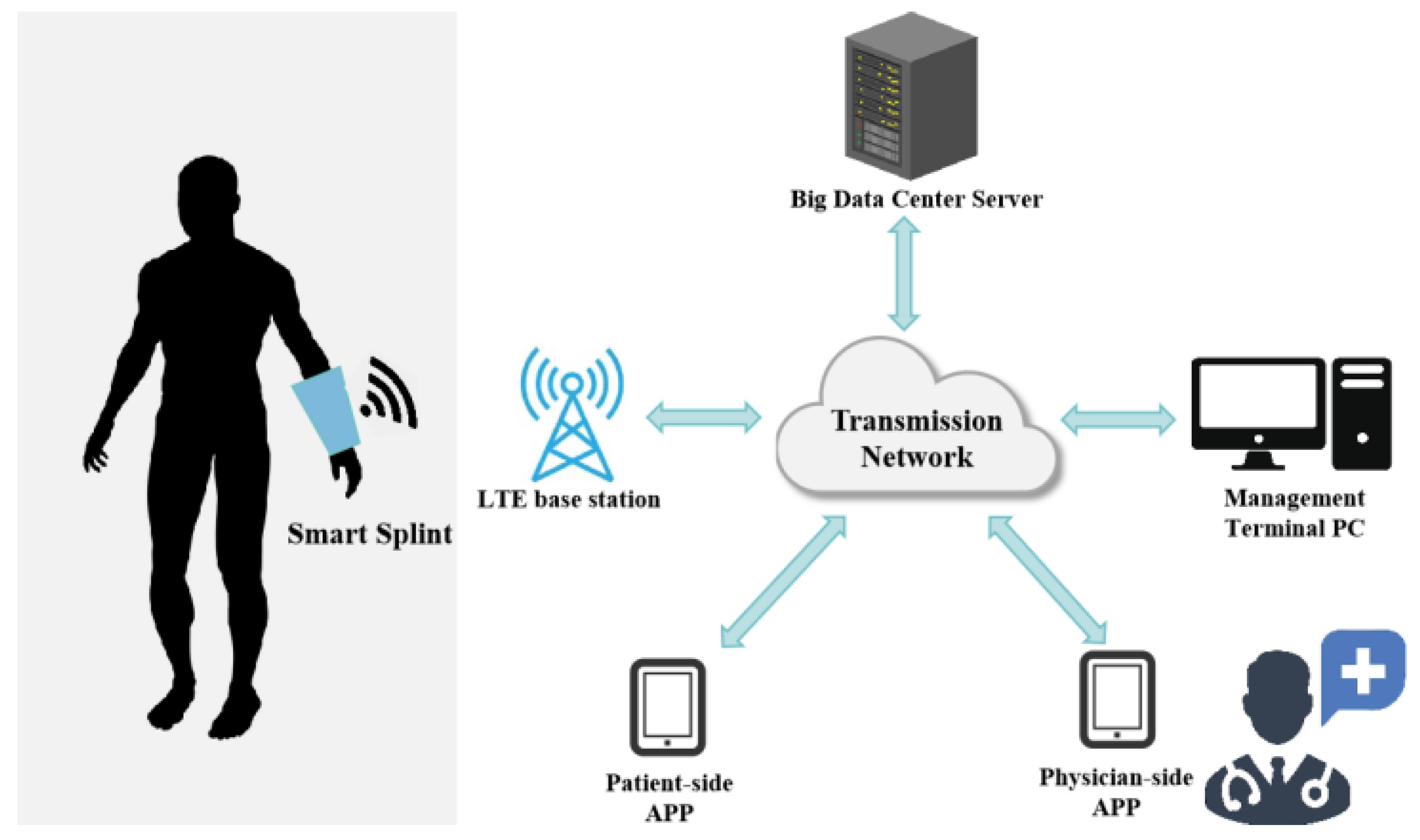
This study presents a client/server (C/S)-based remote monitoring and adjustment system for the intelligent external fixation device. It comprises physician and patient applications (APPs), a physician PC backend, a hospital management backend, and a cloud-based database for managing device assets and limb recovery data. The system enables unified management of device ID, usage status, user information, physician examination and intervention records, and sensor force data (Figure 11). Key advantages include enabling physicians to monitor patient recovery in real-time for timely interventions and treatment adjustments, offering patients a more convenient and personalized medical experience, and providing robust support for research and clinical decision-making through data accumulation and analysis, thereby enhancing treatment efficacy and patient satisfaction. System design prioritized scalability and compatibility using a standardized open architecture. It currently supports physician and patient mobile operations, with a modular design allowing future multi-platform expansion (e.g., tablets, smartwatches) for cross-platform data synchronization. Adherence to HL7/FHIR medical data exchange standards ensures seamless integration with hospital Electronic Medical Record (EMR) and Picture Archiving and Communication Systems (PACS). Treatment data, such as fracture reduction parameters and pressure regulation records, automatically synchronizes to patient EMRs, supporting multidisciplinary collaborative care. A DICOM 3.0 and ISO/HL7 27932 compliant database design ensures bidirectional interaction of pressure sensor data, imaging parameters, and other medical information systems, and supports federated queries and cross-institutional data sharing. Furthermore, microservice-based functional extension interfaces allow for future development of value-added modules like remote rehabilitation guidance, AI-driven dynamic training plan generation, and 3D fracture healing visualization, laying a technical foundation for expanding clinical applications of intelligent traditional Chinese medicine (TCM) devices.
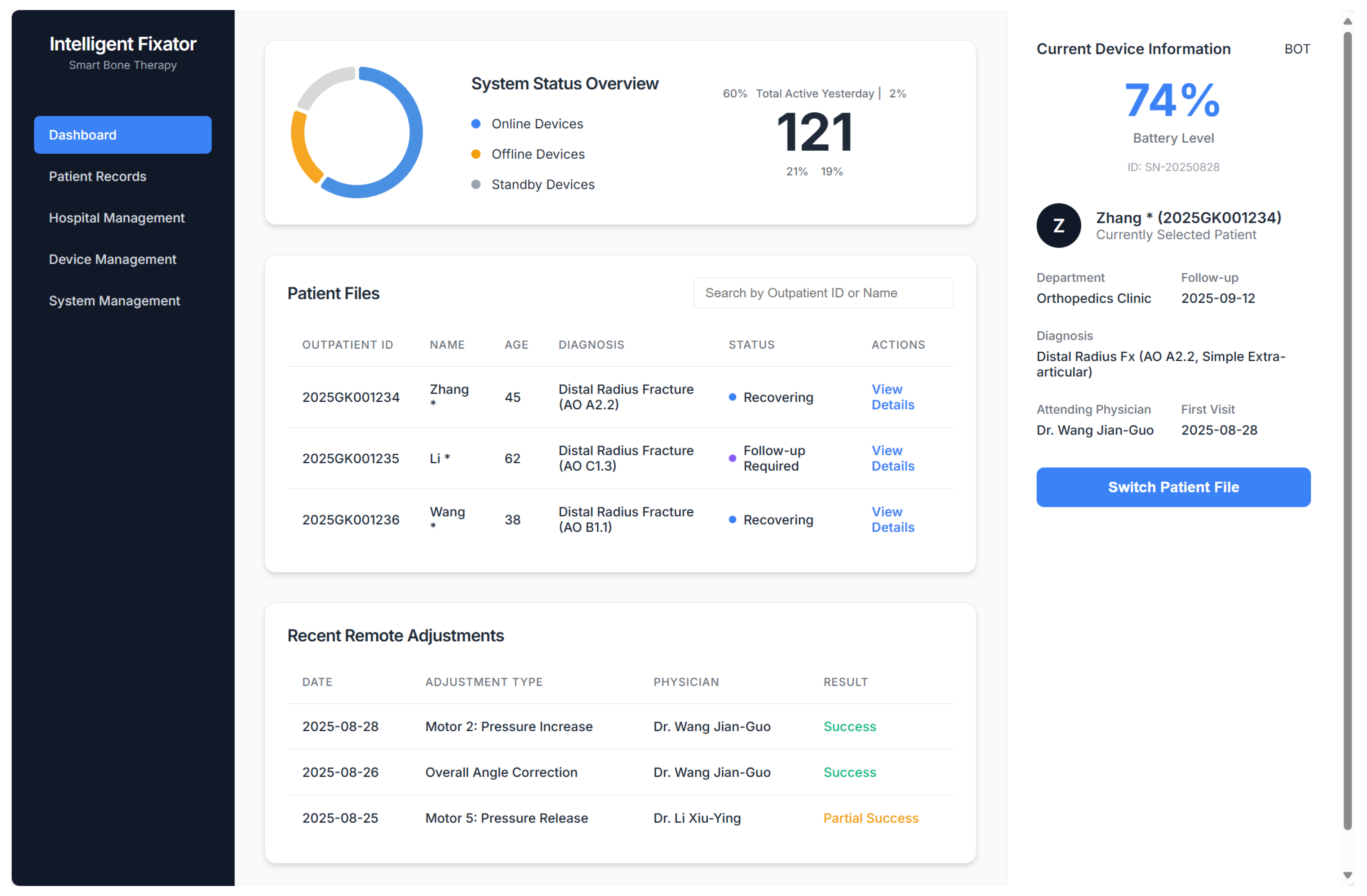
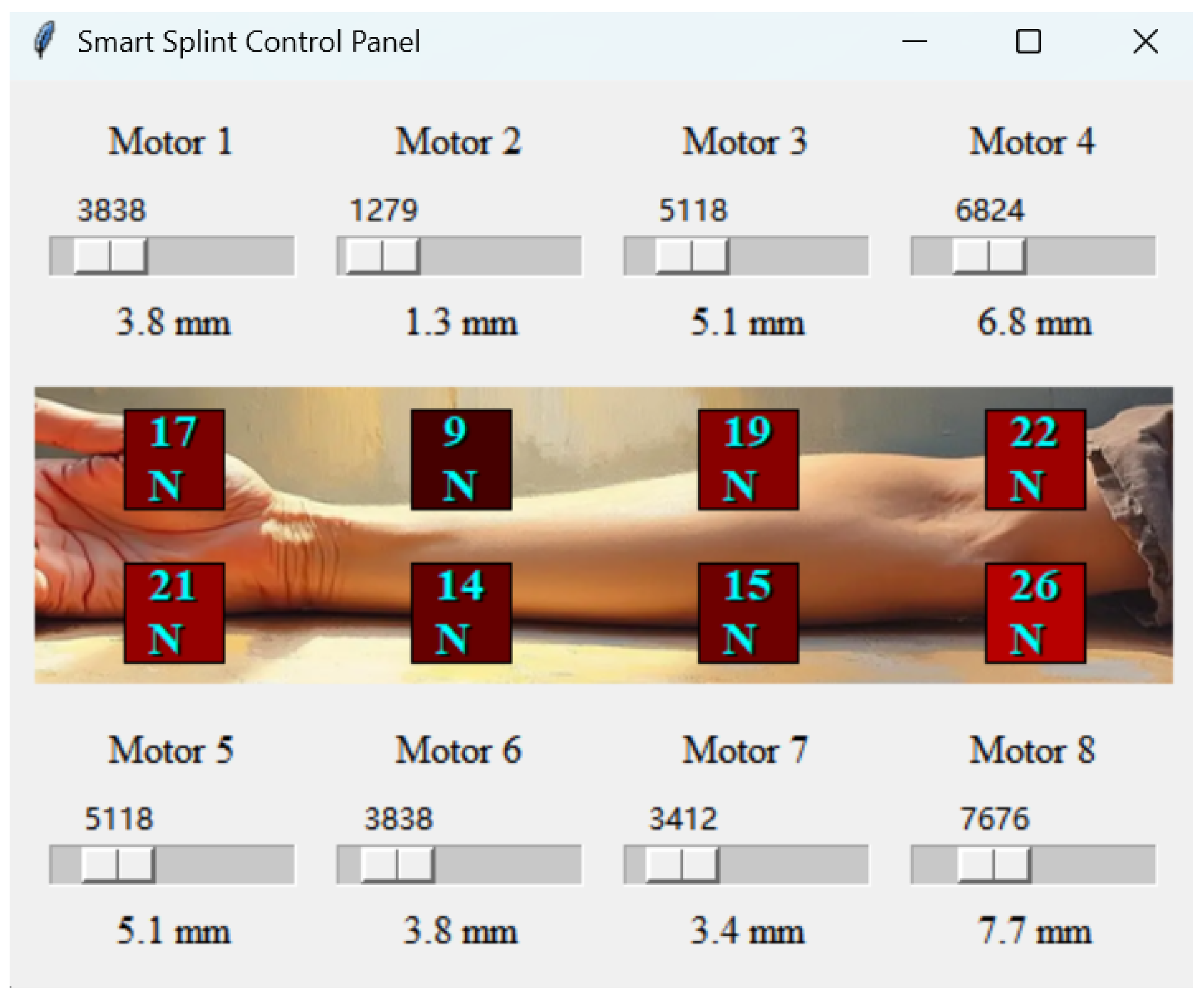
For data acquisition and processing, the system performs real-time collection and storage of pressure sensor data from the intelligent external fixation device. Subsequent statistical analysis and visualization provide physicians with a scientific basis for remote pressure adjustments and fracture recovery prediction, enhancing treatment efficacy and safety. Implementation involves constructing an entity model from sensor and patient data, with data mining and modeling ensuring accuracy and reliability. Data processing adapts to display and system requirements. A SpringBoot framework, valued for its stability and scalability, underpins backend data access APIs for physician, patient, PC, and App interfaces, supporting efficient operation and future expansion. A Vue-based front-end, with Element-Plus UI components and Echarts for dynamic data visualization, creates an intuitive display platform. This API connectivity enables a visual remote monitoring and regulation platform, allowing convenient multi-terminal data access and operation for physicians and patients. To illustrate the system’s functionality, screenshots of the physician-facing user interfaces are provided. Figure 12 shows the high-level clinical management dashboard, which provides an overview of all connected devices, a list of patient files, and a log of recent remote adjustments. Figure 13 displays the low-level control panel, which integrates real-time data visualization and remote control capabilities, showing both the pressure readings (in N) from the eight sensors and the corresponding displacement (in mm) of the eight servo motors.
The data transmission pathway is designed for low power consumption, high reliability, and robust security, adhering to modern standards for medical IoT devices. The workflow is implemented as a two-stage communication architecture to optimize the performance of the wearable device. In the first stage, sensor data processed by the ESP32 microcontroller is transmitted to the patient’s dedicated smartphone application via the integrated Bluetooth Low Energy (BLE) 5.0 module. This local wireless link is secured using AES-128 bit end-to-end encryption to protect data integrity and patient privacy. In the second stage, the smartphone application serves as a secure gateway, relaying the received data to the cloud-based backend server through the phone’s primary internet connection (e.g., LTE-CAT1 or Wi-Fi). To ensure security over the public internet, all communication is protected using the Transport Layer Security (TLS) 1.3 protocol, preventing data tampering and man-in-the-middle attacks. Furthermore, a multi-layered security strategy is implemented for data access and privacy. Access control for real-time pressure data is managed through a secure authentication process, requiring a combination of the unique device ID and a dynamic verification code. All patient data is anonymized in compliance with international privacy regulations such as GDPR and HIPAA. This comprehensive security architecture, designed in compliance with the IEEE P29383 standard (“Medical Internet of Things Security Technical Specifications”), significantly mitigates data transmission risks and ensures patient confidentiality.

3.5. Design of Herbal Pads in Traditional Chinese Medicine (TCM)

In TCM orthopedics, fracture treatment prioritizes the “three-stage differentiation” principle, employing distinct therapeutic approaches tailored to the specific stages of fracture healing to facilitate bone union and functional restoration. This idea is based on a deep comprehension of the fracture healing process and vast clinical experience in traditional Chinese medicine and has considerable guiding significance. In the early phase of fracture healing (1–2 weeks post-injury), patients frequently encounter localized edema, discomfort, and blood stasis, which impedes blood circulation. The principal objective of treatment at this juncture is to enhance blood circulation, eliminate stasis, diminish edema, and relieve pain to reinstate the local flow of qi and blood. We developed a TCM herbal pad using components such as Angelica sinensis (Danggui), Paeonia lactiflora (Chishao), Ligusticum chuanxiong (Chuanxiong), Boswellia carterii (Ruxiang), and Commiphora myrrha (Moyao). These herbs are recognized for enhancing blood circulation, eliminating blood stasis, diminishing swelling, and alleviating pain, hence facilitating early fracture healing.
As the fracture stabilizes and progresses to the intermediate stage (3–4 weeks post-injury), the predominant concerns transition to muscular rigidity and impaired circulation, resulting in diminished local functionality. The therapy objective at this stage is to relax tendons, stimulate collaterals, and harmonize qi and blood to promote the healing of soft tissues and functional recovery. We developed a TCM herbal pad using components such as Cinnamomum cassia (Guizhi), Chaenomeles speciosa (Mugua), Lycopodium japonicum (Shenjincao), Carthamus tinctorius (Honghua), and Salvia miltiorrhiza (Danshen). These herbs facilitate tendon relaxation, activate collaterals, promote local blood circulation, augment nutrition delivery to bones and soft tissues, expedite fracture healing, and improve limb functionality.
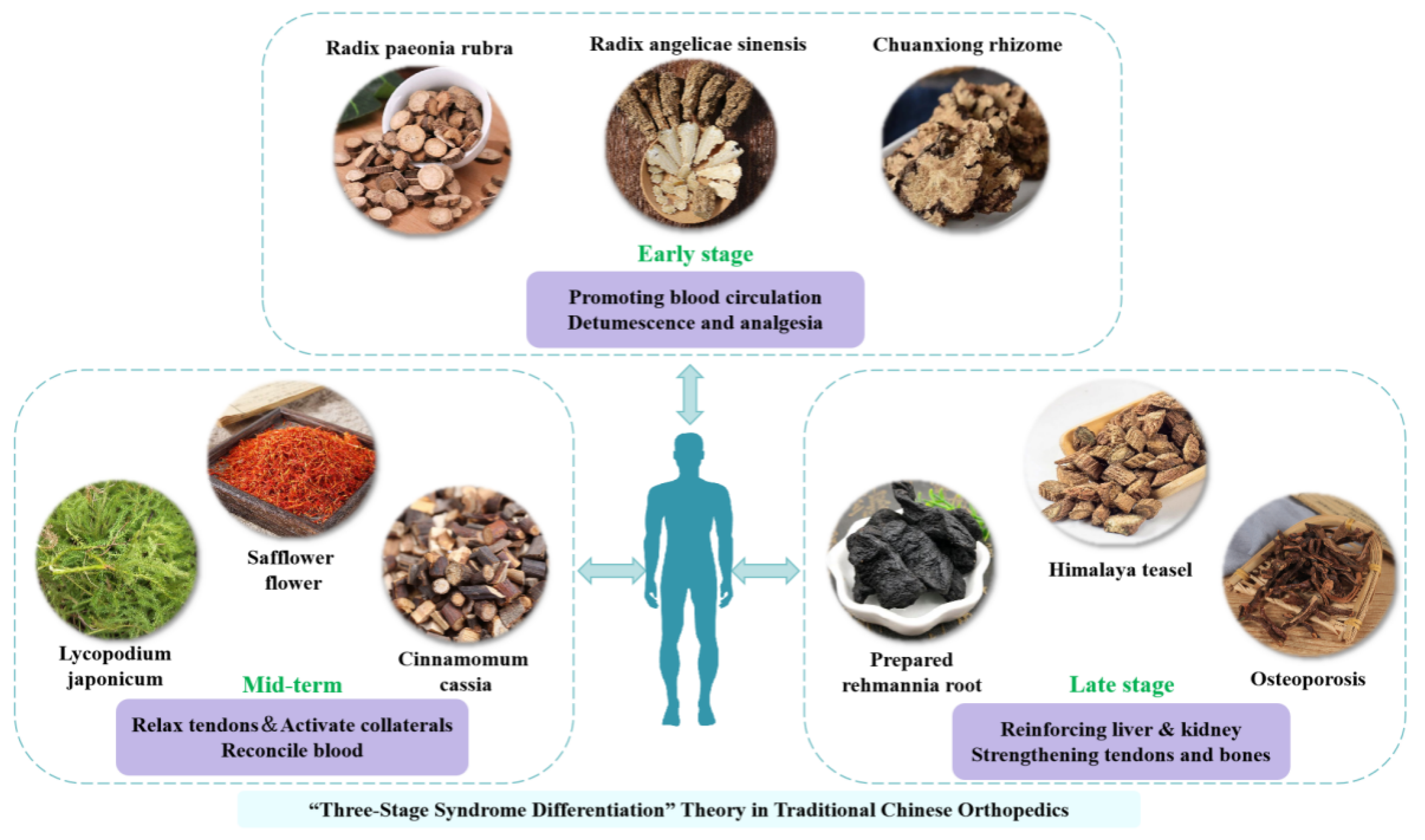
In the advanced phase of fracture healing (one month post-injury), the principal concerns are liver and kidney insufficiency, resulting in compromised bone and tendon integrity, potentially obstructing full fracture healing and functional rehabilitation. The current therapy objective is to invigorate the liver and kidneys, fortify bones and tendons, and improve the body’s overall constitution to facilitate comprehensive fracture healing. We developed a TCM herbal pad with substances such as Rehmannia glutinosa (Shudihuang), Drynaria fortunei (Gusuibu), Eucommia ulmoides (Duzhong), Dipsacus asperoides (Xuduan), and Epimedium brevicornum (Yinyanghuo). These herbs invigorate the liver and kidneys, fortify bones and tendons, enhance the body’s immunity and self-repair mechanisms, and facilitate the last phases of fracture recovery, allowing patients to return to their daily activities and employment. Integrating TCM herbal pads, formulated based on the three-stage differentiation theory, into the intelligent external fixation system allows patients to utilize pads with distinct therapeutic effects during different phases of fracture recovery. In conjunction with the device’s dynamic pressure adjustment and remote monitoring capabilities, this method can more efficiently facilitate fracture healing, mitigate pain, restore wrist joint function, and enhance patients’ quality of life (Figure 14).

3.6. Clinical Trial Design

3.6.1. Clinical Data

This study prospectively enrolled 60 patients diagnosed with extension-type distal radius fractures, confirmed via standardized radiographic examinations, at the Department of Orthopedics and Traumatology of Traditional Chinese Medicine, Third Affiliated Hospital of Beijing University of Chinese Medicine, between July 2023 and July 2024. Participants were randomized using a random number table to either a control group ( n = 30 ) or an experimental group ( n = 30 ). The control group comprised 17 males and 13 females (mean age, 49.8 ± 3.7 years; range, 20–77 years), and the experimental group included 18 males and 12 females (mean age, 48.6 ± 3.8 years; range, 19–75 years). Baseline demographic characteristics were well-matched between the groups, with no statistically significant differences observed (p > 0.05), ensuring comparability for subsequent analyses (Table 2). All participants provided written informed consent in accordance with the Declaration of Helsinki, and the study protocol was approved by the Ethics Committee of the Third Affiliated Hospital of Beijing University of Chinese Medicine.

3.6.2. Diagnostic Criteria

The diagnostic criteria were established based on the Management of Distal Radius Fractures Evidence-Based Clinical Practice Guideline [29]. Key diagnostic indicators included the following: (1) a history of wrist trauma, typically the result of a fall onto an outstretched hand; (2) clinical findings of wrist pain, swelling, restricted mobility, and localized tenderness, with potential crepitus in cases of significant fracture displacement; and (3) radiographic evidence of a fracture within 2.0–3.0 cm of the distal radius, with clear delineation of fracture type, displacement direction, and associated injuries such as ulnar styloid fracture or distal radioulnar joint dislocation.

3.6.3. Therapeutic Method

Control Group
The control group received manual reduction along with conventional small splint fixation. Four small splints were fabricated according to the length of the patient’s forearm. The patient was positioned supine, with the affected limb abducted and the elbow flexed at a 90-degree angle. The forearm was placed in a pronated position, and manual reduction was executed utilizing techniques including palpation, lifting, and rotation. The wrist was positioned in palmar flexion and ulnar deviation for Colles’ fractures or in dorsiflexion and radial deviation for Smith’s fractures. Pads were positioned on the dorsal aspect of the distal fracture and the volar aspect of the proximal fracture, with splints secured using cotton bandages. The splint was positioned at the mid-upper forearm, with the dorsal component extending beyond the wrist joint. The splints were secured with straps, and the arm was maintained in a neutral position with the elbow flexed. Following reduction, the affected limb was elevated to monitor blood circulation, and X-ray imaging was employed to verify the reduction. A second manual reduction was conducted if required, and the splints’ tightness was modified accordingly. No traditional Chinese medicine pads were used throughout the treatment.
Experimental Group
After manual reduction, splint fixation was applied similarly to the traditional splint group, but the conventional four-splint setup was replaced by the novel intelligent splint. Postoperatively, a physician inserted the traditional Chinese medicine (TCM) herbal pad, and the automatic pressure adjustment function of the new intelligent splint was activated, allowing a chip to autonomously regulate the appropriate pressure. Real-time data from the pressure sensors were uploaded to the cloud, enabling physicians to set suitable pressure values for the patient via a corresponding application. The forearm was supported in a neutral position, flexed at 90°, with a triangular bandage sling across the chest. Postoperative X-rays were taken to confirm that the fracture reduction met the required standards. Both groups underwent X-ray examination immediately after fixation to check the reduction status, with follow-up X-rays performed at 3 days, 1 week, 2 weeks, and 4 weeks postoperatively. Patients followed medical advice, sequentially using the three types of TCM herbal pads and changing them themselves at the prescribed times.

3.6.4. Therapeutic Evaluation

Radiographic Evaluation
Standardized posteroanterior and lateral radiographs were acquired at baseline and 6-week follow-up to assess three radiographic parameters: ulnar inclination angle ( θ U I ), volar tilt angle ( θ V T ), and radial height ( H R ). Definitions, normal ranges, and geometric formulas are systematically summarized in Table 3.
Visual Analog Scale (VAS)
Pain intensity is assessed using a 100-mm Visual Analog Scale (VAS) (0 = no pain; 10 = unbearable pain) at standardized timepoints: pre-reduction (T0), post-reduction (T1), and postoperative days 3, 7, and 14 (T2–T4). Clinically significant improvement is defined as a reduction in VAS score by at least 50%. The formula for calculating the percentage improvement is as follows:
Δ VAS = VAS T 0 VAS T x VAS T 0 × 100 %
Single-blinded documentation by a designated physician ensured uniformity of assessments.
Cooney Wrist Function Score
Wrist functional recovery is assessed using the Cooney score, which systematically quantifies four clinical domains: Daily Activity, Grip Strength, Range of Motion, and Discomfort Intensity. The measurement protocols and scoring criteria are detailed in Table 4.
Criteria for Clinical Effectiveness
Clinical efficacy was evaluated according to the criteria established in the Management of Distal Radius Fractures Evidence-Based Clinical Practice Guideline. Outcomes were categorized as follows: Excellent: Radiographic evidence of good fracture reduction and union, normal wrist and forearm function, and absence of local pain or deformity. Good: Radiographic evidence of good fracture reduction and union, normal wrist and forearm function, and no significant local pain or deformity. Fair: Radiographic evidence of fracture union, mild limitation of wrist and forearm function, with local deformity or pain not impairing daily activities. Poor: Radiographic evidence of fracture union, severe limitation of wrist and forearm function, accompanied by significant local swelling, deformity, and pain. The rate of excellent or good outcomes was recorded to determine overall clinical efficacy.

3.6.5. Statistical Analysis

All statistical analyses were performed using SPSS software (version 25.0). Normality of continuous data was assessed using the Shapiro–Wilk test, and homogeneity of variances was evaluated using Levene’s test. Continuous variables that conformed to a normal distribution (p > 0.05) and exhibited homogeneity of variances (p > 0.10) were compared between groups using independent samples t-tests. The Mann–Whitney U test was employed for data that were not normally distributed or showed unequal variances. Categorical variables were described as frequencies and percentages; inter-group differences were analyzed using Pearson’s chi-squared test or Fisher’s exact test, as appropriate (the latter when expected cell frequencies were <5). A two-sided alpha level of 0.05 was set for statistical significance. For multiple comparisons involving radiographic parameters, Visual Analog Scale (VAS) scores, and Cooney scores, the Bonferroni method was applied to mitigate the risk of false positives. Specifically, the original alpha level ( α = 0.05 ) was divided by the number of simultaneous analyses (n) to establish an adjusted significance threshold ( α = 0.05 / n ). For example, when comparing three radiographic parameters, α was set at 0.0167, and a p-value < α was required to declare statistical significance. This error rate control and p-value adjustment were managed via the SPSS Post Hoc test module. Effect sizes were calculated using internationally accepted metrics: Cohen’s d was used for continuous variables to quantify the magnitude of inter-group differences (defined as the difference between two means divided by the pooled standard deviation), while Hedges’ g was applied for non-parametric data to correct for potential small sample bias. All analyses were conducted in accordance with STROBE statement guidelines. By incorporating multiple comparison adjustments and effect size analyses, while preserving traditional reporting features, we aimed to enhance the credibility of our findings and ensure the robustness of our conclusions.

4. Results

All patients were monitored for 12 months, and all 60 instances of distal radius fractures attained clinical union within 4 to 6 weeks.

4.1. Radiographic Evaluation

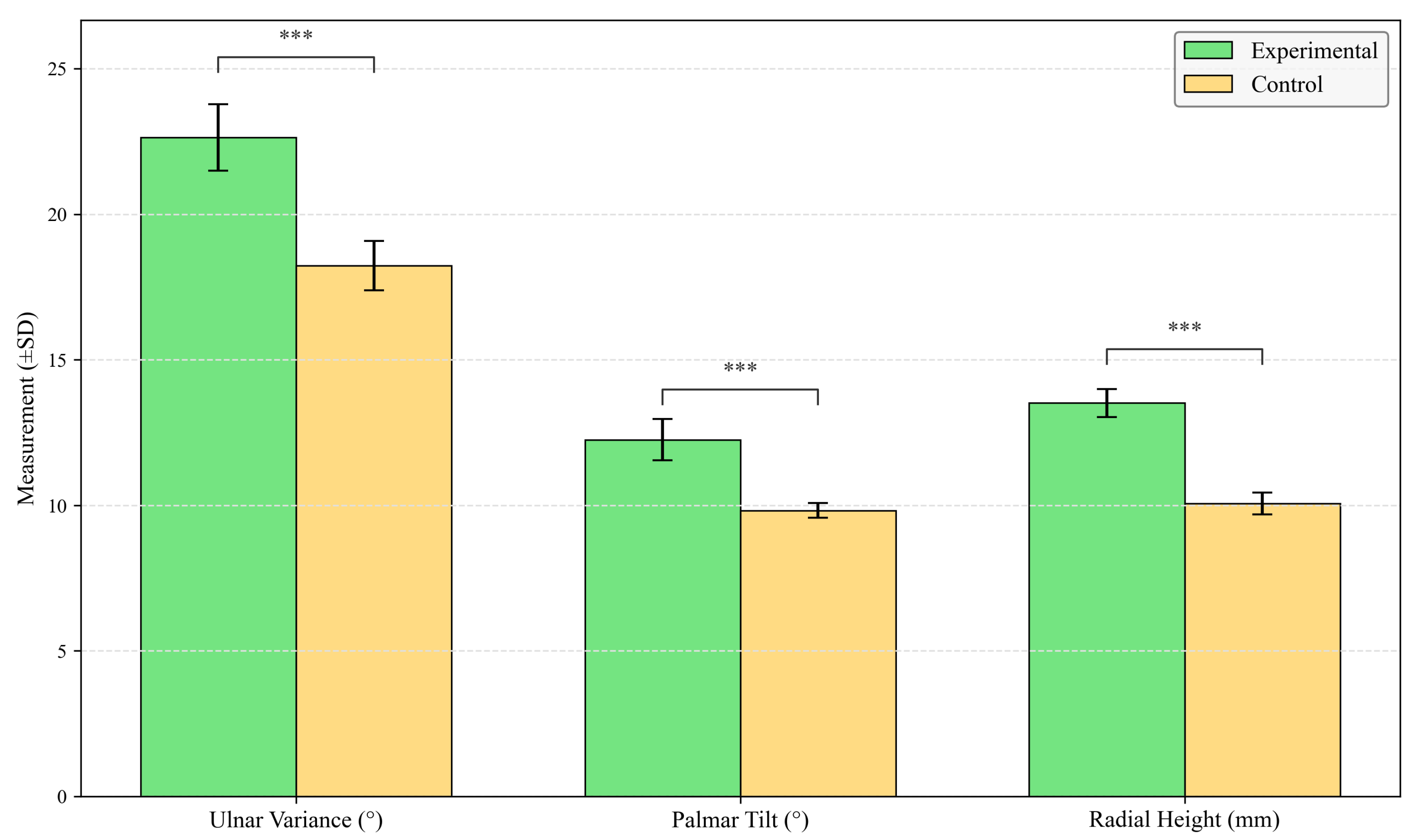
The experimental group demonstrated significantly superior radiographic outcomes compared to the control group (Table 5; Figure 15). Enhanced anatomical restoration was achieved with the experimental intervention, as evidenced by greater ulnar inclination (mean difference 3.45, 95% CI [3.26, 3.64]; t = 36.450, p < 0.001, Cohen’s d = 2.21), increased volar tilt (mean difference 2.43, 95% CI [2.15, 2.71]; t = 20.671, p < 0.001, Cohen’s d = 1.87), and improved radial height (mean difference 4.40 mm, 95% CI [3.92, 4.88]; t = 19.813, p < 0.001, Cohen’s d = 1.94). These data indicate a more complete anatomical recovery with the experimental device.

4.2. Evaluation of VAS and Cooney Scores Pre- and Post-Treatment

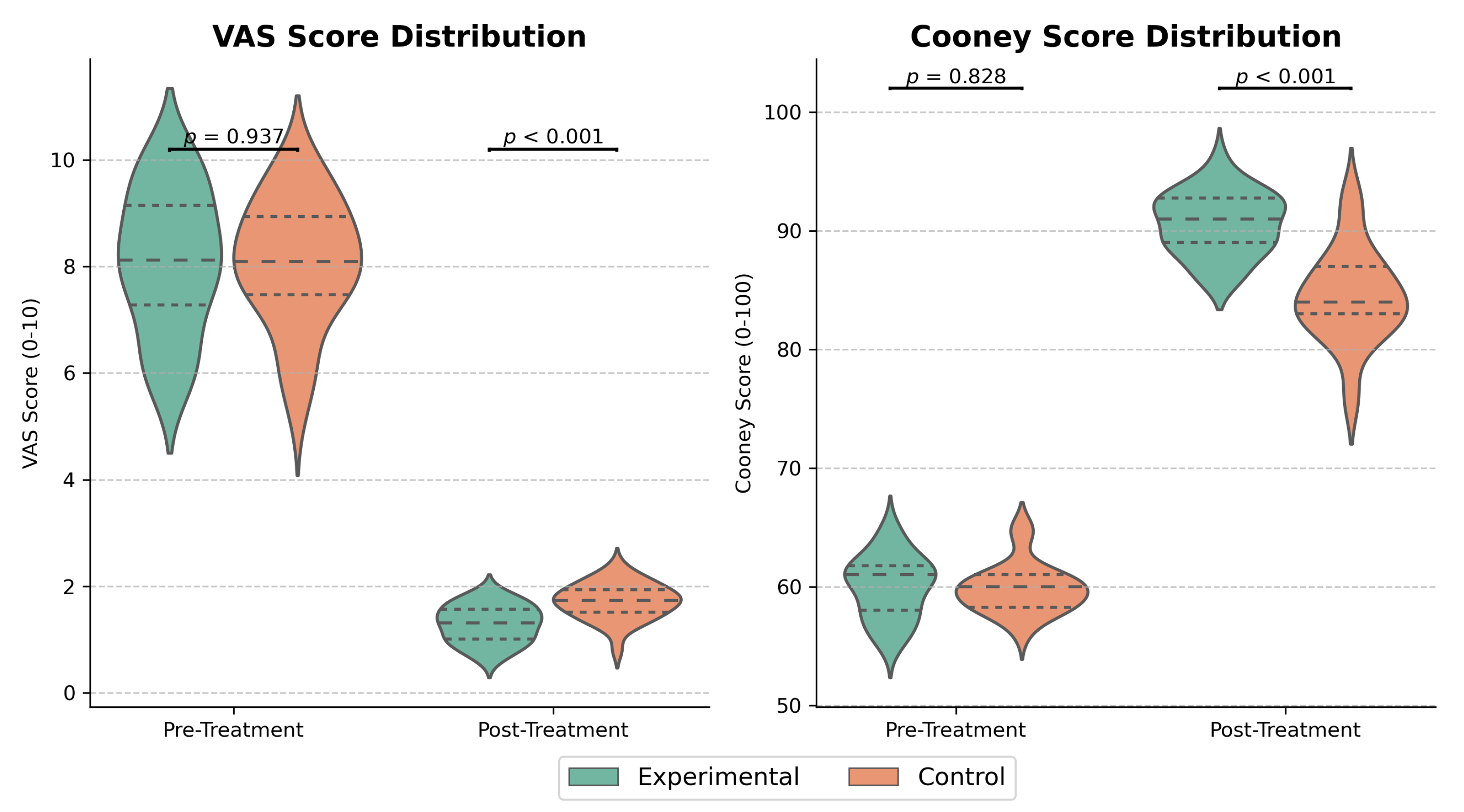
At baseline, there were no significant differences in either the Visual Analog Scale (VAS) for pain or the Cooney wrist function scores between the experimental and control groups ( p > 0.05 for both).
Following the intervention, the experimental group demonstrated significantly better outcomes (Table 6). Specifically, post-treatment VAS scores were substantially lower in the experimental group, indicating significant pain reduction (Mean Difference [95% CI]: −0.73 [−0.98, −0.48]; Z = 3.491 , p < 0.001 ). Furthermore, the experimental group achieved markedly higher Cooney scores, reflecting superior functional recovery (Mean Difference [95% CI]: 8.50 [6.50, 10.50]; t = 4.370 , p < 0.001 ).
These between-group differences remained statistically significant after Bonferroni correction (adjusted p < 0.025 for both). The large effect sizes observed for both VAS (Cohen’s d = 1.23 ) and Cooney scores (Cohen’s d = 1.97 ) underscore the clinical importance of these improvements (Figure 16).

4.3. Comparison of Clinical Effectiveness

The clinical efficacy in the experimental group was significantly higher than in the control group ( p < 0.05 ). In the experimental group, 86.7% (26/30) of patients achieved an excellent or good therapeutic outcome, which was significantly superior to the 63.3% (19/30) observed in the control group. Fisher’s exact test confirmed that the difference in the excellent/good outcome rate between the groups was statistically significant (two-sided p = 0.043 ). The effect size, Cramer’s V = 0.269, indicated a moderate strength of clinical association (Table 7). The one case with a “poor” therapeutic outcome in the control group presented with joint stiffness and malunion.

5. Discussion

This study details the design and preliminary validation of a smart, IoT-enabled splint, showing its potential therapeutic advantages over conventional splinting for distal radius fractures. Our results suggest that the integrated intelligent treatment protocol can achieve statistically superior anatomical restoration and functional recovery. This was highlighted by the experimental group attaining a mean Cooney wrist function score of 91.65 ± 2.38 , substantially higher than the control group’s 83.15 ± 3.17 (p < 0.001). This enhanced performance is attributed to the splint’s core innovation: a fundamental shift from static immobilization to dynamic, closed-loop biomechanical regulation. By using an integrated system of pressure sensors and servo-actuators to maintain therapeutic pressure within an optimal range, the smart splint directly mitigates critical risks of traditional splinting, such as pressure sores or loss of reduction. In the context of prior research, while studies have explored 3D-printed personalized splints, most remain static devices that improve fit but do not address dynamic physiological changes like detumescence. Similarly, the emerging field of smart orthotics has primarily focused on passive monitoring rather than active therapeutic intervention. Our work advances this field by demonstrating the clinical feasibility of an integrated system that not only senses but also autonomously regulates biomechanical forces. This approach aligns with the broader international clinical trend towards personalized medicine and remote patient monitoring. By enabling continuous, data-driven management outside the hospital setting, the system has the potential to reduce clinical workload and minimize hospital visits, which are key goals in modern value-based healthcare systems.
However, we must acknowledge several significant limitations inherent to this proof-of-concept study, which span both its technical validation and clinical methodology. From a technical standpoint, this work lacks exhaustive benchtop validations, such as standardized sensor reliability and drift testing, as well as head-to-head experimental comparisons of the control algorithm against standard controllers (e.g., PID) under simulated physiological conditions. On the clinical and methodological front, the primary limitation remains the inclusion of TCM herbal pads as a confounding variable, which prevents the isolation of the smart splint’s independent mechanical effect. Further limitations include the single-center nature of the trial, its focus on less complex fracture types, and an insufficient follow-up period for assessing long-term outcomes. Additionally, formal biocompatibility and mechanical fatigue testing are essential prerequisites for a full clinical validation. The absence of these comprehensive validations collectively reinforces the study’s position as a preliminary but promising proof-of-concept.
Addressing these limitations will form the basis of our future work. To de-isolate the independent contributions of the dynamic mechanical fixation versus the medicinal intervention, our next step is to design a rigorous four-arm, multicenter randomized controlled trial. This future trial will also validate the splint’s broader efficacy and generalizability across diverse patient populations and more complex fracture cases (e.g., C-type). Prior to this large-scale clinical investigation, comprehensive splint validation is required. This includes formal biocompatibility testing according to ISO 10993 standards and standardized high-cycle mechanical fatigue testing to ensure long-term structural integrity. Concurrently, the system’s technical performance will be rigorously characterized through exhaustive benchtop validations, involving long-term sensor reliability and drift testing and head-to-head experimental comparisons of the control algorithm against standard controllers.
In conclusion, this study successfully demonstrates the proof-of-concept for this intelligent system. By demonstrating a clear clinical potential in moving beyond passive fixation toward data-driven, active therapeutic intervention, this work establishes a viable pathway for the future development and translation of smart orthopedic technologies that can significantly improve patient outcomes.

Author Contributions

Conceptualization, X.L. and Y.M.; methodology, Y.M., B.W. and X.L.; software, H.T. and J.L.; validation, Y.M. and B.W.; formal analysis, H.T.; investigation, Y.M. and B.W.; resources, X.L.; data curation, Y.M. and B.W.; writing—original draft preparation, Y.M., H.T. and J.L.; writing—review and editing, all authors; visualization, H.T.; supervision, X.L.; project administration, X.L.; funding acquisition, Y.M. All authors have read and agreed to the published version of the manuscript.

Funding

This research was funded by the Beijing University of Chinese Medicine Fund, grant number BZYSY-2022-XYYF-02. The APC was funded with the same grant.

Institutional Review Board Statement

The study was conducted in accordance with the Declaration of Helsinki and approved by the Ethics Committee of the Third Affiliated Hospital of Beijing University of Chinese Medicine.

Informed Consent Statement

Informed consent was obtained from all subjects involved in the study. Written informed consent has been obtained from the patient(s) to publish this paper.

Data Availability Statement

The datasets generated and/or analysed during the current study are available from the corresponding author on reasonable request.

Conflicts of Interest

The authors declare no conflict of interest. The funders had no role in the design of the study; in the collection, analyses, or interpretation of data; in the writing of the manuscript; or in the decision to publish the results.

Abbreviations

The following abbreviations are used in this manuscript:
DRFDistal Radius Fracture
IoTInternet of Things
IT2-FLCInterval Type-2 Fuzzy Logic Control
TCMTraditional Chinese Medicine
CRPSComplex Regional Pain Syndrome
PCBPrinted Circuit Board
QPSOQuantum Particle Swarm Optimization
PIDProportional-Integral-Derivative
EMRElectronic Medical Record
PACSPicture Archiving and Communication System
BLEBluetooth Low Energy
VASVisual Analog Scale

References

  1. Koo, K.O.; Tan, D.M.; Chong, A.K. Distal radius fractures: An epidemiological review. Orthop. Surg. 2013, 5, 209–213. [Google Scholar] [CrossRef]
  2. Nellans, K.W.; Kowalski, E.; Chung, K.C. The epidemiology of distal radius fractures. Hand Clin. 2012, 28, 113–125. [Google Scholar] [CrossRef]
  3. Zhang, X.H.; Xiao, Z.Q.; Wang, A.M.; Zhang, H.L.; Li, H.J.; Huang, S.Q. Comparison study of small splint fixation and plaster slab fixation for the treatment of distal radius fractures. Zhongguo Gu Shang = China J. Orthop. Traumatol 2010, 23, 578–580. [Google Scholar] [PubMed]
  4. Cooper, C.; Atkinson, E.J.; Jacobsen, S.J.; O’Fallon, W.M.; Melton, L.J., III. Population-based study of survival after osteoporotic fractures. Am. J. Epidemiol. 1993, 137, 1001–1005. [Google Scholar] [CrossRef]
  5. Kanis, J.A.; Johnell, O.; Oden, A.; Sernbo, I.; Redlund-Johnell, I.; Dawson, A.; Jonsson, B. Long-term risk of osteoporotic fracture in Malmö. Osteoporos. Int. 2000, 11, 669–674. [Google Scholar] [CrossRef]
  6. Eastell, R.; O’Neill, T.W.; Hofbauer, L.C.; Langdahl, B.; Reid, I.R.; Gold, D.T.; Cummings, S.R. Postmenopausal osteoporosis. Nat. Rev. Dis. Prim. 2016, 2, 16069. [Google Scholar] [CrossRef]
  7. Hutchinson, T.; Polansky, S.; Feinstein, A. Post-menopausal oestrogens protect against fractures of hip and distal radius: A case-control study. Lancet 1979, 314, 705–709. [Google Scholar] [CrossRef]
  8. Heinonen, A.; Kannus, P.; Sievänen, H.; Oja, P.; Pasanen, M.; Rinne, M.; Vuori, I. Randomised controlled trial of effect of high-impact exercise on selected risk factors for osteoporotic fractures. Lancet 1996, 348, 1343–1347. [Google Scholar] [CrossRef]
  9. Riggs, B.L.; Melton, L.J., III. The worldwide problem of osteoporosis: Insights afforded by epidemiology. Bone 1995, 17, S505–S511. [Google Scholar] [CrossRef] [PubMed]
  10. Riggs, B.L.; Melton, L.J., III; Robb, R.A.; Camp, J.J.; Atkinson, E.J.; Peterson, J.M.; Khosla, S. Population-based study of age and sex differences in bone volumetric density, size, geometry, and structure at different skeletal sites. J. Bone Miner. Res. 2004, 19, 1945–1954. [Google Scholar] [CrossRef] [PubMed]
  11. Levin, L.S.; Rozell, J.C.; Pulos, N. Distal radius fractures in the elderly. JAAOS 2017, 25, 179–187. [Google Scholar] [CrossRef]
  12. Mauck, B.M.; Swigler, C.W. Evidence-based review of distal radius fractures. Orthop. Clin. N. Am. 2018, 49, 211–222. [Google Scholar] [CrossRef]
  13. Mehta, S.P.; MacDermid, J.C.; Richardson, J.; MacIntyre, N.J.; Grewal, R. Baseline pain intensity is a predictor of chronic pain in individuals with distal radius fracture. J. Orthop. Sport. Phys. Ther. 2015, 45, 119–127. [Google Scholar] [CrossRef]
  14. Jo, Y.H.; Kim, K.; Lee, B.G.; Kim, J.H.; Lee, C.H.; Lee, K.H. Incidence of and risk factors for complex regional pain syndrome type 1 after surgery for distal radius fractures: A population-based study. Sci. Rep. 2019, 9, 4871. [Google Scholar] [CrossRef]
  15. MacDermid, J.C.; Roth, J.H.; Richards, R.S. Pain and disability reported in the year following a distal radius fracture: A cohort study. BMC Musculoskelet. Disord. 2003, 4, 24. [Google Scholar] [CrossRef]
  16. van Leerdam, R.H.; Huizing, F.; Termaat, F.; Kleinveld, S.; Rhemrev, S.J.; Krijnen, P.; Schipper, I.B. Patient-reported outcomes after a distal radius fracture in adults: A 3–4 years follow-up. Acta Orthop. 2019, 90, 129–134. [Google Scholar] [CrossRef]
  17. Hargens, A.R.; Mubarak, S.J.; Owen, C.A.; Garetto, L.P.; Akeson, W.H. Interstitial fluid pressure in muscle and compartment syndromes in man. Microvasc. Res. 1977, 14, 1–10. [Google Scholar] [CrossRef] [PubMed]
  18. Lafontaine, M.; Hardy, D.; Delince, P.H. Stability assessment of distal radius fractures. Injury 1989, 20, 208–210. [Google Scholar] [CrossRef] [PubMed]
  19. Handoll, H.H.; Cameron, I.D.; Mak, J.C.; Panagoda, C.E.; Finnegan, T.P. Multidisciplinary rehabilitation for older people with hip fractures. Cochrane Database Syst. Rev. 2021, CD007125. [Google Scholar] [CrossRef]
  20. Tabrizi, J.S.; Behghadami, M.A.; Saadati, M.; Söderhamn, U. Self-care ability of older people living in urban areas of northwestern Iran. Iran. J. Public Health 2018, 47, 1899. [Google Scholar] [PubMed Central]
  21. Travers, J.L.; Hirschman, K.B.; Naylor, M.D. Older adults’ goals and expectations when using long-term services and supports. J. Appl. Gerontol. 2022, 41, 709–717. [Google Scholar] [CrossRef] [PubMed]
  22. Gutiérrez-Espinoza, H.; Araya-Quintanilla, F.; Olguín-Huerta, C.; Gutiérrez-Monclus, R.; Valenzuela-Fuenzalida, J.; Román-Veas, J.; Campos-Jara, C. Effectiveness of surgical versus conservative treatment of distal radius fractures in elderly patients: A systematic review and meta-analysis. Orthop. Traumatol. Surg. Res. 2022, 108, 103323. [Google Scholar] [CrossRef]
  23. Zhou, J.; Tang, W.; Li, D.; Wu, Y. Morphological characteristics of different types of distal radius die-punch fractures based on three-column theory. J. Orthop. Surg. Res. 2019, 14, 390. [Google Scholar] [CrossRef]
  24. Song, J.; Yu, A.X.; Li, Z.H. Comparison of conservative and operative treatment for distal radius fracture: A meta-analysis of randomized controlled trials. Int. J. Clin. Exp. Med. 2015, 8, 17023. [Google Scholar] [PubMed Central]
  25. Ba, C.; Ni, X.H.; Zhu, G.P.; Yu, J.L.; Cheng, J.J.; Wu, J.S.; Zhao, Q.M. Clinical effect of open reduction and internal fixation with a steel plate through the triceps approach in the treatment of fractures of the middle and lower 1/3 of the humerus. Eur. Rev. Med. Pharmacol. Sci. 2022, 26, 7607–7614. [Google Scholar] [CrossRef]
  26. Uhthoff, H.K.; Poitras, P.; Backman, D.S. Internal plate fixation of fractures: Short history and recent developments. J. Orthop. Sci. 2006, 11, 118–126. [Google Scholar] [CrossRef]
  27. Yu, G.S.; Lin, Y.B.; Le, L.S.; Zhan, M.F.; Jiang, X.X. Internal fixation vs conservative treatment for displaced distal radius fractures: A meta-analysis of randomized controlled trials. Turk. J. Trauma Emerg. Surg. 2016, 22, 233–240. [Google Scholar] [CrossRef]
  28. Shauver, M.J.; Chung, K.C. The economic impact of distal radius fractures. Hand Clin. 2011, 27, 137–143. [Google Scholar]
  29. American Academy of Orthopaedic Surgeons. Management of Distal Radius Fractures Evidence-Based Clinical Practice Guideline. Published 4 December 2020. Available online: https://www.aaos.org/quality/quality-programs/clinical-practice-guidelines/distal-radius-fractures/ (accessed on 1 July 2024).
  30. Li, L.; Liu, X.; Patel, M.; Zhang, L. Depth camera-based model for studying the effects of muscle loading on distal radius fracture healing. Comput. Biol. Med. 2023, 164, 107292. [Google Scholar] [CrossRef]
  31. Wei, L.Y.; Zhang, H.W.; Dong, D.H.; Zuo, J.Z.; Li, L.; Wang, G.Q.; Zhang, J. Manual reduction with traditional small splints for distal radius fracture in older patients. J. Acute Dis. 2021, 10, 78–82. [Google Scholar] [CrossRef]
  32. Chen, Y.J.; Lin, H.; Zhang, X.; Huang, W.; Shi, L.; Wang, D. Application of 3D-printed and patient-specific cast for the treatment of distal radius fractures: Initial experience. 3D Print. Med. 2017, 3, 11. [Google Scholar] [CrossRef]
  33. Yan, W.; Ding, M.; Kong, B.; Xi, X.; Zhou, M. Lightweight splint design for individualized treatment of distal radius fracture. J. Med. Syst. 2019, 43, 284. [Google Scholar] [CrossRef]
  34. Blaya, F.; Pedro, P.S.; Silva, J.L.; D’Amato, R.; Heras, E.S.; Juanes, J.A. Design of an orthopedic product by using additive manufacturing technology: The arm splint. J. Med. Syst. 2018, 42, 54. [Google Scholar] [CrossRef]
  35. Ma, H.; Ruan, B.; Li, J.; Zhang, J.; Wu, C.; Tian, H.; Xi, X. Topology-optimized splints vs casts for distal radius fractures: A randomized clinical trial. JAMA Netw. Open 2024, 7, e2354359. [Google Scholar] [CrossRef]
  36. Berner, S.H.; Willis, F.B. Dynamic splinting in wrist extension following distal radius fractures. J. Orthop. Surg. Res. 2010, 5, 53. [Google Scholar] [CrossRef]
  37. Wu, H.Y.; Wang, Y.R.; Gui, J.; Wen, G.W.; Tang, Z.Y.; Liu, P.; Wu, J.H. Shi’s Yi-Qi Bu-Shen Tong-Luo decoction combined with manipulation in the management of wrist stiffness after distal radius fracture: Study protocol clinical trial (SPIRIT Compliant). Medicine 2020, 99, e19308. [Google Scholar] [CrossRef]
  38. Yuan, Z. Assessment of the safety and efficacy of the Chinese herbal formula (CHF) in fracture treatment. In Proceedings of the 12th International Conference on Biomedical Engineering and Technology; ACM: New York, NY, USA, 2022; pp. 161–168. [Google Scholar] [CrossRef]
  39. McQueen, M.M.; Gaston, P. Acute compartment syndrome: Who is at risk? J. Bone Jt. Surg. 2000, 82, 200–203. [Google Scholar] [CrossRef]
  40. Ax, M.; Palola, V.; Ponkilainen, V.; Launonen, A.P.; Mattila, V.M. Duration of sick leave after operated and non-operated distal radial fracture: A Finnish cohort study of 19,995 patients. J. Hand Surg. 2024, 49, 316–321. [Google Scholar] [CrossRef]
  41. De Agustín Del Burgo, J.M.; Blaya Haro, F.; D’Amato, R.; Juanes Méndez, J.A. Development of a smart splint to monitor different parameters during the treatment process. Sensors 2020, 20, 4207. [Google Scholar] [CrossRef] [PubMed]
  42. Kim, G.J.; Parnandi, A.; Eva, S.; Schambra, H. The use of wearable sensors to assess and treat the upper extremity after stroke: A scoping review. Disabil. Rehabil. 2022, 44, 6119–6138. [Google Scholar] [CrossRef] [PubMed]
  43. Borchani, W.; Aono, K.; Lajnef, N.; Chakrabartty, S. Monitoring of postoperative bone healing using smart trauma-fixation device with integrated self-powered piezo-floating-gate sensors. IEEE Trans. Biomed. Eng. 2015, 63, 1463–1472. [Google Scholar] [CrossRef] [PubMed]
  44. ISO 10993-1:2018; Biological Evaluation of Medical Devices—Part 1: Evaluation and Testing Within a Risk Management Process. International Organization for Standardization—ISO: Geneva, Switzerland, 2018. [CrossRef]
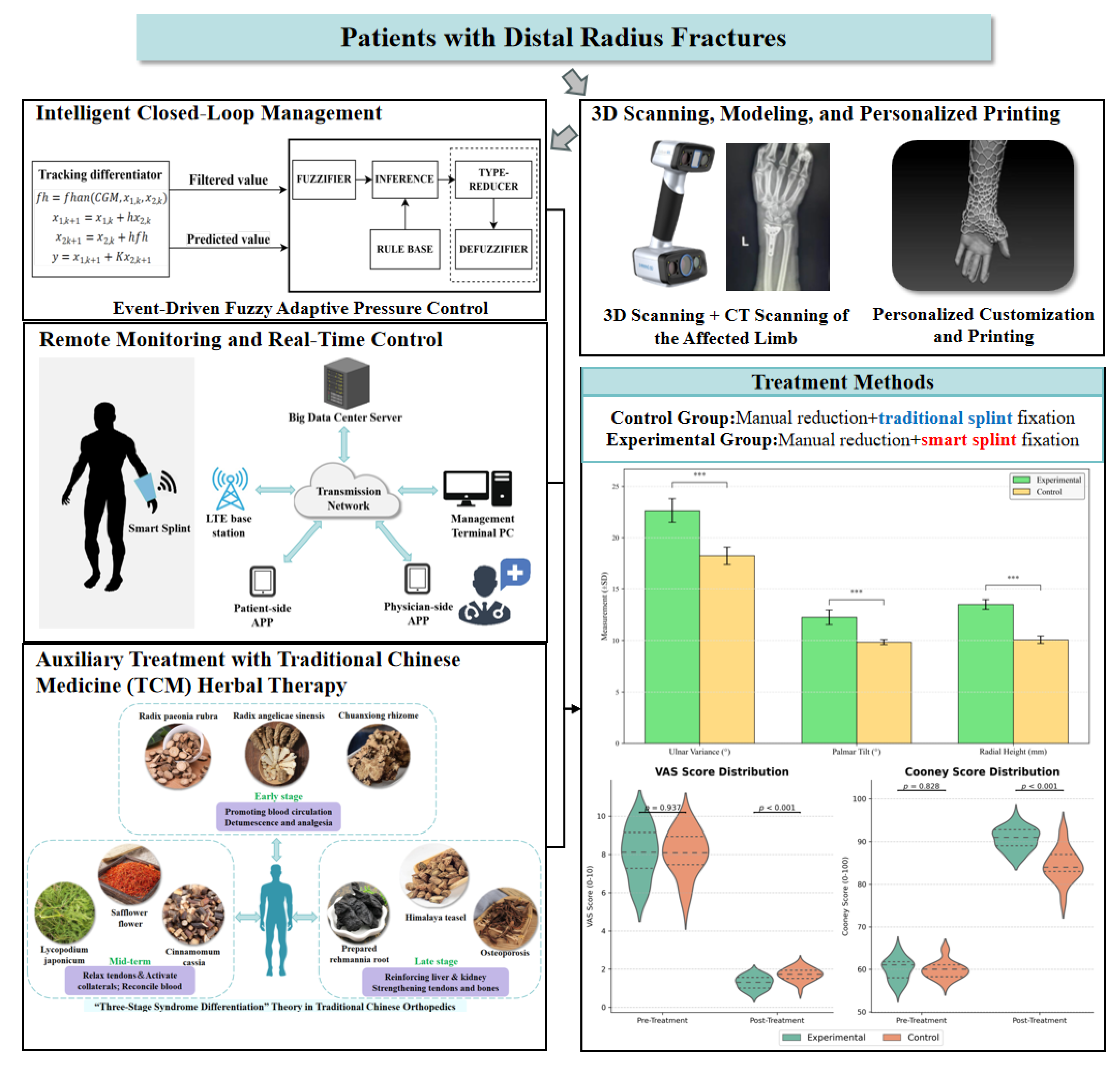
Figure 1. Overall Research Design.The triple asterisks (***) indicate a statistically significant result (p < 0.001).
Electronics 14 03542 g001
Figure 2. 3D scanning of the forearm (left) and 3D-modeled external fixation device (right).
Electronics 14 03542 g002
Figure 3. Thin-film pressure sensor.
Electronics 14 03542 g003
Figure 4. Schematic illustrating the strategic placement of the eight pressure sensors. The specific anatomical locations are as follows: (A) lunate bone (dorsal), (B) triquetral bone, (C) proximal to the fracture line, (D) ulnar styloid process, (E) pisiform bone, (F) scaphoid bone, (G) distal ulna, and (H) radial artery. These sites are shown from four views: (a) dorsal, (b) radial, (c) volar, and (d) ulnar.
Electronics 14 03542 g004
Figure 5. Photograph of the complete smart splint system. Key components are indicated: (A) Integrated PCB Housing, (B) Pressure Sensor Array, and (C) Linear Servo Actuator.
Electronics 14 03542 g005
Figure 6. Linear Servo Driver.
Electronics 14 03542 g006
Figure 7. PCB Control Board.
Electronics 14 03542 g007
Figure 8. The one-touch emergency power-off button located on the exterior of the splint for easy patient access.
Electronics 14 03542 g008
Figure 9. Flowchart of adaptive pressure regulation based on event-driven model.
Electronics 14 03542 g009
Figure 10. Linear servo driver integrated with the small splint.
Electronics 14 03542 g010
Figure 11. Remote monitoring and adjustment system.
Electronics 14 03542 g011
Figure 12. The user interface of the physician-facing “Intelligent Fixator” management dashboard, showcasing the system status overview, patient files, and recent remote adjustment logs.
Electronics 14 03542 g012
Figure 13. The user interface of the “Smart Splint Control Panel”. The central panel visualizes real-time pressure data (in N) from the eight sensors, while the upper and lower panels display the control status for each of the eight servo motors, including their current displacement (in mm).
Electronics 14 03542 g013
Figure 14. TCM Herbal Pad Design based on the “Three-Stage Syndrome Differentiation” Theory in Traditional Chinese Orthopedics.
Electronics 14 03542 g014
Figure 15. Comparison of fracture healing outcomes. The experimental group showed significantly better results in Ulnar Variance, Palmar Tilt, and Radial Height. *** denotes p < 0.001 .
Electronics 14 03542 g015
Figure 16. Comparison of VAS and Cooney scores. Violin plots showing the distribution of VAS scores and Cooney scores for both groups before and after treatment. Post-treatment scores show significant improvement in the experimental group compared to the control group ( p < 0.001 ).
Electronics 14 03542 g016
Table 1. The Fuzzy Rule Base for the IT2-FLC. Input variables are error (e) and change in error ( Δ e ). The output variables are as follows: NB (Negative Big), NS (Negative Small), ZE (Zero), PS (Positive Small), PB (Positive Big) for inputs, and FL (Fast Loosen), SL (Slow Loosen), NC (No Change), ST (Slow Tighten), FT (Fast Tighten) for the output.
Change in Error ( Δ e )NBNSZEPSPB
Error (e)
NBFLFLFLSLNC
NSFLSLSLNCST
ZESLSLNCSTST
PSSLNCSTSTFT
PBNCSTFTFTFT
Table 2. Comparison of baseline characteristics between the two groups [( x ¯ ± s ), n].
GroupNumber of CasesAge ( x ¯ ± s , years)Gender (n)
MaleFemale
Control3049.8 ± 3.71713
Experimental3048.6 ± 3.81812
t−0.6490.040
p0.5821.000
Table 3. Radiographic parameters and mathematical definitions.
ParameterNormal RangeFormulaDefinitions of Variables
Ulnar Inclination ( θ U I )20°–25° θ U I = tan 1 h w h: Vertical distance from radial styloid tip to ulnar articular surface; w: Horizontal width of radial metaphysis
Volar Tilt ( θ V T )10°–15° θ V T = tan 1 d 1 d 2 L d 1 : Dorsal cortex height; d 2 : Volar cortex height; L: Longitudinal axis length of radius
Radial Height ( H R )10–15 mm H R = ( x 2 x 1 ) 2 + ( y 2 y 1 ) 2 P 1 ( x 1 , y 1 ) : Radial styloid apex coordinates; P 2 ( x 2 , y 2 ) : Projection point on ulnar articular surface line (Perpendicular distance)
Table 4. Cooney Wrist Function Score assessment framework.
DomainScore RangeMeasurement MethodOperational Definition
Daily Activity0–25 points10-item questionnaire assessing functional tasksTasks include: cup holding, key turning, object grasping
Grip Strength0–25 pointsDynamometer-measured ratio (affected/unaffected limb) G S = S a S u × 25 , S a : Affected limb; S u : Unaffected
Range of Motion0–25 pointsFlexion/extension arc proportion R O M = θ F E 130 × 25 , θ F E : Measured arc
Discomfort Intensity0–25 pointsPatient-reported VAS (0–100 mm) D I = 25 VAS 4 , VAS: Visual Analog Scale score
Total Score0–100Sum of all subdomainsTotal = GS + ROM + DI + DA
Table 5. Comparison of radiographic outcomes between groups.
GroupUlnar Inclination (°)Volar Tilt (°)Radial Height (mm)
Experimental 22.63 ± 1.14 12.25 ± 0.71 13.51 ± 0.48
Control 18.23 ± 0.85 9.82 ± 0.25 10.06 ± 0.37
Mean Difference [95% CI]4.40 [3.92, 4.88]2.43 [2.15, 2.71]3.45 [3.26, 3.64]
t19.81320.67136.450
Cohen’s d1.941.872.21
p<0.001<0.001<0.001
Table 6. Evaluation of VAS and Cooney scores pre- and post-treatment ( x ¯ ± s ).
VAS ScoreCooney Score
GroupPre-TreatmentPost-TreatmentPre-TreatmentPost-Treatment
Experimental 8.12 ± 1.25 1.15 ± 0.28 60.68 ± 2.47 91.65 ± 2.38
Control 8.14 ± 1.30 1.88 ± 0.31 60.79 ± 2.45 83.15 ± 3.17
Mean Difference
[95% CI]
−0.73
[−0.98, −0.48]
8.50
[6.50, 10.50]
t/Z0.074Z = 3.4910.2904.370
Cohen’s d1.231.97
p-value0.941 < 0.001 0.744 < 0.001
Table 7. Comparison of clinical effectiveness (%). Verified using Fisher’s exact test, χ 2 = 3.9048 , p < 0.05.
GroupExcellentGoodFairPoorExcellent/Good Rate
Experimental18 (60.0)8 (26.7)4 (13.3)026 (86.7)
Control10 (33.3)9 (30.0)10 (33.3)1 (3.3)19 (63.3)
Disclaimer/Publisher’s Note: The statements, opinions and data contained in all publications are solely those of the individual author(s) and contributor(s) and not of MDPI and/or the editor(s). MDPI and/or the editor(s) disclaim responsibility for any injury to people or property resulting from any ideas, methods, instructions or products referred to in the content.