Article

FEM-Based Modelling and AI-Enhanced Monitoring System for Upper Limb Rehabilitation

by
Filippo Laganà
1,*,
Diego Pellicanò
2,
Mariangela Arruzzo
2,
Danilo Pratticò
3,*,
Salvatore A. Pullano
1 and
Antonino S. Fiorillo
1
1
Laboratory of Biomedical Applications Technologies and Sensors (BATS), Department of Health Science, “Magna Græcia” University, 88100 Catanzaro, Italy
2
ITIS Conte M. M. Milano School, 89024 Polistena, Italy
3
DICEAM Department, “Mediterranea” University, 89122 Reggio Calabria, Italy
*
Authors to whom correspondence should be addressed.
Electronics 2025, 14(11), 2268; https://doi.org/10.3390/electronics14112268
Submission received: 8 May 2025 / Revised: 26 May 2025 / Accepted: 30 May 2025 / Published: 31 May 2025
(This article belongs to the Special Issue Circuit Design for Embedded Systems)

Abstract

:
The integration of physical modelling, artificial intelligence (AI), and embedded electronics represents a promising direction in the development of intelligent systems for rehabilitation monitoring. Most existing approaches, however, treat biomechanical simulation and sensor-based AI separately, without leveraging their potential synergy. This study introduces a hybrid framework for upper limb rehabilitation that combines finite element modelling (FEM), AI-based trend classification, and a custom-designed electronic system for real-time signal acquisition and wireless data transmission. A mechanical model, developed in COMSOL 6.2 Multiphysics, simulates the interaction between a robotic glove and a deformable latex sphere. The latex material is described using a two-parameter Mooney–Rivlin hyperelastic formulation to capture large nonlinear deformations under realistic contact conditions. The high-fidelity simulation data are used to validate the signal acquisition chain and to train a supervised AI algorithm capable of classifying rehabilitation progress—whether improving or worsening—based on biomechanical features. An integrated electronic prototype enables seamless data flow to a cloud-based monitoring platform, supporting real-time feedback and adaptability. The classification algorithm demonstrates robust performance across different test conditions, while the electronic system confirms its applicability in rehabilitation settings. The novelty of this paper lies in the closed-loop integration of FEM-based simulation, AI-driven analysis, and embedded electronics into a unified monitoring architecture. This intelligent and non-invasive approach provides a scalable tool for tracking motor recovery and enhancing therapy effectiveness through adaptive, feedback-driven interventions.

1. Introduction

Motor disabilities in the arms, caused by neurological issues like stroke, spinal cord injuries, multiple sclerosis, and diseases that affect the nervous system such as Parkinson’s disease or ALS, are a major reason for people losing their ability to function independently around the world. Deficits severely compromise the quality of life of patients, hindering the execution of essential daily activities, and contribute to an increasing socio-health burden for families and healthcare systems [1,2,3,4]. Neuro-motor rehabilitation of the upper limbs plays a crucial role in the attempt to partially restore compromised motor functions, leveraging the neuroplasticity mechanisms of the central nervous system [5,6].
Scientific studies provide a fundamental overview of the principles behind neurorehabilitation, motor control, and neuroplasticity [7,8]. Functional recovery is based on the repeated, intensive, and variable execution of specific exercises capable of stimulating cortical reorganisation and the formation of new synaptic connections [9]. To this end, traditional rehabilitation protocols involve a combination of passive and active techniques, strengthening exercises, occupational therapy, functional electrical stimulation, and increasingly, the use of advanced technologies such as robotics and virtual reality [10,11]. Among the various rehabilitation exercises employed, gripping and manipulating a latex ball with the affected hand plays a clinically relevant role [
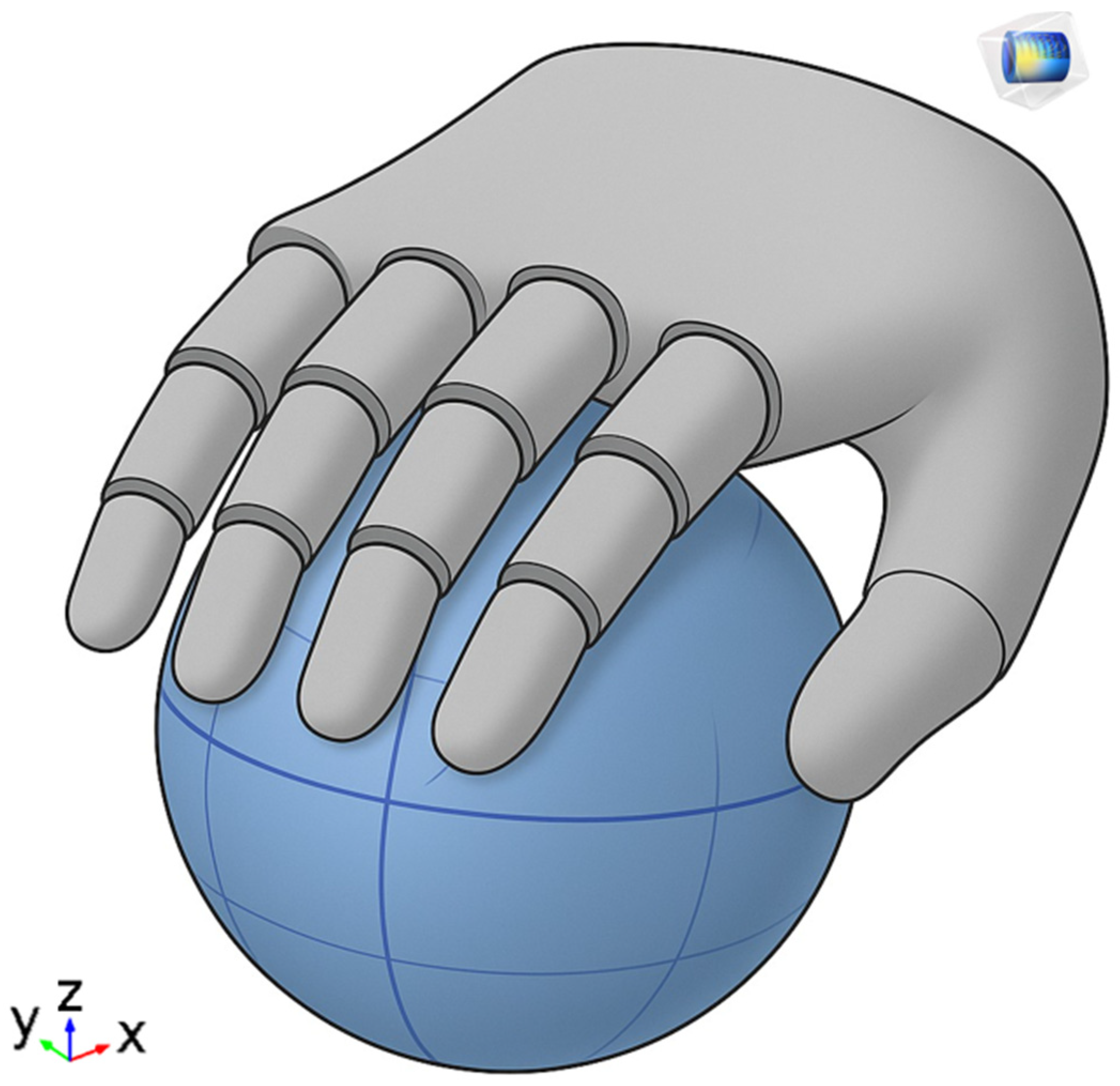
12]. The selection of this exercises for this study is motivated by both its clinical significance and its technical suitability for sensor-based analysis. Gripping tasks provide a robust foundation for biomechanical modelling due to their well-defined contact interactions, and they are frequently included in standardized rehabilitation protocols. This makes them not only a realistic target for monitoring but also a generalizable proxy for evaluating hand motor function recovery.
This simple yet highly effective task stimulates fine motor control, improving grip strength and enhancing proprioceptive feedback. Patients are typically asked to perform repetitive compression movements, as well as coordinated movements such as rolling, rotating, or transferring the ball, which activate multiple muscle groups and joints in a functional and controlled manner [13]. This exercise is often included in both the early and advanced stages of rehabilitation and serves as an important reference for assessing the patient’s progress over time [14]. An important role in the rehabilitation process is played by the physiotherapist, who not only guides and adapts exercises based on the patient’s functional level but also takes care of the continuous monitoring of progress through validated scales (e.g., Fugl-Meyer Assessment and Box and Block Test) and clinical observations [15]. However, in home settings or environments without direct supervision, this monitoring function becomes complex, with the risk of losing therapeutic effectiveness or the emergence of harmful compensatory movements [16,17,18]. In recent years, home-based neurological rehabilitation has undergone a radical transformation thanks to the increasing integration of advanced digital technologies, capable of overcoming many of the limitations of traditional therapeutic models [19,20]. Wearable devices, artificial intelligence (AI), and telemonitoring systems have emerged as strategic tools capable of supporting motor recovery in a continuous, personalized, and high-intensity manner, even outside structured clinical settings [21,22]. Wearable devices integrated with inertial and force sensors allow for the continuous monitoring of upper limb movements, providing real-time feedback and guidance [23,24]. The integration of virtual and augmented reality (VR/AR) in rehabilitation has enabled immersive, engaging environments that improve motor learning and increase patient adherence [25,26,27]. Adaptive rehabilitation scenarios using gamification and AI-driven personalization have shown improved clinical outcomes by increasing engagement and adjusting difficulty levels to patient progress [28,29,30,31,32]. The adoption of telemonitoring platforms also allows therapists to access their patients’ clinical and kinematic data in real-time or asynchronously, facilitating remote follow-up, timely modification of the rehabilitation plan, and proactive management of the care pathway [33,34]. This approach also allows overcoming geographical barriers and offering quality treatments to patients in remote areas or with mobility difficulties, contributing to greater equity in access to care. The reduction in distances represents only one important aspect of the application on patients. In fact, studies have highlighted the effectiveness of these tools in promoting functional recovery post-stroke or in other chronic neurological conditions. The improvements achieved are tested in terms of strength, dexterity, coordination, and independence in daily activities [35]. The quantitative data collected in the conducted studies allowed for an objective analysis of rehabilitative progress, overcoming the limits of subjective evaluations and increasing precision in defining therapeutic goals [36]. Despite their potential, current systems face challenges in usability, interoperability, and the need for large-scale validation [37]. The combination of wearable devices, virtual reality, and artificial intelligence today represents one of the most promising avenues for revolutionising upper-limb home rehabilitation, making it more intensive, personalised, accessible, and sustainable. The technological approach does not aim to replace the therapist but to enhance their capabilities, extending their effectiveness even in unsupervised contexts. This study addresses these challenges by presenting an integrated system that combines physical modelling, AI-based analysis, and wearable technology to support adaptive and personalized home rehabilitation (see Section 2 for system details). The synergistic integration of biomechanical modelling, machine learning algorithms, and embedded electronic systems represents a challenge to be implemented for continuous monitoring and prediction of motor patterns. The use of finite element modelling (FEM) in the rehabilitation field can offer a detailed representation of joint and muscular dynamics, allowing for more precise adaptation of therapeutic protocols to individual morphological and functional characteristics [
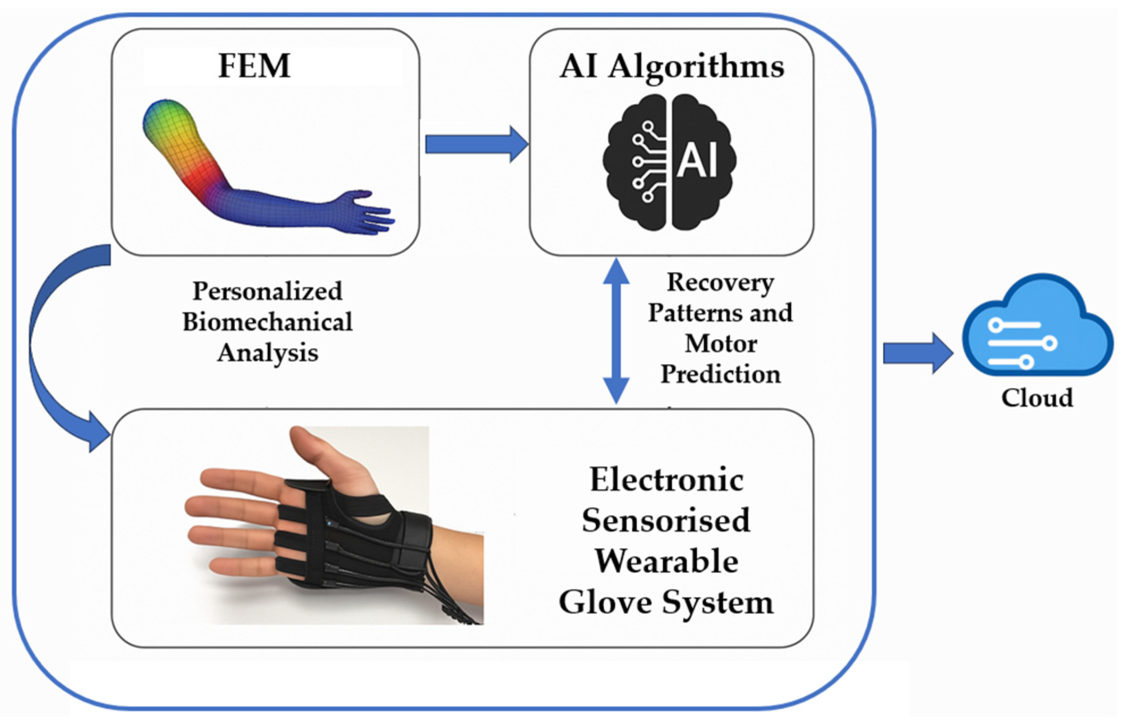
38]. This research fits into this scenario with the aim of developing and validating a hybrid intelligent system that combines the following: physical modelling (FEM) of the upper limbs for personalized biomechanical analysis, artificial intelligence algorithms for the automatic identification of recovery patterns and the prediction of motor evolution, and an integrated electronic sensorised system for the collection and transmission of real-time data. This approach aims to overcome the limitations of current solutions, offering a truly adaptive system, accessible even in home settings, and capable of providing advanced decision support to the physiotherapist. Specifically, the use of FEM modelling allows for the high-detail simulation of the mechanical properties and physiological dynamics of the musculoskeletal structures of the arm and hand. Thanks to FEM, it is possible to construct personalized three-dimensional models derived from individual anatomical data (for example, from MRI or 3D scans) and capable of accurately representing the tensions, forces, deformations, and joint interactions during the execution of an exercise [39,40]. In a rehabilitative context, these models can be used to simulate the biomechanical load exerted during specific therapeutic movements, identify areas subject to overload or at risk of postural compensations, or optimize the selection and intensity of exercises to be assigned to the patient. Such a level of detail allows for a deep personalization of protocols, based not only on the general functional level but also on the actual biomechanical characteristics of the individual user, with the aim of maximizing effectiveness and minimizing risks. At the same time, the application of artificial intelligence (AI) algorithms allows for the analysis of large volumes of motor data collected during rehabilitation sessions. Such algorithms are capable of recognizing rehabilitative patterns, classifying motor trajectories based on their quality (fluency, accuracy, and amplitude), and above all, predicting the functional evolution of the patient based on historical data [41]. The integration of AI enables the automatic evaluation of daily motor performance, the early detection of stagnations or regressions, and the dynamic personalization of the protocol, adapted in real-time to the emerging needs of the patient. This predictive capability offers the physiotherapist quantitative and up-to-date decision support, which can be used to recalibrate the therapeutic intervention more precisely and promptly, optimizing clinical outcomes and maintaining high patient motivation. The third pillar of the proposed study is represented by an embedded platform integrated into a wearable glove, equipped with Inertial Measurement Unit (IMU) sensors, flexion and pressure sensors, and wireless transmission modules. These systems continuously collect data during the execution of exercises, without the need for direct supervision or complex equipment. The raw data are pre-processed locally (edge computing) to reduce latency and then sent to a cloud platform for analysis via AI. The main advantage of this architecture is the ability to operate in real-time and in home environments, without requiring specialized infrastructure, ensuring patient autonomy, monitoring treatment adherence, and detecting any deviations from the prescribed therapeutic protocol. The integration of these three components into a hybrid intelligent system represents a qualitative leap compared to traditional approaches. The combination of a detailed biomechanical model (FEM), an AI-based predictive analytical engine, and an embedded sensorised interface allows not only the ability to observe and evaluate but also to anticipate, adapt, and optimize every aspect of home rehabilitation. This paper is organised as follows: Section 2 provides an overview of the implemented system. Section 3 describes the system components: Section 3.1, the COMSOL Multiphysics model; Section 3.2, the integrated electronic and acquisition system; and Section 3.3, the computational model. Section 4 presents the experimental results, followed by conclusions and future developments.

2. Intelligent Hybrid System

The proposed system is a hybrid and intelligent platform that integrates biomechanical simulation, artificial intelligence, and embedded electronics to support the rehabilitation of upper limbs in both clinical and home settings. Its architecture consists of three closely integrated modules, formed by a FEM model, the design of an integrated sensorised glove, and the implementation of an AI module on simulated data (Figure 1).
The proposed system consists of 3 steps, one sequentially following the other. The first step involves the implementation of a detailed physical model of the human hand interacting with a lattice sphere. Implemented in COMSOL Multiphysics, the simulation uses a two-parameter Mooney–Rivlin hyperelastic model to describe the behaviour of the lattice material under large deformations. This component provides biomechanical insights and supports the calibration of the detection hardware. The simulation accounts for the anisotropic and nonlinear response of soft tissues, enabling the evaluation of stress distribution across finger joints during grasping. Boundary conditions are defined based on realistic hand postures, while contact mechanics are employed to capture interactions between the hand surface and the deformable structure. The output of this step directly informs the design specifications for the embedded sensor array, ensuring optimal sensitivity and mechanical coupling. The second step involves the electronic design of an acquisition board necessary for the development of a wearable glove prototype. The electronic system is equipped with IMU sensors, flex sensors, and pressure sensors, which enable real-time acquisition of movement and force data during rehabilitation exercises. The system is designed for usability, autonomy, and wireless data transmission through low-latency modules, allowing continuous monitoring outside of clinical supervision. The acquired data are processed locally and in the cloud through machine learning algorithms trained to classify motor performance trends. The artificial intelligence system identifies patterns that indicate improvement or stagnation in rehabilitation and provides adaptive feedback. This dynamic personalisation enhances patient engagement and allows therapists to refine therapeutic protocols remotely. The set of components forms a closed-loop system capable of providing adaptive and high-intensity rehabilitation, improving the accuracy of assessments and supporting the decision-making process through real-time data-driven insights. The integration of wearable sensors and cloud-based platforms further enables continuous monitoring and long-term tracking of patient progress. Moreover, the system fosters a more proactive approach to care by alerting clinicians to anomalies or deviations from expected recovery trajectories. The following section delves into the specifics of each individual research phase, analysing and describing every single aspect addressed during the study phase.

3. Modelling and Analysis of AI-Driven Monitoring for Upper Limb Rehabilitation

3.1. System Modelling

3.1.1. Physical Problem Description

The objective of the model is to simulate the mechanical interaction between a robotic hand and a latex ball. The use of biomechanical modelling in rehabilitation is based on texts on musculoskeletal mechanics and finite element modelling [42,43]. The choice of latex is motivated by its high elasticity and nonlinear behaviour, making it ideal for capturing large deformations under load. The simulation focuses on the distribution of internal stress, contact pressure, and displacement fields, thus enabling a quantitative understanding of the mechanical response under realistic loading conditions.

3.1.2. Geometrical and Material Parameters

The parametric definition, adopted to geometry and material properties to allow flexible adjustments in different test scenarios, has certain main aspects. First, the latex sphere is modelled as a solid sphere of a radius of 5 cm and a shell thickness of 0.5 cm. The fingers are idealised as rigid cylinders with a length of 10 cm and diameter of 2 cm. The materials used in the modelling for the robotic hand were rigid plastics with a high Young’s modulus and low Poisson’s ratio. The latex ball is defined using a two-parameter Mooney–Rivlin hyperelastic model with material constants C10 = 0.1 MPa and C01 = 0.03 MPa. The Poisson’s ratio was ν = 0.49 (near incompressibility). The geometry, shown in Figure 2, has a sphere centred in the origin and five fingers (cylinders) positioned and rotated to simulate a gripping configuration.
A rigid hand block connects the fingers through Boolean operations.

3.1.3. Mathematical Formulation

The Mooney–Rivlin constitutive model describes the mechanical behaviour of latex well, which is considered a hyperelastic material. Research studies [44,45,46] use this model to represent the large nonlinear deformations of incompressible or nearly incompressible elastomers. The model expresses the strain energy density function W, which represents the stored elastic energy per unit volume, as a function of the principal invariants of the right Cauchy–Green strain tensor C. Equation (1) defines the deformation energy function in the two-parameter Mooney–Rivlin model.
W = C 10 I 1 3 + C 01 I 2 3
where W is the strain energy density [Pa], C10 and C01 are material constants [Pa], I1 and I2 are the first and second invariants of the deviatoric part of the deformation tensor C, and the constant values “3” are subtracted to ensure that W = 0 in the undeformed configuration. In the context of nonlinear solid mechanics, hyperplastic models do not directly utilise classical stresses and strains but are based on strain tensor invariants. These invariants are scalars that do not change due to rotations of the reference system and are usually invariant with respect to orthogonal transformations. Equations (2) and (3) define the two invariants I1 and I2.
I 1 = t r C = λ 1 2 + λ 2 2 + λ 3 2
Equation (2) associates the first invariant I1, the dominant term in quasi-incompressible materials, with the energy of pure elongation along the three main axes.
I 2 = 1 2 t r C 2 t r C 2 = λ 1 2 λ 2 2 + λ 2 2 λ 3 2 + λ 3 2 λ 1 2
where λ1, λ2, and λ3 are the principal stretch ratios along the three orthogonal directions. Equation (3) measures cross-quadratic combinations between different principal elongations. I2 plays a more pronounced role in complex deformations (e.g., multi-axis shear or tension). In quasi-incompressible materials such as latex, the determinant of the strain gradient J is assumed to be approximately equal to one. Therefore, we neglect the volumetric contribution to the deformation and attribute the mechanical response to changes in shape (isochronous deformation). Equation (4) provides the Cauchy stress tensor σ for an incompressible hyperelastic material adopting the Mooney–Rivlin model.
σ = p I + 2 C 10 B C 01 B 1
The left Cauchy–Green deformation tensor is defined as B = FFT, while its inverse is denoted as B−1. The Lagrange multiplier p is introduced to enforce the incompressibility constraint, and the identity tensor is denoted by I. This formulation highlights how the model simultaneously accounts for both the distortional and volumetric components of the mechanical response, although the volumetric contribution is suppressed under incompressibility conditions. The coefficient C10 governs the material response to shear and distortional deformations, whereas C01 becomes increasingly significant in complex multiaxial deformation states or when dealing with moderately compressible materials. Since C10, for most natural and synthetic elastomers, is significantly larger than C01, the Mooney–Rivlin model was adopted. Therefore, the governing equations for nonlinear elasticity are expressed by Equation (5)
σ + f = ρ u ¨
with σ as the stress tensor, ρ as the material density, f as the body force, and u ¨ as the displacement field.

3.1.4. Mesh Generation and Quality Analysis

To ensure accurate and reliable results from the finite element analysis, a high-quality tetrahedral mesh was generated. Particular attention was given to the resolution of curved surfaces and slender geometries, while maintaining computational efficiency. The main mesh characteristics and generation settings are summarised in Table 1 and Table 2.
The mesh is composed of over 73,000 tetrahedral elements, which provide a volumetric discretisation of the domain, and about 19,000 triangular surface elements, which accurately capture the boundary conditions and interface details. A moderate number of edge and vertex elements ensures a proper resolution of geometric discontinuities and sharp features. Despite the presence of some poorly formed elements—as indicated by a very low minimum element quality of 2.596 × 10⁻¹⁰—the overall quality of the mesh is acceptable, with an average element quality of 0.6343. This suggests that, although localised refinement may be necessary in some critical regions, the mesh is globally well constructed and suitable for numerical computation. The quality of the elements is evaluated based on shape metrics such as the ratio between the volume of the element and the cube of its circumscribed radius or similar criteria based on distortion. To better capture the surface curvature, particularly around spherical and cylindrical features, a curvature resolution factor of 0.6 was adopted. This value allows for a greater density of elements in curved regions without excessively increasing the total mesh size. Similarly, a resolution factor of 0.5 was employed for elongated geometries to ensure adequate discretisation of slender structures, such as fingers or probes, which can have high aspect ratios and be subject to mesh distortions. The minimum and maximum element sizes were set to 0.229 mm and 1.27 mm, respectively, allowing for local refinement where necessary while keeping computational costs manageable. A maximum element growth rate of 1.5 was imposed to ensure a uniform gradation of element sizes throughout the domain, reducing the risk of numerical instability caused by abrupt changes in mesh density. The presented mesh is structured to support high-fidelity simulations while maintaining the computational efficiency of the implemented system.

3.2. Integrated Electronic System

The implemented computational model provided essential feedback to determine the specifications useful for both the design of the integrated electronic system and the subsequent implementation phase of the AI algorithm. The idea best suited to different types of patients consists in the design of an acquisition system developed through a wearable interface in the form of a glove, equipped with four resistive strain gauges (Figure 3).
The arrangement of the strain gauges in a full Wheatstone bridge configuration offers improved temperature compensation and sensitivity. Each bridge arm, which includes a symmetrically positioned strain gauge, suppresses thermal drift and mechanical artefacts. The use of the full-bridge structure improves common-mode noise rejection, while the HX711 amplifier module offers good stability. The input impedance of approximately 10 MΩ and the common mode rejection ratio (CMRR) of over 100 dB ensure high signal fidelity even under fluctuating mechanical or thermal conditions.
The sensors are interconnected in a Wheatstone bridge configuration to provide a specific solution for increasing sensitivity, improving temperature compensation and reducing unwanted mechanical deformation components [47,48,49]. The operating principle is based on the variation of the electrical resistance of a conductor subjected to mechanical deformation, as described by Equation (6).
ε = Δ R G · R
where ΔR is the change in resistance, R is the initial resistance, G is the gauge factor (a measure of the sensor’s sensitivity), and ε is the mechanical strain.
Assuming elastic material behaviour, Hooke’s law, Equation (7) relates the deformation to the applied force.
ε = σ E = F E · A  
where F is the applied force, E is Young’s modulus, and A is the cross-sectional area. Combining Equations (6) and (7) gives a direct relationship (8) between the applied force and the change in resistance:
Δ R = G · R · F E · A  
Due to the low amplitude of the signal generated by the strain gauges (in the millivolt range), a high-precision instrumentation amplifier is used. Therefore, the HX711 module, which is widely used in force and weight-measurement applications, is chosen [50,51]. The circuit configuration of the HX711 module is illustrated in Figure 4.
The circuit diagram is designed to amplify the analogue signal and forward it to the controller for processing. The ESP32 microcontroller is chosen for its high processing capacity, low power consumption, and integrated wireless connectivity (Wi-Fi and Bluetooth) [52,53,54]. The microcontroller’s functionality consists of real-time system and data control, pre-processing, and secure transport of information via HTTPS to a remote server or local network client. The acquired data, accessible to several hosts via the network, are stored in a centralised database. Figure 5 shows the network architecture and data flow in general.
To meet the high interference conditions, the wireless transmission is tested in congested 2.4 GHz channel environments. The average latency achieved by the system had end-to-end values of less than 35 ms and maintained a packet loss rate of only 2.1 per 1000 transmissions. A linear relationship was observed between the received signal strength indicator (RSSI) and data throughput (R² = 0.94) in the range of −45 dBm to −75 dBm. To mitigate interruptions caused by transient connectivity problems, an offline caching routine stores up to 500 time-stamped records in non-volatile memory, which are automatically retransmitted once the connection is restored. A customised, embedded acquisition algorithm has been developed in the microcontroller’s firmware. The algorithm enables real-time monitoring of the mechanical stresses experienced by the user’s fingers. The designed system has the potential to support various applications in biomedical monitoring, rehabilitation, and smart home environments.
The schematic in Figure 6 includes the power regulation unit, the signal conditioning circuit, and the microcontroller-based data processing and transmission block. Specifically, the system is powered via a USB interface and stabilised via an LM2596S step-down voltage regulator, which ensures a constant 5 V supply to the ESP32 microcontroller and connected sensors. The ESP32, located in the centre of the diagram, serves as the primary control and communication unit. It receives the analogue signals from the resistive strain gauges configured in a Wheatstone bridge (outside the diagram), which are connected via the SENSOR_IN port. The analogue signals, after being amplified and digitised via the HX711 module, are processed and then transmitted wirelessly via HTTPS. The presence of the decoupling capacitors (C17 and C18) and the ferrite inductor (L2) minimises noise and ensures supply line stability and signal integrity.
The importance of the diagram in Figure 6 lies in its fundamental role as the basic infrastructure of the wearable system. The integrated system, which constitutes the electronic part of the glove, is a critical component within the overall framework of the study conducted, underpinning both the experimental validation and potential future use of the system in real-world monitoring applications.

3.3. Computational Model

3.3.1. Dataset Description

The dataset employed in this study was collected from patients undergoing upper-limb rehabilitation therapy. The core of the dataset is derived from real-time measurements using a custom lattice ball sensor system, capable of simultaneously capturing three-dimensional spatial positioning—X, Y, and Z coordinates in mm—and the principal strain deformation that quantifies mechanical deformation during grip actions.
A longitudinal protocol was adopted, where a cohort of 10 patients was continuously monitored over a 100-day rehabilitation period. The cohort was demographically balanced, comprising 50% male and 50% female participants, with ages ranging between 40 and 80 years—representative of typical post-stroke or post-trauma rehabilitation populations. The progression of each patient’s grip performance was characterized by calculating daily scaling factors applied to the strain signal. These factors followed nonlinear trajectories consistent with real-world recovery, featuring a general improvement trend interspersed with random fluctuations to simulate physiological variability and setbacks due to fatigue, pain, or inconsistent adherence.
Each rehabilitation session generated 2.048 sequential sensor samples to reflect high-frequency, temporally resolved motor activity. To replicate measurement and biological variability, Gaussian noise was injected into the spatial features, introducing inter-session and inter-subject dispersion. The core signal—solid.ep3—was then statistically compared to its value on the previous day to determine the nature of functional progression. Based on the percentage change in mean deformation, each session was labelled according to five clinically interpretable classes: Severe deterioration (Δ < −7%), Mild deterioration (−7% ≤ Δ < −2%), Neutral (−2% ≤ Δ ≤ +2%), Mild improvement (+2% < Δ ≤ +7%), and Severe improvement (Δ > +7%).
The final dataset encompasses 1,000 labelled daily sessions, each represented as a high-resolution, multivariate time series paired with a categorical label suitable for supervised learning tasks. Table 3 provides a statistical summary of the dataset, including standard deviation, minimum, and maximum values for each spatial coordinate and the solid.ep3 deformation signal.
Measurement noise emulation involved adding a white Gaussian perturbation (standard deviation of 0.5 mm for positions and 0.002 for strain). Daily strain changes (Δ) from the previous day provided ordinal labels (Severe deterioration to Severe improvement). This yielded one thousand labelled sequences, with descriptive statistics presented in Table 3.
Clinically meaningful feedback was session-level; however, algorithm training utilised overlapping 128-sample windows (~1.28 s), decomposing sessions into sixteen segments. Majority voting from window-level predictions enabled holistic daily judgments.

3.3.2. Signal Conditioning and Window-Level Pre-Processing

Window-level preprocessing included positional mean-centering and a sixth-order, zero-phase Butterworth filter (7 Hz cutoff), reducing inter-subject variability and physiological noise. Strain normalisation relative to day 1 baselines allowed predictors to focus on relative rather than absolute changes. These steps ensured stationary input distributions for the convolutional layers, accelerated training convergence, and had minimal computational load (execution < 200 µs on STM32F746).

3.3.3. Network Architecture: LITE-BT Hybrid

The backbone of the classifier, henceforth referred to as LITE-BT (Light Inception with boosTing), separates representation learning from decision-making while remaining within the 64 kB flash/RAM envelope of the STM32F746. Three Mini-Inception modules perform multiscale feature extraction. Each module contains four parallel branches, three one-dimensional convolutions with kernels of one, three, and five taps, plus a three-sample max-pooling path followed by a 1 × 1 convolution. The concatenated outputs are normalised and passed through a ReLU nonlinearity before the next module. After the third module, the channel depth reaches forty-eight; a global average pooling layer then collapses the temporal axis, yielding a 128-dimensional embedding. Because the convolutional parameters have been quantised to eight bits, the entire backbone occupies only 19 kB of flash. The embedding is subsequently classified by a five-fold LightGBM ensemble whose JSON representation adds a further 44 kB, still leaving headroom for the acquisition firmware. For clarity, the layer-by-layer specification of the backbone is presented in Table 4.
Rather than employ a dense classification head, the embedding is passed to a LightGBM ensemble consisting of the five models trained during cross-validation. Gradient boosting offers several practical benefits in this context. First, tree-based decision boundaries cope naturally with the mild class imbalance that was observed in the data. Secondly, inference through a tree ensemble is almost free of floating-point arithmetic and is thus extremely attractive for deployment on a floating-point-constrained microcontroller. Finally, by decoupling feature learning from classification, the pipeline supports post-hoc interpretation via Shapley values or similar techniques, an aspect that is increasingly valued by rehabilitation clinicians. This hybrid CNN–LightGBM design was deliberately chosen to combine efficient multiscale, temporal feature extraction with robust and interpretable classification, providing high accuracy while maintaining real-time responsiveness on embedded systems. The use of LightGBM enables deployment in floating-point-constrained environments and offers resilience to noisy or imbalanced rehabilitation data, making the system suitable for diverse clinical contexts.

3.3.4. Training Procedure and Regularisation Strategy

All models were implemented in Python 3.11, relying on TensorFlow 2.16 for the convolutional components and LightGBM 4.3 for the boosting stage. The CNN was optimised with AdamW. The learning rate followed a five-epoch linear warm-up to 3 × 10⁻³ and then decayed according to a cosine schedule whose floor was set at 3 × 10⁻⁵. Early stopping with a patience of eight epochs on the validation loss guarded against overfitting.
A categorical focal loss with a focusing parameter γ = 1.5 supplanted standard cross-entropy. This choice proved essential for amplifying the gradient signal associated with the relatively rare Severe deterioration and Severe improvement cases. In parallel, three data-augmentation techniques—strain-scaling by ±10%, time-warping by ±8%, and a small spatial jitter—were found to improve the macro-F1 score by approximately 2.5 percentage points.
These strategies, together with the use of a stratified cross-validation scheme and a LightGBM ensemble capable of handling nonlinear feature interactions and class imbalance, improve the model’s robustness across varied patient profiles. Future work will include validating this framework on a more diverse cohort and applying transfer learning to accelerate adaptation to unseen rehabilitation trajectories. During LightGBM training, the 128-dimensional embeddings were first z-normalised; the tree ensemble was then grown using a learning rate of 0.03, a maximum of 31 leaves, and a L1 regularisation of 0.1. Each fold was permitted a maximum of 600 boosting iterations, with early stopping after twenty rounds without improvement in macro-F1.

3.3.5. Quantisation and Deployment

After training, the CNN weights and activations were quantised to full-integer eight-bit precisions using TensorFlow Lite. Static quantisation was preferred over dynamic because it eliminates the need for run-time scale extraction, thereby reducing latency by roughly 9%. The LightGBM ensemble was exported in JSON format and ingested by a lightweight fixed-point interpreter written in C. End-to-end inference for a 128-sample window executes in 4.6 ms on an STM32F746 running at 216 MHz, during which peak RAM usage does not exceed 64 kB. These figures include both the TensorFlow Lite arena and the tree interpreter, confirming that real-time feedback is feasible on a wearable glove without recourse to external computation. To assess multitasking performance, the firmware was benchmarked under FreeRTOS with concurrent sensor acquisition, AI inference, and Wi-Fi transmission. Task execution traces revealed that AI inference (4.6 ms) and data acquisition (3.1 ms) operated without deadline violations at a 50 Hz sampling rate. Stack memory consumption per task remained below 60% of the allocated size, and no overflows or memory faults were detected. These results demonstrate the system’s robustness for real-time embedded operation. Our task scheduling and memory profiling approach was inspired by recent developments in embedded AI and hybrid real-time systems, such as travel demand forecasting models based on spatial–temporal task orchestration and edge computing frameworks [55,56].

4. Results

The FEM focused on analysing the mechanical response of a soft rehabilitation ball subjected to a compressive force representative of a patient’s grip strength (approximately 10 N). The model reproduces the time-dependent deformation behaviour of elastomeric materials commonly used in upper-limb rehabilitation tools.
The evolution of the deformation within the ball over a 9 s interval is represented in Figure 7.
The deformation curve demonstrates how the viscoelastic nature of the material highlights a nonlinearity of the force applied to the latex ball. The rapid increase in deformation in the first few seconds, followed by a gradual approach to a steady state, attests to the behaviour, with the exponential viscoelastic response, expected from soft polymeric materials. The greatest deformation occurs along the vertical axis (Z), aligning with the direction of the applied force.
A detailed quantitative analysis is provided in Table 5, which reports the values of deformation and displacement along the X, Y, and Z axes at one-second intervals.
Initially, the deformation is negligible, but it increases to 0.834 by the ninth second, indicating significant material compliance under sustained load. The displacement along the Z-axis increases from 0.0091 m at 1 s to over 0.0417 m at 9 s, while the lateral displacements in the X and Y directions remain relatively minor, peaking below 3 mm. This directional asymmetry confirms the predicted axial deformation model under vertical compression. The simulated results validate the model’s ability to replicate realistic mechanical interactions between the user’s hand and the rehabilitation device. The nonlinear viscoelastic trend captured in the simulation is essential for accurately characterising the patient’s effort and progress, especially when integrating the data into AI-based monitoring platforms. A test bench equipped with calibrated resistive strain sensors was used to apply controlled compressive forces to the latex ball while recording deformation data. The force–displacement curve extracted from the glove closely matched the FEM-simulated response, with a mean absolute error consistently below 7%. This validates the model’s predictive accuracy and confirms the biomechanical plausibility of the simulation results. In order to assess the reliability of the Mooney–Rivlin material parameters (C10 = 0.1 MPa; C01 = 0.03 MPa), a quantitative comparison between the simulated deformation response and the data acquired during the rehabilitation sessions was carried out. The deformation measurements were processed and temporally aligned with the simulated deformation results.
As illustrated in Figure 8, the experimental deformation closely follows the simulated curve, with a deviation consistently less than 5% throughout the entire rehabilitation sequence. This high degree of correspondence confirms both the suitability of the Mooney–Rivlin model to describe soft-tissue-like behaviour and the validity of the simulation framework in a real-world clinical context.
The quantitative evaluation of LITE-BT was conducted through a stratified five-fold cross-validation, ensuring that windows belonging to the same day remained in the same fold so as not to inflate performance estimates. Unless otherwise stated, all reported metrics refer to the session level, obtained from the majority vote of sixteen window-level predictions. Table 6 condenses the principal macro-averaged metrics. The model attained an accuracy of 0.85 and a macro-F1 of 0.83, while the micro-averaged ROC–AUC reached 0.94. These scores indicate that the classifier is capable of distinguishing clinically meaningful changes in functional status with a reliability comparable to that of in-clinic dynamometry, yet at a fraction of the hardware complexity.
A more granular perspective is offered in Table 7, which breaks the foregoing metrics down by class. It is noteworthy that both Severe deterioration and Severe improvement—the categories that demand immediate therapeutic response—achieved F1-scores above 0.88. By contrast, the Neutral and Mild improvement classes occasionally confused the model; their decision boundary corresponds to a modest ±2% change in mean strain, which also challenges human raters.
The trajectory of the training and validation losses suggests that the combination of a brief warm-up and a cosine decay schedule provides stable optimisation. All five folds converged to their final minima between epochs 41 and 44, after which early stopping was consistently triggered. Validation loss variance across folds remained below 0.006, indicating that the network capacity is well matched to the dataset size. Importantly, no fold invoked the secondary learning-rate reduction mechanism, a sign that the learning-rate schedule had been appropriately tuned.

4.1. Ablation Studies

Three ablation scenarios were explored to gauge the contribution of each architectural component. When the LightGBM head was replaced by a single dense layer, the macro-F1 dropped to 0.78. Eliminating data augmentation reduced the macro-F1 to 0.81, whereas collapsing the heterogeneous Inception kernels into a single k = 3k = 3k = 3 convolution resulted in a macro-F1 of 0.79. These findings, summarised in Table 8, support the architectural decisions itemised in Section 3.3.

4.2. Embedded-System Profiling and Energy Consumption

On-device benchmarks confirmed that the complete inference pipeline consumes 0.36 MFLOPs for a 128-frame window and completes execution in less than 5 ms. Power-monitoring traces revealed an incremental current draw of 8.3 mA during inference peaks, translating into an estimated nine hours of continuous operation on a 500 mAh lithium-polymer battery. Given that rehabilitation exercises rarely exceed one hour per day, the glove can operate for more than a week between charges, satisfying the usability requirements identified in our earlier ethnographic observations.
The dynamic power consumption of the embedded system, analysed over a complete acquisition–inference–transmission cycle, is shown in Figure 9. The measured current waveform highlights distinct operational phases, specifically idle, signal acquisition, AI-based inference, and Wi-Fi transmission. The average current values are 0.99 mA (inactivity), 5.01 mA (acquisition), 10.03 mA (inference), and 120.53 mA (Wi-Fi transmission). The highest current values were observed during the Wi-Fi burst phase, which reflects the high energy demand of data transmission. The total power consumption per cycle was estimated at 1363 mAs (0.379 mAh), corresponding to a theoretical battery life of 502.6 h in idle mode and approximately 4.1 h in continuous Wi-Fi activity, assuming a 500 mAh battery, while the average power consumption during active computing (excluding Wi-Fi) was approximately 8.3 mA, which can be broken down into 5.1 mA for the ESP32 module, 2.3 mA for the sensors, and 0.9 mA for the HX711 amplifier.
To ensure mechanical and electrical reliability during prolonged use, the flexible glove interface was subjected to 15,000 flex cycles using a mechanical actuator applying a constant force of 0.8 N per cycle. Post-test analysis indicated a degradation of strain sensitivity of less than 2.8%, demonstrating good fatigue resistance of the embedded sensing elements. In addition, all electrical interconnections and solder joints were encapsulated with a silicone-based conformal coating [57,58], which ensures stable insulation performance and resistance to moisture, perspiration, and mechanical wear during extended rehabilitation sessions. These results were compared with benchmarks reported in the recent literature [59,60], confirming that the glove system meets established standards for wearable biomedical devices. These results provide direct support for the system’s long-term usability claims and address durability concerns commonly raised in wearable rehabilitation devices. The successful completion of 15,000 flex cycles under controlled loading conditions demonstrates the glove’s readiness for extended use in real-world therapy settings.

4.3. Discussion

The results demonstrate that high-frequency sensor data, when married to an efficient hybrid deep-learning and boosting architecture, can yield clinically actionable feedback entirely on-device. The LITE-BT model is competitive with larger, cloud-based counterparts yet complies with the hardware limitations of an inexpensive off-the-shelf microcontroller. The gradual performance degradation observed as one moves from Severe to Mild classes is unsurprising, owing to the intrinsic difficulty of detecting subtle changes in grip force. Notably, performance remained stable across different rehabilitation phases, with classification precision varying by less than ±5% between early and late sessions. This indicates the model’s capacity to adapt to dynamic motor trajectories. As part of future development, mechanisms for prediction drift detection and online re-calibration will be implemented, along with confidence-based feedback to support clinical decision-making.
We anticipate that enriching the feature space with complementary modalities—such as surface electromyography or tactile pressure mapping—will help close this gap. Despite its strengths, the present study is limited by the modest cohort size. Future work will expand enrolment and explore transfer-learning techniques to reduce the calibration period when the glove is issued to a new patient. To this end, an independent validation cohort including patients with stroke, traumatic injuries, and Parkinson’s disease is currently under recruitment. This will allow a rigorous evaluation of classification performance across a wider clinical spectrum and provide further evidence of the system’s generalizability and robustness in real-world conditions. Additionally, the FEM-derived stress maps discussed in Section 3.1 suggest a fruitful avenue for multi-task learning: the network could be jointly trained to predict both clinical class and localised stress, thereby nudging the embedding towards a more physically grounded representation. While current FEM results rely on a generic anatomical model, preliminary in-lab validation with physiotherapists confirmed the plausibility of simulated stress patterns under realistic rehabilitation conditions. Nevertheless, the model does not yet capture inter-patient variability in tissue properties, joint stiffness, or movement strategies. Despite this, the current FEM implementation demonstrates high mechanical fidelity under typical rehabilitation forces, with a simulated–experimental deviation under 5% in displacement and strain metrics. The hyperelastic Mooney–Rivlin model effectively captures the nonlinear, near-incompressible behaviour of soft tissues, validating its suitability for simulating hand–object interactions in rehabilitation tasks. This limits its ability to fully replicate real-world scenarios. As part of ongoing development, patient-specific FEM models will be generated using MRI-derived geometries and customized boundary conditions. This approach aims to improve biomechanical realism and enable the adaptive personalization of home-based rehabilitation protocols. Importantly, FEM is not intended for continuous runtime use on the wearable device; rather, it informs sensor placement, synthetic data generation, and boundary condition tuning during the offline model’s design phase. Compared to existing solutions that typically rely on either wearable sensing or pure FEM modelling, our integrated framework provides a closed-loop system capable of both biomechanical simulation and real-time classification. This synergy between model-driven simulation and data-driven inference allows for physically grounded personalization while remaining lightweight enough for home-based deployment. This dual approach enhances clinical applicability by delivering interpretable, physics-aware AI predictions and supports scalability through low-cost hardware deployment in home settings. In parallel, the integration of real-time data acquisition with cloud-based monitoring presented challenges related to network latency, packet loss, and energy efficiency. These were addressed through HTTPS caching strategies to reduce transmission overhead, dynamic reconnection protocols to ensure data continuity, and communication optimization using low-power Wi-Fi and BLE modes. Furthermore, all patient data transmissions were secured through encryption and TLS-based protocols, ensuring compliance with medical data protection standards and enhancing the system’s readiness for real-world deployment.

5. Conclusions

This study introduced an innovative framework for upper limb rehabilitation by integrating finite element modelling (FEM), artificial intelligence (AI) algorithms, and an integrated electronic system for signal acquisition and wireless transmission. The FEM simulation accurately modelled the mechanical interaction between a robotic glove and a latex ball, employing a two-parameter Mooney–Rivlin hyperelastic model to represent large nonlinear deformations. The simulation data were essential for validating the acquisition chain and for training a supervised classification algorithm to assess rehabilitation conditions (improvement/deterioration). The design of the developed electronic prototype has demonstrated its effectiveness in providing a continuous flow of data to a cloud-based platform, enabling real-time monitoring and dynamic adaptation of therapeutic plans. The experimental results highlight the effectiveness of this integrated approach, which stands out for its originality in combining physical simulation, intelligent data analysis, and wearable technologies in a smart closed-loop architecture. This system offers a non-invasive, scalable, and adaptive solution for monitoring motor recovery, with the potential to improve rehabilitation outcomes through personalized interventions and real-time feedback. Future work will focus on the construction and clinical validation of the full prototype, as well as on the optimization of data acquisition for seamless integration.

Author Contributions

Conceptualization, F.L., D.P. (Diego Pellicanò) and D.P. (Danilo Pratticò); methodology, F.L., S.A.P., M.A. and D.P. (Danilo Pratticò); software, F.L., D.P. (Diego Pellicanò), and D.P. (Danilo Pratticò); validation, F.L., D.P. (Diego Pellicanò) and D.P. (Danilo Pratticò); formal analysis, F.L., S.A.P., D.P. (Diego Pellicanò) and M.A.; investigation, F.L., S.A.P., D.P. (Diego Pellicanò) and M.A.; resources, F.L., S.A.P., D.P. (Diego Pellicanò) and D.P. (Danilo Pratticò); data curation, F.L., S.A.P., D.P. (Diego Pellicanò) and M.A.; writing—original draft preparation, F.L., M.A., D.P. (Diego Pellicanò) and D.P. (Danilo Pratticò); writing—review and editing, F.L., D.P. (Danilo Pratticò), D.P. (Diego Pellicanò) and A.S.F.; visualization, F.L., S.A.P., D.P. (Diego Pellicanò) and A.S.F.; supervision, F.L., S.A.P. and A.S.F.; project administration, F.L., S.A.P., D.P. (Danilo Pratticò) and M.A. All authors have read and agreed to the published version of the manuscript.

Funding

This research received no external funding.

Data Availability Statement

The data are contained within this article.

Acknowledgments

The support provided by the class 4O of ITIS Conte M.M. Milano Polistena (RC) has significantly contributed to the quality and impact of our research results, specifically by developing the IoT prototyping part and designing the network cloud connection.

Conflicts of Interest

The authors declare no conflicts of interest.

References

  1. Udupa, S.; Kama, V.R.; Menassa, C.C. Shared autonomy in assistive mobile robots: A review. Disabil. Rehabil. Assist. Technol. 2023, 18, 827–848. [Google Scholar] [CrossRef] [PubMed]
  2. Giovannelli, L.; Bari, E.; Jommi, C.; Tartara, F.; Armocida, D.; Garbossa, D.; Segale, L. Mesenchymal stem cell secretome and extracellular vesicles for neurodegenerative diseases: Risk-benefit profile and next steps for the market access. Bioact. Mater. 2023, 29, 16–35. [Google Scholar] [CrossRef] [PubMed]
  3. Tseriotis, V.-S.; Liampas, A.; Lazaridou, I.Z.; Karachrysafi, S.; Vavougios, G.D.; Hadjigeorgiou, G.M.; Papamitsou, T.; Kouvelas, D.; Arnaoutoglou, M.; Pourzitaki, C.; et al. Repulsive Guidance Molecule-A as a Therapeutic Target Across Neurological Disorders: Update. Int. J. Mol. Sci. 2025, 26, 3221. [Google Scholar] [CrossRef] [PubMed]
  4. O’Connell, C.; Guo, M.; Soucy, B.; Calder, M.; Sparks, J.; Plamondon, S. All hands-on deck: The multidisciplinary rehabilitation assessment and management of hand function in persons with neuromuscular disorders. Muscle Nerve 2024, 71, 869–888. [Google Scholar] [CrossRef]
  5. Xiong, J.; Wang, J.T.; Lin, S.; Xie, B.Y. Advances in hemiplegia rehabilitation: Modern therapeutic interventions to enhance activities of daily living. Front. Neurol. 2025, 16, 1555990. [Google Scholar] [CrossRef]
  6. Alsuradi, H.; Hong, J.; Mazi, H.; Eid, M. Neuro-motor controlled wearable augmentations: Current research and emerging trends. Front. Neurorobot. 2024, 18, 1443010. [Google Scholar] [CrossRef]
  7. Kandel, E.R.; Schwartz, J.H.; Jessell, T.M.; Siegelbaum, S.; Hudspeth, A.J.; Mack, S. Principles of Neural Science; McGraw-Hill: New York, NY, USA, 2000; Volume 4, pp. 1227–1246. [Google Scholar]
  8. Pollock, A.; Baer, G.; Langhorne, P.; Pomeroy, V. Physiotherapy treatment approaches for the recovery of postural control and lower limb function following stroke: A systematic review. Clin. Rehabil. 2007, 21, 395–410. [Google Scholar] [CrossRef]
  9. Yan, W.; Lin, Y.; Chen, Y.-F.; Wang, Y.; Wang, J.; Zhang, M. Enhancing Neuroplasticity for Post-Stroke Motor Recovery: Mechanisms, Models, and Neurotechnology. IEEE Trans. Neural Syst. Rehabil. Eng. 2025, 33, 1156–1168. [Google Scholar] [CrossRef]
  10. Banyai, A.D.; Brișan, C. Robotics in Physical Rehabilitation: Systematic Review. Healthcare 2024, 12, 1720. [Google Scholar] [CrossRef]
  11. Minzatanu, D.; Roman, N.A.; Manaila, A.I.; Baseanu, I.C.C.; Tuchel, V.I.; Basalic, E.B.; Miclaus, R.S. Virtual Reality Associated with Functional Electrical Stimulation for Upper Extremity in Post-Stroke Rehabilitation: A Systematic Review. Appl. Sci. 2024, 14, 8248. [Google Scholar] [CrossRef]
  12. Aleixo, P.; Abrantes, J. Proprioceptive and Strength Exercise Guidelines to Prevent Falls in the Elderly Related to Biomechanical Movement Characteristics. Healthcare 2024, 12, 186. [Google Scholar] [CrossRef] [PubMed]
  13. Devries, M.C.; Giangregorio, L. Using the specificity and overload principles to prevent sarcopenia, falls and fractures with exercise. Bone 2023, 166, 116573. [Google Scholar] [CrossRef]
  14. Buckthorpe, M.; Gokeler, A.; Herrington, L.; Hughes, M.; Grassi, A.; Wadey, R.; Della Villa, F. Optimising the early-stage rehabilitation process post-ACL reconstruction. Sports Med. 2024, 54, 49–72. [Google Scholar] [CrossRef]
  15. Marek, K.; Redlicka, J.; Miller, E.; Zubrycki, I. Objectivizing Measures of Post-Stroke Hand Rehabilitation through Multi-Disciplinary Scales. J. Clin. Med. 2023, 12, 7497. [Google Scholar] [CrossRef] [PubMed]
  16. Menniti, M.; Laganà, F.; Oliva, G.; Bianco, M.; Fiorillo, A.S.; Pullano, S.A. Development of Non-Invasive Ventilator for Homecare and Patient Monitoring System. Electronics 2024, 13, 790. [Google Scholar] [CrossRef]
  17. Rodrigues, P.; Quaresma, C.; Costa, M.; Luz, F.; Fonseca, M.M. Virtual Reality-Based Telerehabilitation for Upper Limb Recovery Post-Stroke: A Systematic Review of Design Principles, Monitoring, Safety, and Engagement Strategies. arXiv 2025, arXiv:2501.06899. [Google Scholar]
  18. Menniti, M.; Oliva, G.; Laganà, F.; Bianco, M.G.; Fiorillo, A.S.; Pullano, S.A. Portable Non-Invasive Ventilator for Homecare and Patients Monitoring System. In Proceedings of the 2023 IEEE International Symposium on Medical Measurements and Applications (MeMeA), Jeju, Republic of Korea, 14–16 June 2023; pp. 1–5. [Google Scholar] [CrossRef]
  19. Laganà, F.; Pratticò, D.; Angiulli, G.; Oliva, G.; Pullano, S.A.; Versaci, M.; La Foresta, F. Development of an Integrated System of sEMG Signal Acquisition, Processing, and Analysis with AI Techniques. Signals 2024, 5, 476–493. [Google Scholar] [CrossRef]
  20. Khalid, U.B.; Naeem, M.; Stasolla, F.; Syed, M.H.; Abbas, M.; Coronato, A. Impact of AI-powered solutions in rehabilitation process: Recent improvements and future trends. Int. J. Gen. Med. 2024, 17, 943–969. [Google Scholar] [CrossRef]
  21. Prattico, D.; Laganá, F.; Oliva, G.; Fiorillo, A.S.; Pullano, S.A.; Calcagno, S.; De Carlo, D.; La Foresta, F. Sensors and Integrated Electronic Circuits for Monitoring Machinery on Wastewater Treatment: Artificial Intelligence Approach. In Proceedings of the 2024 IEEE Sensors Applications Symposium (SAS), Naples, Italy, 23–25 July 2024; pp. 1–6. [Google Scholar] [CrossRef]
  22. Laganà, F.; Bibbò, L.; Calcagno, S.; De Carlo, D.; Pullano, S.A.; Pratticò, D.; Angiulli, G. Smart Electronic Device-Based Monitoring of SAR and Temperature Variations in Indoor Human Tissue Interaction. Appl. Sci. 2025, 15, 2439. [Google Scholar] [CrossRef]
  23. Edriss, S.; Romagnoli, C.; Caprioli, L.; Zanela, A.; Panichi, E.; Campoli, F.; Padua, E.; Annino, G.; Bonaiuto, V. The Role of Emergent Technologies in the Dynamic and Kinematic Assessment of Human Movement in Sport and Clinical Applications. Appl. Sci. 2024, 14, 1012. [Google Scholar] [CrossRef]
  24. De Fazio, R.; Mastronardi, V.M.; De Vittorio, M.; Visconti, P. Wearable Sensors and Smart Devices to Monitor Rehabilitation Parameters and Sports Performance: An Overview. Sensors 2023, 23, 1856. [Google Scholar] [CrossRef] [PubMed]
  25. Lind, C.M.; Abtahi, F.; Forsman, M. Wearable Motion Capture Devices for the Prevention of Work-Related Musculoskeletal Disorders in Ergonomics—An Overview of Current Applications, Challenges, and Future Opportunities. Sensors 2023, 23, 4259. [Google Scholar] [CrossRef]
  26. Bonanno, L.; Cannuli, A.; Pignolo, L.; Marino, S.; Quartarone, A.; Calabrò, R.S.; Cerasa, A. Neural Plasticity Changes Induced by Motor Robotic Rehabilitation in Stroke Patients: The Contribution of Functional Neuroimaging. Bioengineering 2023, 10, 990. [Google Scholar] [CrossRef] [PubMed]
  27. Aderinto, N.; Olatunji, G.; Abdulbasit, M.O.; Edun, M.; Aboderin, G.; Egbunu, E. Exploring the efficacy of virtual reality-based rehabilitation in stroke: A narrative review of current evidence. Ann. Med. 2023, 55, 2285907. [Google Scholar] [CrossRef]
  28. Deniz-Garcia, A.; Fabelo, H.; Rodriguez-Almeida, A.J.; Zamora-Zamorano, G.; Castro-Fernandez, M.; Alberiche Ruano, M.D.; Solvoll, T.; Granja, C.; Schopf, T.R.; Callico, G.M.; et al. Quality, usability, and effectiveness of mHealth apps and the role of artificial intelligence: Current scenario and challenges. J. Med. Internet Res. 2023, 25, e44030. [Google Scholar] [CrossRef] [PubMed]
  29. Bell, I.H.; Pot-Kolder, R.; Rizzo, A.; Rus-Calafell, M.; Cardi, V.; Cella, M.; Ward, T.; Riches, S.; Reinoso, M.; Thompson, A.; et al. Advances in the use of virtual reality to treat mental health conditions. Nat. Rev. Psychol. 2024, 3, 552–567. [Google Scholar] [CrossRef]
  30. Rasa, A.R. Artificial intelligence and its revolutionary role in physical and mental rehabilitation: A review of recent advancements. BioMed Res. Int. 2024, 1, 9554590. [Google Scholar] [CrossRef]
  31. Willingham, T.B.; Stowell, J.; Collier, G.; Backus, D. Leveraging Emerging Technologies to Expand Accessibility and Improve Precision in Rehabilitation and Exercise for People with Disabilities. Int. J. Environ. Res. Public Health 2024, 21, 79. [Google Scholar] [CrossRef]
  32. Lobo, P.; Morais, P.; Murray, P.; Vilaça, J.L. Trends and Innovations in Wearable Technology for Motor Rehabilitation, Prediction, and Monitoring: A Comprehensive Review. Sensors 2024, 24, 7973. [Google Scholar] [CrossRef]
  33. Falkowski, P.; Osiak, T.; Wilk, J.; Prokopiuk, N.; Leczkowski, B.; Pilat, Z.; Rzymkowski, C. Study on the Applicability of Digital Twins for Home Remote Motor Rehabilitation. Sensors 2023, 23, 911. [Google Scholar] [CrossRef]
  34. De Carlo, D.; Laganà, F.; Pratticò, D.; Pullano, S.A. Development of an Integrated System for Remote Monitoring of Circuit Integrity in Biomedical Devices. In Proceedings of the 2024 E-Health and Bioengineering Conference (EHB), Iasi, Romania, 14–15 November 2024; pp. 1–6. [Google Scholar] [CrossRef]
  35. Vasu, D.T.; Lim, M.H.; Fong, W.H.; Choong, P.K.; Chou, L.W. Evidence-Based Physiotherapeutic Interventions Enhancing Hand Dexterity, Activities of Daily Living and Quality of Life of Parkinson’s Disease Patients: A Systematic Review. Can. J. Neurol. Sci. 2025, 52, 179–191. [Google Scholar] [CrossRef] [PubMed]
  36. Sardari, S.; Sharifzadeh, S.; Daneshkhah, A.; Nakisa, B.; Loke, S.W.; Palade, V.; Duncan, M.J. Artificial intelligence for skeleton-based physical rehabilitation action evaluation: A systematic review. Comput. Biol. Med. 2023, 158, 106835. [Google Scholar] [CrossRef] [PubMed]
  37. Coman, L.-I.; Ianculescu, M.; Paraschiv, E.-A.; Alexandru, A.; Bădărău, I.-A. Smart Solutions for Diet-Related Disease Management: Connected Care, Remote Health Monitoring Systems, and Integrated Insights for Advanced Evaluation. Appl. Sci. 2024, 14, 2351. [Google Scholar] [CrossRef]
  38. Lloyd, D.G.; Saxby, D.J.; Pizzolato, C.; Worsey, M.; Diamond, L.E.; Palipana, D.; Bourne, M.; De Sousa, A.C.; Mannan, M.M.; Nasseri, A.; et al. Maintaining soldier musculoskeletal health using personalised digital humans, wearables and/or computer vision. J. Sci. Med. Sport 2023, 26, S30–S39. [Google Scholar] [CrossRef]
  39. Diniz, P.; Grimm, B.; Garcia, F.; Fayad, J.; Ley, C.; Mouton, C.; Seil, R. Digital twin systems for musculoskeletal applications: A current concepts review. Knee Surg. Sports Traumatol. Arthrosc. 2025, 33, 1892–1910. [Google Scholar] [CrossRef] [PubMed]
  40. Laganà, F.; Pratticò, D.; De Carlo, D.; Oliva, G.; Pullano, S.A.; Calcagno, S. Engineering Biomedical Problems to Detect Carcinomas: A Tomographic Impedance Approach. Eng 2024, 5, 1594–1614. [Google Scholar] [CrossRef]
  41. Cunha, B.; Ferreira, R.; Sousa, A.S.P. Home-Based Rehabilitation of the Shoulder Using Auxiliary Systems and Artificial Intelligence: An Overview. Sensors 2023, 23, 7100. [Google Scholar] [CrossRef]
  42. Winter, D.A. Biomechanics and Motor Control of Human Movement; John Wiley & Sons: Hoboken, NJ, USA, 2009. [Google Scholar]
  43. Bibbò, L.; Angiulli, G.; Laganà, F.; Pratticò, D.; Cotroneo, F.; La Foresta, F.; Versaci, M. MEMS and IoT in HAR: Effective Monitoring for the Health of Older People. Appl. Sci. 2025, 15, 4306. [Google Scholar] [CrossRef]
  44. Ahmad, D.; Parancheerivilakkathil, M.S.; Kumar, A.; Goswami, M.; Ajaj, R.M.; Patra, K.; Jawaid, M.; Volokh, K.; Zweiri, Y. Recent developments of polymer-based skins for morphing wing applications. Polym. Test. 2024, 135, 108463. [Google Scholar] [CrossRef]
  45. Zhuang, Z.; Gu, C.; Li, S.; Shen, H.; Liu, N.; Li, Z.; Wang, D.; Wang, C.; Liu, L.; Ba, K.; et al. Materials and Structures Inspired by Human Heel Pads for Advanced Biomechanical Function. Biomimetics 2025, 10, 267. [Google Scholar] [CrossRef]
  46. Gurjar, K.V.; Sadangi, A.S.; Kumar, A.; Ahmad, D.; Patra, K.; Collins, I.; Hossain, M.; Ajaj, R.M.; Zweiri, Y. Dielectric elastomer generators: Recent advances in materials, electronic circuits, and prototype developments. Adv. Energy Sustain. Res. 2025, 6, 2400221. [Google Scholar] [CrossRef]
  47. Yang, G.; Deng, J.; Pang, G.; Zhang, H.; Li, J.; Deng, B.; Pang, Z.; Xu, J.; Jiang, M.; Liljeberg, P.; et al. An IoT-enabled stroke rehabilitation system based on smart wearable armband and machine learning. IEEE J. Transl. Eng. Health Med. 2018, 6, 1–10. [Google Scholar] [CrossRef]
  48. Pullano, S.A.; Oliva, G.; Titirsha, T.; Shuvo, M.M.H.; Islam, S.K.; Laganà, F.; La Gatta, A.; Fiorillo, A.S. Design of an Electronic Interface for Single-Photon Avalanche Diodes. Sensors 2024, 24, 5568. [Google Scholar] [CrossRef]
  49. Zambrana, C.; Idelsohn-Zielonka, S.; Claramunt-Molet, M.; Almenara-Masbernat, M.; Opisso, E.; Tormos, J.M.; Miralles, F.; Vargiu, E. Monitoring of upper-limb movements through inertial sensors–preliminary results. Smart Health 2019, 13, 100059. [Google Scholar] [CrossRef]
  50. Quattrocchi, A.; Garufi, G.; Gugliandolo, G.; De Marchis, C.; Collufio, D.; Cardali, S.M.; Donato, N. Handgrip Strength in Health Applications: A Review of the Measurement Methodologies and Influencing Factors. Sensors 2024, 24, 5100. [Google Scholar] [CrossRef]
  51. Mohamed, N.H.; Khan, S.; Jagtap, S. Modernizing Medical Waste Management: Unleashing the Power of the Internet of Things (IoT). Sustainability 2023, 15, 9909. [Google Scholar] [CrossRef]
  52. Herrera, V.A.S.; de Araújo, H.P.; Penteado, C.G.; Gazziro, M.; Carmo, J.P. Low-Cost Embedded System Applications for Smart Cities. Big Data Cogn. Comput. 2025, 9, 19. [Google Scholar] [CrossRef]
  53. Laganà, F.; Britti, D.; Fiorillo, A.S.; Pullano, S.A. New Surface Electrical Charge Detection System for Ecology and Insect Monitoring. In Proceedings of the International Workshop on Biomedical Applications, Technologies and Sensors (BATS), Catanzaro, Italy, 28–29 September 2023; pp. 33–38. [Google Scholar] [CrossRef]
  54. Ioannides, M.G.; Stamelos, A.P.; Papazis, S.A.; Stamataki, E.E.; Stamatakis, M.E. Internet of Things-Based Control of Induction Machines: Specifics of Electric Drives and Wind Energy Conversion Systems. Energies 2024, 17, 645. [Google Scholar] [CrossRef]
  55. Xu, Z.; Lv, Z.; Li, J.; Sun, H.; Sheng, Z. A novel perspective on travel demand prediction considering natural environmental and socioeconomic factors. IEEE Intell. Transp. Syst. Mag. 2022, 15, 136–159. [Google Scholar] [CrossRef]
  56. Li, J.; Lv, Z.; Ma, Z.; Wang, X.; Xu, Z. Optimization of spatial-temporal graph: A taxi demand forecasting model based on spatial-temporal tree. Inf. Fusion 2024, 104, 102178. [Google Scholar] [CrossRef]
  57. Shuvo, I.I.; Shah, A.; Dagdeviren, C. Electronic textile sensors for decoding vital body signals: State-of-the-art review on characterizations and recommendations. Adv. Intell. Syst. 2022, 4, 2100223. [Google Scholar] [CrossRef]
  58. Badawi, M.N.; Kuniyil, M.; Bhatia, M.; Kumar, S.S.A.; Mrutunjaya, B.; Luqman, M.; Adil, S.F. Recent advances in flexible/stretchable hydrogel electrolytes in energy storage devices. J. Energy Storage 2023, 73, 108810. [Google Scholar] [CrossRef]
  59. Duran, M.M.; Moro, G.; Zhang, Y.; Islam, A. 3D printing of silicone and polyurethane elastomers for medical device application: A review. Adv. Ind. Manuf. Eng. 2023, 7, 100125. [Google Scholar] [CrossRef]
  60. Abhinav, V.; Basu, P.; Verma, S.S.; Verma, J.; Das, A.; Kumari, S.; Yadav, P.R.; Kumar, V. Advancements in Wearable and Implantable BioMEMS Devices: Transforming Healthcare Through Technology. Micromachines 2025, 16, 522. [Google Scholar] [CrossRef]
Figure 1. Flowchart intelligent hybrid system.
Electronics 14 02268 g001
Figure 2. Geometric modelling of the monitoring system.
Electronics 14 02268 g002
Figure 3. Wearable acquisition system. The glove includes four resistive strain gauges positioned on the index, middle, ring, and thumb fingers and connected via a full Wheastone bridge circuit to an HX711 module and ESP32 microcontroller.
Electronics 14 02268 g003
Figure 4. Circuit diagram showing the connection between the HX711 module, the Wheatstone bridge-based strain gauges, and the ESP32 microcontroller.
Electronics 14 02268 g004
Figure 5. Network architecture diagram. Sensor data are processed by the ESP32 and transmitted over HTTPS to a shared database.
Electronics 14 02268 g005
Figure 6. Complete schematic of the electronic interface designed for the wearable data acquisition system.
Electronics 14 02268 g006
Figure 7. Temporal evolution of ball deformation under simulated patient grip force.
Electronics 14 02268 g007
Figure 8. Comparison between simulated strain and experimental data collected during patient rehabilitation sessions.
Electronics 14 02268 g008
Figure 9. Dynamic current profile of the embedded system during an acquisition–inference–transmission cycle, with mean current values and estimated energy consumption reported for each phase.
Electronics 14 02268 g009
Table 1. Mesh quality statistics.
ParameterValueUnit
Minimum element quality2.596 × 10−10dimensional
Average element quality0.6343dimensional
Number of tetrahedral elements73.405count
Number of triangular elements19.437count
Number of edge elements2.398count
Number of vertex elements249count
Table 2. Mesh generation parameters.
ParameterValueUnit
Minimum element size0.229mm
Curvature resolution factor0.6dimensional
Slender geometry resolution factor0.5dimensional
Maximum element growth rate1.5dimensional
Table 3. Mesh quality statistics.
FeatureMean ± SDMinimumMaximum
X (mm)24.6 ± 8.32.146.7
Y (mm)10.8 ± 6.9−8.529.9
Z (mm)35.2 ± 7.514.457.0
solid.ep30.018 ± 0.0060.0040.041
Table 4. Layer-wise specification of the LITE-BT backbone (window length = 128 samples; four input features).
IndexLayerCompositionKernel/Pool Size(s)Filters/UnitsOutput Shape
1Input(128, 4)
2Mini-Inception #1Conv1D, Conv1D, Conv1D, MaxPool1D → Conv1D1/3/512 (aggregate)(128, 12)
3Mini-Inception #2as above1/3/524 (aggregate)(128, 24)
4Mini-Inception #3as above1/3/548 (aggregate)(128, 48)
5Batch normalisationchannel-wise(128, 48)
6ReLU activation(128, 48)
7Global average pooling 1Dtemporal(48)
8Linear projectionfully connected128(128)
Table 5. Deformation and spatial displacement of the ball over time.
Time (s)StrainXYZ
10.0000000.0000000.0020000.000000
20.1812690.0009980.0019740.009063
30.3296800.0017980.0019280.016484
40.4511880.0023970.0018650.022559
50.5506720.0027630.0017880.027534
60.6321210.0028820.0016980.031606
70.6988060.0027540.0016010.034940
80.7534040.0023920.0014990.037670
90.7979870.0018190.0013960.039899
Table 6. Macro-averaged performance metrics for session-level predictions.
MetricValue
Accuracy0.85
Precision0.84
Recall0.84
F1-score0.83
ROC–AUC (micro)0.94
Table 7. Per-class precision, recall, and F1-score.
ClassPrecisionRecallF1-Score
Severe deterioration0.920.900.91
Mild deterioration0.830.810.82
Neutral0.780.800.79
Mild improvement0.800.760.78
Severe improvement0.890.870.88
Table 8. Ablation study results.
ConfigurationMacro-F1Δ vs. Full ModelConfiguration
Full LITE-BT0.83Full LITE-BT
CNN + Dense (no LightGBM)0.78–0.05CNN + Dense (no LightGBM)
No augmentations0.81–0.02No augmentations
Single-kernel CNN0.79–0.04Single-kernel CNN
Disclaimer/Publisher’s Note: The statements, opinions and data contained in all publications are solely those of the individual author(s) and contributor(s) and not of MDPI and/or the editor(s). MDPI and/or the editor(s) disclaim responsibility for any injury to people or property resulting from any ideas, methods, instructions or products referred to in the content.

