
Enhancing Cataract Detection through Hybrid CNN Approach and Image Quadration: A Solution for Precise Diagnosis and Improved Patient Care


Abstract
1. Introduction
- Firstly, these models may struggle to identify subtle patterns in the data because they typically use a holistic approach to process the entire image. Retinal fundus images are intricate, containing various features and structures such as retinal blood vessels, anomalies, and spots [9]. These features may exhibit variations across different regions, some of which can be exceedingly subtle and require a high level of visual acuity to capture. By processing the entire image, models may find it challenging to distinguish these subtle nuances due to the amalgamation of all of this information. In contrast, focusing on specific regions may make it more likely for the model to capture these subtle patterns as it can conduct a more in-depth analysis of the areas of interest [10].
- Furthermore, training on the entire image can lead to weaker model resistance to occlusion and noise. Retinal fundus images can be subject to various interferences, such as eyelashes, tears, spots, and the presence of ocular blood vessels, which might partially obscure the structures of interest or introduce noise [11,12]. By training on the entire image, the model might perform poorly in handling these interferences because it may struggle to distinguish which parts are genuine features and which are disturbances.
- Lastly, the model may face challenges in generalizing to new, unseen data as it has been trained on a limited set of examples. Retinal fundus images exhibit a wide diversity, with patients of different ages, ethnicities, and disease stages displaying markedly distinct image characteristics. If the model has only been trained on a specific subset of data, it may have difficulty adapting to the broad variations in retinal fundus images. This can result in a decrease in performance when dealing with new, unseen data as the model lacks the necessary diversity to accommodate variations in different scenarios. Therefore, the model needs better generalization capabilities to handle retinal fundus images from various sources [13].
2. Related Works
2.1. Traditional Image Processing Techniques
2.2. Machine-Learning-Based Approaches
- Supervised Learning: Algorithms like support vector machines (SVMs) and decision trees are trained on labeled datasets to identify cataract-affected regions [17,18,19]. These models rely on the quality and quantity of the training data, and their performance is highly dependent on the features provided by the data.
2.3. Ensemble and Hybrid Models
2.4. Quadration-Based Approaches
2.5. Dataset and Preprocessing Techniques
2.6. Recent Advances in Ophthalmic Disease Diagnosis Techniques
3. Methods
3.1. Experiment Dataset
3.2. Image Preprocessing Methods
3.2.1. Recircle and Crop Black Areas
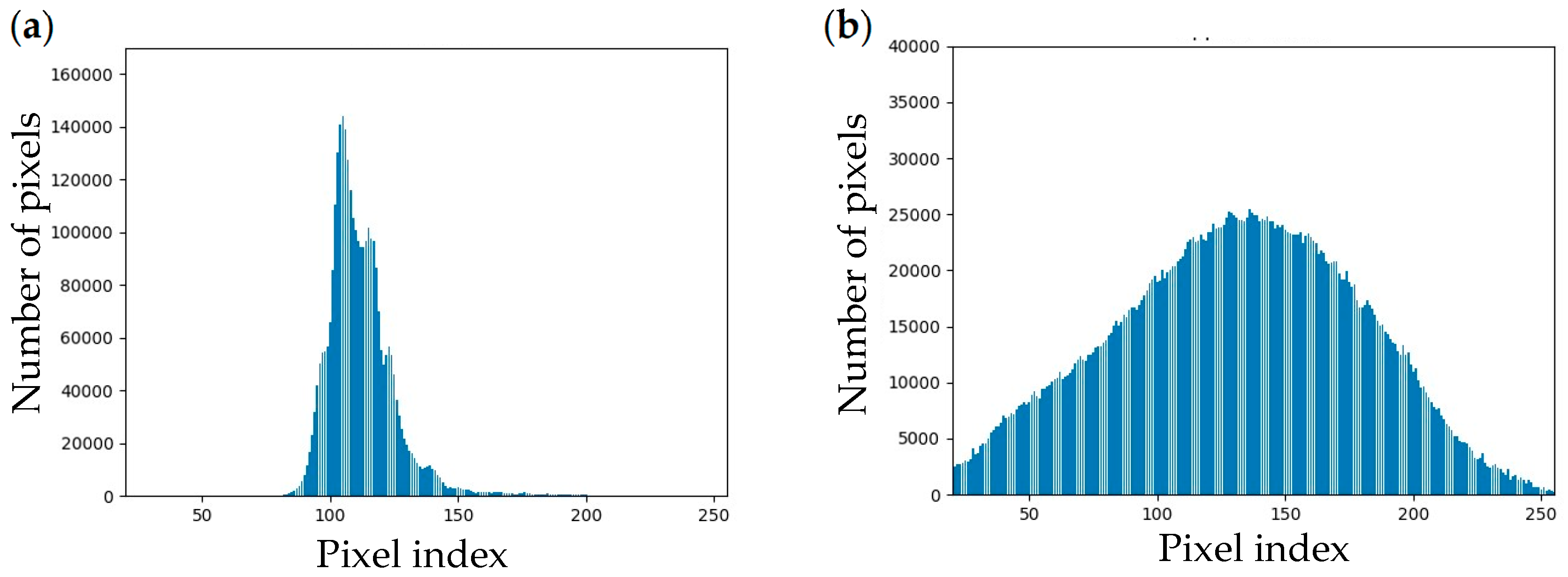
3.2.2. Contrast Limited Adaptive Histogram Equalization (CLAHE) on Green Channel
3.2.3. Image Normalization
3.2.4. Data Augmentation
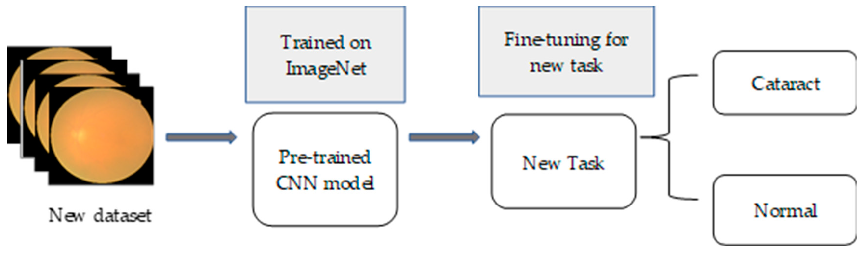
3.3. Transfer Learning with Pretrained CNN Model
3.3.1. CNN Model Selection
3.3.2. Transfer Learning Technique
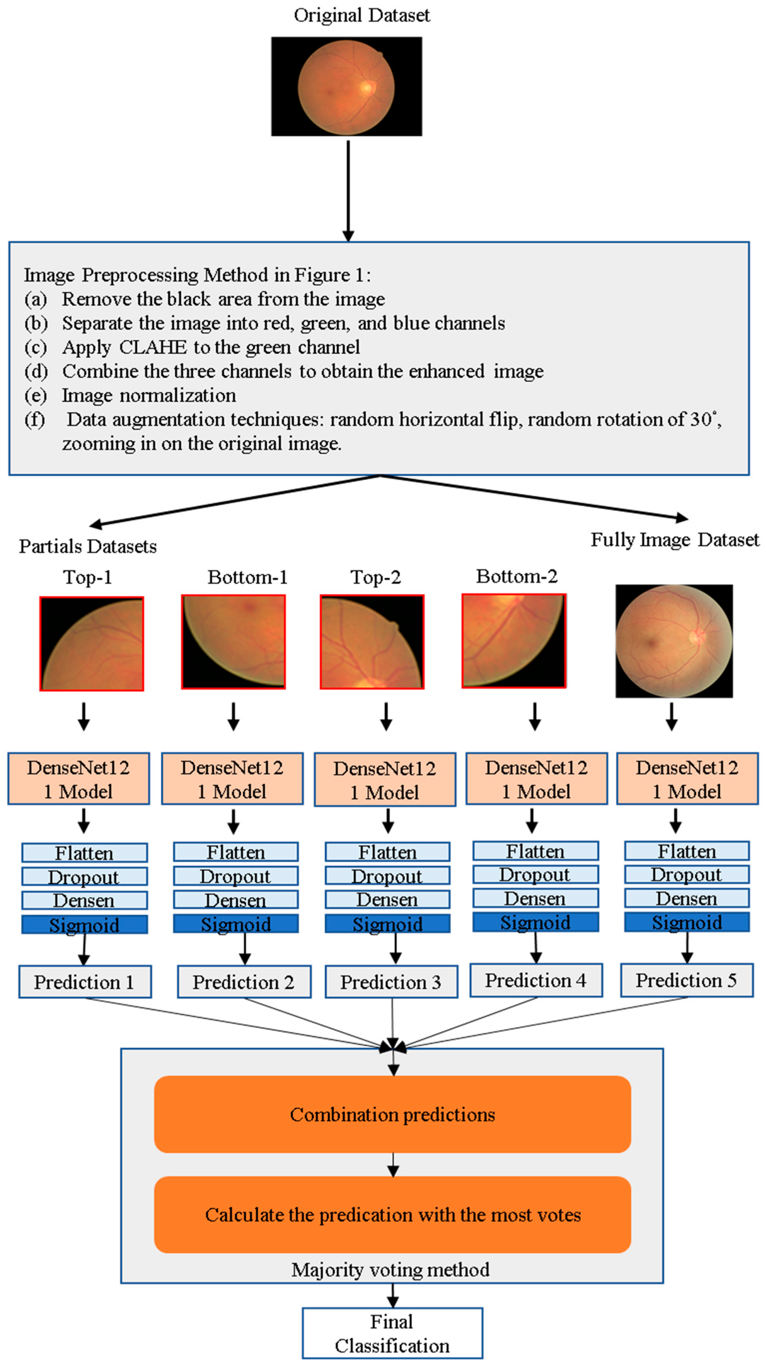
3.4. Proposed Hybrid CNN Models and Cataract Detection Application
3.4.1. Subdivide Original Image into Partials
3.4.2. Hybrid CNN Model Development
3.4.3. Environment and Hyperparameter Setting
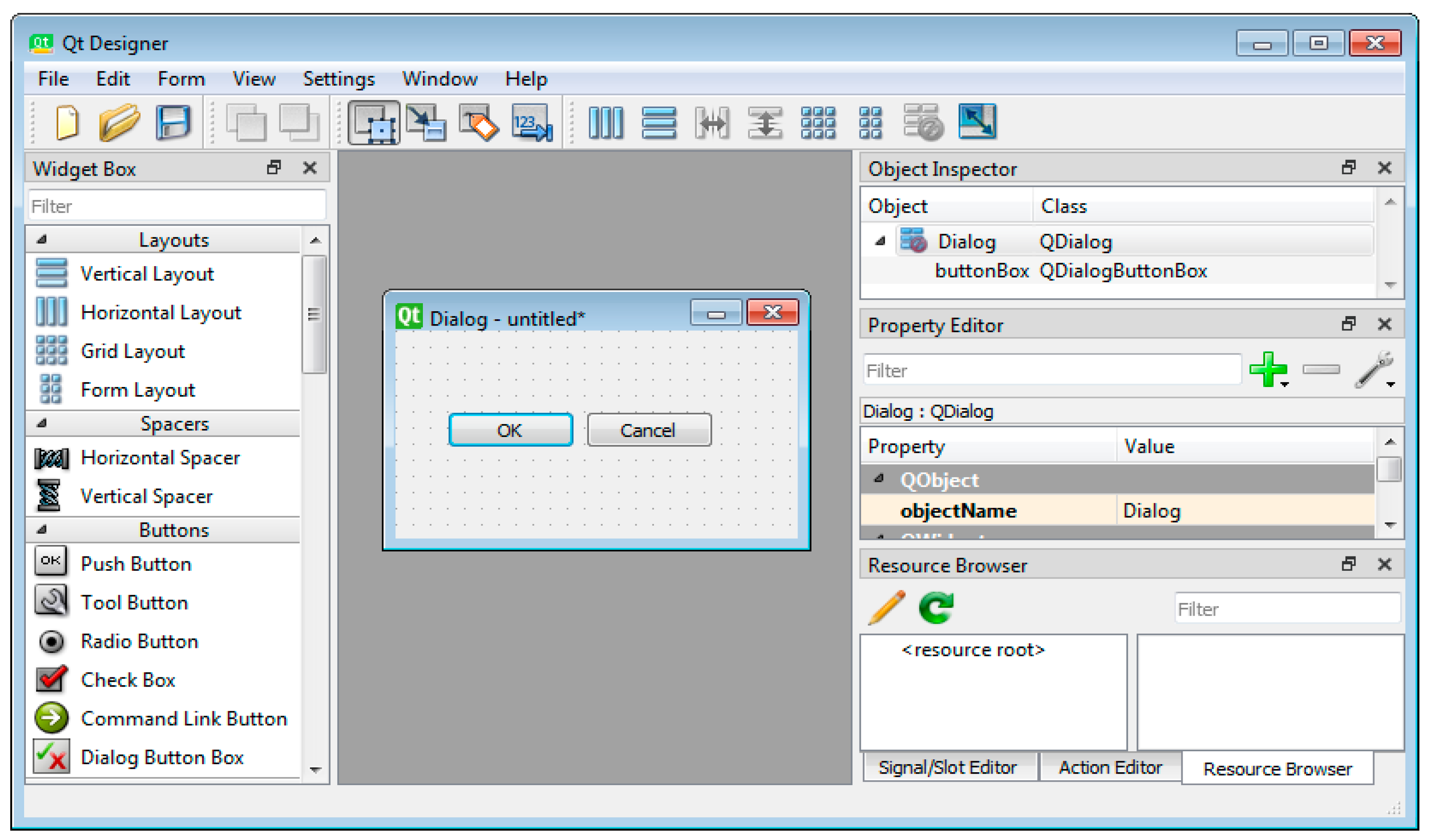
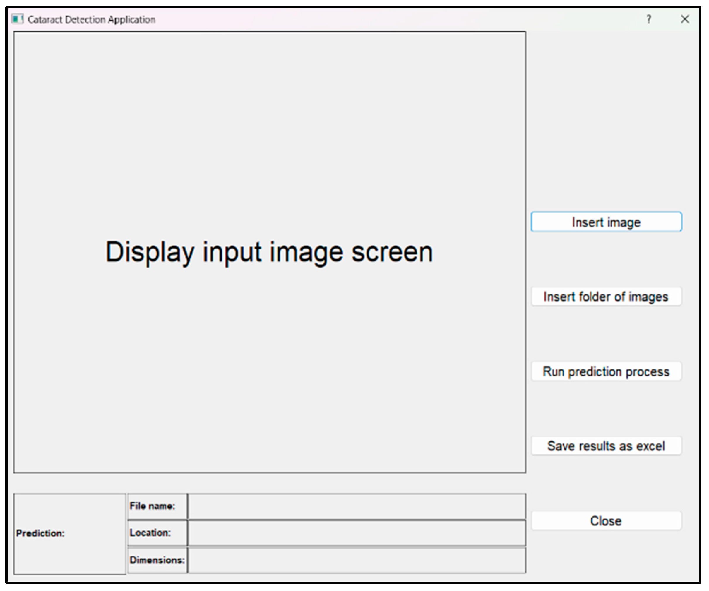
3.4.4. Cataract Detection Application
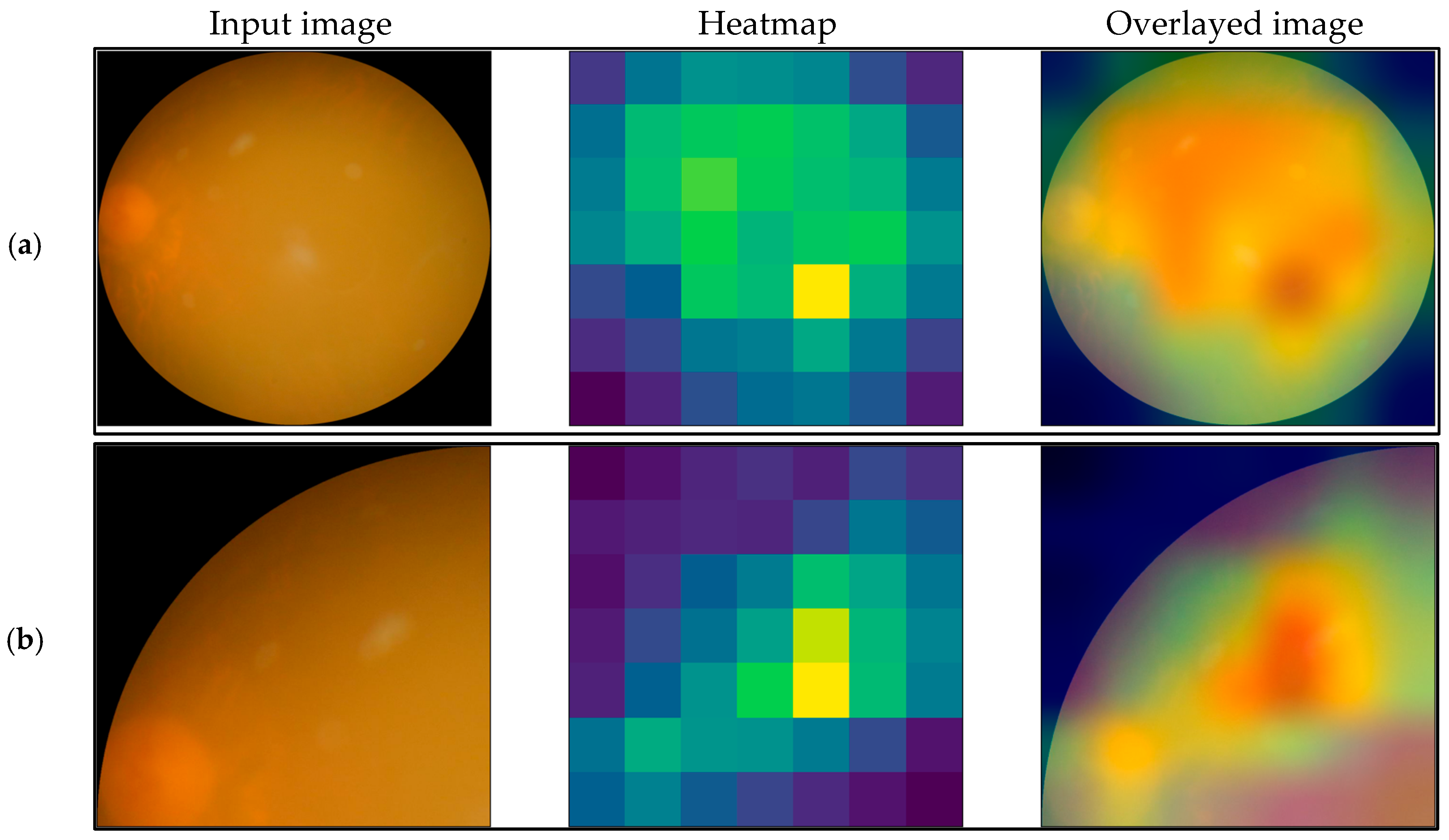
3.5. GradCAM Technique
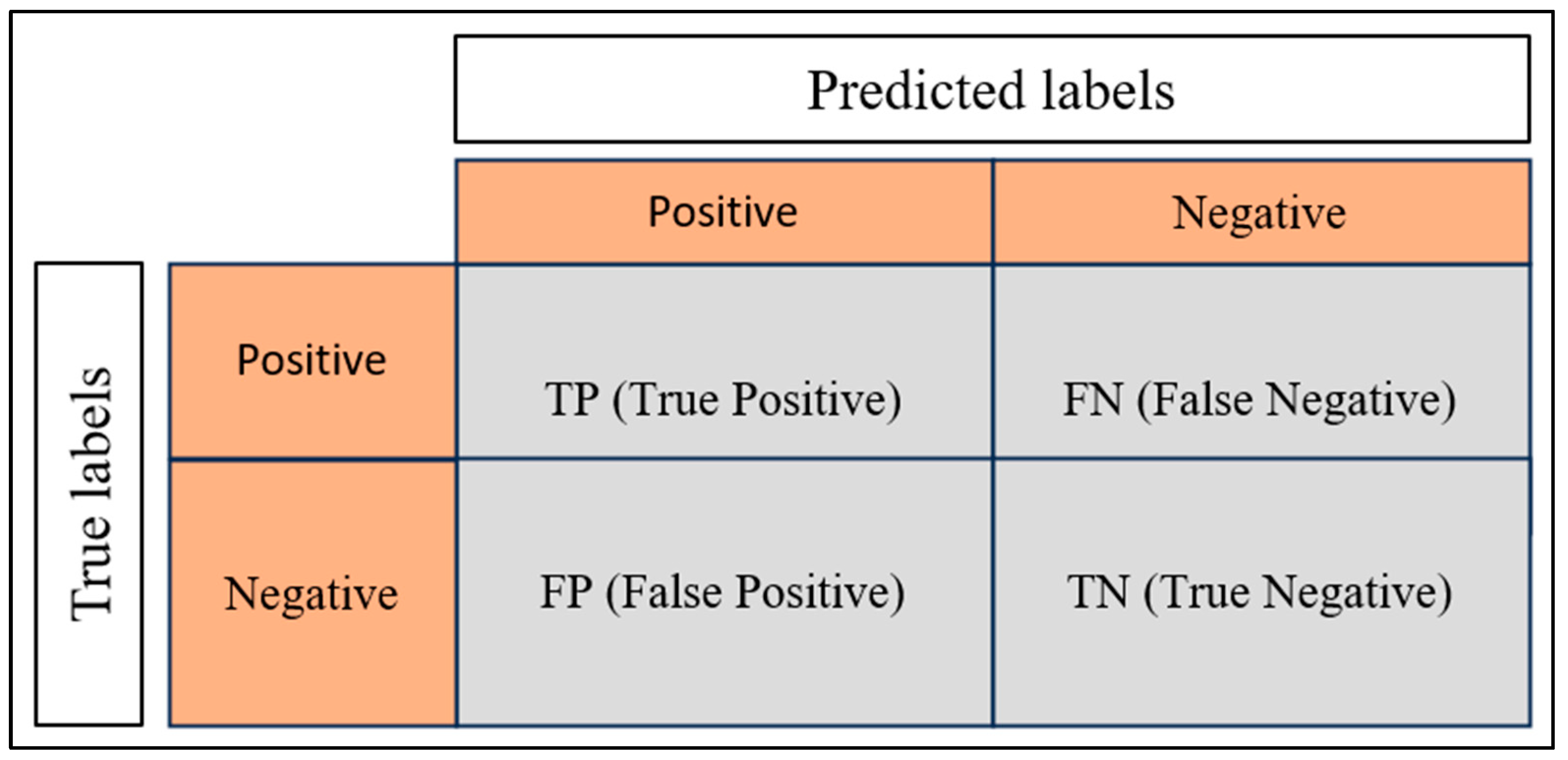
3.6. Performance Evaluation Metrics
4. Results
4.1. GradCAM Technique Consequence
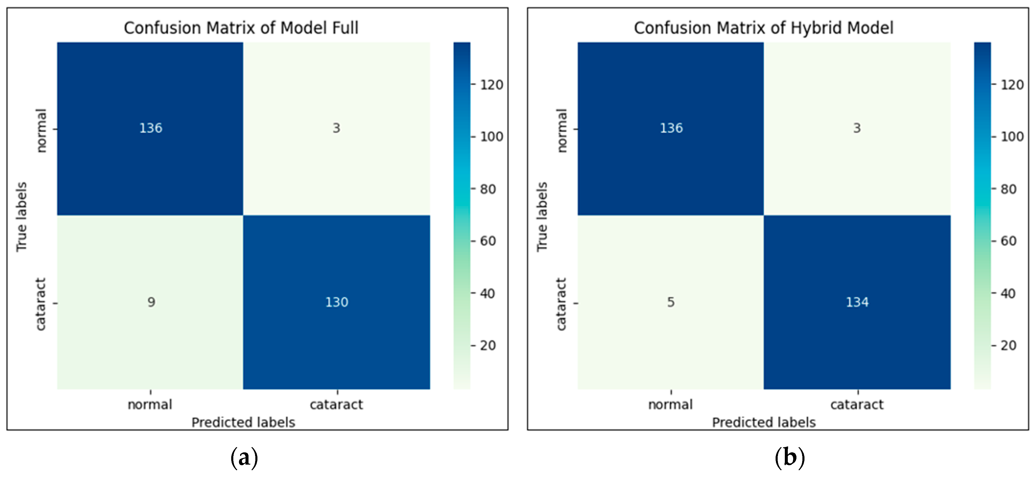
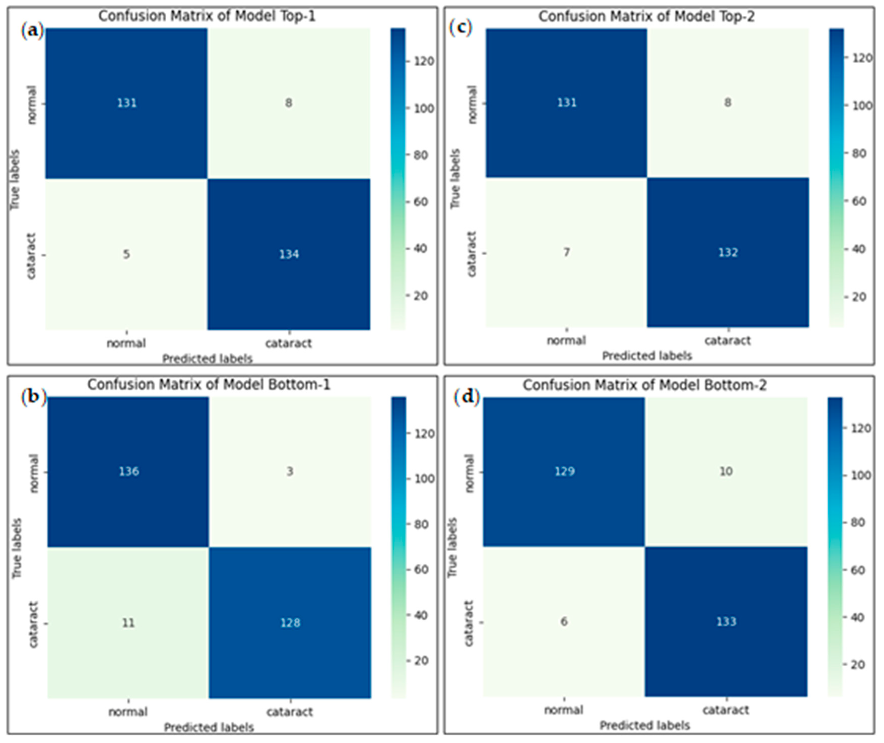
4.2. Confusion Matrix
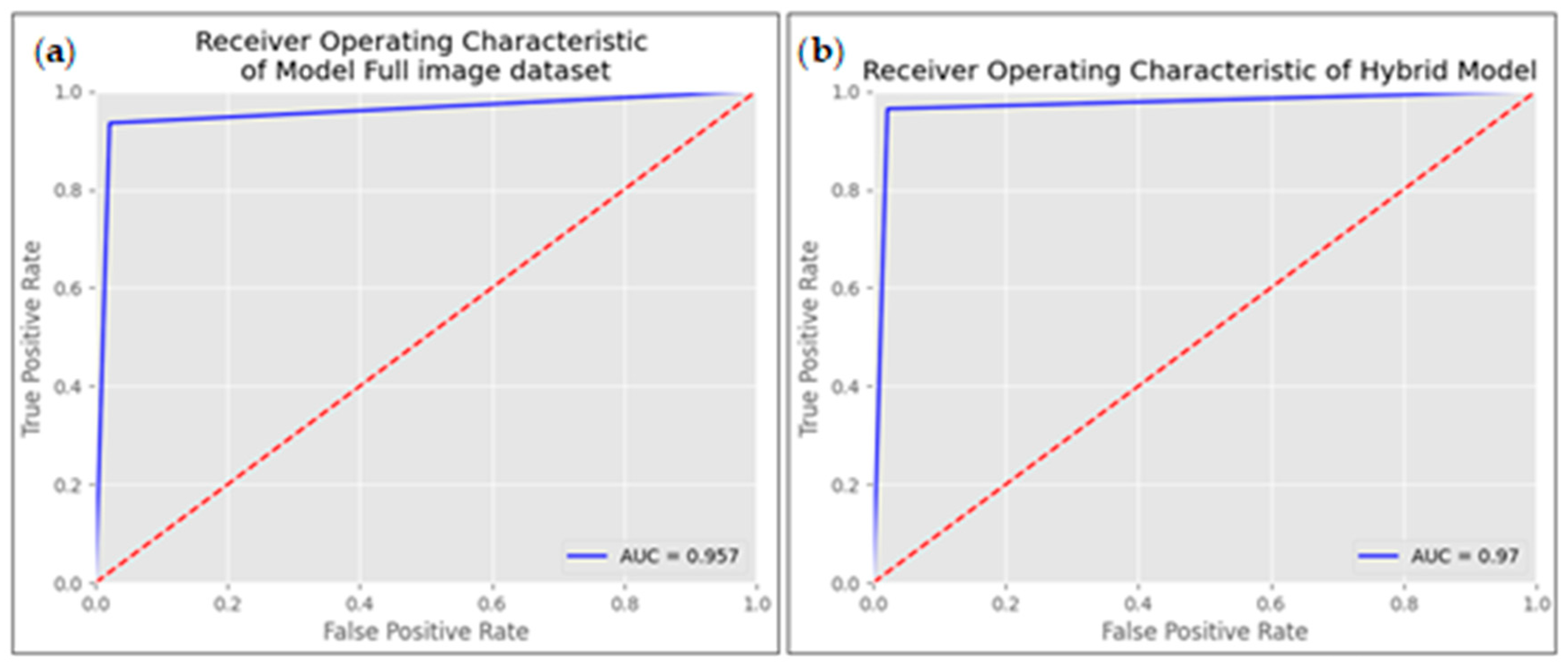
4.3. Receiver Operating Characteristic Curve (ROC Curve)
4.4. Performance Evaluation
4.5. Cataract Detection Application
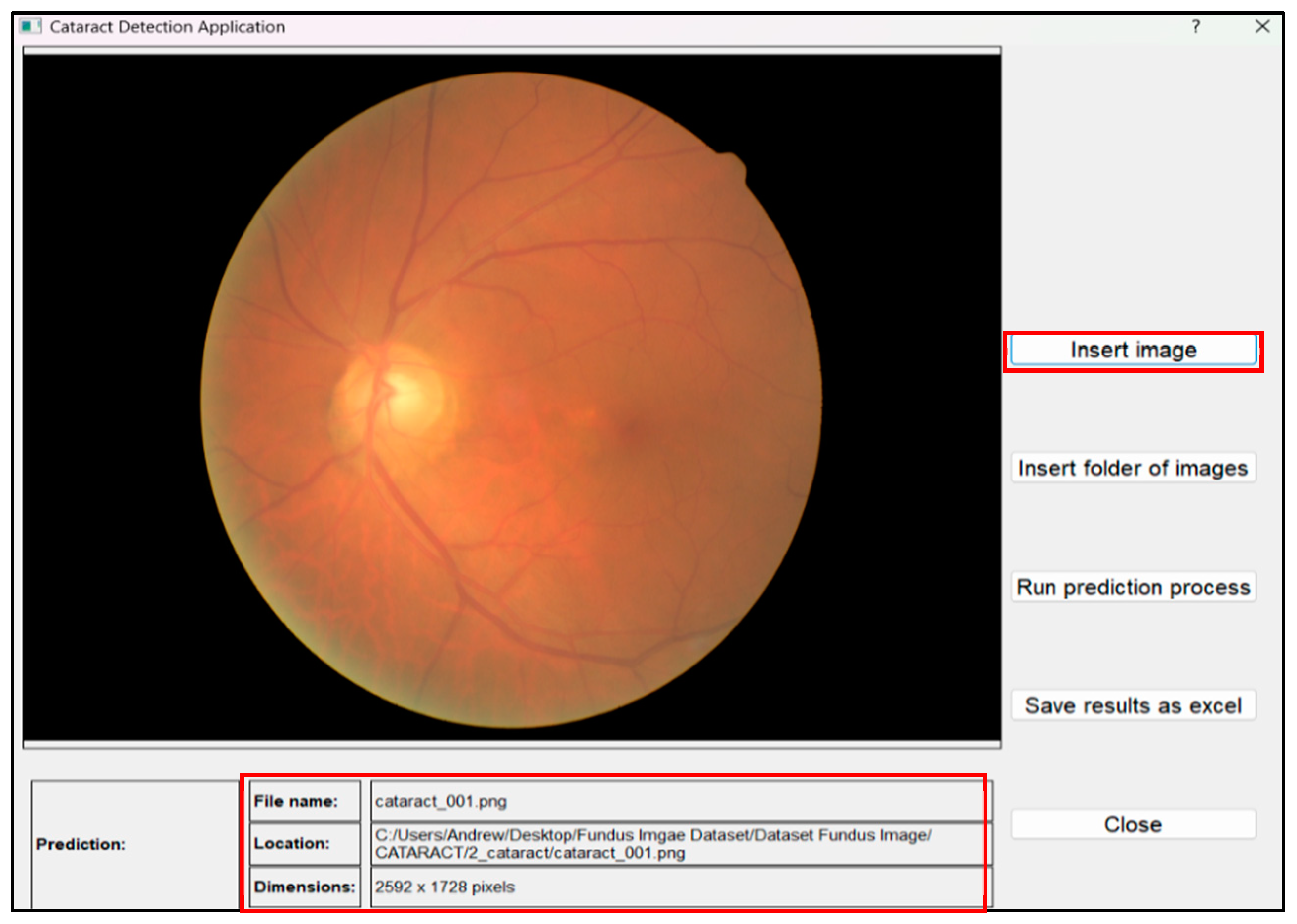
- Inserting fundus images:
- Clicking the “Insert image” button to select and insert a single fundus image.
- Click the “Insert folder of images” button if you have a folder containing multiple fundus images.
- The application will read the input fundus images and display relevant information such as the File name, Location, and Dimensions of the input image (Figure 14).
- Running the prediction process:
- Once the fundus image(s) is(are) inserted, click the [Run prediction process] button.
- The application will execute the program and send the input image to the trained CNN model.
- The program will then analyze the image to detect the presence of cataracts and display the results in the “Prediction” box (Figure 15).
- Save prediction results
- Users can also save the prediction results in an Excel file by clicking the “Save results as Excel” button.
- The application will automatically save the result, and users can choose the location of the saved Excel file.
- The Excel file (Table 9) includes important information such as the file name, prediction results, implementation time, and file location.
- This feature can be helpful for users when they need to perform a further analysis of the prediction results.
5. Conclusions
5.1. Limitations and Future Directions
5.1.1. Limited Dataset Size
5.1.2. Choice of Backbone Architecture
5.1.3. Absence of LOCS III
5.2. Future Research Directions
- Explore Alternative Backbone Architectures: Researchers have the opportunity to broaden their horizons by conducting experiments with a diverse array of backbone architectures. This should include exploration beyond DenseNet121, encompassing models such as ResNet, Inception, and EfficientNet. Comparative assessments of their performance within the constraints of a limited dataset can yield invaluable insights into the most suitable architecture for cataract detection.
- Data Augmentation and Expansion: Expanding the size and diversity of the dataset emerges as a paramount step toward improving model generalization. Techniques for data augmentation, coupled with collaboration with healthcare institutions, can facilitate this goal. Such an approach permits the evaluation of a broader spectrum of model architectures.
- Transfer Learning: Investigating the potential of transfer learning from models pretrained on larger medical image datasets, followed by fine-tuning for cataract detection, presents a promising avenue. Transfer learning leverages pre-existing knowledge and adapts it to the specific intricacies of cataract detection.
- Multi-modality Fusion: The fusion of data from diverse imaging modalities, including optical coherence tomography (OCT) and ultrasound, holds the potential to forge a comprehensive cataract diagnosis system. This approach aspires to bolster accuracy and diagnostic capabilities.
- Evaluation of Operating Efficiency: Although the current study focuses on the methodological advancements and their impact on diagnostic performance, future research should include a comprehensive evaluation of the proposed method’s operating efficiency. This would involve detailed comparisons with other state-of-the-art techniques to ensure that our approach is not only accurate but also computationally efficient and practical for real-world clinical applications.
Author Contributions
Funding
Data Availability Statement
Conflicts of Interest
References
- Mangi, M.; Bashir, M.K.; Inam, M. Outcome of Cataract Surgery at Secondary Eye Care Facility in Karachi. Pak. J. Ophthalmol. 2022, 38. [Google Scholar] [CrossRef]
- Nahar, B. Manual Small-Incision Cataract Surgery. inSIGHT 2022, 2, 7. [Google Scholar]
- Hatch, W.V.; Campbell, E.d.L.; Bell, C.M.; El-Defrawy, S.R.; Campbell, R.J. Projecting the growth of cataract surgery during the next 25 years. Arch. Ophthalmol. 2012, 130, 1479–1481. [Google Scholar] [CrossRef] [PubMed]
- Resnikoff, S.; Lansingh, V.C.; Washburn, L.; Felch, W.; Gauthier, T.-M.; Taylor, H.R.; Eckert, K.; Parke, D.; Wiedemann, P. Estimated number of ophthalmologists worldwide (International Council of Ophthalmology update): Will we meet the needs? Br. J. Ophthalmol. 2020, 104, 588–592. [Google Scholar] [CrossRef]
- Junayed, M.S.; Islam, M.B.; Sadeghzadeh, A.; Rahman, S. CataractNet: An automated cataract detection system using deep learning for fundus images. IEEE Access 2021, 9, 128799–128808. [Google Scholar] [CrossRef]
- Khan, M.S.M.; Ahmed, M.; Rasel, R.Z.; Khan, M.M. Cataract detection using convolutional neural network with VGG-19 model. In Proceedings of the 2021 IEEE World AI IoT Congress (AIIoT), Seattle, WA, USA, 10–13 May 2021; pp. 0209–0212. [Google Scholar]
- Hossain, M.R.; Afroze, S.; Siddique, N.; Hoque, M.M. Automatic detection of eye cataract using deep convolution neural networks (DCNNs). In Proceedings of the 2020 IEEE Region 10 Symposium (TENSYMP), Dhaka, Bangladesh, 5–7 June 2020; pp. 1333–1338. [Google Scholar]
- Lai, C.-J.; Pai, P.-F.; Marvin, M.; Hung, H.-H.; Wang, S.-H.; Chen, D.-N. The use of convolutional neural networks and digital camera images in cataract detection. Electronics 2022, 11, 887. [Google Scholar] [CrossRef]
- Vimala, G.A.G.; Kajamohideen, S. Diagnosis of diabetic retinopathy by extracting blood vessels and exudates using retinal color fundus images. WSEAS Trans. Biol. Biomed. 2014, 11, 20–28. [Google Scholar]
- Tsiknakis, N.; Theodoropoulos, D.; Manikis, G.; Ktistakis, E.; Boutsora, O.; Berto, A.; Scarpa, F.; Scarpa, A.; Fotiadis, D.I.; Marias, K. Deep learning for diabetic retinopathy detection and classification based on fundus images: A review. Comput. Biol. Med. 2021, 135, 104599. [Google Scholar] [CrossRef] [PubMed]
- Zhang, W.; Zhao, X.; Chen, Y.; Zhong, J.; Yi, Z. DeepUWF: An automated ultra-wide-field fundus screening system via deep learning. IEEE J. Biomed. Health Inform. 2020, 25, 2988–2996. [Google Scholar] [CrossRef]
- Szeto, S.K.-H.; Hui, V.W.K.; Siu, V.; Mohamed, S.; Chan, C.K.; Cheung, C.Y.L.; Hsieh, Y.T.; Tan, C.S.; Chhablani, J.; Lai, T.Y. Recent advances in clinical applications of imaging in retinal diseases. Asia-Pac. J. Ophthalmol. 2023, 12, 252–263. [Google Scholar] [CrossRef]
- Li, T.; Bo, W.; Hu, C.; Kang, H.; Liu, H.; Wang, K.; Fu, H. Applications of deep learning in fundus images: A review. Med. Image Anal. 2021, 69, 101971. [Google Scholar] [CrossRef] [PubMed]
- Nguyen, V.-V.; Lin, C.-L. Cataract Detection using Hybrid CNN Model on Retinal Fundus Images. In Proceedings of the 2023 9th International Conference on Applied System Innovation (ICASI), Chiba, Japan, 21–25 April 2023; pp. 42–44. [Google Scholar]
- Zhang, L.; Li, J.; Han, H.; Liu, B.; Yang, J.; Wang, Q. Automatic cataract detection and grading using deep convolutional neural network. In Proceedings of the 2017 IEEE 14th International Conference on Networking, Sensing and Control (ICNSC), Calabria, Italy, 16–18 May 2017; pp. 60–65. [Google Scholar]
- Faizal, S.; Rajput, C.A.; Tripathi, R.; Verma, B.; Prusty, M.R.; Korade, S.S. Automated cataract disease detection on anterior segment eye images using adaptive thresholding and fine tuned inception-v3 model. Biomed. Signal Process. Control 2023, 82, 104550. [Google Scholar] [CrossRef]
- Qiao, Z.; Zhang, Q.; Dong, Y.; Yang, J.-J. Application of SVM based on genetic algorithm in classification of cataract fundus images. In Proceedings of the 2017 IEEE International Conference on Imaging Systems and Techniques (IST), Beijing, China, 18–20 October 2017; pp. 1–5. [Google Scholar]
- Binus, A. Automatic Cataract Detection System based on Support Vector Machine (SVM). In Proceedings of the Second Asia Pacific International Conference on Industrial Engineering and Operations Management, Surakarta, Indonesia, 14–16 September 2021. [Google Scholar]
- Chande, K.; Jha, P.; Aulakh, K.K.; Shinde, S. Cataract detection using textural features and machine learning algorithms. In Proceedings of the 2nd International Conference on Artificial Intelligence: Advances and Applications (ICAIAA 2021), Hyderabad, India, 15–17 April 2021; pp. 595–606. [Google Scholar]
- Maaliw, R.R.; Alon, A.S.; Lagman, A.C.; Garcia, M.B.; Abante, M.V.; Belleza, R.C.; Tan, J.B.; Maaño, R.A. Cataract detection and grading using ensemble neural networks and transfer learning. In Proceedings of the 2022 IEEE 13th Annual Information Technology, Electronics and Mobile Communication Conference (IEMCON), Vancouver, BC, Canada, 12–15 October 2022; pp. 0074–0081. [Google Scholar]
- Ishtiaq, U.; Abdullah, E.R.M.F.; Ishtiaque, Z. A Hybrid Technique for Diabetic Retinopathy Detection Based on Ensemble-Optimized CNN and Texture Features. Diagnostics 2023, 13, 1816. [Google Scholar] [CrossRef] [PubMed]
- Sevani, N.; Tampubolon, H.; Wijaya, J.; Cuvianto, L.; Salomo, A. A Study of Convolution Neural Network Based Cataract Detection with Image Segmentation. In Proceedings of the 2022 IEEE International Conference on Communication, Networks and Satellite (COMNETSAT), Solo, Indonesia, 3–5 November 2022; pp. 216–221. [Google Scholar]
- Deshmukh, S.V.; Roy, A. Retinal Blood Vessel Segmentation Based on Modified CNN and Analyze the Perceptional Quality of Segmented Images. In Proceedings of the International Conference on Advanced Network Technologies and Intelligent Computing, Varanasi, India, 22–24 December 2022; pp. 609–625. [Google Scholar]
- Yadav, S.; Singh Yadav, J.K.P. Enhancing Cataract Detection Precision: A Deep Learning Approach. Trait. Signal 2023, 40, 1413–1424. [Google Scholar] [CrossRef]
- Choi, E.Y.; Han, S.H.; Ryu, I.H.; Kim, J.K.; Lee, I.S.; Han, E.; Kim, H.; Choi, J.Y.; Yoo, T.K. Automated detection of crystalline retinopathy via fundus photography using multistage generative adversarial networks. Biocybern. Biomed. Eng. 2023, 43, 725–735. [Google Scholar] [CrossRef]
- Sahu, S.; Singh, A.K.; Ghrera, S.; Elhoseny, M. An approach for de-noising and contrast enhancement of retinal fundus image using CLAHE. Opt. Laser Technol. 2019, 110, 87–98. [Google Scholar]
- Liang, Y.; He, L.; Fan, C.; Wang, F.; Li, W. Preprocessing study of retinal image based on component extraction. In Proceedings of the 2008 IEEE International Symposium on IT in Medicine and Education, Xiamen, China, 12–14 December 2008; pp. 670–672. [Google Scholar]
- Setiawan, A.W.; Mengko, T.R.; Santoso, O.S.; Suksmono, A.B. Color retinal image enhancement using CLAHE. In Proceedings of the International Conference on ICT for Smart Society, Jakarta, Indonesia, 13–14 June 2013; pp. 1–3. [Google Scholar]
- Alwazzan, M.J.; Ismael, M.A.; Ahmed, A.N. A hybrid algorithm to enhance colour retinal fundus images using a Wiener filter and CLAHE. J. Digit. Imaging 2021, 34, 750–759. [Google Scholar] [CrossRef] [PubMed]
- Shorten, C.; Khoshgoftaar, T.M. A survey on image data augmentation for deep learning. J. Big Data 2019, 6, 1–48. [Google Scholar] [CrossRef]
- Team, K. Keras Documentation: Keras Applications. Available online: https://www.kerasio/api/applications (accessed on 15 May 2023).
- Huang, G.; Liu, Z.; Van Der Maaten, L.; Weinberger, K.Q. Densely connected convolutional networks. In Proceedings of the IEEE Conference on Computer Vision and Pattern Recognition, Honolulu, HI, USA, 21–26 July 2017; pp. 4700–4708. [Google Scholar]
- Xu, X.; Zhang, L.; Li, J.; Guan, Y.; Zhang, L. A hybrid global-local representation CNN model for automatic cataract grading. IEEE J. Biomed. Health Inform. 2019, 24, 556–567. [Google Scholar] [CrossRef] [PubMed]
- Pang, B.; Nijkamp, E.; Wu, Y.N. Deep learning with tensorflow: A review. J. Educ. Behav. Stat. 2020, 45, 227–248. [Google Scholar] [CrossRef]
- Willman, J.M. Creating GUIs with Qt Designer. In Beginning PyQt: A Hands-on Approach to GUI Programming with PyQt6; Springer: Berlin, Germany, 2022; pp. 217–258. [Google Scholar]
- Selvaraju, R.R.; Cogswell, M.; Das, A.; Vedantam, R.; Parikh, D.; Batra, D. Grad-cam: Visual explanations from deep networks via gradient-based localization. In Proceedings of the IEEE International Conference on Computer Vision, Venice, Italy, 22–29 October 2017; pp. 618–626. [Google Scholar]
- Chakraborty, S.; Jana, S. Early Prediction of Cataract using Convolutional Neural Network. In Proceedings of the 2023 IEEE Devices for Integrated Circuit (DevIC), Kalyani, India, 7–8 April 2023; pp. 446–450. [Google Scholar]
- Pratap, T.; Kokil, P. Computer-aided diagnosis of cataract using deep transfer learning. Biomed. Signal Process. Control 2019, 53, 101533. [Google Scholar] [CrossRef]


| Dataset | Normal | Cataract |
|---|---|---|
| Dataset 1 | 2002 | 594 |
| Dataset 2 | 300 | 100 |
| Total | 2302 | 694 |
| Condition | Number of Images | Total Number of Images | Dataset | Number of Images |
|---|---|---|---|---|
| Normal fundus images | 694 | 1388 | Training dataset | 888 |
| Validation dataset | 222 | |||
| Cataract fundus images | 694 | |||
| Testing dataset | 278 |
| Original Training Dataset Size | Augmented Training Dataset Size |
|---|---|
| 888 | 2664 |
| Hyperparameter | Learning Rate | Dropout | Batch Size | Epoch | Patience |
|---|---|---|---|---|---|
| Value | 5 × 10⁻⁵ | 0.5 | 32 | 50 | 15 |
| Data | Performance | Model 1 | Model Top 1 | Model Bottom 1 | Model Top 2 | Model Bottom 2 |
|---|---|---|---|---|---|---|
| Training | Accuracy | 0.9555 | 0.9760 | 0.9675 | 0.9724 | 0.9555 |
| Loss | 0.1542 | 0.0837 | 0.1532 | 0.1310 | 0.1642 | |
| Validation | Accuracy | 0.9496 | 0.9460 | 0.9532 | 0.9424 | 94.60 |
| Loss | 0.1496 | 0.2527 | 0.2089 | 0.1710 | 0.1816 | |
| Testing | Accuracy | 0.9568 | 0.9532 | 0.9496 | 0.9460 | 0.9424 |
| Loss | 0.1030 | 0.2209 | 0.1551 | 0.1877 | 0.1473 |
| Methods | Accuracy (%) |
|---|---|
| Hybrid model (majority voting) | 97.12 |
| Model 1 (full image dataset) | 95.68 |
| Model Top 1 | 95.32 |
| Model Bottom 1 | 94.96 |
| Model Top 2 | 94.60 |
| Model Bottom 2 | 94.24 |
| Methods | Condition | Precision | Recall | F1 Score |
|---|---|---|---|---|
| Model 1 | Cataract | 0.94 | 0.98 | 0.96 |
| Normal | 0.98 | 0.94 | 0.96 | |
| Hybrid Model | Cataract | 0.96 | 0.98 | 0.97 |
| Normal | 0.98 | 0.96 | 0.97 |
| Methods | Year | Dataset Size | Model | Accuracy |
|---|---|---|---|---|
| Hybrid Model | 2024 | 1388 | DenseNet121 | 0.9712 |
| Model 1 | 2023 | 1388 | DenseNet121 | 0.9568 |
| Pratap and Koki [38] | 2019 | 800 | AlexNet and SVM | 0.9291 |
| Hossain et al. [7] | 2020 | 5718 | ResNet-50 | 0.9577 |
| Chakraborty and Jana [37] | 2023 | 1386 | VGG19 | 0.9248 * |
| Junayed et al. [5] | 2021 | 4746 | CataractNet | 0.9913 |
| File Name | Prediction Results | Implementation Time | File Location |
|---|---|---|---|
| 220_left.jpg | Normal | 2023-05-25 15:37:38 | C:/Users/Andrew/Desktop/Fundus Imgae Dataset/Dataset Fundus Image/test_img/220_left.jpg |
| 240_left.jpg | Normal | 2023-05-25 15:37:42 | C:/Users/Andrew/Desktop/Fundus Imgae Dataset/Dataset Fundus Image/test_img/240_left.jpg |
| 260_left.jpg | Cataract | 2023-05-25 15:37:46 | C:/Users/Andrew/Desktop/Fundus Imgae Dataset/Dataset Fundus Image/test_img/260_left.jpg |
| 303_right.jpg | Normal | 2023-05-25 15:37:50 | C:/Users/Andrew/Desktop/Fundus Imgae Dataset/Dataset Fundus Image/test_img/303 _right.jpg |
| 305_right.jpg | Cataract | 2023-05-25 15:37:54 | C:/Users/Andrew/Desktop/Fundus Imgae Dataset/Dataset Fundus Image/test _img/305_right.jpg |
| 330_left.jpg | Cataract | 2023-05-25 15:37:54 | C:/Users/Andrew/Desktop/Fundus Imgae Dataset/Dataset Fundus Image/test_img/330_left.jpg |
| 368_right.jpg | Cataract | 2023-05-25 15:37:58 | C:/Users/Andrew/Desktop/Fundus Imgae Dataset/Dataset Fundus Image/test_img/368_right.jpg |
| 392_left.jpg | Normal | 2023-05-25 15:38:02 | C:/Users/Andrew/Desktop/Fundus Imgae Dataset/Dataset Fundus Image/test_img/392_left.jpg |
| crop_img.jpg | Normal | 2023-05-25 15:38:10 | C:/Users/Andrew/Desktop/Fundus Imgae Dataset/Dataset Fundus Image/test_img/crop_img.jpg |
| NL_012.png | Cataract | 2023-05-25 15:38:13 | C:/Users/Andrew/Desktop/Fundus Imgae Dataset/Dataset Fundus Image/test_img/NL_012.png |
| NL_020.png | Cataract | 2023-05-25 15:38:17 | C:/Users/Andrew/Desktop/Fundus Imgae Dataset/Dataset Fundus Image/test img/NL_020.png |
Disclaimer/Publisher’s Note: The statements, opinions and data contained in all publications are solely those of the individual author(s) and contributor(s) and not of MDPI and/or the editor(s). MDPI and/or the editor(s) disclaim responsibility for any injury to people or property resulting from any ideas, methods, instructions or products referred to in the content. |
© 2024 by the authors. Licensee MDPI, Basel, Switzerland. This article is an open access article distributed under the terms and conditions of the Creative Commons Attribution (CC BY) license (https://creativecommons.org/licenses/by/4.0/).
Share and Cite
Nguyen, V.-V.; Lin, C.-L. Enhancing Cataract Detection through Hybrid CNN Approach and Image Quadration: A Solution for Precise Diagnosis and Improved Patient Care. Electronics 2024, 13, 2344. https://doi.org/10.3390/electronics13122344
Nguyen V-V, Lin C-L. Enhancing Cataract Detection through Hybrid CNN Approach and Image Quadration: A Solution for Precise Diagnosis and Improved Patient Care. Electronics. 2024; 13(12):2344. https://doi.org/10.3390/electronics13122344
Chicago/Turabian StyleNguyen, Van-Viet, and Chun-Ling Lin. 2024. "Enhancing Cataract Detection through Hybrid CNN Approach and Image Quadration: A Solution for Precise Diagnosis and Improved Patient Care" Electronics 13, no. 12: 2344. https://doi.org/10.3390/electronics13122344
APA StyleNguyen, V.-V., & Lin, C.-L. (2024). Enhancing Cataract Detection through Hybrid CNN Approach and Image Quadration: A Solution for Precise Diagnosis and Improved Patient Care. Electronics, 13(12), 2344. https://doi.org/10.3390/electronics13122344