Review

Motion Tracking Algorithms Based on Wearable Inertial Sensor: A Focus on Shoulder

by
Umile Giuseppe Longo
1,2,*,
Sergio De Salvatore
1,2,
Martina Sassi
1,2,
Arianna Carnevale
1,2,3,
Giovanna De Luca
1,2 and
Vincenzo Denaro
1,2
1
Operative Research Unit of Trauma and Orthopaedic Surgery, Fondazione Policlinico Universitario Campus Bio-Medico, Via Alvaro del Portillo 200, 00128 Rome, Italy
2
Research Unit of Trauma and Orthopaedic Surgery, Department of Medicine and Surgery, Università Campus Bio-Medico di Roma, Via Alvaro del Portillo, 00128 Rome, Italy
3
Laboratory of Measurement and Biomedical Instrumentation, Campus Bio-Medico University, Via Alvaro del Portillo 200, 00128 Rome, Italy
*
Author to whom correspondence should be addressed.
Electronics 2022, 11(11), 1741; https://doi.org/10.3390/electronics11111741
Submission received: 19 April 2022 / Revised: 18 May 2022 / Accepted: 27 May 2022 / Published: 30 May 2022
(This article belongs to the Special Issue New Technologies for Biomedical Circuits and Systems)

Abstract

:
Shoulder Range of Motion (ROM) has been studied with several devices and methods in recent years. Accurate tracking and assessment of shoulder movements could help us to understand the pathogenetic mechanism of specific conditions in quantifying the improvements after rehabilitation. The assessment methods can be classified as subjective and objective. However, self-reported methods are not accurate, and they do not allow the collection of specific information. Therefore, developing measurement devices that provide quantitative and objective data on shoulder function and range of motion is important. A comprehensive search of PubMed and IEEE Xplore was conducted. The sensor fusion algorithm used to analyze shoulder kinematics was described in all studies involving wearable inertial sensors. Eleven articles were included. The Quality Assessment of Diagnostic Accuracy Studies-2 was used to assess the risk of bias (QUADAS-2). The finding showed that the Kalman filter and its variants UKF and EKF are used in the majority of studies. Alternatives based on complementary filters and gradient descent algorithms have been reported as being more computationally efficient. Many approaches and algorithms have been developed to solve this problem. It is useful to fuse data from different sensors to obtain a more accurate estimation of the 3D position and 3D orientation of a body segment. The sensor fusion technique makes this integration reliable. This systematic review aims to redact an overview of the literature on the sensor fusion algorithms used for shoulder motion tracking.

1. Introduction

The shoulder joint has three degrees of freedom (DOFs) and is the human joint with the greatest range of motion (ROM) [1,2]. Despite the shoulder’s high degree of freedom, shoulder movements are hard to analyze [3]. This appears to be due to two factors. Firstly, the shoulder joint is controlled by a complicated system of joints, including the shoulder girdle closed chain and its peculiar ‘false scapulothoracic joint’ [4]. Furthermore, the biomechanical complexity of the shoulder joints is related to the musculature that regulates the movements. Since determining the mobility of the shoulder complex is challenging, the scapula is provided with a lot of degrees of freedom depending on the study framework. Recent studies have essentially avoided the issue of shoulder complex mobility estimation methods by kinematically identifying the scapula as an independent object, firmly oriented to the thorax due to three Euler angles [5,6,7], or by assessing an empirical model of the scapula, such as that provided by the recent Stanford University model [8].
In the literature, various kinematic models of the shoulder have been proposed [9,10].
Shoulder ROM has been studied with several devices and methods [11,12,13]. Accurate tracking and assessment of shoulder movements could help us to understand the pathogenetic mechanism of specific conditions or quantifying the improvements after rehabilitation. The assessment methods can be classified as subjective (self-reports or self-evaluation questionnaires) and objective. However, self-reported methods are not accurate, and they do not allow the collection of specific information. Therefore, finding measurement systems that provide quantitative and objective information about shoulder function and ROM is necessary.
Objective systems can be divided into two main classes of motion capture technologies: non-wearable and wearable systems [14]. The former is the most common and includes stereophotogrammetric systems and optoelectronic systems (e.g., VICON, Optotrak, BTS SMART-D), electromagnetic tracking systems (e.g., Fastrak) and ultrasound-based motion analysis systems (e.g., Zebris), nowadays considered as the gold standard [14].
Wearable systems comprise inertial measurement units (IMU) that are composed of an accelerometer and a gyroscope and, in the case of magnetic inertial measurement unit (MIMU), also a magnetometer [12]. These sensors’ measured quantities are acceleration, angular velocity, and magnetic field intensity. Traditionally, the orientation of a segment has been estimated by integrating the angular velocities measured, but this implicates inaccuracies inherent in the measurements.
Many approaches and algorithms have been developed to solve this problem. It is useful to fuse data from different sensors to obtain a more accurate estimation of the 3D position and 3D orientation of a body segment. The sensor fusion technique makes this integration reliable.
This systematic review aims to redact an overview of the literature on the sensor fusion algorithms used for shoulder motion tracking.

2. Materials and Methods

To improve the review’s reporting, the Preferred Reporting Items for Systematic Reviews and Meta-Analyses (PRISMA) were employed.

2.1. Eligibility Criteria

The research question was formulated using the PICOS approach: Patient (P); Intervention (I); Comparator (C); Outcome (O), and Study design (S).
Population: patients with or without shoulder limitations.
Systems and devices to quantify shoulder joint kinematic (O) that used wearable IMU or MIMU sensors (I) were used in patients with or without shoulder limitations (P). All the results were compared to stereophotogrammetric systems to provide information properties of the motion tracking tool and related algorithms. Study design: a validation study, case reports, or clinical trials evaluating 3D motion tracking of the shoulder.

2.1.1. Study Inclusion Criteria

  • Only articles written in English.
  • Articles published in peer-reviewed journals or presented at a conference.
  • Wearable magnetic/inertial sensors to track upper limb kinematics, including the shoulder joint.
  • Studies that developed an algorithm to extract shoulder joint variables of clinical relevance.

2.1.2. Study Exclusion Criteria

  • Reviews, books, cadaver studies;
  • Studies in which the shoulder joint was not included;
  • Patients who underwent previous surgery (hemiarthroplasty, total joint arthroplasty, rotator cuff repair);
  • Patients with neurological pathologies (e.g., stroke);
  • Use of prosthesis, orthoses, exoskeleton or robotic devices;
  • Studies where the analysis was done with a combination of inertial sensors and other types of sensors.

2.2. Search Strategy

The articles included in the study were screened through searches of PubMed and IEEE Xplore databases. The database was screened from its inception to April 2022. The search strategy included free text terms and Mesh (Medical Subject Headings) terms combined with logical Boolean operators (AND, OR). For the research in the databases, isolated or combined keywords and specialized terms were used. The keywords and their synonyms used were: (“shoulder joint*” OR shoulder) AND (“data fusion algorithm*” OR “sensor fusion algorithm”) AND (motion OR movement* OR kinematic*) AND (“wearable sensor* OR “inertial sensor*”).

2.3. Study Selection

Only English-language articles were considered. Two reviewers conducted the initial search (MS and GDL). In case of a disagreement, a third reviewer (UGL) was consulted. CADIMA software was used to conduct the study. The researchers assessed the titles and abstracts, and the full text. The full text of all papers that were not excluded because of their abstract or title was then evaluated. A PRISMA flowchart was used to track the number of articles that were excluded or included (Figure 1). For designing the PRISMA, the rules by Liberati et al. [15] were followed.

2.4. Quality Assessment

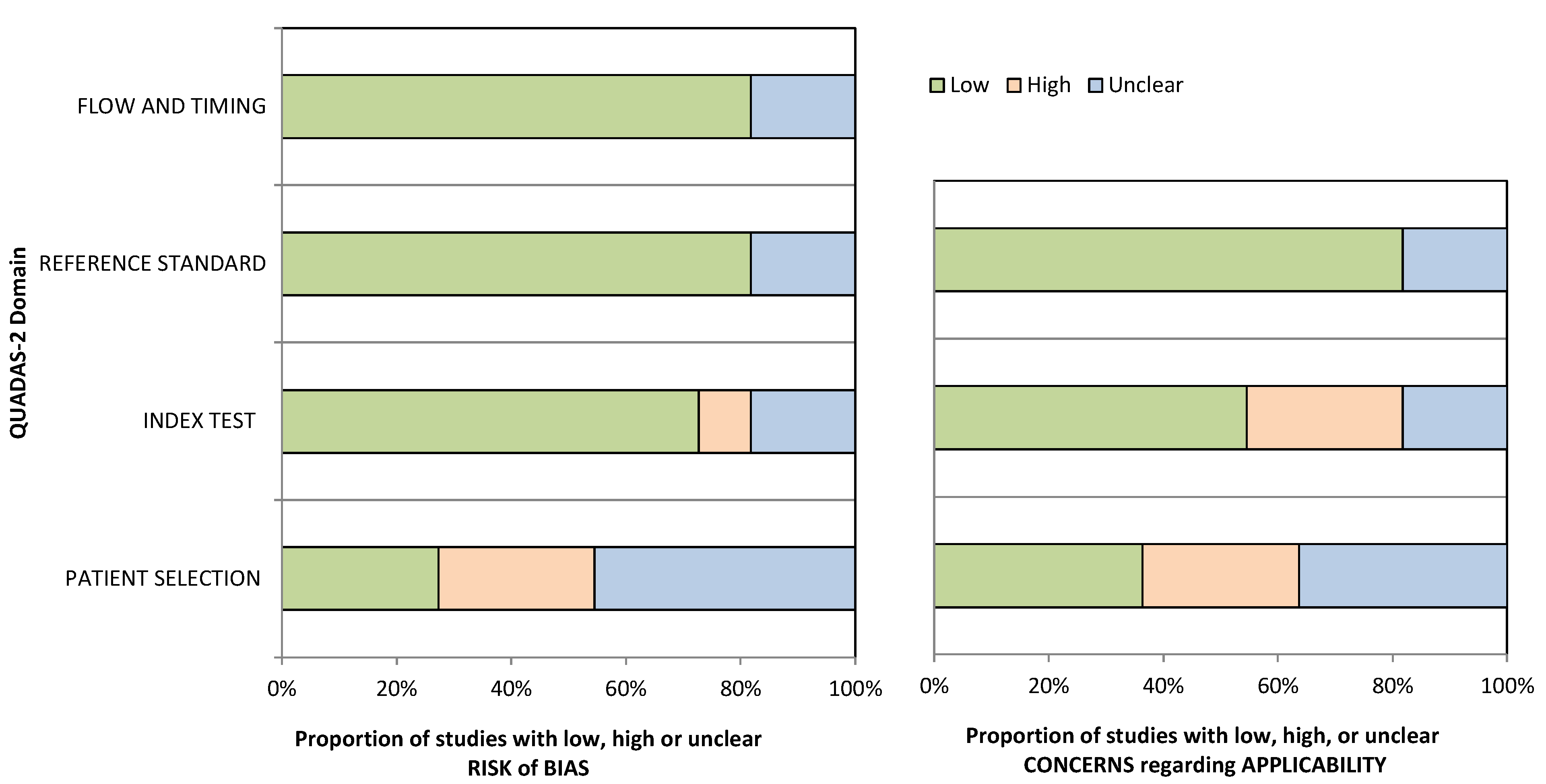
The quality of the study included was assessed by two authors using the quality assessment of Diagnostic Accuracy Studies-2 (QUADAS-2) [16]. QUADAS-2 is a tool developed to assess the quality of diagnostic studies testing the “risk of bias” and the “applicability”. The risk of bias was checked by testing four domains: “patient selection”, “index test”, “reference standard”, and “flow and timing”. Each study’s applicability was graded as “yes, no, or unclear” for the first three categories; “yes” suggested a low risk of bias, “no” signaled a high risk of bias, and “unclear” indicated a lack of sufficient data. Two reviewers handled the domains independently, and a third author resolved any disagreements. The risk of bias is reported in Figure 2.

2.5. Data Synthesis and Analysis

General study characteristics extracted were: author and year of publication; typology and number of sensors; the position of the sensors used to measure or track the joint; upper limb kinematic representation (shoulder DOFs); sensor fusion algorithm; comparator system used to evaluate the performances; movements executed by the participants; shoulder ROM and accuracy. Only qualitative characteristics were reported due to the heterogeneity of the studies. A meta-analysis could not be performed due to the heterogeneity of the included studies.

3. Results

3.1. Study Selection

A flowchart diagram according to the PRISMA protocol was created, and it shows the selection process of the studies (Figure 1).
A total of 213 results were founded with the search on the scientific databases. After the removal of duplicates, 207 were included in the analysis. Of 207 studies, 142 articles were excluded from the study through title and abstract screening. Then, 65 full-text articles were screened. Out of these studies, 53 were excluded, and only 11 studies fulfilled the inclusion criteria. The selection study process is reported in Figure 1.

3.2. Study Characteristics and Risk of Bias Assessment

A summary of the characteristics of the included studies is reported in Table 1 and Table 2.
Several articles used IMU [18,19,20], six studies employed MIMU ([21,22,23,24,25,26]), and one study [27] used accelerometers.
Two studies modelled the upper-limb skeleton structure as a link structure with five DOFs ([20,21,22,23,24,25,26]) and in the other two articles as a link structure with seven DOFs ([23,24,25]).
In order to evaluate the shoulder’s range of motion, some authors asked the participants (healthy subjects) to execute some movements between the flexion/extension, abduction/adduction and internal/external rotation [20,21,22,24,25]. In one study [23] the analysis assessed free movements.
Several sensor fusion techniques were adopted. Complementary filter [22], Neuro-Fuzzy Inference System (ANFIS) [28], Kalman Filter (KF [28], unscented Kalman filter UKF [19,20,23,25], extended Kalman filter EKF [24], quaternion-based gradient descent [21], composite filter [18,22], Lagrangian-based optimization technique [26], and factorized quaternion approach [27] were used.
The population was heterogeneous between studies. The mean age ranged from 23.3 ± 1.33 [22] to 45–73 [21]. The studies by El-Gohary [20], Lee [27] and Zhang [23] did not specify the patient’s characteristics. The remaining studies did include patients. The authors reported no other information about the patient’s shoulders or comorbidities.
The types of movements assessed were reported as follows: El Gohary [19], Hsu [22], Lee [27], Mazomenos [21], Pathirana [24], Peppoloni [25], and Pathirana [24] tested flexion-extension movements. El Gohary [19], Hsu [22], Lee [27], Mazomenos [21], Pathirana [24], and Peppoloni [25] tested internal-external rotation movements. El Gohary [19], Hsu [22], Mazomenos [21], Pathirana [24], Peppoloni [25] assessed abduction-adduction movements. Zhang [23] assessed UKF using free movements. The other studies did not report the type of movements tested.
Quality assessment results of the final 11 articles were as follows: 9/11 studies reported low-risk bias in “flow and timing” and “reference standard” domains, whereas the other two were described as “unclear”. The applicability of the “reference standard” domain was “low” in nine studies and “unclear” in two studies. Instead, 8/11 studies reported low-risk bias in the “index test” domain, whereas one study was considered “low” and two were “unclear”. The applicability of the “index test” domain was “low” in six studies, “unclear” in three, and “high” in two studies. 3/11 studies reported “low” risk bias in the “patient selection” domain, three studies were considered “low”, and five were “unclear”. The applicability of the “patient selection” domain was “low” in four studies, “unclear” in three, and “high” in four studies. The QUADAS-2 is reported in Figure 2.
Table 1. Characteristics of the studies included.
First Author, YearParticipants
(N)
Patients CharacteristicsSensorsSensors (N), PlacementUpper Limb Kinematic Representation
(Shoulder DoF)
AccGyrMagn
El Gohary 2011, [19]--VV IMU (N = 2), APDM Opal sensor
FA (near wrist), UA (between the shoulder and elbow)
-
El-Gohary 2012, [20]HS (N = 8)Not specifiedVV IMU (N = 2), APDM Opal
UA, FA
Upper limb with 5 DOFs,
3 DOFs (Sh)
Hsu 2013, [22]HS (N = 10)8 males, 2 females
Y: 23.3 ± 1.33, a mean height of
171 ± 7.45 cm, and a mean body mass of 62.8 ± 12.1 kg
VVVN = 2 LSM303DLH (acc, magn)
L3G4200D (gyr)
UA, FA
-
Hyde 2008, [18]--VV IMU (N = 2),
Distal end of Sh
-
Lee 2012, [27]HS (N = 1)Not specifiedV Acc (N = 2), MMA7361L (Freescale)
UA (near elb), FA (near wri)
-
Mazomenos 2016, [21]HS (N = 18)
Control group (N = 4)
HS: staff and students from the university, Y: 25–50, both male and female, both left and right arm dominance.
Control group:
stroke survivors, both men and women, Y: 45–73, at different post-stroke rehabilitation stages.
VVVMARG (N = 2),
FA (wri), UA (elb)
2-link limb model of upper limb
3 DOFs (Sh)
Pathirana 2018, [24]HS (N = 10)8 males and 2 femalesVVVMIMU (N = 1), elb3 DOFs (Sh)
Peppoloni 2013, [25]--VVVMIMU (N = 3)Upper limb with 7 DOFs
Salah 2014, [28]--VV IMU (N = 2)
Trunk, thigh
-
Zhang 2011, [23]HS (N = 4)Not specifiedVVVMIMU (N = 3), wlb, wri, SE2 Link limb model (UA, FA), revolute joint (elb)
3 DOFs (Sh)
Zhou 2006, [26]HS (N = 4)Healthy patients, Y = 20–40VVVMIMU (N = 2),
Xsens MT9B
UA (near the wri), FA (elb joint)
Upper limb with 5 DOFs,
HS: healthy subjects, P: patients, Y: range years, S: stroke survivors, acc: accelerometer, gyr: gyroscope, magn: magnetometer, UA: upper arm, FA: forearm, Sh: shoulder, wri: wrist, elb: elbow, Th: thorax, FLX-EXT: flexion-extension; AB-AD abduction-adduction, IER: internal-external rotation, RMSE: root mean square error, M: mean, SD: standard deviation, r: correlation coefficient, UKF: unscented Kalman filter, CF: complementary filter, QUEST: qestimator algorithm, ANFIS: adaptive neuro-fuzzy inference system, O: optical tracking system.
Table 2. Sensors’ characteristics.
First Author, YearSensors Fusion AlgorithmComparatorMovementsShoulder ParametersAccuracy
El Gohary 2011, [19]UKFO (Eagle Analog System)AB-AD (Sh)
IER (Sh)
FLX-EXT (Sh)
-R > 0.9
El-Gohary 2012, [20]UKFO (Vicon)AB-AD (Sh)
IER (Sh)
FLX-EXT (Sh)
-R ≥ 0.95
RMSE < 8°
Hsu 2013, [22]Quaternion-based CFXsens MTw inertial sensorsFLX-EXT, AB, IER RMSE < 3.36°
Hyde 2008, [18]Composite filter-Depression-elevation (sh),
Retraction-protraction (Sh)
Upper limb-orientation<15 Hz
Lee 2012, [27]Factorized quaternion ApproachIMU (MTx Xsens)FLX-EXT (UA)
IER (UA)
Orientation of the UAMean Difference < 3.68°
Mazomenos 2016, [21]Quaternion-based gradient descent-AB-AD (Sh)
IER (Sh)
FLX-EXT (Sh)
Joint angles, position (UA, FA)-
Pathirana 2018, [24]EKFO (Vicon, Kinect)Forward FLX-EXT
AB-AD
Backward FLX-EXT
Horizontal FLX-EXT
-RMSE < 8.46° (Kinect)
RMSE < 6.08° (VICON)
Peppoloni 2013, [25]UKFO (Vicon, Kinect)AB-AD (Sh)
Internal rotation (Sh)
FLX-EXT (Sh)
-RMSE = 7.85° (5 DOFs)
R = 0.93 (5 DOFs)
RMSE = 7.41° (7 DOFs)
R = 0.82 (7 DOFs)
Salah 2014, [28]ANFISVICON--RMSE < 0.018 m
Zhang 2011, [23]UKFMTx sensor units,
BTS SMART-D
Free movementsUpper limb motion
AB-AD (Sh)
IER (Sh)
FLX-EXT (Sh)
RMSE < 0.2276°
R > 0.8912
Zhou 2006, [26]Lagrangian-based optimisation techniqueCODAshrugging-RMSE < 0.004 m
R > 0.96
HS: healthy subjects, P: patients, Y: range years, S: stroke survivors, acc: accelerometer, gyr: gyroscope, magn: magnetometer, UA: upper arm, FA: forearm, Sh: shoulder, wri: wrist, elb: elbow, Th: thorax, FLX-EXT: flexion-extension; AB-AD abduction-adduction, IER: internal-external rotation, RMSE: root mean square error, M: mean, SD: standard deviation, r: correlation coefficient, UKF: unscented Kalman filter, CF: complementary filter, QUEST: quaternion estimator algorithm, ANFIS: adaptive neuro-fuzzy inference system, O: optical tracking system.

3.3. Results of Individual Studies

The psychometric properties analyzed are root mean square (RMSE), correlation coefficient (r), mean (M) and standard deviation (SD) [29]. To evaluate the performance of an algorithm it is necessary to compare the joint angles calculated by the algorithm to ground data derived from a gold standard system (optical tracking system (Vicon ([20,24], BTS [23]), CODA [26], Eagle Analog System [19], Xsens Mtw [22] and Xsens MTx sensor [23,27].
Using the UKF the RMSE value is 0.0564, 0.0230 and 0.0439 respectively for roll, pitch and yaw angles [23], RMSE = 5.5° (FLX-EXT shoulder) and RMSE = 4.4° (AB-AD shoulder) in [20].
The correlation coefficient in El Gohary [19] is consistently greater than 0.9; performing movements at a normal speed FLX-EXT shoulder (correlation = 0.97), AB-AD shoulder (correlation = 0.94), while performing movements at a fast speed FLX-EXT shoulder (correlation = 0.94), AB-AD shoulder (correlation = 0.91).
In another study by El-Gohary et al. [20] the correlation coefficient for FLX-EXT and AB-AD is equal to 0.98 and 0.99 respectively.
In the study by Hsu et al. [22] the average RMS errors are 3.26 degrees for flexion, 3.32 degrees for abduction, 2.34 degrees for extension, 3.12 degrees for external rotation, and 3.36 degrees for internal rotation.
In the study by Hyde et al. [18], the authors reported that accelerometers and gyroscopes can evaluate upper-limb orientation. Moreover, using a three-axis gyroscope for lower limbs resulted in a valid solution. Lastly, using a composite filter was considered a simpler technique than the Kalman filter.
According to Lee et al. [27] the findings reported the superiority of the proposed constraint-augmented Kalman filter compared to the unconstrained Kalman filter models. The accuracy of the former method improved by an average of 1.88–4.18° and 5.85–7.70° compared to the two unconstrained Kalman filter models described.
In the study by Mazomenos and colleagues [21], the authors tested the use of MARG sensors attached to the wrist and elbow using a gradient descent quaternion-based method. Two-link limb model, position vectors, and 3-D tracking were used to detect the upper and forearm positions. Three kinematic parameters (e_fe, s_fe, and vzf), were employed for the kinematic analysis and used to formulate a specific algorithm.
The algorithm reached >88% performance for each task individually and >93% overall across both groups in the stroke group. Moreover, the algorithm successfully identified tasks of different duration with similar accuracy (±6% of the average value) in both groups.
Pathirana and colleagues reported [24] that using a linear formulation in the measurement system yields better results for real-time human kinematic movement estimation. This differs from the traditional approach, which uses extended Kalman filtering or a robust variant of extended Kalman filtering. The linear approach based on measurement conversion increases accuracy. Furthermore, quaternion normalization improves the estimation accuracy of all estimators. Although the suggested approach surpasses traditional approaches for converted measurement Kalman filtering, there is less improvement due to quaternion estimation.
Peppoloni and colleagues [25] proposed a novel 7-DoFs model for reconstructing human upper-limb kinematics in 2 DoFs clavicle motion, 3 DoFs shoulder motion, and 2 DoFs forearm motion. The joint angle estimation for 7 degrees of freedom is slightly better than the other models. Position estimation was also improved; thus, the model presented can precisely track clavicle motion despite its simplicity.
The results obtained by Salah and colleagues [28] show the effectiveness of the proposed algorithm in predicting the human shoulder position with root mean square error of 0.018 m and 0.016 m in the x- and y-direction, respectively.
The algorithm proposed in the study by Zhang and colleagues [23] can capture the upper-limb motion with high accuracy, and the estimation errors are quite low. Most estimation errors between these two systems are less than 0.05 rad. The proposed algorithm showed a small error in rms for computing shoulder movements FLX-EXT (RMSE = 2.4°), AB-AD (RMSE = 0.9°), and IER (RMSE = 2.9°). In addition, there are excellent correlation coefficient values between the method and the BTS system, reflecting a highly linear response.
Experimental results demonstrate that the algorithm proposed in the study by Zhou et al. [26] has RMS position errors that are normally less than 0.01 m, and RMS angle errors that are 2.5–4.8°.

4. Discussion

Objective systems can be divided into two main classes of motion capture technologies: non-wearable and wearable systems. The most common human motion capture techniques are non-wearable systems used to measure joint angles noninvasively during dynamic movements. The advantage of these systems is that they provide very accurate measurements so that they are often taken as a reference (gold standard) to verify the performance of all other motion analysis systems. However, the disadvantages are that they are complicated, expensive and have usability problems because they require a large and structured space to allocate cameras. Therefore, the introduction of wearable systems can overcome the disadvantages of these systems.
Wearable systems can be properly distributed on each body segment providing detailed kinematic parameters. Therefore, they can be attached to the segment of interest without hindering the patient’s movement. Moreover, the low cost and high accuracy increase the interest in these devices being adopted in different applications. Nowadays, wearable systems are used to track human movements during activities of daily living (ADLs).
Usually, the segment’s orientation has been calculated by integrating the angular velocities detected by gyroscopes. The position has been calculated by double-integrating the translational acceleration measured by accelerometers. One of the most significant issues with integration would be that measurement errors rapidly accumulate and decrease accuracy. Roetenberg and colleagues demonstrated that the integration of noisy gyroscope data resulted in a drift of 10–25° after the first minute [30]. Also, the magnetometer presents a limitation: the interference due to the presence of ferromagnetic materials in the proximity of the sensor and environmental magnetic fields. Bachmann et al. observed errors ranging from 12° to 16° due to the effect of ferromagnetic materials [31].
Therefore, many approaches and algorithms to solve these problems have been developed. The sensor fusion technique makes it possible to integrate the information gathered from each sensor.
In literature, there are not many articles about the algorithms used for the analysis of shoulder movement. This systematic review aims to give a brief guide of data fusion techniques and algorithms that can be used to combine wearable sensor data for shoulder motion capture.
The Kalman filter and its variants (UKF and EKF) are used in most of the proposed solutions in the literature. Alternatives based on complementary filters and gradient descent algorithms have been reported as being more computationally efficient. EKF is the most widely employed nonlinear state estimation method. UKF, on the other hand, uses a more accurate method because the calculation of Jacobian matrices is more time-consuming due to the structure and dimension of the process. The high computing load required to develop Kalman-based solutions provides a clear justification for other approaches, including applying fuzzy processing. Also, the composite filter can be considered as a good alternative to the Kalman filter.
Usually, data fusion algorithms use quaternions due to lower computational costs than Euler angles. For example, in the studies by Zhang [22] and Hsu [21] a quaternion-based approach was used. In describing the orientation of a rigid body, quaternions outperform both Euler angles and rotation matrices. Furthermore, the concern of singularities that influence Euler angles and rotation matrices does not exist in quaternion representation, which is known to provide more reliable outcomes during orientation analyses.

4.1. Analysis of Sensor-Fusion Algorithms

Sensor-fusion algorithms can use either deterministic or stochastic methods. To decrease the impact of offsets and drifts in the sensor signals, a complementary filter [22] is used to combine or merge the low-pass filtered accelerometer and magnetometer signals with the high-pass filtered gyroscope data.
Hsu [22] developed a quaternion-based complementary non-linear filter to minimize the cumulative errors and estimate shoulder ROMs using two inertial modules placed on the human upper limb. The effectiveness of this algorithm has been validated by five shoulder motions (flexion, abduction, extension, extra rotation and internal rotation).
The Kalman filter (KF), included in the stochastic approach, is the most widespread sensor fusion algorithm to process data from MIMU or IMU [19]. The KF’s basic principle is to employ recursive processes to estimate sensor orientation, and use the obtained data to modify filter features and anticipate future values of the orientation. This approach has been discovered to produce an output that is less sensitive to noise and fluctuation in sensor data than the complementary filter.
In the literature, the Kalman filter has been adopted in conjunction with another alternative approach, the fuzzy one. Salah [28] employed an adaptive neuro-fuzzy inference system (ANFIS) and a Kalman filter (KF) to estimate in real-time the position of the human shoulder during movements. The Kalman filter analyzes the ANFIS’s performance to reduce the difference between the estimated and real values. The extensive adoption of Kalman-based systems confirms their accuracy and reliability, but they also have a number of disadvantages. For example, the Kalman filter can be applied only when the system is linear. Extensions to this method have been developed, such as EKF and UKF, which work on non-linear systems.
EKF is the most widely used nonlinear state estimation method. EKF uses a first-order Taylor series expansion to linearize the state and observation models [24]. When the distribution is Gaussian, EKF models the state variables with first- and second-order moments. If the dynamics are extremely non-linear and the local linearization does not adequately characterize the relationship, the linearization leads to poor performance. The EKF also necessitates the computation of Jacobian matrices, which can be complex, time-consuming, and error-prone. The EKF’s performance and implementation restrictions can be solved using sequential Monte Carlo algorithms, often known as particle filters.
These algorithms can be used to solve highly non-linear and non-Gaussian estimation problems that require more computing power than the EKF.
UKF uses a more accurate method to characterize the propagation of the state variable distribution through the non-linear models. It accurately estimates probability density functions (PDF) under non-linear transformations [25].
A quaternion-based Kalman filter is an algorithm used to fuse sensor data to reduce the effect of sensor offsets and drifts in the estimate of sensor orientation [32]. It is a development of the complementary filter.
The Lagrangian-based optimization technique [26] integrates the values of acceleration and the estimated value of rotation measured from both the inertial sensors in order to estimate the translation and rotation of the shoulder joint.
Controlling estimation accuracy as a function of frequency is possible with a composite filter [18]. The value of α sets the frequency, in radians/s, below which the blended estimate makes more use of the steady-state estimate based on accelerometers relative to the rate estimate based on the gyroscopes.
Due to the integration of the gyroscope bias, making α too small would result in higher drift. Making α too large, on the other hand, will produce inaccuracies because the low frequency estimator, based on accelerometers, ignores force accelerations.
The study by Hyde et al. [18] used a composite filter for the shoulder’s motion analysis. This filter provides control over estimation accuracy as a function of frequency, setting the composite filter bandwidth to 1 Hz, i.e., α = 2π.
The gradient descent method is simplest to both implement and compute. In the study by Mazomenos and colleagues [21] a quaternion-based gradient descent method was implemented. The upper arm position vector is used to calculate the shoulder flexion/extension and abduction/adduction angles, whereas the forearm position vector is used to determine the shoulder medial/lateral rotation angle.

4.2. Limitations

The present study has some limitations. Firstly, a meta-analysis was impossible due to the heterogeneity and differences (type of sensors and measures adopted) between studies. Moreover, only English studies were included. The patient population was too heterogeneous, and most of the studies included did not report patients’ characteristics. Moreover, the majority of the studies used flexion-extension or internal-external rotation movements. However, the type of movement assessed between studies was different, and no international consensus has been reached regarding the most accurate movements to perform in shoulder analysis. Lastly, due to the few sources about this topic, the quality of evidence of the studies included was low; therefore, it was impossible to draw meaningful and quantifiable conclusions.

5. Conclusions

The biomechanics of the shoulder joint are complex to study. The literature analysis reported heterogeneous studies with a low quality of evidence about shoulder movement analysis. Therefore, further clinical trials involving more patients with different anatomic characteristics are required to obtain significant data on the best motion algorithm to study the shoulder joint.

Author Contributions

Conceptualization, U.G.L. and V.D.; methodology, A.C.; software, A.C.; validation, V.D.; formal analysis, S.D.S.; investigation, G.D.L.; resources, M.S.; data curation, M.S.; writing—original draft preparation, M.S. and G.D.L.; writing—review and editing, S.D.S.; visualization, S.D.S.; supervision, U.G.L.; project administration, U.G.L.; funding acquisition, U.G.L. All authors have read and agreed to the published version of the manuscript.

Funding

This research received no external funding.

Data Availability Statement

The data presented in this study are available on request from the corresponding author.

Acknowledgments

This research did not receive any specific grant from funding agencies in the public, commercial or not-for-profit sectors.

Conflicts of Interest

The authors declare no conflict of interest.

Abbreviations

ANFISadaptive neuro-fuzzy inference system,
Ooptical tracking system
CFcomplementary filter,
QUESTquaternion estimator algorithm,
rcorrelation coefficient,
RMSEroot mean square error,
SDstandard deviation,
UKFunscented Kalman filter,

References

  1. Krishnan, R.; Björsell, N.; Gutierrez-Farewik, E.M.; Smith, C. A survey of human shoulder functional kinematic representations. Med. Biol. Eng. Comput. 2019, 57, 339–367. [Google Scholar] [CrossRef] [PubMed] [Green Version]
  2. Locher, J.; Wilken, F.; Beitzel, K.; Buchmann, S.; Longo, U.G.; Denaro, V.; Imhoff, A.B. Hill-Sachs Off-track Lesions as Risk Factor for Recurrence of Instability after Arthroscopic Bankart Repair. Arthrosc. J. Arthrosc. Relat. Surg. 2016, 32, 1993–1999. [Google Scholar] [CrossRef] [PubMed]
  3. Tondu, B. Estimating Shoulder-Complex Mobility. Appl. Bionics Biomech. 2007, 4, 19–29. [Google Scholar] [CrossRef] [Green Version]
  4. Kapandji, A. Physiology of the Joints; Upper Limb: Churchill Livingstone, UK, 1982; Volume 1. [Google Scholar]
  5. Karduna, A.R.; McClure, P.W.; Michener, L.A. Scapular kinematics: Effects of altering the Euler angle sequence of rotations. J. Biomech. 2000, 33, 1063–1068. [Google Scholar] [CrossRef]
  6. McClure, P.W.; Michener, L.A.; Sennett, B.J.; Karduna, A.R. Direct 3-dimensional measurement of scapular kinematics during dynamic movements in vivo. J. Shoulder Elb. Surg. 2001, 10, 269–277. [Google Scholar] [CrossRef] [Green Version]
  7. Dayanidhi, S.; Orlin, M.; Kozin, S.; Duff, S.; Karduna, A. Scapular kinematics during humeral elevation in adults and children. Clin. Biomech. 2005, 20, 600–606. [Google Scholar] [CrossRef]
  8. Holzbaur, K.R.; Murray, W.M.; Delp, S.L. A model of the upper extremity for simulating musculoskeletal surgery and analyzing neuromuscular control. Ann. Biomed. Eng. 2005, 33, 829–840. [Google Scholar] [CrossRef]
  9. Zhang, C.; Dong, M.; Li, J.; Cao, Q. A Modified Kinematic Model of Shoulder Complex Based on Vicon Motion Capturing System: Generalized GH Joint with Floating Centre. Sensors 2020, 20, 3713. [Google Scholar] [CrossRef]
  10. Carnevale, A.; Schena, E.; Formica, D.; Massaroni, C.; Longo, U.G.; Denaro, V. Skin Strain Analysis of the Scapular Region and Wearables Design. Sensors 2021, 21, 5761. [Google Scholar] [CrossRef]
  11. Beshara, P.; Chen, J.F.; Read, A.C.; Lagadec, P.; Wang, T.; Walsh, W.R. The Reliability and Validity of Wearable Inertial Sensors Coupled with the Microsoft Kinect to Measure Shoulder Range-of-Motion. Sensors 2020, 20, 7238. [Google Scholar] [CrossRef]
  12. Carnevale, A.; Longo, U.G.; Schena, E.; Massaroni, C.; Lo Presti, D.; Berton, A.; Candela, V.; Denaro, V. Wearable systems for shoulder kinematics assessment: A systematic review. BMC Musculoskelet. Disord. 2019, 20, 546. [Google Scholar] [CrossRef] [PubMed]
  13. Longo, U.G.; Huijsmans, P.E.; Maffulli, N.; Denaro, V.; De Beer, J.F. Video analysis of the mechanisms of shoulder dislocation in four elite rugby players. J. Orthop. Sci. 2011, 16, 389–397. [Google Scholar] [CrossRef] [PubMed]
  14. Höglund, G.; Grip, H.; Öhberg, F. The importance of inertial measurement unit placement in assessing upper limb motion. Med. Eng. Phys. 2021, 92, 1–9. [Google Scholar] [CrossRef] [PubMed]
  15. Moher, D.; Liberati, A.; Tetzlaff, J.; Altman, D.G.; Group, P. Preferred reporting items for systematic reviews and meta-analyses: The PRISMA statement. PLoS Med. 2009, 6, e1000097. [Google Scholar] [CrossRef] [PubMed] [Green Version]
  16. Whiting, P.F.; Rutjes, A.W.; Westwood, M.E.; Mallett, S.; Deeks, J.J.; Reitsma, J.B.; Leeflang, M.M.; Sterne, J.A.; Bossuyt, P.M.; QUADAS-2 Group. QUADAS-2: A revised tool for the quality assessment of diagnostic accuracy studies. Ann. Intern. Med. 2011, 155, 529–536. [Google Scholar] [CrossRef] [PubMed]
  17. Haddaway, N.R.; McGuinness, L.A.; Pritchard, C.C. PRISMA2020: R Package and Shinyapp for Producing PRISMA 2020 Compliant Flow Diagram. Zenodo. 2021. Available online: https://zenodo.org/record/5082518#.YpWQJu5Bw2w (accessed on 18 April 2022).
  18. Hyde, R.A.; Ketteringham, L.P.; Neild, S.A.; Jones, R.S. Estimation of upper-limb orientation based on accelerometer and gyroscope measurements. IEEE Trans. Biomed. Eng. 2008, 55, 746–754. [Google Scholar] [CrossRef]
  19. El-Gohary, M.; Holmstrom, L.; Huisinga, J.; King, E.; McNames, J.; Horak, F. Upper limb joint angle tracking with inertial sensors. In Proceedings of the 2011 Annual International Conference of the IEEE Engineering in Medicine and Biology Society, Boston, MA, USA, 30 August–3 September 2011; pp. 5629–5632. [Google Scholar] [CrossRef]
  20. El-Gohary, M.; McNames, J. Shoulder and elbow joint angle tracking with inertial sensors. IEEE Trans. Biomed. Eng. 2012, 59, 2635–2641. [Google Scholar] [CrossRef]
  21. Mazomenos, E.B.; Biswas, D.; Cranny, A.; Rajan, A.; Maharatna, K.; Achner, J.; Klemke, J.; Jobges, M.; Ortmann, S.; Langendorfer, P. Detecting Elementary Arm Movements by Tracking Upper Limb Joint Angles With MARG Sensors. IEEE J. Biomed. Health Inform. 2016, 20, 1088–1099. [Google Scholar] [CrossRef]
  22. Hsu, Y.; Wang, J.; Lin, Y.; Chen, S.; Tsai, Y.; Chu, C.; Chang, C. A wearable inertial-sensing-based body sensor network for shoulder range of motion assessment. In Proceedings of the 2013 1st International Conference on Orange Technologies (ICOT), Tainan, Taiwan, 12–16 March 2013; pp. 328–331. [Google Scholar]
  23. Zhang, Z.; Wong, W.; Wu, J. Ubiquitous Human Upper-Limb Motion Estimation using Wearable Sensors. IEEE Trans. Inf. Technol. Biomed. 2011, 15, 513–521. [Google Scholar] [CrossRef]
  24. Pathirana, P.N.; Karunarathne, M.S.; Williams, G.L.; Nam, P.T.; Durrant-Whyte, H. Robust and Accurate Capture of Human Joint Pose Using an Inertial Sensor. IEEE J. Transl. Eng. Health Med. 2018, 6, 1–11. [Google Scholar] [CrossRef]
  25. Peppoloni, L.; Filippeschi, A.; Ruffaldi, E.; Avizzano, C.A. A novel 7 degrees of freedom model for upper limb kinematic reconstruction based on wearable sensors. In Proceedings of the 2013 IEEE 11th International Symposium on Intelligent Systems and Informatics (SISY), Subotica, Serbia, 26–28 September 2013; pp. 105–110. [Google Scholar]
  26. Zhou, H.; Stone, T.; Hu, H.; Harris, N. Use of multiple wearable inertial sensors in upper limb motion tracking. Med. Eng. Phys. 2008, 30, 123–133. [Google Scholar] [CrossRef] [PubMed]
  27. Lee, G.X.; Low, K. A Factorized Quaternion Approach to Determine the Arm Motions Using Triaxial Accelerometers With Anatomical and Sensor Constraints. IEEE Trans. Instrum. Meas. 2012, 61, 1793–1802. [Google Scholar] [CrossRef]
  28. Salah, O.; Ramadan, A.A.; Sessa, S.; El-Bab, A.M.R.F.; Abo-Ismail, A.; Zecca, M.; Kobayashi, Y.; Takanishi, A.; Fujie, M. Sit to stand sensing using wearable IMUs based on adaptive Neuro Fuzzy and Kalman Filter. In Proceedings of the 2014 IEEE Healthcare Innovation Conference (HIC), Seattle, WA, USA, 8–10 October 2014; pp. 288–291. [Google Scholar]
  29. Filippeschi, A.; Schmitz, N.; Miezal, M.; Bleser, G.; Ruffaldi, E.; Stricker, D. Survey of Motion Tracking Methods Based on Inertial Sensors: A Focus on Upper Limb Human Motion. Sensors 2017, 17, 1257. [Google Scholar] [CrossRef] [Green Version]
  30. Roetenberg, D. Inertial and Magnetic Sensing of Human Motion. Ph.D. Thesis, Universiteit Twente, Enschede, The Netherlands, 2006. [Google Scholar]
  31. Bachmann, E.R.; Yun, X.; Peterson, C.W. An Investigation of the Effects of Magnetic Variations on Inertial/Magnetic Orientation. Sensors 2004, 2, 1115–1122. [Google Scholar]
  32. Yun, X.; Bachmann, E.R. Design, Implementation, and Experimental Results of a Quaternion-Based Kalman Filter for Human Body Motion Tracking. IEEE Trans. Robot. 2006, 22, 1216–1227. [Google Scholar] [CrossRef] [Green Version]
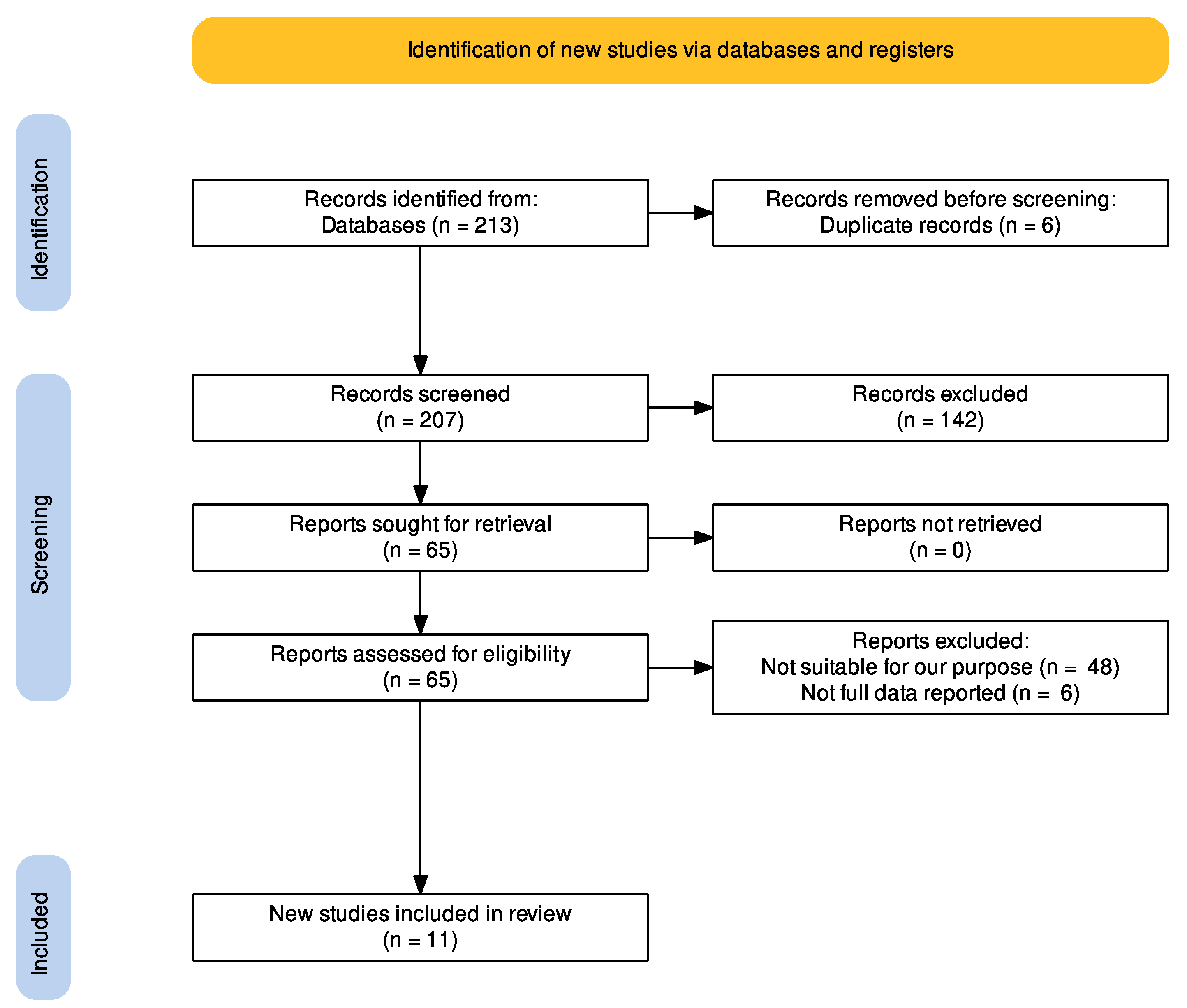
Figure 1. PRISMA 2020 flow diagram [17].
Electronics 11 01741 g001
Figure 2. Quality Assessment of Diagnostic Accuracy Studies-2 (QUADAS-2).
Electronics 11 01741 g002
Publisher’s Note: MDPI stays neutral with regard to jurisdictional claims in published maps and institutional affiliations.