Article

In Vitro Efficacy of Hungarian Propolis against Bacteria, Yeast, and Trichomonas gallinae Isolated from Pigeons—A Possible Antibiotic Alternative?

1
Department of Pharmacology and Toxicology, University of Veterinary Medicine, H-1078 Budapest, Hungary
2
National Laboratory of Infectious Animal Diseases, Antimicrobial Resistance, Veterinary Public Health and Food Chain Safety, University of Veterinary Medicine Budapest, H-1078 Budapest, Hungary
3
Department of Parasitology and Zoology, University of Veterinary Medicine, H-1078 Budapest, Hungary
4
Department of Animal Hygiene, Herd Health and Mobile Clinic, University of Veterinary Medicine, H-1078 Budapest, Hungary
*
Author to whom correspondence should be addressed.
Resources 2023, 12(9), 101; https://doi.org/10.3390/resources12090101
Submission received: 20 May 2023 / Revised: 28 July 2023 / Accepted: 28 August 2023 / Published: 29 August 2023
(This article belongs to the Special Issue Alternative Use of Biological Resources)

Abstract

:
The spread of antimicrobial resistance is one of the most serious human and animal health problems of our time. Propolis is a natural substance with antibacterial, antifungal, and antiparasitic activity, the most active components of which are polyphenols and terpenoids. In the present study, the authors investigated the efficacy of propolis against Staphylococcus spp., Enterococcus spp., Escherichia coli and Salmonella enterica, Candida albicans fungi, and Trichomonas gallinae isolated from pigeons. For each pathogen, the minimum inhibitory concentration (MIC) and minimum eradication concentration (MEC) of eight isolates were determined for 96%, 90%, 80%, 70%, and 60% ethanolic extracts of propolis from the region of Észak-Alföld. Propolis was shown to be effective in inhibiting the growth of Gram-positive bacteria, Candida albicans, and Trichomonas gallinae strains. Propolis showed a much better efficacy against Gram-positive bacteria (1.56–400 µg/mL) than against Gram-negative bacteria (>13,000 µg/mL). For Staphylococcus spp., MIC values ranged within 1.56–400 µg/mL and MEC values within 12.5–3260 µg/mL, while for Enterococcus spp. MIC values ranged within 1.56–400 µg/mL and MEC values within 12.5–800 µg/mL. MIC values > 13,000 µg/mL were found for Escherichia coli and Salmonella enterica species. For Candida albicans, MIC values ranging from 1.56 to 400 µg/mL and MEC values ranging from 3.125 to 800 µg/mL were effective. MEC values between 2.5 and 5 mg/mL were observed for three Trichomonas gallinae strains. The effectiveness against Gram-positive bacteria has, in some cases, approached that of antibiotics, making propolis a potential alternative in the treatment of wound infections. Its outstanding efficacy against Trichomonas gallinae holds promise as a potential alternative for treating this widespread infection in pigeons.

1. Introduction

Propolis is a resinous product consisting of various plant parts and substances produced by honeybees (Apis spp.) [1,2,3,4,5]. To the best of our knowledge, propolis has more than 300 known components, with a composition of approximately 50% resin, 30% wax, 10% essential oil, 5% pollen, and an additional 5% other organic components [1,5,6,7,8]. Propolis is a complex mixture, the exact composition of which depends greatly on the geographical area [9], its flora [1,5,10], the climatic area [11], the time of the year [1,5], the genetic make-up [1,6], and the quality of the bees [12]. All types of propolis have common antibacterial [1,2,3,5,13], antiviral, antioxidant, antiproliferative, antifungal [3,6,13], antiparasitic, anti-inflammatory [6,10], antiprotozoal [14], immunomodulatory [2,13], hepatoprotective [1], antitumor, cytotoxic, and wound healing [10] activity due to their numerous biologically active molecules [1]. However, in addition to the wide range of bioactivities, efforts should be made to standardize their composition, as their chemical constituents are closely related to their efficacy [9]. A recent comprehensive study has already identified more than 800 propolis constituents worldwide, which also supports the observed different biological effects in different origins, again supporting the need for standardization for practical use [15].
We examined the susceptibility of specific pathogenic bacteria found in pigeons, as well as the fungus C. albicans and the parasite T. gallinae. At present, approximately 180,000 meat pigeons are kept in Hungary, and thousands of post and ornamental pigeon breeders are increasing the number of pigeons in the country [16]. Homing pigeons play a key role in the spread of resistance, as they travel hundreds of kilometers in a single race, providing an opportunity for the spread of resistant bacteria [17,18].
In most cases, the antibacterial efficacy of propolis is more pronounced against Gram-positive bacteria than against Gram-negative bacteria [6,19,20,21,22]. Its antibacterial properties are explained via the inhibition of nucleic acid synthesis, reduction in motility [20], functional impairment of the cell membrane, inhibition of energy metabolism [5,23], inhibition of biofilm formation [8,13], damage to cell membrane proteins, alteration of membrane permeability [20,23], and reduction in bacterial resistance [5]. Flavonoids, as the most important phenolic constituents of propolis, typically inhibit nucleic acid synthesis in bacteria, mainly through binding to topoisomerase II. In addition, quercetin is able to inhibit the bacterial enzyme adenosine triphosphatase by binding to the B subunit of DNA gyrase [5]. Furthermore, propolis can form a water layer on its surface (exclusion zone), whose physical barrier is the basis of many of its antibacterial mechanisms of action, due to the hydrophilic groups (-OH, -COOH) present in its many components [7].
Its main mechanism of action against fungi is cell membrane damage via membrane depolarization and induction of apoptosis [20,24]. It also inhibits the expression of several genes involved in pathogenicity, cell adhesion, biofilm formation, and phenotype switching [20,24]. Phenotype switching, i.e., hyphal formation, is one of the most important virulence factors in fungi [24]. Phosphorylated adenosine also reduces nucleotide levels, thereby impairing nucleic acid synthesis and energy metabolism [20]. Formononetin, an important representative of the flavones, has been shown to be very effective against several fungal species [24]. In a study in 2020, the efficacy of different bee products was investigated, where the antifungal activity of propolis was described in response to pinocembrin, pinobanksin, quercetin, and kaempferol; however, the interaction of other compounds, such as caffeic acid, p-coumaric acid, and terpenoid active compounds, with phenolic components is also attributed to the source of antibacterial activity [25,26]. Candida albicans is the most commonly pathogenic yeast, the hyphal form of which is able to penetrate the epithelium and endothelium, causing tissue damage [27].
The antiparasitic action of propolis is also based on several mechanisms [28]. It interferes with phospholipid metabolism, leading to a decrease in the levels of phosphatidylglycerol and phosphatidylinositol, which are constituents of the cell membrane [29]. In addition, another group of flavonoids, chalcones, significantly inhibits parasite growth. The 2,6-dihydroxy, 4-methoxy-chalcone enhances the sterol content and the composition of cell and mitochondrial membranes. This results in altered membrane structure and fluidity [30]. Rosmarinic acid and apigenin cause cell lysis, cytoplasmic condensation, and aggregation of nuclear DNA [31]. Resveratrol acts through damage to the cell organelle of the hydrogenosome. This cellular organelle is responsible for energy production by protozoa and for maintaining redox balance. Apigenin and caffeic acid increase the formation of reactive oxygen species (ROS), consequently causing mitochondrial swelling and apoptosis [32]. Quercetin is an iron chelator and maslinic acid inhibits the protease activity of the parasite surface protein complex, which is essential for entry into the host cell [33,34]. Kaempferol affects the expression of actin and myosin II heavy chain, which inhibits parasite adhesion [35].
T. gallinae is a protozoan that causes yellowish-white, plaque-like deposits in house pigeons, typically in the upper respiratory tract and the pharyngeal cavity. Mortality is very high in chicks and young birds, while adults are often asymptomatic. It is typically transmitted through the drinking water and food of birds, but can also infect chicks through the breast and navel [36,37].
Several studies are investigating the use of antibiotic alternatives and several have demonstrated the potential efficacy of certain agents, such as Campylobacter jejuni strains [38] or E. coli infections [39]. Previously published articles on the effectiveness of propolis indicate its suitability for use as an antibiotic alternative [40].
The antibacterial efficacy of propolis has been described in several studies, but no similar study has been conducted in Hungary so far. The aim of this study was to investigate the antibacterial, antifungal, and antiprotozoal efficacy of propolis using ethanolic extracts of different concentrations and to examine any differences in efficacy between these extracts in vitro. Five different propolis stock extracts were used for the treatment (96%, 90%, 80%, 70%, 60%), because literature sources have noted that different concentrations of ethanol dissolve different flavonoids in high quantities. Gómez-caravaca et al. found that the 80% ethanol extraction contains mainly kaempferide, acacetin, and isorhamnetin, the 70% ethanol extraction contains pinocembrin and sacuranetin, and the 60% ethanol extraction contains isosakuranetin, quercetin, and kaempferol flavaonoids [11]. One of our hypothesis tests was that there is a difference in the efficacy of ethanol extracts of different concentrations. Our next hypothesis was that there is no difference between the antibacterial efficacy of propolis on Gram-positive bacteria and Gram-negative bacteria. Finally, we supposed that propolis effectively inhibits the growth of C. albicans and T. gallinae. Among the pathogens studied, Staphylococcus species have been implicated in purulent dermatitis in pigeons [41], while the role of Enterococcus species has recently been evaluated, particularly in zoonotic and nosocomial infections, specifically in relation to the spread of antimicrobial multi-resistance [42]. The investigation of Escherichia coli and Salmonella species is also of particular importance because of the public health role of pigeons [43] and because these species can cause septicemia in pigeons, and Candida albicans can cause pigeon pus [44,45]. Trichomonas gallinae is mainly responsible for upper respiratory tract, oral cavity, and pigeon pus [46].

2. Materials and Methods

2.1. The Origin and Extraction of Propolis

The raw propolis used for the study was from the region of Észak-Alföld. Five different propolis stock extracts were used for the treatment since, according to literature sources, different concentrations of ethanol dissolve different flavonoids in high quantities, i.e., 80% ethanol mainly contains kaempferide, acacetin, and isorhamnetin, 70% ethanol contains pinocembrin and sakuranetin, and 60% ethanol contains isosakuranetin, quercetin, and kaempferol [11]. For our bacterial and fungal assays, we added 30 mL of ethanol and 10 mL of glycerol to 10 g of propolis when preparing our 96% extract, and 40 mL of 60%, 70%, 80%, and 90% ethanol to 10 g of propolis in each of the other four cases. For propolis extraction, a conventional extraction procedure was used, in which the different ethanol percentage solvents and the powdered crude propolis were allowed to macerate for three weeks at room temperature in a closed vessel protected from light, and finally the undissolved parts were removed with filter paper [47]. The addition of glycerol during the extraction process provided higher active ingredient extraction through a more polar extraction, as described in the literature [48]. In all five cases, a propolis concentration of 250 mg/mL was used. The total flavonoid content of the tested sample was 18.2 mg/g, and the analysis was carried out by our analytical laboratory using the colorimetric method [49].

2.2. Antibacterial and Antifungal Studies

Bacteria and fungi were examined on a 96-well microtiter plate (VWR International, LLC., Debrecen, Hungary), with the effect of solvent being measured under each treated sample; 150 µL propolis extract/ethanol was added to 30 µL sterile broth.
The pathogenic bacterial strains were collected from bacteriological samples taken from sick or dead pigeons in and around Budapest. After several months of sample collection, the species shown in Table 1 were used for our studies. We performed 8 replicates per bacterium and per fungus.
As recommended by Clinical and Laboratory Standards Institute (CLSI), the bacterial and fungal suspension was adjusted to 105 volumes using McFarland standards [50]; breeding was also performed according to a standard [50,51]. The final inoculum volume was 104 colony forming unit (CFU)/mL. We utilized the minimum inhibitory concentration (MIC) determination in our study, which identifies the lowest dilution concentration that effectively inhibits the growth of microorganisms. Additionally, we introduced the concept of the minimum eradication concentration (MEC), which refers to the concentration that reduces the quantity of microorganisms by at least four orders of magnitude.
All wells except the first column of plates were filled with 90 µL tryptone soy broth (TSB) (Biolab Diagnostic Laboratory Ltd., Budapest, Hungary); 30 µL TSB was transferred into the wells of the first column, 150 µL of propolis stock extract was added, 90 µL suspension was transferred into the second column, resuspended, etc.; finally, after the 10th column, the 90 µL excess was discarded with the pipette tip.
Subsequently, 10 µL of the concentrated bacterial suspension was transferred into the columns of the 240 µL TSB-filled inoculum (auxiliary) plate, and 10 µL of bacterial suspension was transferred from the wells of column A–H of column 1 backwards from column 11 of plates containing the dilution series (positive control). The plates were then incubated at 37 °C for 18–24 h and the MIC was determined via the visual method based on the presence/extent of turbidity.
We prepared the same dilution series of ethanol as the solvent for each strain examined. This allowed us to visually determine the point at which ethanol had inhibitory effects and where only the propolis exhibited its activity. The effectiveness of propolis could only be judged from the dilution at which the pathogens were released from ethanol inhibition. As a self-control, 50 µL of each suspension was inoculated from the wells of the first three columns released from ethanol inhibition onto tryptone soy agar (TSA) (Biolab Diagnostic Laboratory Ltd., Budapest, Hungary) to determine the CFU, with a CFU result of zero for wells containing propolis and confluent pathogens in the Petri dish for wells containing ethanol.

2.3. Antiprotozoal Studies

The effect of ethanol on T. gallinae was also investigated in 24-well plates (VWR International, LLC., Debrecen, Hungary), each of which was diluted 2.5-fold with stock extract and solvent, of which 0.3 mL was measured into the wells of the first column.
Samples of T. gallinae were collected in spring 2021 from two flocks of pigeons in Budapest, Hungary, and positive samples were confirmed by polymerase chain reaction (PCR) using a kit from Qiagen (Hilden, Germany). The samples were collected in 8 mL of Trichomonas cysteine-peptone-liver-maltose (CPLM) modified medium (Biolab Diagnostic Laboratory Ltd., Budapest, Hungary) and transported at a controlled temperature. Subsequently, they were incubated at 37 °C using an incubator. For the study, three isolates of T. gallinae were used in parallel.
The first column of plate 24 was filled with 2.7 mL of tap water; 1.5 mL of tap water was added to the other wells, 0.3 mL of propolis tincture was added to wells A and C of the first column, and 0.3 mL of solvent was added to wells B and D below. A dilution series was prepared on a two-well basis, and finally the pipette tip containing 1.5 mL of suspension from the last well was discarded as excess. The plates were then placed in a thermostat at 37 °C for 18 to 24 h and, after incubation, the number of trophozoites was determined using a Bürker chamber [52]. Based on the general formula used for cell counting, the average cell count was determined in large squares: the number of trophozoites was counted in 25 large squares for the initial concentration and in 5 large squares for the treatments, averaged and multiplied by the dilution rate, and finally multiplied by the multiplier 2.5 × 10⁵.

2.4. Statistical Analysis

The results were analyzed using R 4.0.5. The results for bacterial species and fungi were analyzed using a mixed linear model for each species separately due to the correlations. For T. gallinae strains, since there were no correlated data, ANOVA was used with the Tukey post-hoc test.
Our initial assumption, stated as the null hypothesis, was that there would be no variation in efficacy among the various ethanolic propolis extracts. In the protozoa assay, we examined the changes in cell counts of the samples over 24, 48, and 72 h. Using a two-tailed test, we determined whether the cell counts had either increased significantly, indicating successful proliferation, or decreased significantly, compared to the initial cell count.

3. Results

3.1. Efficacy of Propolis against Bacteria and Fungi

Ethanol had negligible or no effect, inhibiting bacterial and fungal growth at concentrations above 1%, whereas it had no effect against T. gallinae at all.
For Staphylococcus and Enterococcus species, 96% and 90% suspensions achieved much better activity in both MIC and MEC values. The potency of the other ethanol extracts was much lower. The MEC is the lowest concentration at which the bacterial count is reduced by at least 4 log values, for example, from 109 cells/mL to at least 105 cells/mL. In our studies, effective treatments resulted in complete eradication, i.e., a reduction to zero.
For E. coli strains, we found the same difference as that described in the literature, i.e., much lower efficacy for Gram-negative bacteria. The MIC was >13,000 µg/mL; concentrations higher than this were not judged due to the inhibitory effect of ethanol. Only for strain 5 did a concentration of 13,000 µg/mL inhibit bacterial growth. For the other strains, 50 µL samples of each of these last columns, already released from the ethanol inhibition and inoculated onto Petri dishes, showed confluent bacterial growth in all cases.
In the case of S. enterica strains, we also found that the assayable concentrations of propolis were not effective in any of their forms. Confluent growth was also observed when 50 µL samples of the first columns released from ethanol inhibition were inoculated onto Petri dishes. The MIC was >13,000 µg/mL; concentrations higher than this were not judged due to the inhibitory effect of ethanol.
In the case of C. albicans strains, only the 96% ethanolic propolis extract showed significant MIC and MEC values. The effectiveness of the other extracts was much lower.
The MIC range, MIC50 value, MIC90 value, MEC range, MEC50, and MEC90 values for each species are summarized in Table 2. The MIC50 value is the minimum concentration of the active substance to which at least 50% of the microbial population tested is sensitive, and this proportion if at least 90% for MIC90.

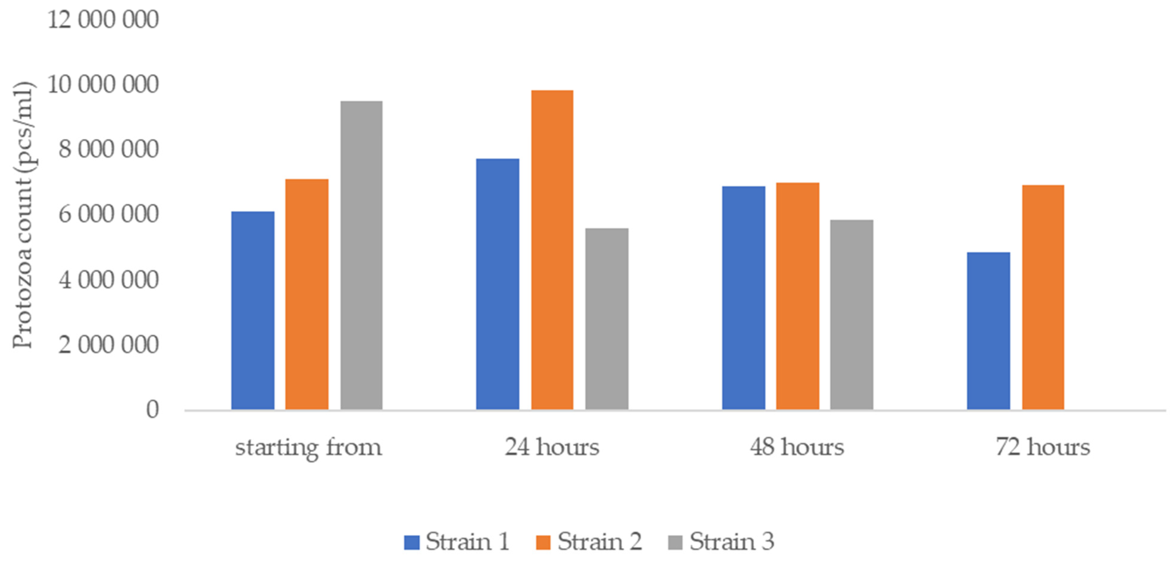
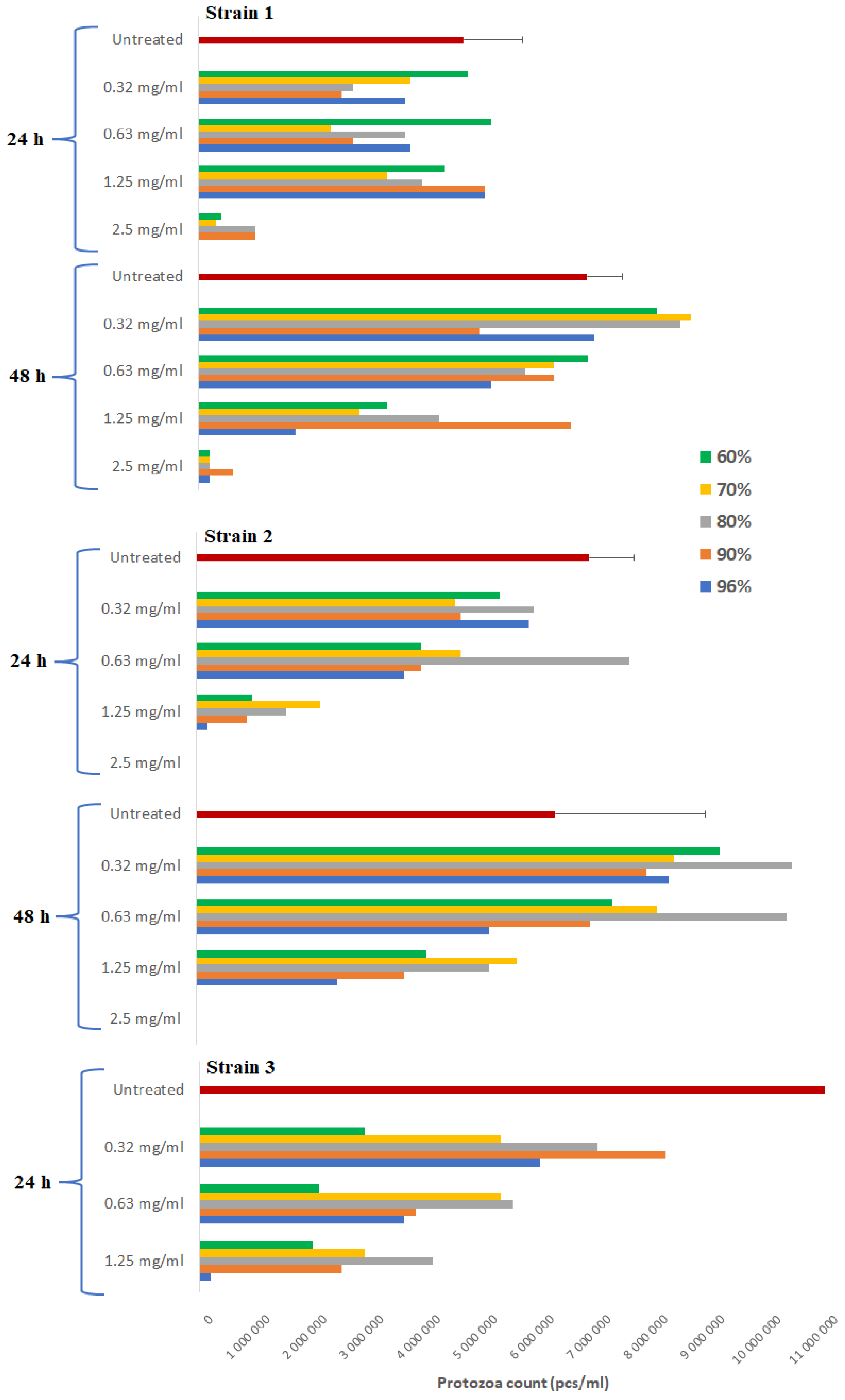
3.2. Viability of Protozoa and Activity of Propolis against T. gallinae

After sample collection, the number of protozoa in the samples was counted after 24 h, 48 h, and 72 h. The results clearly show that in our culture medium, protozoa survival was ensured for 48 h and even their reproduction was observed. However, after 48 h the number of protozoa decreased and they even died in the case of sample 11 (Figure 1). Based on this, the efficacy of propolis was observed after 24 and 48 h of treatment, but the effect lasting for 48 h after introduction could not be assessed for strain 11.
The cell counts of the lines used as controls alongside the treatments also reflect the proliferation of trophozoites, as diluted to 25× they would have been expected to produce approximately 2.8 × 10⁵/mL protozoa. However, after 24 h this value was 8 × 10⁶/mL, which was approximately the same as the initial count. In addition, a steady increase in cell counts was observed in the dilution line up to 1.2 × 10⁷/mL. There are two possible explanations for this: either the increasing amount of nutrients (due to dilution) resulted in more successful multiplication, or the dilutions containing more concentrated ethanol, from which the trophozoites were gradually released, had some inhibitory effect on multiplication.
Three T. gallinae strains were used for the study: strains 6, 10, and 11. For all three strains, it can be stated that the ethanol control treatment had no effect on the viability of the protozoa in both the 24 h and 48 h treatments.
After 24 h of treatment with the 96% extract, complete eradication was observed up to 50× dilution for strain 1 and 100× dilution for strains 2 and 3, so our MEC was 2.5–5 mg/mL. The evolution of the cell count indicates that the minimum parasiticide concentration is equal to the MEC value. At 48 h of treatment, only strains 1 and 2 were detectable, with the former showing complete eradication even at 100× dilution, while the latter showed no change. Thus, the MEC was 2.5 mg/mL and the incubation time did not increase the efficacy of propolis. The results of treatment with the 90%, 80%, 70%, and 60% extracts were fully consistent with those of the 96% extract, so the MEC and the minimum parasiticide concentrations were from 1.1 to 2.5 mg/mL for the 24 h treatment and from 2.5 to 5 mg/mL for the two surviving strains after 48 h of treatment (Figure 2).

3.3. Statistical Analysis

Our first null hypothesis was that propolis is equally effective against Gram-positive and Gram-negative bacteria. There was a significant difference (p < 0.0001) between the MIC values (3.125–400 µg/mL) for Gram-positive bacteria and the MIC values (>13,000 µg/mL) for Gram-negative bacteria. Thus, the null hypothesis is rejected, i.e., our results showed a significant difference between the two groups.
Table 3 illustrates the statistical comparison of MIC and MEC values of each ethanolic extract per bacterial species. Our null hypothesis was that there is no difference between the efficacy of the different ethanolic extracts per bacterial species.
In the case of Staphylococcus species, there is no significant difference in MIC values of the 96% and 90% suspensions (p = 0.9610), and no significant difference in MIC values of the 80% and 70% (p = 0.9910), 80% and 70% (p = 1.0000), and 70% and 60% (p = 0.9910) suspensions. The same was observed for the MEC values. So, for Staphylococcus species, we retained the null hypothesis between the 96–90% and 80–70–60% extracts, but for all other extracts we observed a significant difference in efficacy and rejected the null hypothesis.
For Enterococcus species, no significant difference was observed in MIC values of the 80% and 70% (p = 0.9950), 80% and 60% (p = 0.7810), and 70% and 60% (p = 0.9410) suspensions. There was also no significant difference in MEC values of the same ethanolic extracts. Thus, for Enterococcus species, our null hypothesis is retained between the MIC values of the 80%, 70%, and 60% extracts, and for MEC values, we retained the null hypothesis between the 96% and 90% extracts, and the 80%, 70%, and 60% extracts, and rejected it for all other cases.
Concerning C. albicans, no significant difference was observed in MIC values of the 80% and 70% (p = 0.9984), the 80% and 70% (p = 1.0000), and the 70% and 60% (p = 0.9984) suspensions. However, for the MEC values, there was no significant difference in the efficiencies of the 90% and the 80% ethanolic extracts (p = 0.4530) and the three ethanolic extracts. Thus, regarding C. albicans, we retained our null hypothesis between the MIC values of the 80%, 70%, and 60% extracts, and for MEC values we also retained the null hypothesis between the 90% and 80% extracts, and the 80%, 70% and 60% extracts, but rejected it for all other cases.
In the case of T. gallinae, the success of culturing the parasite was first investigated. Our null hypothesis was that there was no difference in the number of protozoa at the different time points compared to the initial cell count. The initial protozoan count for strain 6 showed a significant increase after 24 h of incubation (p = 0.0028), which tended to stagnate after 48 h (p = 0.3452), and significantly decreased after 72 h (p = 0.0339). Regarding strain 10, the initial protozoan count showed a significant increase after 24 h (p = 0.0050) but, compared to this, the 48 h (p = 0.9993) and 72 h cell counts (p = 0.9961) showed no significant difference. However, a significant decrease was observed for the 48 h (p = 0.0035) and 72 h (p = 0.0025) values compared to the 24 h increase. Furthermore, the trophozoite counts at 48 h and 72 h were stagnant (p = 0.9997). For strain 11, a significant decrease was observed after 24 h, 48 h, and 72 h compared to the initial protozoan count (p < 0.00001) which, when compared to 72 h, also showed a significant decrease (p < 0.00001) at 24 h and 48 h. Only between 24 h and 48 h was the protozoan count similar (p = 0.9740). The results show a significant difference in the initial protozoan counts after 24 h for all strains; thus, our null hypothesis can be rejected for these strains.
Table 4 illustrates the statistical comparison of the MEC values of the different ethanolic extracts per strain. Our null hypothesis was that there is no difference in the efficacy of the different ethanolic extracts for the treatment of T. gallinae.
Significant differences in the MEC values of strain 1 were found between the 96% extract and all the other ethanolic extracts at 24 h and 48 h of treatment (p < 0.0010). In addition, significant differences were observed between the 96% and 60% (p = 0.0479), the 90% and 60% (p = 0.0226), and the 80% and 60% (p = 0.0142) treatments at 24 h. For these, the null hypothesis was rejected and replaced by the counter hypothesis. In the other cases, there was no significant difference, and the null hypothesis was retained.
For the MEC values of strain 2, a significant difference was found between the 96% extract and all the other ethanol extracts at 24 h and 48 h of treatment (p < 0.0010). In addition, the difference was significant between the 24 h 96% and 60% (p = 0.0480) and the 48 h 90% and 60% (p = 0.0479) treatments. For these, the null hypothesis was rejected and replaced by the counter hypothesis. In the other cases there was no significant difference, and the null hypothesis was retained.
In the case of strain 3, we were only able to test the 24 h treatment, as all trophozoites had died by the 48 h time point. A significant difference was found between the 96% extract and all the other ethanolic extracts (p < 0.0010). In addition, the difference was significant between the 96% and 60% treatments (p = 0.0478). For these, the null hypothesis was rejected and replaced by the counter hypothesis. In the other cases, there was no significant difference, and the null hypothesis was retained.

4. Discussion

A concentration of 10–20 µg/mL of Taiwanese green propolis was found to inhibit the growth of S. aureus, while for E. coli, the growth-inhibiting concentration was consistently >640 µg/mL. In the case of methicillin-resistant S. aureus (MRSA), concentrations below 2 µg/mL were also effective [53]. Wojtyczka et al. reported that propolis demonstrated efficacy against S. aureus and MRSA at concentrations of 0.39–0.78 mg/mL [54]. Suleman et al. observed the effectiveness of thirty-nine South African and three Brazilian propolis batches against S. aureus at a concentration of 6 µg/mL [55]. Additionally, Almeida et al. observed effective inhibition of bacterial growth against S. aureus at concentrations of 271.74–543.48 µg/mL of Brazilian red propolis [56].
In a study investigating the efficacy of ethanolic extracts of Brazilian brown, green, and red propolis, the MIC values for S. aureus were determined at concentrations of 400–800 µg/mL for brown propolis, 200–400 µg/mL for green propolis, and 200 µg/mL for red propolis. For E. coli, the MIC was determined at concentrations of 1600–1800 µg/mL for brown propolis, 400–1600 µg/mL for green propolis, and 400 µg/mL for red propolis [57]. In 2019, Grecka et al. investigated the efficacy of Polish propolis and found that concentrations of 128–512 µg/mL were effective against S. aureus [58].
For E. faecalis, low MIC values have been reported in Brazil, South Africa, and Morocco [59], of 49 µg/mL [55] and 70 µg/mL [60]. Propolis of Palestinian and Iranian origins showed moderate effectiveness, with MIC values ranging from 170 to 625 µg/mL [61] and from 250 to 300 µg/mL [62,63], respectively. Higher MIC values (1200–1400 µg/mL) have been described for propolis from Slovenia compared to the average values [64], while studies in Serbia described MIC values of 16,800 µg/mL [65].
In studies involving E. coli, MIC values of 31.2 µg/mL were found for Bolivian propolis extracted in absolute ethanol [66], 31.5 µg/mL for propolis similarly dissolved in Chile [67], 128 µg/mL for Brazilian propolis extracted in ethanol [68], and 169 µg/mL for Omani propolis [69]. The least effective concentrations were described for Serbian extract (10,000 µg/mL) [19] and Brazilian propolis extract (8000 µg/mL) [70].
In studies of S. enterica, concentrations of 62.5 µg/mL for Chilean propolis extract [67] and 125 µg/mL for Bolivian extract [66] were used. However, in most cases, ethanolic propolis tincture proved ineffective, with only 10,000 mg/mL concentration inhibiting growth in the case of Serbian propolis tincture [19]. In the study of propolis of Slovenian origin, the lowest MIC value was 580 µg/mL but, in most cases, values of 1200–1400 µg/mL were reported [57].
The difference in efficacy against Gram-positive and Gram-negative bacteria described in the literature [6,19,20,21] was confirmed by our studies, with MIC values of 3.125–400 µg/mL for the former and >13 mg/mL for the latter. Furthermore, the main reason for the different results compared to the literature is the geographically different chemical composition of propolis.
No significant difference was observed in efficacy between different concentrations of ethanolic extracts on Staphylococcus spp. in terms of MIC values between the 96% and 90% (p = 0.9610), 80% and 70% (p = 0.9910), 80% and 70% (p = 1.0000), and 70% and 60% (p = 0.9910) suspensions. MIC values ranging from 3.125 to 50 µg/mL were observed for the 96% extract, while Mavri et al. described MIC values between 150 and 290 µg/mL [64]. For the 90% extract, MIC values ranging from 1.56 to 400 µg/mL were obtained. No comparative literature is available in this case. Regarding the 80%, 70%, and 60% extracts, there was a visible increase in MIC values compared to the former extracts, with average concentrations of 50–400 µg/mL observed. Similar to our own results, MIC values of 150–250 µg/mL have been described for Iranian propolis using 80% ethanolic extract [62,63]. For 70% Greek propolis tincture, a concentration of 120 µg/mL was found to be effective [36]; however, El Menyiy et al. found it to be effective at a concentration of 2 µg/mL [60], whereas Hegazi et al. described MIC values of 2400 and 4600 µg/mL [63]. MIC values of 20 µg/mL were also described for the 60% extract [53].
For Enterococcus species, no significant difference in MIC values, i.e., efficacy, was observed between the 80% and 70% (p = 0.9950), 80% and 60% (p = 0.7810), and 70% and 60% (p = 0.9410) suspensions. MIC values for the 96% tincture ranged from 6.25 to 50 µg/mL. In contrast, Mavri et al. observed efficacy only at 1200 µg/mL [64]. The 90% extract was also effective at concentrations of 1.56–12.5 µg/mL. No comparative literature is available here. For the 80% tincture, concentrations of 100–400 µg/mL were effective in our experiment; similarly, MIC values of 250–300 µg/mL have been described for Iranian propolis [62,63]. In contrast, in the experiment of Campos et al., the MIC ranged from 880 to 1020 µg/mL [71]. For the 70% extract, concentrations of 100–200 µg/mL were found to be effective in our studies. Similarly, El Menyiy et al. described MIC values of 70 µg/mL [60], and MIC values between 170 and 625 µg/mL were observed for Palestinian propolis [61]. In contrast, only a concentration of 1400 µg/mL was effective for Slovenian propolis [64]. In our studies, concentrations between 50 and 200 µg/mL were effective for the 60% extract.
In studies with E. coli strains, no effective inhibitory concentration could be determined for any of the propolis extracts. MIC values were always higher than 13 mg/mL except in one case, where a concentration of 13 mg/mL inhibited bacterial growth. Similar values were found in the literature. In studies on Serbian propolis, MICs ranged from 2.5 to 10 mg/mL [19]. Hegazi et al. also found a high MIC value of 1.6 mg/mL [72], and for Egyptian and Saudi Arabian propolis, only concentrations of 2.5 and 1.5 mg/mL were effective [73]. In contrast, Nina et al. described an effective inhibitory effect at concentrations as low as 31.2 µg/mL [66,74], and an MIC of 31.5 µg/mL was observed for Chilean propolis [67].
In the case of S. enterica, similarly to E. coli, propolis did not show sufficient efficacy. In all cases, the MIC was greater than 13 mg/mL. In the case of Serbian propolis, a concentration of 10 mg/mL was effective [19], and in the case of Slovenian propolis, MIC values ranged from 580 µg/mL to 1.4 mg/mL [57]. In some cases, MIC values of 62.5 µg/mL [67] and 125 µg/mL [66] were found to be effective.
In an experiment by Ota et al., Candida albicans was the most sensitive to propolis among four Candida species (C. albicans, C. tropicalis, C. krusei, and C. guilliermondii) [75]. The induction of apoptosis has been described as one of the main antifungal mechanisms of action [20], and a pronounced effect against the fungal cell membrane [8] has also been observed. In the case of Brazilian propolis, anti-C. albicans activity was demonstrated (273.43 µg/mL) and was found to inhibit hyphal transformation. When Portuguese and French propolis were tested, efficacy against C. albicans and C. glabrata was observed (15.63–250 µg/mL). In the case of Irish and Czech propolis, the antifungal activity of propolis was pronounced (0.1–5 mg/mL) [73].
In a large-scale Polish study, propolis samples from fifty different geographical locations were described to be effective against C. albicans at a concentration of 630 µg/mL [76]. An ethanolic extract of green propolis was tested in dentistry and found to have fungicidal and anti-biofilm activity against C. albicans, C. parapsilosis, and C. tropicalis at concentrations of 2.5 µg/mL in oral candidiasis [74]. In a previous survey in Hungary, 19–45% of the flocks of geese and ducks examined had esophageal lesions caused by C. albicans, which cause significant economic losses when left untreated [77]. The use of propolis as an alternative in the treatment of these conditions can be considered, as the causative agent is susceptible to propolis.
For C. albicans, no significant difference in MIC values was observed between the 80% and 70% (p = 0.9984), 80% and 70% (p = 1.0000), and 70% and 60% (p = 0.9984) suspensions. The 96% extract was effective at MIC values of 1.56–50 µg/mL. In an experiment by Boisard et al., propolis was similarly effective at a concentration of 31.25 µg/mL [78]. However, only MIC values between 625 µg/mL and 5000 µg/mL were found to be effective for Serbian propolis [19]. The 90% extract was clearly the least effective compared to other ethanolic extracts, with a constant MIC of 400 µg/mL. The 80%, 70%, and 60% extracts showed similar efficacy, with MIC values ranging from 25 to 400 µg/mL, except for one or two outliers. However, in the case of the 80% extract, outstanding efficacy was also described, with MIC values ranging from 11 µg/mL to 14.5 µg/mL [79]. In studies with the 70% extract, a MIC of 31.25 µg/mL was found, similar to the 96% extracts [78]. However, only MIC values between 4048 and 5000 µg/mL were effective in the case of German propolis [49,50].
For Trichomonas vaginalis, ethanol-containing Brazilian brown propolis extract was effective at a concentration of 400 µg/mL [79]. In their study, Sena-Lopes et al. described 100% eradication of trophozoites at a concentration of 500 µg/mL of ethanolic extract [80]. The only test described in the literature for T. gallinae was conducted with an aqueous extract of propolis of Egyptian origin, for which the MIC was 75,000 µg/mL [37].
Significant differences were found between the 96% extract and all other ethanolic extracts for T. gallinae treatments (p < 0.0010). After 24 h of treatment, MEC values varied between 2.5 and 5 mg/mL, regardless of the ethanol concentration used. After 48 h of treatment, MEC values ranged from 2.5 to 5 mg/mL. Comparative literature is only available using an aqueous extract of propolis, where 100% eradication of parasites occurred only at a concentration of 75 mg/mL [37].

5. Conclusions

Due to variations in geographical origin and composition, propolis may exhibit different MIC values in different countries. MEC values are consistently equal to or higher than MIC values. However, the substantial variability observed in propolis may significantly reduce the likelihood of developing resistance compared to antibiotics. The highest efficacy was achieved with extracts containing the highest concentrations of ethanol (96% and 90%). The impact of ethanol was found to be negligible in our studies.
Trichomonas gallinae infection is highly prevalent in pigeons, and our findings indicate that propolis holds promise as an alternative to antibiotics for its treatment: it can potentially serve as a topical treatment option when directly applied to the oral cavity or mixed with drinking water, and it could also replace the use of ronidazole, the only effective nitroimidazole agent, which is prohibited for use in animals intended for food production. Pigeons are legally classified as food-producing animals.
Our research confirmed significantly higher efficacy against Gram-positive bacteria. These results suggest that propolis could serve as an effective alternative treatment for external infections caused by Gram-positive bacteria, such as skin infections. However, further in vivo studies are necessary to validate these findings. Propolis also exhibited remarkable efficacy against C. albicans, but it would be valuable to conduct studies on a broader range of fungal species. The efficacy of propolis against Trichomonas gallinae was less pronounced compared to that found in studies on human Trichomonas vaginalis, and no other comparative studies are currently available for these infections.
A limiting factor in its veterinary use may be the use of an ethanol extractant and its role in possible allergic reactions.

Author Contributions

Conceptualization, Á.K.; methodology, Á.K.; software, Á.K.; validation, Á.J., L.K. and B.T.-S.; formal analysis, Á.K.; investigation, P.C.; resources, P.C.; data curation, Á.K.; writing—original draft preparation, Á.K.; writing—review and editing, Á.K.; visualization, Á.K.; supervision, Á.J.; project administration, P.C.; funding acquisition, Á.J. All authors have read and agreed to the published version of the manuscript.

Funding

Project no. RRF-2.3.1-21-2022-00001 has been implemented with the support provided by the Recovery and Resilience Facility (RRF), financed under the National Recovery Fund budget estimate, RRF-2.3.1-21 funding scheme.

Data Availability Statement

The data presented in this study are available from the corresponding author upon reasonable request.

Acknowledgments

I would like to thank Krisztián Berta for his help in microbiological sample collection, and László Makrai for his help in bacterial isolation. I would also like to thank Sándor Hornok and Barbara Tuska-Szalay for their selfless help in collecting and identifying protozoa. Thanks are due to Zsolt Abonyi-Tóth for his professional assistance in the statistical analysis of the results.

Conflicts of Interest

The authors declare no conflict of interest.

References

  1. Santos, L.M.; Fonseca, M.S.; Sokolonski, A.R.; Deegan, K.R.; Araújo, R.P.; Umsza-Guez, M.A.; Barbosa, J.D.; Portela, R.D.; Machado, B.A. Propolis: Types, composition, biological activities, and veterinary product patent prospecting. J. Sci. Food Agric. 2020, 100, 1369–1382. [Google Scholar] [CrossRef]
  2. Daneshmand, A.; Sadeghi, G.H.; Karimi, A.; Vaziry, A.; Ibrahim, S.A. Evaluating complementary effects of ethanol extract of propolis with the probiotic on growth performance, immune response and serum metabolites in male broiler chickens. Livest. Sci. 2015, 178, 195–201. [Google Scholar] [CrossRef]
  3. Aygun, A.; Sert, D. Effects of prestorage application of propolis and storage time on eggshell microbial activity, hatchability, and chick performance in Japanese quail (Coturnix coturnix japonica) eggs. Poult. Sci. 2013, 92, 3330–3337. [Google Scholar] [CrossRef] [PubMed]
  4. Ding, J.; Matsumiya, T.; Hayakari, R.; Shiba, Y.; Kawaguchi, S.; Seya, K.; Ueno, K.; Imaizumi, T. Daily Brazilian green propolis intake elevates blood artepillin C levels in humans. J. Sci. Food Agric. 2021, 101, 4855–4861. [Google Scholar] [CrossRef]
  5. Almuhayawi, M.S. Propolis as a novel antibacterial agent. Saudi J. Biol. Sci. 2020, 27, 3079–3086. [Google Scholar] [CrossRef]
  6. Przybyłek, I.; Karpiński, T.M. Antibacterial Properties of Propolis. Molecules 2019, 24, 2047. [Google Scholar] [CrossRef] [PubMed]
  7. Kowacz, M.; Pollack, G.H. Propolis-induced exclusion of colloids: Possible new mechanism of biological action. Colloid Interface Sci. Commun. 2020, 38, 100307. [Google Scholar] [CrossRef] [PubMed]
  8. Rivera-Yañez, N.; Rivera-Yañez, C.R.; Pozo-Molina, G.; Méndez-Catalá, C.F.; Reyes-Reali, J.; Mendoza-Ramos, M.I.; Méndez-Cruz, A.R.; Nieto-Yañez, O. Effects of Propolis on Infectious Diseases of Medical Relevance. Biology 2021, 10, 428. [Google Scholar] [CrossRef]
  9. Silva-Carvalho, R.; Baltazar, F.; Almeida-Aguiar, C. Propolis: A Complex Natural Product with a Plethora of Biological Activities That Can Be Explored for Drug Development. Evid.-Based Complement. Altern. Med. 2015, 2015, 206439. [Google Scholar] [CrossRef]
  10. Regueira-Neto, M.d.S.; Tintino, S.R.; Rolón, M.; Coronal, C.; Vega, M.C.; de Queiroz Balbino, V.; de Melo Coutinho, H.D. Antitrypanosomal, antileishmanial and cytotoxic activities of Brazilian red propolis and plant resin of Dalbergia ecastaphyllum (L) Taub. Food Chem. Toxicol. 2018, 119, 215–221. [Google Scholar] [CrossRef]
  11. Gómez-Caravaca, A.M.; Gómez-Romero, M.; Arráez-Román, D.; Segura-Carretero, A.; Fernández-Gutiérrez, A. Advances in the analysis of phenolic compounds in products derived from bees. J. Pharm. Biomed. Anal. 2006, 41, 1220–1234. [Google Scholar] [CrossRef]
  12. Farnesi, A.P.; Aquino-Ferreira, R.; De Jong, D.; Bastos, J.K.; Soares, A.E.E. Effects of stingless bee and honey bee propolis on four species of bacteria. Genet. Mol. Res. 2009, 8, 635–640. [Google Scholar] [CrossRef]
  13. Liberio, S.A.; Pereira, A.L.A.; Dutra, R.P.; Reis, A.S.; Araújo, M.J.A.M.; Mattar, N.S.; Silva, L.A.; Ribeiro, M.N.S.; Nascimento, F.R.F.; Guerra, R.N.M.; et al. Antimicrobial activity against oral pathogens and immunomodulatory effects and toxicity of geopropolis produced by the stingless bee Melipona fasciculata Smith. BMC Complement. Altern. Med. 2011, 11, 108. [Google Scholar] [CrossRef] [PubMed]
  14. Attia, Y.A.; Al-Khalaifah, H.; Ibrahim, M.S.; Al-Hamid, A.E.A.; Al-Harthi, M.A.; El-Naggar, A. Blood Hematological and Biochemical Constituents, Antioxidant Enzymes, Immunity and Lymphoid Organs of Broiler Chicks Supplemented with Propolis, Bee Pollen and Mannan Oligosaccharides Continuously or Intermittently. Poult. Sci. 2017, 96, 4182–4192. [Google Scholar] [CrossRef] [PubMed]
  15. Kasote, D.; Bankova, V.; Viljoen, A.M. Propolis: Chemical diversity and challenges in quality control. Phytochem. Rev. 2022, 21, 1887–1911. [Google Scholar] [CrossRef]
  16. Állatállomány, 1 December 2019. Available online: https://www.ksh.hu/docs/hun/xftp/stattukor/allat/2019/index.html (accessed on 8 October 2021).
  17. Ghanbarpour, R.; Aflatoonian, M.R.; Askari, A.; Abiri, Z.; Naderi, Z.; Bagheri, M.; Jajarmi, M.; Shobeiri, S.; Molaei, R.; Askari, N. Domestic and game pigeons as reservoirs for Escherichia coli harbouring antimicrobial resistance genes. J. Glob. Antimicrob. Resist. 2020, 22, 571–577. [Google Scholar] [CrossRef]
  18. Alhababi, D.A.; Eltai, N.O.; Nasrallah, G.K.; Farg, E.A.; Al Thani, A.A.; Yassine, H.M. Antimicrobial Resistance of Commensal Escherichia coli Isolated from Food Animals in Qatar. Microb. Drug Resist. 2020, 26, 420–427. [Google Scholar] [CrossRef] [PubMed]
  19. Stepanović, S.; Antić, N.; Dakić, I.; Svabić-Vlahović, M. In vitro antimicrobial activity of propolis and synergism between propolis and antimicrobial drugs. Microbiol. Res. 2003, 158, 353–357. [Google Scholar] [CrossRef]
  20. Zulhendri, F.; Chandrasekaran, K.; Kowacz, M.; Ravalia, M.; Kripal, K.; Fearnley, J.; Perera, C.O. Antiviral, Antibacterial, Antifungal, and Antiparasitic Properties of Propolis: A Review. Foods 2021, 10, 1360. [Google Scholar] [CrossRef]
  21. Mirzoeva, O.K.; Grishanin, R.N.; Calder, P.C. Antimicrobial action of propolis and some of its components: The effects on growth, membrane potential and motility of bacteria. Microbiol. Res. 1997, 152, 239–246. [Google Scholar] [CrossRef]
  22. Kerek, Á.; Csanády, P.; Jerzsele, Á. Antibacterial efficiency of propolis—Part 1. Magy. Állatorvosok Lapja 2022, 144, 285–298. [Google Scholar]
  23. Gucwa, K.; Kusznierewicz, B.; Milewski, S.; Van Dijck, P.; Szweda, P. Antifungal Activity and Synergism with Azoles of Polish Propolis. Pathogens 2018, 7, 56. [Google Scholar] [CrossRef]
  24. Kurek-Górecka, A.; Górecki, M.; Rzepecka-Stojko, A.; Balwierz, R.; Stojko, J. Bee Products in Dermatology and Skin Care. Molecules 2020, 25, 556. [Google Scholar] [CrossRef]
  25. Anjum, S.I.; Ullah, A.; Khan, K.A.; Attaullah, M.; Khan, H.; Ali, H.; Bashir, M.A.; Tahir, M.; Ansari, M.J.; Ghramh, H.A.; et al. Composition and functional properties of propolis (bee glue): A review. Saudi J. Biol. Sci. 2019, 26, 1695–1703. [Google Scholar] [CrossRef]
  26. Sudbery, P.E. Growth of Candida albicans hyphae. Nat. Rev. Microbiol. 2011, 9, 737–748. [Google Scholar] [CrossRef] [PubMed]
  27. Ádám, K.; Péter, C.; Ákos, J. Antiprotozoal and antifungal efficiency of propolis—Part 2. Magy. Állatorvosok Lapja 2022, 144, 691–704. [Google Scholar]
  28. Siheri, W.; Ebiloma, G.U.; Igoli, J.O.; Gray, A.I.; Biddau, M.; Akrachalanont, P.; Alenezi, S.; Alwashih, M.A.; Edrada-Ebel, R.; Muller, S.; et al. Isolation of a Novel Flavanonol and an Alkylresorcinol with Highly Potent Anti-Trypanosomal Activity from Libyan Propolis. Molecules 2019, 24, 1041. [Google Scholar] [CrossRef]
  29. Torres-Santos, E.C.; Sampaio-Santos, M.I.; Buckner, F.S.; Yokoyama, K.; Gelb, M.; Urbina, J.A.; Rossi-Bergmann, B. Altered sterol profile induced in Leishmania amazonensis by a natural dihydroxymethoxylated chalcone. J. Antimicrob. Chemother. 2009, 63, 469–472. [Google Scholar] [CrossRef] [PubMed]
  30. Antwi, C.A.; Amisigo, C.M.; Adjimani, J.P.; Gwira, T.M. In vitro activity and mode of action of phenolic compounds on Leishmania donovani. PLoS Negl. Trop. Dis. 2019, 13, e0007206. [Google Scholar] [CrossRef] [PubMed]
  31. Fonseca-Silva, F.; Canto-Cavalheiro, M.M.; Menna-Barreto, R.F.S.; Almeida-Amaral, E.E. Effect of Apigenin on Leishmania amazonensis Is Associated with Reactive Oxygen Species Production Followed by Mitochondrial Dysfunction. J. Nat. Prod. 2015, 78, 880–884. [Google Scholar] [CrossRef]
  32. De Pablos, L.M.; González, G.; Rodrigues, R.; García Granados, A.; Parra, A.; Osuna, A. Action of a Pentacyclic Triterpenoid, Maslinic Acid, against Toxoplasma gondii. J. Nat. Prod. 2010, 73, 831–834. [Google Scholar] [CrossRef]
  33. Maróstica Junior, M.R.; Daugsch, A.; Moraes, C.S.; Queiroga, C.L.; Pastore, G.M.; Parki, Y.K. Comparison of volatile and polyphenolic compounds in Brazilian green propolis and its botanical origin Baccharis dracunculifolia. Food Sci. Technol. 2008, 28, 178–181. [Google Scholar] [CrossRef]
  34. Bolaños, V.; Díaz-Martínez, A.; Soto, J.; Marchat, L.A.; Sanchez-Monroy, V.; Ramírez-Moreno, E. Kaempferol inhibits Entamoeba histolytica growth by altering cytoskeletal functions. Mol. Biochem. Parasitol. 2015, 204, 16–25. [Google Scholar] [CrossRef] [PubMed]
  35. Tully, T.N.; Lawton, M.P.C.; Dorrestein, G.M. (Eds.) Avian Medicine; Butterworth-Heinemann: Oxford, UK; Boston, MA, USA, 2000; ISBN 978-0-7506-3598-1. [Google Scholar]
  36. Arafa, M.I.; Hassan, H.H.; Mahmoed, W.G.M.; Abdel-Rahman, M.F. Study the effect of aequeous extract of propolis on Trichomonas gallinae, in vitro. Assiut Vet. Med. J. 2016, 62, 82–88. [Google Scholar] [CrossRef]
  37. Petrilla, J.; Mátis, G.; Molnár, A.; Jerzsele, Á.; Pál, L.; Gálfi, P.; Neogrády, Z.; Dublecz, K. A butirát antibakteriális hatékonyságának in vitro vizsgálata különféle Campylobacter jejuni törzseken. Magy. Állatorvosok Lapja 2021, 143, 57–64. [Google Scholar]
  38. Pomothy, J.M.; Barna, R.F.; Gere, E. The Effects of the Rosmarinic Acid in Livestock Animals: Literature Review. Magy. Állatorvosok Lapja 2020, 142, 567–576. [Google Scholar]
  39. Skoskiewicz-Malinowska, K.; Kaczmarek, U.; Malicka, B.; Walczak, K.; Zietek, M. Application of Chitosan and Propolis in Endodontic Treatment: A Review. Mini Rev. Med. Chem. 2017, 17, 410–434. [Google Scholar] [CrossRef] [PubMed]
  40. Kizerwetter-Świda, M.; Chrobak-Chmiel, D.; Rzewuska, M.; Antosiewicz, A.; Dolka, B.; Ledwoń, A.; Czujkowska, A.; Binek, M. Genetic characterization of coagulase-positive staphylococci isolated from healthy pigeons. Pol. J. Vet. Sci. 2015, 18, 627–634. [Google Scholar] [CrossRef] [PubMed]
  41. De Vasconcellos, H.V.G.; Silva, K.F.B.; Montenegro, H.; Miguel, C.B.; Tizioto, P.; Agostinho, F.; Araújo, M.C.; Ribas, R.M.; da Silva, M.V.; Soares, S.d.C.; et al. Staphylococcus aureus and Enterococcus faecium isolated from pigeon droppings (Columba livia) in the external environment close to hospitals. Rev. Soc. Bras. Med. Trop. 2022, 55, e0353. [Google Scholar] [CrossRef]
  42. Karim, S.J.I.; Islam, M.; Sikder, T.; Rubaya, R.; Halder, J.; Alam, J. Multidrug-resistant Escherichia coli and Salmonella spp. isolated from pigeons. Vet. World 2020, 13, 2156–2165. [Google Scholar] [CrossRef]
  43. Stenkat, J.; Krautwald-Junghanns, M.-E.; Schmidt, V. Causes of morbidity and mortality in free-living birds in an urban environment in Germany. EcoHealth 2013, 10, 352–365. [Google Scholar] [CrossRef] [PubMed]
  44. Pasmans, F.; Baert, K.; Martel, A.; Bousquet-Melou, A.; Lanckriet, R.; De Boever, S.; Van Immerseel, F.; Eeckhaut, V.; de Backer, P.; Haesebrouck, F. Induction of the carrier state in pigeons infected with Salmonella enterica subspecies enterica serovar typhimurium PT99 by treatment with florfenicol: A matter of pharmacokinetics. Antimicrob. Agents Chemother. 2008, 52, 954–961. [Google Scholar] [CrossRef] [PubMed]
  45. Stabler, R.M. Trichomonas gallinae: A review. Exp. Parasitol. 1954, 3, 368–402. [Google Scholar] [CrossRef] [PubMed]
  46. Cunha, I.B.S.; Sawaya, A.C.H.F.; Caetano, F.M.; Shimizu, M.T.; Marcucci, M.C.; Drezza, F.T.; Povia, G.S.; Carvalho, P.d.O. Factors that influence the yield and composition of Brazilian propolis extracts. J. Braz. Chem. Soc. 2004, 15, 964–970. [Google Scholar] [CrossRef]
  47. Galeotti, F.; Maccari, F.; Fachini, A.; Volpi, N. Chemical Composition and Antioxidant Activity of Propolis Prepared in Different Forms and in Different Solvents Useful for Finished Products. Foods 2018, 7, 41. [Google Scholar] [CrossRef]
  48. Da Silva, L.A.L.; Pezzini, B.R.; Soares, L. Spectrophotometric determination of the total flavonoid content in Ocimum basilicum L. (Lamiaceae) leaves. Pharmacogn. Mag. 2015, 11, 96–101. [Google Scholar] [CrossRef] [PubMed]
  49. Melvin, P. CLSI: Methods for Dilution Antimicrobial Susceptibility Tests for Bacteria That Grow Aerobically, 11th ed.; Köt. CLSI Standards M07; Clinical and Laboratory Standards Institute: Wayne, PA, USA, 2018. [Google Scholar]
  50. James, H.J. CLSI: Methods for Determining Bactericidal Activity of Antimicrobial Agents, 1st ed.; Köt. CLSI Standards M26; Clinical and Laboratory Standards Institute: Wayne, PA, USA, 1999. [Google Scholar]
  51. Gunetti, M.; Castiglia, S.; Rustichelli, D.; Mareschi, K.; Sanavio, F.; Muraro, M.; Signorino, E.; Castello, L.; Ferrero, I.; Fagioli, F. Validation of analytical methods in GMP: The disposable Fast Read 102® device, an alternative practical approach for cell counting. J. Transl. Med. 2012, 10, 112. [Google Scholar] [CrossRef]
  52. Chen, Y.-W.; Ye, S.-R.; Ting, C.; Yu, Y.-H. Antibacterial activity of propolins from Taiwanese green propolis. J. Food Drug Anal. 2018, 26, 761–768. [Google Scholar] [CrossRef]
  53. Wojtyczka, R.D.; Dziedzic, A.; Idzik, D.; Kępa, M.; Kubina, R.; Kabała-Dzik, A.; Smoleń-Dzirba, J.; Stojko, J.; Sajewicz, M.; Wąsik, T.J. Susceptibility of Staphylococcus aureus clinical isolates to propolis extract alone or in combination with antimicrobial drugs. Molecules 2013, 18, 9623–9640. [Google Scholar] [CrossRef] [PubMed]
  54. Suleman, T.; van Vuuren, S.; Sandasi, M.; Viljoen, A.M. Antimicrobial activity and chemometric modelling of South African propolis. J. Appl. Microbiol. 2015, 119, 981–990. [Google Scholar] [CrossRef] [PubMed]
  55. Da Cruz Almeida, E.T.; da Silva, M.C.D.; Oliveira, J.M.D.S.; Kamiya, R.U.; Arruda, R.E.D.S.; Vieira, D.A.; Silva, V.d.C.; Escodro, P.B.; Basílio-Júnior, I.D.; do Nascimento, T.G. Chemical and microbiological characterization of tinctures and microcapsules loaded with Brazilian red propolis extract. J. Pharm. Anal. 2017, 7, 280–287. [Google Scholar] [CrossRef] [PubMed]
  56. Devequi-Nunes, D.; Machado, B.A.S.; Barreto, G.d.A.; Rebouças Silva, J.; da Silva, D.F.; da Rocha, J.L.C.; Brandão, H.N.; Borges, V.M.; Umsza-Guez, M.A. Chemical characterization and biological activity of six different extracts of propolis through conventional methods and supercritical extraction. PLoS ONE 2018, 13, e0207676. [Google Scholar] [CrossRef]
  57. Grecka, K.; Kuś, P.M.; Okińczyc, P.; Worobo, R.W.; Walkusz, J.; Szweda, P. The Anti-Staphylococcal Potential of Ethanolic Polish Propolis Extracts. Molecules 2019, 24, 1732. [Google Scholar] [CrossRef] [PubMed]
  58. Silva, R.P.D.; Machado, B.A.S.; Barreto, G.d.A.; Costa, S.S.; Andrade, L.N.; Amaral, R.G.; Carvalho, A.A.; Padilha, F.F.; Barbosa, J.D.V.; Umsza-Guez, M.A. Antioxidant, antimicrobial, antiparasitic, and cytotoxic properties of various Brazilian propolis extracts. PLoS ONE 2017, 12, e0172585. [Google Scholar] [CrossRef]
  59. El Menyiy, N.; Bakour, M.; El Ghouizi, A.; El Guendouz, S.; Lyoussi, B. Influence of Geographic Origin and Plant Source on Physicochemical Properties, Mineral Content, and Antioxidant and Antibacterial Activities of Moroccan Propolis. Int. J. Food Sci. 2021, 2021, 5570224. [Google Scholar] [CrossRef]
  60. Touzani, S.; Imtara, H.; Katekhaye, S.; Mechchate, H.; Ouassou, H.; Alqahtani, A.S.; Noman, O.M.; Nasr, F.A.; Fearnley, H.; Fearnley, J.; et al. Determination of Phenolic Compounds in Various Propolis Samples Collected from an African and an Asian Region and Their Impact on Antioxidant and Antibacterial Activities. Molecules 2021, 26, 4589. [Google Scholar] [CrossRef]
  61. Nazeri, R.; Ghaiour, M.; Abbasi, S. Evaluation of Antibacterial Effect of Propolis and its Application in Mouthwash Production. Front. Dent. 2019, 16, 1–12. [Google Scholar] [CrossRef]
  62. Jafarzadeh Kashi, T.S.; Kasra Kermanshahi, R.; Erfan, M.; Vahid Dastjerdi, E.; Rezaei, Y.; Tabatabaei, F.S. Evaluating the In-vitro Antibacterial Effect of Iranian Propolis on Oral Microorganisms. Iran. J. Pharm. Res. IJPR 2011, 10, 363–368. [Google Scholar] [PubMed]
  63. Mavri, A.; Abramovič, H.; Polak, T.; Bertoncelj, J.; Jamnik, P.; Smole Možina, S.; Jeršek, B. Chemical Properties and Antioxidant and Antimicrobial Activities of Slovenian Propolis. Chem. Biodivers. 2012, 9, 1545–1558. [Google Scholar] [CrossRef]
  64. Ristivojević, P.; Dimkić, I.; Trifković, J.; Berić, T.; Vovk, I.; Milojković-Opsenica, D.; Stanković, S. Antimicrobial Activity of Serbian Propolis Evaluated by Means of MIC, HPTLC, Bioautography and Chemometrics. PLoS ONE 2016, 11, e0157097. [Google Scholar] [CrossRef]
  65. Nina, N.; Feresin, G.; Giménez, A.; Capusiri, E.; Schmeda-Hirschmann, G. Antibacterial and Leishmanicidal Activity of Bolivian Propolis. Lett. Appl. Microbiol. 2016, 62, 290–296. [Google Scholar] [CrossRef] [PubMed]
  66. Nina, N.; Quispe, C.; Jiménez-Aspee, F.; Theoduloz, C.; Feresín, G.E.; Lima, B.; Leiva, E.; Schmeda-Hirschmann, G. Antibacterial Activity, Antioxidant Effect and Chemical Composition of Propolis from the Región del Maule, Central Chile. Molecules 2015, 20, 18144–18167. [Google Scholar] [CrossRef]
  67. Regueira, M.S.; Tintino, S.R.; da Silva, A.R.P.; Costa, M.d.S.; Boligon, A.A.; Matias, E.F.F.; de Queiroz Balbino, V.; Menezes, I.R.A.; Melo Coutinho, H.D. Seasonal variation of Brazilian red propolis: Antibacterial activity, synergistic effect and phytochemical screening. Food Chem. Toxicol. 2017, 107, 572–580. [Google Scholar] [CrossRef]
  68. Popova, M.; Dimitrova, R.; Al-Lawati, H.T.; Tsvetkova, I.; Najdenski, H.; Bankova, V. Omani propolis: Chemical profiling, antibacterial activity and new propolis plant sources. Chem. Cent. J. 2013, 7, 158. [Google Scholar] [CrossRef]
  69. Sforcin, J.M.; Fernandes, A.; Lopes, C.A.; Bankova, V.; Funari, S.R. Seasonal effect on Brazilian propolis antibacterial activity. J. Ethnopharmacol. 2000, 73, 243–249. [Google Scholar] [CrossRef]
  70. Campos, J.F.; dos Santos, U.P.; dos Santos da Rocha, P.; Damião, M.J.; Balestieri, J.B.P.; Cardoso, C.A.L.; Paredes-Gamero, E.J.; Estevinho, L.M.; de Picoli Souza, K.; dos Santos, E.L. Antimicrobial, Antioxidant, Anti-Inflammatory, and Cytotoxic Activities of Propolis from the Stingless Bee Tetragonisca fiebrigi (Jataí). Evid.-Based Complement. Alternat. Med. 2015, 2015, 296186. [Google Scholar] [CrossRef]
  71. Hegazi, A.G.; Hady, F.K.A.E.; Allah, F.A.M.A. Chemical Composition and Antimicrobial Activity of European Propolis. Z. Naturforschung C 2000, 55, 70–75. [Google Scholar] [CrossRef]
  72. Al-Waili, N.; Al-Ghamdi, A.; Ansari, M.J.; Al-Attal, Y.; Salom, K. Synergistic effects of honey and propolis toward drug multi-resistant Staphylococcus aureus, Escherichia coli and Candida albicans isolates in single and polymicrobial cultures. Int. J. Med. Sci. 2012, 9, 793–800. [Google Scholar] [CrossRef]
  73. Bezerra, C.R.F.; Borges, K.R.A.; Alves, R.d.N.S.; Teles, A.M.; Rodrigues, I.V.P.; da Silva, M.A.C.N.; Nascimento, M.d.D.S.B.; Bezerra, G.F.d.B. Highly efficient antibiofilm and antifungal activity of green propolis against Candida species in dentistry materials. PLoS ONE 2020, 15, e0228828. [Google Scholar] [CrossRef] [PubMed]
  74. Ota, C.; Unterkircher, C.; Fantinato, V.; Shimizu, M.T. Antifungal activity of propolis on different species of Candida. Mycoses 2001, 44, 375–378. [Google Scholar] [CrossRef] [PubMed]
  75. AL-Ani, I.; Zimmermann, S.; Reichling, J.; Wink, M. Antimicrobial Activities of European Propolis Collected from Various Geographic Origins Alone and in Combination with Antibiotics. Medicines 2018, 5, 2. [Google Scholar] [CrossRef]
  76. Domán, M.; Vásárhelyi, B.; Balka, G.; Jantyik, T.; Laukó, T.; Bányai, K.; Makrai, L. Esophageal Mycosis in Hungarian Goose and Duck Flocks. Magy. Állatorvosok Lapja 2021, 143, 667–675. [Google Scholar]
  77. Boisard, S.; Le Ray, A.-M.; Landreau, A.; Kempf, M.; Cassisa, V.; Flurin, C.; Richomme, P. Antifungal and Antibacterial Metabolites from a French Poplar Type Propolis. Evid.-Based Complement. Altern. Med. 2015, 2015, 319240. [Google Scholar] [CrossRef] [PubMed]
  78. Falcão, S.I.; Vale, N.; Cos, P.; Gomes, P.; Freire, C.; Maes, L.; Vilas-Boas, M. In Vitro Evaluation of Portuguese Propolis and Floral Sources for Antiprotozoal, Antibacterial and Antifungal Activity. Phytother. Res. 2014, 28, 437–443. [Google Scholar] [CrossRef]
  79. De Oliveira Dembogurski, D.S.; Silva Trentin, D.; Boaretto, A.G.; Rigo, G.V.; da Silva, R.C.; Tasca, T.; Macedo, A.J.; Carollo, C.A.; Silva, D.B. Brown propolis-metabolomic innovative approach to determine compounds capable of killing Staphylococcus aureus biofilm and Trichomonas vaginalis. Food Res. Int. 2018, 111, 661–673. [Google Scholar] [CrossRef]
  80. Sena-Lopes, Â.; Bezerra, F.S.B.; das Neves, R.N.; de Pinho, R.B.; Silva, M.T.d.O.; Savegnago, L.; Collares, T.; Seixas, F.; Begnini, K.; Henriques, J.A.P.; et al. Chemical composition, immunostimulatory, cytotoxic and antiparasitic activities of the essential oil from Brazilian red propolis. PLoS ONE 2018, 13, e0191797. [Google Scholar] [CrossRef] [PubMed]
Figure 1. Initial, 24 h, 48 h, and 72 h protozoan counts/mL for each Trichomonas gallinae (T. gallinae) strain. It can be clearly seen that the number of protozoa in strains 1 and 2 increased over 24 h and then gradually started to decrease. In the case of strain 3, the decrease in cell number was continuous, and death was noted after 72 h. Treatment was started at hour 24. Therefore, in terms of treatments, the 24 h (48 h) and 48 h (72 h) treatments could be evaluated for strains 1 and 2, and for strain 3 only the 24 h (48 h) treatment was suitable for evaluation.
Resources 12 00101 g001
Figure 2. Results of treatments of T. gallinae strains with different ethanolic propolis extracts. For strain 1, only the 96% extract showed a difference in time between the individual ethanolic propolis extracts. For strain 2, there was no significant difference in time between the different ethanolic propolis extracts. In the case of strain 3, only the 24 h treatment was assessable. For all but the 90% extract, the 24 h treatment clearly showed that the protozoan count increased steadily with decreasing propolis concentration, so that higher concentrations below the MEC value also inhibited parasite reproduction to a lesser extent.
Resources 12 00101 g002
Table 1. Number of strains within the species and the source of samples. Most of the strains were isolated from pigeons with clinical signs after pathology but, in some cases, we had to supplement the sample element count with purchased ATCC strains. In the case of C. albicans, all strains tested were complemented.
Bacterial StrainNumber of StrainsSource of Samples
Staphylococcus aureus2 strains + 2 strains *nasal cavity and egg
Staphylococcus delphini2 strainsrespiratory tract and liver
Staphylococcus sciuri2 strainsrespiratory and intestinal tract
Enterococcus gallinarum1 strain + 3 strains *conjunctiva
Enterococcus columbae2 strainsrespiratory and intestinal tract
Enterococcus hirae1 strainrespiratory tract
Enterococcus cecorum1 strainrespiratory tract
Escherichia coli7 strains + 1 ATCC strain *conjunctiva, intestinal tract, liver
Salmonella enterica8 strainsintestinal tract, liver, joint, testis
Candida albicans8 strains * skin
* Clinical isolates, and including an American Type Culture Collection (ATCC) strain number 25922.
Table 2. Efficacy of different ethanolic propolis extracts against the tested strains. For each species tested, the susceptibility of eight strains was examined; in most cases, the minimum eradication concentration 50 values were higher than the minimum inhibitory concentration 50 values. Enterococcus spp. showed higher sensitivity.
StrainEthanol Extract96%90%80%70%60%
µg/mL
Staphylococcus spp.
(n = 8)
MIC-rangeµg/mL3.125–501.56–40025–40050–40050–400
MIC5012.56.25100100100
MIC902550100200100
MEC-range12.5–10012.5–3260200–3260200–3260200–3260
MEC502550200200400
MEC9010050400800400
Enterococcus spp.
(n = 8)
MIC-range6.25–501.56–12.5100–400100–20050–200
MIC502512.5200200200
MIC905012,5400200200
MEC-range12.5–10012.5–50200–800100–400200–400
MEC5010050400200200
MEC9010050800400400
Candida albicans
(n = 8)
MIC-range1.56–50400–40025–400100–40050–200
MIC5025400100100100
MIC9050400200200200
MEC-range3.125–50400–400100–800100–800100–400
MEC5050400200200200
MEC9050400400200200
MIC-range—minimum inhibitory concentration range; MIC50—minimum inhibitory concentration 50; MIC90—minimum inhibitory concentration 90; MEC-range—minimum eradication concentration range; MEC50—minimum eradication concentration 50; MEC90—minimum eradication concentration 90.
Table 3. Statistical analysis of MIC and MEC values for different ethanolic propolis extracts. For each species, p-values are below the diagonal for MIC values and above the diagonal for MEC values, obtained by comparing treatments with each ethanol extract. No significant difference was found for values marked with an asterisk.
Ethanol
Concentration
96%90%80%70%60%
MICMECMICMECMICMECMICMECMICMEC
Staphylococcus spp.96%MIC --------
MEC-0.2200 *-<0.0010-<0.0010-<0.0010
90%MIC0.9610 *- ------
MEC---<0.0010-<0.0010-<0.0010
80%MIC<0.0001-<0.0001- ----
MEC-----0.9980 *-0.9980 *
70%MIC<0.0001-<0.0001-0.9910 *- --
MEC-------1.0000 *
60%MIC<0.0001-<0.0001-1.0000 *-0.9910 *-
MEC--------
Enterococcus spp.96%MIC --------
MEC-0.1950*-<0.0010-<0.0010-<0.0010
90%MIC<0.0001- ------
MEC---<0.0010-<0.0010-<0.0010
80%MIC<0.0001-<0.0001- ----
MEC-----0.0861 *-0.3734 *
70%MIC<0.0001-<0.0001-0.9950 *- --
MEC-------0.9520 *
60%MIC<0.0001-<0.0001-0.7810 *-0.9410 *-
MEC--------
Candida albicans96%MIC --------
MEC <0.0010-<0.0010-<0.0010-<0.0010
90%MIC<0.0010- ------
MEC---0.4530*-0.0583 *-<0.0010
80%MIC<0.0010-<0.0010- ----
MEC-----0.8547 *-0.8547 *
70%MIC<0.0010-0.0041-0.9984 *- --
MEC-------1.0000 *
60%MIC<0.0010-0.0014-1.0000 *-0.9984 *-
MEC--------
MIC—minimum inhibitory concentration; MEC—minimum eradication concentration.
Table 4. Statistical analysis of MEC values for different ethanolic propolis extracts. For each T. gallinae strain (1, 2, 3), p-values are below the diagonal for the 24 h treatment and above the diagonal for the MEC values corresponding to the 48 h treatment, obtained by comparing the treatments with the different ethanolic extracts. No significant difference was found for values marked with an asterisk.
MEC96%90%80%70%60%
24-h48-h24-h48-h24-h48-h24-h48-h24-h48-h
196% -<0.0010-<0.0010-<0.0010-<0.0010
2-<0.0010-<0.0010-<0.0010-<0.0010
3--------
190%<0.0010- -0.9999 *-0.3809 *-0.0226
2<0.0010--0.1075 *-0.3097 *-0.0479
3<0.0010-- ----
180%<0.0010-<0.1074 *- -0.2951 *-0.0142
2<0.0010-0.1070 *--0.9842 *-0.9975 *
3<0.0010-0.1074 *-----
170%<0.0010-0.3098 *-0.9842 *- -0.7193 *
2<0.0010-0.3100 *-0.9840 *--0.9125 *
3<0.0010-0.3097 *-0.9842 *---
160%<0.0010-0.0479-0.9975 *-0.9125 *-
2<0.0010-0.0480-0.9980 *-0.9120 *-
3<0.0010-0.0478-0.9975 *-0.9125 *-
MECminimum eradication concentration.
Disclaimer/Publisher’s Note: The statements, opinions and data contained in all publications are solely those of the individual author(s) and contributor(s) and not of MDPI and/or the editor(s). MDPI and/or the editor(s) disclaim responsibility for any injury to people or property resulting from any ideas, methods, instructions or products referred to in the content.

