Review

Regulation of EMT Markers, Extracellular Matrix, and Associated Signalling Pathways by Long Non-Coding RNAs in Glioblastoma Mesenchymal Transition: A Scoping Review

by
Dexter Hoi Long Leung
,
Brandon Wee Siang Phon
,
Mageswary Sivalingam
,
Ammu Kutty Radhakrishnan
and
Muhamad Noor Alfarizal Kamarudin
*
Jeffrey Cheah School of Medicine and Health Sciences, Monash University Malaysia, Jalan Lagoon Selatan, Bandar Sunway 47500, Malaysia
*
Author to whom correspondence should be addressed.
Biology 2023, 12(6), 818; https://doi.org/10.3390/biology12060818
Submission received: 27 April 2023 / Revised: 26 May 2023 / Accepted: 31 May 2023 / Published: 4 June 2023
(This article belongs to the Special Issue Epigenetic Regulation of High-Grade Glioma Plasticity)

Abstract

:

Simple Summary

Glioblastoma (GBM) is the most lethal type of brain tumour due to the high invasiveness caused by the process of mesenchymal (MES) transition. This process is modulated by a myriad of biological factors, one of them being long non-coding RNAs (lncRNAs) which remain highly elusive in the GBM. In this scoping review, all recent lncRNAs which were studied to play a regulatory role in GBM cells through modulating EMT markers, transcription factors (TFs), and the affiliated signalling pathways associated with MES transition were identified through a systematic literature search and reviewed. The results provide an update and prospective on the complex interplays of the EMT markers, TFs, and signalling pathways with lncRNAs identified in GBM cell studies.

Abstract

Glioblastoma (GBM) mesenchymal (MES) transition can be regulated by long non-coding RNAs (lncRNAs) via modulation of various factors (Epithelial-to-Mesenchymal (EMT) markers, biological signalling, and the extracellular matrix (ECM)). However, understanding of these mechanisms in terms of lncRNAs is largely sparse. This review systematically analysed the mechanisms by which lncRNAs influence MES transition in GBM from a systematic search of the literature (using PRISMA) performed in five databases (PubMed, MEDLINE, EMBASE, Scopus, and Web of Science). We identified a total of 62 lncRNAs affiliated with GBM MES transition, of which 52 were upregulated and 10 were downregulated in GBM cells, where 55 lncRNAs were identified to regulate classical EMT markers in GBM (E-cadherin, N-cadherin, and vimentin) and 25 lncRNAs were reported to regulate EMT transcription factors (ZEB1, Snai1, Slug, Twist, and Notch); a total of 16 lncRNAs were found to regulate the associated signalling pathways (Wnt/β-catenin, PI3k/Akt/mTOR, TGFβ, and NF-κB) and 14 lncRNAs were reported to regulate ECM components (MMP2/9, fibronectin, CD44, and integrin-β1). A total of 25 lncRNAs were found dysregulated in clinical samples (TCGA vs. GTEx), of which 17 were upregulated and 8 were downregulated. Gene set enrichment analysis predicted the functions of HOXAS3, H19, HOTTIP, MEG3, DGCR5, and XIST at the transcriptional and translational levels based on their interacting target proteins. Our analysis observed that the MES transition is regulated by complex interplays between the signalling pathways and EMT factors. Nevertheless, further empirical studies are required to elucidate the complexity in this process between these EMT factors and the signalling involved in the GBM MES transition.

1. Introduction

IDH-wildtype Glioblastoma (GBM) denotes the most aggressive form of adult-type diffuse glioma with a low survival and high rate of tumour recurrence post-surgical resection [1]. This phenomenon is greatly attributed to the high invasiveness of the glioblastoma (GBM), which makes the complete removal of this tumour difficult, leading to residual tumour foci that can grow and develop resistance to therapy [2]. GBM can also be classified into neural, pro-neural, classical, or mesenchymal subtypes based on differences in genetic signatures, whereby the mesenchymal (MES) subtype is associated with the highest invasive capability [3]. In a healthy brain environment, the dysregulation of various interplays amongst the biological processes at the genetic level may lead to the development of aggressive phenotypes, often seen in malignant tumours [4]. The highly invasive characteristics of the MES subtype of GBM are attributed to a biological event which induces GBM cells of other subtypes to transition to the MES state—an event which is termed as MES transition. MES transition is highly similar to the classic EMT process observed in the epithelial setting and is a major driver that allows GBM cells to exhibit an aggressive phenotype [5].
In the epithelial setting, EMT is a process where the epithelial cells transition into the mesenchymal state, which facilitates cell migration by allowing these cells to detach from a basal membrane [6]. It is a dynamic process that involves disruption of cell–cell adhesion, cytoskeleton remodelling, and changes in the cell–matrix behaviour, all of which ultimately affect tumour progression and metastasis [7]. This process is tightly regulated and is often observed in various cancers. The commonly used markers for the classical EMT process are commonly recognised as E-cadherin, N-cadherin, and vimentin. Induction of the EMT process is characterised by the downregulation of E-cadherin and the upregulation of N-cadherin and vimentin, which are responsible for the loss of cell-to-cell adhesion [8]. The expression of cadherins is governed by several transcription factors; namely ZEB1/2, Snail transcription family (Snail/Slug), Twist1, and Notch [9]. In addition, these transcription factors can also modulate the activation of specific signalling pathways such as mTOR, PI3K/Akt, Wnt/β-catenin, and TGF-β pathways, which affect the EMT process [10]. Because GBMs are not typical epithelial cells due to the absence of a basal membrane within the neural setting and inconsistent expression of E-cadherin, to coin the term “EMT” within the GBM context would be scientifically inaccurate. However, studies in recent years have found that the regulation of the classical EMT markers can induce the GBM cells to the invasive mesenchymal (MES) subtype [11]. These observations have recorded the similarities in genetic profiles observed in both MES transition and the classical EMT process, which includes the classical EMT markers, TFs, and induction of signalling pathways. In the neural setting, the highly hypoxic microenvironment and the extracellular matrix (ECM) are factors which affect GBM MES transition [12]. The highly hypoxic tumour microenvironment stimulates hypoxic adaptive responses, which in turn induces MES transition. Tumour cells will then become increasingly motile where they traverse the ECM which involves cellular interactions with both matrix and cytoskeletal proteins. This will lead to the changes in the integrity of the ECM, leading to cytoskeletal remodelling which further supports the pro-invasion activities of GBM cells.
Looking at the research trend within the past few years, non-coding RNAs (ncRNAs) are known to regulate a wide range of biological processes and signalling pathways, which include cancers and their associated migratory and metastasis mechanisms that include the EMT process. The ncRNAs have garnered interest in cancer research due to their ability to act as molecular “switches” of various signalling pathways in regulating cellular activities through a competitive endogenous RNA (ceRNA) mechanism. The ncRNAs will “sponge” their target through this mechanism, resulting in a silencing effect at both the translational and protein levels [13]. Long non-coding RNAs (lncRNAs) are a class of ncRNAs with more than 200 nucleotides that can sponge with the target microRNAs (miRNAs), DNA, and proteins, which subsequently affect downstream processes. With the growing number of new lncRNAs, their characterisation is essential to complement the current knowledge enshrouding their roles in malignant tumour progression. In cancers, a huge number of lncRNAs and their the involvements in both cancers and EMT are well characterised [14]. Various pro- and anti-EMT lncRNAs have been deliberated in cancers which exhibited differential effects, whereby the regulation of EMT by lncRNA(s) was observed to be present at both the transcriptional and translational levels. The physiological role of lncRNAs is also context-dependent, where a single lncRNA can exhibit differential effects between different cancer subtypes. This multifaceted nature of lncRNAs has posed a challenge to effectively identify and characterise their molecular role in specific cancers. Although the development of lncRNA(s) is still at a very premature stage, they could be elicited as a promising target of prognostic or therapeutical value.
A recent systematic review on colorectal cancer identified several lncRNAs that regulate EMT via the ZEB1, E-cadherin, and Wnt/β-catenin pathways [15]. Although lncRNAs have been widely reported in EMT-related signalling pathways in GBM [16], the specific molecular mechanisms in the GBM MES transition are not fully elucidated. Hence, through this systematic scoping review, we aim to review and analyse the roles of various lncRNAs in regulating common EMT markers in GBM, transcription factors, signalling pathways, and ECM components, which may provide insights into GBM cell invasion via the involvement of the GBM MES transition. This analysis also further identifies their expression and relevancy in clinical samples and further bridges the gaps in understanding GBM MES transition regulation by clinically relevant lncRNAs in GBM.

2. Materials and Methods

2.1. Literature Search Strategy

The Preferred Reporting Items for Systematic Reviews and Meta-analyses (PRISMA) guidelines were used to design, analyse, and report data obtained in this scoping review. Five electronic databases, i.e., PubMed (NLM, Bethesda, MD, USA), MEDLINE (NLM, Bethesda, MD, USA), EMBASE (Elsevier, Amsterdam, The Netherlands), Scopus (Elsevier, Amsterdam, The Netherlands), and Web of Science (Clarivate Analytics, London, UK), were searched systematically for studies related to lncRNAs and their roles in GBM MES transition.

2.2. Study Selection

The search was restricted from 1 January 2011 to 31 October 2021. The following search terms were used: “Glioblastoma”, “Glioma”, “GBM”, “EMT”, “Epithelial-to Mesenchymal”, “Mesenchymal”, “Progression”, “ZEB”, “TWIST”, “SLUG”, “SNAI1”, “lncRNA”, “non-coding RNA”, “long non-coding RNA”. The Boolean search terms “AND” and “OR” were used to define the search. Searches from all databases were collected and duplicate references were removed manually. Only English original articles were included in the final selection for data analysis.

2.3. Inclusion and Exclusion Criteria

Articles were included in the study (i.e., met the inclusion criteria) if the article reported on original research on cell-based models of GBM associated with GBM MES transition or if they were studies investigating the molecular signalling role or involvements of lncRNAs in any type of cellular processes/molecular pathways or factors that will lead to GBM MES transition programme in GBM. In contrast, articles that did not focus on lncRNAs in GBM cell lines, involved in vivo studies, were unrelated to EMT-related mechanisms, or were review articles were excluded from the study (i.e., exclusion criteria).

2.4. Data Extraction and Outcomes

Title and abstract screening, full-text screening, and data extraction were performed by two researchers (DHLL and BWSP). All screenings required consensus between both researchers, and any conflicts were resolved by a senior author (MNAK). The following data were extracted from the selected studies: authors, year of publication, lncRNA, miRNA interaction, further signalling interaction(s), cell lines used, and relevance to GBM MES transition.

2.5. Validation of lncRNAs via TCGA, and GTEx Datasets

For validation, level 3 gene expression profiles of TCGA patient cohort and normal brain samples were obtained from UCSC Xena data portal (https://xenabrowser.net/datapages/, accessed on 20 November 2021) [17]. Specifically, Illumina Hiseq2000 RNASeq log2 transformed HTSeq counts were obtained. Clinical data of the GBM patients from the TCGA database such as gender, age, IDH1 mutation status, and sample type were obtained both from UCSC Xena [17] and cBioPortal (https://www.cbioportal.org, accessed on 20 November 2021) [18,19]. The utilisation of the Toil pipeline allowed for a unified processing workflow between the TCGA and GTEx datasets, with STAR being used to generate alignments and quantifications being performed using RSEM [20]. The recomputation of the raw RNA-Seq data from TCGA and GTEx by the UCSC Xena project makes the two datasets compatible, allowing for the direct expression analyses.

2.6. Gene Set Enrichment Analysis of lncRNAs

Protein interactions associated with the lncRNAs identified in our literature search and available in the TCGA vs. GTEx database were obtained from the RAID v2 database (https://www.rna-society.org/raid2/, accessed on 20 November 2021). Gene set enrichment analysis (GSEA) was then performed through Enrich (https://maayanlab.cloud/Enrichr/, accessed on 20 November 2021), where the Kyoto Encyclopedia of Genes and Genomes (KEGG) pathways, Gene Ontology (GO) Biological Process (BP), and GO Molecular Functions (MF) annotations were utilised.

3. Results

3.1. Literature Search Results of lncRNAs Regulating GBM MES Transition

A total of 1110 original articles were identified from the databases. The search process is illustrated in Figure 1. Of the 1110 papers identified, 738 studies were included after removal of duplicate studies. Then, 639 studies were removed following the initial assessment based on title and abstract screening, which yielded 99 studies which were subjected to full-text review. During full-text review, 21 studies were excluded, leaving 78 studies which were included for data syntheses (Figure 1). In these 78 studies, 52 lncRNAs were reported to be upregulated, and 10 lncRNAs were reported to be downregulated (Table 1). The expression of these lncRNAs was validated (by the respective authors) using (i) quantitative PCR (qPCR), where the authors compared the expression of lncRNA(s) between GBM cell lines or and/or GBM tissues against normal brain tissues/cells, or (ii) through bioinformatics studies using The Cancer Genome Atlas (TCGA) and GTEx databases. Based on these results, the expression of lncRNAs that are oncogenic and promote GBM MES transition were often upregulated and, in contrast, the lncRNAs associated with a tumour suppressive role were downregulated.

3.2. Dysregulated lncRNAs Affect the Expression of Classical EMT Markers

LncRNAs can modulate GBM MES transition through direct or indirect regulation of the expression of common MES-related markers (vimentin, E-cadherin, and N-cadherin) through specific biological pathways. In most studies, the EMT markers commonly used to determine the MES transition are E-cadherin, N-cadherin, and vimentin. In our analysis, 45 upregulated and 10 downregulated lncRNAs can regulate the GBM MES transition through the direct change of expression of these EMT markers (Figure 2A, Table 1). A total of 30 lncRNAs were found to affect the expression of N-cadherin, and 33 and 38 lncRNAs regulated the expression of E-cadherin and vimentin, respectively (Table 1). Seventeen lncRNAs were identified to affect the expression of all three markers (E-cadherin, N-cadherin, and vimentin), while six lncRNAs (LINC00473, AGAP2-AS1, SAMMSON, NNT-AS1, ZFAS-1, and ZEB1-AS1) were found to affect the expression of both N-cadherin and E-cadherin; six lncRNAs (PDIA3P1, LINC00466, PVT1, HSP90AA1-IT1, HOXC13-AS, and SNHG11) were found to affect the expression of both N-cadherin and vimentin; and finally, ten lncRNAs were identified to affect the expression of both vimentin and E-cadherin (Table 1). In all studies, the regulation of E-cadherin is inversely proportional to the regulation of both N-cadherin and vimentin. Still, the regulation of both N-cadherin and vimentin appears directly proportional, signifying their roles as MES markers.
Based on the analysis, the upregulation of the oncogenic lncRNAs demonstrated a positive correlation with the increased expression of N-cadherin and vimentin, with a negative expression of E-cadherin. This observation was consistent with the knockdown of the upregulated lncRNAs via RNAi, demonstrating an increased expression of E-cadherin and a reduction in both mesenchymal markers. Meanwhile, overexpression of tumour suppressive lncRNAs led to the increased expression of E-cadherin and decreased N-cadherin and vimentin. These observations further support the oncogenic or tumour-suppressive roles of these lncRNAs as either a promoter or inhibitor of MES transition in GBM.

3.3. The Expression of Long Non-Coding RNAs Regulates EMT Transcriptional Factors

Next, we identified lncRNAs that regulate transcriptional factors (TFs) of classical EMT biomarkers such as ZEB1, Twist, Notch, and Slug/Snai1 (Table 2). The knockdown of these lncRNAs (oncogenic) downregulated the expression of ZEB1, Twist, Notch, and Slug/Snai1 (Table 2) and vice versa in overexpression studies for the tumour-suppressive lncRNAs. From the studies, 13 lncRNAs were identified to regulate Snai1 and ZEB1, followed by Slug (6 lncRNAs), Twist1 (3 lncRNAs), and Notch (1 lncRNA) (Figure 2B). Some of the lncRNAs were reported to regulate multiple EMT-TFs (Figure 3), namely, (i) the downregulated lncRNA DGCR5 which downregulates TWIST1, Slug, and ZEB1 expression upon inactivation and (ii) upregulated lncRNA ZFAS-1 which upregulates TWIST1, ZEB1, and Snai1 expression upon overexpression. Only one lncRNA (SNHG6) was reported in the regulation of Notch.

3.4. Long Non-Coding RNAs Target EMT-Associated Biological Signalling Pathways

In our analysis, several studies reported that the GBM MES transition by lncRNAs involved specific signalling pathways, namely, Wnt/β-catenin, PI3K/Akt, TGF-β, NF-κB, and mTOR signalling pathways (Table 3). Amongst them, four upregulated (CCAT2, H19, PDIA3P1, SLC8A-AS1) and three downregulated (DGCR5, LINC-PINT, PTCSC3) lncRNAs were identified within Wnt/β-catenin signalling. We identified five upregulated lncRNAs (FOXD2-AS1, GNG12-AS1, SAMMSON, UCA1, and NEAT1) that were shown to regulate the PI3K/Akt/mTOR pathways, while the high expression of PDIA3P1 and UCA1 regulated the TGF-β pathway. Additionally, the upregulated lncRNAs LINC01057 and LOXL-AS1 were reported in the regulation of NF-κB signalling (Figure 4). Two lncRNAs were reported to be involved in regulating more than one signalling pathway, namely, UCA1 was reported to be involved in both PI3K/Akt and TGF-β signalling pathways, and PDIA3P1 was reported to be involved with both Wnt/β-catenin and TGF-β signalling pathways.

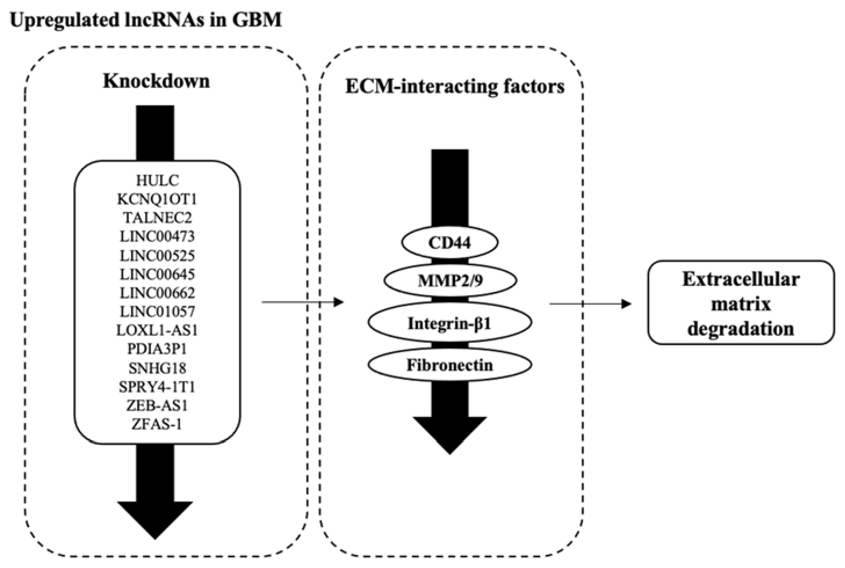
3.5. Factors Facilitating ECM Degradation Are Affected by lncRNAs’ Dysregulation

From the results, we obtained lncRNAs which interact with several factors that are integral in maintaining the integrity of ECM. These factors are CD44, MMP2/9, Integrin, and Fibronectin (Figure 5, Table 4). The downregulation of oncogenic lncRNAs through knockdown led to the subsequent downregulation of these ECM factors. Only one lncRNA, SLC8A1-AS1, was recorded to upregulate claudin expression following knockdown.

3.6. Downregulated lncRNAs and Their Potential Tumour-Suppressive Roles in GBM MES Transition

We identified 10 downregulated lncRNAs, which are tabulated in Table 5. As a contrast to the upregulated lncRNAs, the overexpression of the downregulated lncRNAs demonstrated the increased expression of E-cadherins and decreased expression of N-cadherin and vimentin. An opposite trend was observed where the inactivation of these downregulated lncRNAs is associated with decreased expression of E-cadherins and increased expression of N-cadherin and vimentin. Similarly, changes in the expression of these downregulated lncRNAs showed an opposite trend as well in the case of expression levels of the EMT-TFs (Slug, Snai1, ZEB1, and Twist) and ECM components (MMP2/9, fibronectin) which is inversely proportional to the expression of these downregulated lncRNAs.

3.7. Long Non-Coding RNAs Are Dysregulated in the Clinical Setting

Our analysis has observed a total of 41 lncRNAs from the 62 lncRNAs identified in our systematic search to be present in the clinical dataset (complete data available in File S1). Among these lncRNAs, 17 were found to be upregulated (FC > 2), while 8 were found to be downregulated (FC < 0.5). The top-10 upregulated and top-8 downregulated lncRNAs are tabulated in Table 6 and Table 7, respectively. Analysis was also performed to compare the expression of lncRNAs within GBM samples, including treatment received vs. no treatment received, primary vs. recurrent tumours, and IDH-wildtype versus IDH-mutant. No lncRNA of significance which is affiliated with these parameters was obtained.

3.8. Dysregulated lncRNAs Affect Cell Phenotype through Transcriptional and Translational Regulation

Next, we obtained the protein interactions of these lncRNAs from the RAID database (complete data available in File S1). We performed GSEA for the sets of proteins associated with upregulated lncRNAs (HOXAS3, H19, HOTTIP) and downregulated lncRNAs (MEG3, DGCR5, XIST) (data available in File S2. Figures S1–S6), GSEA through KEGG pathways demonstrated that the pathway “RNA transport” is enriched in three lncRNAs—HOXAS3, DGCR5, and XIST. GSEA through GO-BP and MF showed that the proteins associated with these lncRNAs mostly function as a transcriptional or translational control, with GO BP annotations related to translation, and transcription is highly enriched. GSEA through GO MF showed that these proteins primarily function through binding with RNA or binding at the transcription region.

4. Discussion

To date, the roles of any specific lncRNAs in cancers or specifically in GBM are still debatable. This is due to contradictory results in either pro- or anti-cancer effects or in terms of biological pathways that were observed between different cell lines and between cell and animal models. This was reflected similarly in our results, with the lncRNA MEG3 being an example. As the genetic makeup of cancer cells is highly heterogenous, it would be necessary to pinpoint the exact role of specific lncRNAs affected in any particular biological setting to allow the identification of the mechanism being regulated. Further development of lncRNAs without knowing their exact role within individualised cancer samples would be redundant.

4.1. EMT Markers E- and N-Cadherins Are Commonly Used as Markers of MES Transition in GBM

Both classical cadherins, E- and N-cadherins, were investigated in almost all of the studies as an indication of a GBM MES transition. As previously mentioned, cadherin expression in the neuronal setting has observed contradictory and inconsistent results. Noh et al. (2017) evaluated the expression of both cadherins in glioma tissues and cell lines and concluded their limited prognostic value in gliomas. With this in mind, developing a genomic profile for gliomas would provide a more accurate and indicative alternative to the current cadherin solution for the detection of an EMT-like process. Potential factors that would be a crucial element in such genomic profiles would be lncRNAs, EMT-TFs, and also activities of biological signalling pathways.

4.2. Transcription Factors for EMT Play an Important Role in the Overarching Regulation of MES Transition in GBM

Our analysis observed a huge number of lncRNAs that are capable of regulating the expression and activity of EMT transcription factors (ZEB1, Slug, Snail, Twist1) which modulate the expression of the typical EMT markers (E-cadherin, N-cadherin, and vimentin) and concomitantly induce MES transition in GBM. Among these transcription factors, Snai1 and ZEB1 are the most frequently studied and are regulated by 13 lncRNAs. These transcription factors are found to be the central regulators that modulate the expression of the EMT markers. For instance, Snai1 has been heavily implicated as the master regulator of EMT, where its overexpression can lead to the repression of E-cadherin [96]. Twist, consisting of Twist1/2, on the other hand, can also suppress E-cadherin which increases the expression of mesenchymal markers [97]. Another important transcription factor that drives GBM MES transition is ZEB1 [98].
In our analysis, we observed the interplays amongst the transcription factors, where ZEB1 activity was regulated by both Snai1 and Twist [99]. However, knowing the intrinsic mechanism among the transcription factors and whether their expressions are directly affected by lncRNA cannot be concluded at the current stage. Two lncRNAs identified in this study, DGCR5 and ZFAS-1, were shown to affect the expression of multiple EMT transcription factors. From our clinical database analysis, DGCR5 is one of the downregulated lncRNAs, which may suggest its tumour-suppressive role. To further verify the function of DGCR5, GSEA was performed on the on the proteins which were reported to interact with DGCR5. GSEA analysis inferred the potential translational silencing effect of DGCR5 on oncogenic target proteins via binding with a miRNA target in the canonical lncRNA–miRNA–mRNA signalling axis, which supports its tumour-suppressive role.

4.3. Transcription Factors of EMT Influence Components of Cancer-Associated Signalling Pathways Which Leads to GBM MES Transition

Our current analysis focuses the literature search on several prominent pathways which were reported to be affiliated with the EMT process. We observed several interplays among the signalling pathways that regulate the activity of the EMT transcription factors and EMT markers. PI3K signalling can be hyperactivated through activation of TGF-β, where TGF-β activation happens through the phosphorylation of Twist, leading to the subsequent observed loss of E-cadherin [100]. Downstream activation of PI3K leads to subsequent AKT and mTOR activation, which in turn upregulates Snai1 and also NF-κB. Meanwhile, the downstream activation of NF-κB activates multiple EMT transcription factors such as ZEB1, Slug, and Twist [101,102]. From our analysis, both PI3K and Wnt signalling are regulated by the highest numbers of lncRNAs. However, in these individual studies, the interplays between signalling pathways are often not investigated, which indicates the need for further validations on these lncRNAs to allow the identification of the specific molecular pathways affected. In hindsight, although our analysis demonstrated that lncRNAs regulate EMT transcription factors and EMT-associated biological signalling pathways, their direct association cannot be concluded at the current stage. For example, our systematic literature search demonstrated that the knockdown of H19 downregulated ZEB1 and Wnt signalling [60]. However, the direct correlation of Wnt signalling with ZEB1 regulation was unclear in the study. Moreover, the activity of Wnt can also be regulated through TGF-β, which is ZEB1-dependent. This further signifies the complex nature of lncRNA regulation of EMT at various biological levels.

4.4. Long Non-Coding RNAs Influence GBM Microenvironment through Various ECM Components Which Contribute to MES Transition

From our analysis, we also observed lncRNAs which modulated the expression of the ECM components, including matrix metalloproteins (MMPs), fibronectins, CD44, claudin, and integrin-β1. Matrix metalloproteinases (MMPs) are a family of endoproteases, which function to breakdown various proteins in the extracellular matrix (ECM), ultimately leading to their degradation [103]. Degradation of the ECM is closely associated with EMT and tumour cell invasion. In this sense, MMPs are also referred to as an initiator for EMT. In a clinical study, a low level of MMP2 is associated with longer mean survival amongst GBM patients, and its increased level is positively associated with increased tumour grades and recurrence [104]. In our analysis, the expression of two MMPs, MMP2 and MMP9, was frequently influenced by lncRNAs such as HULC, SNHG11, ZEB-AS1, and ZFAS-1. Additionally, both MMPs have been reported to be involved in ZEB1 signalling and Snai1 [105,106]. Similarly, there were lncRNAs identified (HULC, SNHG18, and ZFAS-1) which influence the expression of MMPs and are reported to regulate the EMT transcription factors. Based on this, a linkage between the lncRNAs that regulate the activity of the transcription factors (Snail and ZEB1), which in turn regulate the ECM components (including MMPs), can be observed during MES transition in GBM. Figure 6 illustrates the tight regulations of EMT-TFs and ECM-interacting factors by the signalling pathways highlighted in this study pertaining to the MES process in GBM. LncRNAs were reported to exhibit regulatory activity at various levels in this overarching signalling axis, which emphasised the importance of the accurate characterisation of lncRNAs’ roles within the GBM setting.

4.5. Research Is Lacking Outside of Cell Studies to Observe Clinical Relevance of the lncRNAs in GBM MES Transition

To obtain further insights into lncRNAs within the clinical setting, our analysis observed lncRNAs such as HOXC13-AS being similarly overexpressed in vitro and up to 68-fold in GBM tissues. Similarly, the downregulated expression patterns of MEG3, DGCR5, and LINC-PINT recorded from our literature search also match with the expression data in our analysis of GBM clinical samples (0.11, 0.12, and 0.50-fold, respectively). Interestingly, several upregulated lncRNAs obtained from our systematic search were found to be downregulated instead in the clinical setting. Examples of such lncRNAs are NEAT1 and UCA1, where both of which are widely studied oncogenic lncRNAs. A possible explanation for these inconsistencies could be attributed to the limitations of the current GBM experimental setup. Furthermore, additional analysis was performed to compare the expression of lncRNAs obtained in this study among various GBM clinical sample categories, including parameters such as intervention received and primary or recurrent tumours. From the analysis, only one lncRNA, LINC00645, was upregulated in temozolomide-treated GBM samples compared to non-treated GBM samples. This suggest that the exposure to TMZ may induce molecular interactions with the lncRNAs. Additionally, the exposure of TMZ was known to induce epigenetic changes, which may further affect or influence the expression of the lncRNAs. As for other lncRNAs subjected to TMZ-related analysis, no results of significance were obtained. This can be due to the following: (i) the expression of lncRNAs might not be affected by these parameters, (ii) the sample sizes of GBM clinical samples with these specific criteria are limited, and (iii) the out-of-date statuses of these publicly available expression profiles of GBM clinical samples have greatly limited the information available on the lncRNAs to be effectively compared.
Perhaps the most exigent need for EMT studies is to understand the in-depth process attributed to the high cell invasive phenotype of GBM, and this would expedite the development of anti-EMT therapies to palliate the current negative outcomes among GBM patients. Further, the EMT-like process and the associated factors discussed previously are also heavily implicated with the acquired chemoresistance, especially temozolomide (TMZ) resistance, in GBM. Although whether the acquisition of TMZ resistance and EMT are linked events remains unclear in our current understanding, some studies have reported the involvement of EMT factors in TMZ resistance, which include ZEB1, Slug, and vimentin [107,108,109].

4.6. Future Perspective in lncRNA Studies: Moving forward from Cell Studies

Practical application of lncRNAs in the GBM therapeutic setting is currently still at a mature stage. There are several early clinical trials which were carried out in other cancers but not in GBM [110]. Considering the heterogeneity observed within the genetic profiles between GBM patients, alongside the current practice for GBM diagnosis, development of an effective approach for GBM diagnosis is imperative. In this sense, lncRNAs serve as a potential tool that could be further developed for GBM diagnosis. However, much more effort is still required in characterising novel lncRNAs to allow a more in-depth understanding of the common genetic representation in GBM to allow the effective development of lncRNAs as reliable GBM genetic biomarkers. Additionally, it was seen that most research tends to focus on the upregulated, oncogenic lncRNAs compared to the tumour-suppressive lncRNAs. Future research can focus on tumour-suppressive lncRNAs as they can also be a potential molecular target in the EMT-like process in GBM. Additionally, the different expression profiles of some lncRNAs within in vitro models compared with those in a clinical setting accentuate the need for future studies using a GBM model with a closer-to-truth representation of genomic studies within the brain setting. There are several considerations that in vitro models will not be able to address fully, as they do not emulate the actual brain environment which will deaden the veracity of such studies. In contrast, 3D culture models could be a potential bridging tool to facilitate the convergence of 2D cell models to animal models. A systematic review performed by Phon et al. has found dissimilarities between genomic profiles of 3D and 2D culture models, where 3D cultures can simulate the brain microenvironment to a closer degree [111]. This further reinforces the need to verge upon the junctions between various pre-clinical models to allow the excogitation of lncRNA functions to a more precise level.

5. Conclusions

Our systematic analysis has identified upregulated and downregulated lncRNAs that confer oncogenic or tumour-suppressive properties that influence the GBM MES transition in GBM with a complex signalling regulation between the various EMT transcription factors, ECM components, and classical EMT markers (vimentin, E-cadherin, and N-cadherin). This GBM MES transition by lncRNAs is regulated by complex interplays between Wnt/β-catenin, PI3K/Akt, TGF-β, and NF-κB pathways. Nevertheless, the current understanding of the interplay between the signalling pathways and EMT factors is still immature. The complexity of various interplays between these factors implicated the importance of further validation studies to confirm the direct relation and crosstalk amongst factors in the signalling involved. Additionally, the disparity of lncRNAs’ expression further warrants the usage of more suitable pre-clinical models to allow the elucidation of these regulatory mechanisms relevant to the clinical settings.

Supplementary Materials

The following supporting information can be downloaded at: https://www.mdpi.com/article/10.3390/biology12060818/s1, File S1: List of dysregulated lncRNAs identified in clinical dataset and all lncRNA-protein interactions obtained from RAID database for selected lncRNAs; File S2: GSEA of lncRNAs (HOXAS3, H19, HOTTIP, MEG3, DGCR5, and XIST) through GO-BP, GO-MF, and KEGG Pathways.

Author Contributions

D.H.L.L. and M.N.A.K. conceptualised the project, where D.H.L.L. developed the study and M.N.A.K. oversaw overall direction and planning. M.N.A.K., A.K.R. and M.S. administered the project equally. D.H.L.L. performed the literature search and D.H.L.L., B.W.S.P. and M.N.A.K. screened the data. D.H.L.L. and B.W.S.P. extracted and verified the data and conflicts were resolved by M.N.A.K. and D.H.L.L. synthesised study data, created figures and tables, while D.H.L.L. and B.W.S.P. performed bioinformatic analyses, which all were reviewed by M.N.A.K., M.S., A.K.R. and D.H.L.L. drafted the manuscript with contributions from B.W.S.P., M.N.A.K., M.S. and A.K.R. in the review and critical editing. All authors have read and agreed to the published version of the manuscript.

Funding

This study was funded by a collaborative research grant from the Jeffrey Cheah School of Medicine and Health Sciences, Monash University Malaysia (Project Code: SED-000019).

Institutional Review Board Statement

Not applicable.

Informed Consent Statement

Not applicable.

Data Availability Statement

Not applicable.

Acknowledgments

Figure 3 is created by Cytoscape. D.H.L.L. and B.W.S.P. are supported by a Ph.D. scholarship funded by Monash University Malaysia. The authors would also like to thank Jeffrey Cheah School of Medicine and Health Science, Monash University for the support given.

Conflicts of Interest

The authors declare no conflict of interest.

References

  1. Louis, D.N.; Perry, A.; Wesseling, P.; Brat, D.J.; Cree, I.A.; Figarella-Branger, D.; Hawkins, C.; Ng, H.K.; Pfister, S.M.; Reifenberger, G.; et al. The 2021 WHO Classification of Tumors of the Central Nervous System: A summary. Neuro Oncol. 2021, 23, 1231–1251. [Google Scholar] [CrossRef] [PubMed]
  2. So, J.S.; Kim, H.; Han, K.S. Mechanisms of Invasion in Glioblastoma: Extracellular Matrix, Ca(2+) Signaling, and Glutamate. Front. Cell Neurosci. 2021, 15, 663092. [Google Scholar] [CrossRef] [PubMed]
  3. Verhaak, R.G.; Hoadley, K.A.; Purdom, E.; Wang, V.; Qi, Y.; Wilkerson, M.D.; Miller, C.R.; Ding, L.; Golub, T.; Mesirov, J.P.; et al. Integrated genomic analysis identifies clinically relevant subtypes of glioblastoma characterized by abnormalities in PDGFRA, IDH1, EGFR, and NF1. Cancer Cell 2010, 17, 98–110. [Google Scholar] [CrossRef] [PubMed] [Green Version]
  4. Liu, C.; Zong, H. Developmental origins of brain tumors. Curr. Opin. Neurobiol. 2012, 22, 844–849. [Google Scholar] [CrossRef] [PubMed] [Green Version]
  5. Vollmann-Zwerenz, A.; Leidgens, V.; Feliciello, G.; Klein, C.A.; Hau, P. Tumor Cell Invasion in Glioblastoma. Int. J. Mol. Sci. 2020, 21, 1932. [Google Scholar] [CrossRef] [Green Version]
  6. Iwadate, Y. Epithelial-mesenchymal transition in glioblastoma progression. Oncol. Lett. 2016, 11, 1615–1620. [Google Scholar] [CrossRef] [Green Version]
  7. Roche, J. The Epithelial-to-Mesenchymal Transition in Cancer. Cancers 2018, 10, 52. [Google Scholar] [CrossRef] [Green Version]
  8. Zeisberg, M.; Neilson, E.G. Biomarkers for epithelial-mesenchymal transitions. J. Clin. Investig. 2009, 119, 1429–1437. [Google Scholar] [CrossRef] [Green Version]
  9. Ansieau, S.; Collin, G.; Hill, L. EMT or EMT-Promoting Transcription Factors, Where to Focus the Light? Front. Oncol. 2014, 4, 353. [Google Scholar] [CrossRef] [Green Version]
  10. Liu, X.; Yun, F.; Shi, L.; Li, Z.H.; Luo, N.R.; Jia, Y.F. Roles of Signaling Pathways in the Epithelial-Mesenchymal Transition in Cancer. Asian Pac. J. Cancer Prev. 2015, 16, 6201–6206. [Google Scholar] [CrossRef] [Green Version]
  11. Noh, M.G.; Oh, S.J.; Ahn, E.J.; Kim, Y.J.; Jung, T.Y.; Jung, S.; Kim, K.K.; Lee, J.H.; Lee, K.H.; Moon, K.S. Prognostic significance of E-cadherin and N-cadherin expression in Gliomas. BMC Cancer 2017, 17, 583. [Google Scholar] [CrossRef] [PubMed] [Green Version]
  12. Leung, D.H.L. Long non-coding RNA in glioblastoma invasion: Angiogenesis and mesenchymal transition via PI3K and Wnt signalling. Asia Pac. J. Mol. Biol. Biotechnol. 2023, 31, 36–52. [Google Scholar] [CrossRef]
  13. Statello, L.; Guo, C.-J.; Chen, L.-L.; Huarte, M. Gene regulation by long non-coding RNAs and its biological functions. Nat. Rev. Mol. Cell Biol. 2021, 22, 96–118. [Google Scholar] [CrossRef] [PubMed]
  14. Gugnoni, M.; Ciarrocchi, A. Long Noncoding RNA and Epithelial Mesenchymal Transition in Cancer. Int. J. Mol. Sci. 2019, 20, 1924. [Google Scholar] [CrossRef] [Green Version]
  15. O’Brien, S.J.; Bishop, C.; Hallion, J.; Fiechter, C.; Scheurlen, K.; Paas, M.; Burton, J.; Galandiuk, S. Long non-coding RNA (lncRNA) and epithelial-mesenchymal transition (EMT) in colorectal cancer: A systematic review. Cancer Biol. 2020, 21, 769–781. [Google Scholar] [CrossRef] [PubMed]
  16. Xu, Q.; Deng, F.; Qin, Y.; Zhao, Z.; Wu, Z.; Xing, Z.; Ji, A.; Wang, Q.J. Long non-coding RNA regulation of epithelial–mesenchymal transition in cancer metastasis. Cell Death Dis. 2016, 7, e2254. [Google Scholar] [CrossRef]
  17. Goldman, M.J.; Craft, B.; Hastie, M.; Repečka, K.; McDade, F.; Kamath, A.; Banerjee, A.; Luo, Y.; Rogers, D.; Brooks, A.N.; et al. Visualizing and interpreting cancer genomics data via the Xena platform. Nat. Biotechnol. 2020, 38, 675–678. [Google Scholar] [CrossRef]
  18. Cerami, E.; Gao, J.; Dogrusoz, U.; Gross, B.E.; Sumer, S.O.; Aksoy, B.A.; Jacobsen, A.; Byrne, C.J.; Heuer, M.L.; Larsson, E.; et al. The cBio cancer genomics portal: An open platform for exploring multidimensional cancer genomics data. Cancer Discov. 2012, 2, 401–404. [Google Scholar] [CrossRef] [Green Version]
  19. Gao, J.; Aksoy, B.A.; Dogrusoz, U.; Dresdner, G.; Gross, B.; Sumer, S.O.; Sun, Y.; Jacobsen, A.; Sinha, R.; Larsson, E.; et al. Integrative analysis of complex cancer genomics and clinical profiles using the cBioPortal. Sci. Signal. 2013, 6, pl1. [Google Scholar] [CrossRef] [Green Version]
  20. Vivian, J.; Rao, A.A.; Nothaft, F.A.; Ketchum, C.; Armstrong, J.; Novak, A.; Pfeil, J.; Narkizian, J.; Deran, A.D.; Musselman-Brown, A.; et al. Toil enables reproducible, open source, big biomedical data analyses. Nat. Biotechnol. 2017, 35, 314–316. [Google Scholar] [CrossRef] [Green Version]
  21. Li, J.; Wang, Y.M.; Song, Y.L. Knockdown of long noncoding RNA AB073614 inhibits glioma cell proliferation and migration via affecting epithelial-mesenchymal transition. Eur. Rev. Med. Pharmacol. Sci. 2016, 20, 3997–4002. [Google Scholar] [PubMed]
  22. Sun, Y.; Shen, Y.; Li, X. Knockdown of long non-coding RNA AGAP2-AS1 suppresses the proliferation and metastasis of glioma by targeting microRNA-497-5p. Bioengineered. 2021. [Google Scholar] [CrossRef] [PubMed]
  23. Zhang, D.L.; Zhou, H.G.; Liu, J.; Mao, J. Long Noncoding RNA ASB16-AS1 Promotes Proliferation, Migration, and Invasion in Glioma Cells. Biomed Res. Int. 2019, 2019, 10. [Google Scholar] [CrossRef] [PubMed] [Green Version]
  24. Li, X.S.; Qi, S.J.; Ma, D.Z.; Fan, J.B.; Wang, J.T. Long non-coding RNA BLACAT1 promotes the proliferation and invasion of glioma cells via Wnt/beta-catenin signaling. Exp. Ther. Med. 2019, 17, 4703–4708. [Google Scholar] [CrossRef] [Green Version]
  25. Cui, B.Z.; Li, B.S.; Liu, Q.; Cui, Y.Q. lncRNA CCAT1 Promotes Glioma Tumorigenesis by Sponging miR-181b. J. Cell. Biochem. 2017, 118, 4548–4557. [Google Scholar] [CrossRef] [PubMed]
  26. Zeng, J.; Du, T.P.; Song, Y.F.; Gao, Y.; Li, F.Y.; Wu, R.M.; Chen, Y.J.; Li, W.; Zhou, H.; Yang, Y.; et al. Knockdown of Long Noncoding RNA CCAT2 Inhibits Cellular Proliferation, Invasion, and Epithelial-Mesenchymal Transition in Glioma Cells (vol 25, pg 913, 2017). Oncol. Res. 2020, 28, 551–552. [Google Scholar] [CrossRef]
  27. Li, Y.F.; Zong, J.; Zhao, C. lncRNA CTBP1-AS2 promotes proliferation and migration of glioma by modulating miR-370-3p-Wnt7a-mediated epithelial-mesenchymal transition. Biochem. Cell Biol. 2020, 98, 661–668. [Google Scholar] [CrossRef]
  28. Yang, J.X.; Sun, Y.; Gao, L.; Meng, Q.; Yang, B.Y. Long non-coding RNA DANCR facilitates glioma malignancy by sponging miR-33a-5p. Neoplasma 2018, 65, 790–798. [Google Scholar] [CrossRef] [Green Version]
  29. Lv, Q.L.; Wang, L.C.; Li, D.C.; Lin, Q.X.; Shen, X.L.; Liu, H.Y.; Li, M.; Ji, Y.L.; Qin, C.Z.; Chen, S.H. Knockdown lncRNA DLEU1 Inhibits Gliomas Progression and Promotes Temozolomide Chemosensitivity by Regulating Autophagy. Front. Pharmacol. 2020, 11, 15. [Google Scholar] [CrossRef]
  30. Xu, D.W.; Liu, R.H.; Meng, L.; Zhang, Y.; Lu, G.J.; Ma, P.J. Long non-coding RNA ENST01108 promotes carcinogenesis of glioma by acting as a molecular sponge to modulate miR-489. Biomed. Pharmacother. 2018, 100, 20–28. [Google Scholar] [CrossRef]
  31. Dong, H.X.; Cao, W.; Xue, J. Long noncoding FOXD2-AS1 is activated by CREB1 and promotes cell proliferation and metastasis in glioma by sponging miR-185 through targeting AKT1. Biochem. Biophys. Res. Commun. 2019, 508, 1074–1081. [Google Scholar] [CrossRef] [PubMed]
  32. Shen, F.Z.; Chang, H.G.; Gao, G.J.; Zhang, B.; Li, X.S.; Jin, B.Z. Long noncoding RNA FOXD2-AS1 promotes glioma malignancy and tumorigenesis via targeting miR-185-5p/CCND2 axis. J. Cell. Biochem. 2019, 120, 9324–9336. [Google Scholar] [CrossRef]
  33. Zhao, J.; Zeng, X.B.; Zhang, H.Y.; Xiang, J.W.; Liu, Y.S. Long non-coding RNA FOXD2-AS1 promotes cell proliferation, metastasis and EMT in glioma by sponging miR-506-5p. Open Med. 2020, 15, 921–931. [Google Scholar] [CrossRef] [PubMed]
  34. Xiang, Z.J.; Lv, Q.L.; Chen, X.R.; Zhu, X.T.; Liu, S.K.; Li, D.C.; Peng, X.D. Lnc GNG12-AS1 knockdown suppresses glioma progression through the AKT/GSK-3 beta/beta-catenin pathway. Biosci. Rep. 2020, 40, 10. [Google Scholar] [CrossRef] [PubMed]
  35. Li, J.Y.; Ji, X.J.; Wang, H.D. Targeting Long Noncoding RNA HMMR-AS1 Suppresses and Radiosensitizes Glioblastoma. Neoplasia 2018, 20, 456–466. [Google Scholar] [CrossRef]
  36. Zhang, S.Y.; Wang, W.W.; Liu, G.X.; Xie, S.L.; Li, Q.X.; Li, Y.R.; Lin, Z.Y. Long non-coding RNA HOTTIP promotes hypoxia-induced epithelial-mesenchymal transition of malignant glioma by regulating the miR-101/ZEB1 axis. Biomed. Pharmacother. 2017, 95, 711–720. [Google Scholar] [CrossRef]
  37. Chen, W.H.; Li, Q.Y.; Zhang, G.L.; Wang, H.; Zhu, Z.H.; Chen, L.K. LncRNA HOXA-AS3 promotes the malignancy of glioblastoma through regulating miR-455-5p/USP3 axis. J. Cell. Mol. Med. 2020, 24, 11755–11767. [Google Scholar] [CrossRef]
  38. Liu, N.; Wang, Z.Y.; Liu, D.C.; Xie, P. HOXC13-AS-miR-122-5p-SATB1-C-Myc feedback loop promotes migration, invasion and EMT process in glioma. Oncotargets Ther. 2019, 12, 7165–7172. [Google Scholar] [CrossRef] [Green Version]
  39. Gao, T.H.; Gu, G.Y.; Tian, J.X.; Zhang, R.; Zheng, X.R.; Wang, Y.A.; Pang, Q.; Liu, Q. LncRNA HSP90AA1-IT1 promotes gliomas by targeting miR-8855p-CDK2 pathway. Oncotarget 2017, 8, 75284–75297. [Google Scholar] [CrossRef] [Green Version]
  40. Yin, T.T.; Wu, J.; Hu, Y.C.; Zhang, M.; He, J. Long non-coding RNA HULC stimulates the epithelial-mesenchymal transition process and vasculogenic mimicry in human glioblastoma. Cancer Med. 2021, 10, 5270–5282. [Google Scholar] [CrossRef]
  41. Ding, P.F.; Liang, B.; Shou, J.X.; Wang, X.J. lncRNA KCNQ1OT1 promotes proliferation and invasion of glioma cells by targeting the miR-375/YAP pathway. Int. J. Mol. Med. 2020, 46, 1983–1992. [Google Scholar] [CrossRef] [PubMed]
  42. Li, W.; Soufiany, I.; Lyu, X.; Lu, C.; Wei, Y.; Shi, Z.; You, Y. SP1-upregulated LBX2-AS1 promotes the progression of glioma by targeting the miR-491-5p/LIF axis. J. Cancer 2021, 13, 6989–7002. [Google Scholar] [CrossRef] [PubMed]
  43. Tang, J.; Yu, B.; Li, Y.; Zhang, W.; Alvarez, A.A.; Hu, B.; Cheng, S.Y.; Feng, H. TGF-β-activated lncRNA LINC00115 is a critical regulator of glioma stem-like cell tumorigenicity. EMBO Rep. 2019, 20, e48170. [Google Scholar] [CrossRef] [PubMed]
  44. Brodie, S.; Lee, H.K.; Jiang, W.; Cazacu, S.; Xiang, C.L.; Poisson, L.M.; Datta, I.; Kalkanis, S.; Ginsberg, D.; Brodie, C. The novel long non-coding RNA TALNEC2, regulates tumor cell growth and the stemness and radiation response of glioma stem cells. Oncotarget 2017, 8, 31798–31814. [Google Scholar] [CrossRef] [Green Version]
  45. Cai, J.Q.; Zhang, J.W.; Wu, P.F.; Yang, W.T.; Ye, Q.L.; Chen, Q.; Jiang, C.L. Blocking LINC00152 suppresses glioblastoma malignancy by impairing mesenchymal phenotype through the miR-612/AKT2/NF-B pathway. J. Neuro-Oncol. 2018, 140, 225–236. [Google Scholar] [CrossRef]
  46. Reon, B.J.; Karia, B.T.R.; Kiran, M.; Dutta, A. LINC00152 Promotes Invasion through a 3′-Hairpin Structure and Associates with Prognosis in Glioblastoma. Mol. Cancer Res. 2018, 16, 1470–1482. [Google Scholar] [CrossRef] [Green Version]
  47. Li, F.; Shen, Z.Z.; Xiao, C.M.; Sha, Q.K. YY1-mediated up-regulation of lncRNA LINC00466 facilitates glioma progression via miR-508/CHEK1. J. Gene Med. 2021, 23, 15. [Google Scholar] [CrossRef]
  48. Zhang, Q.S.; Wang, G.W.; Xu, L.P.; Yao, Z.Q.; Song, L.J. Long non-coding RNA LINC00473 promotes glioma cells proliferation and invasion by impairing miR-637/CDK6 axis. Artif. Cells Nanomed. Biotechnol. 2019, 47, 3896–3903. [Google Scholar] [CrossRef] [Green Version]
  49. Du, X.L.; Tu, Y.M.; Liu, S.; Zhao, P.Z.; Bao, Z.Y.; Li, C.; Li, J.H.; Pan, M.H.; Ji, J. LINC00511 contributes to glioblastoma tumorigenesis and epithelial-mesenchymal transition via LINC00511/miR-524-5p/YB1/ZEB1 positive feedback loop. J. Cell. Mol. Med. 2020, 24, 1474–1487. [Google Scholar] [CrossRef] [Green Version]
  50. Liu, Z.; Tao, B.; Li, L.; Liu, P.; Xia, K.; Zhong, C. LINC00511 knockdown suppresses glioma cell malignant progression through miR-15a-5p/AEBP1 axis. Brain Res. Bull. 2021, 173, 82–96. [Google Scholar] [CrossRef]
  51. Wan, Y.; Liang, F.; Wei, M.J.; Liu, Y. Long non-coding RNA LINC00525 regulates the proliferation and epithelial to mesenchymal transition of human glioma cells by sponging miR-338-3p. AMB Express 2020, 10, 9. [Google Scholar] [CrossRef]
  52. Li, C.L.; Zheng, H.S.; Hou, W.L.; Bao, H.B.; Xiong, J.S.; Che, W.L.; Gu, Y.F.; Sun, H.M.; Liang, P. Long non-coding RNA linc00645 promotes TGF-beta-induced epithelial-mesenchymal transition by regulating miR-205-3p-ZEB1 axis in glioma. Cell Death Dis. 2019, 10, 17. [Google Scholar] [CrossRef] [Green Version]
  53. Geng, Y.B.; Wu, Y.L.; Xu, C.; Li, T.; Zhang, L.W. Long Non-Coding RNA LINC00662 Regulated Proliferation and Migration by Targeting miR-34a-5p/LMAN2L Axis in Glioma. Oncotargets Ther. 2020, 13, 10161–10172. [Google Scholar] [CrossRef] [PubMed]
  54. Tang, G.D.; Luo, L.Y.; Zhang, J.L.; Zhai, D.F.; Huang, D.Q.; Yin, J.; Zhou, Q.; Zhang, Q.; Zheng, G.P. lncRNA LINC01057 promotes mesenchymal differentiation by activating NF-kappa B signaling in glioblastoma. Cancer Lett. 2021, 498, 152–164. [Google Scholar] [CrossRef] [PubMed]
  55. Lulli, V.; Buccarelli, M.; Ilari, R.; Castellani, G.; De Dominicis, C.; Di Giamberardino, A.; D’Alessandris, Q.G.; Giannetti, S.; Martini, M.; Stumpo, V.; et al. Mir-370-3p Impairs Glioblastoma Stem-Like Cell Malignancy Regulating a Complex Interplay between HMGA2/HIF1A and the Oncogenic Long Non-Coding RNA (lncRNA) NEAT1. Int. J. Mol. Sci. 2020, 21, 21. [Google Scholar] [CrossRef] [PubMed]
  56. Yu, H.; Xu, A.; Wu, B.; Wang, M.; Chen, Z. Long noncoding RNA NEAT1 promotes progression of glioma as a ceRNA by sponging miR-185-5p to stimulate DNMT1/mTOR signaling. J. Cell Physiol. 2021, 236, 121–130. [Google Scholar] [CrossRef]
  57. Pan, T.; Xue, M. LncRNA-NNT-AS1 contributes to the progression of glioma by miR-582-5p/EZH2 axis. Cytotechnology 2021, 73, 473–482. [Google Scholar] [CrossRef]
  58. Wang, S.B.; Qi, Y.H.; Gao, X.; Qiu, W.; Liu, Q.L.; Guo, X.F.; Qian, M.Y.; Chen, Z.H.; Zhang, Z.P.; Wang, H.Z.; et al. Hypoxia-induced lncRNA PDIA3P1 promotes mesenchymal transition via sponging of miR-124-3p in glioma. Cell Death Dis. 2020, 11, 17. [Google Scholar] [CrossRef] [Green Version]
  59. Bi, Y.Y.; Ji, J.; Zhou, Y.X. LncRNA-PVT1 indicates a poor prognosis and promotes angiogenesis via activating the HNF1B/EMT axis in glioma. J. Cancer 2021, 12, 5732–5744. [Google Scholar] [CrossRef]
  60. Chang, L.; Wang, J.; Zhou, F.; Wang, D.; Chen, R.; Zhang, Y.; Zhang, J. LncRNA RP11-84E24.3 drives tumorigenesis and epithelial-to-mesenchymal transition of glioma cells by promoting TFAP2C-mediated activation of SNAI1. J. Neurooncol. 2021, 151, 157–171. [Google Scholar] [CrossRef]
  61. Ni, H.Z.; Wang, K.; Xie, P.; Zuo, J.D.; Liu, W.G.; Liu, C. LncRNA SAMMSON Knockdown Inhibits the Malignancy of Glioblastoma Cells by Inactivation of the PI3K/Akt Pathway. Cell. Mol. Neurobiol. 2021, 41, 79–90. [Google Scholar] [CrossRef] [PubMed]
  62. He, L.; Yang, H.; Zhu, X.L.; Zhang, Y.; Lv, K. Knockdown of long non-coding RNA SLC8A1-AS1 attenuates cell invasion and migration in glioma via suppression of Wnt/beta-catenin signaling pathways. Brain Res. Bull. 2021, 176, 112–120. [Google Scholar] [CrossRef] [PubMed]
  63. Geng, Y.B.; Xu, C.; Wang, Y.; Zhang, L.W. Long non-coding RNA SNHG11 promotes cell proliferation, invasion and migration in glioma by targeting miR-154-5p. Eur. Rev. Med. Pharmacol. Sci. 2020, 24, 4901–4908. [Google Scholar] [PubMed]
  64. Zheng, R.; Yao, Q.; Li, X.; Xu, B. Long Noncoding Ribonucleic Acid SNHG18 Promotes Glioma Cell Motility via Disruption of α-Enolase Nucleocytoplasmic Transport. Front. Genet. 2019, 10, 1140. [Google Scholar] [CrossRef] [PubMed] [Green Version]
  65. Meng, Q.; Yang, B.Y.; Liu, B.; Yang, J.X.; Sun, Y. Long non-coding RNA SNHG6 promotes glioma tumorigenesis by sponging miR-101-3p. Int. J. Biol. Mrk. 2018, 33, 148–155. [Google Scholar] [CrossRef] [Green Version]
  66. Nie, J.; Feng, Y.; Wang, H.; Lian, X.Y.; Li, Y.F. Long Non-Coding RNA SNHG6 Supports Glioma Progression Through Upregulation of Notch1, Sox2, and EMT. Front. Cell Dev. Biol. 2021, 9, 7. [Google Scholar] [CrossRef]
  67. Liu, H.; Lv, Z.; Guo, E. Knockdown of long noncoding RNA SPRY4-IT1 suppresses glioma cell proliferation, metastasis and epithelial-mesenchymal transition. Int. J. Clin. Exp. Pathol. 2015, 8, 9140–9146. [Google Scholar]
  68. Lou, J.Y.; Luo, J.; Yang, S.C.; Ding, G.F.; Liao, W.; Zhou, R.X.; Qiu, C.Z.; Chen, J.M. Long non-coding RNA SUMO1P3 promotes glioma progression via the Wnt/β-catenin pathway. Eur. Rev. Med. Pharm. Sci. 2020, 24, 9571–9580. [Google Scholar] [CrossRef]
  69. Chen, H.Y.; Liu, Z.Y.; Chang, H.; Gao, J.B.; Li, Y.J.; Chen, L.H.; Luo, Y.C.; Xu, R.X. Knockdown of long noncoding RNA UCA1 inhibits glioma cell metastasis via reduction of epithelial-mesenchymal transition. Int. J. Clin. Exp. Pathol. 2016, 9, 12621–12626. [Google Scholar]
  70. Huang, Z.; Zhao, X.Y.; Wu, X.W.; Xiang, L.; Yuan, Y.N.; Zhou, S.; Yu, W.F. LncRNA UCA1 facilitated cell growth and invasion through the miR-206/CLOCK axis in glioma. Cancer Cell Int. 2019, 19, 9. [Google Scholar] [CrossRef]
  71. Li, Z.; Liu, H.; Zhong, Q.; Wu, J.; Tang, Z. LncRNA UCA1 is necessary for TGF-β-induced epithelial-mesenchymal transition and stemness via acting as a ceRNA for Slug in glioma cells. FEBS Open Bio 2018, 8, 1855–1865. [Google Scholar] [CrossRef] [PubMed] [Green Version]
  72. Li, Z.G.; Xiang, W.C.; Shui, S.F.; Han, X.W.; Guo, D.; Yan, L. 11 Long noncoding RNA UCA1 functions as miR-135a sponge to promote the epithelial to mesenchymal transition in glioma. J. Cell. Biochem. 2020, 121, 2447–2457. [Google Scholar] [CrossRef] [PubMed]
  73. Liang, C.; Yang, Y.; Guan, J.; Lv, T.; Qu, S.; Fu, Q.; Zhao, H. LncRNA UCA1 sponges miR-204-5p to promote migration, invasion and epithelial-mesenchymal transition of glioma cells via upregulation of ZEB1. Pathol. Res. Pract. 2018, 214, 1474–1481. [Google Scholar] [CrossRef]
  74. Luo, C.; Quan, Z.; Zhong, B.; Zhang, M.; Zhou, B.; Wang, S.; Luo, X.; Tang, C. LncRNA XIST promotes glioma proliferation and metastasis through miR-133a/SOX4. Exp. Ther. Med. 2020, 19, 1641–1648. [Google Scholar] [CrossRef] [PubMed] [Green Version]
  75. Lv, Q.L.; Hu, L.; Chen, S.H.; Sun, B.; Fu, M.L.; Qin, C.Z.; Qu, Q.; Wang, G.H.; He, C.J.; Zhou, H.H. A long noncoding RNA ZEB1-AS1 promotes tumorigenesis and predicts poor prognosis in glioma. Int. J. Mol. Sci. 2016, 17, 1431. [Google Scholar] [CrossRef]
  76. Gao, K.; Ji, Z.W.; She, K.; Yang, Q.Y.; Shao, L.B. Long non-coding RNA ZFAS1 is an unfavourable prognostic factor and promotes glioma cell progression by activation of the Notch signaling pathway. Biomed. Pharmacother. 2017, 87, 555–560. [Google Scholar] [CrossRef]
  77. Lv, Q.L.; Chen, S.H.; Zhang, X.; Sun, B.; Hu, L.; Qu, Q.; Huang, Y.T.; Wang, G.H.; Liu, Y.L.; Zhang, Y.Y.; et al. Upregulation of long noncoding RNA zinc finger antisense 1 enhances epithelial-mesenchymal transition in vitro and predicts poor prognosis in glioma. Tumour Biol. 2017, 39, 1010428317695022. [Google Scholar] [CrossRef] [Green Version]
  78. Chen, X.; Li, Y.; Zuo, C.; Zhang, K.; Lei, X.; Wang, J.; Yang, Y.; Zhang, J.; Ma, K.; Wang, S.; et al. Long Non-Coding RNA H19 Regulates Glioma Cell Growth and Metastasis via miR-200a-Mediated CDK6 and ZEB1 Expression. Front. Oncol. 2021, 11, 4556. [Google Scholar] [CrossRef]
  79. Hu, Q.; Yin, J.; Zeng, A.; Jin, X.; Zhang, Z.; Yan, W.; You, Y. H19 Functions as a Competing Endogenous RNA to Regulate EMT by Sponging miR-130a-3p in Glioma. Cell. Physiol. Biochem. 2018, 50, 150–168. [Google Scholar] [CrossRef]
  80. Jia, L.; Tian, Y.; Chen, Y.; Zhang, G. The silencing of LncRNA-H19 decreases chemoresistance of human glioma cells to temozolomide by suppressing epithelial-mesenchymal transition via the Wnt/β-Catenin pathway. Onco Targets 2018, 11, 313–321. [Google Scholar] [CrossRef] [Green Version]
  81. Dong, N.; Guo, J.X.; Han, S.; Bao, L.; Diao, Y.; Lin, Z.X. Positive feedback loop of lncRNA HOXC-AS2/miR-876-5p/ZEB1 to regulate EMT in glioma. Oncotargets Ther. 2019, 12, 7601–7609. [Google Scholar] [CrossRef] [PubMed] [Green Version]
  82. Li, H.; Yuan, X.; Yan, D.; Li, D.; Guan, F.; Dong, Y.; Wang, H.; Liu, X.; Yang, B. Long Non-Coding RNA MALAT1 Decreases the Sensitivity of Resistant Glioblastoma Cell Lines to Temozolomide. Cell. Physiol. Biochem. 2017, 42, 1192–1201. [Google Scholar] [CrossRef] [PubMed] [Green Version]
  83. Wang, H.; Li, L.; Yin, L. Silencing LncRNA LOXL1-AS1 attenuates mesenchymal characteristics of glioblastoma via NF-κB pathway. Biochem. Biophys. Res. Commun. 2018, 500, 518–524. [Google Scholar] [CrossRef] [PubMed]
  84. Wang, J.; Qin, C.; Zhong, C.; Wen, Y.; Ke, S.; Liao, B. Long non-coding RNA CASC2 targeting miR-18a suppresses glioblastoma cell growth, metastasis and EMT in vitro and in vivo. J. Biosci. 2020, 45, 107. [Google Scholar] [CrossRef] [PubMed]
  85. He, Z.; Long, J.; Yang, C.; Gong, B.; Cheng, M.; Wang, Q.; Tang, J. LncRNA DGCR5 plays a tumor-suppressive role in glioma via the miR-21/Smad7 and miR-23a/PTEN axes. Aging 2020, 12, 20285–20307. [Google Scholar] [CrossRef]
  86. Yang, F.; Huang, Y.L. DGCR5 suppresses the EMT of pediatric primary glioblastoma multiforme cell and serves as a prognostic biomarker. Eur. Rev. Med. Pharmacol. Sci. 2019, 23, 10024–10034. [Google Scholar] [CrossRef]
  87. Zhu, X.P.; Pan, S.A.; Chu, Z.; Zhou, Y.X.; Huang, Y.K.; Han, D.Q. LncRNA GAS5 regulates epithelial-mesenchymal transition and viability of glioma cells by targeting microRNA-106b and regulating PTEN expression. Neurosci. Res. 2021, 170, 32–40. [Google Scholar] [CrossRef]
  88. Zhu, H.; Chen, Z.; Shen, L.; Tang, T.; Yang, M.; Zheng, X. Long Noncoding RNA LINC-PINT Suppresses Cell Proliferation, Invasion, and EMT by Blocking Wnt/β-Catenin Signaling in Glioblastoma. Front. Pharm. 2020, 11, 586653. [Google Scholar] [CrossRef]
  89. Lei, J.; Zhou, Z. Effect of long intergenic non-coding RNA 00312 on regulating biological behaviors of glioma cells by targeting microRNA-21-3p. Int. J. Clin. Exp. Med. 2021, 14, 852–863. [Google Scholar]
  90. Fu, Q.; Li, S.S.; Zhou, Q.J.; Yalikun, K.; Yisireyili, D.; Xia, M. Low LINC00599 expression is a poor prognostic factor in glioma. Biosci. Rep. 2019, 39, 9. [Google Scholar] [CrossRef] [Green Version]
  91. Lu, X.W.; Xu, N.; Zheng, Y.G.; Li, Q.X.; Shi, J.S. Increased expression of long noncoding RNA LINC00961 suppresses glioma metastasis and correlates with favorable prognosis. Eur. Rev. Med. Pharmacol. Sci. 2018, 22, 4917–4924. [Google Scholar] [PubMed]
  92. Gong, X.; Huang, M.Y. Tumor-Suppressive Function of lncRNA-MEG3 in Glioma Cells by Regulating miR-6088/SMARCB1 Axis. Biomed Res. Int. 2020, 2020, 15. [Google Scholar] [CrossRef] [PubMed]
  93. Yang, Z.H.; Bian, E.B.; Xu, Y.D.; Ji, X.H.; Tang, F.; Ma, C.C.; Wang, H.L.; Zhao, B. Meg3 Induces EMT and Invasion of Glioma Cells via Autophagy. Oncotargets Ther. 2020, 13, 989–1000. [Google Scholar] [CrossRef] [PubMed] [Green Version]
  94. Xia, S.; Ji, R.; Zhan, W. Long noncoding RNA papillary thyroid carcinoma susceptibility candidate 3 (PTCSC3) inhibits proliferation and invasion of glioma cells by suppressing the Wnt/beta-catenin signaling pathway. BMC Neurol. 2017, 17, 30. [Google Scholar] [CrossRef] [PubMed] [Green Version]
  95. Tang, C.; Wang, Y.; Zhang, L.; Wang, J.; Wang, W.; Han, X.; Mu, C.; Gao, D. Identification of novel LncRNA targeting Smad2/PKCα signal pathway to negatively regulate malignant progression of glioblastoma. J. Cell Physiol. 2020, 235, 3835–3848. [Google Scholar] [CrossRef] [Green Version]
  96. Du, L.; Tang, J.-H.; Huang, G.-H.; Xiang, Y.; Lv, S.-Q. The progression of epithelial-mesenchymal transformation in gliomas. Chin. Neurosurg. J. 2017, 3, 23. [Google Scholar] [CrossRef] [Green Version]
  97. Vesuna, F.; van Diest, P.; Chen, J.H.; Raman, V. Twist is a transcriptional repressor of E-cadherin gene expression in breast cancer. Biochem. Biophys. Res. Commun. 2008, 367, 235–241. [Google Scholar] [CrossRef] [Green Version]
  98. Chandra, A.; Jahangiri, A.; Chen, W.; Nguyen, A.T.; Yagnik, G.; Pereira, M.P.; Jain, S.; Garcia, J.H.; Shah, S.S.; Wadhwa, H.; et al. Clonal ZEB1-Driven Mesenchymal Transition Promotes Targetable Oncologic Antiangiogenic Therapy Resistance. Cancer Res. 2020, 80, 1498–1511. [Google Scholar] [CrossRef]
  99. Sánchez-Tilló, E.; Liu, Y.; de Barrios, O.; Siles, L.; Fanlo, L.; Cuatrecasas, M.; Darling, D.S.; Dean, D.C.; Castells, A.; Postigo, A. EMT-activating transcription factors in cancer: Beyond EMT and tumor invasiveness. Cell Mol. Life Sci. 2012, 69, 3429–3456. [Google Scholar] [CrossRef]
  100. Yang, J.; Mani, S.A.; Donaher, J.L.; Ramaswamy, S.; Itzykson, R.A.; Come, C.; Savagner, P.; Gitelman, I.; Richardson, A.; Weinberg, R.A. Twist, a Master Regulator of Morphogenesis, Plays an Essential Role in Tumor Metastasis. Cell 2004, 117, 927–939. [Google Scholar] [CrossRef] [Green Version]
  101. Crespo, S.; Kind, M.; Arcaro, A. The role of the PI3K/AKT/mTOR pathway in brain tumor metastasis. J. Cancer Metastasis Treat. 2016, 2, 80–89. [Google Scholar] [CrossRef] [Green Version]
  102. Dong, P.; Xiong, Y.; Yue, J.; Hanley, S.J.B.; Kobayashi, N.; Todo, Y.; Watari, H. Long Non-coding RNA NEAT1: A Novel Target for Diagnosis and Therapy in Human Tumors. Front. Genet. 2018, 9, 471. [Google Scholar] [CrossRef] [PubMed] [Green Version]
  103. Scheau, C.; Badarau, I.A.; Costache, R.; Caruntu, C.; Mihai, G.L.; Didilescu, A.C.; Constantin, C.; Neagu, M. The Role of Matrix Metalloproteinases in the Epithelial-Mesenchymal Transition of Hepatocellular Carcinoma. Anal. Cell. Pathol. 2019, 2019, 9423907. [Google Scholar] [CrossRef] [Green Version]
  104. Zhou, W.; Yu, X.; Sun, S.; Zhang, X.; Yang, W.; Zhang, J.; Zhang, X.; Jiang, Z. Increased expression of MMP-2 and MMP-9 indicates poor prognosis in glioma recurrence. Biomed. Pharmacother. 2019, 118, 109369. [Google Scholar] [CrossRef] [PubMed]
  105. Zhang, Y.; Xu, L.; Li, A.; Han, X. The roles of ZEB1 in tumorigenic progression and epigenetic modifications. Biomed. Pharmacother. 2019, 110, 400–408. [Google Scholar] [CrossRef] [PubMed]
  106. Shan, Y.; Zhang, L.; Bao, Y.; Li, B.; He, C.; Gao, M.; Feng, X.; Xu, W.; Zhang, X.; Wang, S. Epithelial-mesenchymal transition, a novel target of sulforaphane via COX-2/MMP2, 9/Snail, ZEB1 and miR-200c/ZEB1 pathways in human bladder cancer cells. J. Nutr. Biochem. 2013, 24, 1062–1069. [Google Scholar] [CrossRef]
  107. Oh, S.J.; Ahn, E.J.; Kim, O.; Kim, D.; Jung, T.Y.; Jung, S.; Lee, J.H.; Kim, K.K.; Kim, H.; Kim, E.H.; et al. The Role Played by SLUG, an Epithelial-Mesenchymal Transition Factor, in Invasion and Therapeutic Resistance of Malignant Glioma. Cell. Mol. Neurobiol. 2019, 39, 769–782. [Google Scholar] [CrossRef]
  108. Siebzehnrubl, F.A.; Silver, D.J.; Tugertimur, B.; Deleyrolle, L.P.; Siebzehnrubl, D.; Sarkisian, M.R.; Devers, K.G.; Yachnis, A.T.; Kupper, M.D.; Neal, D.; et al. The ZEB1 pathway links glioblastoma initiation, invasion and chemoresistance. EMBO Mol. Med. 2013, 5, 1196–1212. [Google Scholar] [CrossRef]
  109. Lai, S.W.; Huang, B.R.; Liu, Y.S.; Lin, H.Y.; Chen, C.C.; Tsai, C.F.; Lu, D.Y.; Lin, C. Differential Characterization of Temozolomide-Resistant Human Glioma Cells. Int. J. Mol. Sci. 2018, 19, 127. [Google Scholar] [CrossRef] [Green Version]
  110. Wu, X.; Yang, L.; Wang, J.; Hao, Y.; Wang, C.; Lu, Z. The Involvement of Long Non-Coding RNAs in Glioma: From Early Detection to Immunotherapy. Front. Immunol. 2022, 13, 2229. [Google Scholar] [CrossRef]
  111. Phon, B.W.S.; Kamarudin, M.N.A.; Bhuvanendran, S.; Radhakrishnan, A.K. Transitioning pre-clinical glioblastoma models to clinical settings with biomarkers identified in 3D cell-based models: A systematic scoping review. Biomed. Pharmacother. 2022, 145, 112396. [Google Scholar] [CrossRef] [PubMed]
Figure 1. PRISMA flow diagram for systematic search of articles from PubMed, MEDLINE, EMBASE, Scopus, and Web of Science literature databases. Articles were then further scrutinised for suitability to be included in the study based on the inclusion and exclusion criteria. Finally, the included studies were extracted and analysed in this study.
Biology 12 00818 g001
Figure 2. (A) The number of upregulated lncRNAs identified to regulate N-cadherin, E-cadherin, and vimentin expression. (B) The number of upregulated (↑) and downregulated (↓) lncRNAs identified through the systematic literature search which regulate EMT-TFs; Snai1, ZEB1, Slug, Twist1, and Notch.
Biology 12 00818 g002
Figure 3. Network visualisation of various lncRNAs in regulating of EMT transcription factors. DGCR5 and ZFAS-1 (red box) were observed to be lncRNAs which were shown to be involved in the regulation of multiple EMT-TFs, namely Twist1, Slug, and ZEB1 (for DGCR5) and Twist1, Snai1, and ZEB1 (for ZFAS-1). Several lncRNAs (orange box) were identified to regulate two EMT-TFs, while lncRNAs in pink box were shown to only regulate a specific EMT-TF. Figure was created using Cytoscape software version 3.10.0.
Biology 12 00818 g003
Figure 4. Upregulated (↑) and downregulated (↓) lncRNAs identified through systematic literature search which regulate biological signalling pathways prominent in GBM and cancers.
Biology 12 00818 g004
Figure 5. Downregulation of lncRNAs through knockdown led to the observation of decreased expression of factors integral to ECM integrity. The dysregulation of ECM factors and components will lead to the degradation of the ECM, which is part of the cytoskeletal remodelling process that promotes the invasive phenotype observed in GBM cells.
Biology 12 00818 g005
Figure 6. Pathway model in which various biological signalling pathways, including Wnt/β-catenin (red arrow), PI3K/Akt (blue arrow), TGF-β (green arrow), and NF-κB (brown arrow) signalling pathways will affect EMT-TFs and ECM-interacting factors which will subsequently induce the MES transition in GBM that is positively correlated with the expression of EMT markers, N-cadherin, and vimentin and negatively correlated with the expression of E-cadherin. The overarching regulation of lncRNA(s) was observed at all levels, which further emphasised the importance of accurate characterisation of the lncRNAs in future studies to allow the development of a promising lncRNA target(s) as either a biomarker or therapeutic target.
Biology 12 00818 g006
Table 1. Systematic literature search of upregulated lncRNAs that are involved in the regulation of GBM MES transition in GBM observed through the change in expression of EMT markers. The expression of these upregulated lncRNAs was inversely proportional to the expression of N-cadherin and vimentin, where an opposite trend in expression was observed in the case of E-cadherin.
lncRNA miRNA InteractionsKnockdown or Overexpression StudiesEffect on EMT Markers ExpressionReferences
AB073614NAKnockdown↑ E-cadherin
↓ Vimentin
[21]
AGAP2-AS1 mi-497-5pKnockdown↑ E-cadherin
↓ N-cadherin
[22]
ASB16-AS1NAKnockdown↑ E-cadherin
↓ Vimentin, N-cadherin
[23]
BLACAT1NAKnockdown↑ E-cadherin
↓ Vimentin//N-cadherin
[24]
CCAT1miR-181bKnockdown↑ E-cadherin
↓ Vimentin
[25]
CCAT2NAKnockdown↑ E-cadherin
↓ Vimentin//N-cadherin
[26]
CTBP1-AS2miR-370-3pKnockdown↑ E-cadherin
↓ Vimentin//N-cadherin
[27]
DANCRmiR-33a-5pKnockdown↑ E-cadherin
↓ Vimentin
[28]
DLEU1 NAKnockdown↓ N-cadherin[29]
ENST01108miR-489Overexpression↑ Vimentin
↓ E-cadherin
[30]
FOXD2-AS1miR-506-5p, miR-185Knockdown↑ E-cadherin
↓ Vimentin//N-cadherin
[31,32,33]
GNG12-AS1NAKnockdown↓ Vimentin [34]
HMMR-AS1 NABothKnockdown↓
Vimentin
[35]
HOTTIPmiR-101Knockdown↑ Vimentin
↓ E-cadherin
[36]
HOXA-AS3miR-455-5pKnockdown↑ E-cadherin
↓ Vimentin//N-cadherin
[37]
HOXC13-ASmiR-122-5pKnockdown↓ Vimentin//N-cadherin[38]
HSP90AA1-IT1miR-885-5pKnockdown↓ Vimentin//N-cadherin[39]
HULCNABothKnockdown
↑ E-cadherin
↓ Vimentin//N-cadherin
Overexpression
↑ N-cadherin//Vimentin
↓ E-cadherin
[40]
KCNQ1OT1miR-375Knockdown↑ E-cadherin
↓ Vimentin//N-cadherin
[41]
LBX2-AS1miR-491-5pKnockdown↑ E-cadherin
↓ Vimentin//N-cadherin
[42]
LINC00115miR-200sKnockdown↑ E-cadherin
↓ Vimentin
[43]
LINC00152miR-612 and miR-107BothOverexpression
↑ N-cadherin//Vimentin
↓ E-cadherin
[44,45,46]
LINC00466miR-598Knockdown↓ Vimentin//N-cadherin [47]
LINC00473miR-637Knockdown↑ E-cadherin
↓ N-cadherin
[48]
LINC00511miR-524-5p
miR-15a-5p
Knockdown↑ E-cadherin
↓ Vimentin//N-cadherin
[49,50]
LINC00525miR-338-3pKnockdown↑ E-cadherin
↓ Vimentin//Fibronectin
[51]
LINC00645miR-205-3pBothOverexpression
↑ Vimentin
[52]
LINC00662miR-34a-5pKnockdown↓ Vimentin [53]
LINC01057NAKnockdown↓ Vimentin[54]
NEAT1Mir-370-3p, miR-185-5pBothKnockdown
↑ E-cadherin
↓ Vimentin
[55,56]
NNT-AS1miR-582-5pKnockdown↑ E-cadherin
↓ N-cadherin
[57]
PDIA3P1miR-124-3pBothKnockdown
↓ N-cadherin//Vimentin
Overexpression
↑ Vimentin
[58]
PVT1miR-1207-3pKnockdown↓ N-cadherin//Vimentin[59]
RP11-84E24.3NABothOverexpression
↑ N-cadherin//Vimentin
↓ E-cadherin
[60]
SAMMSONNAKnockdown↑ E-cadherin
↓ N-cadherin
[61]
SLC8A1-AS1NAKnockdown↑ E-cadherin
↓ N-cadherin//Vimentin
[62]
SNHG11 miR-154-5p Knockdown↓ N-cadherin//Vimentin[63]
SNHG18NAKnockdown↑ N-cadherin//Vimentin
↓ E-cadherin
[64]
SNHG6miR-101-3pKnockdown↑ E-cadherin
↓ Vimentin
[65,66]
SPRY4-IT1 NAKnockdown↑ E-cadherin
↓ Vimentin
[67]
SUMO1P3NABothKnockdown
↑ E-cadherin
↓ N-cadherin//Vimentin
[68]
UCA1miR-1, miR-203a, miR-204-5p, miR-135a, miR-206BothKnockdown
↑ E-cadherin
↓ N-cadherin//Vimentin
Overexpression
↑ N-cadherin//Vimentin
↓ E-cadherin
[69,70,71,72,73]
XISTmiR-133aKnockdown↑ E-cadherin
↓ N-cadherin//Vimentin
[74]
ZEB1-AS1NAKnockdown↑ E-cadherin
↓ N-cadherin
[75]
ZFAS-1NAKnockdown↑ E-cadherin
↓ N-cadherin
[76,77]
lncRNA: long non-coding RNA; miRNA: microRNA. : downregulated; : upregulated.
Table 2. Systematic search of the upregulated lncRNAs in regulating EMT transcription factors associated with GBM MES transition in GBM. The expression of these upregulated lncRNAs was observed to be directly proportional to the expression of the EMT-TFs listed.
lncRNAmiRNA InteractionsKnockdown or overexpression StudiesEffect of lncRNA Expression on EMT-TFsReferences
AGAP2-AS1 mi-497-5pKnockdown↓ Snai1[22]
CCAT2NAKnockdown↓ Twist//Snail//β-catenin[26]
CTBP1-AS2miR-370-3pKnockdown↓ Snai1[27]
DLEU1 NAKnockdown↓ ZEB1//Snai1[29]
H19miR-130a-3p, miR-200aBothOverexpression:
↑ ZEB1
[78,79,80]
HMMR-AS1 NABothKnockdown:
↓ ZEB1
[35]
HOTTIPmiR-101KnockdownHOTTIP indirectly upregulates ZEB1[36]
HOXC-AS2miR-876-5pKnockdown↑ ZEB1, lncRNA also positively regulated by ZEB1 feedback loop[81]
HULCNABothOverexpression:
↑ Slug//Snai1
[40]
LINC00115miR-200sKnockdown↓ ZEB1[43]
LINC00152miR-612 and miR-107BothOverexpression:
↑ Snai1
[44,45,46]
LINC00511miR-524-5p
miR-15a-5p
Knockdown↓ Snai1 [49,50]
LINC00645miR-205-3pBothKnockdown:
↓ ZEB1
[52]
MALAT1NABothOverexpression:
↑ ZEB1
[82]
RP11-84E24.3NABothKnockdown:
↓ Snai1
[60]
SNHG18NAOverexpression↑ Snai1//Slug [64]
SNHG6miR-101-3pKnockdown↓ Notch1[65,66]
SUMO1P3NABothKnockdown:
↓ Slug//Snai1
[68]
UCA1miR-1, miR-203a, miR-204-5p, miR-135a, miR-206BothKnockdown:
↓ Slug1//ZEB1
[69,70,71,72,73]
ZFAS-1NAKnockdown↓ Snai1//ZEB1//Twist [76,77]
lncRNA: long non-coding RNA; miRNA: microRNA. : downregulated; : upregulated.
Table 3. List of upregulated lncRNAs which regulate EMT-associated signalling pathways.
lncRNA Knockdown or Overexpression StudiesEffect of lncRNA ExpressionReferences
CCAT2Knockdown↓ β-catenin[25]
FOXD2-AS1Knockdown↓ AKT1 [31,32,33]
GNG12-AS1Knockdown↓ P-AKT[34]
H19BothLncRNA affects Wnt/β-catenin pathway[78,79,80]
LINC01057BothLncRNA activates NF-κB[54]
LOXL1-AS1KnockdownLncRNA activates NF-κB indirectly through RelB repression[83]
NEAT1BothOverexpression:
↓ DNMT1
DNMT1 affects mTOR signalling
[55,56]
PDIA3P1BothKnockdown:
↓ β-catenin//TGF-β
Overexpression:
↑ TGF-β
RELA expression directly proportional to NF-κB activity
[58]
SAMMSONKnockdownKnockdown inactivated PI3K/Akt pathway[61]
SLC8A1-AS1KnockdownSLC8A1-AS1 knockdown impairs Wnt/β-catenin signalling[62]
UCA1BothKnockdown:
↓ p-AKT
LncRNA involves in TGF-β signalling
[69,70,71,72,73]
lncRNA: long non-coding RNA; miRNA: microRNA. : downregulated; : upregulated.
Table 4. Upregulated lncRNAs which regulate factors and components that affect the integrity of the extracellular matrix.
lncRNA Knockdown or Overexpression StudiesEffect of lncRNA ExpressionReferences
HULCBothOverexpression:
↑ MMP2//MMP9
[40]
KCNQ1OT1Knockdown↓ MMP9 [41]
TALNEC2Knockdown ↓ Fibronectin[44]
LINC00473Knockdown↓ MMP9//CDK6[48]
LINC00525Knockdown↓ Fibronectin[51]
LINC00645BothOverexpression:
↑ Fibronectin
[52]
LINC00662Knockdown↓ Fibronectin[53]
LINC01057Both↓ CD44[54]
LOXL1-AS1Knockdown↓ CD44[83]
PDIA3P1BothKnockdown:
↓ CD44
[58]
SLC8A1-AS1Knockdown↑ Claudin[62]
SNHG18Overexpression↑ MMP2//MMP9[64]
SPRY4-IT1Knockdown↓ Fibronectin[67]
ZEB1-AS1Knockdown↓ MMP2//MMP9//Integrin-β1[75]
ZFAS-1Knockdown↓ MMP2//MMP9//Integrin-β1 [76,77]
lncRNA: long non-coding RNA; MMP: Matrix Metalloprotein. : downregulated; : upregulated.
Table 5. Downregulated lncRNAs which act with a tumour-suppressive role in the GBM MES transition in GBM.
lncRNA miRNA InteractorKnockdown or Overexpression StudiesEMT Suppressed/ActivatedReferences
CASC2miR-18a Knockdown↑ Vimentin//N-cadherin
↓ E-cadherin
[84]
DGCR5miR-21 and miR-23aOverexpression↑ E-cadherin//ZO-1//β-catenin
↓ Vimentin//Snai2//Twist//ZEB1//Fibronectin
[85,86]
GAS5miR-106bOverexpression↑ E-cadherin
↓ Slug//Vimentin
GAS5 regulates PTEN through miR-106b
[87]
LINC-PINTNAOverexpression↓ N-cadherin//Vimentin//Slug
LINC-PINT also suppresses Wnt/β-catenin signalling
[88]
LINC00312miR-21-3pOverexpression↑ E-cadherin
↓ Vimentin//N-cadherin//MMP2//MMP9
[89]
LINC00599NAOverexpression↑ E-cadherin
↓ Vimentin
[90]
LINC00961NAOverexpression↑ E-cadherin
↓ Vimentin//N-cadherin
[91]
MEG3miR-6088BothKnockdown:
↑ E-cadherin
↓ Vimentin//N-cadherin//Snai1
Overexpression:
↑ ZEB1//ZEB2
Regulates SMARCB1 through miR-6088
Also reported to have both oncogenic and tumour-suppressive effects in two separate studies
[92,93]
PTCSC3NAOverexpression↑ E-cadherin
↓ Fibronectin//Snai1//ZEB1
Overexpression also inhibits Wnt/β-catenin pathway
[94]
TCONS_00020456NAKnockdown↑ N-cadherin//Vimentin
↓ E-cadherin
[95]
lncRNA: long non-coding RNA; miRNA: microRNA. : downregulated; : upregulated.
Table 6. Top-10 upregulated lncRNAs in GBM clinical setting.
lncRNAFold Change
HOXC13-AS68.26
HOXA-AS350.15
H1931.32
HOXC-AS226.98
HOTTIP11.36
SLC8A1-AS19.88
AGAP2-AS19.49
LBX2-AS16.07
LINC005115.44
DLEU14.57
Table 7. Top-8 downregulated lncRNAs in GBM clinical setting.
lncRNAFold Change
MEG30.11
DGCR50.12
MALAT10.30
XIST0.33
BLACAT10.34
KCNQ1OT10.36
SAMMSON0.46
LINC-PINT0.50
Disclaimer/Publisher’s Note: The statements, opinions and data contained in all publications are solely those of the individual author(s) and contributor(s) and not of MDPI and/or the editor(s). MDPI and/or the editor(s) disclaim responsibility for any injury to people or property resulting from any ideas, methods, instructions or products referred to in the content.

