Article

Aldanella attleborensis (Mollusca) from Cambrian Stage 2 of the Three Gorges Area and Its Stratigraphic Implications

1
Key Laboratory of Western Mineral Resources and Geological Engineering, Ministry of Education, School of Earth Science and Resources, Chang’an University, Xi’an 710054, China
2
State Key Laboratory of Palaeobiology and Stratigraphy, Nanjing Institute of Geology and Palaeontology and Center for Excellence in Life and Paleoenvironment, Chinese Academy of Sciences, Nanjing 210008, China
3
Shaanxi Key Laboratory of Early Life and Environments, Department of Geology, Northwest University, Xi’an 710069, China
*
Authors to whom correspondence should be addressed.
Biology 2023, 12(2), 261; https://doi.org/10.3390/biology12020261
Submission received: 31 December 2022 / Revised: 23 January 2023 / Accepted: 2 February 2023 / Published: 7 February 2023

Abstract

:

Simple Summary

Aldanella attleborensis, is a mollusk fossil found worldwide; its first appearance datum (FAD) has been suggested as one of the potential markers for defining the base of Cambrian Stage 2. However, A. attleborensis was uncommonly recovered in the Cambrian strata of South China, and if occurring, was usually described as Aldanella yanjiaheensis or an indeterminate species in the literature due to lack of detailed taxonomic work. That is an obvious obstacle to correlate and subdivide the Cambrian strata in South China and worldwide. Recently, numerous specimens of Aldanella were collected from Member 5 of the Yanjiahe Formation in the Three Gorges area, South China, which is the type locality of A. yanjiaheensis. Morphological and taxonomic study of these specimens supports that A. yanjiaheensis is a junior synonym of A. attleborensis. Shell microstructure and muscle scar imprints on its internal mold surface indicate that A. attleborensis may belong to the gastropods. The occurrence of A. attleborensis in Member 5 of the Yanjiahe Formation suggests that this stratigraphic unit shall be of Cambrian Stage 2 in age, and can be correlated with the Dahai Member of the Zhujiaqing Formation in Yunnan, uppermost Kuanchuanpu Formation in Shaanxi, and the uppermost Tianzhushan Member of the Dengying Formation in Hubei, South China. Meanwhile, using the FAD of A. attleborensis as a bio-marker, the upper Terreneuvian strata in South China, Siberia, Avalonia, and Estonia can be well correlated.

Abstract

Some small shelly fossils are important index fossils for global stratigraphic subdivisions and correlations of the Cambrian Terreneuvian. The first appearance datum (FAD) of the cosmopolitan mollusk Aldanella attleborensis has been suggested as one of the potential markers for defining the base of Cambrian Stage 2. Aldanella fossils were uncommon in South China, and if occurring, were often described as Aldanella yanjiaheensis, A. attleborensis, or indeterminate species in the literature, while A. yanjiaheensis was often taken as a junior synonym of A. attleborensis. Nevertheless, a detailed taxonomic revision of A. yanjiaheensis based on material from its type locality awaits to be made. In this study, we systematically re-evaluated A. yanjiaheensis based on the numerous specimens collected from the base of Member 5 of the Yanjiahe Formation in the Three Gorges area, western Hubei Province of South China. Detailed taxonomic comparison further substantiates that A. yanjiaheensis is a junior synonym of A. attleborensis, signifying its strong potential for a global correlation across paleocontinents. Morphological parameter analyses indicate that the length and width of shell tube of A. attleborensis shows allometric growth. The nearly cosmopolitan distribution and characteristic morphology of A. attleborensis indicate that it can play a significant role in the subdivision and correlation of Cambrian Stage 2. The co-occurrence of A. attleborensis and Watsonella crosbyi from the base of Member 5 of the Yanjiahe Formation corroborates that Member 5 belongs to Cambrian Stage 2.

1. Introduction

The Cambrian radiation is one of the most significant bio-revolutions in Earth’s history, and the sudden appearance of Small Shelly Fossils (SSFs) represents a crucial episode of this radiation event [1,2,3]. Microscopic mollusks, which are the major components of SSF assemblages, are important for studying the origin and early evolution of mollusks [4,5,6,7,8] as well as for the subdivision and correlation of pre-trilobitic Cambrian strata [4,9]. At present, the definition of the base of Cambrian Stage 2 (Terreneuvian Series) has not been determined yet. Given the cosmopolitan distribution of two molluscan taxa in Cambrian Terreneuvian strata, the first appearance datum (FAD) of Aldanella attleborensis Shaler and Foerste, 1888 [10] or Watsonella crosbyi Grabau, 1900 [11], has been suggested as a potential marker for defining the base of Cambrian Stage 2 [12,13,14,15,16,17,18,19,20,21,22].
Aldanella Vostokova, 1962 [23], with its dextral or sinistral, turbospirally coiled shell, is a characteristic mollusk with morphological variation and has a worldwide distribution in the late Terreneuvian strata. Although its systematic position is controversial (gastropods, helcionelloids, hyoliths, or polychaetes), Aldanella has been more often considered as the earliest gastropod [13,24,25,26,27]. Owing to the over-evaluation of the morphological variations, 15 species had been described under this genus on the basis of worldwide materials [10,13,28,29,30,31,32,33,34,35,36,37,38]. After several rounds of taxonomic revisions during the last few decades [13,39,40,41], the genus Aldanella was suggested to include only six valid species: A. attleborensis (type species), A. crassa, A. golubevi, A. utchurica, A. operosa, and A. sibirica [13]. However, the supposedly diagnostic features of the different species (e.g., conch height, whorl expansion rate, and whorl number) were still considered to be within the range of intraspecific variation by Landing et al. [41].
Although there are numerous mollusk records in the Cambrian Terreneuvian strata on the Yangtze Platform (e.g., eastern Yunnan, western Hubei, southern Shaanxi, and southwestern Sichuan), the occurrence of Aldanella is quite scarce. Owing to the scarcity and poor preservation of specimens, the importance of Aldanella in stratigraphy has not been well studied in South China. The first report of Aldanella in South China was from the Yanjiahe Formation in the western Hubei Province, and a new species, Aldanella yanjiaheensis Chen, 1984 was erected [33]. Subsequently, A. yanjiaheensis (or described as an indeterminate species or A. attleborensis) was sporadically recovered in other localities in China, such as the Dahai Member of the Zhujiaqing Formation of Yunnan [9,42], Kuanchuanpu Formation of Shaanxi [43], Yanjiahe Formation [19,20,44,45,46] and Huangshandong Member (=Tianzhushan Member) of the Dengying Formation [47] of Hubei, and Yurtus Formation of Tarim [48,49]. Although A. yanjiaheensis was tentatively regarded as a junior synonym of A. attleborensis based on the morphological features depicted in the low-resolution illustrations in the literature [13,39,41], a detailed comparative study has not been conducted systematically yet because there was no re-examination of the type specimens and no new specimens were recovered from the type locality until recently. Consequently, the taxonomic status of A. yanjiaheensis is not formally resolved yet and the species requires a detailed taxonomic revision based on more specimens from the Yanjiahe Formation [50].
Recently, hundreds of specimens of Aldanella were collected from the Yanjiahe Formation in the Three Gorges Area. Based on these new materials, the purpose of this study is to give a detailed description of these specimens and to investigate the taxonomy of Aldanella in order to formally verify whether A. yanjiaheensis is a junior synonym of A. attleborensis. Additionally, we will highlight the stratigraphic importance of A. attleborensis in South China, which is also taken as a potential index fossil for defining the base of the Cambrian Stage 2 like Watsonella crosbyi.

2. Geological Setting, Materials, and Methods

All the specimens in this study were collected from the base of Member 5 of the Yanjiahe Formation in the Yanjiahe and Gunziao sections, Yichang, Hubei province (for the geological setting, see figure 1 in [19] and figure 2 in [20]). The Yanjiahe Formation is approximately 40 m thick and is mainly exposed around the southern and western flanks of the Huangling anticline [19,20,46,51]. Overlain by the Shuijingtuo Formation, the Yanjiahe Formation disconformably overlies the Baimatuo Member of the Dengying Formation (Ediacaran), and can be lithologically subdivided into five members (originally described as 5 beds, see [19]). The SSFs of the Yanjiahe Formation were mainly recovered in Members 2, 3, and 5. Member 2 is approximately 1.9 m thick, and consists of siliceous-phosphatic, intraclastic dolostone with SSFs belonging to the Anabarites trisulcatusProtohertzina anabarica Assemblage Zone [19,33]. Member 3 (15.7 m thick) consists of gray-black limestone interbedded with silty shale, and yields SSFs of the Purella antiqua Assemblage Zone in siliceous-phosphatic nodules [19]. In addition, the silty shale of this member is abundant in macrofossils, such as Protoconites minor and Yanjiahella biscarpa [52,53,54]. Member 5 (1.1 m thick) consists of siliceous-phosphatic, intraclastic limestone, and yields SSFs of the Watsonella crosbyi Assemblage Zone (or Aldanella attleborensis Assemblage Zone) [20]. Although there was no chronology documentation of the Yanjiahe Formation, the U–Pb age from the base of the Shuijingtuo Formation in the Yangtze Gorges area gives an age of 526.4 ± 5.4 Ma [55], and the Yanjiahe Formation was suggested as recording the biogeochemical history of approximately the first 15 Ma of the Cambrian Period [56]. Overlain by the Shuijingtuo Formation, the age of the Yanjiahe Formation must be older than the upper Shuijingtuo Formation. According to the age of base of Cambrian Stage 2 in the International Chronostratigraphic Chart (ca. 529 Ma), herein estimates the age of Member 5 of the Yanjiahe Formation approximately 529 Ma based on the co-occurring of A. attleborensis and W. crosbyi. Abundant SSFs have been found in Member 5 of the Yanjiahe Formation, such as cnidarians (Septuconularia yanjiaheensis [51], Octapyrgites elongatus [57], Decimoconularia isofacialis [58], Septuconularia crassiformis [59]), mollusks (Watsonella, [20], Anabarella [20,60], Oelandiella [20], Igorella [19,60]), and sclerites [61], as well as a mass of tubes, embryos, and shells [unpublished data].
Approximately 10 tons of rock samples were dissolved in 10% acetic acid. The insoluble residues were washed and collected using sieves. More than 100 specimens of Aldanella were manually picked from the residues using a binocular microscope. Selected specimens were coated with gold and photographed using an FEI Quanta 650 Scanning Electron Microscope (SEM). Up to 1000 micro-computed tomographic (micro-CT) images (.tiff format) of the specimen CUBar22-4 were obtained using a Zeiss Xradia 520 at the State Key Laboratory of Continental Dynamics, Northwest University, Xi’an, China. The micro-CT data were processed using VG Studio 3.0 Max for 3D volume rendering.
All the specimens were cataloged and deposited at Chang’an University (CU), Xi’an, China. The scheme of measurements (Figure 1A1–A3) and anatomical terminology (Figure 1A3,B) used herein follow those of Parkhaev and Karlova [13].

3. Results

Systematic Palaeontology

Phylum Mollusca Cuvier, 1797 [62]
Class Gastropoda Cuvier, 1797 [62]
Order Pelagielliformes Mackinnon, 1985 [63]
Family Aldanellidae Linsley and Kier, 1984 [64]
Genus Aldanella Vostokova, 1962 [23]
1962 Aldanella Vostokova, pp. 65–66 [23].
2011 Aldanella Vostokova; Parkhaev and Karlova, p. 1174 (and synonymy list therein) [13].
2017 Aldanella Vostokova; Kouchinsky et al., p. 345 [21].
Type species. Pleurotomaria (Raphistoma) attleborensis Shaler and Foerste, 1888 [10]; Cambrian Stage 2; Hoppin Hill, Massachusetts, USA.
Species included. Fossils of Aldanella have been reported widely from the lower Cambrian of the main continents. Owing to considerable morphological variation, 15 species had been erected under Aldanella (or its junior synonym), including A. attleborensis Shaler and Foerste, 1888 [10], A. crassa Missarzhevsky in Rozanov et al., 1969 [30], A. golubevi Parkhaev, 2007 [38], A. utchurica Missarzhevsky in Rozanov et al., 1969 [30], A. operosa Missarzhevsky in Rozanov and Missarzhevsky, 1966 [29], A. rozanovi Missarzhevsky in Rozanov and Missarzhevsky, 1966 [29], A. kolymica Barskova, 1988 [34], A. costata Missarzhevsky, 1989 [35], A. patelliforma Bokova, 1990 [36], A. kunda Öpik, 1926 [28], A. polonica Lendzion, 1977 [32], A. yanjiaheensis Chen, 1984 [33], A. iberica Gubanov in Gubanov et al., 2004 [37], A. sibirica Parkhaev and Karlova, 2011 [13], and Paraaldanella kotuica Golubev, 1976 [31]. Through a detailed examination of these species, Parkhaev and Karlova [13] considered existence of species over-splitting under Aldanella and made a detailed taxonomic revision of these species. After their revision, Aldanella included six valid species, i.e., A. attleborensis, A. crassa, A. golubevi, A. utchurica, A. operosa, and A. sibirica, while A. yanjiaheensis was considered a junior synonym of the type species.
Emended diagnosis. (After [13]) Shell small, low-conical, turbospirally coiled, dextral or sinistral. Whorl surface flattened or slightly convex, and periphery rounded. Aperture oval in outline, oblique to periphery. Spire projecting onto the last whorl surface. Umbilicus varying in width, with or without opening umbilical perforation. Small, cap-shaped protoconch faintly separated from teleoconch. One or two septa occasionally developed in spire. Shell ornamented with growth lines and folds.
Remarks. The genus Aldanella was originally established by Vostokova in 1962 [23] based on material from Siberian Platform and on previous figured specimens from Massachusetts while the latter was inappropriately filed under the genus Pleurotomaria [10]. Although the morphology was very similar to that of Aldanella, Golubev [31] erected the genus Paraaldanella (type species P. kotuica) based on specimens from the western Anabar region. Parkhaev and Karlova [13] regarded the diagnostic features of P. kotuica within the intraspecific variation of Aldanella utchurica, and considered P. kotuica as a junior subjective synonym of A. utchurica. Therefore, Paraaldanella has been treated as a junior synonym of Aldanella. The low-conical shell of Aldanella is similar to that of dextral Philoxenella Vostokova, 1962 [23] and sinistral Barskovia Golubev, 1976 [31], but the difference is that both Philoxenella and Barskovia have circular apertures. From the apical view, Aldanella is similar to Pelagiella Matthew, 1895 [65], but the difference is that the latter has a flattened spire, and a larger, approximately triangular aperture.
Occurrence. Cambrian Fortunian to Stage 3, South China, Tarim, Siberia, North America, Europe, Mongolia (?), Australia.
Aldanella attleborensis Shaler and Foerste, 1888 [10]
  • 1888 Pleurotomaria (Raphistoma) attleborensis Shaler and Foerste, pp. 30–31, pl. 2, figure 2 [10].
  • 1962 Aldanella attleborensis Vostokova, p. 66–67, pl. 2, figure 12a,b [23].
  • 2011 Aldanella attleborensis; Parkhaev and Karlova, p. 1187, 1189, 1191, pl. 1, figures 1–6; pl. 2, figures 1–8; pl. 3, figures 1–10; pl. 4, figures 1–10; pl. 5, figures 1–8; pl. 17, figures 1 and 2 (and the synonymy list therein) [13].
  • 2013 Aldanella attleborensis; Dzik and Mazurek, figures 2A–G, 3A–F and 5A–I [66].
  • 2014 Aldanella yanjiaheensis; Guo et al., figure 4a [19].
  • 2014a Aldanella attleborensis; Parkhaev, figure 1 [67].
  • 2017 Aldanella attleborensis; Kouchinsky et al., pp. 347–351, figures 20A–F, 21A–G and 22A,B [21].
  • 2020 Aldanella attleborensis; Steiner et al., figure 6A–E [46].
  • 2021 Aldanella attleborensis; Guo et al., figure 3A–D [20].
Holotype. MCZ, no. 101331; United States, Nahant section; Lower Cambrian, Weymouth Formation. Designated by Shaler and Foerste [10].
Material. All the specimens (more than 100) studied herein were recovered from the base of Member 5 of the Yanjiahe Formation in the Yanjiahe and Gunziao sections. The original shells of A. attleborensis were dissolved by either diagenesis or acid maceration in the lab. Most specimens were preserved as phosphatic steinkerns exhibiting a hollow apical part and a stuffed apertural part (Figure 2E,F3), but some specimens had partial phosphatic external coatings. Because phosphate usually precipitates first from the internal surface of the shell/skeleton during diagenesis [68], internal molds are generally compactly preserved with a fine surface and occasionally with imprints of shell microstructures.
Diagnosis. Shell turbospiral, low-conical, and dextral. Umbilicus broad, upper whorl surface flattened, and periphery rounded. Aperture oblique oval. Expansion ratio of the shell moderate. Shell ornamented with growth lines. Protoconch small, cap-like with a mucro. Distinction between protoconch and teleoconch inconspicuous.
Description. Turbospirally coiled, dextral shell (Figure 2A1,B1,C1,D1,F1,G1,H1,I,J1), intermediate in size (approximately 2000 μm in diameter), consists of up to 2.6 tightly coiling whorls. The Kexp is moderate, (approximately 2.75), and the Kiso is small (approximately 1.27) (see Supplementary Table S1). The spire protrudes slightly above the surface of the last whorl (Figure 2A3,B3,C3,D3,E,F3,G3,H3). The upper whorl surface is flattened or slightly convex (Figure 2A3,D3,E,C3,G3). The periphery is rounded (Figure 2A2,B2,D2), and the base is slightly convex (Figure 2C3,D3,G3). The aperture is oblique oval in outline, with a width/height ratio of approximately 1.5. The umbilicus is wide (Figure 2A2,B2,C2,D2,F2,G2,H2), and some specimens exhibit opening umbilical perforations (Figure 2A,B,H).
The internal molds are mostly smooth (Figure 2A–D,F), or with more or less ornamentation of the transverse folds on the upper surface of last whorl (Figure 2G1,I,J). The transverse folds are clear on the middle of the whorl and fade away to the suture and periphery (Figure 2G1,I). However, some growth lines are clearly present on the external coating of the upper surface of the whorls and umbilical areas (Figure 3A–G). Growth lines occur from the protoconch margin and become clear at the aperture. They are expressed in an arched course and gradually become “S” shaped on the last whorl surface (Figure 3B–F). The spire of the internal mold is smooth.
The protoconch is cap-like, and faintly separates from the teleoconch. It is approximately 100–200 μm in length and always bears a mucro (Figure 3D–F and Figure 4A–H). In some specimens, the protoconch could be demarcated from the teleoconch by a slight groove (Figure 5B–D) or the absence of growth lines (Figure 5A).
A hollow space exists between the internal mold and external coating (Figure 3A,D–G and Figure 4G3–G6), and it may reflect the morphological space of the original shell. The calcareous shells were dissolved away after acid maceration in the lab or during diagenesis. The shell wall is thin (approximately 10 μm) compared with the diameter of the whorl and always thickens in the umbilical area (Figure 4G5).
Remarks. Aldanella attleborensis was originally described as Pleurotomaria (Raphistoma) attleborensis by Shaler and Foerste [10] from the Cambrian strata of Massachusetts. When the genus Aldanella was established by Vostokova in 1962 [23] based on samples from the Siberian Platform, Pleurotomaria (Raphistoma) attleborensis was designated as its type species. Owing to the specific morphological features, Aldanella can be easily distinguished from other Cambrian mollusks. However, the endemic features and morphologic variations have led to an over-splitting taxonomy of Aldanella. After several rounds of revisions [13,39], A. kunda, A. rozanovi, A. costata, A. patelliforma, and A. yanjiaheensis have been regarded as junior synonyms of A. attleborensis based on the similar diagnostic features that were considered to be intraspecific variations. However, a detailed analysis of A. yanjiaheensis based on the materials from its type locality has not been performed.
In South China, specimens of Aldanella were first recovered from the Yanjiahe Formation of the Three Gorges area by Chen [33], and a new species, Aldanella yanjiaheensis, was established because these specimens were different from Aldanella ex. gr. attleborensis by having a smaller shell diameter. On account of the similarities in morphology, Landing [39] considered A. yanjiaheensis as a junior synonym of A. attleborensis. Although Aldanella has been occasionally reported in many locations in China [9,19,20,44,45,46,47,48,49,69], the scarcity of specimens has limited paleontologists to give a detailed taxonomic revision.
In this study, 109 specimens of Aldanella collected from the ‘A. yanjiaheensis’ type locality were studied and measured (Supplementary Table S1). All the specimens are dextral, and low-conical, the shells diameter ranges from 1000 to 2400 μm, and the expansion ratio is moderate. The protoconch of the specimens is small, about 100–200 μm long, and always bears a mucro. The shell whorl surface is more flattened, and ornamented with traverse folds or growth lines. Except for the larger shell diameter, the morphological features and ornamentation of Aldanella specimens from the Yanjiahe Formation are consistent with A. attleborensis. So, our current study formally confirmed that the species A. yanjiaheensis Chen, 1984 [33] is a junior synonym of A. attleborensis [13,39].
Aldanella attleborensis differs from A. golubevi by the dextral shell and smaller expansion ratio (Figure 6C); from A. sibirica by the larger (6A) and higher (6F) shell, and the more flattened upper whorl surface; from A. crassa by the smaller shell diameter (6A), smaller expansion ratio (6C), and higher whorl number of the shell (6B); from A. operosa by the absence of septum structure on the apical part, and much larger shell diameter (6A); and from A. utchurica by its much lower shell.

4. Discussion

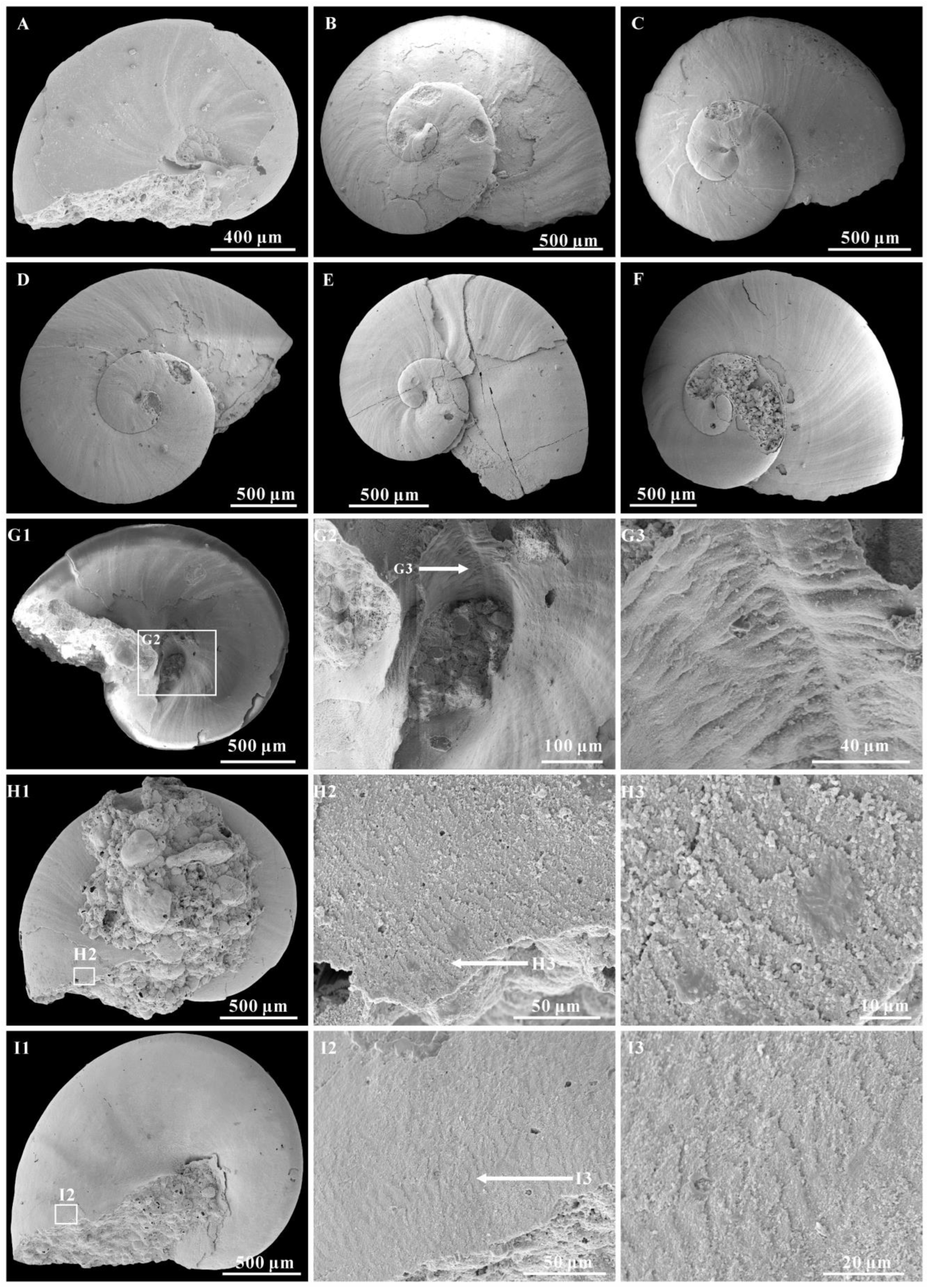
4.1. Shell Microstructures of Aldanella attleborensis

Shell microstructures could be remarkably well preserved on the Cambrian mollusk shells [20,70,71], and numerous types of shell microstructures have been identified, such as prismatic, nacre, crossed lamellar, and foliated. However, only convex polygons [21,40,72,73,74,75] and lamellar microstructures [70] have been occasionally reported on internal molds of Aldanella.
The convex polygons appear on the spire of internal mold surface, and are about 20–60 μm in diameter. The convex polygons are varying in shape, mainly circular or nearly circular, and always separated by shallow grooves [21,72,73,75]. Conversely, in some literature, the space between convex polygons is large and appears as concave polygons. Hence the convex and concave polygons seem to alternate on the spire of internal mold of Aldanella [40,74]. Similar convex polygonal impressions were described on the apex of Oelandiella and Securiconus, and were mostly interpreted as imprints of the prismatic layer [40,73,75]. The imprints of the prismatic layer always preserved as negative or positive reliefs on the molluscan internal molds [71]. Different to the prismatic layer imprints, the convex polygons of Aldanella are restricted to the apical areas [21,40,72,73,74,75] and no tubercles in the polygons. In addition, the polygons that are interpreted as imprints of the prismatic shell layer are much smaller (10–20 μm), although the varied size of convex polygons has been attributed to the growth stages and competition for space [73]. The convex polygons of Aldanella have been interpreted as the impressions of cells of the outer epithelium [13,74], but this view remains to be investigated.
Inner layer laminar microstructure, the only preserved microstructure on the A. attleborensis of the Yanjiahe Formation, appears on the internal mold surface near the aperture (Figure 2J2 and Figure 3H,I). Different to the laminar microstructures of Pojetaia with a rounded edge, the lamella of herein shows two different forms: straight edge (Figure 3H) or sharp-toothed edge (Figure 2J2 and Figure 3I). The orientation of the lamella points to the suture on the upper whorl surface and to the umbilicus on the base. This type of microstructure is common in Cambrian mollusks, such as Anabarella, Watsonella, Fordilla, Pojetaia, and Pelagiella [20,70,71,76], and is interpreted as a foliated aragonite [70].

4.2. Muscle Scars

Muscle scars provide important information to study the morphology and phylogeny of ancient univalved mollusks. Compared to most cap-shaped mollusks that have paired muscle scars on the dorsal margin, apical area, or sub-apical area [77,78], the muscle scars are always difficult to see in coiled gastropod shells where the muscle attachment site is always restricted on the umbilical area, as a columellar scar [79].
On the internal mold surface of A. attleborensis, a spiral structure is found on the umbilical area (Figure 2H4). The spiral structure appears as about three-quarters of a whorl and vanishes near the aperture. The spiral structure is ribbon-shaped, and the width increases in the progress of growth. A similar structure has been interpreted as columellar muscle attachment on the shell umbilicus of A. rozanovi and A. kunda (junior synonyms of A. attleborensis) [26,40]. The difference is that on A. rozanovi, the spiral structure consists of honeycomb patterns with a diameter up to 6–8 μm. Isakar and Peel [40] urged caution to compare the honeycomb patterns on A. rozanovi umbilicus [26] with gastropod retractor muscles, because the honeycomb pattern is similar to the convex polygons on the spire of the internal molds. However, they ignored the fact that the convex polygons are much larger than the honeycomb patterns. In addition, on the apical surface of A. kunda, there exists an equivocal spiral ridge interpreted as the trace of a spirally migrating shell attachment muscle [40]. In Paleozoic helically coiled gastropods, ribbon-like muscle scar appeared on the umbilical area of internal molds, such as Ordovician Tropidodiscus [80], Silurian Megalomphala [81], Carboniferous Porcellia [82], and Bellerophon [81], but it is rare in Cambrian gastropods. The muscle scar on Tropidodiscus appeared on the umbilical shoulder and was preserved as a slightly depressed zone [80]. The position and the shape of Tropidodiscus muscle scar is consistent with that of Aldanella. The muscle scar of Megalomphala was preserved as smooth area on the umbilical wall of the internal mold, separated by two spiral ridges from the internal mold [81]. On the Porcellia, a single ribbon-like muscle scar is longer and narrower, and lies on the basal surface of the whorl [82]. The muscle scar of Bellerophon is located on the umbilical shoulder, but the difference is that Bellerophon has a pair of muscle scars and the scars were preserved as a raised zone [81].
Besides Aldanella, similar ribbon-like structures also have been described on Cambrian coiled mollusk Pelagiella [63,83,84]. That is an important characteristic to support these genera as gastropods [26,27]. Comparing the ribbon-like structure position and shape of A. attleborensis with that of the muscle scars on the upper Paleozoic gastropods, as well as the columellar muscles on living spirally coiled gastropods, it is herein considered that the spiral structure on the umbilicus of Aldanella is the shell muscle attachment area, although some of upper Paleozoic gastropods have a pair of muscle scars.

4.3. Systematic Position of Aldanella

The systematic position of the genus Aldanella is controversial, and has been suggested to gastropods [13,24,25,26,27], helcionelloids [40,85], hyoliths [66], or polychaetes [86,87]. The phosphatic preservation of the shell and the absence of the soft body provide extremely limited evidence to study the phylogeny and affinity of this genus. The imprints of the shell microstructures and the shell muscle attachments have been recognized as significant information to reveal the systematic position of early mollusks [20,70,71,88,89], and therefore, it is necessary to research these features on Aldanella.
The presence of laminar microstructure on the internal molds of A. attleborensis provides crucial information to analyze its systematic position. As the laminar microstructure reported in Pelagiella has been suggested to be nacreous by Thomas et al. [27], the presumption that Pelagielliformes microstructure is comparable to that of hyoliths in lacking nacre is inappropriate. Most importantly, the operculum, preserved in hyoliths, has never been found in Pelagielliformes. Apparently, Aldanella is not a hyolith. The septa presented in the initial shell of A. operosa exclude the possibility that Aldanella is a polychaete [26]. Furthermore, laminar microstructure was never discovered in polychaetes [90]. In addition, Aldanella was suggested as helcionelloids because they share an endogastric shell. However, the muscle scar preserved on the umbilicus area of A. attleborensis is more similar to that of Paleozoic gastropods than the paired muscle scars of helcionelloids. Hence, according to the muscle scars similarity of Aldanella and upper Paleozoic gastropods in position and shape, the appearance of laminar microstructure on the Aldanella shell, the presence of protoconch and regular septa in the initial part of the shell, as well as the asymmetrically turbospiral shell, Aldanella is more likely to be a gastropod than helcionelloids, hyoliths, or polychaetes.

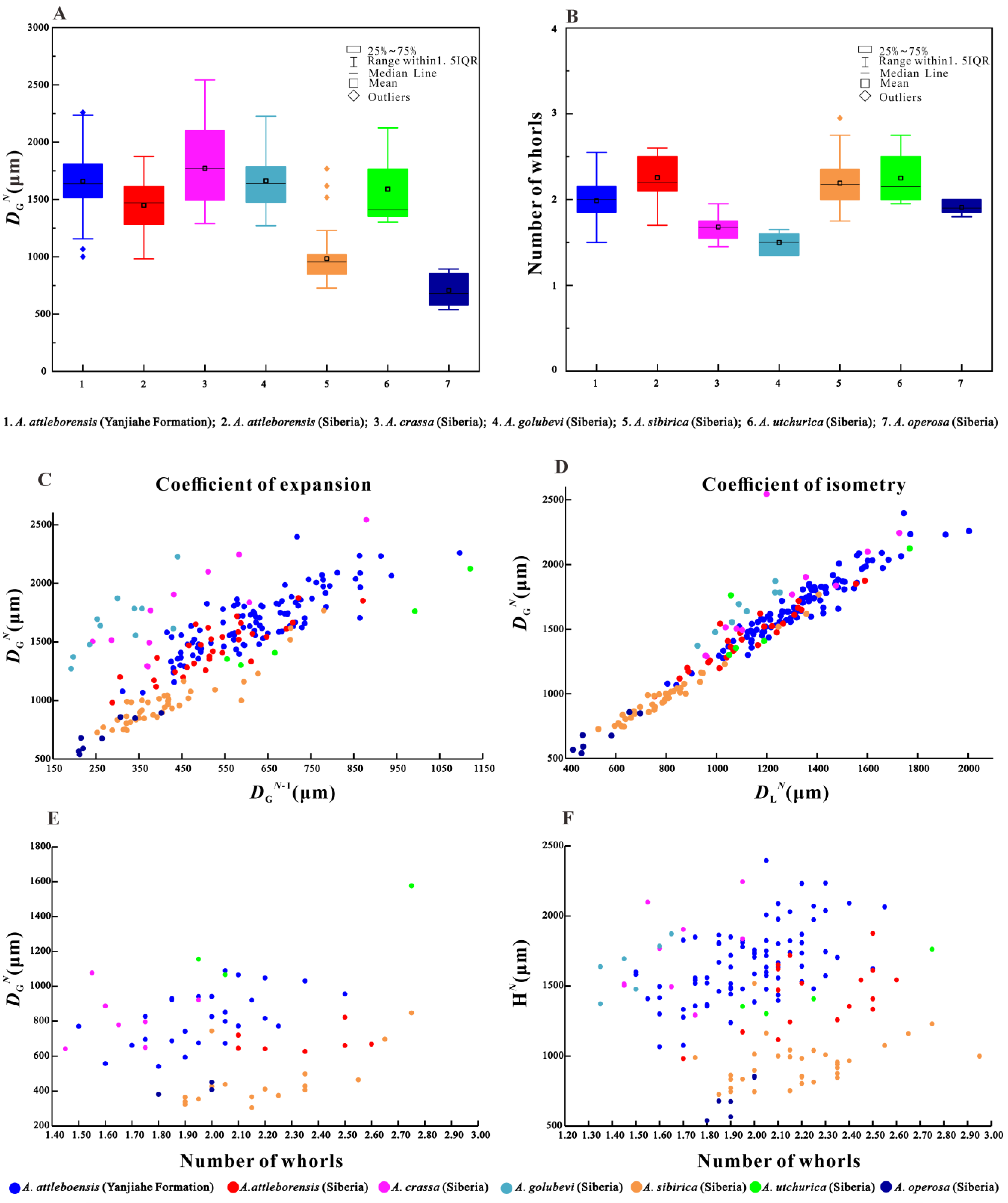
4.4. Ontogeny of Aldanella attleborensis

Ontogenetic analysis is a useful method for studying the origin and early evolution of gastropods. Based on the fossil materials of Paleozoic, many achievements on gastropod ontogeny have been made [91,92]. However, the ontogeny of early Cambrian gastropods, such as Aldanella and Pelagiella, is rarely systematically studied due to limited specimens. The shell of Aldanella was ontogenetically identified in two (protoconch and teleoconch) or three (protoconch I, protoconch II, and teleoconch) developmental stages [93].
The morphology of A. attleborensis analyzed in this study can be compared with Siberian materials, and the protoconch and teleoconch of the shell is faintly separated. The protoconch of A. attleborensis is small, approximately 100–200 μm in length. The small size of the protoconch may indicate the existence of the planktonic larval stage during development [24,91,93,94], which is consistent with its worldwide distribution in the lower Cambrian strata. The protoconch always bears a mucro on those specimens preserved with external coating, but the protoconch of the internal mold is smoothly rounded (Figure 4A2,H2). This is a unique character that has been found on the external coating of Aldanella [66,72]. The size and shape of the mucro is variable, and it looks like a short column (Figure 4C2,E2,F2) or blunt cusp (Figure 4A2,B2,D2,G2). As shown in the Micro-CT images (Figure 4G4–G6), when the original shell is dissolved, the mucro is expressed as a hollow space. This means that the mucro is an original solid protrusion on the protoconch. Moreover, the mucro has no surface ornamentation. Although the function of the mucro remains unknown, there is no ground to conclude that it may represent a discrete stage of larval shell development, as suggested by Dzik [95] and Thomas et al. [27].
Most of the specimens studied herein were partially broken, especially at the protoconch and aperture. Nevertheless, the well-preserved inner lip of the aperture on some specimens with phosphatic coating (Figure 2G2 and Figure 3A) indicates that the steinkerns almost represent complete specimens, not just broken off pieces of larger shells [66]. The specimens of A. attleborensis described herein range from 1067 μm to 2397 μm in diameter (Figure 6A), and from 1.50 to 2.55 in the number of whorls (Figure 6B). The wide range of sizes and whorl numbers of the shells suggests that both juvenile and adult stages are preserved. In addition, the box-plot of shell diameter and whorl numbers showed that the adult shell size mainly ranges from 1400 μm to 1900 μm, and the whorl numbers correspond to approximately 2. The strong linear regressions of the expansion and isometry rates (Figure 6C,D) indicate that the shell tube width (the straight-line distance from suture to periphery) of A. attleborensis constantly increases. Owing to the turbospirally coiled shell, the length of shell tube (the helical-line distance from protoconch to aperture) is difficult to measure. Compared with the diameter of the shell, the number of whorls could be more accurate in estimating the longitudinal growth rate of the shell tube. Although the general trend is positive, the weak linear regression of the shell diameter and number of whorls (Figure 6E) suggests that the final shell size is not strongly correlated with the whorl number, and the length of shell tube of the whorl is allometric growth relative to its width. In addition, the shell height had a weak relationship with the number of whorls (Figure 6F). The shell height might be more affected by the obliquity and oblateness of shell tube cross section.

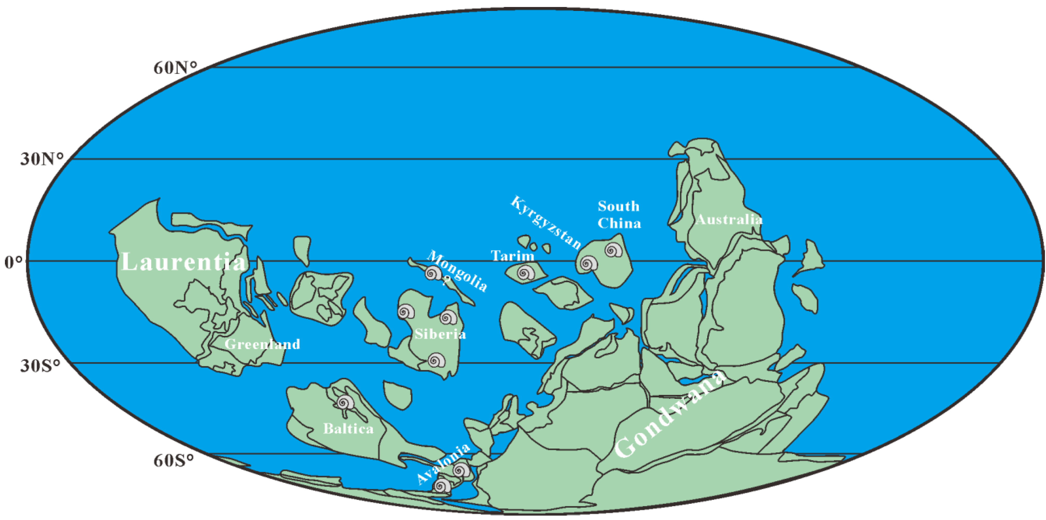
4.5. Stratigraphic Implications of Aldanella attleborensis

With the progress of Cambrian stratigraphy subdivision, more than half of the Global Boundary Stratotype Section and Points (GSSPs) of Cambrian stages have been ratified. However, due to high levels of endemism and the facies-restricted distribution of fossils, as well as the absence of the pelagic skeletonized metazoans used for global correlation, formal ratification of Stage 2, Stage 3, and Stage 4 is difficult [41,96]. Nevertheless, SSFs play an important biostratigraphic role for the subdivision and correlation of pre-trilobitic (Terreneuvian) Cambrian strata [4,9,15,16,17,18]. For example, the widely distributed A. attleborensis or W. crosbyi both have been suggested as a potential marker for defining the base of Cambrian Stage 2.
In this study, numerous specimens of A. attleborensis, co-occurring with W. crosbyi, have been discovered from Member 5 of the Yanjiahe Formation of the Three Gorges area, South China. Besides the Yanjiahe Formation, A. attleborensis was also discovered from the uppermost Huangshandong Member (Tianzhushan Member) of the Dengying Formation in Tianzhushan of the Three Gorges area [47]. The thickness of the Huangshandong Member is extremely varied at different sections in the Three Gorges area (0.1-8 m) [43,44,97], and the SSF assemblages are often mixed [43,44], or SSF zone 1 and 2 could be recognized [46,97]. However, besides A. attleborensis, W. crosbyi was also found in the uppermost Tianzhushan Member from the Tianzhushan Section [97], Huangshandong Section [43,97], and Songlinpo Section (unpublished data). This may indicate that the SSF zone 3 occasionally exists in the uppermost Tianzhushan Member at some sections with a relatively thick unmixed sequence of this lithological unit.
The similar phenomenon also appears in southern Shaanxi. The Kuanchuanpu Formation is well-exposed in southern Shaanxi and contains abundant SSFs. Nevertheless, scarce materials of A. attleborensis and W. crosbyi were only recovered in the uppermost Kuanchuanpu Formation at the Yuanjiaping Section of Ningqiang County [43]. This indicates that the sediments containing SSF zone 3 are rare in the Kuanchuanpu Formation of southern Shaanxi. In addition, Aldanella attleborensis was discovered from the Dahai Member of the Zhujiaqing Formation in Eastern Yunnan [9,42], but its first occurrence horizon is much higher than that of W. crosbyi [42].
Using A. attleborensis as a biostratigraphic marker, Member 5 of the Yanjiahe Formation, uppermost Tianzhushan Member, uppermost Kuanchuanpu Formation, and the Dahai Member can be well correlated. Therefore, the FAD of A. attleborensis has a potential to correlate the pre-trilobitic strata in South China. In addition, A. attleborensis was documented in the upper part of the Yurtus Formation, Aksu-Wushi region, Tarim [48,49,69], but the age of the uppermost Yurtus Formation has been suggested to be Qiongzhusian equivocally [49]. Therefore, in Tarim the detailed stratigraphic correlation still needs to be further confirmed.
Figure 7. Paleogeographical distribution of Aldanella attleborensis from Cambrian Stage 2. Paleogeographic map modified from [98] and [20]; the distribution data from [9,13,21,33,39,40,49,99,100] and herein.
Biology 12 00261 g007
With the exception of South China, Aldanella attleborensis is also widely distributed in the pre-trilobitic strata of most paleocontinents, such as Avalonia, Baltica, Siberia, etc. (Figure 7). The materials of A. attleborensis in Siberia are extremely rich, and its stratigraphic range were suggested to be the whole Tommotian [13]. However, A. attleborensis may appear much lower than W. crosbyi in Siberia [21,50] and the hiatus between the Pestrotsvet Formation and the underlying Ust-Yudoma Formation [101] makes it difficult to evaluate the accuracy of the FAD [12]. The FADs of W. crosbyi and A. attleborensis in Avalonia correspond to the base of the Tommotian, but the temporal range of W. crosbyi and A. attleborensis is much longer and their LADs (last appearance data) might not be limited to the Tommotian [41]. In western Mongolia, the original description of W. crosbyi assemblage zone suggested a Tommotian age [100], but it was revised to be pre-Tommotian in age by Smith et al. [102] based on chemostratigraphic data. Aldanella in western Mongolia was uncommon, appeared much higher than W. crosbyi, and only one specimen, assigned to Aldanella sp., was reported from the probable Botoman Stage [100]. Although this indeterminate species has been revised to A. attleborensis [13], the horizon and taxonomy of A. attleborensis in western Mongolia [100] is questionable [21]. Aldanella attleborensis has been reported in the Kestla Member of the Lontova Formation in northern Estonia [40], and its age is limited to the Tommotian [67,99].
In summary, A. attleborensis is one of the most widely distributed SSFs and is an important fossil for both regional and global correlation of Cambrian Terreneuvian (Figure 8). Based on its wide geographic distribution and distinct turbospirally coiled shell, A. attleborensis has the potential to be an index fossil for defining the base of Cambrian Stage 2 [13,14,67]. In addition to A. attleborensis, another mollusk, W. crosbyi, which is broadly similar in stratigraphic distribution to A. attleborensis, such as South China, Siberia, Avalonia, Australia, Armorican France, and Mongolia (see [12,20] and references cited therein), has also been suggested as a potential marker for defining the base of Cambrian Stage 2 [12,103].

5. Conclusions

Numerous specimens of Aldanella were collected from Member 5 of the Yanjiahe Formation at the Yanjiahe and Gunziao sections in the Three Gorges area, South China. This study shows that A. yanjiaheensis is morphologically consistent with A. attleborensis in many aspects, such as the small turbospiral shell, moderate expansion rate, up to 2.5 tightly coiled whorls, oblique oval aperture, and growth lines occurring on the shell. Our work formally confirms that the species A. yanjiaheensis Chen, 1984 is a junior synonym of A. attleborensis.
Two developmental stages, protoconch and teleoconch, of the shell of A. attleborensis were identified. The small protoconch of the shell bears a distinct mucro. The strong linear regressions of the expansion and isometry rates indicated that the shell tube width constantly increases. However, the weak linear regression of the shell diameter and number of whorls suggests that the shell tube of the whorl is allometric growth. The spiral muscle scar structure further confirms that Aldanella is more likely to be a gastropod.
Aldanella attleborensis widely occurred in Cambrian paleocontinents (South China, Mongolia, Siberian, Baltica, Avalonia, etc.) from low to high paleolatitudes. The stratigraphic range of A. attleborensis indicates that it was mainly restricted to Cambrian Stage 2. Aldanella attleborensis can be easily distinguished by its characteristic features from other congeneric species. Therefore, similar to W. crosbyi, the FAD of A. attleborensis could also be a potential candidate marker for defining the base of Cambrian Stage 2. The co-occurrence of A. attleborensis and W. crosbyi at the base of Member 5 of the Yanjiahe Formation indicates that Member 5 belongs to Cambrian Stage 2.

Supplementary Materials

The following supporting information can be downloaded at: https://www.mdpi.com/article/10.3390/biology12020261/s1, Table S1: The datum of Aldanella attleborensis measurements.

Author Contributions

Writing—original draft preparation, Y.Q.; writing—review and editing, J.G., G.L., J.H. and Z.Z.; materials administration, Z.S., J.P. and J.S. Project administration, Supervision, J.G. All authors have read and agreed to the published version of the manuscript.

Funding

This research was funded by the Natural Science Foundation of China (Nos. 42172016 to Guo, J.F.; 41890844 to Zhang, Z.F., Li, G.X. and Guo, J.F.; 41621003 to Han, J.), the Strategic Priority Research Program of the Chinese Academy of Sciences (No. XDB26000000 to Han, J. and Guo, J.F.), Key Scientific and Technological Innovation Team Project in Shaanxi Province, State Key Laboratory of Palaeobiology and Stratigraphy (Nanjing Institute of Geology and Palaeontology, CAS) (Nos. 203106 to Guo, J.F.; 163107 to Han, J.).

Institutional Review Board Statement

Not applicable.

Informed Consent Statement

Not applicable.

Data Availability Statement

Not applicable.

Acknowledgments

We thank Bing Pan (Nanjing Institute of Geology and Palaeontology, Chinese Academy of Sciences) for collection of literature. We also thank reviewers and editors for their comments and suggestions.

Conflicts of Interest

There are no conflict of interest to declare.

References

  1. Shu, D.G. Cambrian explosion: Birth of tree of animals. Gondwana Res. 2008, 14, 219–240. [Google Scholar] [CrossRef]
  2. Maloof, A.C.; Porter, S.M.; Moore, J.L.; Dudás, F.Ö.; Bowring, S.A.; Hoggins, J.A.; Fike, D.A.; Eddy, M.P. The earliest Cambrian record of animals and ocean geochemical change. Geol. Soc. Am. Bull. 2010, 122, 1731–1774. [Google Scholar] [CrossRef]
  3. Erwin, D.H.; Laflamme, M.; Tweedt, S.M.; Sperling, E.A.; Pisani, D.; Peterson, K.J. The Cambrian conundrum: Early divergence and later ecological success in the early history of animals. Science 2011, 334, 1091–1097. [Google Scholar] [CrossRef] [PubMed]
  4. Qian, Y.; Chen, M.E.; He, T.G.; Zhu, M.Y.; Yin, G.Z.; Feng, W.M.; Xu, J.T.; Jiang, Z.W.; Liu, D.Y.; Li, G.X.; et al. Taxonomy and Biostratigraphy of Small Shelly Fossils in China; Science Press: Beijing, China, 1999; pp. 1–247. (In Chinese) [Google Scholar]
  5. Parkhaev, P.Y. The Functional Morphology of the Cambrian Univalved Mollusks–Helcionellids: 1. Paleontol. J. 2000, 34, 32–38. [Google Scholar]
  6. Parkhaev, P.Y. The Functional Morphology of the Cambrian Univalved Mollusks–Helcionellids: 2. Paleontol. J. 2001, 35, 470–475. [Google Scholar]
  7. Parkhaev, P.Y. Phylogenesis and the System of the Cambrian Univalved Mollusks. Paleontol. J. 2002, 36, 25–36. [Google Scholar]
  8. Parkhaev, P.Y. The Early Cambrian radiation of Mollusca. In Phylogeny and Evolution of the Mollusca; Ponder, W.F., Lindberg, D.R., Eds.; University California Press: Berkeley, CA, USA, 2008; pp. 33–69. [Google Scholar]
  9. Steiner, M.; Li, G.X.; Qian, Y.; Zhu, M.Y.; Erdtmann, B.D. Neoproterozoic to early Cambrian small shelly fossil assemblages and a revised biostratigraphic correlation of the Yangtze Platform (China). Palaeogeogr. Palaeoclimatol. Palaeoecol. 2007, 254, 67–99. [Google Scholar] [CrossRef]
  10. Shaler, N.S.; Foerste, A.F. Preliminary description of North Attleborough fossils. Bull. Museum Compar. Zool. 1888, 16, 27–41. [Google Scholar]
  11. Grabau, A.W. Palaeontology of the Cambrian terranes of the Boston Basin. Occas. Pap. Boston Soc. Natur. Hist. 1900, 4, 601–694. [Google Scholar]
  12. Li, G.X.; Zhao, X.; Gubanov, A.P.; Zhu, M.Y.; Na, L. Early Cambrian mollusc Watsonella crosbyi: A potential GSSP index fossil for the base of the Cambrian Stage 2. Acta Geol. Sin. 2011, 85, 309–319. [Google Scholar] [CrossRef]
  13. Parkhaev, P.Y.; Karlova, G.A. Taxonomic revision and evolution of Cambrian mollusks of the genus Aldanella Vostokova, 1962 (Gastropoda: Archaeobranchia). Paleontol. J. 2011, 45, 1145–1205. [Google Scholar] [CrossRef]
  14. Parkhaev, P.Y.; Karlova, G.A.; Rozanov, A.Y. Taxonomy, stratigraphy and biogeography of Aldanella attleborensis–A possible candidate for defining the base of Cambrian Stage 2. Bull. Museum Northern Arizona 2011, 67, 298–300. [Google Scholar]
  15. Devaere, L.; Clausen, S.; Steiner, M.; Álvaro, J.J.; Vachard, D. Chronostratigraphic and palaeogeographic significance of an early Cambrian microfauna from the Heraultia Limestone, northern Montagne Noire, France. Palaeontol. Electron. 2013, 16, 1–91. [Google Scholar] [CrossRef] [PubMed]
  16. Yang, B.; Steiner, M.; Li, G.X.; Keupp, H. Terreneuvian small shelly faunas of East Yunnan (South China) and their biostratigraphic implications. Palaeogeogr. Palaeoclimatol. Palaeoecol. 2014, 398, 28–58. [Google Scholar] [CrossRef]
  17. Betts, M.J.; Paterson, J.R.; Jago, J.B.; Jacquet, S.M.; Skovsted, C.B.; Topper, T.P.; Brock, G.A. New lower Cambrian shelly fossil assemblage zones in the lower Hawker Group, Arrowie Basin, South Australia. Gondwana Res. 2016, 36, 163–195. [Google Scholar] [CrossRef]
  18. Betts, M.J.; Paterson, J.R.; Jacquet, S.M.; Andrew, A.S.; Hall, P.A.; Jago, J.B.; Jagodzinski, E.A.; Preiss, W.V.; Crowley, J.L.; Brougham, T.; et al. Early Cambrian chronostratigraphy and geochronology of South Australia. Earth-Sci. Rev. 2018, 185, 498–543. [Google Scholar] [CrossRef] [Green Version]
  19. Guo, J.F.; Li, Y.; Li, G.X. Small shelly fossils from the Early Cambrian Yanjiahe Formation, Yichang, Hubei, China. Gondwana Res. 2014, 25, 999–1007. [Google Scholar] [CrossRef]
  20. Guo, J.F.; Li, G.X.; Qiang, Y.Q.; Song, Z.C.; Zhang, Z.F.; Han, J.; Wang, W.Z. Watsonella crosbyi from the lower Cambrian (Terreneuvian, Stage 2) Yanjiahe Formation in Three Gorges Area, South China. Palaeoworld 2021, 30, 1–19. [Google Scholar] [CrossRef]
  21. Kouchinsky, A.; Bengtson, S.; Landing, E.; Steiner, M.; Vendrasco, M.; Ziegler, K. Terreneuvian stratigraphy and faunas from the Anabar Uplift, Siberia. Acta Palaeontol. Pol. 2017, 62, 311–440. [Google Scholar] [CrossRef]
  22. Jacquet, S.M.; Brougham, T.; Skovsted, C.B.; Jago, J.B.; Laurie, J.R.; Betts, M.J.; Topper, T.P.; Brock, G.A. Watsonella crosbyi from the lower Cambrian (Terreneuvian, Stage 2) Normanville Group in South Australia. Geol. Mag. 2017, 154, 1088–1104. [Google Scholar]
  23. Vostokova, V.A. Cambrian Gastropods of the Siberian Platform and Taimyr. Sbornik Statei Paleontol. Biostratigrafii 1962, 28, 51–74. (In Russian) [Google Scholar]
  24. Runnegar, B.; Pojeta, J.J. Molluscan phylogeny: The paleontological viewpoint. Science 1974, 186, 311–317. [Google Scholar] [CrossRef]
  25. Pojeta, J.J.; Runnegar, B. The paleontology of rostroconch mollusks and the early history of the phylum Mollusca. US Geol. Sur. Prof. Pap. 1976, 968, 1–88. [Google Scholar]
  26. Parkhaev, P.Y. New data on the morphology of ancient gastropods of the genus Aldanella vostokova, 1962 (archaeobranchia, pelagielliformes). Paleontol. J. 2006, 40, 244–252. [Google Scholar] [CrossRef]
  27. Thomas, R.D.K.; Runnegar, B.; Matt, K. Pelagiella exigua, an early Cambrian stem gastropod with chaetae: Lophotrochozoan heritage and conchiferan novelty. Palaeontology 2020, 63, 1–27. [Google Scholar] [CrossRef]
  28. Öpik, A. Über den estländischen blauen Ton. Publ. Geol. Inst. Univ. Tartu 1926, 6, 39–46. [Google Scholar]
  29. Rozanov, A.Y.; Missarzhevsky, V.V. Biostratigraphy and fauna of Lower Cambrian horizons. Tr. Geol. Inst. Akad. Nauk SSSR 1966, 148, 1–126. (In Russian) [Google Scholar]
  30. Rozanov, A.Y.; Missarzhevsky, V.V.; Volkova, N.A.; Voronova, L.G.; Krylov, I.N.; Keller, B.M.; Korolyuk, I.K.; Lendzion, K.; Michniak, R.; Pyhova, N.G.; et al. The Tommotian Stage and the Cambrian lower boundary problem. Tr. Geol. Inst. Akad. Nauk SSSR 1969, 206, 1–380. (In Russian) [Google Scholar]
  31. Golubev, S.N. Ontogenetic changes and evolutionary trends in Early Cambrian spiral gastropods of the superfamily Pelagiellacea. Paleontol. J. 1976, 10, 143–149. [Google Scholar]
  32. Lendzion, K. First gastropod fauna from the Klimontovian Stage (Lower Cambrian) of the South-Eastern Poland. Kwartalnik Geol. 1977, 21, 239–243. [Google Scholar]
  33. Chen, P. Discovery of Lower Cambrian small shelly fossils from Jijiapo, Yichang, west Hubei and its significance. Prof. Pap. Stratigr. Palaeontol. 1984, 13, 49–64. (In Chinese) [Google Scholar]
  34. Barskova, M.I. New Mollusks from the Lower Cambrian Deposits of the Kolyma Uplift. Paleontol. J. 1988, 1, 101–105. (In Russian) [Google Scholar]
  35. Missarzhevsky, V.V. Oldest skeletal fossils and stratigraphy of Precambrian and Cambrian boundary beds. Tr. Geol. Inst. Akad. Nauk SSSR 1989, 443, 1–237. (In Russian) [Google Scholar]
  36. Bokova, A.R. New Lower Cambrian gastropods from the Siberian Platform. Paleontol. J. 1990, 24, 134–136. [Google Scholar]
  37. Gubanov, A.P.; Fernández Remolar, D.C.; Peel, J.S. Early Cambrian molluscs from Sierra de Córdoba. Geobios 2004, 37, 199–215. (In Spain) [Google Scholar] [CrossRef]
  38. Parkhaev, P.Y. Shell chirality in Cambrian gastropods and sinistral members of the genus Aldanella Vostokova, 1962. Paleontol. J. 2007, 41, 233–240. [Google Scholar] [CrossRef]
  39. Landing, E. Lower Cambrian of eastern Massachusetts: Stratigraphy and small shelly fossils. J. Paleontol. 1988, 62, 661–695. [Google Scholar]
  40. Isakar, M.; Peel, J.S. Lower Cambrian helcionelloid molluscs from Estonia. GFF 2007, 129, 255–262. [Google Scholar] [CrossRef]
  41. Landing, E.; Geyer, G.; Brasier, M.D.; Bowring, S.A. Cambrian evolutionary radiation: Context, correlation, and chronostratigraphy-Overcoming deficiencies of the first appearance datum (FAD) concept. Earth-Sci. Rev. 2013, 123, 133–172. [Google Scholar] [CrossRef]
  42. Li, G.X.; Xiao, S.H. Micrina and Tannuolina (Tannuolinidae) from the lower Cambrian of eastern Yunnan, South China. J. Paleontol. 2004, 78, 900–913. [Google Scholar] [CrossRef]
  43. Xing, Y.S.; Ding, Q.X.; Luo, H.L.; He, T.G.; Wang, Y.G. The Sinian-Cambrian boundary of China; Geological Publishing House: Beijing, China, 1984; pp. 1–262. (In Chinese) [Google Scholar]
  44. Ding, L.F.; Zhang, L.Y.; Li, Y.; Dong, J.S.; Chen, H.X.; Tian, S.H. The Study of Late Sinian–Early Cambrian Biotas from the Northern Margin of Yangtze Platform; Scientific and Technical Documents Publishing House: Beijing, China, 1992; pp. 1–156. (In Chinese) [Google Scholar]
  45. Guo, J.F.; Li, Y.; Han, J.; Zhang, X.L.; Zhang, Z.F.; Ou, Q.; Liu, J.N.; Shu, D.G.; Maruyama, S.; Komiya, T. Fossil association from the lower Cambrian Yanjiahe Formation in the Yangtze Gorges area, Hubei, South China. Acta Geol. Sin. 2008, 82, 1124–1132. [Google Scholar]
  46. Steiner, M.; Yang, B.; Hohl, S.; Zhang, L.; Chang, S. Cambrian small skeletal fossil and carbon isotope records of the southern Huangling Anticline, Hubei (China) and implications for chemostratigraphy of the Yangtze Platform. Palaeogeogr. Palaeoclimatol. Palaeoecol. 2020, 554, 1–19. [Google Scholar] [CrossRef]
  47. Yu, W. New molluscan naterials of the Tethys. In 2nd International Symposium on Shallow Tethys; Mckenzie, K.G., Ed.; A A Balkema Publisher: Rotterdam, The Netherlands, 1987; pp. 51–59. [Google Scholar]
  48. Qian, J.X.; Xiao, B. An Early Cambrian Small Shelly Fauna from Aksu-Wushi Region, Xinjiang. Prof. Pap. Stratigr. Palaeontol. 1984, 13, 65–90. (In Chinese) [Google Scholar]
  49. Qian, Y.; Feng, W.M.; Li, G.X.; Yang, A.H.; Feng, M.; Zhao, X.; Xiao, B. Taxonomy and biostratigraphy of the early Cambrian univalved Mollusc fossils from Xinjiang. Acta Micropalaeontol. Sin. 2009, 26, 193–210. (In Chinese) [Google Scholar]
  50. Yang, B.; Steiner, M. Terreneuvian bio- and chemostratigraphy of the South Sichuan Region (South China). J. Geol. Soc. London 2021, 178, 1–15. [Google Scholar] [CrossRef]
  51. Guo, J.F.; Han, J.; Van Iten, H.; Wang, X.; Qiang, Y.Q.; Song, Z.C.; Wang, W.Z.; Zhang, Z.F.; Li, G.X. A fourteen-faced hexangulaconulariid from the early Cambrian (Stage 2) Yanjiahe Formation, South China. J. Paleontol. 2020, 94, 45–55. [Google Scholar] [CrossRef]
  52. Guo, J.F.; Li, Y.; Han, H.P.; Ou, Q.; Zhou, J.R.; Zheng, Y.J. New macroscopic problematic fossil from the early Cambrian Yanjiahe biota, Yichang, Hubei, China. Acta Geol. Sin. 2012, 86, 791–798. [Google Scholar]
  53. Guo, J.F.; Chen, Y.L.; Song, Z.C.; Zhang, Z.F.; Qiang, Y.Q.; Betts, M.J.; Zheng, Y.J.; Yao, X.Y. Geometric morphometric analysis of Protoconites minor from the Cambrian (Terreneuvian) Yanjiahe Formation in Three Gorges, South China. Palaeontol. Electron. 2020, 23, a48. [Google Scholar] [CrossRef]
  54. Topper, T.P.; Guo, J.F.; Clausen, S.; Skovsted, C.B.; Zhang, Z.F. A stem group echinoderm from the basal Cambrian of China and the origins of Ambulacraria. Nat. Commun. 2019, 10, 1366. [Google Scholar] [CrossRef]
  55. Okada, Y.; Sawaki, Y.; Komiya, T.; Hirata, T.; Takahata, N.; Sano, Y.; Han, J.; Maruyama, S. New chronological constraints for cryogenian to Cambrian rocks in the Three Gorges, Weng’an and Chengjiang areas, South China. Gondwana Res. 2014, 25, 1027–1044. [Google Scholar] [CrossRef]
  56. Ahn, S.Y.; Zhu, M.Y. Lowermost Cambrian acritarchs from the Yanjiahe Formation, South China: Implication for defining the base of the Cambrian in the Yangtze Platform. Geol. Mag. 2017, 1, 1–15. [Google Scholar] [CrossRef]
  57. Guo, J.; Han, J.; Iten, H.V.; Song, Z.C.; Qiang, Y.Q.; Wang, W.Z.; Zhang, Z.F.; Li, G.X.; Sun, Y.F.; Sun, j. A new tetraradial olivooid (Medusozoa) from the lower Cambrian (Stage 2) Yanjiahe Formation, South China. J. Paleontol. 2020, 94, 457–466. [Google Scholar] [CrossRef]
  58. Guo, J.F.; Han, J.; Iten, H.V.; Song, Z.C.; Qiang, Y.Q.; Wang, W.Z.; Zhang, Z.F.; Li, G.X. A ten-faced hexangulaconulariid from Cambrian Stage 2 of South China. J. Paleontol. 2021, 95, 957–964. [Google Scholar] [CrossRef]
  59. Song, Z.C.; Guo, J.F.; Han, J.; Iten, H.V.; Qiang, Y.Q.; Peng, J.X.; Sun, J.; Zhang, Z.F. A new species of Septuconularia (Hexangulaconulariidae, Cnidaria) from Cambrian Stage 2, South China. Acta Geol. Sin. 2022, 96, 757–765. [Google Scholar] [CrossRef]
  60. Qiang, Y.Q.; Peng, J.X.; Song, Z.C.; Sun, J.; Zhao, X.F.; Li, G.X.; Han, J.; Guo, J.F. Early Cambrian Anabarella plana from Three Gorges area, South China. Front. Earth Sci. 2023, 10, 2591. [Google Scholar] [CrossRef]
  61. Song, Z.C.; Guo, J.F.; Pan, B.; Qiang, Y.Q.; Li, G.X.; Peng, J.X.; Sun, J.; Han, J. Ocruranus–Eohalobia Sclerites from the Cambrian Stage 2 Yanjiahe Formation in South China: Scleritome Reconstruction and Zoological Affinity. Biology 2022, 11, 1648. [Google Scholar] [CrossRef]
  62. Cuvier, G. Tableau Élementaire de L’historie Naturelle des Animaux; Baudouin: Paris, France, 1797; p. 710. [Google Scholar]
  63. Mackinnon, D.I. New Zealand late Middle Cambrian molluscs and the origin of Rostroconchia and Bivalvia. Alcheringa 1985, 9, 65–81. [Google Scholar] [CrossRef]
  64. Linsley, R.M.; Kier, W.M. The Paragastropoda: A proposal for a new Class of Paleozoic Mollusca. Malacologia 1984, 25, 241–254. [Google Scholar]
  65. Matthew, G.F. The Protolenus fauna. Trans. N. Y. Acad. Sci. 1895, 14, 101–153. [Google Scholar] [CrossRef]
  66. Dzik, J.; Mazurek, D. Affinities of the alleged earliest Cambrian gastropod Aldanella. Can. J. Zool. 2013, 91, 914–923. [Google Scholar] [CrossRef]
  67. Parkhaev, P.Y. On the stratigraphy of Aldanella attleborensis–potential index species for defining the base of Cambrian Stage 2. In Extended Summary, IGCP Project 591 Field Workshop; Zhan, R.B., Huang, B., Eds.; Nanjing University Press: Nanjing, China, 2014; pp. 102–105. [Google Scholar]
  68. Krajewski, K.P. Early diagenetic phosphate cements in the Albian condensed glauconitic limestone of the Tatra Mountains, Western Carpathians. Sedimentology 1984, 31, 443–470. [Google Scholar] [CrossRef]
  69. Yue, Z.; Gao, L.Z. Paleontology, biostratigraphy and geological significance of the early Cambrian Protoconodonts and other skeletal microfossils from Aksu-Wushi region, Xinjiang, China. Bull. Inst. Geol. Chinese Acad. Geol. Sci. 1992, 23, 133–160. (In Chinese) [Google Scholar]
  70. Vendrasco, M.J.; Checa, A.G.; Kouchinsky, A.V. Shell microstructure of the early bivalve Pojetaia and the independent origin of nacre within the mollusca. Palaeontology 2011, 54, 825–850. [Google Scholar] [CrossRef]
  71. Kouchinsky, A. Shell microstructures of the Early Cambrian Anabarella and Watsonella as new evidence on the origin of the Rostroconchia. Lethaia 1999, 32, 173–180. [Google Scholar] [CrossRef]
  72. Kouchinsky, A.; Alexander, R.; Bengtson, S.; Bowyer, F.; Clausen, S.; Holmer, L.E.; Kolesnikov, K.A.; Korovnikov, I.V.; Pavlov, V.; Skovsted, C.B.; et al. Early–middle Cambrian stratigraphy and faunas from northern Siberia. Acta Palaeontol. Pol. 2022, 67, 341–464. [Google Scholar] [CrossRef]
  73. Kouchinsky, A. Shell microstructures in Early Cambrian molluscs. Acta Palaeontol. Pol. 2000, 45, 119–150. [Google Scholar]
  74. Ushatinskaya, G.T.; Parkhaev, P.Y. Preservation of imprints and casts of Cambrian brachiopods, mollusks, and problematics. Paleontol. J. 2005, 39, 29–39. [Google Scholar]
  75. Vendrasco, M.J.; Porter, S.M.; Kouchinsky, A.V.; Li, G.X.; Fernandez, C.Z. Shell microstructures in early mollusks. Festivus. 2010, 42, 43–53. [Google Scholar]
  76. Li, L.Y.; Zhang, X.L.; Yun, H.; Li, G.X. Complex hierarchical microstructures of Cambrian mollusk Pelagiella: Insight into early biomineralization and evolution. Sci. Rep. 2017, 7, 1935. [Google Scholar] [CrossRef]
  77. Parkhaev, P.Y. Muscle scars of the Cambrian univalved mollusks and their significance for systematics. Paleontol. J. 2002, 36, 453–459. [Google Scholar]
  78. Parkhaev, P.Y. Structure of shell muscles in the Cambrian gastropod genus Bemella (Gastropoda: Archaeobranchia: Helcionellidae). Paleontol. J. 2014, 48, 17–25. [Google Scholar] [CrossRef]
  79. Peel, J.S. Muscle scars in euomphaline gastropods from the Ordovician of Baltica. Est. J. Earth Sci. 2019, 68, 88–100. [Google Scholar] [CrossRef]
  80. Horný, R.J. Circumbilical retractor muscle attachment areas found in Tropidodiscus (Gastropoda, Bellerophontoidea). J. Czech Geol. Soc. 2019, 44, 126–130. [Google Scholar]
  81. Peel, J.S. Muscle scars and mode of life of Carinaropsis (Bellerophontoidea, Gastropoda) from the Ordovician of Tennessee. J. Paleont. 1993, 67, 528–534. [Google Scholar] [CrossRef]
  82. Peel, J.S. Muscle scars in Porcellia (Gastropoda; Pleurotomariacea) from the Carboniferous of England. Bull. geol. Soc. Denmark. 1986, 35, 53–58. [Google Scholar] [CrossRef]
  83. Runnegar, B.; Jell, P.A. Australian Middle Cambrian molluscs and their bearing on early molluscan evolution. Alcheringa 1976, 1, 109–138. [Google Scholar] [CrossRef]
  84. Runnegar, B. Muscle scars, shell form and torsion in Cambrian and Ordovician univalved molluscs. Lethaia 1981, 14, 311–322. [Google Scholar] [CrossRef]
  85. Gubanov, A.P.; Peel, J.S. Cambrian monoplacophoran molluscs (Class Helcionelloida). Am. Malacol. Bull. 2000, 15, 139–145. [Google Scholar]
  86. Yochelson, E. Discussion of early Cambrian ‘‘molluscs”. J. Geol. Soc. 1975, 131, 661–662. [Google Scholar]
  87. Yochelson, E. An alternative approach to the interpretation of the phylogeny of ancient mollusks. Malacologia 1978, 17, 165–191. [Google Scholar]
  88. Runnegar, B. Shell microstructures of Cambrian molluscs replicated by phosphate. Alcheringa 1985, 9, 245–257. [Google Scholar] [CrossRef]
  89. Li, L.Y.; Zhang, X.L.; Skovsted, C.B.; Yun, H.; Li, G.X.; Pan, B. Shell microstructures of the helcionelloid mollusc Anabarella australis from the lower Cambrian (Series 2) Xinji Formation of North China. J. Syst. Palaeontol. 2019, 1, 1–11. [Google Scholar]
  90. Li, L.Y.; Skovsted, C.B.; Topper, T.P. Deep origin of the crossed-lamellar microstructure in early Cambrian molluscs. Palaeontology 2022, 65, 1–14. [Google Scholar] [CrossRef]
  91. Nützel, A.; Frýda, J. Paleozoic plankton revolution: Evidence from early gastropod ontogeny. Geology 2003, 31, 829–831. [Google Scholar] [CrossRef]
  92. Dzik, J. Decline in diversity of early Palaeozoic loosely coiled gastropod protoconchs. Lethaia 2019, 53, 32–46. [Google Scholar] [CrossRef]
  93. Parkhaev, P.Y. Protoconch morphology and peculiarities of the early ontogeny of the Cambrian Helcionelloid Mollusks. Paleontol. J. 2014, 48, 369–379. [Google Scholar] [CrossRef]
  94. Jablonski, D.; Lutz, R.A. Larval ecology of marine benthic invertebrates: Paleobiological implications. Biol. Rev. 1983, 58, 21–89. [Google Scholar] [CrossRef]
  95. Dzik, J. Evolution of “small shelly fossils” assemblages of the early Paleozoic. Acta Palaeontol. Pol. 1994, 39, 247–313. [Google Scholar]
  96. Zhu, M.Y.; Yang, A.H.; Yuan, J.L.; Li, G.X.; Zhang, J.M.; Zhao, F.C.; Ahn, S.Y.; Miao, L.Y. Cambrian integrative stratigraphy and timescale of China. Sci. China: Earth Sci. 2019, 62, 25–60. [Google Scholar] [CrossRef]
  97. Qian, Y.; Chen, M.E.; Chen, Y.Y. Hyolithids and other small shelly fossils from the lower Cambrian Huangshandong formation in the eastern part of the Yangtze Gorge. Acta Palaeontol. Sin. 1979, 18, 207–229. (In Chinese) [Google Scholar]
  98. Torsvik, T.H.; Cocks, L.R.M. New global palaeogeographical reconstructions for the Early Palaeozoic and their generation. In Early Palaeozoic Biogeography and Palaeogeography; Harper, D.A.T., Servais, T., Eds.; Geological Society: London, UK, 2013; pp. 5–24. [Google Scholar]
  99. Bengtson, S.; Fletcher, T.P. The oldest sequence of skeletal fossils in the Lower Cambrian of southeastern Newfoundland. Can. J. Earth Sci. 1983, 20, 525–536. [Google Scholar] [CrossRef]
  100. Esakova, N.V.; Zhegallo, E.A. Biostratigraphy and fauna of the lower Cambrian of Mongolia. Tr. Sovm. Sovet. Mongol. Paleontol. Eksped. 1996, 46, 1–216. (In Russian) [Google Scholar]
  101. Gubanov, A.P. Early Cambrian palaeogeography and the probable Iberia–Siberia connection. Tectonophysics 2002, 352, 153–168. [Google Scholar] [CrossRef]
  102. Smith, E.F.; Macdonald, F.A.; Petach, T.A.; Bold, U.; Schragg, D.P. Integrated stratigraphic, geochemical, and paleontological late Ediacaran to early Cambrian records from southwestern Mongolia. Geol. Soc. Am. Bull. 2016, 128, 442–468. [Google Scholar] [CrossRef]
  103. Li, G.X.; Zhu, M.Y.; Zhang, J.M.; Steiner, M. Bio- and carbon isotope stratigraphy of the lower Cambrian in eastern Yunnna and the subdivision of the 1st Series of the Cambrian. In Proceedings of the Geological Society of Australia, XI International Conference of the Cambrian Stage Subdivision Working Group, South Australia, Australia, 14–24 August 2006; Jago, J., Ed.; p. 20. [Google Scholar]
  104. Landing, E.; Kouchinsky, A. Correlation of the Cambrian evolutionary radiation: Geochronology, evolutionary stasis of earliest Cambrian (Terreneuvian) small shelly fossil (SSF) taxa, and chronostratigraphic significance. Geol. Mag. 2016, 153, 750–756. [Google Scholar] [CrossRef]
Figure 1. Scheme of measurements and anatomical terminology for Aldanella attleborensis. (A), internal molds, CUBar186-11; (A1), the measurement scheme of number of whorls, (A2), the measurement scheme of diameter, (A3), the measurement scheme of height; (B), internal mold, CUBar250-2. Different color definitions: green—aperture; red—mucro; pink—protoconch; yellow—last whorl; pink and purple—spire. Symbols and abbreviations are as follows: DGN—greater shell diameter at the stage of N whorls; DLN—lesser shell diameter at the stage of N whorls; DGN−0.5—greater shell diameter at the stage of N−0.5 whorls; DGN−1—greater diameter at the stage of N−1; HN—shell height at the stage of N whorls. HSN—spire height at the stage of N whorls; HAN—height of aperture at the stage of N whorls; WA—aperture width; WLW—width of shell tube of the last whorl; Kexp—ratio of greater shell diameter to the diameter of the previous whorl; K—ratio of shell height to greater diameter of shell; Kiso—ratio of greater to lesser diameters of shell.
Biology 12 00261 g001
Figure 2. Different stages of Aldanella attleborensis from the Yanjiahe Formation in Yanjiahe Section. (A), internal molds, CUBar207-4; (A1) apical view, (A2) umbilical view, (A3) apertural view. (B), internal molds, CUBar208-2; (B1) apical view, (B2) umbilical view, (B3) apertural view. (C), internal molds, CUBar207-11; (C1) apical view, (C2) apertural view, (C3) umbilical view. (D), internal molds, CUBar206-12; (D1) apical view, (D2) umbilical view, (D3) apertural view. (E), internal mold, CUBar206-3, apertural view, showing the hollow. (F), internal molds, CUBar207-1; (F1) apical view, (F2) umbilical view, (F3) apertural view, showing the hollow internal mold. (G), internal molds, CUBar186-9; (G1) apical view, (G2) umbilical view, showing the well-preserved inner lip of aperture, (G3) apical view. (H), internal molds, CUBar206-9; (H1) apical view, (H2) umbilical view, (H3) apertural view, (H4) magnification of (H2), showing the spiral muscle scar. (I), internal molds, apical view, CUBar115-4. (J), internal molds, apical view, CUBar158-6; (J2) magnification of (J1), showing the laminar microstructure.
Biology 12 00261 g002
Figure 3. The growth lines and microstructure on the Aldanella attleborensis. (A), internal mold with external coating, umbilical view, CUBar11-10. (B), internal mold with external coating, apical view, CUBar249-7. (C), internal mold with external coating, apical view, CUBar249-8. (D), internal mold with external coating, apical view, CUBar249-17. (E), internal mold with external coating, apical view, CUBar250-3. (F), internal mold with external coating, apical view, CUBar250-4. (G), internal mold with external coating, umbilical view, CUBar98-10; (G2,G3) magnifications of (G1) showing the special ornamentation of umbilical areas. (H), internal mold, umbilical view, CUBar208-8; (H2,H3) magnifications of (H1) showing the laminar microstructure. (I) internal mold, umbilical view, CUBar208-4; (I2,I3) magnifications of (I1) showing the laminar microstructure. (A,GI) from Yanjiahe Section, (BF) from Gunziao Section.
Biology 12 00261 g003
Figure 4. The mucro on the protoconch of Aldanella attleborensis. (AH), internal molds with partial external coating, apical views, CUBar207-9, CUBar207-14, CUBar98-33, CUBar115-14, CUBar208-9, CUBar98-26, CUBar208-7, and CUBar22-4, respectively; (A2,B2,C2,D2,E2,F2,G2,H2) magnifications of (A1,B1,C1,D1,E1,F1,G1,H1), showing variation of mucros. (G3G6), Micro-CT tomography of the (G1); (G3), 3D volume rendering; (G4), virtual intersection, indicated by the blue flat in (G3); (G5), virtual intersection traversed the mucro, indicated by the line in (G1); (G6), virtual intersection along the mucro, indicated by the line in (G1), white arrows indicate the mucro position. Specimens from the Yanjiahe Section.
Biology 12 00261 g004
Figure 5. The protoconch of Aldanella attleborensis. (A), internal molds with external coating, apical views, CUBar79-26; (A2) magnification of (A1) showing the absence of growth lines on the protoconch. (B), internal molds, apical views, CUBar206-5; (B2) magnifications of (B1) showing the slight groove between the protoconch and teleoconch. (C), internal molds, apical views, CUBar14-12; (C2) magnifications of (C1) showing the slight groove between the protoconch and teleoconch. (D), internal molds, apical views, CUBar115-3; (D2) magnifications of (D1) showing the slight groove between the protoconch and teleoconch. White arrows indicate the position that distinguishes protoconch and teleoconch. Specimens from Yanjiahe Section.
Biology 12 00261 g005
Figure 6. Morphological parameter analyses of Aldanella. (A), box-plot of greater shell diameter, including A. attleborensis (Yanjiahe Formation, N = 108), A. attleborensis (Siberia, N = 31), A. crassa (Siberia, N = 11), A. golubevi (Siberia, N = 11), A. sibirica (Siberia, N = 41), A. utchurica (Siberia, N = 5), A. operosa (Siberia, N = 8). (B), box-plot of number of whorls, including A. attleborensis (Yanjiahe Formation, N = 81), A. attleborensis (Siberia, N = 17), A. crassa (Siberia, N = 10), A. golubevi (Siberia, N = 7), A. sibirica (Siberia, N = 34), A. utchurica (Siberia, N = 4), A. operosa (Siberia, N = 6). (C), greater shell diameter/lesser shell diameter ratio showing the coefficient of isometry. (D), greater shell diameter at stage of N whorls/greater shell diameter at stage of N-1 whorls ratio, showing the coefficient of expansion. (E), linear regression between the number of whorls and the greater shell diameter. (F), linear regression between the number of whorls and the shell height. Siberian data from Parkhaev and Karlova [13], tables 1–6.
Biology 12 00261 g006
Figure 8. Global chronostratigraphic correlation chart of Fortunian and Stage 2, Terreneuvian. Biostratigraphic and biozone data from [9,12,20,21,40,41,67,104].
Biology 12 00261 g008
Disclaimer/Publisher’s Note: The statements, opinions and data contained in all publications are solely those of the individual author(s) and contributor(s) and not of MDPI and/or the editor(s). MDPI and/or the editor(s) disclaim responsibility for any injury to people or property resulting from any ideas, methods, instructions or products referred to in the content.

