Article

Gut Bacteriomes and Ecological Niche Divergence: An Example of Two Cryptic Gastropod Species

1
Department of Invertebrate Zoology, St. Petersburg State University, 199034 St. Petersburg, Russia
2
Center for Bioinformatics and Algorithmic Biotechnology, St. Petersburg State University, 199034 St. Petersburg, Russia
3
Department of Epidemiology, St. Petersburg Pasteur Institute, Mira Street 14, 197101 St. Petersburg, Russia
*
Author to whom correspondence should be addressed.
Biology 2023, 12(12), 1521; https://doi.org/10.3390/biology12121521
Submission received: 26 October 2023 / Revised: 4 December 2023 / Accepted: 8 December 2023 / Published: 13 December 2023

Abstract

:

Simple Summary

Nowadays, it is well known that the gut bacterial microbiome is crucially important for the adaptation of multicellular organisms to their environment. In this study, we aimed to identify the differences between the bacteriomes of two closely related marine snail species living in sympatry but feeding in different micro-niches. Although there were significant interspecies differences detected during the summer season, we did not observe this dissimilarity during the cold season. Moreover, the diversity of bacterial communities associated with snails decreased in autumn. We suggest that bacteria predominantly associated with one species degrade its toxic feeding substrate. These results help to understand the role of bacteriomes in the adaptation and divergence of closely related species.

Abstract

Symbiotic microorganisms may provide their hosts with abilities critical to their occupation of microhabitats. Gut (intestinal) bacterial communities aid animals to digest substrates that are either innutritious or toxic, as well as support their development and physiology. The role of microbial communities associated with sibling species in the hosts’ adaptation remains largely unexplored. In this study, we examined the composition and plasticity of the bacteriomes in two sibling intertidal gastropod species, Littorina fabalis and L. obtusata, which are sympatric but differ in microhabitats. We applied 16S rRNA gene metabarcoding and shotgun sequencing to describe associated microbial communities and their spatial and temporal variation. A significant drop in the intestinal bacteriome diversity was revealed during the cold season, which may reflect temperature-related metabolic shifts and changes in snail behavior. Importantly, there were significant interspecies differences in the gut bacteriome composition in summer but not in autumn. The genera Vibrio, Aliivibrio, Moritella and Planktotalea were found to be predominantly associated with L. fabalis, while Granulosicoccus, Octadecabacter, Colwellia, Pseudomonas, Pseudoalteromonas and Maribacter were found to be mostly associated with L. obtusata. Based on these preferential associations, we analyzed the metabolic pathways’ enrichment. We hypothesized that the L. obtusata gut bacteriome contributes to decomposing algae and detoxifying polyphenols produced by fucoids. Thus, differences in the sets of associated bacteria may equip their closely phylogenetically related hosts with a unique ability to occupy specific micro-niches.

1. Introduction

Microorganisms associated with metazoans can powerfully influence the adaptive scope of their hosts. The gut (intestinal) microorganisms of both vertebrates and invertebrates can supply the host with enzymes, antimicrobials, nutrients and other metabolites [1,2,3]. The survival of a multicellular organism or its feeding specialization may directly depend on cooperation with bacteria [1,2,4,5,6]. There is evidence that microorganisms participate in the digestion of non-nutritive and even toxic substrates. For instance, the snails Rubyspira osteovora live in the deep-sea whale falls and feed on whale bones. The intestinal microbial community provides these snails with the ability to ferment the bone tissue [6]. The pest species of coffee plantations, the beetle Hypothenemus hampei, feeds on coffee bushes, despite the presence of the toxic alkaloid caffeine. Ceja-Navarro et al. [4] showed that caffeine can be degraded in the gut of H. hamppei and that experimental inactivation of the gut microbiota eliminates this ability. Bacterial symbionts may also be involved in the proper development of the digestive, immune, circulatory and nervous systems [7,8,9,10,11]. Moreover, prokaryotic symbionts significantly change the physiology and behavior of their hosts [12,13,14,15,16,17,18]. This explicitly demonstrates the critical importance of analyzing associated bacteriomes when studying how metazoans adapt to their ecological niches. Intestinal bacteria are an important mediator of hosts’ adaptation to the conditions of the inhabited niche, which has been shown in vertebrate [19] and invertebrate [20,21] animals.
Regarding recently diverged sympatric sister-species, their ecological specialization to different parts of the environment contributes to their reproductive isolation. In addition, it provides an advantage in alleviating interspecies competition and promoting more efficient exploitation of environmental resources [22,23,24]. The subdivision of ecological niches within the same biocoenosis requires different adaptive complexes in young species, which may be dependent on the associated bacteriome [20,21,25,26,27,28,29]. Nevertheless, the evolutionary fate of the associated microbial community during ecological speciation is still poorly investigated. Here, we analyzed the composition and plasticity of the bacterial microbiomes in two sister-species of intertidal gastropods (Caenogastropoda, Mollusca).
Marine intertidal mollusks from the genus Littorina are used as an informative model for studies on invertebrate evolutionary biology, ecology and adaptation [30,31,32]. Atlantic snails of the subgenus Littorina (Neritrema) have been comprehensively studied in the context of post hoc and ad hoc sympatric speciation events along environmental gradients [33]. While various trends accompanying divergence at the levels of whole genomes [34] and single genes [32] of the Littorina snails have been broadly investigated, their bacteriomes had been nearly ignored until recently. Over the past few years, general descriptions of bacterial microbiome analysis of the Littorina (Neritrema) species [35] and the ecotypes of L. saxatilis [36] were published. Moreover, a hypothesis was suggested about the role of symbionts in the reproductive barriers’ formation in this group of species [37]. Our study focuses on two sister-species within L. (Neritrema): L. fabalis and L. obtusata.
Littorina fabalis (Turton 1825) and L. obtusata (Linnaeus 1758) (Littorinidae: Littorina: Neritrema) are intertidal micro-grazers living together on the rocky shores of the North Atlantic [38]. Even being similar morphologically and physiologically, these species show different patterns in micro-biotopic distribution. L. fabalis prefers to stay on Fucus serratus in the lower intertidal and the upper subtidal zones while L. obtusata prefers Fucus vesiculosis and Ascophyllum nodosum at the lower and the middle intertidal levels. Although the distributions of these species partially overlap [38,39,40,41,42], they are considered to be informative models for understanding the factors, mechanisms and consequences of ecological speciation [35,42,43].
In this study, we applied 16S rRNA gene metabarcoding and shotgun sequencing to compare the bacteriomes of L. fabalis and L. obtusata in different parameters. The metabarcoding method has already been successfully implemented for the molluscan bacteriome analysis. In particular, the impact of location and host species on freshwater mussel gut bacterial bacteriome composition [44]; the snails’ ability to accumulate new bacterial communities depending on the bacteria availability [45]; and the possibility of OTU-based metagenomic functional predictions on snails’ gut bacteriome [46] were convincingly demonstrated. In previous studies, we have shown the geographic and micro-niche variability in Littorina bacteriomes as well as the difference between these snails’ gut and body surface bacterial microbiomes and between associated and environmental ones [35,36]. In this project, we aimed at comparing the composition and spatial/temporal plasticity of the gut bacteriomes between the two sympatric sister-species in search of a possible role of commensal bacteria in host adaptation to their specific niches. We revealed significant between-season and between-region variability and described interspecies differences in the gut bacteriome composition. Particularly, we established several bacterial genera predominantly associated with one snail species and suggested hypothetical explanations for the adaptive significance of this enrichment.

2. Materials and Methods

2.1. 16S rDNA-Metabarcoding

2.1.1. Sampling

The study was designed to assess interspecies variation in the gut- and body-surface-associated bacteriomes of two sibling intertidal gastropod species, Littorina obtusata and Littorina fabalis, to identify seasonal and geographic variability in bacteriomes associated with different Littorina populations and their habitats. To assess the geographic variability of bacteriomes, sampling was performed in two regions: the Barents Sea, Dalnye Zelentsy and the White Sea, Kartesh cape. The samples from the Barents Sea region were collected at two sites: Oscar Bay (Barents#1, 69°07′01.0″ N 36°04′07.5″ E) and Yarnyshnaya Bay (Barents#2, 69°06′22.0″ N 36°03′35.0″ E) during the period 3–9 August 2021 (sampling period average water temperature (Ta) = 12 °C, average salinity (Sa) = 33.5‰; these data are consistent with typical values for the region [47]). In the White Sea region, sampling was performed at one site, Levaya Bay (66°20′16.7″ N 33°39′36.3″ E). To estimate the seasonal impact on bacterial microbiomes’ composition, the White Sea specimens were collected in different seasons: autumn (1–8 November 2021; sampling period average water temperature (Ta) = 2 °C, average salinity (Sa) = 24.5‰, these data are consistent with typical values for the region [48,49]) and early summer (1–7 June 2022; Ta = 10 °C, Sa = 24.8‰).
The samples were collected during low tide. Pooled environmental samples were obtained by numerous scrapings from natural substrates (fucoids and stones); a minimum of three replicates were made for each sample. The dissection of collected snails was carried out on the day of sampling in the field laboratory and included anatomical species identification [38] which was sufficient in this cryptic species group as a concordance between molecular data and anatomical features has been observed [50,51]. Each mollusk was dissected individually. This dissection procedure was practiced in the snail bacteriome studies [46,52]. For this process, two aseptically treated Petri dishes were used. In the first dish, soft tissues were taken out of the shell, and the shell fragments were washed away with sterile water. Then, in the second dish, genital morphology was first examined for species identification, then the tentacles were excised to be pooled from four individuals, and the resected intestinal fragments (midgut and hindgut) from every snail were fixed individually. Immature snails and individuals infected with trematodes were excluded from the analysis. After each dissection, Petri dishes and the instruments were routinely disinfected to avoid cross-sample contamination. To restrain any contamination, a negative control sample was obtained by using a sterile tampon to wipe the cleaned Petri dishes. All samples were fixed in 96% ethanol (environmental ones were fixed directly on the shore; snails’ samples were fixed during dissection).

2.1.2. Library Preparation and Sequencing

Library preparation and sequencing were performed as described before [35,36]. In short: genomic DNA was extracted using PowerSoil Pro Kit (QIAGEN, Hilden, Germany) according to the manufacturer’s protocol. Then, a dual-indexing 16S rDNA library was prepared for sequencing on the Illumina MiSeq platform. Library preparation included locus-specific PCR, indexing PCR, and final pooling. Amplicons after either the inner or the outer PCR were cleaned-up with magnetic beads (Evrogen, Moscow, Russia). DNA concentration was measured using a Qubit 2.0 fluorometer (ThermoFisher Scientific, Waltham, MA, USA) with the QuDye dsDNA HS Assay Kit (Lumiprobe RUS, Moscow, Russia). The library preparation was based on the L. Hugerth protocol [53]. The 16S rRNA gene V4 region was amplified using 515F (5′–TGCCAGCMGCCGCGGTAA–3′) and 806R (5′–GGACTACHVGGGTWTCTAAT–3′) primers [54]. A total of 137 samples (Table 1 and Table 2) were sequenced on the Illumina MiSeq platform by Evrogen.

2.1.3. Bioinformatic and Statistical Analysis

The reads were received demultiplexed, with adapter sequences trimmed. The quality of the data was evaluated using the FastQC version 0.12.0 tool [55]. The data were imported into the QIIME2 [56] denoising algorithm and amplicon sequence variants’ (ASVs) formations were conducted in DADA2 [57] wrapped as QIIME 2 plugin. Reads shorter than 250 bp, with number of expected errors higher than 2, and chimeras were discarded. The taxonomy of the representative sequences was determined using the sklearn-based classifier [58] trained on the sequences extracted from SILVA 16S rRNA gene database v. 138 [59]. Then, the data were filtered to exclude mitochondria and chloroplast sequences. Additional data filtering and normalization to the median library size was conducted in R (R Core Team 2021) using the phyloseq package [60].
To estimate alpha diversity, we used the Shannon–Wiener diversity and the Pielou evenness indices [61,62,63]. Beta diversity at the ASV level was visualized using the non-metric multidimensional scaling (nMDS) based on Bray–Curtis dissimilarity matrix [64]. The quality of ordinations was checked using the stress value [65]. Distances between the White Sea gut samples were additionally shown as boxplots grouped by host species and season. nMDS plots and boxplots were drawn in ggplot2 [66]. The effects of the host species, season, region and sample type factors on the bacteriome composition were tested, relying on permutational analysis using a linear model evaluation with a randomized residual permutation procedure (lm.rrppp function from the RRPP package (version 1.2.3) in R [67]) with the default parameters. The taxonomic composition of the samples was visualized, reflecting the names and relative abundances of the most abundant genera for samples averaged by sample type, on barplots built in the fantaxtic package [68]. We used the Random Forest classifier [69,70] in the MicrobiomeAnalyst web-service [71] to identify taxa important to particular sample compositions. For each pairwise comparison, a constant random seed (123456) was used and 500 decision trees were constructed to draw the final result. The relative abundance of the specific genera for each of the samples was visualized using barplots constructed in phyloseq. After statistical processing of the entire dataset, gut samples were subsampled and normalized separately to be run through the same analysis to avoid the loss of rare taxa and less prominent differences.

2.2. Shotgun-Sequencing and Metabolic Pathway Annotation

2.2.1. Sampling

For each species, L. fabalis and L. obtussata, pooled fecal samples (feces of 15 individuals in one pool) were collected. Pooling material of several individuals is a generally accepted approach that allows for obtaining DNA concentrations sufficient for analysis [72,73,74]. Sampling was performed at the Oscar Bay site, the Barents Sea, in September 2022. Feces was collected via aseptic dissection, pooled in a tube for a species and fixed with 96% ethanol.

2.2.2. Sequencing

DNA was extracted using PowerSoil Pro Kit (QIAGEN, Hong Kong, China) according to the manufacturer’s protocol. The DNA samples were transported to the Evrogen commercial service sequencing lab. The quality of genomic DNA was checked using agarose gel electrophoresis. Genomic DNA samples were prepared for sequencing using the Illumina DNA Prep kit. The quality of the resulting library pool was checked using an Agilent 2200 TapeStation instrument. Quantitative analysis of the pool was performed using qPCR. After quality control and DNA quantity assessment, the libraries were sequenced on the Illumina MiSeq device (read length 300 bp on both sides of the fragments) using MiSeq Reagent Kit v3 (600 cycles).

2.2.3. Assembly and Annotation

The program FastQC (v. 0.12.0) [55] was used to check the quality of the obtained reads. Then, based on the quality results, the program fastp (v. 0.23.4) was used to remove sequences with total phred scores below 20, as well as adapter sequences [75]. Complete metagenomes were assembled using the SPAdes program in metaspades mode with a k-mer size of 23 and a minimum contig length of 200 [76]. Next, the program Kaiju (v. 1.9.2) was used for taxonomic classification [77]. The subset of NCBI BLAST nr database containing Archaea, bacteria and viruses (10 May 2023 data version) was used as the database. Taxonomic classification in Kaiju resulted in the generation of tables for the microbial genera and species associated with L. obtusata and L. fabalis. For each taxonomic unit in the table, information about its percentage composition relative to the entire sample, the number of reads mapped to the respective taxon, and its NCBI identifier were included. Species with an abundances of less than 0.01% were filtered out as noise. In the Kaiju output, for each target genus with a composition greater than 1% based on 16S data, corresponding species were identified. GCF (Genome Reference Consortium) files in fasta format were downloaded for each of these species. Subsequently, these files were annotated using the Prokka tool, v. 1.11 [78]. The program output yielded fasta files containing information on annotated protein sequences predicted from the genomic data. The annotation results for each genome were uploaded to the KEGG Automatic Annotation Server (KAAS) [79] for the prediction of metabolic pathways. Based on the predicted data, a table was compiled that provided information on the number of annotated genes related to the metabolic pathway present in the genome of the species of interest. At least two-fold differences in gene numbers involved in a particular metabolic pathway between genera of different comparison groups were accepted as significant (metabolic pathways represented by one or two genes were not considered).

3. Results and Discussion

3.1. General Patterns

The bacteriomes (bacterial microbiomes) from different sources were analyzed: the environmental biofilms (EB) from diverse living (algae) and non-living (stones) substrates of the intertidal zone, and the communities associated with body surfaces (BSB) and the guts (GB) of two periwinkle species, L. fabalis and L. obtusata. A comparison of the bacterial species’ compositions showed that the GB grouped mostly separately from the EB, while the BSB were located on the ordination in the middle zone between the GB and EB samples (Figure 1). Generally, this is consistent with the previously published thorough comparative analyses of the bacteriomes associated with the Littorina species [35,36]. In this study, we focused on the detailed comparison between the GBs of the cryptic sister-species L. fabalis and L. obtusata. Notably, the BSB and GB of both host species partially overlapped with the EB samples from A. nodosum, F. serratus and stones—the substrates where snails of both species can be found [42]. Only samples of L. obtusata tended to be ordinated closer to the EB of the F. vesiculosus from the middle shore level, where L. fabalis do not live (Figure 1). This corroborates the dependency of the composition of the snail-associated bacteriomes on the EB of a particular micro-biotope [35].

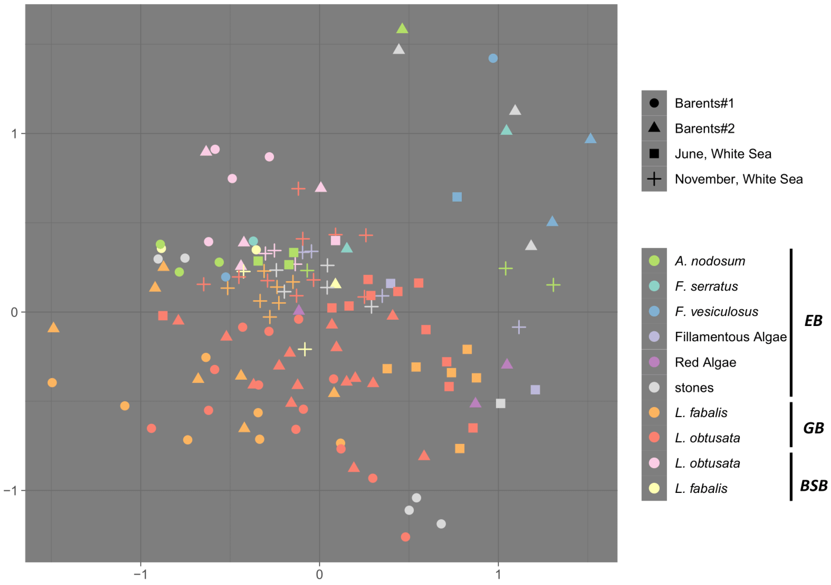
3.2. Sources of Variability in the Bacteriome Composition

We evaluated the variability of the GB composition in space and time: there were two collection sites in the same season (at the Barents Sea) and one site in two different seasons (at the White Sea) in our analysis. The dominant bacterial genera and overall community characteristics (species richness and evenness) remained consistent between the two Barents Sea sites. Moreover, there were no significant effects of the collection site (PERMANOVA p > 0.05). Notably, the differences between host species were found to be significant for bacteriome composition at the Barents Sea (PERMANOVA p = 0.012) (Figure 1 and Figure S1). The analyzed sites were not a long distance from each other and one of them (the Oscar Bay) was in close proximity to human habitation and to a docking area, which were expected to have impacts on coastal microbiota composition (e.g., high polyaromatic hydrocarbon concentrations were registered in the intertidal and subtidal sediment of the Oscar Bay, [80]). Accordingly, bacteria of the genus Acinetobacter were highly abundant in the EB and the BSB samples from the Oscar Bay exclusively; these bacteria were also registered in the GB samples of both host species, though were less abundant (Figure S2). The genus Acinetobacter is a common inhabitant of soil and ocean sediment, including polar regions, being enriched in the hydrocarbon-polluted sites [81]. Since the 1990s, it has also been recognized as an important infectious agent in humans worldwide [82], being associated with skin and gastrointestinal diseases and transmitted, e.g., via feces [83]. Thus, though some signs of anthropogenic impact in the Oscar Bay were registered, the scale of differences did not reach a significant level in this case.
On the White Sea coast, the gut bacteriome samples obtained from the same site in early summer and in autumn differed drastically (Figure 1 and Figure S3; PERMANOVA p = 0.001). These between-season compositional alterations were accompanied by a drop in the mean bacterial diversity (both alpha and beta diversity; Figure 2a,b); moreover, the bacteriomes’ compositional differences due to the host species became insignificant in autumn, though were detectable in early summer (PERMANOVA p < 0.01). Importantly, although the microbial diversity in the GB decreased in autumn, the dominant genera were detected in both seasons (Figure 2a and Figure 3).
Studies on the seasonal variation in the gut bacteriome composition of marine invertebrates are still quite rare. Season was revealed to be a significant predictor of microbial community structure in guts of Eastern oysters Crassostrea virginica [84,85] and the urochordate ascidian Halocynthia roretzi [86], with the cold season being inferior to the summer months in the community richness in both cases. In the former case, the abundance of heterotrophic bacteria and carbon source utilization in the mollusk-associated bacteriomes correlated with the seawater temperature. The role of temperature as a factor structuring the bacteriome associated with the hemolymph of the Crassostrea oysters was also demonstrated in scales of temporal and spatial variation, as well as experimental conditions, with bacterial alpha diversity increasing along with warming [85,87,88]. Interestingly, we recalculated data published earlier [35], and revealed that the values of the gut bacteriome alpha diversity in three Littorina species (L. littorea, L. saxatilis and L. fabalis) tended to be lower in the northern collection sites (Norway, Tromso) compared to the southern ones (Sweden, Tjarno) (Figure 2c).
Obviously, temperature can affect the bacterial community, directly influencing its metabolic activity, production rate and assortment of organic compounds, and thus the bacterial diversity, which was demonstrated both within microbial mats and in plankton [89,90]. Such effects of environmental temperature are fairly expected also for bacterial communities associated with poikilothermic animals. Temperature also affects metabolic rates and moving activity (including foraging and grazing) of the poikilothermic hosts. The feeding activity of intertidal grazers has seasonality in temperate and polar latitudes. For instance, the Patella limpets demonstrated a clear decline in grazing activity during the cold season and its general positive correlation with the seawater temperature [91]. Seasonal patterns are also known in periwinkles; moreover, it was hypothesized that the winter decline in gastropod grazing activity may be a factor eliciting the recovery of algal abundances [41]. A decrease in feeding activity suggests a reduction in the engulfment of bacteria as a food substrate, which may impoverish the diversity of the gut-associated community.
Another important point is that movement restraints caused by low temperatures affect the foraging activity of snails, limiting the diversity of microbiotopes where every particular individual can be encountered. Moreover, both L. fabalis and L. obtusata are prone to abandon the macroalgal canopy and hide at the base of the stones and rocks during the cold season [40]. That is, during autumn and winter, the two flat periwinkle species not only make contact with a lesser amount and a lesser diversity of EB bacteria, but also the sets of bacteria these species make contact with are more similar than during the summer season (due to the similarity of the occupied microbiotope). This explains why L. fabalis and L. obtusata harbor non-differing bacteriomes of relatively low diversity in the autumn samples. In addition, the cold-season-lowered foraging activity can explain the diminishing of not only the alpha but also the beta diversity observed in this study (Figure 2b).

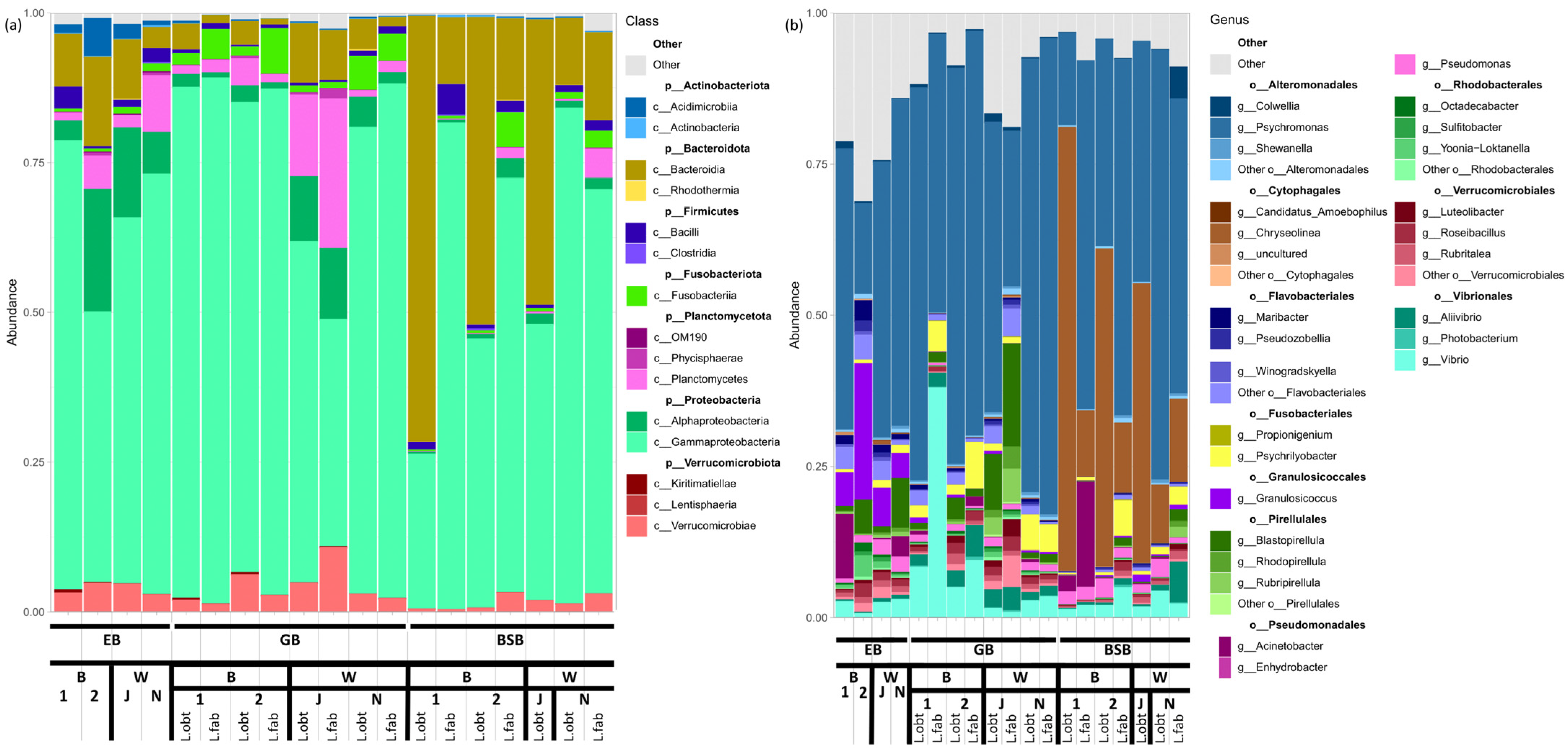
3.3. Associated Bacteriome Composition

The general composition of the gut bacteriomes associated with the Littorina snails was described in detail earlier [35,36]. Our present results on L. obtusata and L. fabalis corroborated the tendencies established previously for the Littorina (Neritrema) and Littorina (Littorina) species:
  • The community is dominated by a limited number of bacterial lineages (3–5);
  • The most abundant taxon is Proteobacteria, especially Gammaproteobacteria;
  • Those less abundant, but inevitably present in the GB groups, are Fusobacteria (Fusobacteriales), Bacteroides (Flavobacteriales) and Planctomycetes (Pirellulales);
  • Cytophagales are predominantly enriched in the BSB (Figure 3a).
The principal bacterial genera in the GB were Psychromonas (Gammaproteobacteria, Alteromonadales), Psychrilyobacter (Fusobacteria), Vibrio and Aliivibrio (Gammaproteobacteria, Vibrionales), and the less abundant Pseudomonas (Gammaproteobacteria, Pseudomonadales), Blastopirellula (Planctomycetes, Pirellulales), Luteolibacter (Verrucomicrobia), as well as the unclassified Alphaproteobacteria g. sp., Flavobacteria g. sp., Verrucomicrobia g. sp. and Pirellulales g. sp.

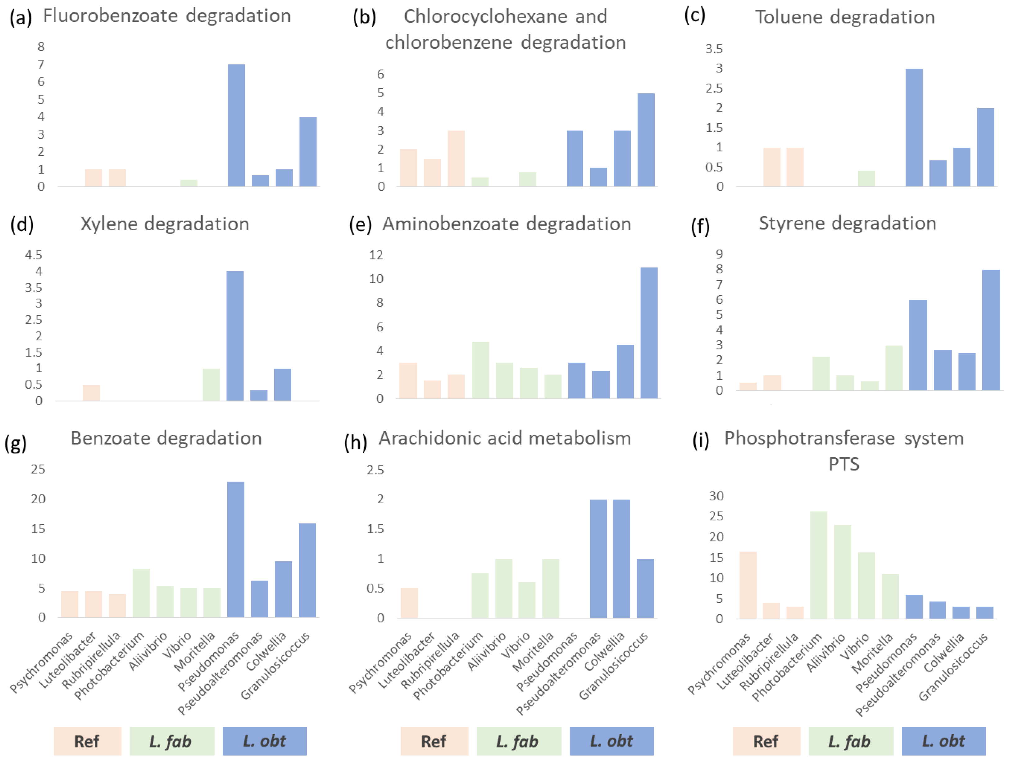
3.4. Interspecies Differences in the Gut Bacteriome Composition

During the summer season, we analyzed the between-species differences of the gut bacteriome in two geographic regions: the White Sea and the Barents Sea. We expected that in the White Sea the between-host-species differences in the associated bacteriome composition may be less pronounced compared to in the Barents Sea. We hypothesized this based on the differences between the Barents and the White seas in the distribution patterns of fucoids which the host snails are predominantly associated with. For instance, F. serratus (preferentially inhabited by L. fabalis) in the White Sea is present only sporadically and does not form zones of high density because it is sensitive to low salinity [92,93]. Indeed, we revealed distribution patterns for both L. obtusata and L. fabalis in the White Sea to be a little dissimilar to those in the Barents Sea. However, there still were clear ecological differences between them in both regions. In particular, in the White Sea, L. obtusata mainly inhabited the depth of the F. vesiculosus canopies (but A. nodosum in the Barents Sea, [42]), while L. fabalis was most often associated with A. nodosum, both on its surface and under the canopy (Figure S4), but preferred F. serratus in the Barents Sea [42].
Although, generally, the composition of high-rank taxa and the list of abundant genera in the GB of both host species were similar (Figure 3a,b), the statistically significant differences due to the host species were registered in both the White and the Barents Sea, which corresponds to the ecological differences described above. We applied the Random Forest analysis to identify some marker bacterial lineages strictly specific to the host species (exclusively detected in either L. fabalis or L. obtusata on a regular basis) and failed to find any. Yet, several bacterial genera tended to be enriched in one of the two host species, being the so-called soft markers (Figure S5). We considered only genera showing non-contradictory trends in different collection sites (even if not in all sites a particular genus was identified as a marker by the Random Forest). In this way, Vibrio, Aliivibrio, Moritella and Planktotalea were revealed as being predominantly associated with L. fabalis, while Granulosicoccus, Octadecabacter, Colwellia, Pseudomonas, Pseudoalteromonas and Maribacter were found to be primarily associated with L. obtusata.
We applied the Kaiju classifier to the shotgun DNA-sequencing data to establish the exact species in each target genus with relative abundances in the GB above 0.01 in at least one collection site. Then, based on the full bacterial lineage taxonomy, we performed an analysis of the enrichment of metabolic pathways in the soft marker genera compared to other abundant genera (≥0.01 mean relative abundance, with no clear distribution trends) using the number of genes involved in a certain pathway as a variable. Only one pathway (‘Phosphotransferase system PTS’, ID 02060), related to the uptake of carbohydrates, particularly hexoses, hexitols, and disaccharides, was found to be specifically enriched in bacterial genera associated with L. fabalis. In contrast, there were a number of pathways enriched in the bacteria predominantly present in the L. obtusata gut (Figure 4; Table S3).
Among those pathways was, for instance, ‘Arachidonic acid metabolism’ (ID 00590). Interestingly, arachidonic acid was revealed through metabolomic profiling as being specifically abundant in the L. obtusata compared to other Littorina species [42]. The list of pathways revealed now as specifically enriched in the L. obtusata were ‘Fluorobenzoate degradation’ (ID 00364), ‘Benzoate degradation’ (ID 00362), ‘Aminobenzoate degradation’ (ID 00627), ‘Chlorocyclohexane and chlorobenzene degradation’ (ID 00361), ‘Styrene degradation’ (ID 643), ‘Toluene degradation’ (ID 00623) and ‘Xylene degradation’ (ID 00622). These pathways are associated with the genera Pseudomonas and Granulosicoccus; the former is most abundantly present in the L. obtusata GB of the White Sea samples, while the latter is in those of the Barents Sea; though both genera were registered in all populations analyzed (Figures S6 and S7).
The genus Granulosicoccus was reported from marine environments, including the seashore zone in the polar regions. It is known to be associated with brown macroalgae such as F. vesiculosus [94,95,96]; it was not previously reported in association with metazoans. Granoulosicoccus species are photoheterotrophs equipped with several metabolic functions of high relevance, such as nitrogen- and sulfur-transformation, the potential to synthesize cobalamin (B12), etc. [97]. Bacteria of the Pseudomonas genus are involved in antagonistic interactions and a variety of metabolic abilities related to synthesis of toxins and degradation of xenobiotics [98]. Species of the Pseudomonas genus were described in the gut-associated bacterial microbiomes of marine invertebrates [99,100]. The mentioned metabolic competencies rely on diverse pathways, including those listed above. Among the products/intermediates/substrates that can be processed in those metabolic pathways are phenolic and polyphenolic compounds [101,102,103]. This fact is of high importance with regard to the ecological differences between the host snail species L. fabalis and L. obtusata.
L. fabalis and L. obtusata demonstrate different, though partially overlapping, preferences to the shore levels, the host fucoid species and the placement in (on/under) an algal canopy [40,42]. One more feature differing in these two snail species is the structure of the radula, reflecting their divergence in feeding behavior [38,41]. While the diet of L. fabalis mainly includes epiphytic microbiota of the macroalgal surface, L. obtusata is able to excavate the depth of the seaweed thalli because its radular outer marginal teeth are armed with numerous angular cusps [40,41]. Brown algae (Phaeophyta) are well-known producers of diverse toxic phenolic and polyphenolic metabolites (e.g., phlorotannins), which defend them against fouling and grazing by micro- and macro-organisms [104,105,106]. Moreover, F. vesiculosus and A. nodosum, preferred by L. obtusata, contain higher concentrations of toxic polyphenols compared to F. serratus, which L. fabalis is predominantly associated with [41,107]. This poses the question about the mechanisms of tolerance of L. obtusata to the toxic action of tannins.
To our best knowledge, there are no data evidencing the ability of the Granulosicoccus or Pseudomonas bacteria (or their combination) to metabolize the phlorotannins of fucoids. Nevertheless, the well-established competencies of these micro-organisms to metabolically transform and degrade phenolic and polyphenolic compounds provides a basis on which to hypothesize their involvement in detoxification and conveying the adaptation of a host to its feeding practices. The possibility that the commensals of the Pseudomonas genus participate in the inactivation of the food toxins in the gut of the invertebrate herbivores was described in the example of the coffee berry borer beetle, Hypothenemus hampei [4].
The specific abundance of bacteria from genera Granulosicoccus and Pseudomonas in the intestinal bacterial community of L. obtusata, as well as the enrichment of polyphenol degradation metabolic pathways associated with these bacteria, may be a factor endowing these snails with the resistance to the toxic fucoids’ metabolites, such as phlorotannins, and allowing this species to occupy its specific niche. However, the details of the possible roles of Granulosicoccus or Pseudomonas in the degradation of food polyphenols in the guts of the Littorina snails are awaiting clarification in future studies.

4. Conclusions

We performed a detailed analysis of the bacteriomes associated with the cryptic sister-species of intertidal snails. Although there were significant inter-regional and inter-seasonal differences in the bacteriome composition, the genera dominant in abundance remained stable and similar between the host species. This is consistent with the idea of the Littorina-associated bacteriome conservatism stated previously.
We registered differences in the gut bacterial microbiome, body surface bacteriome and the environmental bacteriome, which were consistent with our previous results.
We revealed significant differences between L. obtusata and L. fabalis bacteriomes during the warmer season.
The decrease in the gut bacteriomes interspecific differences in autumn (and probably winter), as well as differences in alpha and beta diversity rates between seasons, indicates profound seasonal changes in the snails’ behavior. We expect these behavioral and compositional shifts to be less prominent in regions with warmer climates.
The predominant enrichment of the bacteriome with certain bacterial genera—especially for the L. obtusata gut bacteriome—is both a consequence of snail microhabitat features and their adaptations to the environment.
The enrichment of metabolic pathways involving the biodegradation of phenolic compounds in the genera Granulosicoccus and Pseudomonas predominantly associated with L. obtusata may be critical in mediating the tolerance of this species to fucoid-derived toxic substances (which L. obtusata adult snails engulf in a greater amount than their sister-species L. fabalis). In turn, such differences in bacteriome composition may act as a factor maintaining the differentiation of ecological niches between species.

Supplementary Materials

The following supporting information can be downloaded at: https://www.mdpi.com/article/10.3390/biology12121521/s1, Figure S1: Comparison of the Barents Sea bacteriomes; Figure S2: Relative abundance of the Acinetobacter sp. in all analyzed samples; Figure S3: Comparison of the White Sea bacteriomes; Figure S4: Littorina micro-niche distribution; Figure S5: The Random Forest analysis results; Figure S6: Relative abundance of the bacteria, predominantly abundant in L. fabalis; Figure S7: Relative abundance of the bacteria, predominantly abundant in L. obtusata; Table S1: Details on 16S libraries. Per sample sequencing depth and resulting ASV number; Table S2: Shotgun. Details on shotgun samples. The amount of reads and taxa; Table S3: Pathways. Number of genes in metabolic pathways by KEGG category in principal bacterial genera present in gut bacteriome of L. fabalis and L. obtusata.

Author Contributions

Conceptualization, A.M. and A.G.; methodology, formal analysis and investigation, E.G. (Elizaveta Gafarova), D.K., K.S., I.G., D.P., E.Z., E.G. (Elena Golikova) and A.M.; writing—original draft preparation, A.M., E.G. (Elizaveta Gafarova), D.K. and K.S.; writing—review and editing, A.M. and A.G.; supervision, A.M. and A.G. All authors have read and agreed to the published version of the manuscript.

Funding

The study was supported by the grant of the Russian Science Foundation (RSF) No 19-14-00321 (PI Andrei I. Granovitch). The participation of Sogomonyan K. S was supported by St. Petersburg State University (grant ID: 94030965).

Institutional Review Board Statement

Institutional Review Board approval is not applicable to the study. Periwinkles are not endangered or protected species and our sampling was performed outside protected areas, so no special permission for the material collection was required. Moreover, all applicable international, national, and institutional guidelines for the care and use of animals were followed by the authors.

Informed Consent Statement

Not applicable.

Data Availability Statement

Raw data are available for downloading from the NCBI database. 16S rRNA gene metabarcoding reads are deposited as PRJNA1044559 and shotgun metagenomics raw sequences are deposited as PRJNA1044926.

Acknowledgments

We thank A. Gonchar for the language editing, M. Varfolomeeva for the comprehensive advice on the design of the statistical experiments, and L. Danilov for the valuable recommendations on shotgun assembly and annotation. The wet-lab work was conducted with the equipment of the Resource Centers of St. Petersburg State University “Development of Molecular and Cell Technologies” and “Environmental Safety Observatory”. We are grateful to the White Sea Biological Station “Kartesh” (Zoological Institute RAS) for the accommodation, field lab facilities and provided data from the standard pelagic monitoring station D-1. We thank the “Dalnye Zelentsy” diving center for the accommodation and the kind help during the sampling expedition on the eastern Murman coast.

Conflicts of Interest

The authors declare no conflict of interest.

References

  1. Attardo, G.M.; Lohs, C.; Heddi, A.; Alam, U.H.; Yildirim, S.; Aksoy, S. Analysis of milk gland structure and function in Glossina morsitans: Milk protein production, symbiont populations and fecundity. J. Insect Physiol. 2008, 54, 1236–1242. [Google Scholar] [CrossRef] [PubMed]
  2. Hansen, A.K.; Moran, N.A. Aphid genome expression reveals host-symbiont cooperation in the production of amino acids. Proc. Natl. Acad. Sci. USA 2011, 108, 2849–2854. [Google Scholar] [CrossRef] [PubMed]
  3. Flint, H.J.; Scott, K.P.; Duncan, S.H.; Louis, P.; Forano, E. Microbial degradation of complex carbohydrates in the gut. Gut Microbes 2012, 3, 289–306. [Google Scholar] [CrossRef] [PubMed]
  4. Ceja-Navarro, J.A.; Vega, F.E.; Karaoz, U.; Hao, Z.; Jenkins, S.; Lim, H.C.; Kosina, P.; Infante, F.; Northen, T.R.; Brodie, E.L. Gut microbiota mediate caffeine detoxification in the primary insect pest of coffee. Nat. Commun. 2015, 6, 7618. [Google Scholar] [CrossRef] [PubMed]
  5. Shapira, M. Gut Microbiotas and Host Evolution: Scaling up Symbiosis. Trends Ecol. Evol. 2016, 31, 539–549. [Google Scholar] [CrossRef] [PubMed]
  6. Aronson, H.S.; Zellmer, A.J.; Goffredi, S.K. The specific and exclusive microbiome of the deep-sea bone-eating snail, Rubyspira osteovora. FEMS Microbiol. Ecol. 2017, 93, fiw250. [Google Scholar] [CrossRef]
  7. O’Hara, A.M.; Shanahan, F. The gut flora as a forgotten organ. EMBO Rep. 2006, 7, 688–693. [Google Scholar] [CrossRef]
  8. Gerardo, N.M.; Altincicek, B.; Anselme, C.; Atamian, H.; Barribeau, S.M.; de Vos, M.; Duncan, E.J.; Evans, J.D.; Gabaldon, T.; Ghanim, M.; et al. Immunity and other defenses in pea aphids, Acyrthosiphon pisum. Genome Biol. 2010, 11, R21. [Google Scholar] [CrossRef]
  9. Laughton, A.M.; Garcia, J.R.; Altincicek, B.; Strand, M.R.; Gerardo, N.M. Characterisation of immune responses in the pea aphid, Acyrthosiphon pisum. J. Insect Physiol. 2011, 57, 830–839. [Google Scholar] [CrossRef]
  10. Sommer, F.; Backhed, F. The gut microbiota--masters of host development and physiology. Nat. Rev. Microbiol. 2013, 11, 227–238. [Google Scholar] [CrossRef]
  11. Kamada, N.; Seo, S.U.; Chen, G.Y.; Nunez, G. Role of the gut microbiota in immunity and inflammatory disease. Nat. Rev. Immunol. 2013, 13, 321–335. [Google Scholar] [CrossRef] [PubMed]
  12. Sharon, G.; Segal, D.; Ringo, J.M.; Hefetz, A.; Zilber-Rosenberg, I.; Rosenberg, E. Commensal Bacteria Play a Role in Mating Preference of Drosophila melanogaster. Proc. Natl. Acad. Sci. USA 2010, 107, 20051–20056. [Google Scholar] [CrossRef] [PubMed]
  13. Foster, J.A.; McVey Neufeld, K.-A. Gut–Brain Axis: How the Microbiome Influences Anxiety and Depression. Trends Neurosci. 2013, 36, 305–312. [Google Scholar] [CrossRef] [PubMed]
  14. Hsiao, E.Y.; McBride, S.W.; Hsien, S.; Sharon, G.; Hyde, E.R.; McCue, T.; Codelli, J.A.; Chow, J.; Reisman, S.E.; Petrosino, J.F.; et al. Microbiota Modulate Behavioral and Physiological Abnormalities Associated with Neurodevelopmental Disorders. Cell 2013, 155, 1451–1463. [Google Scholar] [CrossRef] [PubMed]
  15. Lize, A.; McKay, R.; Lewis, Z. Gut microbiota and kin recognition. Trends Ecol. Evol. 2013, 28, 325–326. [Google Scholar] [CrossRef] [PubMed]
  16. Desbonnet, L.; Clarke, G.; Shanahan, F.; Dinan, T.G.; Cryan, J.F. Microbiota Is Essential for Social Development in the Mouse. Mol. Psychiatry 2014, 19, 146–148. [Google Scholar] [CrossRef]
  17. Cryan, J.F.; Dinan, T.G. More than a Gut Feeling: The Microbiota Regulates Neurodevelopment and Behavior. Neuropsychopharmacol 2015, 40, 241–242. [Google Scholar] [CrossRef]
  18. Valles-Colomer, M.; Falony, G.; Darzi, Y.; Tigchelaar, E.F.; Wang, J.; Tito, R.Y.; Schiweck, C.; Kurilshikov, A.; Joossens, M.; Wijmenga, C.; et al. The neuroactive potential of the human gut microbiota in quality of life and depression. Nat. Microbiol. 2019, 4, 623–632. [Google Scholar] [CrossRef]
  19. Greene, L.K.; Williams, C.V.; Junge, R.E.; Mahefarisoa, K.L.; Rajaonarivelo, T.; Rakotondrainibe, H.; O’Connell, T.M.; Drea, C.M. A role for gut microbiota in host niche differentiation. ISME J. 2020, 14, 1675–1687. [Google Scholar] [CrossRef]
  20. Duperron, S.; Pottier, M.A.; Leger, N.; Gaudron, S.M.; Puillandre, N.; Le Prieur, S.; Sigwart, J.D.; Ravaux, J.; Zbinden, M. A tale of two chitons: Is habitat specialisation linked to distinct associated bacterial communities? FEMS Microbiol. Ecol. 2013, 83, 552–567. [Google Scholar] [CrossRef]
  21. Derycke, S.; De Meester, N.; Rigaux, A.; Creer, S.; Bik, H.; Thomas, W.K.; Moens, T. Coexisting Cryptic Species of the Litoditis marina Complex (Nematoda) Show Differential Resource Use and Have Distinct Microbiomes with High Intraspecific Variability. Mol. Ecol. 2016, 25, 2093–2110. [Google Scholar] [CrossRef] [PubMed]
  22. Gause, G.F. The Struggle for Existence; Williams and Wilkins: Baltimore, MD, USA, 1934; p. 163. [Google Scholar]
  23. Schluter, D. The Ecology of Adaptive Radiation; Oxford University Press: New York, NY, USA, 2000; p. 296. [Google Scholar]
  24. Ackerly, D.D. Community Assembly, Niche Conservatism, and Adaptive Evolution in Changing Environments. Int. J. Plant Sci. 2003, 164, S165–S184. [Google Scholar] [CrossRef]
  25. Haygood, M.G.; Schmidt, E.W.; Davidson, S.K.; Faulkner, D.J. Microbial symbionts of marine invertebrates: Opportunities for microbial biotechnology. J. Mol. Microbiol. Biotechnol. 1999, 1, 33–43. [Google Scholar] [PubMed]
  26. Reveillaud, J.; Maignien, L.; Eren, A.M.; Huber, J.A.; Apprill, A.; Sogin, M.L.; Vanreusel, A. Host-Specificity among Abundant and Rare Taxa in the Sponge Microbiome. ISME J. 2014, 8, 1198–1209. [Google Scholar] [CrossRef] [PubMed]
  27. Weiland-Brauer, N.; Neulinger, S.C.; Pinnow, N.; Kunzel, S.; Baines, J.F.; Schmitz, R.A. Composition of Bacterial Communities Associated with Aurelia aurita Changes with Compartment, Life Stage, and Population. Appl. Environ. Microbiol. 2015, 81, 6038–6052. [Google Scholar] [CrossRef]
  28. Bost, A.; Martinson, V.G.; Franzenburg, S.; Adair, K.L.; Albasi, A.; Wells, M.T.; Douglas, A.E. Functional variation in the gut microbiome of wild Drosophila populations. Mol. Ecol. 2018, 27, 2834–2845. [Google Scholar] [CrossRef]
  29. Jehrke, L.; Stewart, F.A.; Droste, A.; Beller, M. The impact of genome variation and diet on the metabolic phenotype and microbiome composition of Drosophila melanogaster. Sci. Rep. 2018, 8, 6215. [Google Scholar] [CrossRef]
  30. Johannesson, K.; Panova, M.; Kemppainen, P.; André, C.; Rolan-Alvarez, E.; Butlin, R.K. Repeated evolution of reproductive isolation in a marine snail: Unveiling mechanisms of speciation. Philos. Trans. R. Soc. B Biol. Sci. 2010, 365, 1735–1747. [Google Scholar] [CrossRef]
  31. Butlin, R.K.; Saura, M.; Charrier, G.; Jackson, B.; André, C.; Caballero, A.; Coyne, J.A.; Galindo, J.; Grahame, J.W.; Hollander, J. Parallel evolution of local adaptation and reproductive isolation in the face of gene flow. Evolution 2014, 68, 935–949. [Google Scholar] [CrossRef]
  32. Panova, M.; Johansson, T.; Canbäck, B.; Bentzer, J.; Rosenblad, M.A.; Johannesson, K.; Tunlid, A.; André, C. Species and gene divergence in Littorina snails detected by array comparative genomic hybridization. BMC Genom. 2014, 15, 687. [Google Scholar] [CrossRef]
  33. Mittermayer, F.; Helmerson, C.; Duvetorp, M.; Johannesson, K.; Panova, M. The molecular background of the aspartate aminotransferase polymorphism in Littorina snails maintained by strong selection on small spatial scales. Gene 2023, 876, 147517. [Google Scholar] [CrossRef] [PubMed]
  34. Johannesson, K.; Butlin, R.K.; Panova, M.; Westram, A.M. Mechanisms of adaptive divergence and speciation in Littorina saxatilis: Integrating knowledge from ecology and genetics with new data emerging from genomic studies. In Population Genomics: Marine Organisms; Springer: Cham, Switzerland, 2020; pp. 277–301. [Google Scholar] [CrossRef]
  35. Maltseva, A.L.; Varfolomeeva, M.A.; Gafarova, E.R.; Panova, M.A.Z.; Mikhailova, N.A.; Granovitch, A.I. Divergence together with microbes: A comparative study of the associated microbiomes in the closely related Littorina species. PLoS ONE 2021, 16, e0260792. [Google Scholar] [CrossRef] [PubMed]
  36. Panova, M.A.Z.; Varfolomeeva, M.A.; Gafarova, E.R.; Maltseva, A.L.; Mikhailova, N.A.; Granovitch, A.I. First Insights into the Gut Microbiomes and the Diet of the Littorina Snail Ecotypes, a Recently Emerged Marine Evolutionary Model. Evol. Appl. 2023, 16, 365–378. [Google Scholar] [CrossRef] [PubMed]
  37. Maltseva, A.; Lobov, A.; Pavlova, P.; Panova, M.; Gafarova, E.; Marques, J.; Danilov, L.; Granovitch, A. Orphan gene in Littorina: An unexpected role of symbionts in the host evolution. Gene 2022, 824, 146389. [Google Scholar] [CrossRef] [PubMed]
  38. Reid, D.G. Systematics and Evolution of Littorina; Ray Society: London, UK, 1996; p. 463. [Google Scholar]
  39. Williams, G.A. The comparative ecology of the flat periwinkles littorina obtusata l. and littorina mariae sacchi et rastelli. Field Study 1990, 7, 469–482. [Google Scholar]
  40. Watson, D.C.; Norton, T.A. The habitat and feeding preferences of Littorina obtusata (L.) and L. mariae Sacchi et Rastelli. J. Exp. Mar. Biol. Ecol. 1987, 112, 61–72. [Google Scholar] [CrossRef]
  41. Norton, T.A.; Hawkins, S.J.; Manley, N.L.; Williams, G.A.; Watson, D.C. Scraping a Living: A Review of Iittorinid Grazing. In Progress in Littorinid and Muricid Biology; Developments in Hydrobiology; Springer: Dordrecht, The Netherlands, 1990; Volume 56, pp. 117–138. [Google Scholar] [CrossRef]
  42. Maltseva, A.L.; Varfolomeeva, M.A.; Ayanka, R.V.; Gafarova, E.R.; Repkin, E.A.; Pavlova, P.A.; Shavarda, A.L.; Mikhailova, N.A.; Granovitch, A.I. Linking Ecology, Morphology, and Metabolism: Niche Differentiation in Sympatric Populations of Closely Related Species of the Genus Littorina (Neritrema). Ecol. Evol. 2021, 11, 11134–11154. [Google Scholar] [CrossRef]
  43. Carvalho, J.; Pereira, C.; Sotelo, G.; Costa, D.; Galindo, J.; Faria, R. De Novo Isolation of 17 Microsatellite Loci for Flat Periwinkles (Littorina fabalis and L. Obtusata) and Their Application for Species Discrimination and Hybridization Studies. J. Mollus. Stud. 2015, 81, 421–425. [Google Scholar] [CrossRef]
  44. Weingarten, E.A.; Atkinson, C.L.; Jackson, C.R. The gut microbiome of freshwater Unionidae mussels is determined by host species and is selectively retained from filtered seston. PLoS ONE 2019, 14, e0224796. [Google Scholar] [CrossRef]
  45. Schols, R.; Vanoverberghe, I.; Huyse, T.; Decaestecker, E. Host-bacteriome transplants of the schistosome snail host Biomphalaria glabrata reflect species-specific associations. FEMS Microbiol. Ecol. 2023, 99, fiad101. [Google Scholar] [CrossRef]
  46. Hu, Z.; Chen, X.; Chang, J.; Yu, J.; Tong, Q.; Li, S.; Niu, H. Compositional and predicted functional analysis of the gut microbiota of Radix auricularia (Linnaeus) via high-throughput Illumina sequencing. PeerJ 2018, 6, e5537. [Google Scholar] [CrossRef] [PubMed]
  47. Zonn, I.S.; Kostianoy, A.G.; Semenov, A.V. The Western Arctic Seas Encyclopedia; Springer International Publishing: Cham, Switzerland, 2017; p. 547. [Google Scholar]
  48. Golikova, E.; Varfolomeeva, M.; Yakovis, E.; Korsun, S. Saltmarsh foraminifera in the subarctic White Sea: Thrive in summer, endure in winter. Estuar. Coast. Shelf Sci. 2020, 238, 106685. [Google Scholar] [CrossRef]
  49. White Sea Hydrology and Zooplankton Time-Series: Kartesh D1. 2020. Available online: https://www.st.nmfs.noaa.gov/copepod/time-series/ru-10101/ (accessed on 24 November 2023).
  50. Costa, D.; Sotelo, G.; Kaliontzopoulou, A.; Carvalho, J.; Butlin, R.; Hollander, J.; Faria, R. Hybridization patterns between two marine snails, Littorina fabalis and L. obtusata. Ecol. Evol. 2020, 10, 1158–1179. [Google Scholar] [CrossRef] [PubMed]
  51. Carvalho, J.; Sotelo, G.; Galindo, J.; Faria, R. Genetic characterization of flat periwinkles (Littorinidae) from the Iberian Peninsula reveals interspecific hybridization and different degrees of differentiation. Biol. J. Linn. Soc. 2016, 118, 503–519. [Google Scholar] [CrossRef]
  52. Neu, A.T.; Allen, E.E.; Roy, K. Diversity and composition of intertidal gastropod microbiomes across a major marine biogeographic boundary. Environ. Microbiol. Rep. 2019, 11, 434–447. [Google Scholar] [CrossRef]
  53. Hugerth, L. MiSeq Amplicon Sequencing Sample Prep with Dual Barcoding. Available online: https://github.com/EnvGen/LabProtocols/blob/master/Amplicon_dual_index_prep_EnvGen.rst (accessed on 23 October 2023).
  54. Caporaso, J.G.; Lauber, C.L.; Walters, W.A.; Berg-Lyons, D.; Lozupone, C.A.; Turnbaugh, P.J.; Fierer, N.; Knight, R. Global Patterns of 16S rRNA Diversity at a Depth of Millions of Sequences per Sample. Proc. Natl. Acad. Sci. USA 2011, 108, 4516–4522. [Google Scholar] [CrossRef] [PubMed]
  55. Andrews, S. FastQC: A Quality Control Tool for High throughput Sequence Data. 2010. Available online: https://www.bioinformatics.babraham.ac.uk/projects/fastqc (accessed on 23 October 2023).
  56. Bolyen, E.; Rideout, J.R.; Dillon, M.R.; Bokulich, N.A.; Abnet, C.C.; Al-Ghalith, G.A.; Alexander, H.; Alm, E.J.; Arumugam, M.; Asnicar, F.; et al. Reproducible, interactive, scalable and extensible microbiome data science using QIIME 2. Nat. Biotechnol. 2019, 37, 852–857. [Google Scholar] [CrossRef]
  57. Callahan, B.J.; McMurdie, P.J.; Rosen, M.J.; Han, A.W.; Johnson, A.J.; Holmes, S.P. DADA2: High-resolution sample inference from Illumina amplicon data. Nat. Methods 2016, 13, 581–583. [Google Scholar] [CrossRef]
  58. Pedregosa, F.; Varoquaux, G.; Gramfort, A.; Michel, V.; Thirion, B.; Grisel, O.; Blondel, M.; Prettenhofer, P.; Weiss, R.; Dubourg, V.; et al. Scikit-Learn: Machine Learning in Python. J. Mach. Learn. Res. 2011, 12, 2825–2830. [Google Scholar]
  59. Quast, C.; Pruesse, E.; Yilmaz, P.; Gerken, J.; Schweer, T.; Yarza, P.; Peplies, J.; Glockner, F.O. The SILVA ribosomal RNA gene database project: Improved data processing and web-based tools. Nucleic Acids Res. 2013, 41, D590–D596. [Google Scholar] [CrossRef]
  60. McMurdie, P.J.; Holmes, S. phyloseq: An R package for reproducible interactive analysis and graphics of microbiome census data. PLoS ONE 2013, 8, e61217. [Google Scholar] [CrossRef] [PubMed]
  61. Shannon, C.E. A mathematical theory of communication. Bell Syst. Tech. J. 1948, 27, 379–423. [Google Scholar] [CrossRef]
  62. MacArthur, R. Fluctuations of Animal Populations and a Measure of Community Stability. Ecology 1955, 36, 533. [Google Scholar] [CrossRef]
  63. Pielou, E.C. Shannon’s Formula as a Measure of Specific Diversity: Its Use and Misuse. Am. Nat. 1966, 100, 463–465. [Google Scholar] [CrossRef]
  64. Bray, J.R.; Curtis, J.T. An Ordination of the Upland Forest Communities of Southern Wisconsin. Ecol. Monogr. 1957, 27, 325–349. [Google Scholar] [CrossRef]
  65. Kruskal, J.B. Multidimensional Scaling by Optimizing Goodness of Fit to a Nonmetric Hypothesis. Psychometrika 1964, 29, 1–27. [Google Scholar] [CrossRef]
  66. Wickham, H. A Layered Grammar of Graphics. J. Comput. Graph. Stat. 2010, 19, 3–28. [Google Scholar] [CrossRef]
  67. Collyer, M.L.; Adams, D.C. RRPP: An r package for fitting linear models to high-dimensional data using residual randomization. Methods Ecol. Evol. 2018, 9, 1772–1779. [Google Scholar] [CrossRef]
  68. Teunisse, G.M. Fantaxtic: Fantaxtic Plots for Phyloseq Data. 2018. Available online: https://github.com/gmteunisse/fantaxtic (accessed on 23 October 2023).
  69. Breiman, L.; Breiman, L.; Cutler, R.A. Random Forests Machine Learning. J. Clin. Microbiol. 2001, 2, 199–228. [Google Scholar]
  70. Dang, T.; Kishino, H. Forward variable selection improves the power of random forest for high-dimensional microbiome data. bioRxiv 2020. [Google Scholar] [CrossRef]
  71. Dhariwal, A.; Chong, J.; Habib, S.; King, I.L.; Agellon, L.B.; Xia, J. MicrobiomeAnalyst: A web-based tool for comprehensive statistical, visual and meta-analysis of microbiome data. Nucleic Acids Res. 2017, 45, W180–W188. [Google Scholar] [CrossRef] [PubMed]
  72. Andersen, V.D.; Jensen, M.S.; Munk, P.; Vigre, H. Robustness in quantifying the abundance of antimicrobial resistance genes in pooled faeces samples from batches of slaughter pigs using metagenomics analysis. J. Glob. Antimicrob. Resist. 2021, 24, 398–402. [Google Scholar] [CrossRef] [PubMed]
  73. Ray, K.J.; Cotter, S.Y.; Arzika, A.M.; Kim, J.; Boubacar, N.; Zhou, Z.; Zhong, L.; Porco, T.C.; Keenan, J.D.; Lietman, T.M. High-throughput sequencing of pooled samples to determine community-level microbiome diversity. Ann. Epidemiol. 2019, 39, 63–68. [Google Scholar] [CrossRef] [PubMed]
  74. Rodríguez-Ruano, S.M.; Juhaňáková, E.; Vávra, J.; Nováková, E. Methodological insight into mosquito microbiome studies. Front. Cell. Infect. Microbiol. 2020, 10, 86. [Google Scholar] [CrossRef]
  75. Chen, S. Ultrafast One-pass FASTQ Data Preprocessing, Quality Control, and Deduplication Using Fastp. iMeta 2023, 2, e107. [Google Scholar] [CrossRef]
  76. Nurk, S.; Meleshko, D.; Korobeynikov, A.; Pevzner, P.A. metaSPAdes: A new versatile metagenomic assembler. Genome Res. 2017, 27, 824–834. [Google Scholar] [CrossRef] [PubMed]
  77. Menzel, P.; Ng, K.L.; Krogh, A. Fast and sensitive taxonomic classification for metagenomics with Kaiju. Nat. Commun. 2016, 7, 11257. [Google Scholar] [CrossRef]
  78. Seemann, T. Prokka: Rapid Prokaryotic Genome Annotation. Bioinformatics 2014, 30, 2068–2069. [Google Scholar] [CrossRef]
  79. Moriya, Y.; Itoh, M.; Okuda, S.; Yoshizawa, A.C.; Kanehisa, M. KAAS: An automatic genome annotation and pathway reconstruction server. Nucleic Acids Res. 2007, 35, W182–W185. [Google Scholar] [CrossRef]
  80. Kursheva, A.V.; Morgunova, I.P.; Petrova, V.I.; Litvinenko, I.V. Hydrocarbons in the Littoral Sediments and March Soils of the Southwestern Coast of the Barents Sea. Geochem. Int. 2023, 61, 972–988. [Google Scholar] [CrossRef]
  81. Jung, J.; Park, W. Acinetobacter Species as Model Microorganisms in Environmental Microbiology: Current State and Perspectives. Appl. Microbiol. Biotechnol. 2015, 99, 2533–2548. [Google Scholar] [CrossRef] [PubMed]
  82. Munoz-Price, L.S.; Weinstein, R.A. Acinetobacter infection. N. Engl. J. Med. 2008, 358, 1271–1281. [Google Scholar] [CrossRef] [PubMed]
  83. Manchanda, V.; Sanchaita, S.; Singh, N. Multidrug resistant acinetobacter. J. Glob. Infect. Dis. 2010, 2, 291–304. [Google Scholar] [CrossRef] [PubMed]
  84. Pierce, M.L.; Ward, J.E.; Holohan, B.A.; Zhao, X.; Hicks, R.E. The Influence of Site and Season on the Gut and Pallial Fluid Microbial Communities of the Eastern Oyster, Crassostrea Virginica (Bivalvia, Ostreidae): Community-Level Physiological Profiling and Genetic Structure. Hydrobiologia 2016, 765, 97–113. [Google Scholar] [CrossRef]
  85. Pierce, M.L.; Ward, J.E. Gut Microbiomes of the Eastern Oyster (Crassostrea virginica) and the Blue Mussel (Mytilus edulis): Temporal Variation and the Influence of Marine Aggregate-Associated Microbial Communities. mSphere 2019, 4, e00730-19. [Google Scholar] [CrossRef]
  86. Wei, J.; Gao, H.; Yang, Y.; Liu, H.; Yu, H.; Chen, Z.; Dong, B. Seasonal dynamics and starvation impact on the gut microbiome of urochordate ascidian Halocynthia roretzi. Anim. Microbiome 2020, 2, 30. [Google Scholar] [CrossRef]
  87. Lokmer, A.; Mathias Wegner, K. Hemolymph microbiome of Pacific oysters in response to temperature, temperature stress and infection. ISME J. 2015, 9, 670–682. [Google Scholar] [CrossRef]
  88. Lokmer, A.; Goedknegt, M.A.; Thieltges, D.W.; Fiorentino, D.; Kuenzel, S.; Baines, J.F.; Wegner, K.M. Spatial and Temporal Dynamics of Pacific oyster Hemolymph Microbiota across Multiple Scales. Front. Microbiol. 2016, 7, 1367. [Google Scholar] [CrossRef]
  89. Everroad, R.C.; Otaki, H.; Matsuura, K.; Haruta, S. Diversification of Bacterial Community Composition along a Temperature Gradient at a Thermal Spring. Microb. Environ. 2012, 27, 374–381. [Google Scholar] [CrossRef]
  90. Liu, H.; Zhang, H.; Powell, J.; Delgado-Baquerizo, M.; Wang, J.; Singh, B. Warmer and Drier Ecosystems Select for Smaller Bacterial Genomes in Global Soils. iMeta 2023, 2, e70. [Google Scholar] [CrossRef]
  91. Jenkins, S.; Arenas, F.; Arrontes, J.; Bussell, J.; Castro, J.; Coleman, R.; Hawkins, S.; Kay, S.; Martínez, B.; Oliveros, J.; et al. European-Scale Analysis of Seasonal Variability in Limpet Grazing Activity and Microalgal Abundance. Mar. Ecol. Prog. Ser. 2001, 211, 193–203. [Google Scholar] [CrossRef]
  92. Malavenda, S.V. The tolerance of the Barents Sea fucoids for varying salinity. Bot. Zhurnal 2011, 96, 342–349. [Google Scholar]
  93. Wahl, M.; Jormalainen, V.; Eriksson, B.K.; Coyer, J.A.; Molis, M.; Schubert, H.; Dethier, M.; Karez, R.; Kruse, I.; Lenz, M.; et al. Stress ecology in fucus: Abiotic, biotic and genetic interactions. Adv. Mar. Biol. 2011, 59, 37–105. [Google Scholar] [CrossRef] [PubMed]
  94. Lachnit, T.; Meske, D.; Wahl, M.; Harder, T.; Schmitz, R. Epibacterial community patterns on marine macroalgae are host-specific but temporally variable. Environ. Microbiol. 2011, 13, 655–665. [Google Scholar] [CrossRef] [PubMed]
  95. Park, S.; Jung, Y.T.; Won, S.M.; Park, J.M.; Yoon, J.H. Granulosicoccus undariae sp. nov., a member of the family Granulosicoccaceae isolated from a brown algae reservoir and emended description of the genus Granulosicoccus. Antonie Van Leeuwenhoek 2014, 106, 845–852. [Google Scholar] [CrossRef] [PubMed]
  96. Kang, I.; Lim, Y.; Cho, J.C. Complete genome sequence of Granulosicoccus antarcticus type strain IMCC3135(T), a marine gammaproteobacterium with a putative dimethylsulfoniopropionate demethylase gene. Mar. Genom. 2018, 37, 176–181. [Google Scholar] [CrossRef]
  97. Weigel, B.L.; Miranda, K.K.; Fogarty, E.C.; Watson, A.R.; Pfister, C.A. Functional Insights into the Kelp Microbiome from Metagenome-Assembled Genomes. mSystems 2022, 7, e01422-21. [Google Scholar] [CrossRef]
  98. Chiellini, C.; Lombardo, K.; Mocali, S.; Miceli, E.; Fani, R. Pseudomonas Strains Isolated from Different Environmental Niches Exhibit Different Antagonistic Ability. Ethol. Ecol. Evol. 2019, 31, 399–420. [Google Scholar] [CrossRef]
  99. Zhang, M.; Sun, Y.; Liu, Y.; Qiao, F.; Chen, L.; Liu, W.-T.; Du, Z.; Li, E. Response of Gut Microbiota to Salinity Change in Two Euryhaline Aquatic Animals with Reverse Salinity Preference. Aquaculture 2016, 454, 72–80. [Google Scholar] [CrossRef]
  100. Cicala, F.; Lago-Lestón, A.; Gomez-Gil, B.; Gollas-Galván, T.; Chong-Robles, J.; Cortés-Jacinto, E.; Martínez-Porchas, M. Gut Microbiota Shifts in the Giant Tiger Shrimp, Penaeus Monodon, during the Postlarvae, Juvenile, and Adult Stages. Aquac. Int. 2020, 28, 1421–1433. [Google Scholar] [CrossRef]
  101. Medić, A.B.; Karadžić, I.M. Pseudomonas in environmental bioremediation of hydrocarbons and phenolic compounds-key catabolic degradation enzymes and new analytical platforms for comprehensive investigation. World J. Microbiol. Biotechnol. 2022, 38, 165. [Google Scholar] [CrossRef] [PubMed]
  102. Yesankar, P.J.; Patil, A.; Kapley, A.; Qureshi, A. Catalytic resilience of multicomponent aromatic ring-hydroxylating dioxygenases in Pseudomonas for degradation of polycyclic aromatic hydrocarbons. World J. Microbiol. Biotechnol. 2023, 39, 166. [Google Scholar] [CrossRef] [PubMed]
  103. Zhang, S.; An, Z.; Su, X.; Hou, A.; Liu, L.; Zhang, L.; Lai, J. Phenol degradation at high salinity by a resuscitated strain Pseudomonas sp. SAS26: Kinetics and pathway. J. Environ. Chem. Eng. 2023, 11, 110182. [Google Scholar] [CrossRef]
  104. Ragan, M.A.; Craigie, J.S. Physodes and the phenolic compounds of brown algae. Isolation and characterization of phloroglucinol polymers from Fucus vesiculosus (L.). Can. J. Biochem. 1976, 54, 66–73. [Google Scholar] [CrossRef]
  105. Imbs, T.I.; Zvyagintseva, T.N. Phlorotannins Are Polyphenolic Metabolites of Brown Algae. Russ. J. Mar. Biol. 2018, 44, 263–273. [Google Scholar] [CrossRef]
  106. Lomartire, S.; Cotas, J.; Pacheco, D.; Marques, J.C.; Pereira, L.; Goncalves, A.M.M. Environmental Impact on Seaweed Phenolic Production and Activity: An Important Step for Compound Exploitation. Mar. Drugs 2021, 19, 245. [Google Scholar] [CrossRef]
  107. Bate-Smith, E.C. Haemanalysis of Tannins: The Concept of Relative Astringency. Phytochemistry 1973, 12, 907–912. [Google Scholar] [CrossRef]
Figure 1. Comparison of bacteriome composition of all samples. nMDS ordination of the bacteriome in different sample types. EB—environmental bacteriome, GB—gut bacteriome, BSB—body surface bacteriome.
Biology 12 01521 g001
Figure 2. Diversity overview. (a) Shannon diversity index values. The index was calculated for the White Sea samples on the subsampled gut dataset. Index values for samples from a host species are combined into a boxplot. (b) Beta diversity measures. Bray–Curtis dissimilarity indices for the White sea samples. Distances were calculated on the subsampled gut dataset and shown as boxplots grouped by host species and season. (c) Shannon diversity index values (previously published data [32]). The general trend of the associated bacterial community alpha diversity value to decrease and can be demonstrated by latitudinal comparison. The Shannon diversity index values for our previously published data on three Littorina species (L. littorea, L. saxatilis and L. obtusata) are in consistency with this trend.
Biology 12 01521 g002
Figure 3. Relative abundance of the most abundant bacterial taxa averaged for each sample type. (a) Relative abundance at the class level. Bacterial classes are grouped into phyla. (b) Relative abundance at the genus level. Bacterial genera are grouped into orders.
Biology 12 01521 g003
Figure 4. The mean number of genes involved in particular metabolic pathways. (a) Fluorobenzoate degradation. (b) Chlorocyclohexane and chlorobenzene degradation. (c) Toluene degradation. (d) Xylene degradation. (e) Aminobenzoate degradation. (f) Styrene degradation. (g) Benzoate degradation. (h) Arachidonic acid metabolism. Interestingly, a slight trend for genes of this pathway to be enriched in L. obtusata is compatible with the results of a recent metabolomics study, where arachidonic acid was identified as a compound specifically enriched in L. obtusata [42]. (i) Phosphotransferase system (PTS). The numbers of considered species were in the reference group (ref)—Psychromonas: 2 (P. ingrahamii, P. sp. CNPT3); Luteolibacter: 2 (L. ambystomatis, L. luteus); and Rubripirellula 1 (R. lacrimiformis). In the group of species predominantly present in L. fabalis (L. fab)—Photobacterium 4 (P. gaetbulicola Gung47, P. damselae, P. profundum, P. ganghwense); Aliivibrio 3 (A. fischeri ATCC 7744, A. fischeri MJ11, A. salmonicida); Vibrio 5 (V. breoganii, V. sp VB16, V. algicola, V. ciclytrophicus, V. splendidus); and Moritella 1 (M. marina). In the group of species predominantly present in L. obtusata (L.obt)Pseudomonas 1 (P. stutzeri); Pseudoalteromonas 3 (P. carrageenovora, P. sp. A25, P. tunicata); Colwellia 1 (C. psychrerythraea); and Granulosicoccus 1 (G. antarcticus).
Biology 12 01521 g004
Table 1. A list of the successfully sequenced samples collected on the Barents Sea coast.
SampleBarents#1Barents#2
EnvironmentF. vesiculosus scraping (×2)F. vesiculosus scraping (×2)
A. nodosum scraping (×3)A. nodosum scraping (×1)
F. serratus scraping (×1)F. serratus scraping (×2)
Stones scraping (×5)Stones scraping (×3)
Red Algae scraping (×3)
SnailsL. fabalis (gut ×7, tent. ×2)L. fabalis (gut ×8, tent. ×2)
L. obtusata (gut ×13, tent. ×4)L. obtusata (gut ×15, tent. ×4)
Table 2. A list of the successfully sequenced samples collected on the White Sea coast.
SampleWhite, JuneWhite, November
EnvironmentF. vesiculosus scraping (×1)A. nodosum scraping (×3)
A. nodosum scraping (×3)Filamentous Algae scraping (×4)
Filamentous Algae scraping (×2)Stones scraping (×5)
Stones scraping (×1)
SnailsL. fabalis (gut ×6)L. fabalis (gut ×7, tent. ×2)
L. obtusata (gut ×11, tent. ×1)L. obtusata (gut ×12, tent. ×3)
Disclaimer/Publisher’s Note: The statements, opinions and data contained in all publications are solely those of the individual author(s) and contributor(s) and not of MDPI and/or the editor(s). MDPI and/or the editor(s) disclaim responsibility for any injury to people or property resulting from any ideas, methods, instructions or products referred to in the content.

