Article

Isolation, Identification and Hyperparasitism of a Novel Cladosporium cladosporioides Isolate Hyperparasitic to Puccinia striiformis f. sp. tritici, the Wheat Stripe Rust Pathogen

1
Department of Biological Engineering, Yangling Vocational & Technical College, Xianyang 712100, China
2
State Key Laboratory of Crop Stress Biology for Arid Areas, College of Plant Protection, Northwest A&F University, Xianyang 712100, China
*
Author to whom correspondence should be addressed.
Biology 2022, 11(6), 892; https://doi.org/10.3390/biology11060892
Submission received: 5 May 2022 / Revised: 20 May 2022 / Accepted: 7 June 2022 / Published: 10 June 2022
(This article belongs to the Section Plant Science)

Abstract

:

Simple Summary

Obligate biotrophic pathogen Puccinia striiformis f. sp. tritici (Pst) is a major threat to wheat production. Parasites of Pst can be used to develop biological agents for environmentally friendly control of this fungal disease. Here, we report a hyperparasitic fungus isolated from taupe-colored uredinia of Pst and identified as Cladosporium cladosporioides through molecular and morphological characterizations. The hyperparasitic isolate was able to reduce the production and viability of Pst. Therefore, Cladosporium cladosporioides may have potential in biological control of stripe rust on wheat.

Abstract

Wheat rust outbreaks have caused significantly economic losses all over the world. Puccinia striiformis f. sp. tritici (Pst) is an obligate biotrophic fungus causing stripe rust on wheat. Application of fungicides may cause environmental problems. The effects of hyperparasites on plant pathogens are the basis for biological control of plant pathogenic fungi and parasites of Pst have great value in biological agents development. Here, we report the isolation and characterization of isolate of Cladosporium cladosporioides from Pst based on morphological characterization and analysis of molecular markers. The hyperparasitic isolate was isolated from taupe-colored uredinia of Pst. Upon artificial inoculation, the hyperparasitic isolate was able to reduce the production and germination rate of Pst urediospores, and Pst uredinia changed color from yellow to taupe. Scanning electron microscopy demonstrated that the strain could efficiently colonize Pst urediospores. Therefore, the isolate has the potential to be developed into a biological control agent for managing wheat stripe rust.

1. Introduction

Wheat stripe rust (also called yellow rust), caused by Puccinia striiformis f. sp. tritici (Pst), poses a great threat to wheat production worldwide [1]. In 2000, 9 of the 64 major wheat producing countries reported severe losses in wheat yields caused by stripe rust [2]. In 2000–2012, about 88% of the world’s wheat-producing areas were affected by stripe rust [3]. In China, the disease can reduce the yield of wheat by 10–20%, and even more than 60% in extremely severe epidemic years [4,5].
Wheat stripe rust is currently controlled mainly by developing resistant cultivars and applying pesticides when needed. However, race-specific resistance is often circumvented by new races of the pathogen, and pesticides application adds extra cost and may adversely affect the environment [6,7,8]. Therefore, other strategies such as biological control could provide additional options for managing stripe rust. Hyperparasitic fungi have been reported for various plant pathogenic fungi [9,10,11], and some of them have been used in control of plant diseases [11,12,13]. Ampelomyces mycoparasites suppressed the sporulation rate of powdery mildew, and the infected plants regained vigor after Ampelomyces killed the pathogens [14]. The cell wall protein fraction of Pythium oligandrum, a parasite of pathogen Rhizoctonia solani AG-3, enhanced the expression of defense-related genes in potato. Application of P. oligandrum oospore suspension at 104 or 105 mL−1 to potato seed tuber pieces, the black scurf disease severity index reduced about 35% compared with untreated controls in field [15]. More than 30 genera of hyperparasitic fungi parasitic in rust fungi have been reported, including Alternaria spp., Aphanocladium spp., Fusarium spp., Lecanicillium spp., and Scytalidium spp. [16,17,18,19,20,21]. However, only six species, Alternaria alternata, Cladosporium cladosporioides, Lecanicillium Lecanii, Microdochium nivale, Simplicillium obclavatum and Typhula idahoensis have been reported to infect uredinia and urediospores of Pst [18,21,22,23].
Pst is an obligate biotrophic basidiomycete fungus. The fungus produces yellow to orange uredinia on susceptible host plants. During an Pst urediospores inoculation experiment in a growth chamber, we found that yellow to orange uredinia turned taupe. Based on this phenomenon, we further isolated a novel Pst hyperparasite. Based on morphological characterization and analysis of molecular marker, we identified the hyperparasitic isolate as C. cladosporioides (Fresen.) G.A. de Vries. Furthermore, we demonstrated that this isolate was able to reduce the production and viability of Pst urediospores. Thus, the isolate may have potential in biological control of wheat stripe rust.

2. Materials and Methods

2.1. Isolation of the Hyperparasite from Pst-Infected Leaves

Wheat cultivar “Fielder” inoculated with Pst urediospores were kept in a growth chamber at about 16 °C and 80–90% relative humidity. When Pst was sporulating 14 days after inoculation, Pst uredinia started to change color from yellow to taupe. Leaves bearing taupe pustules were cut off the plants, surface-sterilized with 75% alcohol for 1 min, and transferred to a Petri dish containing PDA medium. The dish was incubated in darkness at 25 °C for 5 days [21]. A mycelial tip was transferred to a new dish and incubated under the same condition for obtaining a pure culture.

2.2. Morphological Identification

The obtained pure culture isolate, C. cladosporioides R23Bo, was grown on PDA as described above, and a diameter of 5 mm mycelial disk was placed at the center of a new PDA plate and cultured at the same conditions. Colonies, hyphae, conidiophores, and conidia were observed and measured under a light microscope.
To study the isolate’s ultrastructure, the samples were prepared using the previously described method [21]. First, the samples were fixed in a glutaraldehyde fixative solution overnight at 4℃, rinsed with PBS buffer for 10 min for 4 times, and dehydrated for 15–20 min with five concentration gradients (30%, 50%, 70%, 80%, 90%) ethanol. The dehydrated samples were soaked in isoamyl acetate for 10–20 min, and then processed with carbon dioxide drier. Finally, the samples were treated by spray-gold [24]. The samples were observed under a SEM.

2.3. Molecular Characterization

Mycelia of isolate R23Bo were collected from colonies cultured at 25 °C in darkness for 5 days. DNA was extracted from the mycelia using the cetyl trimethylammonium bromide (CTAB) method [25]. The generic primers of ITS (eukaryotic ribosomal DNA) (ITS1: TCCGTAGGTGAACCTGCG; ITS4: TCCTCCGCTTATTGATATGC) were used in PCR amplification. PCR procedure was conducted as follows: 94 °C for 4 min; 94 °C for 30 s, 55 °C for 30 s, 72 °C for 30 s, 35 cycles; 72 °C for 10 min. The PCR products were separated in 1.5% agarose gel and collected and purified using the agarose gel DNA extraction purification Kit (Takara, Dalian, China). The amplified fragments were sequenced by the AuGCT company (Beijing, China).

2.4. Phylogenetic Analysis

The sequences of five species in genus Cladosporium were retrieved from GenBank (Table 1), and aligned using software MEGA7.0.26 (https://www.megasoftware.net/ (accessed on 5 May 2022)) [26]. Phylogenetic analysis was conducted using the neighbor-joining (NJ) method, and bootstrap analysis was conducted to determine the robustness of branches using 1000 replications.

2.5. Pathogenicity and Hyperparasite Tests

The inoculations of Pst were performed following the previously described methods [21]. Briefly, wheat plants (cv. Fielder) grown in a greenhouse for 20 days were first inoculated with urediospores of Pst race CYR31 collected from Su11 wheat. The collected urediospores of Pst race CYR31 were diluted with water to 20 mg·mL−1 and inoculated by brush. The Pst-inoculated plants were incubated in a dew chamber at 12 °C in dark for 24 h, and then grown in a growth chamber at 16 °C with 16 h light photoperiod. Three, five, seven and nine days after Pst inoculation, the plants in different pots were inoculated with the conidian suspension (1.0 × 10⁶ spores/mL) of C. cladosporioides isolate R23Bo, kept in a dew chamber at 16 °C in dark for 24 h, and then returned to the growth chamber for growth under the same conditions. Plants inoculated only with Pst urediospores were used as a control. Fourteen days after Pst inoculation, symptoms and signs were recorded and yellow colored uredinia were counted using pictures analyzed with Image J number counting software (National Institutes of Health, 1.48u, Bethesda, MD, USA). Samples for SEM observation were collected at 3, 5, 7, and 9 days after hyperparasite inoculation (dai). Microscopic observations were conducted using a SEM. Genomic DNA of the samples were extracted from the infected leaf tissue to determine the fungi biomass (Pst DNA/wheat DNA ratio) at 3, 5, 7, 9 dai. Quantitative PCR (qPCR) was performed in a CFX96 Connect Real-Time PCR Detection System (Bio-Rad, Hercules, CA, USA) to determine the Pst DNA content in the infected wheat leaves using a TB Green Premix DimerEraser (Perfect Real Time) (TaKaRa, Dalian, China). The fungal Pst-EF1 (Pst elongation factor 1) and wheat TaEF1-α (wheat elongation factor 1 alpha) fusion plasmids were diluted into a serial concentrations (103, 104, 105, 106, 107, 108, and 109 fmol·cotL−1) for generation of the standard curves (Figure S1). Wheat-EF1 primers (F: TGGTGTCATCAAGCCTGGTATGGT; R: ACTCATGGTGCATCTCAACGGACT) and Pst-EF1 primers (F: TTCGCCGTCCGTGATATGAGACAA; R: ATGCGTATCATGGTGGTGGAGTGA) were used for qPCR analysis. The experiment include three independently biological repeats. Germination rate of Pst assay was performed following the previously described methods [21] with some modifications. Freshly collected hyperparasited urediospores were cultured on sterile water at 9 °C for 6 h, then placed on slides to count the numbers of germinated urediospores using an Olympus BX51T-32P01 optical microscope (Tokyo, Japan). A germ tube length up to the one-half spore diameter was defined as germination. The germination rate was calculated as germinated urediospores/100 urediospores. One hundred urediospores were selected randomly, and all experiments were performed three times.

3. Results

3.1. SEM Observations of Pst Uredinia Parasitized by the Hyperparasite

Observations using a scanning electronic microscope (SEM) showed that without hyperparasite infection, Pst uredinia had a normal shape and structure (Figure 1A). The Pst urediospores were shriveled at the early infection stage of the hyperparasite (Figure 1B–D). Soon, the hyperparasite hyphae invaded the urediospores (Figure 1E,F), and the urediospores were completely covered by hyperparasite conidian and hyphae and eventually disappeared (Figure 1G,H).

3.2. Morphological Characterization of the Hyperparasite

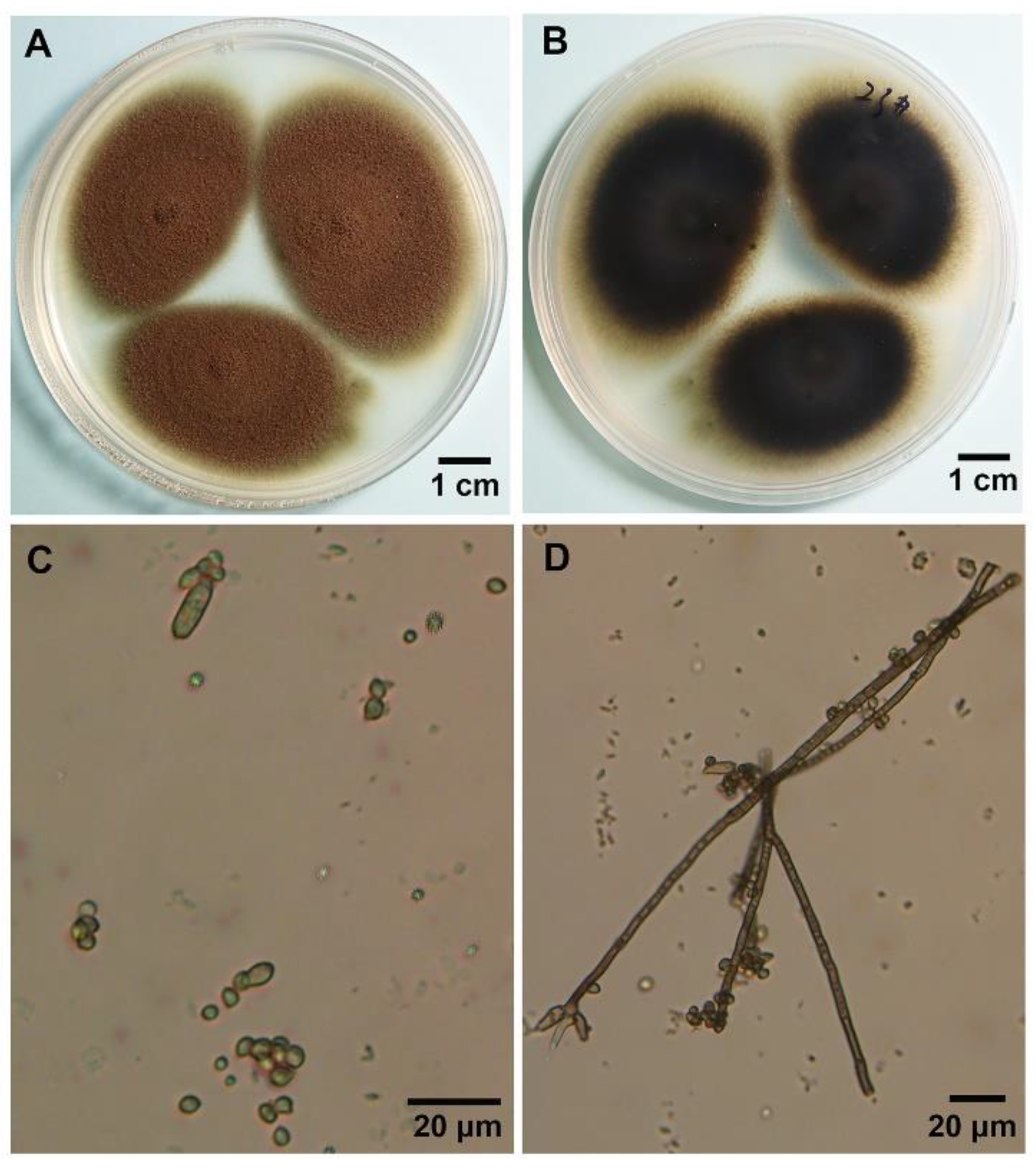
The morphological characteristics of the hyperparasite cultured on media were studied using a light microscope. The front side of the fungal colonies was taupe-colored and the reverse side was brown to black (Figure 2A,B). Mycelia grew fast and were dense. Colonies reached 20–30 mm in diameter on potato dextrose agar (PDA), at 25 °C in 7 days (Figure 2A). The conidiophores were light brown and branched, and conidia varied in size, ranging from 5 µm to 15 µm (mean 12 µm) in length and from 1 µm to 5 µm (mean 2.5 µm) in width (Figure 2C,D).
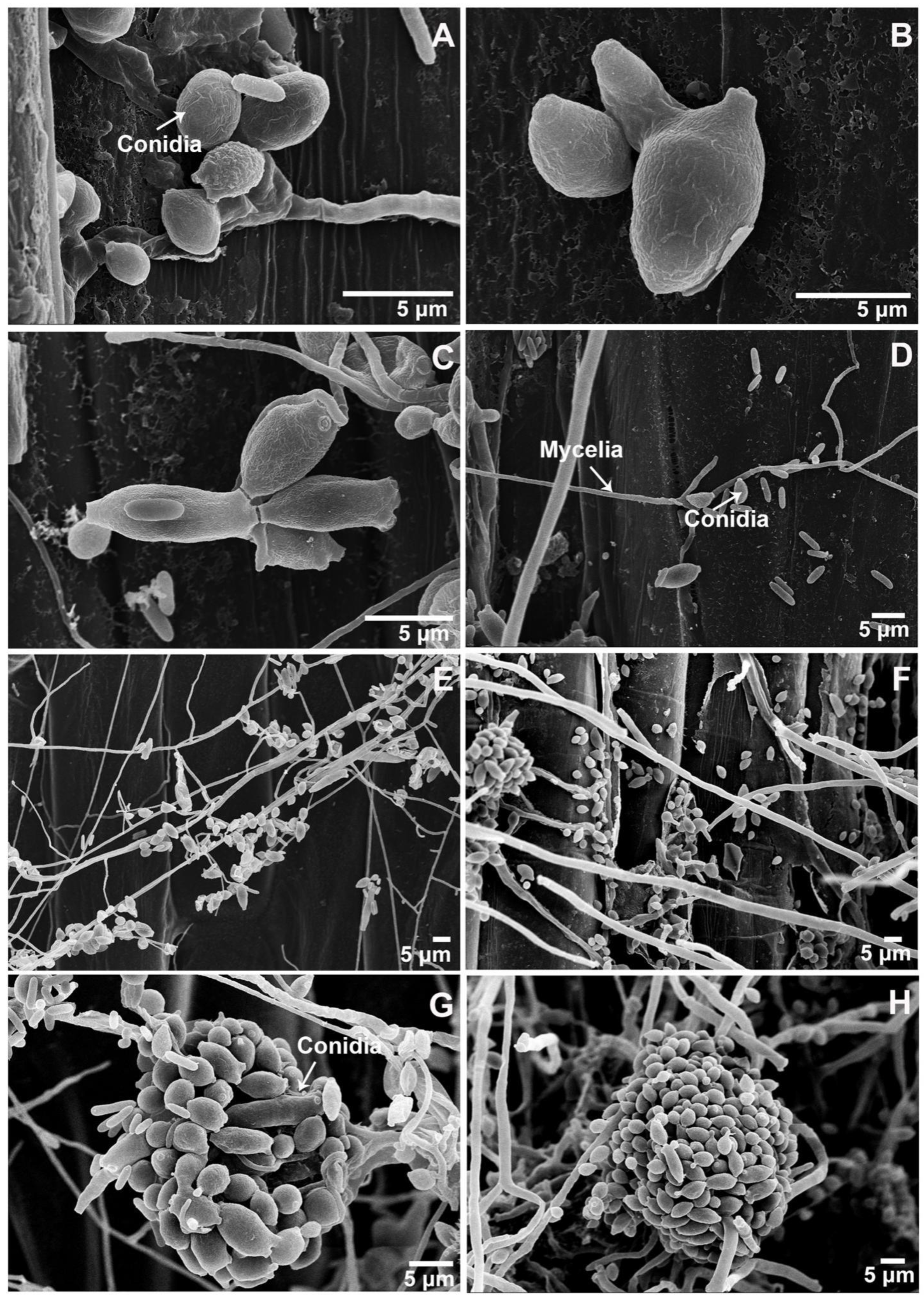
More detailed morphological features of the hyperparasite were revealed by SEM observations. Ramoconidia had one or more conidial scars (Figure 3A–D). Numerous conidia eventually form clusters (Figure 3E–H). According to the above morphology characteristics, the hyperparasite was identified as Cladosporium cladosporioides (Fresen.) G.A. de Vries.

3.3. Molecular Characterization of the C. cladosporioides Isolate

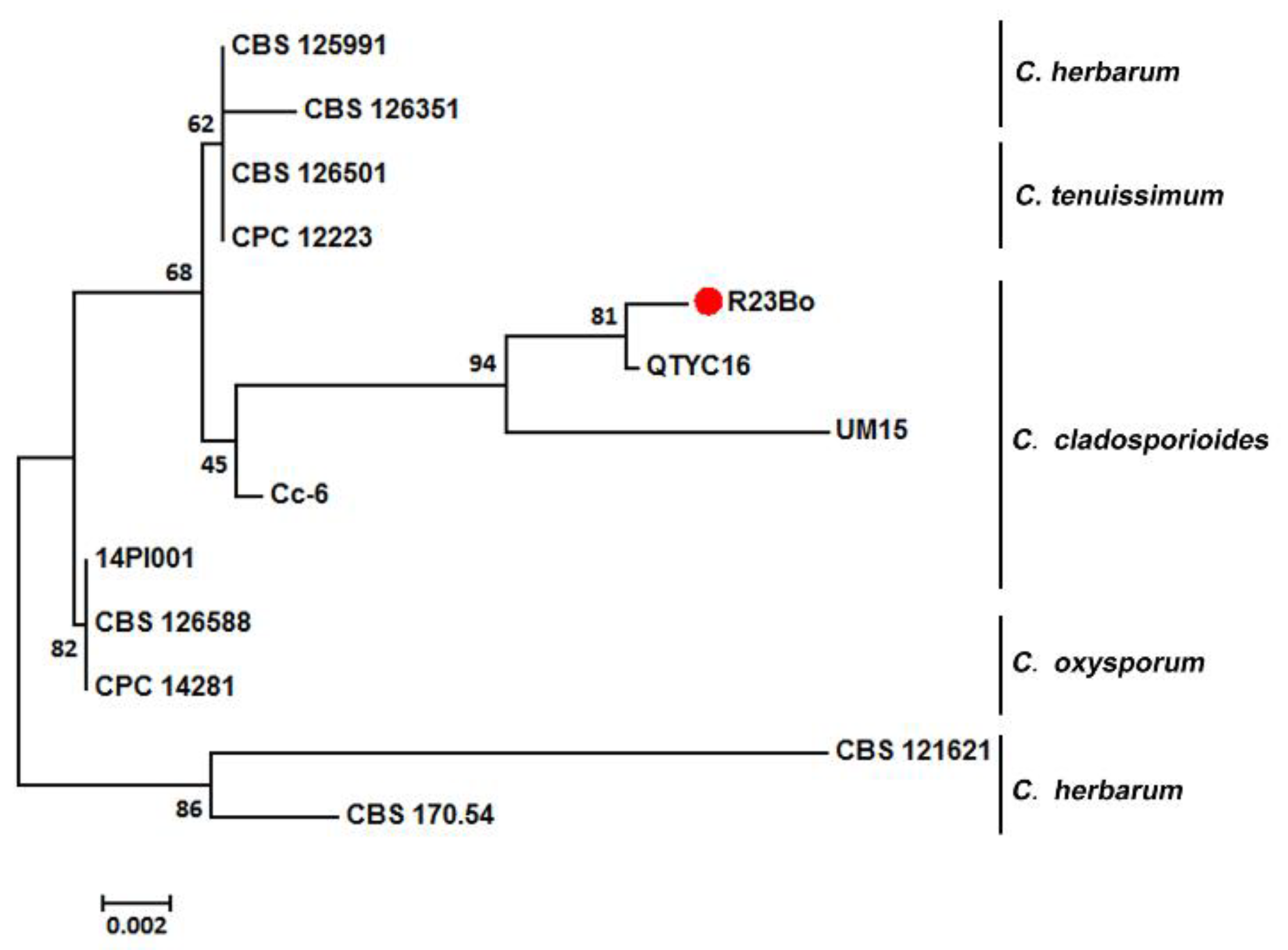
A neighbor-joining (NJ) tree was constructed for with Cladosporium species based on the internal transcribed spacer (ITS) sequences using software MEGA7 (Figure 4). The Cladosporium species used for the phylogenetic analysis are provided in Table 1. Our isolate R23Bo was most closely related to isolate QTYC16 of C. cladosporioides previously isolated from Pantala flavescens larvae, but not closely related to 14PI001, an isolate of C. cladosporioides previously isolated from Pst (Figure 4) Isolate.

3.4. Confirmation of the C. cladosporioides Isolate Parasitizing Pst

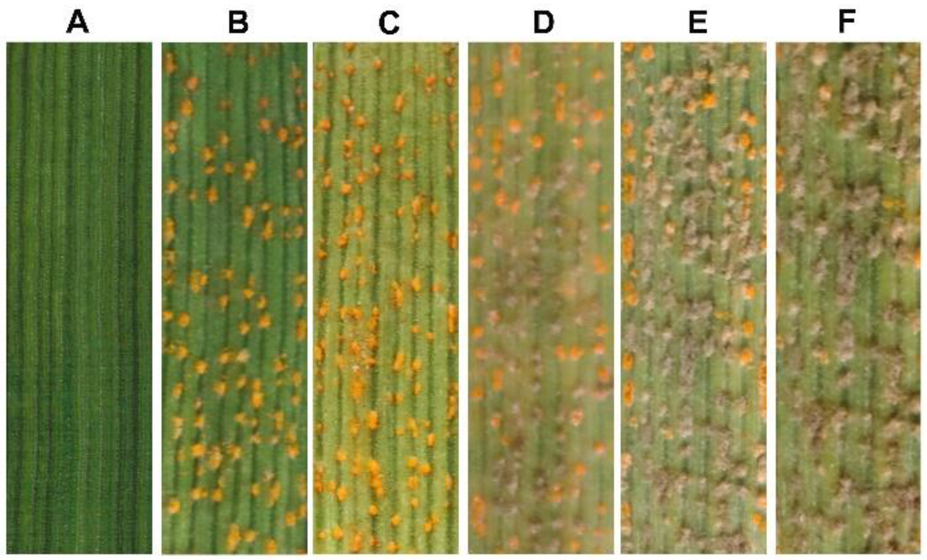
The hyperparasitic ability of isolate R23Bo of C. cladosporioides was confirmed by co-inoculation of wheat plants with Pst and the isolate. Wheat leaves inoculated only with the conidian suspension of isolate R23Bo did not show any symptoms or signs of fungal infection (Figure 5A). When inoculated with only the Pst urediospores suspension, yellow colored uredinia with urediospores formed on the inoculated leaves 12 days post inoculation (dpi) (Figure 5B). When wheat leaves were inoculated with Pst urediospores followed by inoculation with the conidia suspension of the C. cladosporioides isolates different days after Pst inoculation, Pst yellow-colored uredinia were changed to taupe (Figure 5A,C–F). The longer C. cladosporioides grew together with Pst, the fewer yellow uredinia or the more taupe pustules.
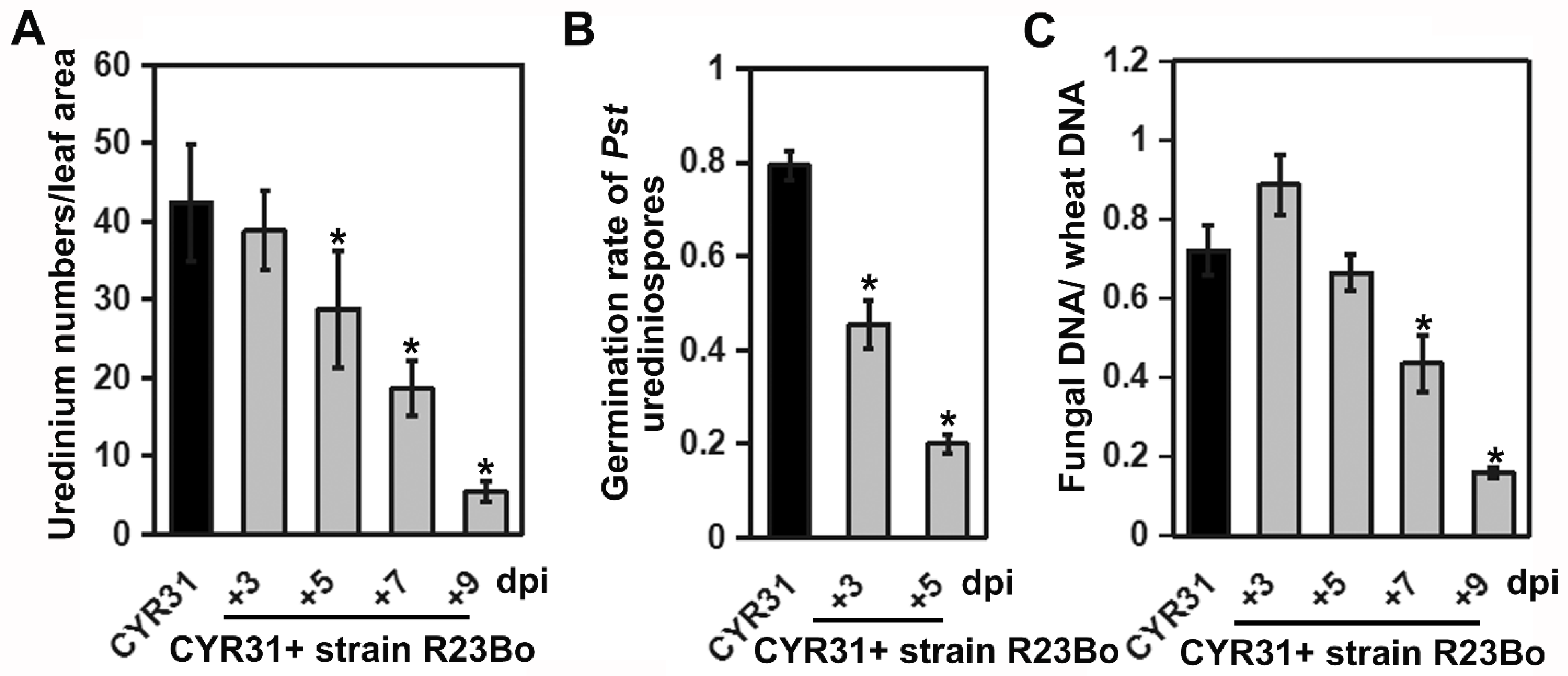
When the number of yellow uredinia were counted using ImageJ number counting software, 12 days after Pst inoculation, the number of yellow uredinia per cm2 leaves was the lowest in the treatment of C. cladosporioides 9 days after Pst inoculation (Figure 6A). R23Bo-strain-treated pustules showed impact the production of urediospores, and the fertility of spores is seriously affected, as is obviously exhibited in that the ratio of the spores germination reduces by 65% at 3 dpi and 80% at 5 dpi (Figure 6B). The biomass of the Pst, measured by the Pst DNA/wheat DNA ratio, decreased as the treatment with C. cladosporioides lengthened (Figure 6C). The results showed that isolate of C. cladosporioides is able to parasitize Pst, leading to the reduction in Pst urediospore production.
SEM observation further illustrated that isolate R23Bo could efficiently parasitize Pst. At 36 hai, C. cladosporioides conidian produced germ tubes which contacted with Pst urediospores and then grew into the urediospores (Figure 7B). The parasitic fungus grew inside and produced hyphae and conidiophores from the urediospores (Figure 7C), and it completely destroyed the urediospores at 120 h after the parasite treatment (Figure 7C,D).

4. Discussion

The identification of new hyperparasites is useful to understanding the biodiversity of mycoparasites, and it provides the potential to develop new strategies for biological control of plant diseases [13,32]. In the present study, we isolated and identified a fungal isolate from Pst uredinia. The hyperparasitic isolate is able to reduce Pst infection. Furthermore, the isolate can reduce Pst urediospore production and viability. Thus, the isolate has a potential value in biological prevention of wheat stripe rust.
In the morphological identification, the spore size is an important classification criterion of Cladosporium spp. As the spore dimensions of the most species in the genus overlap, it is difficult to identify species of Cladosporium using only morphological characters, especially the size of conidian [28]. Using only ITS sequences is also not reliable to identify Cladosporium spp. [33]. In the present study, we use ITS sequence analysis and morphological features to identify the hyperparasitic isolate as C. cladosporioides. Several fungal species have been reported to parasitize Pst, including C. cladosporioides [22]. However, the ITS sequence analysis showed that the C. cladosporioides isolate obtained in this study is clearly different from the isolate reported in Zhan et al. [22]. It is interesting that our isolate is most closely related to a C. cladosporioides isolate obtained from Pantala flavescens larvae [29]. This relationship may suggest that the isolate we obtained from Pst uredinia may have other hosts to parasitize and/or natural substrates to grow on.
Biocontrol strategies have potential to achieve efficacy in preventing and treating diseases under environmentally friendly conditions. Some studies have been conducted to explore hyperparasites to control rusts. For example, Cladosporium spp. was found to parasitize Melampsora spp. [34]. Several fungal species including C. cladosporioides were identified as hyperparasites of Pst [18,21,22]. The isolate R23Bo of C. cladosporioides identified in the present study is able to reduce or stop the growth of Pst urediospores by growing into uredinia. The isolate is fast-growing and easy to culture. Additionally, this parasitic ability makes R23Bo a potential biological control agent which could be developed into a biocontrol agent for managing wheat stripe rust.
Stripe rust is started by urediospore infection of host plants and continually develops by producing more urediospores and consequently more infections. Therefore, it is crucial to reduce urediospores for combating the rust disease. In the present study, we observed that after the inoculation of Pst urediospores and C. cladosporioides conidian on wheat leaves, urediospores were first produced on the wheat leaves, and then C. cladosporioides began to grow on urediospores. The exact invasion or parasitism stage cannot be determined at present. It is only clear that C. cladosporioides parasitizes in the sporulation stage of Pst. In order to develop the isolate as a biocontrol agent, further studies should be conducted to its effect on other plants, humans, animals, and environment, as well as to develop methods for producing and applying the biocontrol agent.

5. Conclusions

Identification of parasites infecting cereal pathogenic fungi is essential for developing biological control strategies for managing plant diseases. In this study, we report the discovery of a fungal strain isolated from Pst. Through molecular and morphological characterizations, we identified the hyperparasitic fungus as species Cladosporium cladosporioides. We demonstrated that the fungus was able to parasitize the obligate biotrophic rust fungus. Our experiments showed that Cladosporium cladosporioides was able to impair Pst sporulation and reduce urediospores germination. Collectively, Cladosporium cladosporioides may be harnessed for controlling stripe rust, and these results shed new light on biological control agent for managing plant pathogens.
The present study identified Cladosporium cladosporioides as a new hyperparasite of Pst. Although the fungus has the potential utility value as a biological control agent for control stripe rust, additional research is needed to determine if the hyperparasite is environmentally friendly and further to explore its potential to control other rust pathogens.

Supplementary Materials

The following are available online at https://www.mdpi.com/article/10.3390/biology11060892/s1, Figure S1: Standard curves derived from the serial dilutions for each Pst-EF1 (Pst elongation factor 1) and TaEF1α (wheat elongation factor 1 alpha).

Author Contributions

H.Z., N.W. designed the research. H.Z., M.H, X.F., L.D., S.Z. and Z.H. performed the experiments. H.Z., N.W. and M.H. analyzed the data. H.Z. and N.W. wrote the manuscript. All authors have read and agreed to the published version of the manuscript.

Funding

This study was supported by the China Agriculture Research System (CARS-3), International Science and technology cooperation project of Shaanxi provincial key R&D plan-key project (2020KWZ-009).

Institutional Review Board Statement

Not applicable.

Informed Consent Statement

Not applicable.

Data Availability Statement

The relevant datasets supporting the results of this article are included within the article and its additional files.

Acknowledgments

We sincerely thank all the researchers who contributed to the wheat genome database the wheat expression database. We thank reviewers for checking our manuscript and the editors for editing the paper.

Conflicts of Interest

The authors declare no conflict of interest.

References

  1. Wellings, C.R. Global status of stripe rust: A review of historical and current threats. Euphytica 2011, 179, 129–141. [Google Scholar] [CrossRef]
  2. Wellings, C.R. Puccinia striiformis in Australia: A review of the incursion, evolution, and adaptation of stripe rust in the period 1979–2006. Aust. J. Agric. Res. 2007, 58, 567–575. [Google Scholar] [CrossRef]
  3. Beddow, J.M.; Pardey, P.G.; Chai, Y.; Hurley, T.M.; Kriticos, D.J.; Braun, H.J.; Park, R.F.; Cuddy, W.S.; Yonow, T. Research investment implications of shifts in the global geography of wheat stripe rust. Nat. Plants 2015, 1, 15132. [Google Scholar] [CrossRef]
  4. Chen, W.; Wu, L.; Liu, T.; Xu, S.; Jin, S.; Peng, Y.; Wang, B. Race Dynamics, Diversity, and Virulence Evolution in Puccinia striiformis f. sp. tritici, the Causal Agent of Wheat Stripe Rust in China from 2003 to 2007. Plant Dis. 2009, 93, 1093–1101. [Google Scholar] [CrossRef] [Green Version]
  5. Gao, L.; Yu, H.X.; Kang, X.H.; Shen, H.M.; Li, C.; Liu, T.G.; Liu, B.; Chen, W.Q. Development of SCAR Markers and an SYBR Green Assay to Detect Puccinia striiformis f. sp. tritici in Infected Wheat Leaves. Plant Dis. 2016, 100, 1840–1847. [Google Scholar]
  6. Chen, X.; Kang, Z. Integrated Control of Stripe Rust. In Stripe Rust; Springer: Dordrecht, The Netherlands, 2017. [Google Scholar]
  7. Ansari, I.; El-Kady, M.M.; Arora, C.; Sundararajan, M.; Maiti, D.; Khan, A. A review on the fatal impact of pesticide toxicity on environment and human health. Glob. Clim. Chang. 2021, 361–391. [Google Scholar] [CrossRef]
  8. Aktar, W.; Sengupta, D.; Chowdhury, A. Impact of pesticides use in agriculture: Their benefits and hazards. Interdiscip. Toxicol. 2009, 2, 1–12. [Google Scholar] [CrossRef] [Green Version]
  9. Ainsworth, G.C.; Bisby, G.R. Ainsworth & Bisby’s dictionary of the fungi. Mycologist 1995, 17, 17–19. [Google Scholar]
  10. Köhl, J.; Molhoek, W.; Haas, G.D.; Geijn, G. Selection and orchard testing of antagonists suppressing conidia production of the apple scab pathogen Venturia inaequalis. Eur. J. Plant Pathol. 2009, 123, 401–414. [Google Scholar] [CrossRef]
  11. Berendsen, R.L.; Kalkhove, S.I.C.; Lugones, L.G.; Baars, J.P.; Wösten, H.A.M.; Bakker, P.A. Effects of fluorescent Pseudomonas spp. isolated from mushroom cultures on Lecanicillium fungicola. Biol. Control 2012, 63, 210–221. [Google Scholar] [CrossRef]
  12. Adhikari, A.; Nandi, S.; Dutta, S.; Bhattacharya, I.; Mandal, T. Study of morphology and Mycoparasitism of some antagonists of Trichoderma sp. from West Bengal, India. J. Mt. Sinai Hosp. N. Y. 2014, 22, 91. [Google Scholar]
  13. Zhong, X.; Li, S.S.; Peng, Q.Y.; Zhang, J.S.; Kan, X.T.; Zhang, G.R.; Liu, X. A Polycephalomyces hyperparasite of Ophiocordyceps sinensis leads to shortened duration of production and reduced numbers of host ascospores. Fungal Ecol. 2016, 21, 24–31. [Google Scholar] [CrossRef]
  14. Kiss, L.; Russell, J.C.; Szentiványi, O.; Xu, X.; Jeffries, P. Biology and biocontrol potential of Ampelomyces mycoparasites, natural antagonists of powdery mildew fungi. Biocontrol Sci. Technol. 2004, 14, 635–651. [Google Scholar] [CrossRef]
  15. Ikeda, S.; Shimizu, A.; Shimizu, M.; Takahashi, H.; Takenaka, S. Biocontrol of black scurf on potato by seed tuber treatment with Pythium oligandrum. Biol. Control 2012, 60, 297–304. [Google Scholar] [CrossRef]
  16. Kapooria, R.G.; Sinha, S. Phylloplane mycoflora of pearl millet and its influence on the development of Puccinia penniseti. Trans. Br. Mycol. Soc. 1969, 53, 153–155. [Google Scholar] [CrossRef]
  17. Koçl, N.K.; Défago, G. Studies on the Host Range of the Hyperparasite Aphanocladium album. J. Phytopathol. 2010, 107, 214–218. [Google Scholar]
  18. Cummins, G.B.; Littlefield, L.J. Biology of the plant rusts: An introduction. Bioence 1984, 34, 116. [Google Scholar]
  19. Moricca, S.; Ragazzi, A.; Mitchelson, K.R.; Assante, G. Antagonism of the Two-Needle Pine Stem Rust Fungi Cronartium flaccidum and Peridermium pini by Cladosporium tenuissimum In Vitro and In Planta. Phytopathology 2001, 91, 457–468. [Google Scholar] [CrossRef] [Green Version]
  20. Tsuneda, A.; Hiratsuka, Y.; Maruyama, P.J. Hyperparasitism of Scytalidium uredinicola on western gall rust, Endocronartium harknessii. Can. J. Bot. 1980, 58, 1154–1159. [Google Scholar] [CrossRef]
  21. Li, Z.; Jie, Z.; Liang, X.; Zhan, G.; Kang, Z. Identification of a Novel Alternaria alternata Strain Able to Hyperparasitize Puccinia striiformis f. sp. tritici, the Causal Agent of Wheat Stripe Rust. Front. Microbiol. 2017, 8, 71. [Google Scholar]
  22. Zhan, G.; Tian, Y.; Wang, F.; Chen, X.; Guo, J.; Jiao, M.; Huang, L.; Kang, Z. A novel fungal hyperparasite of Puccinia striiformis f. sp. tritici, the causal agent of wheat stripe rust. PLoS ONE 2014, 9, e111484. [Google Scholar]
  23. Wang, N.; Fan, X.; Zhang, S.; Liu, B.; Wang, X. Identification of a Hyperparasitic Simplicillium obclavatum strain affecting the infection dynamics of Puccinia striiformis f. sp. tritici on wheat. Front. Microbiol. 2020, 11, 1277. [Google Scholar] [CrossRef]
  24. Bomblies, K.; Shukla, V.; Graham, C. Scanning Electron Microscopy (SEM) of Plant Tissues. Cold Spring Harb. Protoc. 2008, 2008, pdb.prot4933. [Google Scholar] [CrossRef]
  25. Wang, Y.B.; Yu, H.; Dai, Y.D.; Wu, C.K.; Zeng, W.B.; Yuan, F.; Liang, Z.Q. Polycephalomyces agaricus, a new hyperparasite of Ophiocordyceps sp. infecting melolonthid larvae in southwestern China. Mycol. Prog. 2015, 14, 70. [Google Scholar] [CrossRef]
  26. Kumar, S.; Stecher, G.; Tamura, K. MEGA7: Molecular Evolutionary Genetics Analysis Version 7.0 for Bigger Datasets. Mol. Biol. Evol. 2016, 33, 1870–1874. [Google Scholar] [CrossRef] [Green Version]
  27. Chen, R.S.; Wang, W.L.; Li, J.C.; Wang, Y.Y.; Tsay, J.G. First Report of Papaya Scab Caused by Cladosporium cladosporioides in Taiwan. Plant Dis. 2009, 93, 426. [Google Scholar] [CrossRef]
  28. Bensch, K.; Groenewald, J.Z.; Dijksterhuis, J.; Starink-Willemse, M.; Andersen, B.; Summerell, B.A.; Shin, H.D.; Dugan, F.M.; Schroers, H.J.; Braun, U. Species and ecological diversity within the Cladosporium cladosporioides complex (Davidiellaceae, Capnodiales). Stud. Mycol. 2010, 67, 1–94. [Google Scholar] [CrossRef]
  29. Shao, M.W.; Lu, Y.H.; Miao, S.; Zhang, Y.; Chen, T.T.; Zhang, Y.L. Diversity, Bacterial Symbionts and Antibacterial Potential of Gut-Associated Fungi Isolated from the Pantala flavescens Larvae in China. PLoS ONE 2015, 10, e0134542. [Google Scholar] [CrossRef]
  30. Schubert, K.; Groenewald, J.Z.; Braun, U.; Dijksterhuis, J.; Starink, M.; Hill, C.F.; Zalar, P.; de Hoog, G.S.; Crous, P.W. Biodiversity in the Cladosporium herbarum complex (Davidiellaceae, Capnodiales), with standardisation of methods for Cladosporium taxonomy and diagnostics. Stud. Mycol. 2007, 58, 105–156. [Google Scholar] [CrossRef]
  31. Vu, D.; Groenewald, M.; de Vries, M.; Gehrmann, T.; Stielow, B.; Eberhardt, U.; Al-Hatmi, A.; Groenewald, J.Z.; Cardinali, G.; Houbraken, J.; et al. Large-scale generation and analysis of filamentous fungal DNA barcodes boosts coverage for kingdom fungi and reveals thresholds for fungal species and higher taxon delimitation. Stud Mycol. 2019, 92, 135–154. [Google Scholar] [CrossRef]
  32. Vandermeer, J.; Perfecto, I.; Liere, H. Evidence for hyperparasitism of coffee rust (Hemileia vastatrix) by the entomogenous fungus, Lecanicillium lecanii, through a complex ecological web. Plant Pathol. 2010, 58, 636–641. [Google Scholar] [CrossRef] [Green Version]
  33. Dugan, F.; Braun, U.; Groenewald, J.; Crous, P. Morphological plasticity in Cladosporium sphaerospermum. Persoonia 2008, 21, 9–16. [Google Scholar] [CrossRef] [Green Version]
  34. Heather, W.A.; Sharma, J.K. Some Aspects of Poplar Rust Research in Australia. Aust. For. 1977, 40, 28–43. [Google Scholar] [CrossRef]
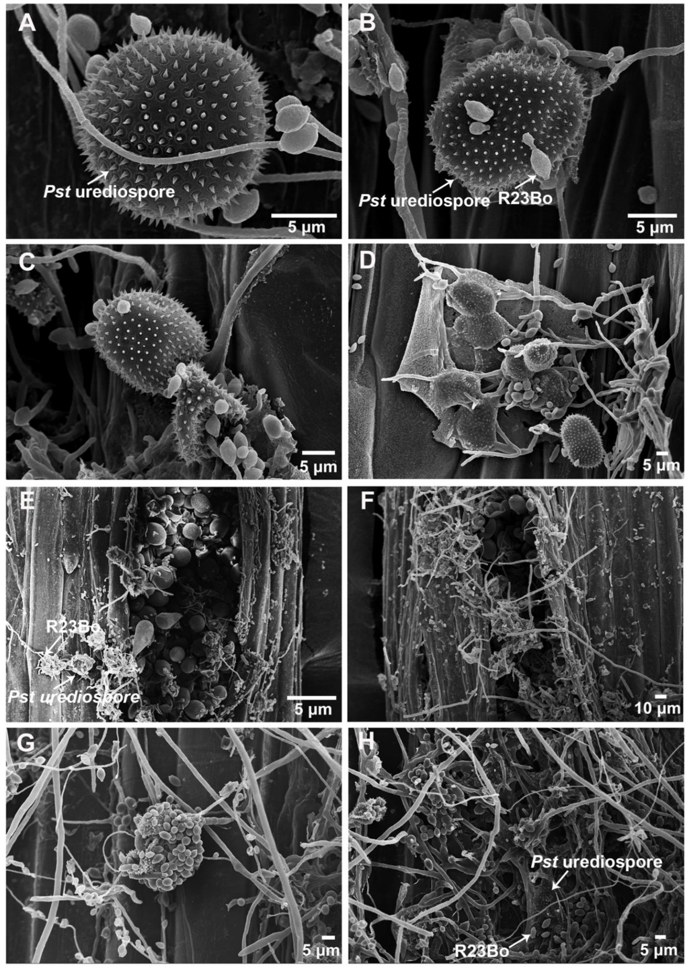
Figure 1. The hyperparasite Cladosporium cladosporioides isolate parasitized Puccinia striiformis f. sp. tritici (Pst) urediospores and uredinium. (A), urediospores of the Pst before C. cladosporioides was parasitized (×4000). (BD), At the early hyperparasitic stage, the urediospores were shriveled (×3000, ×2200, ×1000, respectively). (E,F), At the middle hyperparasitic stage, the hyperparasite mycelia and conidia invaded the Pst urediospores (×400, ×400, respectively). (G,H), At the late hyperparasitic stage, the urediospores of the Pst are completely destroyed (×1000, ×800, respectively).
Biology 11 00892 g001
Figure 2. Morphological characteristics of the Cladosporium cladosporioides isolate under a light microscope. (A,B), C. cladosporioides cultured on PDA medium at 25 °C for 9 days, the front side and the reverse side, respectively. (C,D), Septate hyphae and conidia.
Biology 11 00892 g002
Figure 3. Morphologic characteristics of the Cladosporium cladosporioides isolate under a scanning electron microscope. (AC), Conidia (×6000, ×13,000 and ×5000, respectively). (DF), Mycelia and conidia (×2000, ×1000, and ×1000, respectively). (G,H), Conidia (×2500 and ×1500, respectively).
Biology 11 00892 g003
Figure 4. A neighbor-joining (NJ) tree constructed for 13 isolates of 5 Cladosporium species using software MEGA7. The Puccinia striiformis hyperparasitic isolate (R23Bo) of C. cladosporioides is marked with a red dot. The values are the frequencies in percentage for the branches with 1000 bootstrap replications.
Biology 11 00892 g004
Figure 5. The Cladosporium cladosporioides isolate hyperparasitizing Puccinia striiofrmis f. sp. tritici (Pst). (A), Wheat leaf inoculated only with the conidian suspension of C. cladosporioides isolate R23Bo; (B), At 12 days post inoculation (dpi), wheat leaves inoculated only with Pst urediospores; (CF), Inoculations with the conidian suspension of C. cladosporioides after wheat leaves inoculated with Pst (pictures taken 12 dpi of Pst). C–F are Pst-infected leaves inoculated with C. cladosporioides at 3, 5, 7 and 9 dpi of Pst, respectively.
Biology 11 00892 g005
Figure 6. (A), Comparison of yellow-colored Pst uredinium numbers among the different Pst inoculation and time-point treatments of C. cladosporioides R23Bo. Yellow-colored uredinia were counted 12 dpi of Pst race CYR31. In the legend, CYR31 means only inoculated with CYR31; and CYR31+ 3, 5, 7, and 9 means CYR31 plus inoculation of C. cladosporioides R23Bo at 3, 5, 7, and 9 days after the Pst inoculation. (B), Pst urediospores germination rate. (C), The Puccinia striiformis f. sp. tritici biomass changed when hyperparasite C. cladosporioides R23Bo parasitized, measured by qRT-PCR. (+) means after C. cladosporioides R23Bo treatment 3, 5, 7, and 9 days. Statistical analysis was performed by Student’s t-tests. Asterisks indicate p value < 0.05.
Biology 11 00892 g006
Figure 7. Morphologic characterization of Puccinia striiformis f. sp. tritici (Pst) urediospores infected by Cladosporium cladosporioides under a scanning electron microscope. (A), Conidia of C. cladosporioides on the surface of a Pst urediospore (×2000) at 12 hai (hours after inoculation); (B), at 36 hai, a C. cladosporioides conidium generated a germ tube (×4000); (C), at 72 hai, t C. cladosporioides produce conidiophores from the Pst urediospore (×3000); (D), at 120 hai, C. cladosporioides has completely colonized the Pst urediospore and the urediospore is destroyed (×1500).
Biology 11 00892 g007
Table 1. Accession and GenBank numbers of Cladosporium species used in the molecular characterization of this study.
SpeciesAccession NumberGenBank Number (ITS, EF, ACT)Substrate (Including Host)CountryReference
C. cladosporioidesCc-6EU935608.1Carica papayaChinaChen et al., 2009 [27]
C. cladosporioidesUM15HQ148094.1Echinacea purpureaUSABensch et al., 2010 [28]
C. cladosporioidesQTYC16KM103301.1Pantala flavescens larvaeChinaShao et al., 2015 [29]
C. cladosporioidesFCBP:1493KY290222.1Solanum melongenaPakistan-
C. cladosporioidesCBS 112388HM148003 HM148244 HM148490Indoor airGermanyBensch et al., 2010 [28]
C. cladosporioidesCBS 143.35HM148011 HM148252 HM148498Pisum sativumSouth AfricaBensch et al., 2010 [28]
C. cladosporioidesCBS 144.35HM148012 HM148253 HM148499P. sativumUSABensch et al., 2010 [28]
C. cladosporioidesCBS 101367HM148002 HM148489 HM148243SoilBrazilBensch et al., 2010 [28]
C. cladosporioidesCPC 14018 MRC 10810HM148040 HM148281 HM148527Triticum aestivumSouth AfricaBensch et al., 2010 [28]
C. cladosporioidesCPC 14244HM148044 HM148285 HM148531Magnolia spp.USABensch et al., 2010 [28]
C. cladosporioidesCBS 145.35HM148013 HM148254 HM148500P. sativumGermanyBensch et al., 2010 [28]
C. cladosporioides14PI001 CGMCC 7.175KJ598781 KM281945 KJ598781Puccinia striiformisChinaZhan et al., 2014 [22]
C. herbarumCBS 121621EF679363Hordeum vulgareNetherlandsSchubert et al., 2007 [30]
C. herbarumCBS 132.29HM148010 HM148251 HM148497-a-Bensch et al., 2010 [28]
C. herbarumCBS 170.54MH857281 IMI025324 NCTC6740-EnglandVu et al., 2018 [31]
C. oxysporumCBS 125991 CPC 14371 BA 1738HM148118 HM148362 HM148607Soil, near the terracottaChinaBensch et al., 2010 [28]
C. oxysporumCBS 126351 CPC 14308 BA 1707HM148119 HM148363 HM148608Indoor airVenezuelaBensch et al., 2010 [28]
C. pseudocladosporioidesCBS 149.66HM148161 HM148405 HM148650T. aestivumUSABensch et al., 2010 [28]
C. pseudocladosporioidesCBS 176.82HM148162 HM148406 HM148651Pteridium aquilinumRomaniaBensch et al., 2010 [28]
C. salinaeCBS 119413 EXF-335DQ780374 JN906993 EF101390Hypersaline waterSloveniaBensch et al., 2010 [28]
C. tenuissimumCPC 12223HM148208 HM148453 HM148698RustBrazilBensch et al., 2010 [28]
C. tenuissimumCBS 126501 CPC 14410HM148199 HM148444 HM148689Musa spp.Ivory CoastBensch et al., 2010 [28]
C. xylophilumCBS 126588HM148231 HM148477 HM148722Salix viminalis twigsItalyBensch et al., 2010 [28]
C. xylophilumCPC 14281HM148233 HM148479 HM148724LeavesFranceBensch et al., 2010 [28]
a Not available.
Publisher’s Note: MDPI stays neutral with regard to jurisdictional claims in published maps and institutional affiliations.