Article

An Anaerobic Environment Drives the Harboring of Helicobacter pylori within Candida Yeast Cells

by
Kimberly Sánchez-Alonzo
1,
Luciano Arellano-Arriagada
1,
Humberto Bernasconi
2,
Cristian Parra-Sepúlveda
1,
Víctor L. Campos
3,
Fabiola Silva-Mieres
1,
Katia Sáez-Carrillo
4,
Carlos T. Smith
1 and
Apolinaria García-Cancino
1,*
1
Laboratory of Bacterial Pathogenicity, Department of Microbiology, Faculty of Biological Sciences, Universidad de Concepcion, Concepcion 4070386, Chile
2
Laboratorio Pasteur, Concepcion 4030635, Chile
3
Laboratory of Environmental Microbiology, Department of Microbiology, Faculty of Biological Sciences, Universidad de Concepcion, Concepcion 4070386, Chile
4
Department of Statistics, Faculty of Physical and Mathematical Sciences, Universidad de Concepcion, Concepcion 4070386, Chile
*
Author to whom correspondence should be addressed.
Biology 2022, 11(5), 738; https://doi.org/10.3390/biology11050738
Submission received: 18 April 2022 / Accepted: 7 May 2022 / Published: 12 May 2022

Abstract

:

Simple Summary

Helicobacter pylori is a pathogen that is associated with a number of gastric pathologies and has adapted to the gastric environment. Outside this organ, stress factors such as oxygen concentration affect the viability of this bacterium. This study aimed to determine if changes in oxygen concentration promoted the entry of H. pylori into the interior of yeast cells of the Candida genus. Co-cultures of H. pylori and Candida strains in Brucella broth plus 5% fetal bovine serum were incubated under microaerobic, anaerobic, or aerobic conditions. Bacteria-like bodies (BLBs) were detected within yeast cells (Y-BLBs) by optical microscopy, identified by molecular techniques, and their viability evaluated by SYTO-9 fluorescence. Co-cultures incubated under the three conditions showed the presence of Y-BLBs, but the highest Y-BLB percentage was present in H. pylori J99 and C. glabrata co-cultures incubated under anaerobiosis. Molecular techniques were used to identify BLBs as H. pylori and SYTO-9 fluorescence confirmed that this bacterium remained viable within yeast cells. In conclusion, although without apparent stress conditions H. pylori harbors within Candida yeast cells, its harboring increases significantly under anaerobic conditions. This endosymbiotic relationship also depends mostly on the H. pylori strain used in the co-culture.

Abstract

Helicobacter pylori protects itself from stressful environments by forming biofilms, changing its morphology, or invading eukaryotic cells, including yeast cells. There is little knowledge about the environmental factors that influence the endosymbiotic relationship between bacterium and yeasts. Here, we studied if oxygen availability stimulated the growth of H. pylori within Candida and if this was a bacterial- or yeast strain-dependent relationship. Four H. pylori strains and four Candida strains were co-cultured in Brucella broth plus 5% fetal bovine serum, and incubated under microaerobic, anaerobic, or aerobic conditions. Bacteria-like bodies (BLBs) within yeast cells (Y-BLBs) were detected by microscopy. H. pylori was identified by FISH and by PCR amplification of the 16S rRNA gene of H. pylori from total DNA extracted from Y-BLBs from H. pylori and Candida co-cultures. BLBs viability was confirmed by SYTO-9 fluorescence. Higher Y-BLB percentages were obtained under anaerobic conditions and using H. pylori J99 and C. glabrata combinations. Thus, the H. pyloriCandida endosymbiotic relationship is strain dependent. The FISH and PCR results identified BLBs as intracellular H. pylori. Conclusion: Stressful conditions such as an anaerobic environment significantly increased H. pylori growth within yeast cells, where it remained viable, and the bacterium–yeast endosymbiotic relationship was bacterial strain dependent with a preference for C. glabrata.

1. Introduction

Helicobacter pylori is a Gram-negative, neutrophilic and microaerophilic bacterium with a helical morphology [1,2] and it infects above 50% of the population worldwide. An infection with this bacterium is mostly acquired during infancy and it is estimated that one third of the infant population is infected [3]
Differences in life conditions, such as hygiene levels, are probably responsible for the higher percentages of occurrences in developing countries [4,5]. This pathogenic microorganism colonizes the gastric epithelium, where it releases effector proteins, such as CagA and VacA, causing morphological changes in epithelial cells of the host and also stimulating the release of cytoplasmic components and interleukins, resulting in a strong inflammatory response which can lead to cell apoptosis with tissue damage [6,7]. The constant deterioration of the gastric epithelium, as a consequence of H. pylori infection, causes pathologies of the gastrointestinal tract, such as acute and chronic gastritis, peptic ulcer, gastric mucosa-associated lymphoid tissue (MALT) lymphoma, and gastric cancer [8,9], and the clinical manifestations are dependent on the immune condition of the host and on the virulence factors of H. pylori [3]. At present, there are multiple extra-gastric clinical manifestations attributed to infections by this pathogen, such as ischemic heart disease, anemia, insulin resistance, type 2 diabetes mellitus, and idiopathic thrombocytopenic purpura [9,10]. Thus, this pathogen is considered to have a high negative impact on human health, making it necessary to search for new treatment alternatives, such as probiotics or natural extracts (i.e., polyphenols) to confront the increase of H. pylori strains resistant to antibiotics [11,12,13,14,15,16]. However, despite its importance and the years of research dedicated to it, the dissemination routes and survival strategies used by this microorganism are still not completely understood [17], making it difficult to generate effective preventive measures to decrease the prevalence of its infection.
The stomach of humans is the natural habitat for H. pylori, which proliferates usually in the superior section of the intestine and in the gastric mucosa and causes the above-mentioned associated pathologies [11]. This pathogen possesses the metabolic machinery to grow in the gastric environment and to survive under the harsh conditions that exist there. The enzymatic arsenal that favors the growth of H. pylori in this anatomical site includes enzymes such as urease, carbonic anhydrases, catalase, peroxidase, and superoxide dismutase [11,18,19,20]. Since H. pylori adapts to the gastric conditions, extra-gastric conditions such as changes in pH and variations in oxygen concentration constitute stressors that impact its morphology and survival [21,22]. To survive in these environments, it can become part of a biofilm, develop a stage of viable but non-culturable bacteria (VBNC), or grow within eukaryotic microorganisms such as amoebas or yeasts [23,24,25,26,27], which could provide shelter from imminent threats to its viability.
Regarding the intracellular relationship of H. pylori and yeasts, it has been established that this bacterium can invade yeasts from different sources including the environment, foods, or the vaginal and oral microbiota of humans [28,29,30]. However, the environmental conditions that support the growth of H. pylori within these fungal cells still needs to be investigated in detail. Recently, it was reported that alterations in pH resulted in a stressful environment for H. pylori and increased its entry into yeast cells of the genus Candida, especially at low pH [31] values. Variations in the concentration of nutrients, temperatures outside the optimal range, as well as antibiotics such as amoxicillin have been shown to increase the growth of H. pylori within Candida cells [32,33,34]. There is limited knowledge about the relationship of intra-yeast H. pylori with the yeast cells that shelter it or how yeast cells might provide protection and serve as transmission vehicles for the bacterium, and therefore, its transmission routes should be investigated. It is also essential to determine the environmental factors involved in this pathogen harboring within yeast cells of the genus Candida. Thus, the aims of this work are: (i) to determine whether aerobic or anaerobic conditions are stressors which support the growth of H. pylori within Candida yeast cells, (ii) to identify if the endosymbiotic association between yeast and bacteria is dependent on the strain of both microorganisms, and (iii) to determine if H. pylori remains viable within yeast cells, sheltering it against unfavorable environmental oxygen variations.

2. Materials and Methods

2.1. Culture Conditions

In this work, the H. pylori strains used were the reference strains J99 (also referred as ATCC 700824), G-27 and SS-1, and the clinical strain H707 (gastric biopsy origin). In addition, two Candida reference strains (Candida albicans ATCC 90028 and Candida glabrata ATCC 90030) and two clinical strains of the same genus (C. albicans VT-3 (vaginal discharge origin) and C. glabrata LEO-37 (oral cavity origin)) were considered to be yeasts representative of the genus Candida. These strains are all maintained at the culture collection of the Laboratory of Bacterial Pathogenicity, Department of Microbiology, University of Concepcion, Chile.
The H. pylori strains were cultured on Columbia agar (CA) (OXOID, Basingstoke, UK) plus 5% FBS (Biological Industries, Cromwell, CT, USA) and the plates were incubated in a microaerobic incubator (10% CO2 and 5% O2) (Thermo Scientific, Waltham, MA, USA) at 37 °C from 48 h to 72 h [35]. Regarding the Candida strains, they were cultured in Sabouraud agar (SA) (Merck, Darmstadt, Germany) plus chloramphenicol (CHL) (OXOID, Basingstoke, UK), in accordance with the instructions of the manufacturer. This medium will be hereafter referred as SA-CHL. Plates were incubated in an aerobic incubator (ZHICHENG, Shanghai, China) at 37 °C for 24 h. To confirm the purity of the cultures of both microorganisms, the Gram staining was performed, and then observed using an optical microscope. Additionally, urease, oxidase, and catalase tests were performed to corroborate the purity of the cultures of the H. pylori strains. For the Candida strain cultures, the purity was verified culturing random yeast cells in CHROMagar (Difco, Wokingham, UK), conducting urease tests, and observing wet mounts, using the oil immersion objective lens of an optical microscope to confirm the absence of extracellular bacteria or of bacteria-like bodies (BLBs) contained in the vacuole of yeast cells [36].

2.2. Growth of H. pylori Strains and Candida Strains Cultured under Aerobic, Microaerobic, or Anaerobic Conditions

This assay was performed as described by Sánchez-Alonzo et al. [33] with modifications. Each strain of H. pylori and Candida strain was suspended at an optical density (O.D.) of 0.1 at 600 nm in Brucella broth (BB) (Difco, Wokingham, UK) plus 5% FBS (BB-5%FBS) and placed in an Infinite M200 PRO microplate reader (TECAN, Männedorf, Switzerland) at time zero. A 200 µL aliquot of each suspension was transferred to wells of flat bottom 96-well plates (Thomas Scientific, Swedesboro, NJ, USA). Different plates were used for each strain under three different conditions (aerobic, microaerobic, or anaerobic) which were incubated in the Infinite M200 PRO reader. The microaerobic and anaerobic conditions were generated using CampyGen and Anaerogen sachets (Thermo Scientific, Waltham, MA, USA), respectively. The microaerobic condition corresponded to 10% CO2 and 5% O2. To evaluate the growth of the strains, the absorbances of the cultures were measured at a wavelength of 600 nm in the same Infinite M200 PRO reader every 2 h for yeast strains and every 8 h for H. pylori strains. These measurements were made for 50 h and 72 h for the yeast and the bacterium, respectively. Growth curves for each strain were performed in triplicate. The methodology of this section is shown in Figure S1.

2.3. Co-Cultures of H. pylori Strains with Candida Strains

These assays were performed independently co-culturing each one of the H. pylori strains with each one of the different Candida strains, following the protocol described by Sánchez-Alonzo et al. [33]. Suspensions of each yeast and bacterial strain were adjusted to an O.D. of 0.1 at 600 nm in 0.89% saline solution (SS). Then, 500 µL of H. pylori strain suspension and 500 µL of Candida strain suspension were placed in the well of 12-well plates (Thomas Scientific, Swedesboro, NJ, USA) containing 4 mL BB-5%FBS. The plates were kept at 37 °C for 48 h under the aforementioned conditions. The microaerobic and anaerobic conditions were also achieved using CampyGen or Anaerogen sachets, respectively, as described in the section above. Each co-culture was performed in triplicate. The methodology of this section is shown in Figure S2.

2.4. Search for Intra-Yeast Bacteria-like Bodies (BLBs)

This assay was performed as described by Sánchez-Alonzo et al. [33] with modifications. At the same time of incubating the co-cultures, wet mounts of the co-cultures were prepared taking 20 µL aliquots from each co-culture at 0, 1, 3, 6, 12, 24, and 48 h. Each one of the aliquots was laid on a glass slide and observations were made using an optical photomicroscope (Leica, Wetzlar, Germany) using the 100X objective lens to search for mobile BLBs within yeast cells. If yeast cells harboring bacteria-like bodies (Y-BLBs) were observed, 20 µL of the respective co-culture were transferred to Sabouraud agar plus chloramphenicol (SA-CHL) and the cultures were incubated under aerobic conditions at 37 °C for 24 h. Once the incubation period was completed, wet mounts of yeast cells obtained from random colonies were observed to verify the presence of Y-BLBs in the cultures. Then, colonies from the same cultures were taken and placed in Eppendorf tubes containing 1 mL of 1X phosphate buffered saline (PBS) pH 7.4 plus 0.015 µL mL clarithromycin, and the tubes incubated at 37 °C for 24 h under microaerobic conditions (10% CO2, 5% O2). The yeast cells were washed using 1 mL of 1X PBS and centrifuging at 6700× g (Eppendorf, San Diego, CA, USA). An aliquot of 20 µL was obtained and streaked in SA-CHL containing plates which were incubated at 37 °C for 24 h. Once the yeast cells grew, wet mounts and Gram-staining of yeast cells were performed to verify the absence of extracellular bacteria. The methodology of this section is shown in Figure S3.

2.5. Identification of Intra-Yeast BLBs Using the FISH Technique

One milliliter of sterile 1X PBS was added to 2 mL Eppendorf tubes (Hauppauge, NY, USA), and then yeast cells were added from colonies selected at random from the cultures in SA-CHL in which co-cultures were positive for BLBs until their turbidity was similar to that of Tube 3 as the McFarland Standard. The tubes were centrifuged at 6700× g for 2 min, and this last step was repeated once. One mL of PBS was added to each one of the pellets and vortexed for 5 s (DLAB, Ontario, CA, USA). Then, 100 µL of each yeast cell suspension was placed on a glass slide, and allowed to dry for 20 min. Next, they were fixed and dehydrated as described by Böckelmann and coworkers [37], and dried at room temperature. One hundred µL of hybridization solution (270 µL 5 M NaCl, 30 µL 1 M TRIS-HCl, 525 µL of 37.7% deionized formamide, 675 µL of MiliQ water, and 1.5 µL of 10% SDS) and then 6 µL of 5 ng µL−1 Hpy probe 5′-CACACCTGACTGACTATCCCG-3′ labeled with Cy3 [38] were added to each smear. Hybridization and washing were also performed as described by the above-mentioned authors. After the last washing, the slides were allowed to dry and 100 µL of 1 mg mL−1 aniline blue was added, and then the slides were incubated at room temperature, for 10 min. The smears were washed two times using 1 mL of 1X PBS and they were allowed to dry in the darkness. The slides were observed using a camera-equipped fluorescence microscope (Motic, Viking Way, Richmond, BC, Canada) fitted with TRIC (AT540/605) and DAPI (AT395/460) filters (Motic, Viking Way, Richmond, BC, Canada). The images captured were processed and combined using the ImageJ software (NIH Image, Bethesda, MD, USA). The methodology of this section is shown in Figure S4.

2.6. Detection of the 16S rRNA Gene of H. pylori in the Total DNA of Yeast Cells

This assay was performed as described by Sánchez-Alonzo et al. [33] with modifications. Yeast cells from random colonies were taken from cultures on SA-CHL in which the presence of Y-BLBs was detected. Yeast cells were added to 1 mL of SS until an O.D. of 0.1 at 600 nm was obtained; each suspension was centrifuged at 6700× g for 2 min. Then, each pellet was resuspended in 1 mL of 10 mM Tris EDTA (TE) buffer adjusted to a pH of 8.0 and then agitated in a vortex for 5 s. Then, each solution was centrifuged at 11,300× g during 5 min, the supernatants discarded and 200 µL of TE buffer were added. Next, each tube was subjected to a heat shock which included freezing at −80 °C for 30 min and thawing in a thermoblock at 100 °C for 10 min; repeating this cycle thrice. After the third cycle, the tubes were incubated at −80 °C for 24 h, and then incubated at 70 °C during 2 h. Finally, the total yeast’s DNA was extracted using the commercial NucleoSpin Tissue kit (MACHEREY-NAGEL, Düren, Germany) following the manufacturer’s instructions. After the total DNA was extracted, the 16S rRNA gene of H. pylori was amplified by PCR and the amplicons visualized in an agarose gel electrophoresis to detect the presence of the expected 110 pb amplicon, all this as described by Sánchez-Alonzo and coworkers [33], with few modifications: 1.5 μL of DNA samples were added, 30 amplification cycles were programmed and 5 mL of the amplified product were loaded in each lane. Finally, the amplicons were recorded exposing the gel to ultraviolet light in a ENDURO model photodocumenter (Labnet, Edison, NJ, USA). The design of the assays of this Section is shown in Figure S5.

2.7. H. pylori Viability Assay

This assay was performed as described by Sánchez-Alonzo et al. [33] with modifications. Yeast cells cultured in SA-CHL in which Y-BLBs were observed, a suspension with a turbidity similar to 0.5 MacFarland Standard was made in 1 mL of SS. Then, 1 µL of the working solution of a LIVE/DEAD BacLight Bacterial Viability Kit L-7012 (ThermoFisher, Waltham, MA, USA) was added. Suspensions were incubated for 15 min in the darkness, and agitated in a vortex at minimum speed for 3 s (DLAB, Ontario, CA, USA). Then, the pellets were resuspended, and 10 µL of each suspension were added to a glass slide which was placed under the 100x objective lens of a camera-equipped fluorescence microscope (Motic, Viking Way, Richmond, BC, Canada). The filters fitted to the microscope were FITC (AT480/535) and TRIC (AT540/605) filters. The images were processed and combined using the ImageJ software version 1.53 (NIH Image, Bethesda, MD, USA). The design of the assays of this section is shown in Figure S6.

2.8. Statistical Analysis

Data collected were analyzed using the SPSS 24.0 software (IBM Company, Armonk, NY, USA). The Tukey´s test was used to verify whether or not differences were significant. Values of p ≤ 0.05 were considered to be significant, while those ≤ 0.0001 were considered to be highly significant. Different letters in tables or figures indicate that, in accordance with the Tukey´s test, the results are significantly different.

3. Results

3.1. Growth Curves of H. pylori and Candida Strains Cultured under Aerobic, Microaerobic, or Anaerobic Conditions

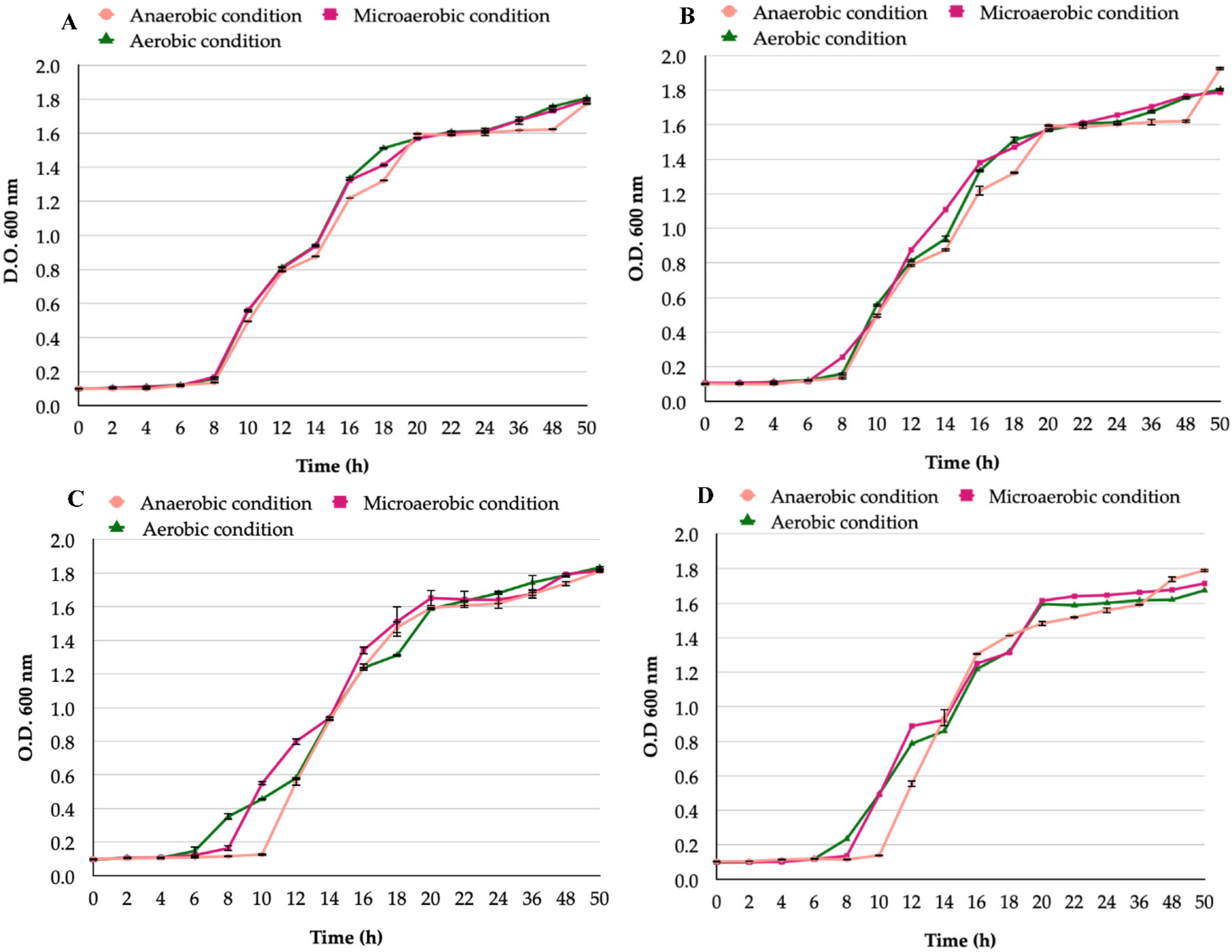
There was no significant difference when comparing the in vitro growth curves of all H. pylori strains incubated under microaerobic or anaerobic condtions (Figure 1). The bacterial growth incubated under aerobic conditions was compared with that obtained under anaerobic or microaerobic conditions and highly significant inhibition (p < 0.0001) was observed (Figure 1). The growth curves of different H. pylori strains under the same incubation conditions were compared and there were no significant differences observed (p = 0.1).
Regarding the growth of yeasts belonging to the Candida genus, all strains grew when incubated under the three conditions evaluated (Figure 2) with no significant differences in the growth obtained under each condition. The growth of all the Candida strains incubated in either of the three conditions tested showed no significant differences (p = 0.2).

3.2. Detection of Bacteria-like Bodies (Y-BLBs) within Yeasts

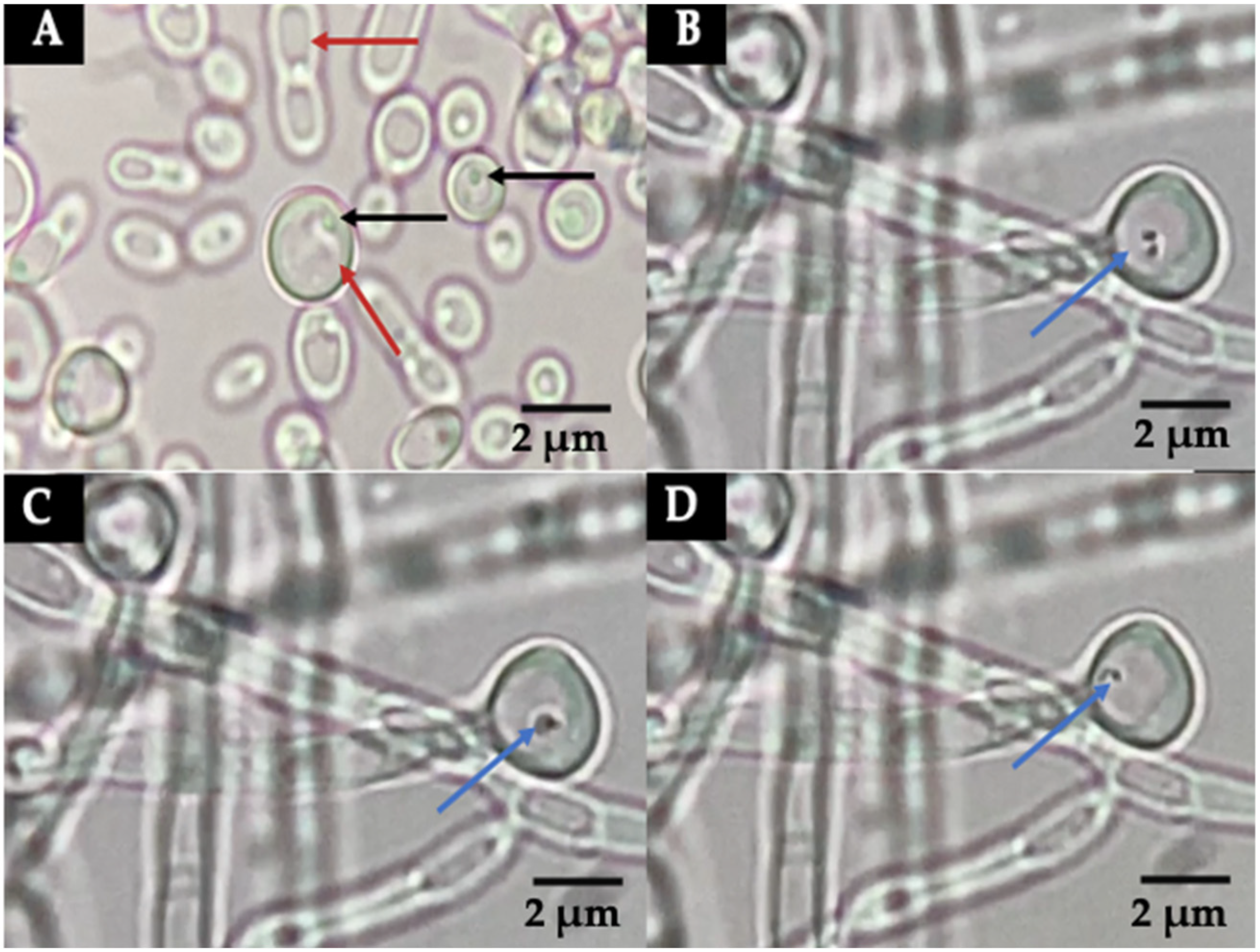
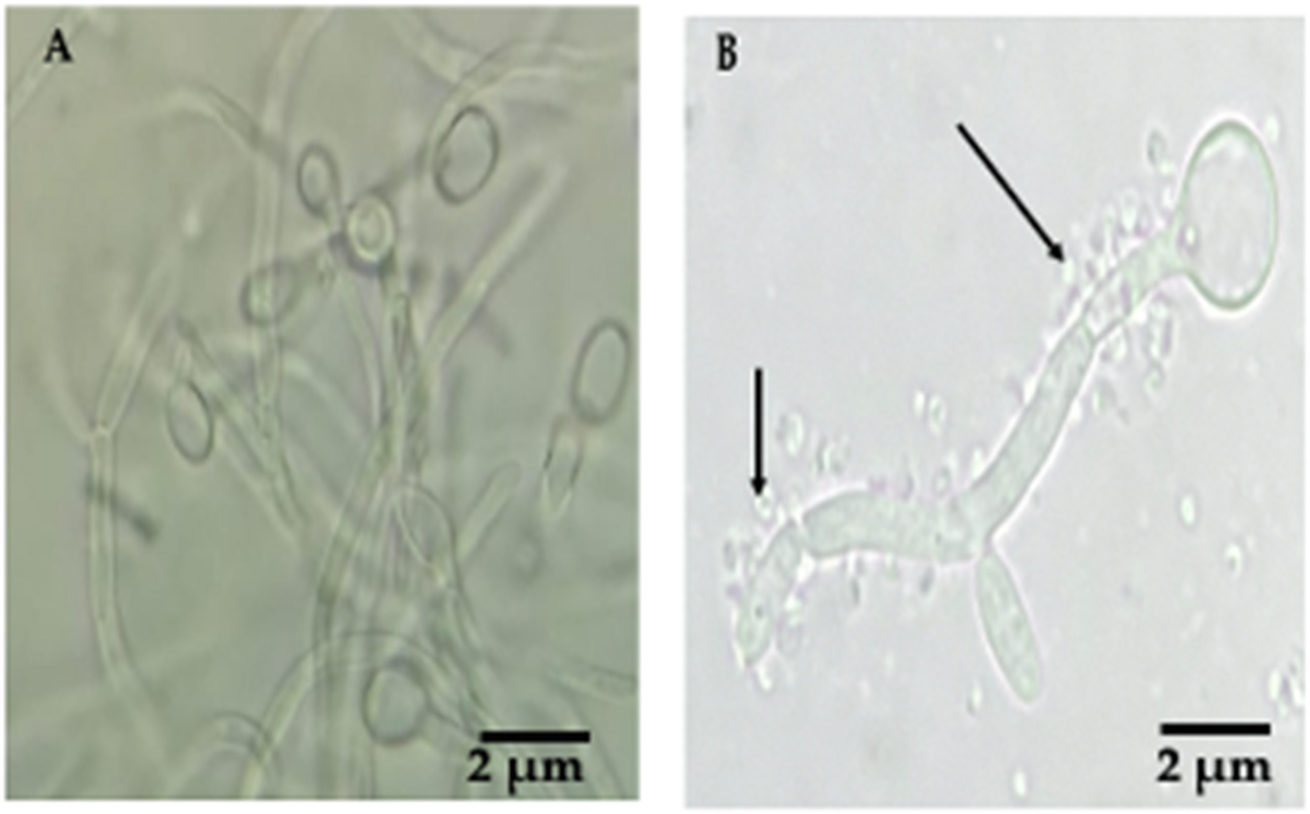
Mobile Y-BLBs were detected in the wet mounts of all co-cultures incubated under the three different conditions, starting at time 1 h. The movements of BLBs were ascertained observing the changes in positions of the BLBs within vacuoles of yeast cells (Figure 3B–D). Wet mounts also revealed H. pylori cells adhering to pseudohyphae of C. albicans cells (Figure 4).
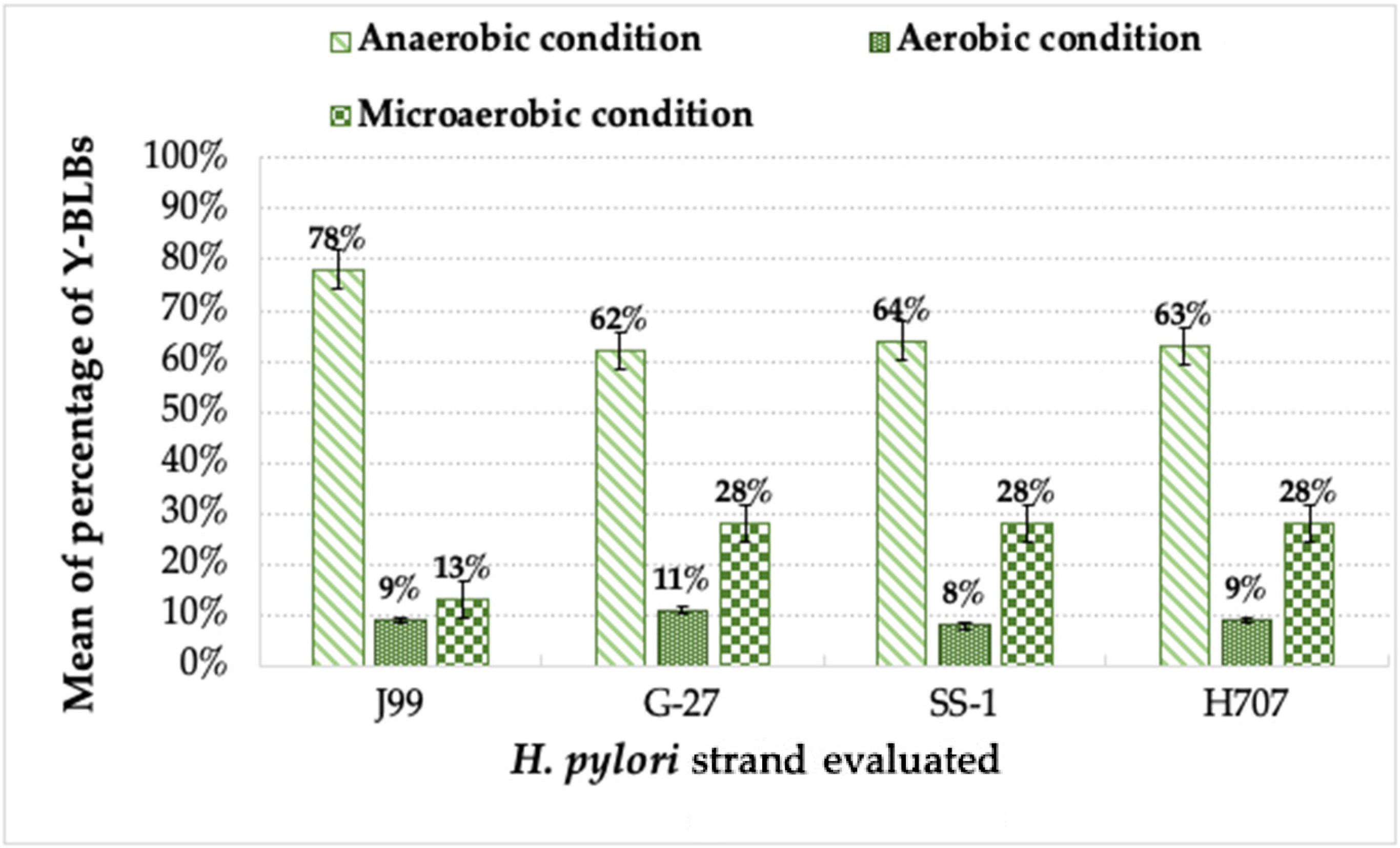
When analyzing the percentages of Y-BLBs present in the different co-cultures, the total number of Y-BLBs present in the different incubation conditions, and the strain of H. pylori being evaluated, the higher Y-BLB percentages were obtained when incubation was conducted under anaerobic conditions (62% to 78%), followed by incubations under microaerobic (13% to 28%) or aerobic conditions (8% to 11%). The means of the Y-BLB percentages also varied according to the strain of H. pylori assayed. The higher means of the Y-BLB percentages were found in those co-cultures in which the H. pylori J99 strain was incubated under anaerobic conditions (Figure 5), being highly significantly different from the means of the Y-BLB percentages obtained when incubations were under aerobic or microaerobic conditions. The Y-BLB percentages found in co-cultures incubated under microaerobic conditions were, in all cases, significantly higher than those obtained when incubated under aerobic conditions (p = 0.03) (Figure 5).
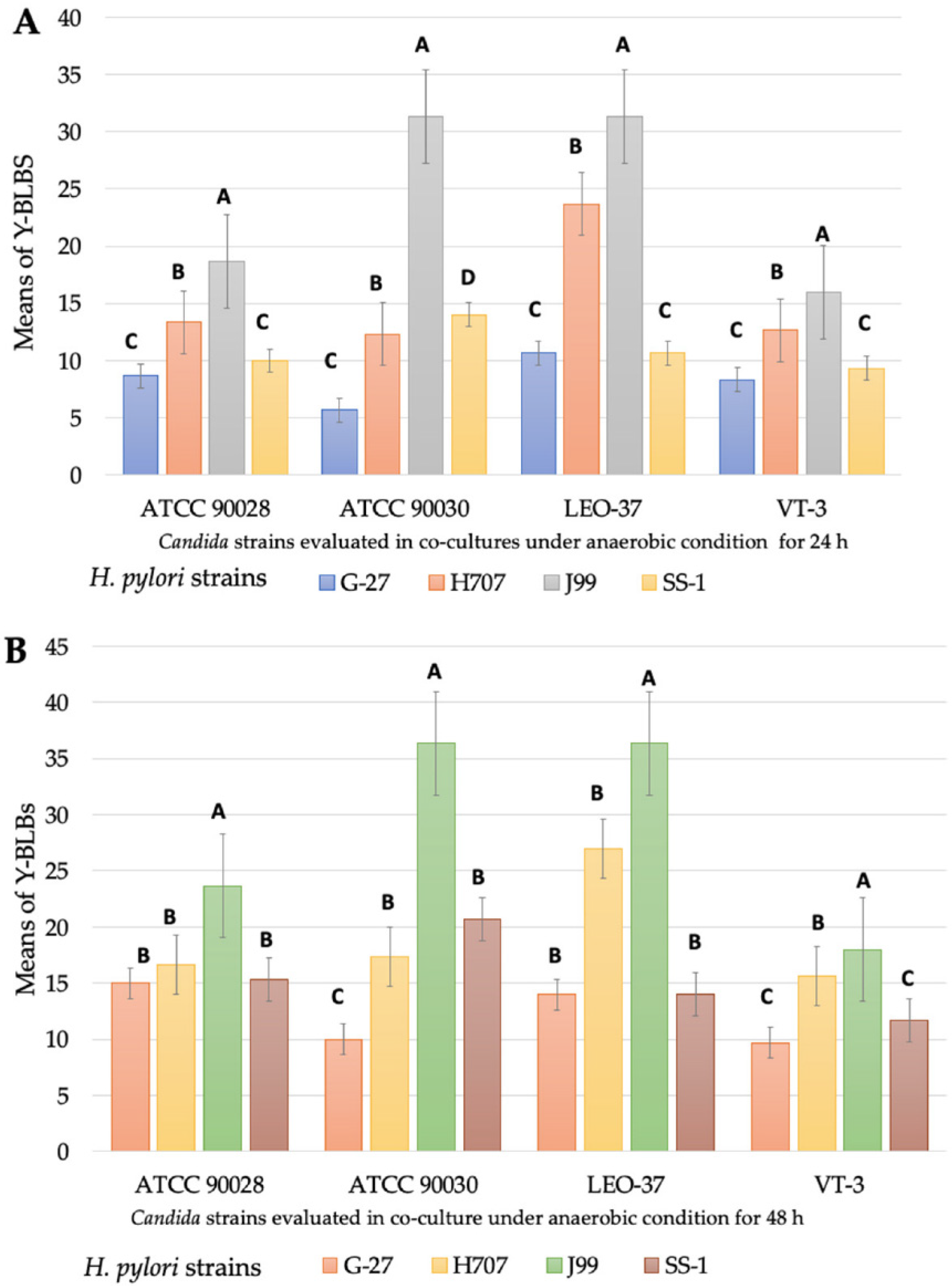
When the means of the Y-BLB percentages obtained per co-incubation time were analyzed, it was observed that the higher means were obtained at 24 h and 48 h under anaerobic conditions (Figure 6). This information allowed us to establish that the H. pylori-Candida co-culture combination produced the higher means of Y-BLB percentages. The co-cultures combining the C. glabrata strain ATCC 90030 or the C. glabrata strain LEO-37 plus the H. pylori strain J99 resulted in the higher means of Y-BLB percentages (Figure 7).

3.3. Identification, at the Species Level, of Intra-Yeast Bacteria-like Bodies (BLBs)

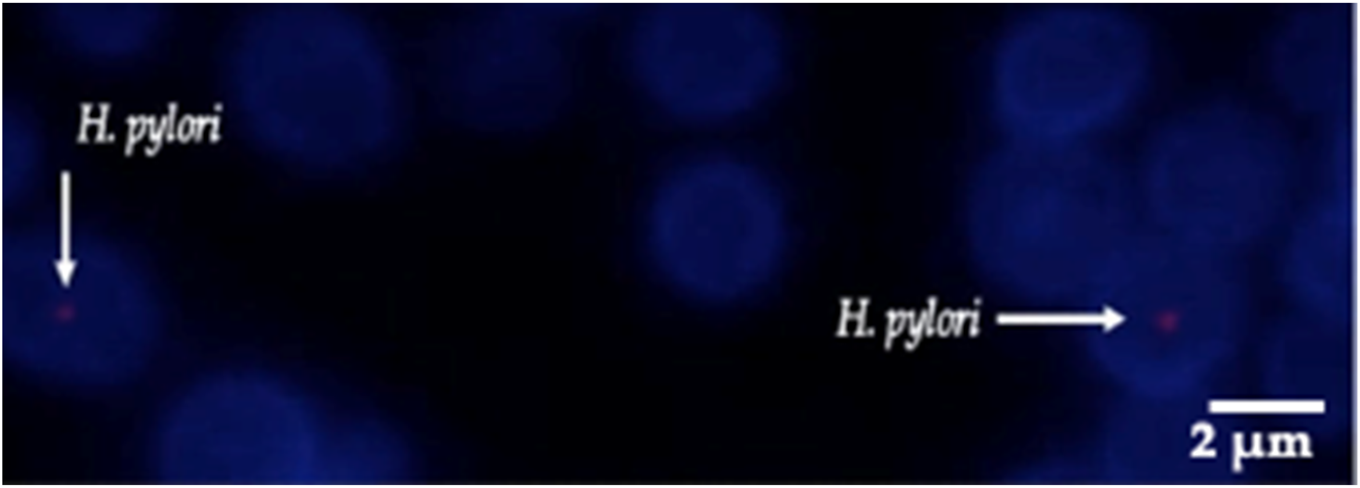
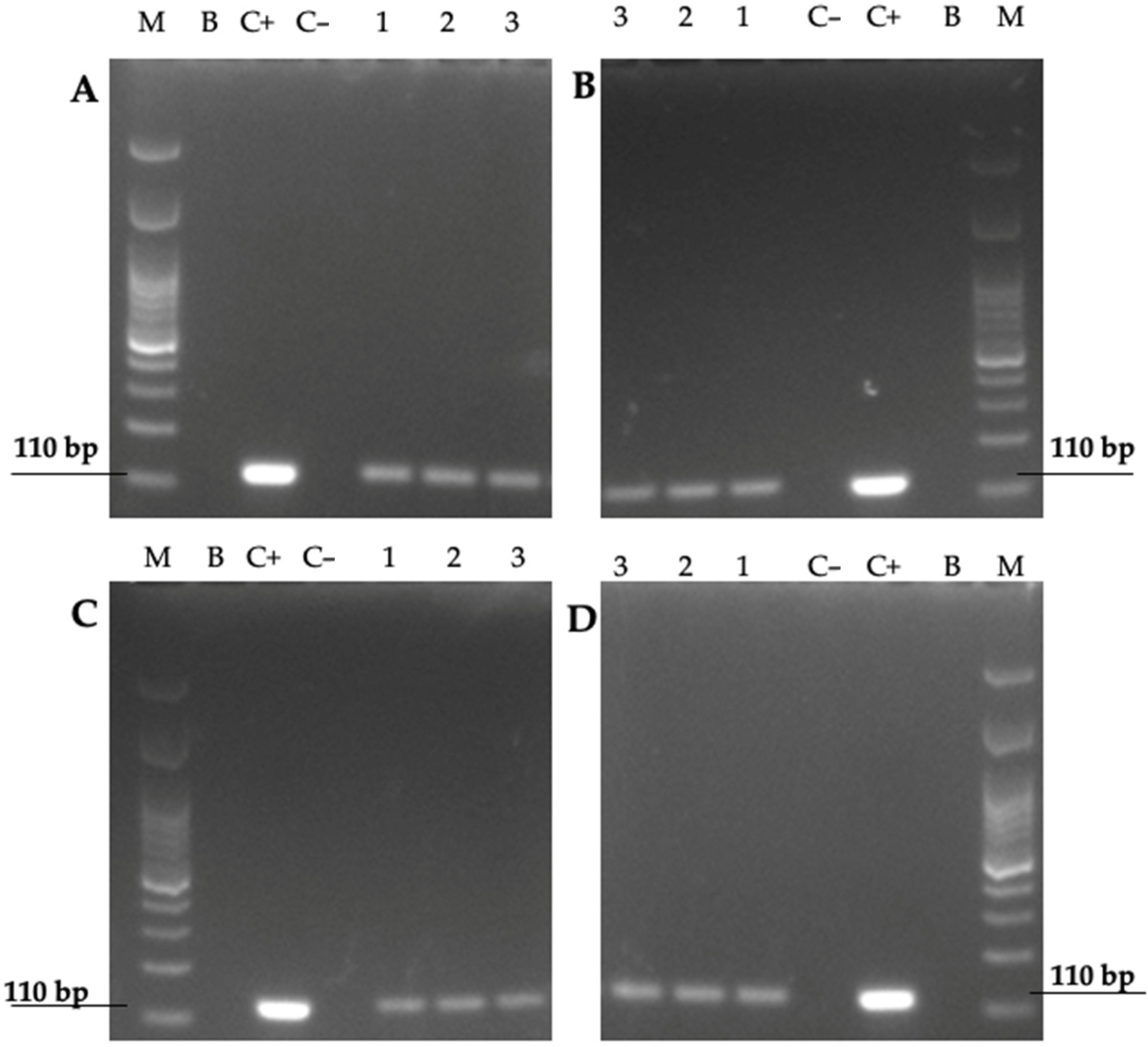
The fluorescent in situ hybridization (FISH) technique revealed that yeast cells previously found to contain BLBs showed the expected red fluorescence corresponding to the Cy3-labeled fluorescent probe specific for H. pylori DNA. This assay allowed us to identify BLBs as cells belonging to the species H. pylori growing inside the Candida cells (Figure 8). Furthermore, the identification, at the species level, of Y-BLBs as H. pylori was also supported by the amplification of the H. pylori gene codifying its 16S rRNA in the total DNA extracted from yeast cells which were previously co-incubated with H. pylori cells, and then the presence of Y-BLBs was confirmed (Figure 9). Together, both assays confirmed that the BLBs observed under an optical microscope corresponded to intracellular H. pylori within Candida cells.

3.4. Viability Assessment of H. pylori Cells Harboring in Yeast Cells

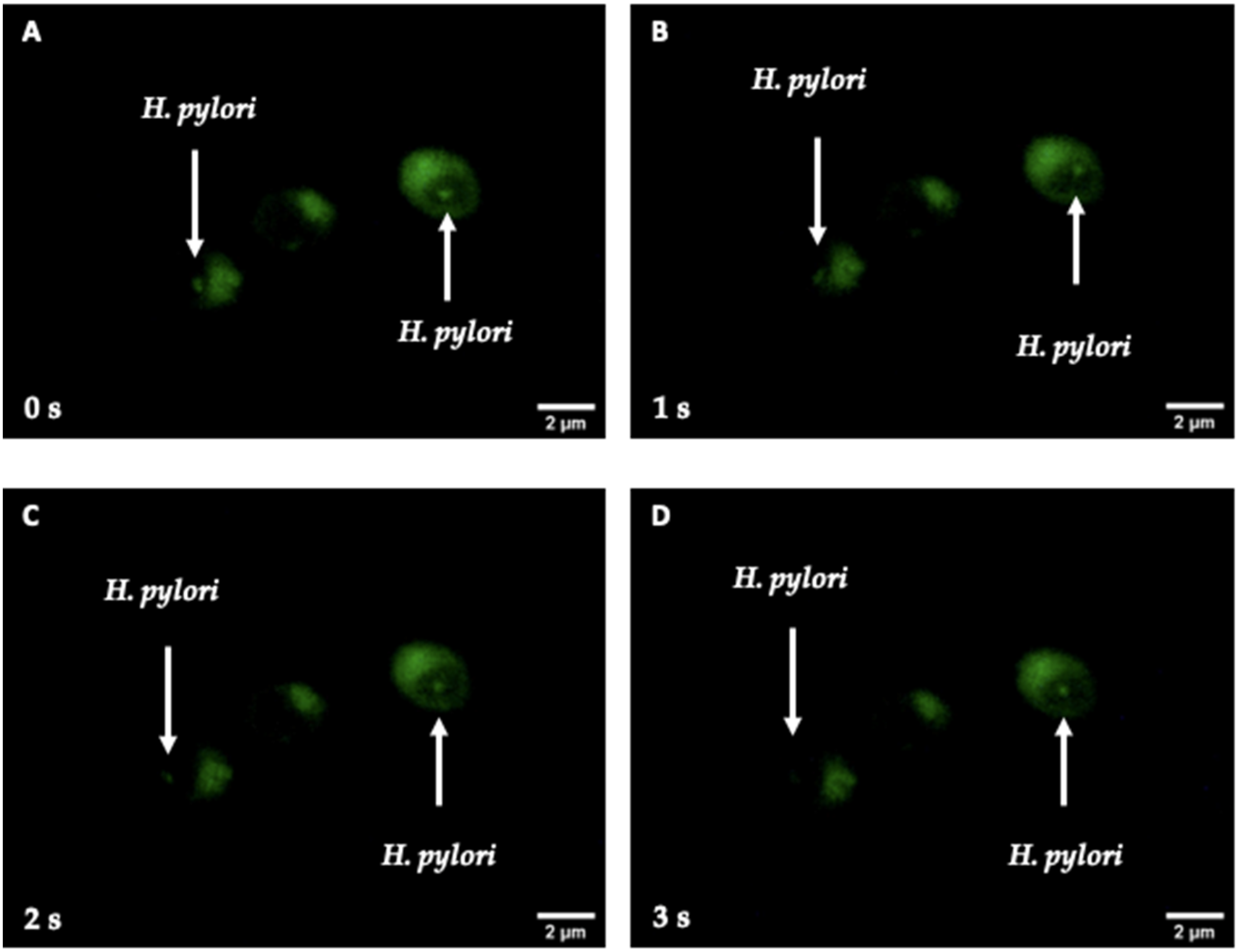
The LIVE/DEAD BacLight Bacterial Viability Kit demonstrated, by the SYTO 9 green fluorescence present in the interior of the vacuoles of yeast cells, that viable H. pylori cells harbor within yeast cells. Thus, intra-yeast H. pylori (or BLBs) remained viable after their entry into yeast cells and reseeding of the Y-BLBs in Sabouraud plus chloramphenicol (SA-CHL) (Figure 10). Furthermore, this same figure shows, at 1 s intervals, the change of position of the green fluorescent bodies (H. pylori cells) contained by the vacuoles of yeast cells, which indicated that bacteria were mobile.

4. Discussion

H. pylori is a pathogen that is well adapted to survive gastric conditions [22,39], and is difficult to grow in culture [40,41,42,43]. In vitro, this fastidious pathogen grows under microaerophilic and capnophilic conditions [44]. H. pylori growth under an aerobic condition showed a highly significant inhibition (p < 0.0001) as compared with the growth obtained when it was incubated under microaerobic or anaerobic conditions. Since H. pylori cells were able to survive under anaerobic conditions for up to 72 h, our results are in agreement with the literature [45]. It was not surprising that none of the H. pylori strains grew when incubated under aerobic conditions. Regarding Candida strains, all strains grew under the three incubation conditions studied. Our results are supported by studies in the literature reporting that although yeasts belonging to this fungal genus are classified as aerophilic, they have the ability to adapt to different environmental conditions, such as different oxygen concentrations [46,47,48]. Thus, Candida yeasts can be members of the normal human microbiota of the skin, mouth, vagina, and gastrointestinal tract [46,47,49].
H. pylori and Candida co-cultures incubated under anaerobic conditions showed that H. pylori J99 produced higher means of Y-BLB percentages regardless of the Candida strain used in the co-culture. Although, currently, there is no explanation for this, it has been reported that the invasiveness of the H. pylori J99 strain is higher than that of the G-27 and SS-1 strains [50]. Different bacteria synthesize chitinase which is used to degrade chitin to obtain carbon and nitrogen, and has also been reported to be a virulence factor which allows Legionella pneumophila to move across the alveolar mucosa [51,52]. It would be interesting to investigate if H. pylori strains produce chitinase to degrade chitin, a component of the yeast wall, favoring its entry into yeast cells.
Yeast factors could also be implicated in the H. pylori and Candida cell interactions. In the present work, the higher means of Y-BLB percentages were obtained when H. pylori J99 cells were co-cultured with the C. glabrata strains (LEO-37 or ATCC 90030). In 1998, Ansorg and coworkers reported [53] an H. pylori relationship with C. glabrata, describing the preference of bacterial adhesion towards non-albicans Candida yeasts. To date, the reason for the affinity between H. pylori and C. glabrata has not been clarified; however, it is possible to attribute it to the high production of adhesins by C. glabrata cells, which might provide a wide range of possibilities for bacteria to adhere to yeast. Furthermore, C. glabrata cells may have receptors on their surfaces that detect microbe-associated molecular patterns (MAMPs), as reported for the fungus Fusarium graminearum [54], which may lead Candida cells, after recognizing H. pylori MAMPs, to trigger a signaling cascade triggering bacterial endocytosis by the fungal cell; however, so far this is just a hypothesis. It is known that when subjected to varying environmental conditions, such as hypoxic conditions, yeast cells, including those of C. glabrata, express moonlighting proteins on their cell surfaces that participate in both adhesions and interactions with other cells and in the formation of a biofilm [55], which would favor interactions between bacteria and this species of yeast. The presence of hyphae in C. albicans favors bacterial adhesion to this yeast species and contributes to disseminate mobile bacteria [56], such as H. pylori. Furthermore, it has been described that hyphae could be a rich source of nutrients for bacteria [56].
In the present study, we report that higher means of Y-BLB percentages were obtained when co-culturing bacterial and yeast cells under anaerobic conditions. When oxygen in the environment is less than 1%, H. pylori morphologically transforms to a coccoid form [45]. However, as mentioned above, we achieved anaerobic conditions using AnaeroGen sachets, which generated a concentration from 7% to 15% CO2 and less than 0.1% oxygen. This may favor the growth of H. pylori outside yeast cells; however, the lack of the oxygen concentration required by this bacterium may be stressful for it, promoting its entry into yeast cells.
The second incubation condition evaluated was microaerobic condition, where a reduction in the means of Y-BLB percentages was observed as compared with those obtained under anaerobic conditions. These results are in agreement with the literature, since a microaerobic condition is considered to be the optimal environment for the growth of this pathogen, and therefore, H. pylori bacteria enters yeasts of the genus Candida without any apparent stress factor forcing the bacteria to search for shelter. These results are similar to those obtained in a previous study by our research group [31], where it was observed that under optimal pH growth conditions for H. pylori (pH 7), this bacterium enters into yeast cells, but at a lower rate as compared with conditions that threaten bacterial viability, such as acidic pH (pH 3 or 4). Similar results were observed under conditions of variations in nutrient concentrations [33].
The third incubation condition assayed was the aerobic condition, where percentages from only 8% to 11% Y-BLBs were obtained, percentages even lower than those recorded under anaerobic conditions. H. pylori is a bacterium which is sensitive to oxygen concentrations (from 20% to 21%) [57] that promote a coccoid form, VBNC condition, and also inhibit bacterial growth, clearly stressing this pathogen. Although no growth of H. pylori was observed under this environmental condition, there are studies in the literature that support its viability in the presence of oxygen from 6 h to 12 h. The above, together with the results of the present study, allow us to suggest that viable H. pylori cells could have entered into the yeast cells during the first hours of incubation of the co-cultures. This suggestion is supported by our observations of wet mounts prepared during the initial hours of co-cultures incubated under aerobic conditions that showed extracellular H. pylori cells with a bacillar morphology.
The identification, at the species level, of the BLBs using the FISH and PCR techniques together with the cell viability assay, allowed us to confirm that the intra-yeast BLBs observed in the wet mounts corresponded, in fact, to H. pylori viable cells with the ability to remain viable within this eukaryotic microorganism. This observation provides the basis for future research on the mechanism involved in the endosymbiosis between these two microorganisms, and to elucidate whether this mechanism is, in fact, another route of transmission for H. pylori.

5. Conclusions

H. pylori enters yeast cells of the genus Candida in a basal amount even if nonapparent stress conditions are present; however, anaerobic conditions significantly promote the entry of this bacterium into yeast cells. Moreover, under an anaerobic environment, the endosymbiotic relationship between H. pylori and Candida cells is H. pylori strain dependent, i.e., C. glabrata is preferred over C. albicans by H. pylori, particularly by H. pylori J99. Moreover, H. pylori is not only able to enter into Candida cells, mostly when subjected to a stress, but also remains viable within yeast cells, which could be used as a shelter.

Supplementary Materials

The following supporting information can be downloaded at https://www.mdpi.com/article/10.3390/biology11050738/s1, Video S1: Wet mount of yeast cells harboring bacteria-like bodies (Y-BLBs) observed in a H. pylori H707-C. albicans VT-3 co-culture incubated under an anaerobic condition https://youtu.be/5iRM_Clkk4Y (accessed on 9 April 2022), Figure S1: Schematic representation of the methodology to obtain the growth curves of H. pylori and Candida strains cultured under aerobic, microaerobic, or anaerobic conditions, Figure S2: Schematic representation of the methodology to co-culture H. pylori strains with Candida strains, Figure S3: Schematic representation of the methodology to search for intra-yeast bacteria-like bodies (BLBs), Figure S4: Schematic representation of the methodology to identify the intra-yeast bacteria-like bodies (BLBs) using the FISH technique, Figure S5: Schematic representation of the methodology to detect the 16S rRNA gene of H. pylori in the total DNA of yeast cells, Figure S6: Schematic representation of the methodology to confirm the viability of intra-vacuolar H. pylori within yeast cells.

Author Contributions

Conceptualization, K.S.-A. and A.G.-C.; methodology, K.S.-A. and A.G.-C.; validation, K.S.-A., A.G.-C. and C.T.S.; formal analysis, K.S.-A., H.B., L.A.-A., F.S.-M. and A.G.-C.; investigation, K.S.-A., C.P.-S. and A.G.-C.; resources, A.G.-C., H.B. and V.L.C.; data curation, K.S.-A., H.B. and K.S.-C.; writing—original draft preparation, K.S.-A., C.T.S., L.A.-A. and A.G.-C.; writing—review and editing, K.S.-A., C.T.S., L.A.-A. and A.G.-C.; visualization, K.S.-A., C.T.S., L.A.-A., F.S.-M., V.L.C. and A.G.-C.; supervision, A.G.-C. and V.L.C.; project administration, A.G.-C. and C.P.-S.; funding acquisition, A.G.-C. All authors have read and agreed to the published version of the manuscript.

Funding

This work was supported by Grant VRID-Enlace 218.036.047-1.0, University of Concepcion, Concepcion, Chile.

Informed Consent Statement

Not applicable.

Data Availability Statement

Not applicable.

Acknowledgments

The collaboration and support of Laboratorio Pasteur is acknowledged.

Conflicts of Interest

The authors declare no conflict of interest.

References

  1. Abdi, Y.M.; Young, K.A.; Rampton, D.S.; Hardie, J.M.; Feldman, R.A.; Banatvala, N. Comparison of the Effects of Anaerobic and Micro-Aerophilic Incubation on Resistance of Helicobacter pylori to Metronidazole. J. Med. Microbiol. 1999, 48, 407–410. [Google Scholar] [CrossRef] [PubMed] [Green Version]
  2. Bravo, D.; Hoare, A.; Soto, C.; Valenzuela, M.A.; Quest, A.F. Helicobacter pylori in Human Health and Disease: Mechanisms for Local Gastric and Systemic Effects. World J. Gastroenterol. 2018, 24, 3071–3089. [Google Scholar] [CrossRef] [PubMed]
  3. Araújo, G.R.L.; Marques, H.S.; Santos, M.L.C.; da Silva, F.A.F.; da Brito, B.B.; Correa Santos, G.L.; de Melo, F.F. Helicobacter pylori Infection: How Does Age Influence the Inflammatory Pattern? World J. Gastroenterol. 2022, 28, 402–411. [Google Scholar] [CrossRef] [PubMed]
  4. Bury-Moné, S.; Kaakoush, N.O.; Asencio, C.; Mégraud, F.; Thibonnier, M.; De Reuse, H.; Mendz, G.L. Is Helicobacter pylori a True Microaerophile? Helicobacter 2006, 11, 296–303. [Google Scholar] [CrossRef] [PubMed]
  5. Hooi, J.K.Y.; Lai, W.Y.; Ng, W.K.; Suen, M.M.Y.; Underwood, F.E.; Tanyingoh, D.; Malfertheiner, P.; Graham, D.Y.; Wong, V.W.S.; Wu, J.C.Y.; et al. Global Prevalence of Helicobacter pylori Infection: Systematic Review and Meta-Analysis. Gastroenterology 2017, 153, 420–429. [Google Scholar] [CrossRef] [Green Version]
  6. Atherton, J.C.; Cao, P.; Peek, R.M.; Tummuru, M.K.; Blaser, M.J.; Cover, T.L. Mosaicism in Vacuolating Cytotoxin Alleles of Helicobacter pylori. Association of Specific VacA Types with Cytotoxin Production and Peptic Ulceration. J. Biol. Chem. 1995, 270, 17771–17777. [Google Scholar] [CrossRef] [Green Version]
  7. Gilani, A.; Razavilar, V.; Rokni, N.; Rahimi, E. VacA and CagA Genotypes Status and Antimicrobial Resistance Properties of Helicobacter pylori Strains Isolated from Meat Products in Isfahan Province, Iran. Iran. J. Vet. Res. 2017, 18, 97–102. [Google Scholar]
  8. Sharndama, H.C.; Mba, I.E. Helicobacter pylori: An up-to-Date Overview on the Virulence and Pathogenesis Mechanisms. Braz. J. Microbiol. 2022, 53, 33–50. [Google Scholar] [CrossRef]
  9. Robinson, K.; Atherton, J.C. The Spectrum of Helicobacter-Mediated Diseases. Annu. Rev. Pathol. 2021, 16, 123–144. [Google Scholar] [CrossRef]
  10. Franceschi, F.; Covino, M.; Baudron, C.R. Review: Helicobacter pylori and Extragastric Diseases. Helicobacter 2019, 24, e12636. [Google Scholar] [CrossRef] [Green Version]
  11. Guerra-Valle, M.; Orellana-Palma, P.; Petzold, G. Plant-Based Polyphenols: Anti-Helicobacter pylori Effect and Improvement of Gut Microbiota. Antioxidants 2022, 11, 109. [Google Scholar] [CrossRef] [PubMed]
  12. Merino, J.S.; García, A.; Pastene, E.; Salas, A.; Saez, K.; González, C.L. Lactobacillus fermentum UCO-979C Strongly Inhibited Helicobacter pylori SS1 in Meriones Unguiculatus. Benef. Microbes 2018, 9, 625–627. [Google Scholar] [CrossRef] [PubMed]
  13. Salas-Jara, M.J.; Sanhueza, E.A.; Retamal-Díaz, A.; González, C.; Urrutia, H.; García, A. Probiotic Lactobacillus fermentum UCO-979C Biofilm Formation on AGS and Caco-2 Cells and Helicobacter pylori Inhibition. Biofouling 2016, 32, 1245–1257. [Google Scholar] [CrossRef] [PubMed]
  14. García, A.; Navarro, K.; Sanhueza, E.; Pineda, S.; Pastene, E.; Quezada, M.; Henríquez, K.; Karlyshev, A.; Villena, J.; González, C. Characterization of Lactobacillus fermentum UCO-979C, a Probiotic Strain with a Potent Anti-Helicobacter pylori Activity. Electron. J. Biotechnol. 2017, 25, 75–83. [Google Scholar] [CrossRef]
  15. Baker, D.A. Plants against Helicobacter pylori to Combat Resistance: An Ethnopharmacological Review. Biotechnol. Rep. Amst. Neth. 2020, 26, e00470. [Google Scholar] [CrossRef] [PubMed]
  16. Bonifácio, B.V.; dos Santos Ramos, M.A.; da Silva, P.B.; Bauab, T.M. Antimicrobial Activity of Natural Products against Helicobacter Pylori: A Review. Ann. Clin. Microbiol. Antimicrob. 2014, 13, 54. [Google Scholar] [CrossRef] [Green Version]
  17. Kayali, S.; Manfredi, M.; Gaiani, F.; Bianchi, L.; Bizzarri, B.; Leandro, G.; Di Mario, F.; De’ Angelis, G.L. Helicobacter pylori, Transmission Routes and Recurrence of Infection: State of the Art. Acta Bio-Med. Atenei Parm. 2018, 89, 72–76. [Google Scholar] [CrossRef]
  18. Olivera-Severo, D.; Uberti, A.F.; Marques, M.S.; Pinto, M.T.; Gomez-Lazaro, M.; Figueiredo, C.; Leite, M.; Carlini, C.R. A New Role for Helicobacter pylori Urease: Contributions to Angiogenesis. Front. Microbiol. 2017, 8, 1883. [Google Scholar] [CrossRef] [Green Version]
  19. Hazell, S.L.; Evans, D.J.; Graham, D.Y. Helicobacter pylori Catalase. J. Gen. Microbiol. 1991, 137, 57–61. [Google Scholar] [CrossRef] [Green Version]
  20. Mori, M.; Suzuki, H.; Suzuki, M.; Kai, A.; Miura, S.; Ishii, H. Catalase and Superoxide Dismutase Secreted from Helicobacter pylori. Helicobacter 1997, 2, 100–105. [Google Scholar] [CrossRef]
  21. Nilsson, H.-O.; Blom, J.; Al-Soud, W.A.; Ljungh, Å.; Andersen, L.P.; Wadström, T. Effect of Cold Starvation, Acid Stress, and Nutrients on Metabolic Activity of Helicobacter pylori. Appl. Environ. Microbiol. 2002, 68, 11–19. [Google Scholar] [CrossRef] [PubMed] [Green Version]
  22. Mouery, K.; Rader, B.A.; Gaynor, E.C.; Guillemin, K. The Stringent Response Is Required for Helicobacter pylori Survival of Stationary Phase, Exposure to Acid, and Aerobic Shock. J. Bacteriol. 2006, 188, 5494–5500. [Google Scholar] [CrossRef] [PubMed] [Green Version]
  23. Hathroubi, S.; Servetas, S.L.; Windham, I.; Merrell, D.S.; Ottemann, K.M. Helicobacter pylori Biofilm Formation and Its Potential Role in Pathogenesis. Microbiol. Mol. Biol. Rev. 2018, 82, e00001-18. [Google Scholar] [CrossRef] [PubMed] [Green Version]
  24. Andersen, L.P.; Rasmussen, L. Helicobacter pylori-Coccoid Forms and Biofilm Formation. FEMS Immunol. Med. Microbiol. 2009, 56, 112–115. [Google Scholar] [CrossRef] [PubMed] [Green Version]
  25. Aktas, D.; Bagirova, M.; Allahverdiyev, A.M.; Abamor, E.S.; Safarov, T.; Kocazeybek, B.S. Resuscitation of the Helicobacter pylori Coccoid Forms by Resuscitation Promoter Factor Obtained from Micrococcus luteus. Curr. Microbiol. 2020, 77, 2093–2103. [Google Scholar] [CrossRef]
  26. Moreno-Mesonero, L.; Moreno, Y.; Alonso, J.L.; Ferrús, M.A. Detection of Viable Helicobacter pylori inside Free-Living Amoebae in Wastewater and Drinking Water Samples from Eastern Spain. Environ. Microbiol. 2017, 19, 4103–4112. [Google Scholar] [CrossRef] [Green Version]
  27. Matamala-Valdés, L.; Sánchez-Alonzo, K.; Parra, C.; Sáez, K.; Aguayo-Reyes, A.; García, A.; Matamala-Valdés, L.; Sánchez-Alonzo, K.; Parra, C.; Sáez, K.; et al. Detection of Intracellular Helicobacter pylori in Candida spp. from Neonate Oral Swabs. Rev. Assoc. Méd. Bras. 2018, 64, 928–935. [Google Scholar] [CrossRef]
  28. Salmanian, A.-H.; Siavoshi, F.; Akbari, F.; Afshari, A.; Malekzadeh, R. Yeast of the Oral Cavity Is the Reservoir of Helicobacter pylori. J. Oral Pathol. Med. 2008, 37, 324–328. [Google Scholar] [CrossRef]
  29. Siavoshi, F.; Salmanian, A.H.; Akbari, F.; Kbari, F.A.; Malekzadeh, R.; Massarrat, S. Detection of Helicobacter pylori-Specific Genes in the Oral Yeast. Helicobacter 2005, 10, 318–322. [Google Scholar] [CrossRef]
  30. Sánchez-Alonzo, K.; Parra-Sepúlveda, C.; Vergara, L.; Bernasconi, H.; García-Cancino, A.; Sánchez-Alonzo, K.; Parra-Sepúlveda, C.; Vergara, L.; Bernasconi, H.; García-Cancino, A. Detection of Helicobacter pylori in Oral Yeasts from Students of a Chilean University. Rev. Assoc. Med. Bras. 2020, 66, 1509–1514. [Google Scholar] [CrossRef]
  31. Sánchez-Alonzo, K.; Parra-Sepúlveda, C.; Vega, S.; Bernasconi, H.; Campos, V.L.; Smith, C.T.; Sáez, K.; García-Cancino, A. In Vitro Incorporation of Helicobacter pylori into Candida albicans Caused by Acidic PH Stress. Pathogens 2020, 9, 489. [Google Scholar] [CrossRef] [PubMed]
  32. Sánchez-Alonzo, K.; Belmar, L.; Parra-Sepúlveda, C.; Bernasconi, H.; Campos, V.L.; Smith, C.T.; Sáez, K.; García-Cancino, A. Antibiotics as a Stressing Factor Triggering the Harboring of Helicobacter pylori J99 within Candida albicans ATCC10231. Pathogens 2021, 10, 382. [Google Scholar] [CrossRef] [PubMed]
  33. Sánchez-Alonzo, K.; Silva-Mieres, F.; Arellano-Arriagada, L.; Parra-Sepúlveda, C.; Bernasconi, H.; Smith, C.T.; Campos, V.L.; García-Cancino, A. Nutrient Deficiency Promotes the Entry of Helicobacter pylori Cells into Candida Yeast Cells. Biology 2021, 10, 426. [Google Scholar] [CrossRef] [PubMed]
  34. Sánchez-Alonzo, K.; Arellano-Arriagada, L.; Castro-Seriche, S.; Parra-Sepúlveda, C.; Bernasconi, H.; Benavidez-Hernández, H.; Campos, V.L.; Sáez, K.; Smith, C.T.; García-Cancino, A. Temperatures Outside the Optimal Range for Helicobacter pylori Increase Its Harboring within Candida Yeast Cells. Biology 2021, 10, 915. [Google Scholar] [CrossRef] [PubMed]
  35. Andersen, L.P.; Wadström, T. Basic Bacteriology and Culture. In Helicobacter pylori: Physiology and Genetics; Mobley, H.L., Mendz, G.L., Hazell, S.L., Eds.; ASM Press: Washington, DC, USA, 2001; ISBN 978-1-55581-213-3. [Google Scholar]
  36. Olver, W.J.; Stafford, J.; Cheetham, P.; Boswell, T.C. Comparison of Candida ID Medium with Sabouraud-Chloramphenicol Agar for the Isolation of Yeasts from Clinical Haematology Surveillance Specimens. J. Med. Microbiol. 2002, 51, 221–224. [Google Scholar] [CrossRef]
  37. Böckelmann, U.; Manz, W.; Neu, T.R.; Szewzyk, U. Investigation of Lotic Microbial Aggregates by a Combined Technique of Fluorescent In Situ Hybridization and Lectin-Binding-Analysis. J. Microbiol. Methods 2002, 49, 75–87. [Google Scholar] [CrossRef]
  38. Rüssmann, H.; Kempf, V.A.J.; Koletzko, S.; Heesemann, J.; Autenrieth, I.B. Comparison of Fluorescent In Situ Hybridization and Conventional Culturing for Detection of Helicobacter pylori in Gastric Biopsy Specimens. J. Clin. Microbiol. 2001, 39, 304–308. [Google Scholar] [CrossRef] [Green Version]
  39. Yang, I.; Nell, S.; Suerbaum, S. Survival in Hostile Territory: The Microbiota of the Stomach. FEMS Microbiol. Rev. 2013, 37, 736–761. [Google Scholar] [CrossRef]
  40. Ranjbar, R.; Khamesipour, F.; Jonaidi-Jafari, N.; Rahimi, E. Helicobacter pylori Isolated from Iranian Drinking Water: VacA, CagA, IceA, OipA and BabA2 Genotype Status and Antimicrobial Resistance Properties. FEBS Open Bio 2016, 6, 433–441. [Google Scholar] [CrossRef]
  41. Zamani, M.; Vahedi, A.; Maghdouri, Z.; Shokri-Shirvani, J. Role of Food in Environmental Transmission of Helicobacter pylori. Casp. J. Intern. Med. 2017, 8, 146–152. [Google Scholar] [CrossRef]
  42. Saeidi, E.; Sheikhshahrokh, A. VacA Genotype Status of Helicobacter pylori Isolated from Foods with Animal Origin. BioMed Res. Int. 2016, 2016, 8701067. [Google Scholar] [CrossRef] [PubMed] [Green Version]
  43. Agarwal, S.; Jithendra, K.D. Presence of Helicobacter pylori in Subgingival Plaque of Periodontitis Patients with and without Dyspepsia, Detected by Polymerase Chain Reaction and Culture. J. Indian Soc. Periodontol. 2012, 16, 398–403. [Google Scholar] [CrossRef] [PubMed]
  44. Park, S.; Hu, J.Y. Assessment of the Extent of Bacterial Growth in Reverse Osmosis System for Improving Drinking Water Quality. J. Environ. Sci. Health Part A 2010, 45, 968–977. [Google Scholar] [CrossRef] [PubMed]
  45. Donelli, G.; Matarrese, P.; Fiorentini, C.; Dainelli, B.; Taraborelli, T.; Di Campli, E.; Di Bartolomeo, S.; Cellini, L. The Effect of Oxygen on the Growth and Cell Morphology of Helicobacter pylori. FEMS Microbiol. Lett. 1998, 168, 9–15. [Google Scholar] [CrossRef]
  46. Biswas, S.K.; Chaffin, W.L. Anaerobic Growth of Candida albicans Does Not Support Biofilm Formation under Similar Conditions Used for Aerobic Biofilm. Curr. Microbiol. 2005, 51, 100–104. [Google Scholar] [CrossRef]
  47. Dumitru, R.; Hornby, J.M.; Nickerson, K.W. Defined Anaerobic Growth Medium for Studying Candida albicans Basic Biology and Resistance to Eight Antifungal Drugs. Antimicrob. Agents Chemother. 2004, 48, 2350–2354. [Google Scholar] [CrossRef] [Green Version]
  48. Rosa, E.A.R.; Rached, R.N.; Ignácio, S.A.; Rosa, R.T.; José da Silva, W.; Yau, J.Y.Y.; Samaranayake, L.P. Phenotypic Evaluation of the Effect of Anaerobiosis on Some Virulence Attributes of Candida albicans. J. Med. Microbiol. 2008, 57, 1277–1281. [Google Scholar] [CrossRef]
  49. Cottier, F.; Mühlschlegel, F.A. Sensing the Environment: Response of Candida albicans to the X Factor. FEMS Microbiol. Lett. 2009, 295, 1–9. [Google Scholar] [CrossRef] [Green Version]
  50. Liu, H.; Semino-Mora, C.; Dubois, A. Mechanism of H. pylori Intracellular Entry: An In Vitro Study. Front. Cell. Infect. Microbiol. 2012, 2, 13. [Google Scholar] [CrossRef] [Green Version]
  51. Rathore, A.S.; Gupta, R.D. Chitinases from Bacteria to Human: Properties, Applications, and Future Perspectives. Available online: https://www.hindawi.com/journals/er/2015/791907/ (accessed on 9 March 2021).
  52. Rehman, S.; Grigoryeva, L.S.; Richardson, K.H.; Corsini, P.; White, R.C.; Shaw, R.; Portlock, T.J.; Dorgan, B.; Zanjani, Z.S.; Fornili, A.; et al. Structure and Functional Analysis of the Legionella Pneumophila Chitinase ChiA Reveals a Novel Mechanism of Metal-Dependent Mucin Degradation. PLoS Pathog. 2020, 16, e1008342. [Google Scholar] [CrossRef]
  53. Ansorg, R.; Schmid, E.N. Adhesion of Helicobacter pylori to Yeast Cells. Zentralbl. Bakteriol. Int. J. Med. Microbiol. 1998, 288, 501–508. [Google Scholar] [CrossRef]
  54. Ipcho, S.; Sundelin, T.; Erbs, G.; Kistler, H.C.; Newman, M.-A.; Olsson, S. Fungal Innate Immunity Induced by Bacterial Microbe-Associated Molecular Patterns (MAMPs). G3 Genes Genomes Genet. 2016, 6, 1585–1595. [Google Scholar] [CrossRef] [PubMed] [Green Version]
  55. Shirtliff, M.E.; Peters, B.M.; Jabra-Rizk, M.A. Cross-Kingdom Interactions: Candida albicans and Bacteria. FEMS Microbiol. Lett. 2009, 299, 1–8. [Google Scholar] [CrossRef] [Green Version]
  56. Deveau, A.; Brulé, C.; Palin, B.; Champmartin, D.; Rubini, P.; Garbaye, J.; Sarniguet, A.; Frey-Klett, P. Role of Fungal Trehalose and Bacterial Thiamine in the Improved Survival and Growth of the Ectomycorrhizal Fungus Laccaria Bicolor S238N and the Helper Bacterium Pseudomonas fluorescens BBc6R8. Environ. Microbiol. Rep. 2010, 2, 560–568. [Google Scholar] [CrossRef]
  57. Wang, G.; Alamuri, P.; Maier, R.J. The Diverse Antioxidant Systems of Helicobacter pylori. Mol. Microbiol. 2006, 61, 847–860. [Google Scholar] [CrossRef] [PubMed]
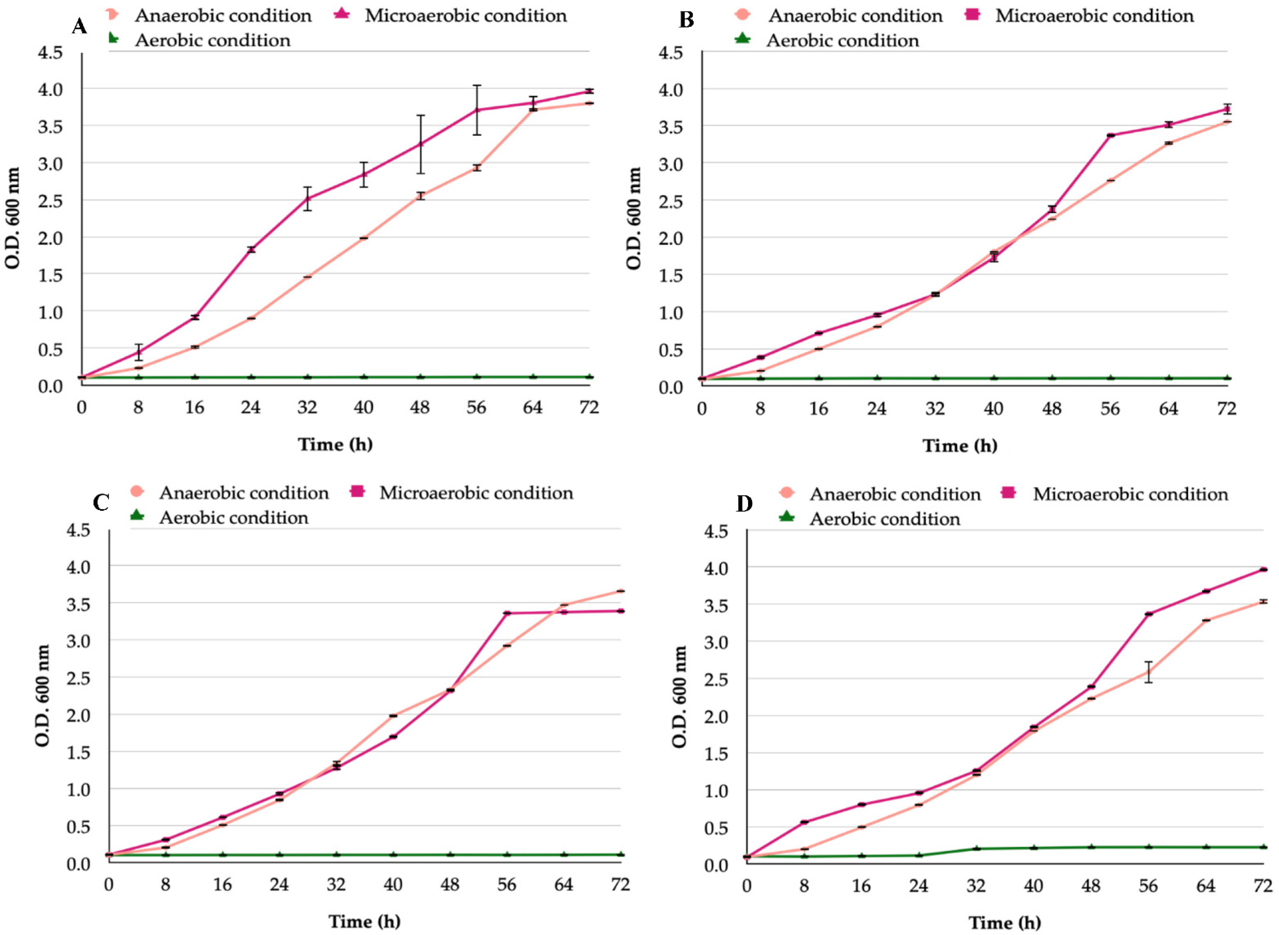
Figure 1. Growth curves of H. pylori strains incubated in the presence of an aerobic, microaerobic, or anaerobic condition: (A) H. pylori reference strain J99; (B) H. pylori reference strain G-27; (C) H. pylori SS-1 reference strain; (D) H. pylori H707 clinical strain.
Biology 11 00738 g001
Figure 2. Growth curves of Candida strains under aerobic, microaerobic, or anaerobic conditions: (A) C. albicans ATCC 90028 reference strain; (B) C. glabrata ATCC 90030 reference strain; (C) C. albicans VT-3 clinical strain; (D) C. glabrata LEO-37 clinical strain. No significant differences were present among these growth curves (p = 0.5).
Biology 11 00738 g002
Figure 3. Wet mounts of yeast cells harboring bacteria-like bodies (Y-BLBs). Images, similar to observations of other co-cultures, correspond to a co-culture of H. pylori H707 strain and C. albicans VT-3 strain, incubated under an anaerobic condition: (A) Yeast cells from a pure culture of the C. albicans VT-3 cells lacking BLBs in their vacuole (red arrow), nuclei of the yeast cells indicated with black arrows; (BD) show the changes in positions of BLBs in the vacuoles (blue arrows) in images taken at 1 s intervals. The actual movement of the BLBs is shown in Video S1.
Biology 11 00738 g003
Figure 4. (A) Wet mount of a pure culture of C. albicans VT-3 cells, no extracellular bacteria nor bacteria adhered to the surface of hyphae can be seen; (B) wet mount of a co-culture of H. pylori G-27 strain and C. albicans VT-3 strain incubated under an anaerobic condition showing extracellular H. pylori bacteria adhered to the filamentous yeast structures (black arrow).
Biology 11 00738 g004
Figure 5. Means of the percentages of yeast cells harboring bacteria-like bodies (Y-BLBs) in co-cultures of H. pylori strains plus Candida strains incubated for 48 h under anaerobic, microaerobic, or aerobic conditions. The higher means of Y-BLB percentages were found in co-cultures including the H. pylori J99 strain incubated under anaerobic conditions.
Biology 11 00738 g005
Figure 6. Means of yeast cells including bacteria-like bodies (Y-BLBs) found in the different co-cultures under the different incubation conditions. The higher means of Y-BLB percentages were reached at 24 h and 48 h of co-culture incubation under anaerobic conditions.
Biology 11 00738 g006
Figure 7. Means of intra-yeast bacteria-like bodies (Y-BLBs) found in H. pylori strains and Candida strains, co-cultured under anaerobic conditions for 24 h (A) or 48 h (B). At both incubation times, the higher means of Y-BLB percentages were found when the H. pylori J99 strain was co-cultured with C. glabrata strains. Different letters indicate significant differences (p < 0.05). Strains ATCC 90028 and VT-3 belong to C. albicans species and strains ATCC 90030 and LEO-37 belong to C. glabrata species.
Biology 11 00738 g007
Figure 8. Yeast cells obtained from a H. pylori J99-C. glabrata LEO-37 co-culture incubated under anaerobic conditions in Brucella broth medium plus 5% fetal bovine serum. The red fluorescence observed within yeast cells is a consequence of the hybridization of the H. pylori-specific fluorescent probe (white arrow). Blue fluorescence corresponds to aniline blue bound to yeast 1,3 β-glucans.
Biology 11 00738 g008
Figure 9. Images of 2% agarose gel showing the amplicons resulting from the amplification, by PCR, of the 16S rRNA gene of H. pylori from the total DNA of different Candida strains previously co-cultured under different oxygen concentrations with H. pylori J99 strain: (A) H. pylori J99 strain and C. albicans ATCC 90028 strain co-culture; (B) H. pylori J99 strain and C. glabrata ATCC 90030 strain co-culture; (C) H. pylori J99 strain and C. albicans VT-3 strain co-culture; (D) H. pylori J99 strain and C. glabrata LEO-37 strain co-culture. M, markers of molecular weight; B, blank (master mix, primers, and PCR grade water); C−, negative control (pure C. glabrata LEO-37 strain DNA); C+, positive control (pure H. pylori J99 strain DNA). Lanes 1, 2 and 3: amplicons amplified from the total DNA extracted from yeast cells previously co-cultured with H. pylori under aerobic, microaerobic, or anaerobic conditions, respectively.
Biology 11 00738 g009
Figure 10. Fluorescence microscopy of C. glabrata LEO-37 strain cells co-cultured with H. pylori G-27 strain cells under microaerobic condition for 48 h, and then cultured on Sabouraud agar plus chloramphenicol for 24 h to eradicate bacteria extracellularly located. The viability of bacteria harboring within Candida cells was demonstrated by SYTO 9 green fluorescence within yeast cells (white arrows). Images (AD) were obtained from the same microscopic field at 1 s intervals showing the change of position of bacteria located in the vacuole of Candida cells.
Biology 11 00738 g010
Publisher’s Note: MDPI stays neutral with regard to jurisdictional claims in published maps and institutional affiliations.