Article

The First Survival Score for Patients Aged ≥80 Years Irradiated for Brain Metastases

1
Department of Radiation Oncology, University of Lubeck, 23562 Lubeck, Germany
2
Department of Radiation Oncology, Mayo Clinic, Scottsdale, AZ 85259, USA
3
Department of Oncology, Vejle Hospital, University Hospital of Southern Denmark, 7100 Vejle, Denmark
4
Research Department, Zealand University Hospital, 4600 Køge, Denmark
5
Medical Practice for Radiotherapy and Radiation Oncology, 30161 Hannover, Germany
*
Author to whom correspondence should be addressed.
Biology 2022, 11(10), 1434; https://doi.org/10.3390/biology11101434
Submission received: 24 August 2022 / Revised: 27 September 2022 / Accepted: 29 September 2022 / Published: 30 September 2022

Abstract

:

Simple Summary

Decisions regarding personalized treatment for brain metastases should consider the patient’s lifespan, which can be estimated with survival scores. Since very elderly patients (≥80 years) are different from other patients, e.g., due to a higher comorbidity index and decreased organ functions, these patients likely benefit from specific scores. A survival score was generated in 94 patients aged ≥ 80 years who were irradiated (whole-brain radiotherapy) for metastases of the brain. The score achieved high accuracy with respect to the prediction of death up to 6 months and survival for ≥1 and ≥2 months following treatment. When compared to an existing tool, the new score was more precise regarding death ≤ 1 month following radiotherapy and survival (all time points). Therefore, the new score appears preferable.

Abstract

Survival scores facilitate personalized cancer treatment. Due to demographic changes, very elderly patients are more prevalent than in the past. A score was developed in 94 patients aged ≥ 80 years undergoing whole-brain radiotherapy for brain metastases. Dose fractionation, treatment period, age, sex, performance score (ECOG-PS), tumor type, count of lesions, metastases outside the brain, and interval tumor diagnosis to radiotherapy were retrospectively evaluated. Independent predictors of survival were used for the score. Based on individual scoring points obtained from 3-month survival rates, prognostic groups were designed. Additionally, the score was compared to an existing tool developed in patients ≥ 65 years. ECOG-PS, count of lesions, and extra-cranial metastases were independent prognostic factors. Three groups were created (7, 10, and 13–16 points) with 3-month survival of 6%, 25%, and 67% (p < 0.001), respectively. Positive predictive values (PPVs) regarding death ≤ 3 and survival ≥ 3 months were 94% and 67% (new score) vs. 96% and 48% (existing tool), respectively. PPVs for survival ≥1 and ≥2 months were 88% and 79% vs. 63% and 58%, respectively. Both tools were accurate in predicting death ≤2, ≤3, and ≤6 months. The new score was more precise regarding death ≤1 month and survival (all time periods) and appeared preferable. However, it still needs to be validated.

1. Introduction

A substantial number of adult cancer patients experience brain metastases [1]. Many of them receive radiotherapy, either alone or following neurosurgical resection. Several types of radiotherapy are available, including whole-brain radiotherapy (WBRT) and local irradiation, namely, single-fraction radiosurgery (SRS) and fractionated stereotactic radiotherapy (FSRT) [1]. In most studies, SRS and FSRT were limited to 1–4 cerebral lesions. Although studies from Japan showed that SRS also appeared safe for 5–10 lesions, the majority of these patients receive WBRT alone, particularly if they are older or frail, or if uncontrolled extra-cranial metastases exist [1,2]. Moreover, local therapies are generally limited to lesions with a diameter of ≤4 cm [1]. In the case of a high risk of new brain metastases outside of the irradiated sites, local therapies and WBRT may be combined to improve intracerebral control [3]. However, one should be aware that adding WBRT is associated with more pronounced neurocognitive decline [4].
For WBRT alone, several dose-fractionation regimens are available [1]. The most appropriate individual regimen depends on several factors including the survival prognosis. Generally, patients with a very limited life expectancy should receive a less time-consuming regimen to ensure that they spend as little time as possible of their remaining lifespan receiving WBRT. Patients with very limited survival times may not benefit from WBRT at all and be treated with best supportive care (BSC) including corticosteroids [5]. In contrast, for patients with more favorable prognoses, longer-course regimens with higher total doses and lower doses per fraction are more appropriate [1]. Higher total doses may lead to longer-lasting intracerebral control, and lower doses per fraction are associated with a decreased risk of late sequelae including neurocognitive decline [6,7,8,9]. Since the risk of late effects increases with time, this endpoint is more important for longer-term survivors. These considerations illustrate that it is important for the treating physicians to estimate individual survival prognoses most precisely prior to the selection of an individual therapy. This estimation will be considerably facilitated with scoring instruments. Several instruments are available for the radiation treatment of brain metastases including a survival score for patients aged ≥ 65 years [10]. Such a score is reasonable, since patients of this age group are different from younger patients, e.g., due to higher degrees of comorbidity including cardiovascular disease and decreased organ functions. This holds particularly true for the group of very elderly patients. Several authors defined very elderly as ≥80 years, and some of them compared this age group to patients aged 70 to 79 years [11,12,13,14,15,16,17]. Due to the demographic change, the group of very elderly patients is growing and will come increasingly into focus [18]. To provide a very good personalized treatment for these patients, the development of specific prognostic tools for this group appears reasonable. An instrument has now been created in cancer patients ≥ 80 years assigned to radiotherapy of brain metastases. To reduce the risk of a selection bias due to radiotherapy type, only WBRT alone was allowed for eligibility. The novel score was primarily designed to predict 3-month survival probability, but should also be usable to estimate the survival at 1, 2, and 6 months. In addition, the score was compared for accuracy with a previous tool developed for patients aged ≥ 65 years.

2. Patients and Methods

Ninety-four patients aged 80 years or older, who received WBRT without neurosurgical resection or radiosurgery at two German radiotherapy departments between 2000 and 2021, were retrospectively evaluated. The dose-fractionation regimen of radiotherapy and seven characteristics were evaluated for associations with survival following radiotherapy. Performance status was represented by the Eastern Cooperative Oncology Group performance score (ECOG-PS), which was obtained from the patient records. In addition, since systemic treatments have changed over time, which might have had an impact on the survival outcomes, patients treated between 2000 and 2012 (n = 48) were compared to those treated between 2013 and 2021 (n = 46). The distribution of the dose-fractionation regimens and the other characteristics are summarized in Table 1.
Univariate analyses were performed using the Kaplan–Meier method and the log-rank test. The characteristics that achieved significance (p < 0.05) were analyzed for independence using a Cox proportional hazards model. p-values < 0.05 indicated significance. The characteristics identified as independent predictors of survival were incorporated in the scoring tool; the 3-month survival rates given in percent were divided by 10. The resulting points were summed for each individual to obtain patient scores. Higher scores represented better survival outcomes. Depending on the 3-month survival rates of the patient scores, three groups were designed.
In addition, the new score was compared to an existing tool (Evers-Score), previously developed for elderly patients aged 65 years or older [10]. Comparisons for diagnostic accuracy were performed using the positive predictive values (PPVs) to correctly identify individuals dying within 1, 2, 3, and 6 months, respectively (worst prognostic groups), and individuals surviving for 1, 2, 3, and 6 months, respectively (best prognostic groups).
The PPVs for the prediction of death were calculated using the following equation:
PPV = [patients dying within n* mos./(patients dying within n* mos. + patients not dying within n* mos.)] × 100
The PPVs for the prediction of survival were calculated using the following equation:
PPV = [patients surviving for n mos./(patients surviving for n* mos. + patients not surviving for n* mos.)] × 100
[mos. = months; *n = 1, 2, 3 or 6 months, respectively].

3. Results

The median survival was 2.0 months in the whole series. On univariate analyses, a better ECOG performance score (p = 0.003), lower number of brain metastases (p = 0.014), and no metastases outside the brain (p = 0.034) were significantly associated with improved survival (Table 2). Since survival rates at 3 months were similar for ECOG-PS 0–1 and ECOG-PS 2 and the score was based on the 3-month survival rate, these were combined, although ECOG 0–1 achieved better outcomes at 6 months (Table 2). The three significant characteristics, i.e., ECOG-PS (0–2 vs. 3; hazard ratio 1.52; 95% confidence interval 1.18–1.95; p = 0.001), number of brain metastases (1 vs. 2–3 vs. ≥4; 1.32; 1.10–1.58; p = 0.003) and extra-cranial metastases (no vs. yes; 2.47; 1.42–4.29; p = 0.001), were also significant in the multivariate analysis and incorporated into the survival score. The scoring points are given in Table 3. After the addition of the points for individual patients, the scores were 7 (n = 17), 10 (n = 53), 13 (n = 21), and 16 (n = 3) points, respectively. The corresponding 3-month survival rates were 6%, 25%, 62%, and 100% (p < 0.001). Since the number of patients with 16 points was small, 13 and 16 points were combined and three groups were created: 7 points (n = 17), 10 points (n = 53), and 13–16 points (n = 24). The median survival times were 1.0, 2.0, and 6.5 months, respectively (Figure 1, p < 0.001). The survival rates at 1, 2, 3, and 6 months of these groups are shown in Table 4. When performing intergroup comparisons for 3-month survival (Fisher’s exact test), the difference between the 10-point and the 13–16-point groups was significant (p < 0.001), and the comparison between the 7-point and the 10-point groups showed a trend (p = 0.16). In the 10-point group, seven patients survived >6 months. Six of these patients were younger than 85 years, five patients had less than four lesions, and five patients an ECOG-PS of 0–1.
The new tool was compared to the Evers-Score, which originally included four prognostic groups (3–6, 7–9, 10–12, and 13 points, respectively) [10]. Since only two patients of the present cohort achieved 13 points on the Evers-Score, the two best prognostic scores (10–12 and 13 points) were combined to one group. This procedure facilitated the comparability of both tools. The PPVs for both correct prediction of death within 1, 2, 3, and 6 months and the survival of at least 1, 2, 3, and 6 months are summarized in Table 5. The Evers-Score differentiated between the involvement of 0 (4 scoring points), 1 (3 points), or ≥2 (1 point) extra-cranial organs, and not between the absence or presence of extra-cranial metastases (present score). Since, in the present study, the count of involved extracranial organs was not available for 42 patients (45%), patients with extra-cranial metastases were assigned 2 points when calculating the Evers-Score. Given this limitation, both scores performed almost equally with respect to the prediction of death within 2, 3, and 6 months. The new score appeared more accurate regarding death within 1 month and survival for 1, 2, 3, and 6 months. Thus, it appears preferable for patients of 80 years or older. However, since the survival was poor in the entire series, the new instrument achieved high PPVs to predict survival ≥1 month (88%) and ≥2 months (79%), but not ≥3 months (67%) and ≥6 months (50%).

4. Discussion

The number of very elderly cancer patients (≥80 years) is constantly growing. Patients of this age group often have significant comorbidities. Moreover, liver and kidney function and bone marrow reserve are often worse than in younger patients. Therefore, very elderly patients may be less resistant to aggressive cancer therapies and less compliant. For example, in a study of breast cancer patients, women ≥80 years were significantly less likely to receive the recommended adjuvant radiotherapy than patients aged 70–79 years [19]. To avoid over- or undertreatment and provide optimal treatment for very elderly patients, they should be considered a separate group. This also applies to palliative situations such as the occurrence of brain metastases.
Several improvements regarding the treatment of brain metastases have been achieved during the last one or two decades, including high-precision radiotherapy techniques and novel targeted systemic therapies [1,20]. Another important and comparably novel approach is the design of personalized treatment regimens. This concept particularly considers an individual patient’s needs, preferences, and social situation. Moreover, the patient’s survival prognosis is important. In case of very limited prognosis, the patients should receive a treatment that is both short and little burdensome, or BSC including dexamethasone and analgesics [5]. The more favorable the survival prognosis, the more important local control, survival, and longer-term treatment-related toxicity become. Therefore, it is relevant to be able to estimate an individual patient’s remaining lifespan. For patients with brain metastases, several survival scores are available including a tool for elderly patients aged ≥65 years [10,21,22,23]. Until now, no survival score was developed specifically for very elderly patients with brain metastases. The present study was performed to close this gap. In 94 patients aged ≥80 years who underwent WBRT alone, a survival score including three prognostic groups was created.
Patients of the 7-point group had very limited survival times. Only 12% and 6% survived ≥2 and ≥3 months, respectively, and all patients died within 4 months. Therefore, these patients should receive BSC alone, including dexamethasone and analgesics. In the Quality of Life after Treatment for Brain Metastases (QUARTZ) trial, performed in 538 patients with brain metastases from lung cancer, WBRT when added to BSC plus dexamethasone provided very little clinical benefit [5]. The quality-adjusted life-years were only 46.4 and 41.7 days, respectively. There were no significant differences regarding serious adverse events, survival, overall quality of life, and administration of dexamethasone.
Patients of the 10-point group also had unfavorable prognoses with a median survival of 2.0 months, and 3- and 6-month survival rates of 25% and 13%, respectively. The median survival time of 2.0 months is similar to those times observed in the QUARTZ trial (9.2 weeks in the WBRT group and 8.5 weeks in the BSC-alone group, respectively) [5]. Thus, many patients of the 10-point group appear to be suitable candidates for BSC including dexamethasone and analgesics. Selected patients may receive WBRT with 5 × 4 Gy. In a retrospective study of 442 patients treated with WBRT for >3 lesions, 5 × 4 Gy was no less effective than 10 × 3 Gy regarding 6-month survival (24% vs. 27%, p = 0.29) and intracerebral control (50% vs. 37%, p = 0.07) [24]. WBRT in addition to BSC should be considered particularly for patients younger than 85 years, patients with <4 cerebral lesions, and/or patients with an ECOG-PS of 0–1.
Patients of the 13–16-point group had the best survival, e.g., a 6-month survival rate of 50%. Moreover, 27% of these patients survived for ≥12 months. Therefore, these patients can benefit from longer-course radiotherapy such as 20 × 2 Gy. In a study from 1989, neurocognitive deficits following WBRT were observed, when doses per fraction of ≥3 Gy were used [6]. Shibamoto et al. recommended 20 × 2 Gy in their review article [7]. In 2019, Gaspar et al. considered 10 × 3 Gy the standard regimen and recommended regimens including higher doses per fraction (e.g., 5 × 4 Gy) only in cases of a poor performance status or very limited survival due to concerns regarding neurocognitive decline [8]. In contrast, a post hoc analysis of a phase 3 trial that compared the neurosurgical resection of a single lesion followed by SRS (n = 102) or WBRT (n = 92), the WBRT regimen (15 × 3.5 Gy or 10 × 3 Gy) showed no significant impact on neurocognitive function [25]. However, since these patients only had a single lesion and 20 × 2 Gy was not used, these results may not apply to patients not suitable for neurosurgery or SRS, but WBRT alone. Patients of the 13–16-point group with 1–4 lesions may also be considered for SRS or FSRT alone. Two retrospective studies suggested that SRS was effective and feasible in patients aged ≥ 80 years [16,17].
When following the recommendations made above, one has to be aware of the limitations of this study. Despite the fact that only patients receiving WBRT were included to reduce the risk of bias caused by the type of radiotherapy, such a risk remains due to the retrospective study character. Due to the smaller number of subjects in the age group ≥80 years compared to other age groups, the present score could not be validated. There might be a significant difference in survival between ECOG-PS 0–1 and ECOG-PS 2 in a larger cohort. Moreover, despite a notable survival difference of 19% at 3 months between the 7-point and the 10-point groups, this difference did not achieve significance. However, despite these limitations, the new score, which is the first tool specifically designed for very elderly patients irradiated for cerebral metastases, appeared more accurate than a previous score developed in elderly patients aged ≥ 65 years [10].

5. Conclusions

The first survival score specifically designed for patients aged ≥ 80 years with brain metastases is presented. Given its limitations, the new instrument was very accurate in predicting death ≤1, ≤2, ≤3, and ≤6 months following radiotherapy and survival for ≥1 and ≥2 months. Since the survival was comparably poor in the entire cohort, the PPVs regarding the prediction of survival for ≥3 and ≥6 months were lower. When compared to a previous tool created for patients aged ≥65 years, the new score was more accurate in predicting death ≤1 month and survival within all four periods of time. Thus, the new instrument was considered preferable for patients aged ≥80 years. It can help physicians to design the optimal individual therapy for patients of this age group. Since the new instrument was specifically designed for patients ≥ 80 years of age, it should not be used for younger patients, particularly not for patients < 65 years. Moreover, one has to be aware that the score still requires validation, ideally in a larger prospective cohort, which is important for its introduction into clinical routine.

Author Contributions

Conceptualization, D.R., C.D., C.K., S.T., and S.J.; methodology, D.R., C.D., S.E.S., C.K., S.T., and S.J.; validation, D.R., C.D., and S.J.; formal analysis, D.R. and S.E.S.; data collection, D.R., C.D., and S.J.; resources, D.R., S.T., and S.J.; writing—original draft preparation, D.R. and S.E.S.; writing—review and editing, D.R., C.D., S.E.S., C.K., S.T., and S.J.; visualization, D.R., C.D., S.E.S., C.K., S.T., and S.J.; project administration, D.R, and S.T.; funding acquisition, D.R., S.T., and S.J. All authors have read and agreed to the published version of the manuscript.

Funding

This study is part of the Interreg-project TreaT, funded by the European Regional Development Fund through the Interreg Deutschland–Danmark program (no. 148-1.1-21). Specific external funding for this study was not received.

Institutional Review Board Statement

The study was approved by the local ethics committee (University of Lübeck, no. 22-062).

Informed Consent Statement

Since the current study is retrospective in nature, written informed consent was not required from patients alive at the time of the study.

Data Availability Statement

The data analyzed for this paper cannot be shared due to data protection regulations.

Conflicts of Interest

The authors declare no conflict of interest.

References

  1. Gondi, V.; Bauman, G.; Bradfield, L.; Burri, S.H.; Cabrera, A.R.; Cunningham, D.A.; Eaton, B.R.; Hattangadi-Gluth, J.A.; Kim, M.M.; Kotecha, R.; et al. Radiation therapy for brain metastases: An ASTRO clinical practice guideline. Pract. Radiat. Oncol. 2022, 12, 265–282. [Google Scholar] [CrossRef] [PubMed]
  2. Serizawa, T.; Yamamoto, M.; Higuchi, Y.; Sato, Y.; Shuto, T.; Akabane, A.; Jokura, H.; Yomo, S.; Nagano, O.; Kawagishi, J.; et al. Local tumor progression treated with Gamma Knife radiosurgery: Differences between patients with 2–4 versus 5–10 brain metastases based on an update of a multi-institutional prospective observational study (JLGK0901). J. Neurosurg. 2019, 132, 1480–1489. [Google Scholar] [CrossRef] [PubMed]
  3. Huttenlocher, S.; Dziggel, L.; Hornung, D.; Blanck, O.; Schild, S.E.; Rades, D. A new prognostic instrument to predict the probability of developing new cerebral metastases after radiosurgery alone. Radiat. Oncol. 2014, 9, 215. [Google Scholar] [CrossRef]
  4. Brown, P.D.; Jaeckle, K.; Ballman, K.V.; Farace, E.; Cerhan, J.H.; Anderson, S.K.; Carrero, X.W.; Barker, F.G., 2nd; Deming, R.; Burri, S.H.; et al. Effect of radiosurgery alone vs radiosurgery with whole brain radiation therapy on cognitive function in patients with 1 to 3 brain metastases: A randomized clinical trial. JAMA 2016, 316, 401–409. [Google Scholar] [CrossRef]
  5. Mulvenna, P.; Nankivell, M.; Barton, R.; Faivre-Finn, C.; Wilson, P.; McColl, E.; Moore, B.; Brisbane, I.; Ardron, D.; Holt, T.; et al. Dexamethasone and supportive care with or without whole brain radiotherapy in treating patients with non-small cell lung cancer with brain metastases unsuitable for resection or stereotactic radiotherapy (QUARTZ): Results from a phase 3, non-inferiority, randomised trial. Lancet 2016, 388, 2004–2014. [Google Scholar]
  6. DeAngelis, L.M.; Delattre, J.Y.; Posner, J.B. Radiation-induced dementia in patients cured of brain metastases. Neurology 1989, 39, 789–796. [Google Scholar] [CrossRef] [PubMed]
  7. Shibamoto, C.; Sugie, C.; Iwata, H. Radiotherapy for metastatic brain tumors. Int. J. Clin. Oncol. 2009, 14, 281–288. [Google Scholar] [CrossRef] [PubMed]
  8. Gaspar, L.E.; Prabhu, R.S.; Hdeib, A.; McCracken, D.J.; Lasker, G.F.; McDermott, M.W.; Kalkanis, S.N.; Olson, J.J. Congress of neurological surgeons systematic review and evidence-based guidelines on the role of whole brain radiation therapy in adults with newly diagnosed metastatic brain tumors. Neurosurgery 2019, 84, E159–E162. [Google Scholar] [CrossRef] [PubMed]
  9. Rades, D.; Panzner, A.; Dziggel, L.; Haatanen, T.; Lohynska, R.; Schild, S.E. Dose-escalation of whole-brain radiotherapy for brain metastasis in patients with a favorable survival prognosis. Cancer 2012, 118, 3852–3859. [Google Scholar] [CrossRef] [PubMed]
  10. Evers, J.N.; Schild, S.E.; Segedin, B.; Nagy, V.; Khoa, M.T.; Trang, N.T.; Rades, D. A new score predicting survival prognosis after whole-brain radiotherapy alone for brain metastases in elderly patients. Anticancer Res. 2014, 34, 2455–2458. [Google Scholar] [PubMed]
  11. Fujimoto, D.; Taniguchi, K.; Takashima, J.; Miura, F.; Kobayashi, H. Validity and safety of laparoscopic gastrectomy with D1+ lymphadenectomy for very elderly advanced gastric cancer patients; retrospective cohort study. Jpn. J. Clin. Oncol. 2022, 3, hyac126. [Google Scholar] [CrossRef] [PubMed]
  12. Fukushima, T.; Oyamada, Y.; Ikemura, S.; Nukaga, S.; Inoue, T.; Arai, D.; Ohgino, K.; Kuroda, A.; Ishioka, K.; Sakamaki, F.; et al. Real-world clinical practice for advanced non-small-cell lung cancer in the very elderly: A retrospective multicenter analysis. Clin. Lung Cancer 2022, 23, 532–541. [Google Scholar] [CrossRef] [PubMed]
  13. Depboylu, B. Treatment and patient related quality of life issues in elderly and very elderly breast cancer patients. Transl. Cancer Res. 2020, 9 (Suppl. S1), S146–S153. [Google Scholar] [CrossRef]
  14. Clérigo, V.; Hasmucrai, D.; Teixeira, E.; Alves, P.; Vilariça, A.S.; Sotto-Mayor, R. Characterization and management of elderly and very elderly patients with non-small cell lung cancer. Clin. Respir. J. 2020, 14, 683–686. [Google Scholar] [CrossRef]
  15. Wilson, T.; Fleischer, L.; Patel, S.; Bhatnagar, A.; Ahmad, N.; Kubicek, G. Outcomes of curative treatment for head and neck squamous cell carcinoma in very elderly adults ≥80 years old. Head Neck 2022. [Google Scholar] [CrossRef] [PubMed]
  16. Watanabe, S.; Yamamoto, M.; Sato, Y.; Kawabe, T.; Higuchi, Y.; Kasuya, H.; Yamamoto, T.; Matsumura, A.; Barfod, B.E. Stereotactic radiosurgery for brain metastases: A case-matched study comparing treatment results for patients 80 years of age or older versus patients 65–79 years of age. J. Neurosurg. 2014, 121, 1148–1157. [Google Scholar] [CrossRef] [PubMed]
  17. Yomo, S.; Hayashi, M. Is upfront stereotactic radiosurgery a rational treatment option for very elderly patients with brain metastases? A retrospective analysis of 106 consecutive patients age 80 years and older. BMC Cancer 2016, 16, 948. [Google Scholar] [CrossRef] [PubMed]
  18. Peters, E.; Pritzkuleit, R.; Beske, F.; Katalinic, A. Demographic change and disease rates. A projection until 2050. Bundesgesundheitsblatt 2010, 53, 417–426. [Google Scholar] [CrossRef] [PubMed]
  19. Strader, L.A.; Helmer, S.D.; Yates, C.L.; Tenofsky, P.L. Octogenarians: Noncompliance with breast cancer treatment recommendations. Am. Surg. 2014, 80, 1119–1123. [Google Scholar] [CrossRef] [PubMed]
  20. Vogelbaum, M.A.; Brown, P.D.; Messersmith, H.; Brastianos, P.K.; Burri, S.; Cahill, D.; Dunn, I.F.; Gaspar, L.E.; Gatson, N.T.N.; Gondi, V.; et al. Treatment for brain metastases: ASCO-SNO-ASTRO Guideline. J. Clin. Oncol. 2022, 40, 492–516. [Google Scholar] [CrossRef]
  21. Gaspar, L.; Scott, C.; Rotman, M.; Asbell, S.; Phillips, T.; Wasserman, T.; McKenna, W.G.; Byhardt, R. Recursive partitioning analysis (RPA) of prognostic factors in three Radiation Therapy Oncology Group (RTOG) brain metastases trials. Int. J. Radiat. Oncol. Biol. Phys. 1997, 37, 745–751. [Google Scholar] [CrossRef]
  22. Sperduto, P.W.; Berkey, B.; Gaspar, L.E.; Mehta, M.; Curran, W. A new prognostic index and comparison to three other indices for patients with brain metastases: An analysis of 1960 patients in the RTOG database. Int. J. Radiat. Oncol. Biol. Phys. 2008, 70, 510–514. [Google Scholar] [CrossRef] [PubMed]
  23. Rades, D.; Dziggel, L.; Nagy, V.; Segedin, B.; Lohynska, R.; Veninga, T.; Khoa, M.T.; Trang, N.T.; Schild, S.E. A new survival score for patients with brain metastases who received whole-brain radiotherapy (WBRT) alone. Radiother. Oncol. 2013, 108, 123–127. [Google Scholar] [CrossRef] [PubMed]
  24. Rades, D.; Kieckebusch, S.; Lohynska, R.; Veninga, T.; Stalpers, L.J.; Dunst, J.; Schild, S.E. Reduction of overall treatment time in patients irradiated for more than three brain metastases. Int. J. Radiat. Oncol. Biol. Phys. 2007, 69, 1509–1513. [Google Scholar] [CrossRef] [PubMed]
  25. Trifiletti, D.M.; Ballman, K.V.; Brown, P.D.; Anderson, S.K.; Carrero, X.W.; Cerhan, J.H.; Whitton, A.C.; Greenspoon, J.; Parney, I.F.; Laack, N.N.; et al. Optimizing whole brain radiation therapy dose and fractionation: Results from a prospective phase 3 trial (NCCTG N107C [Alliance]/CEC.3). Int. J. Radiat. Oncol. Biol. Phys. 2020, 106, 255–260. [Google Scholar] [CrossRef] [Green Version]
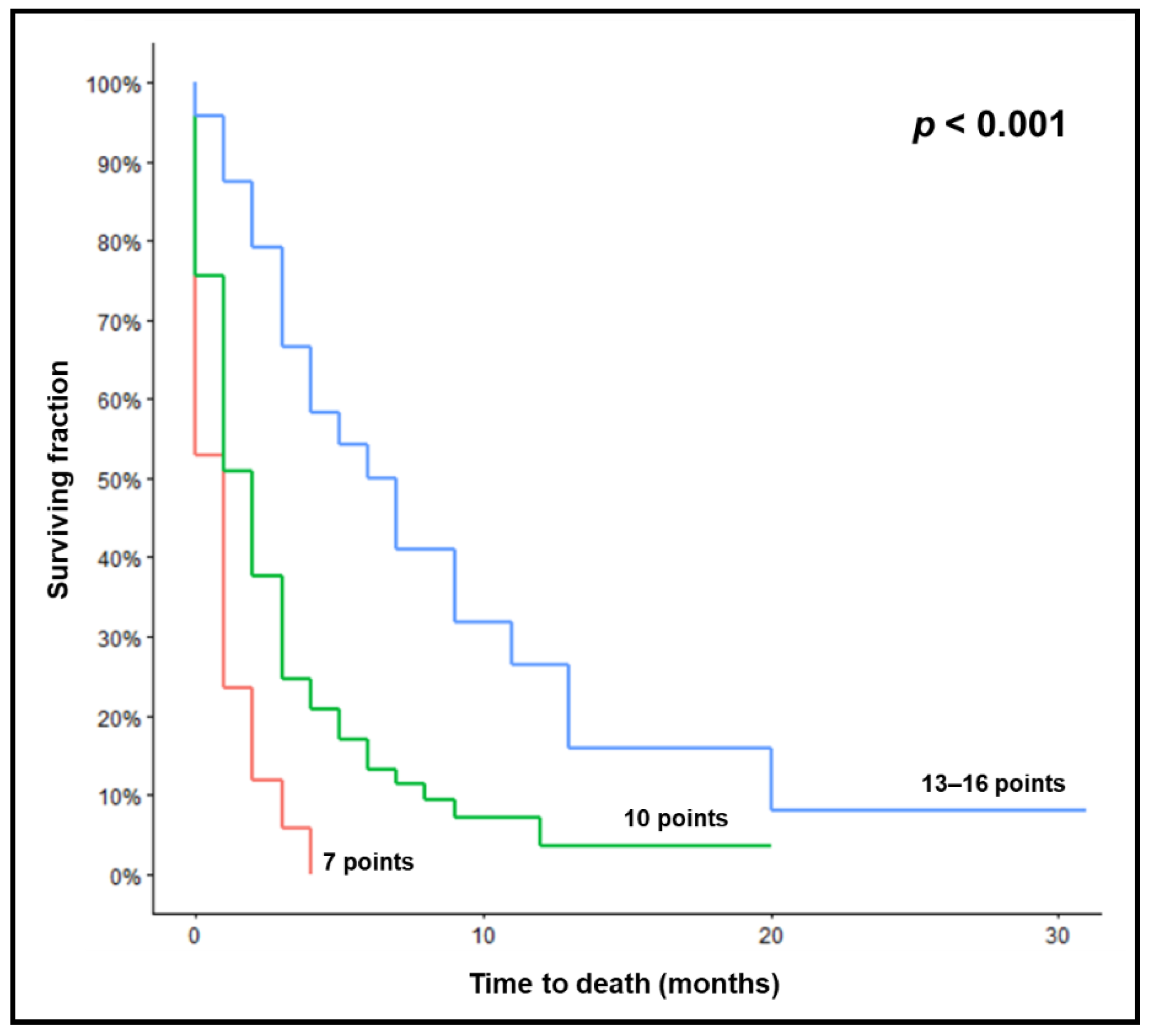
Figure 1. Kaplan–Meier curves for survival of the three prognostic groups (7, 10, and 13–16 points). p-value was calculated using the log-rank test.
Biology 11 01434 g001
Table 1. Characteristics evaluated for survival.
CharacteristicNumber of
Subjects
Proportion (%)
Age
80–84 years6872
85–90 years2628
Gender
Female3941
Male5559
ECOG performance score
0–13032
23638
32830
Tumor type
Breast cancer1011
Non-small cell lung cancer4245
Small-cell lung cancer910
Less radiosensitive tumors66
Cancer of unknown primary66
Gastrointestinal cancers 910
Other malignancies1213
Number of brain metastases
11819
2–32426
≥45255
Extra-cranial metastases
No2021
Yes7479
Interval tumor diagnosis to WBRT
≤12 months6569
>12 months2931
Treatment period (years)
2000–20124851
2013–20214649
WBRT regimen
20 Gy in 5 fractions1213
30 Gy in 10 fractions4043
>30 Gy in 11–20 fractions4245
ECOG: Eastern Cooperative Oncology Group; WBRT: whole-brain radiotherapy.
Table 2. Survival rates at 1, 2, 3, and 6 months following radiotherapy related to patient characteristics, treatment period, and dose-fractionation regimen.
CharacteristicSurvival Rates (%)p-Value
At 1 MonthAt 2 MonthsAt 3 MonthsAt 6 Months
Age 0.54
80–84 years50443522
85–90 years69422315
Gender 0.83
Female51463123
Male58423318
ECOG performance score 0.003
0–157504033
261534225
34625110
ECOG performance score <0.001
0–259524129
34625110
Tumor type 0.73
Breast cancer50404030
Non-small cell lung cancer55452924
Small-cell lung cancer44442211
Less radiosensitive tumors67503317
Cancer of unknown primary100836717
Gastrointestinal cancers44222211
Other malignancies50333317
Number of brain metastases 0.014
194785644
2–350382925
≥444352510
Extra-cranial metastases 0.034
No80655530
Yes49382618
Interval from tumor diagnosis to WBRT 0.11
≤12 months54422814
>12 months59484134
Treatment period (years) 0.29
2000–201263483521
2013–202148392820
Dose-fractionation regimen 0.66
20 Gy in 5 fractions8358508
30 Gy in 10 fractions55403020
>30 Gy in 11–20 fractions48432924
ECOG: Eastern Cooperative Oncology Group; WBRT: whole-brain radiotherapy.
Table 3. Survival rates at 3 months and scoring points.
CharacteristicSurvival Rate
at 3 Months (%)
Scoring
Points
ECOG performance score
0–2414
3111
Number of brain metastases
1566
2–3293
≥4253
Extra-cranial metastases
No556
Yes263
ECOG: Eastern Cooperative Oncology Group.
Table 4. Three prognostic groups and corresponding survival rates at 1, 2, 3, and 6 months.
Survival Rates (%)p-Value
At 1 MonthAt 2 MonthsAt 3 MonthsAt 6 Months
Prognostic group <0.001
7 points241260
10 points51382513
13–16 points88796750
Bold p-value is significant.
Table 5. Comparison of the new instrument and the previous Evers-Score [10] with respect to accuracy to predict death within 1, 2, 3, and 6 months (worst prognostic groups) and survival for at least 1, 2, 3, and 6 months (best prognostic groups).
EndpointPPVs of the New ScorePPVs of the Evers-Score
Death
within 1 month76%56%
within 2 months88%80%
within 3 months94%96%
within 6 months100%100%
Survival
for at least 1 month88%63%
for at least 2 months79%58%
for at least 3 months67%48%
for at least 6 months50%35%
PPVs: positive predictive values.
Publisher’s Note: MDPI stays neutral with regard to jurisdictional claims in published maps and institutional affiliations.