Article

Influence of Annealing Temperatures on Raman and Optical Absorption Spectra of TiO2 Nanorod Thin Film Coatings

1
School of Electronic Engineering, Huainan Normal University, Huainan 232038, China
2
School of Materials, Huaibei Normal University, Huaibei 235099, China
*
Author to whom correspondence should be addressed.
Coatings 2025, 15(11), 1338; https://doi.org/10.3390/coatings15111338
Submission received: 18 September 2025 / Revised: 7 November 2025 / Accepted: 10 November 2025 / Published: 18 November 2025
(This article belongs to the Section Thin Films)

Abstract

Titanium dioxide (TiO2) is an important semiconductor material widely used in both fundamental studies and technological applications. Herein, TiO2 nanorod thin film coatings were fabricated on transparent conductive fluorine-doped tin oxide (FTO) substrates using a hydrothermal synthesis approach, followed by annealing at various temperatures. The effects of annealing temperatures on the Raman and optical absorption spectra were systematically investigated to elucidate the behavior of Raman-active lattice vibrations and optical transitions. As the annealing temperatures increased, both the full width at half maximum of the Raman vibrational modes and the band gap of the TiO2 nanorod thin films decreased. These trends indicate enhanced crystallinity and phonon lifetimes at higher annealing temperatures. The longer phonon lifetimes contribute to reduced electron–hole recombination, while the narrower band gap extends the optical absorption range into the visible region. This study provides valuable insights into the relationship between annealing temperatures and the structural, vibrational and optical properties of rutile TiO2 nanorod thin film coatings, highlighting their potential for improved performance in photoelectrocatalytic and optoelectronic applications.

1. Introduction

Metal oxides are widely employed as photoelectrocatalytic materials for water splitting and pollutant degradation, with notable examples including ZnO, RuO2, and TiO2 [1,2,3]. Among transition metal oxides, TiO2 stands out as one of the most significant photocatalytic materials [1,3]. Its extensive study and application over the past few decades can be attributed to its unique optical, structural, and electrical properties, such as excellent chemical and thermal stability, corrosion resistance, and remarkable catalytic activity [4]. TiO2 has been extensively investigated as a key photocatalyst and semiconductor material in environmental purification and solar-driven hydrogen generation [1,3,5,6,7]. However, TiO2 is a kind of wide-band-gap semiconductor material (3.0–3.2 eV) that primarily absorbs ultraviolet light, which constitutes <5% of the solar spectrum [6,8,9]. To improve its photocatalytic efficiency under solar irradiation, numerous strategies have been explored [10,11,12], including surface morphology modification [13,14,15], coupling with other phases or compounds [16,17,18,19,20,21,22], and elemental doping [23,24,25,26,27]. In addition to these approaches, thermal annealing is a crucial post-synthesis treatment for optimizing the electronic and structural properties of TiO2. Annealing can influence its Raman spectral features, crystallinity, and photoelectrochemical activity, thereby enhancing its solar spectrum utilization [28,29,30,31]. A comprehensive understanding of how annealing affects the physical and optical characteristics of rutile TiO2 nanorod thin film coatings is crucial for elucidating their synergistic effects and further improving photocatalytic performance.
Post-annealed rutile TiO2 represents the most stable and commonly observed crystal structure among its three polymorphic forms: anatase, rutile, and brookite [31]. However, thermal treatment can significantly influence the electronic, optical, and structural properties of rutile TiO2, as demonstrated in numerous studies. Mathpal et al. [30] investigated the effect of annealing temperatures on the Raman spectral data of TiO2 nanoparticles. Hou et al. [29] revealed the distinctive photoelectrochemical behavior of annealed TiO2 nanorod arrays, highlighting the critical role of post-annealing in modifying material properties. Khan et al. [31] further reported the structural and optoelectronic evolution of nanostructured TiO2 thin films upon annealing. Moreover, asymmetric lattice vibrations in rutile TiO2 have been shown to exhibit reversible trends with varying laser powers [10]. Temperature-dependent Raman shifts and symmetric broadening are often attributed to thermal expansion and anharmonic phonon–phonon coupling [10].
Herein, the impact of annealing temperatures on the Raman and absorption spectra of TiO2 nanorod thin film coatings prepared by the hydrothermal approach is systematically investigated. Four Raman-active vibrational modes of post-annealed rutile TiO2 nanorod thin films were analyzed. The results reveal that a high annealing temperature decreases both the optical band gap and the full width at half maximum (FWHM) of the Raman modes. The reduction in the band gap enhances the material’s ability to harvest solar energy, while the narrowing of the FWHM is attributed to suppressed phonon broadening resulting from longer phonon lifetimes [32]. Extended phonon lifetimes strengthen phonon–electron interactions, facilitating the formation and separation of photoexcited charge carriers during photoelectrochemical processes. These findings contribute to a deeper understanding of the physical mechanisms regulating the photocatalytic performance of annealed rutile TiO2 nanorod thin films.

2. Experimental Section

2.1. Synthesis of TiO2 Nanorod Thin Film Coatings

TiO2 nanorod thin film coatings were prepared via a hydrothermal approach. First, deionized water (30 mL) was mixed with concentrated hydrochloric acid (36–38% wt, 30 mL) in a beaker under ambient conditions and stirred for 10 min, in which the hydrogen ion concentration is approximately 6 mol/L at room temperature. Subsequently, titanium butoxide (TBOT, ≥99.0%, 1 mL) was slowly added dropwise to the solution, followed by continuous stirring for another 10 min to ensure homogeneity. Three fluorine-doped tin oxide (FTO) glass substrates were then placed at an angle inside a 100 mL Teflon-lined stainless-steel autoclave, with the conductive sides facing inward and the shorter edges in contact with the bottom of the liner. The prepared precursor solution was transferred into the autoclave, which was sealed and heated at 150 °C for 12 h in a drying oven. The resulting TiO2-coated FTO substrates were thoroughly rinsed with deionized water and dried at ambient temperature. Further details of the synthesis process can be found in [26].

2.2. Annealing Treatment

Five sample groups were prepared: pristine (unannealed) and annealed at 150, 300, 450, and 600 °C in air, respectively. Each annealing process was conducted for 2 h under controlled atmospheric conditions. Slight variations in the actual sample temperature may have occurred while characterizing the Raman spectra.

2.3. Characterization

The surface morphology and microstructure of the as-prepared and annealed TiO2 nanorod films were analyzed using Raman imaging coupled to field emission scanning electron microscope (FE-SEM, TESCAN-MAIA/WITEC MAIA3 GMU, Czech Republic) operated at 5 kV. The crystalline structure was examined by X-ray diffraction (XRD, D8 ADVANCE, Bruker, Germany) using Cu-Kα radiation (λ = 0.15406 nm) at 40 mA and 40 kV, with a scanning range of 20–80° and a step size of 0.02°. Characterization of optical properties was performed using a UV-Vis spectrophotometer (Lambda 950, PerkinElmer, USA). Raman spectral data were obtained using a confocal microscopic Raman spectrometer (Renishaw, UK) with a 532 nm excitation laser at 25 mW output power and a 10 s acquisition time for each sample. All measurements were performed at room temperature.

3. Results and Discussion

3.1. Surface Topography

The SEM morphologies of TiO2 nanorod thin film coatings annealed at various temperatures are shown in Figure 1. Figure 1a corresponds to the as-prepared (unannealed) TiO2 sample, while Figure 1b–d represent samples annealed at 150, 450, and 600 °C for 2 h in air, respectively. The insets present low-magnification SEM images for a broader view of the surface morphology. The specimen annealed at 300 °C is not displayed due to its strong morphology similarity to that annealed at 150 °C. Overall, the TiO2 nanorod arrays are uniformly distributed, vertically aligned, and densely packed across the whole surface of FTO substrates. The nanorods exhibit nearly square cross-sections in the top-view images, with smooth and well-defined sidewalls [29]. These structural characteristics are consistently observed in all samples. At lower annealing temperatures (Figure 1a,b), numerous protrusions or apex-like features appear at the tips of the nanorods. As the annealing temperature increases (Figure 1c,d), these apexes gradually diminish, and the rod tops become progressively smoother. At 600 °C, the nanorod tips exhibit a distinctly flattened morphology, indicating a thermal smoothing effect. This morphological evolution can be attributed to thermally induced surface diffusion and densification processes, leading to the compaction or slight shrinkage of the nanorods with increasing annealing temperatures [29,33]. Furthermore, the mean diameter of the square column nanorods post-annealing is slightly smaller than that of the as-prepared sample, and the inter-rod spacing appears to increase. These changes in nanorod geometry and surface compactness are expected to influence both the optical absorption and photocatalytic behavior of the TiO2 nanorod thin film coatings by altering light-scattering characteristics and charge transport pathways.

3.2. XRD Characterization

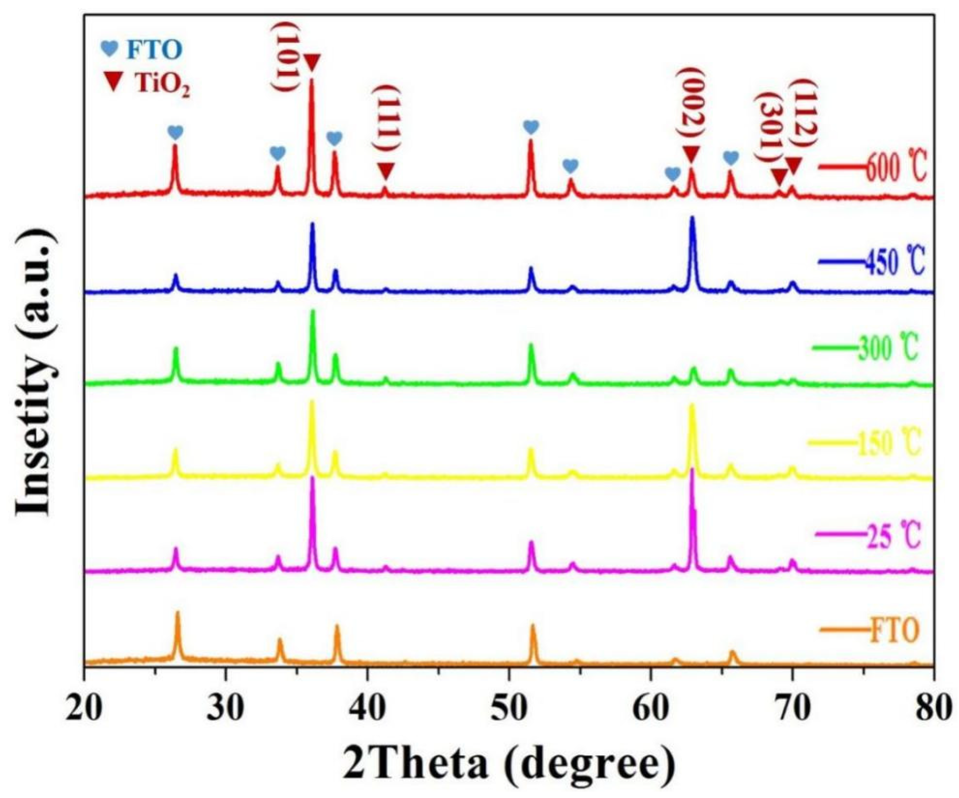
The XRD patterns of all TiO2 samples, along with the blank FTO substrate, are presented in Figure 2. The diffraction peaks corresponding to the FTO substrates are identified according to JCPDS card NO. 41-1445 and are indicated by an orange-yellow line and heart-shaped symbols in the figure. Both the pristine and annealed TiO2 nanorod films exhibit a rutile crystal structure (JCPDS No. 21-1276; space group P42/mnm (136); lattice parameters a = b = 0.4593 nm, and c = 0.2595 nm), in agreement with previous reports [34,35]. The characteristic diffraction peaks, excluding those from FTO substrates, appear at 2θ values of 36.16°, 41.36°, 62.30°, 69.36°, and 70.21°, which correspond to the (101), (111), (002), (301), and (112) planes of the rutile TiO2, respectively. The XRD patterns of all samples, both unannealed and annealed, show identical peak positions, indicating that the rutile phase remains stable throughout the annealing process. This confirms that no phase transformation or lattice distortion occurs under the applied thermal conditions. However, the intensity of the diffraction peaks, particularly those at 36.16° and 41.36°, increases slightly with higher annealing temperatures. This enhancement suggests improved crystallinity and a subtle textural reorientation of the TiO2 nanorods, which may influence their Raman and optical absorption properties. Overall, the results demonstrate that the TiO2 nanorod thin film coatings maintain a robust rutile monocrystalline structure even after high-temperature annealing, which is consistent with previous findings [28,36]. These structural characteristics also correlate well with the SEM observations, further confirming the thermal stability and uniformity of the TiO2 nanorod arrays [29].

3.3. Optical Absorption Spectra

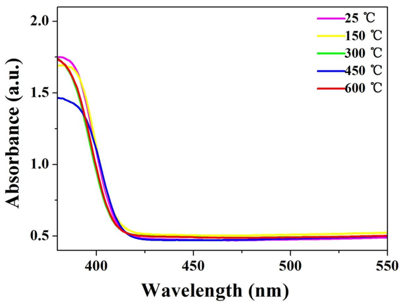
To examine the optical properties of TiO2 nanorod thin films at different temperatures, UV-Vis absorbance spectra were recorded, as shown in Figure 3. Overall, the absorbance spectra of all TiO2 nanorod thin film coatings exhibit similar profiles. The absorption onset occurs near 415 nm, corresponding to an electronic band gap of approximately 3.0 eV, consistent with the literature values for rutile TiO2 [1,5,6]. A sharp decrease in absorbance is observed around 400 nm. Notably, with an increasing annealing temperature, the intrinsic absorption edge shows a gradual redshift (Figure 3), indicating enhanced visible light absorption. This shift is due to the reduced lattice spacing and improved crystallinity induced by the annealing process, which leads to a slight narrowing of the band gap in the TiO2 nanorod arrays. These structural modifications, in line with the SEM analysis, are expected to enhance the optical response and photocatalytic efficiency of the films, particularly for pollutant degradation and photoelectrochemical water-splitting applications.
The corresponding Tauc plots for the TiO2 nanorod thin film coatings are displayed in Figure 4. For crystalline semiconductor thin films, the relationship between the photon energy (hν) and the absorbance (α) can be expressed by the following function [37,38,39]:
(αhν)2 = A(hν − Eg)
where α, h, ν, A, and Eg are the absorbance, Planck’s constant (h = 6.626 × 10⁻³⁴ J·s), photon frequency, proportionality constant, and Eg for the energy band gap, respectively. The estimated band gap of the pristine TiO2 nanorod thin film coating is approximately 3.06 eV, which is relatively consistent with previously reported values [40]. The band gap of the TiO2 coating annealed at 450 °C is about 3.01 eV, which is slightly narrower than that of the pristine TiO2 coating. However, the band gap of the sample annealed at 600 °C in air becomes wider, which can be changed in its internal structure. Although the difference between not annealing and annealing is not very remarkable, it is considered that the annealing treatment may reduce the energy gap and increase the generation rate of charge carriers when TiO2 nanorod array coatings are exposed to light. This can improve the photoelectric and catalytic performances of the TiO2 nanorod film coating semiconductor material. The observed optical evolution is in good agreement with the previously discussed SEM results.

3.4. Raman Spectra

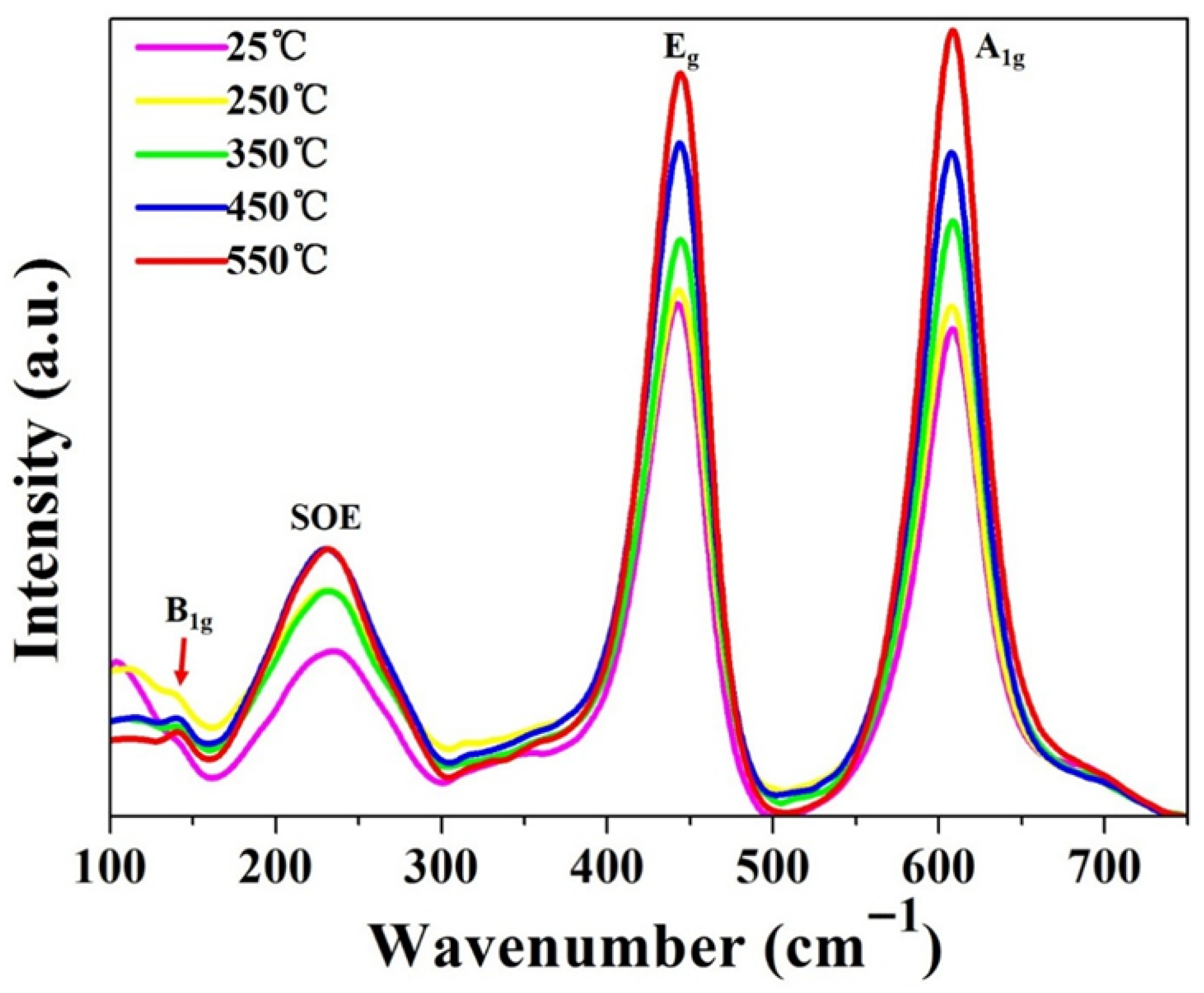
Raman spectroscopy is a promising technique for probing the structural and vibrational properties of TiO2 semiconductor materials, particularly the rutile phase. The Raman spectra of TiO2 nanorod thin film coatings grown hydrothermally on FTO substrates and subsequently annealed at different temperatures in air are presented in Figure 5. Rutile TiO2, which crystallizes in the tetragonal P4/mnm space group, exhibits 15 optical vibrational modes. Among these, four characteristic modes are typically observed, including B1g (145 cm−1), the second-order effect (240 cm−1), Eg (445 cm−1), and A1g (610 cm−1) [10,41]. The three first-order Raman-active modes (A1g + B1g + Eg) correspond to symmetric and asymmetric lattice vibrations of oxygen atoms within the TiO6 octahedra [10,42]. All TiO2 samples, both pristine and annealed, exhibit the same characteristic Raman peaks, confirming that the rutile crystal structure is maintained during annealing. However, several notable trends are observed with increasing annealing temperatures. First, the Raman peak intensities increase progressively, with the unannealed sample showing the weakest signal and the 600 °C-annealed sample showing the strongest. The peak positions remain nearly unchanged, indicating the structural stability of the rutile phase. The enhanced intensity is attributed to improved crystallinity and a reduced defect concentration, consistent with the XRD analysis. Second, the FWHM of each Raman mode narrows as the annealing temperature increases. This narrowing reflects reduced phonon scattering and diminished disorder within the TiO2 lattice, corresponding to longer phonon lifetimes [32]. Extended phonon lifetimes imply slower vibrational relaxation and reduced phonon broadening, suggesting improved lattice coherence.
From a physical perspective, longer phonon lifetimes enhance the coupling between phonons and conduction band electrons, effectively reducing electron–hole recombination rates. This improved charge separation can enhance photocatalytic efficiency in processes such as pollutant degradation and photoelectrochemical water splitting. Overall, the Raman results demonstrate that annealing improves the structural order and phonon dynamics of rutile TiO2 nanorod thin films without altering their crystal phase. These findings are consistent with the SEM and UV-Vis analyses, collectively confirming that controlled annealing strengthens the optical and vibrational properties of the TiO2 nanorod arrays, thereby promoting their photocatalytic performance.

4. Conclusions

Herein, rutile TiO2 nanorod arrays were successfully synthesized on FTO substrates using a one-step hydrothermal approach, followed by annealing in air at various temperatures (range: 150–600 °C) for 2 h. The effects of annealing temperatures on the surface morphology, crystal structure, optical absorption spectra, band gap energy, and Raman spectroscopic features of the TiO2 nanorod films were systematically investigated. The results demonstrate that annealing enhances the optical absorption of TiO2 by extending the absorption edge toward longer wavelengths and slightly narrowing the band gap, thereby improving solar light utilization. The Raman spectra show that while the peak positions remain unchanged, confirming the preservation of the rutile phase, the overall Raman intensity increases with the temperature, indicating improved crystallinity. Furthermore, the FWHM of the Raman-active modes decreases as the annealing temperature increases, suggesting reduced phonon scattering and longer phonon lifetimes. Extended phonon lifetimes promote stronger phonon–electron coupling and suppress electron–hole recombination, which is beneficial for enhancing photocatalytic efficiency. These findings provide deeper insight into the intrinsic relationship between thermal treatments and the optical–vibrational behavior of rutile TiO2 nanorod thin films, offering valuable guidance for optimizing their performance in photocatalytic and photoelectrochemical applications such as water splitting and pollutant degradation.

Author Contributions

Conceptualization, S.C. and H.L.; Methodology, S.C.; Resources, S.C.; Data curation, S.C. and H.L.; Writing—original draft, S.C. All authors have read and agreed to the published version of the manuscript.

Funding

This research was funded by Key Project of the Education Department of Anhui Province grant number [2024AH051749].

Institutional Review Board Statement

Not applicable.

Informed Consent Statement

Not applicable.

Data Availability Statement

Data is contained within the article.

Conflicts of Interest

The authors declare no conflicts of interest.

References

  1. Fujishima, A.; Honda, K. Electrochemical photolysis of water at a semiconductor electrode. Nature 1972, 238, 37–38. [Google Scholar] [CrossRef]
  2. Li, C.L.; Chen, S.R.; Gao, X.X.; Zhang, W.; Wang, Y.F. Fabrication, characterization and photoelectrochemical properties of CdS/CdSe nanofilm co-sensitized ZnO nanorod arrays on Zn foil substrate. J. Colloid Interface Sci. 2021, 588, 269–282. [Google Scholar] [CrossRef]
  3. Yao, Y.H.; Tang, Y.; Zhou, Y.L.; Cui, D.; Wang, P.P.; Luo, D.J.; Wang, Q.; Tian, B.; Pan, K.K.; Yu, F. Visible light-driven high efficient water hydrogen evolution using Ru-RuO2/TiO2 catalyst. Int. J. Hydrogen Energy 2025, 145, 473–484. [Google Scholar] [CrossRef]
  4. Murugesam, S.; Kuppusami, P.; Mohandas, E. Rietveld X-ray diffraction analysis of nanostructured rutile films of titania prepared by pulsed laser deposition. Mater. Res. Bull. 2010, 45, 6–9. [Google Scholar] [CrossRef]
  5. Chen, X.; Mao, S.S. Titanium dioxide nanomaterials: Synthesis, properties, modifications, and applications. Chem. Rev. 2007, 107, 2891–2959. [Google Scholar] [CrossRef] [PubMed]
  6. Chen, X.; Liu, L.; Yu, P.Y.; Mao, S.S. Increasing solar absorption for photocatalysis with black hydrogenated titanium dioxide nanocrystals. Science 2011, 331, 746–750. [Google Scholar] [CrossRef]
  7. Tuntithavornwat, S.; Saisawang, C.; Ratvijitvech, T.; Watthanaphanit, A.; Hunsom, M.; Kannan, M.A. Recent development of black TiO2 nanoparticles for photocatalytic H2 production: An extensive review. Int. J. Hydrogen Energy 2024, 55, 1559–1593. [Google Scholar] [CrossRef]
  8. Zhang, H.; Besteiro, L.V.; Liu, L.; Wang, C.; Selopal, G.S.; Chen, Z.; Barba, D.; Wang, Z.M.; Zhao, H.; Lopinski, G.P. Efficient and stable photoelectrochemical hydrogen generation using optimized colloidal heterostructured quantum dots. Nano Energy 2021, 79, 105416. [Google Scholar] [CrossRef]
  9. Cheng, X.F.; Leng, W.H.; Liu, D.H.; Zhan, J.Q.; Cao, C.N. Enhanced photoelectrocatalytic performance of Zn-doped WO3 photocatalysts for nitrite ions degradation under visible light. Chemosphere 2007, 68, 1976–1984. [Google Scholar] [CrossRef] [PubMed]
  10. Zhang, Y.L.; Harris, C.X.; Wallenmeyer, P.; James, M.; Chen, X.B. Asymmetric lattice vibrational characteristics of rutile TiO2 as revealed by laser power dependent Raman spectroscopy. J. Phy. Chem. C 2013, 117, 24015–24022. [Google Scholar] [CrossRef]
  11. Ghorai, N.; Yang, Z.C.; Gebre, S.T.; Wu, S.X.; Zhao, F.Y.; Ivanov, I.N.; Lian, T.Q. Efficient size-dependent hot electron transfer from Au to TiO2 nanoparticles. Nano Lett. 2025, 25, 3253–3258. [Google Scholar] [CrossRef] [PubMed]
  12. Chen, Y.F.; Li, C.Y.; Campos, R.F.; Esplandiu, M.J.; Fraxedas, J.; Liguori, N.; Villa, K. Unraveling charge transport in heterostructured nanomotors for efficient photocatalytic motion. Nano Lett. 2025, 25, 10169–10177. [Google Scholar] [CrossRef] [PubMed]
  13. Wolcott, A.; Smith, W.A.; Kuykendall, T.R.; Zhao, Y.; Zhang, J.Z. Photoelectrochemical water splitting using dense and aligned TiO2 nanorod arrays. Small 2009, 5, 104–111. [Google Scholar] [CrossRef]
  14. Yao, H.; Wu, Y.F.; Yang, H.; Ma, J.; Sun, M.; Chen, Y.; Zhang, W.; Di, W.; Lv, P.; Li, M. Vertical growth of two-dimensional TiO2 nanosheets array films and enhanced photoelectrochemical properties sensitized by CdS quantum dots. Electrochim. Acta 2014, 125, 258–265. [Google Scholar] [CrossRef]
  15. Cho, I.S.; Choi, J.; Zhang, K.; Kim, S.J.; Jeong, M.J.; Cai, L.; Park, T.; Zheng, X.; Park, J.H. Highly efficient solar water splitting from transferred TiO2 nanotube arrays. Nano Lett. 2015, 15, 5709–5715. [Google Scholar] [CrossRef]
  16. Sreedhar, A.; Jung, H.; Kwon, J.H.; Yi, J.; Sohn, Y.; Gwag, J.S. Novel composite ZnO/TiO2 thin film photoanodes for enhanced visible-light-driven photoelectrochemical water splitting activity. J. Electroanal. Chem. 2017, 804, 92–98. [Google Scholar] [CrossRef]
  17. Liu, C.; Yang, Y.; Li, J.; Chen, S. Phase transformation synthesis of TiO2/CdS heterojunction film with high visible light photoelectrochemical activity. Nanotechnology 2018, 29, 265401. [Google Scholar] [CrossRef]
  18. Chen, S.R.; Li, C.L.; Hou, Z.Y. A novel in situ synthesis of TiO2/CdS heterojunction for improving photoelectrochemical water splitting. Int. J. Hydrogen Energy 2019, 44, 25473–25485. [Google Scholar] [CrossRef]
  19. Yu, H.J.; Zhao, Y.; Chao, Z.L.; Shang, Y.; Cao, Y.; Li, Z.W.; Tung, C.H.; Zhang, T. Carbon quantum dots/TiO2 composites for efficient photocatalytic hydrogen evolution. J. Mater. Chem. A 2014, 2, 3344–3351. [Google Scholar] [CrossRef]
  20. Zhang, H.; Zhang, D.; Qin, X.; Cheng, C. Three-dimensional CdS-sensitized sea urchin like TiO2-ordered arrays as efficient photoelectrochemical anodes. J. Phys. Chem. C 2015, 119, 27875–27881. [Google Scholar] [CrossRef]
  21. Mehta, M.; Singh, A.P.; Kumar, S.; Krishnamurthy, S.; Wickman, B.; Basu, S. Synthesis of MoS2-TiO2 nanocomposite for enhanced photocatalytic and photoelectrochemical performance under visible light irradiation. Vacuum 2018, 155, 675–681. [Google Scholar] [CrossRef]
  22. Naidu, P.Y.; Kumar, M.; Meena, B.; Challapalli, S. Synergistic integration of TiO2/Mn:CdS/CZTS ternary heterojunction photoanode for superior solar-driven hydrogen evolution. Int. J. Hydrogen Energy 2025, 166, 151000. [Google Scholar] [CrossRef]
  23. Yuan, J.; Chen, M.; Shi, J.; Shang, G.W. Preparations and photocatalytic hydrogen evolution of N-doped TiO2 from urea and titanium tetrachloride. Int. J. Hydrogen Energy 2006, 31, 1326–1331. [Google Scholar] [CrossRef]
  24. Asahi, R.; Morikawa, T.; Ohwaki, T.; Aoki, K.; Taga, Y. Visible-light photocatalysis in nitrogen-doped titanium oxides. Science 2001, 293, 269–271. [Google Scholar] [CrossRef] [PubMed]
  25. Hong, X.; Wang, Z.; Cai, W.; Feng, L.; Zhang, J.; Yang, Y.; Ma, N.; Liu, Y. Visible-light-activated nanoparticle photocatalyst of iodine-doped titanium dioxide. Chem. Mater. 2005, 17, 1548–1552. [Google Scholar] [CrossRef]
  26. Ming, X.; Da, P.; Hao, Y.W.; Zhao, D.; Zheng, G. Controlled Sn-doping in TiO2 nanowire photoanodes with enhanced photoelectrochemical conversion. Nano Lett. 2012, 12, 1503–1508. [Google Scholar]
  27. Cho, I.S.; Lee, C.H.; Feng, Y.; Logar, M.; Rao, P.M.; Cai, L.; Kim, D.R.; Sinclair, R.; Zheng, X. Co-doping titanium dioxide nanowires with tungsten and carbon for enhanced photoelectrochemical performance. Nat. Commun. 2013, 4, 1723. [Google Scholar] [CrossRef]
  28. Zhang, S.; Gu, X.; Zhao, Y.; Qiang, Y. Enhanced photoelectrochemical performance of TiO2 nanorod arrays by a 500 °C annealing in air: Insights into the mechanism. J. Electron. Mater. 2016, 45, 648–653. [Google Scholar] [CrossRef]
  29. Chen, S.R.; Li, C.L.; Hou, Z.Y. The novel behavior of photoelectrochemical property of annealing TiO2 nanorod arrays. J. Mater. Sci. 2020, 55, 5969–5981. [Google Scholar] [CrossRef]
  30. Mohan, C.M.; Anand, K.T.; Manish, K.S.; Gairola, S.P.; Pandey, S.N.; Arvind, A. Effect of annealing temperature on Raman spectra of TiO2 nanoparticles. Chem. Phys. Lett. 2013, 555, 182–186. [Google Scholar] [CrossRef]
  31. Khan, A.F.; Mehmood, M.; Durrani, S.K.; Ali, M.L.; Rahim, N.A. Structural and optoelectronic properties of nanostructured TiO2 thin films with annealing. Mat. Sci. Semicon. Proc. 2015, 29, 161–169. [Google Scholar] [CrossRef]
  32. Lan, T.; Tang, X.; Fultz, B. Phonon anharmonicity of rutile TiO2 studied by Raman spectrometry and molecular dynamics simulations. Phys. Rev. B 2012, 85, 094305. [Google Scholar] [CrossRef]
  33. Xie, Z.; Shuang, S.; Ma, L.W.; Zhu, F.; Liu, X.X.; Zhang, Z.J. Annealing effect on the photoelectrochemical and photocatalytic performance of TiO2 nanorod arrays. RSC Adv. 2017, 7, 51382–51390. [Google Scholar] [CrossRef]
  34. Feng, W.J.; Lin, L.Y.; Li, H.J.; Chi, B.; Pu, J.; Li, J. Hydrogenated TiO2/ZnO heterojunction nanorod arrays with enhanced performance for photoelectrochemical water splitting. Int. J. Hydrogen Energy 2017, 42, 3938–3946. [Google Scholar] [CrossRef]
  35. Jaramillo-Quintero, O.A.; Triana, M.A.; Rincon, M.E. Optimization of charge transfer and transport processes at the CdSe quantum dots/TiO2 nanorod interface by TiO2 interlayer passivation. J. Phys. D Appl. Phys. 2017, 50, 235305. [Google Scholar] [CrossRef]
  36. Fàbrega, C.; Andreu, T.; Tarancón, A.; Flox, C.; Morata, A.; Calvo-Barrio, L.; Morante, J.R. Optimization of surface charge transfer processes on rutile TiO2 nanorods photoanodes for water splitting. Int. J. Hydrogen Energy 2013, 38, 2979–2985. [Google Scholar] [CrossRef]
  37. Liu, Y.; Jiang, J.G.; Xu, Q.; Li, M.T.; Guo, L.J. Photoelectrochemical performance of CdS nanorods grafted vertically aligned TiO2 nanorods. Mater. Res. Bull. 2013, 48, 4548–4554. [Google Scholar] [CrossRef]
  38. Cheng, C.; Wang, H.S.; Li, J.C.; Yang, H.; Xie, A.J.; Chen, P.; Li, S.K.; Huang, F.Z.; Shen, Y.H. Ordered macroporous CdS-sensitized N-doped TiO2 inverse opals films with enhanced photoelectrochemical performance. Electrochim. Acta 2014, 146, 378–385. [Google Scholar] [CrossRef]
  39. Teng, W.; Wang, Y.M.; Huang, H.H.; Li, X.Y.; Tang, Y.B. Enhanced photoelectrochemical performance of MoS2 nanobelts-loaded TiO2 nanotube arrays by photo-assisted electrodeposition. Appl. Surf. Sci. 2017, 425, 507–517. [Google Scholar] [CrossRef]
  40. Liu, B.K.; Wang, D.J.; Wang, L.L.; Sun, Y.J.; Lin, Y.H.; Zhang, X.Q.; Xie, T.F. Glutathione-assisted hydrothermal synthesis of CdS-decorated TiO2 nanorod arrays for quantum dot-sensitized solar cells. Electrochim. Acta 2013, 113, 661–667. [Google Scholar] [CrossRef]
  41. Wu, P.C.; Song, X.Y.; Si, S.Y.; Ke, Z.J.; Cheng, L.; Li, W.Q.; Xiao, X.H.; Jiang, C.Z. Significantly enhanced visible light response in single TiO2 nanowire by nitrogen ion implantation. Nanotechnology 2018, 29, 184005. [Google Scholar] [CrossRef] [PubMed]
  42. Ahmad, M.K.; Mokhtar, S.M.; Soon, C.F.; Nafarizal, N. Raman investigation of rutile-phased TiO2 nanorods/nanoflowers with various reaction times using one step hydrothermal method. J. Mater. Sci. Mater. Electron. 2016, 27, 7920–7926. [Google Scholar] [CrossRef]
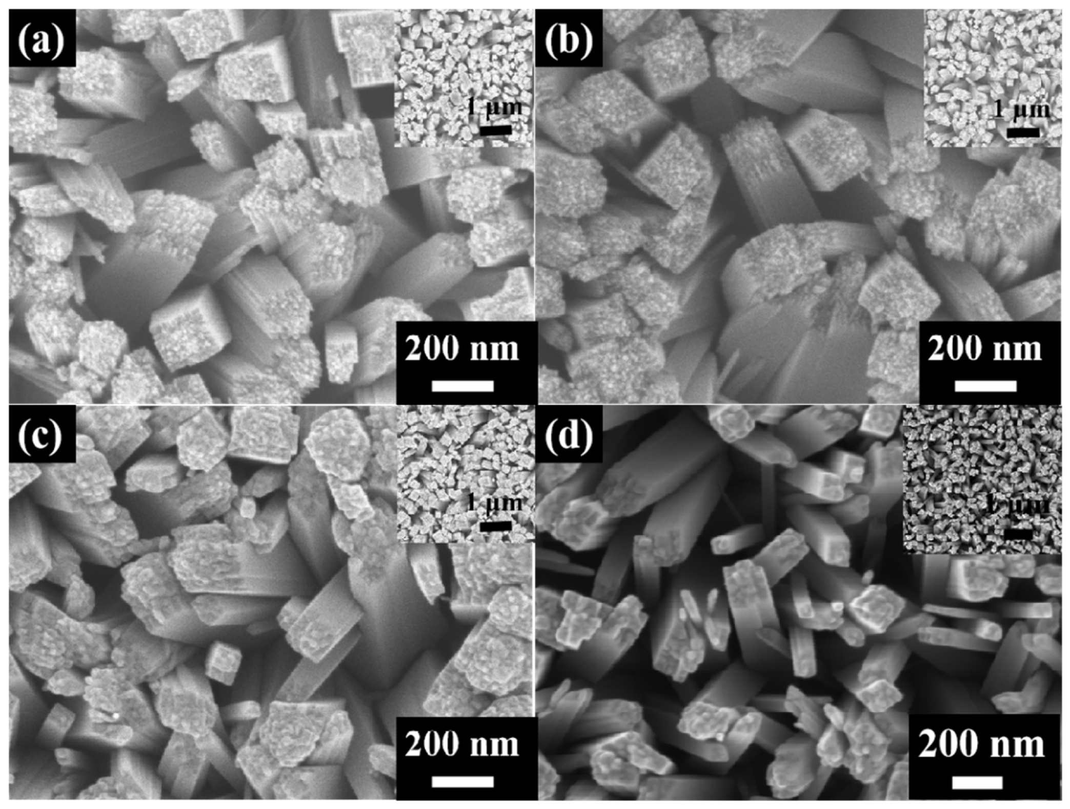
Figure 1. The SEM topographies of the original and annealing TiO2 specimens: (a) for pristine, (b) for 150 °C, (c) for 450 °C, and (d) for 600 °C; the insets are low-magnification SEM morphologies.
Coatings 15 01338 g001
Figure 2. XRD patterns of FTO, the original, and various annealed samples nanorod thin film coatings.
Coatings 15 01338 g002
Figure 3. UV-Vis absorbance spectra of all the TiO2 nanorod thin film coatings.
Coatings 15 01338 g003
Figure 4. Energy gap of TiO2 samples.
Coatings 15 01338 g004
Figure 5. The Raman spectra of TiO2 nanorod thin film coatings.
Coatings 15 01338 g005
Disclaimer/Publisher’s Note: The statements, opinions and data contained in all publications are solely those of the individual author(s) and contributor(s) and not of MDPI and/or the editor(s). MDPI and/or the editor(s) disclaim responsibility for any injury to people or property resulting from any ideas, methods, instructions or products referred to in the content.