Article

An Investigation into Electrodeposited Co−Ni−TiO2 Films with Improved Mechanical and Corrosion Properties

School of Materials Science and Engineering, Jiangsu University of Science and Technology, Zhenjiang 212000, China
*
Author to whom correspondence should be addressed.
Coatings 2023, 13(4), 783; https://doi.org/10.3390/coatings13040783
Submission received: 28 February 2023 / Revised: 4 April 2023 / Accepted: 15 April 2023 / Published: 18 April 2023

Abstract

:
This investigation proposes the use of sol-enhanced electrodeposition to create a range of Co−Ni−TiO2 films. The addition of TiO2 sol controls the nucleation process and the properties of the composite films by generating TiO2 nanoparticles in situ in the electrodeposition process. The transmission electron microscopy (TEM) and zeta potential analyses revealed a relatively homogenous distribution with particle size in the range below 100 nm for the TiO2 nanoparticles produced. Microstructure, phase composition, hardness, friction, and corrosion resistance of Co−Ni−TiO2 films were thoroughly investigated in relation to TiO2 sol concentration. The results show that the addition of a limited content of TiO2 sol upgraded Co−Ni films by producing a Co−Ni−TiO2 film with a high dispersion of TiO2 nanoparticles. On the other hand, too much TiO2 sol could cause agglomeration and hinder the metal deposition process, resulting in surface pores and the deterioration of film performance.

1. Introduction

Due to their excellent mechanics, wear, corrosion, and other advantages, Co−Ni alloys are frequently used in protective coatings in various industries [1,2,3,4]. The main preparation techniques for cobalt-nickel coatings include electrodeposition, physical vapor deposition, chemical vapor deposition, and plasma spraying [5,6,7,8]. Electroplating technology has been regarded as a simple and efficient technique for preparing Co−Ni films [9,10]. The performance of a Co−Ni film cannot further satisfy the demands in some challenging service conditions, and it is of great scientific significance to further strengthen the Co−Ni film. Current methods for improving the performance of Co−Ni alloys include heat treatment, the pulse electrodeposition process, and composite electrodeposition technology [11,12,13]. Composite electrodeposition technology, one of the industry’s most widely employed reinforcement methods, can significantly enhance the Co−Ni alloy’s comprehensive properties [14,15].
Nanoparticles such as SiC [16], ZrO2 [17], TN [18], WC [19], TiO2 [20,21,22], and Al2O3 [23] have recently been utilized as the second reinforcing phase for composite electrodeposition. In traditional composite electrodeposition techniques, nanometer-sized inert particles are added to the electrolytes to create films. For instance, recent research by Rasooli et al. explored the influence of Cr2O3 nanoparticles on the Ni−Co−Cr2O3 coatings’ microstructure, mechanical properties, and corrosion-related properties, and found the incorporation of Cr2O3 nanoparticles could increase the nucleation amount of Co and improve the coating’s performance [24]. In another report, by examining the SiC-doped Ni−Co coating, Bakhit et al. stated that embedding SiC nanoparticles improved the Ni−Co film’s compactness and corrosion resistance [25].
However, nanoparticles with a high specific surface region and surface energy can easily result in agglomeration in a bath and greatly impair the performance of the resultant composite coating. To overcome this issue, our research group developed TiO2 sol-enhanced electrodeposition to reach nanoparticles’ excellent dispersion and chemical stability in electrolytes. The TiO2-incorporated composite coatings show improvements in mechanical and corrosion performance. When adding the TiO2 sol to the plating bath, TiO2 nanoparticles could be generated and directly co-deposited on the substrate’s surface with metal ions, effectively preventing nanoparticle agglomeration. The excellent dispersion of nanoparticles gives rise to a uniform, compact surface structure and an excellent coating performance.
Diverse Co−Ni−TiO2 films with superior performance were electrodeposited using TiO2 sol at various contents. Microstructure, phase composition, mechanical properties, and corrosion performance of Co−Ni−TiO2 films have been examined at diverse contents of TiO2 sols. The Co−Ni film’s mechanical properties and corrosion resistance can be greatly improved if TiO2 sol is present in a suitable amount, enhancing its utility in industrial use.

2. Experimental

2.1. TiO2 Sol Preparation

All reagents were purchased from Beijing Chemical Ltd. (Beijing, China) and used at analytical grade without further purification. The method of preparing TiO2 sol comprises the following three steps: (1) preparation of solution A: adding 5.64 mL of C4H11NO2 (diethanolamine) to 70 mL of C2H5OH (ethanol) with adequate stirring, followed by slowly dripping 19.74 mL of C16H36O4Ti (tetrabutyl titanate) into the as-prepared solution under a stirring speed of 300 rpm; (2) preparation of solution B: mix 9 mL of C2H5OH and 0.9 mL of distilled water; and (3) solution B was added dropwise into solution A, after which the bath was stirred 500 rpm for 30 min to produce TiO2 sol.
This study mixed 10 mL of TiO2 sol with 100 mL of di-ionized (DI) water under ultrasonic agitating, with the purpose of producing and determining the in situ created TiO2 nanoparticles. After the solution was dropped on a copper mesh and dried, the fresh nanoparticle could be observed.

2.2. Electrodeposition of Co−Ni−TiO2 Films

This experiment produced Co−Ni−TiO2 films in the electrodeposition process and used the brass sheet as a cathode and the high-purity cobalt board as an anode. To generate TiO2 nanoparticles in situ, TiO2 sol was slowly added to the Co−Ni electrolyte at magnetic stirring prior to electrodeposition. It should be noted that the attached organic chains in the freshly formed nanoparticles further avoid possible agglomerations.
The electrolytes used in the experiment consisted of 70 g/L NiSO4 (nickel sulfate), 50 g/L NiCl (nickel chloride), 60 g/L CoSO4 (cobalt sulfate), 50 g/L H3BO3 (boric acid), 0.015 g/L C12H25SO4Na (sodium dodecyl sulfate), and 0.15 g/L C7H5NO3S (saccharin). The electrodepositing temperature was 50 °C and the stirring rate was 300 rpm. Composite electrodeposition was performed at the current density of 25 mA/cm2 for 40 min with various concentrations of TiO2 sol.

2.3. Sample Characterization

This work used a transmitting electron microscope to characterize the newly generated nanoparticles (HRTEM, FEI Talos F200X G2, Thermo Fisher Scientific, Santa Clara, CA, USA). The samples’ surface morphology, cross-sectional morphology, and wear track morphology were observed using a Gemini SEM 300 electron microscope with an embedded EDS detector manufactured by Zeiss Company, Jena, Germany. A 90-plus zeta potential analyzer (Malvern Zetasizer Nano ZS, Malvern, UK) was employed to investigate the zeta potential of a composite plating solution in order to determine the dispersibility and stability of TiO2 nanoparticles. The phase constituents of the Co−Ni−TiO2 films were analyzed by Cu−Kα radiation (XRD, Bruker, Saarbrucken, Germany).
The Co−Ni−TiO2 film’s hardness was determined by a Vickers microhardness analyzer using 50 g for 15 s. The wear performance of the Co-Ni-TiO2 films was determined using a micro-tribometer (UMT−2, CETR, New York, NY, USA). In the wear tests, steel balls 4 mm in diameter and a load of 5 N were used under a wear speed of 10 mm/s.
With the aid of an electrochemical workstation (CS2350, Corrtest, Wuhan, China), the Co−Ni−TiO2 films’ electrochemical performance was evaluated. The platinum plate with dimensions of 10 mm × 10 mm × 0.3 mm served as the counter electrode, while a saturated KCl electrode served as the reference electrode. When the open circuit potential had been stabilized, the samples were tested at room temperature at a scanning speed of 1 mV/s using 3.5 wt.% NaCl solution.

3. Results and Discussion

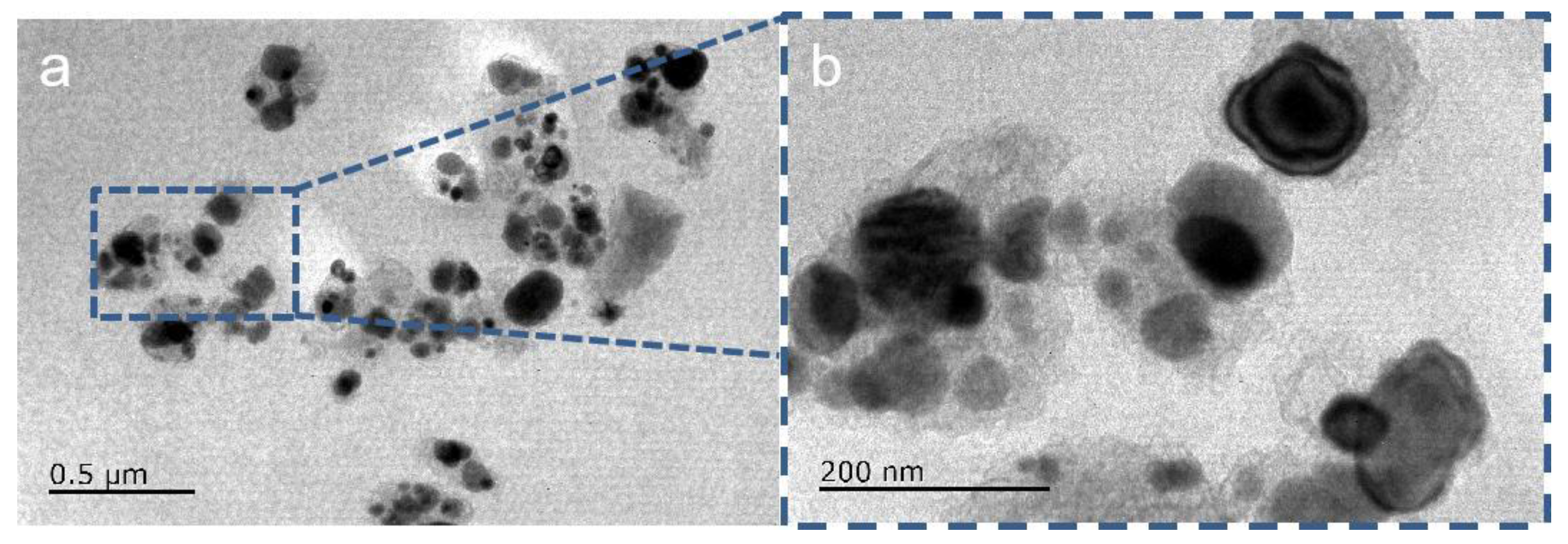
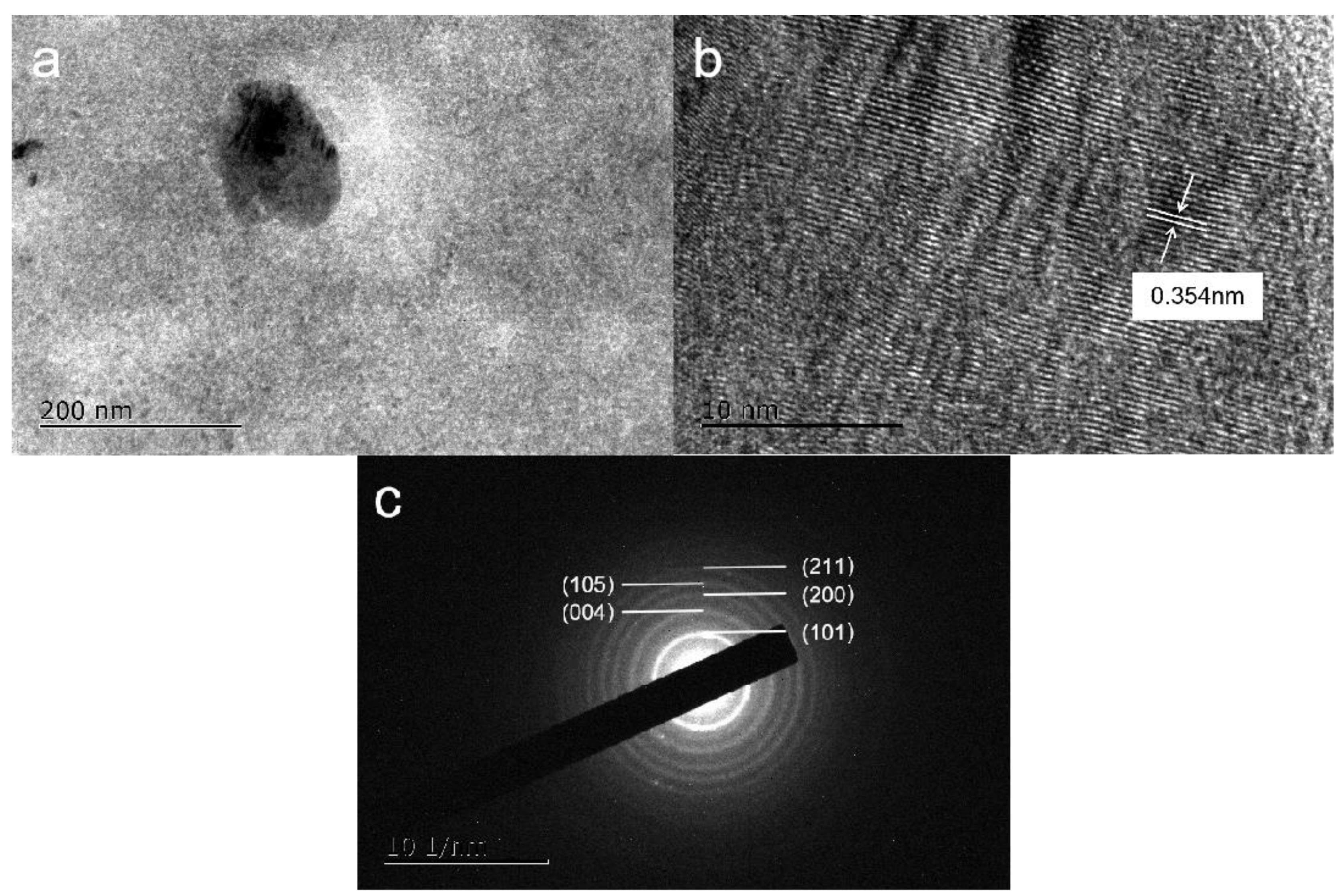
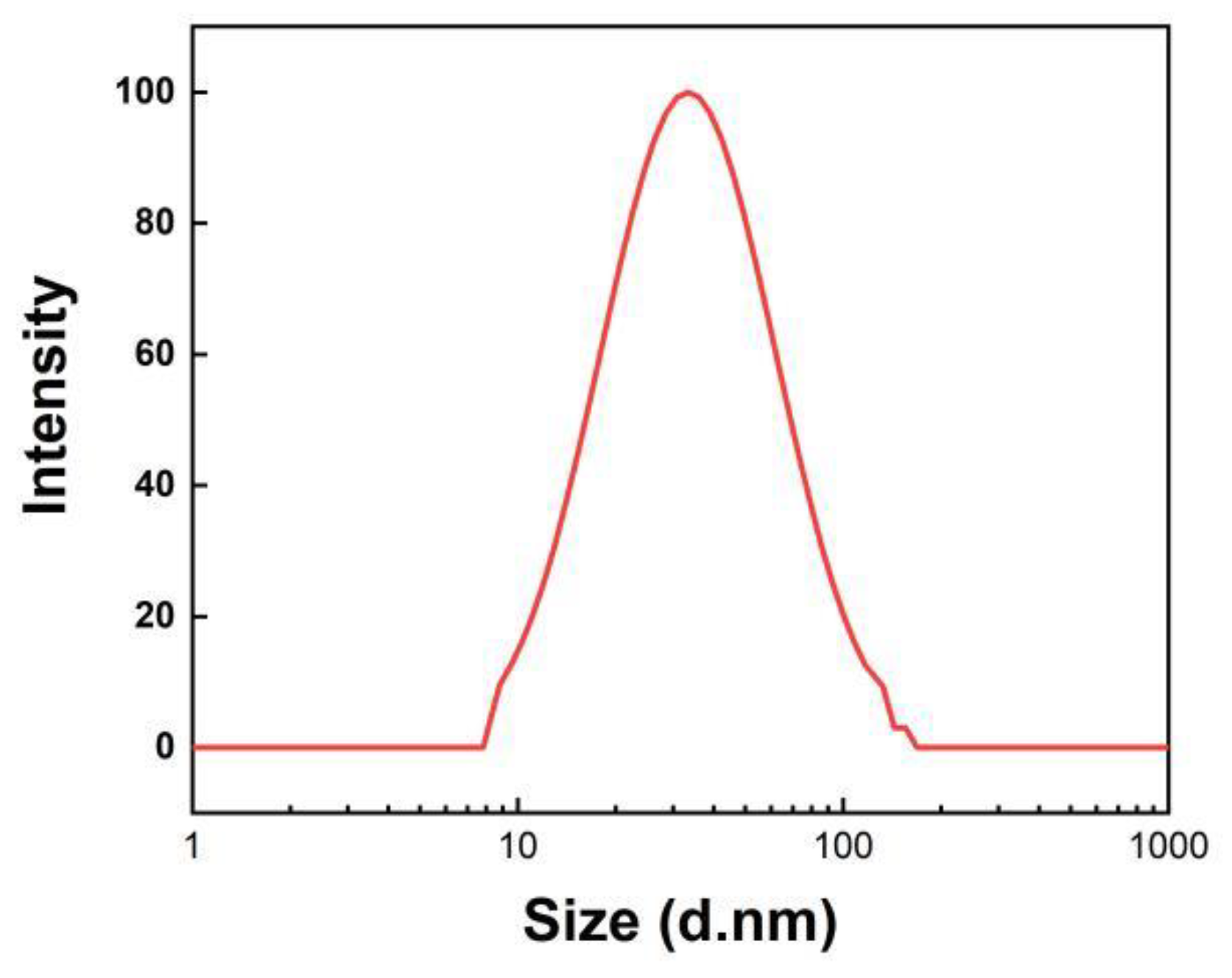
3.1. Characterization of TiO2 Sol

Figure 1 presents the TEM images recorded on the generated TiO2 nanoparticles made by adding sol into DI water. Figure 1a shows that the TiO2 nanoparticles are agglomerated in the drying process, but some individual nanoparticles can still be identified. The TiO2 nanoparticles agglomerate into various-sized spheres under the TEM observations shown in Figure 1b. Some TiO2 nanoparticle agglomerates can be found, and the separated TiO2 nanoparticles show a particle diameter of no more than 100 nm.
To confirm the existence of the produced nanoparticles, the individual TiO2 nanoparticle was observed and analyzed. The particle size that was observed is approximately 80 nm, as illustrated in Figure 2a, with an interplanar space of 0.354 nm. Moreover, the electron diffraction pattern reveals the presence of anatase-type nanoparticles, as depicted in Figure 2b,c.
The particle size distribution of TiO2 nanoparticles generated in DI water is further determined in Figure 3. The recorded size distribution map indicates that the average size of TiO2 nanoparticles ranges from ~20 to ~100 nm in distilled water. This is consistent with the findings from the above TEM microscopic morphology observation. We note that the zeta potential measured a slightly smaller size distribution than the TEM images, which may be the result of aggregation occurring in the drying process prior to the TEM observation.

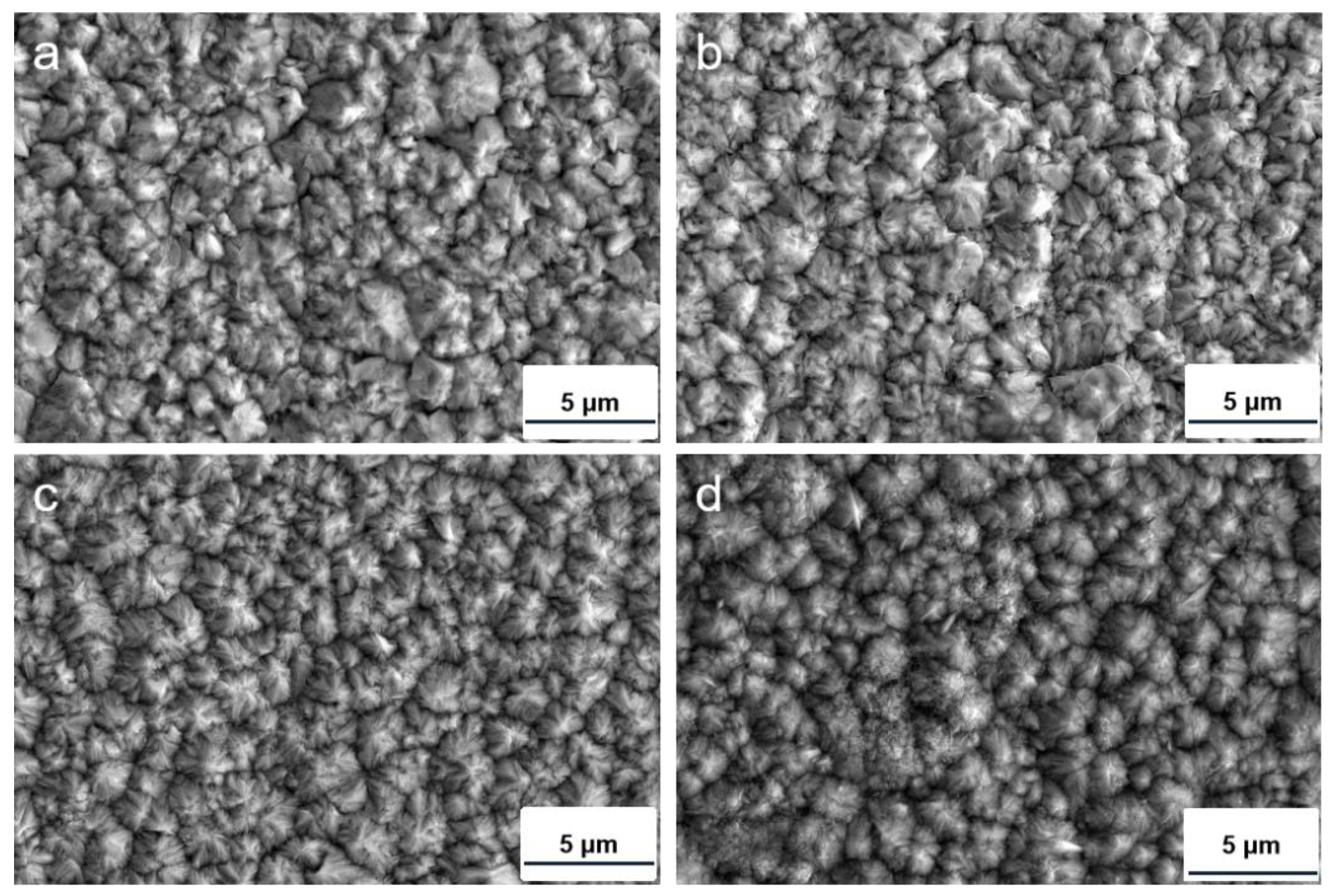
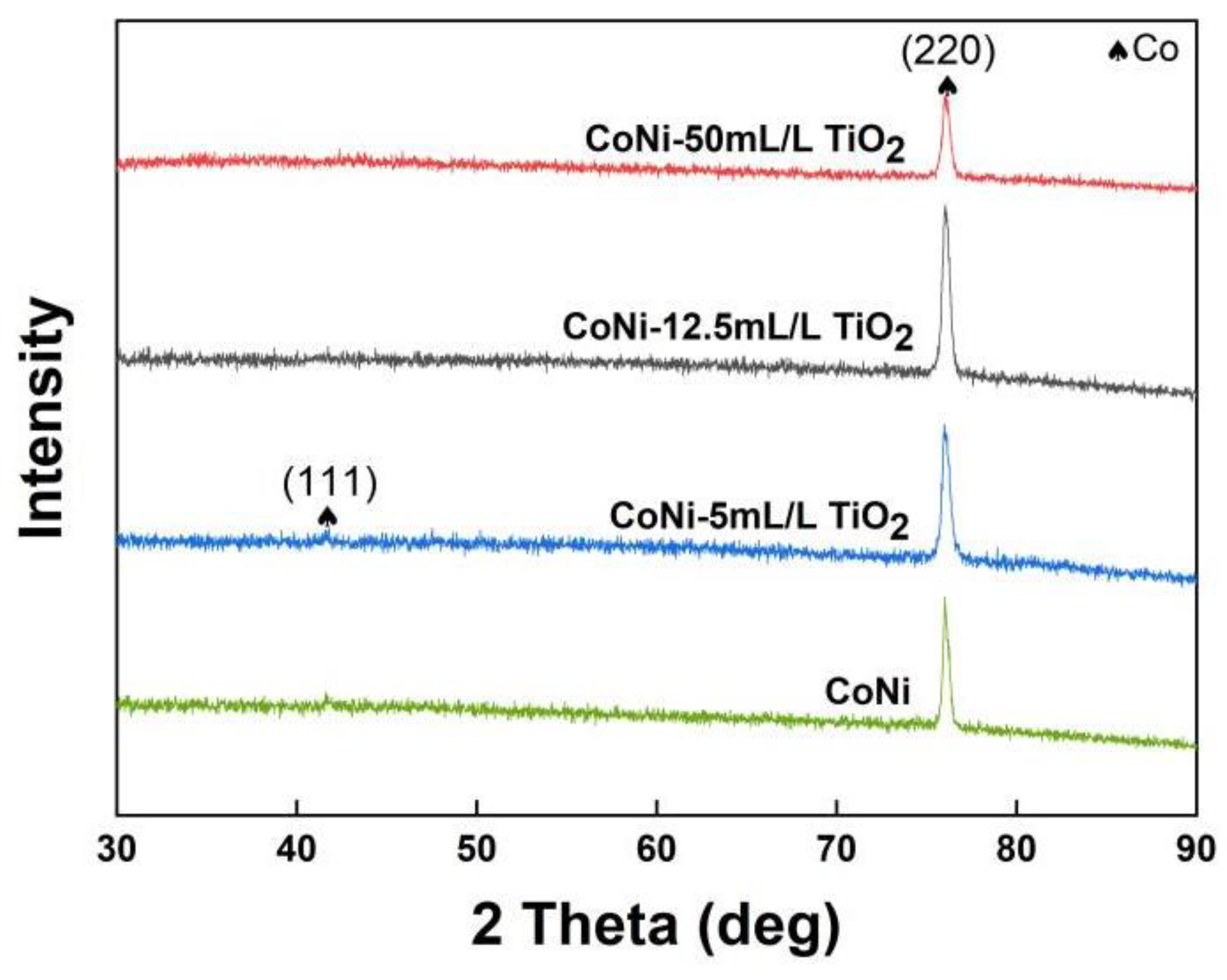
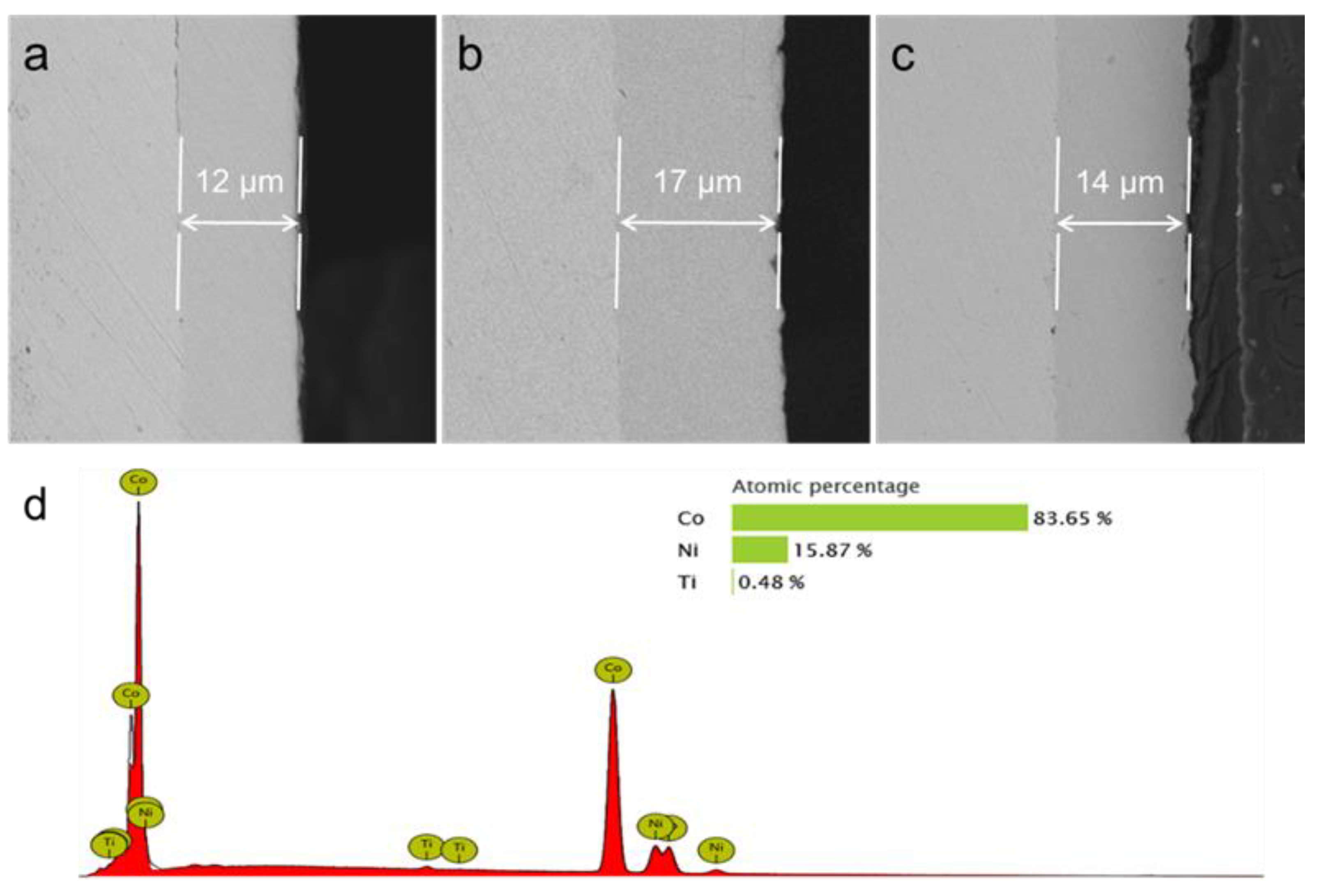
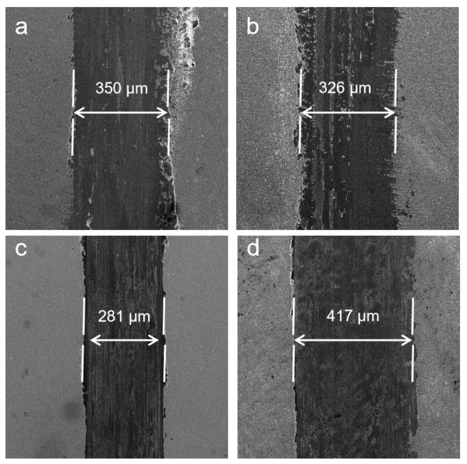
3.2. Structural Characterizations of Co−Ni−TiO2 Films

Figure 4 depicts the microscopic surface morphologies of Co−Ni−TiO2 films produced by varied TiO2 concentrations. The surface of the Co−Ni−TiO2 films is refined and has the most significant surface uniformity and coverage at 12.5 mL/L TiO2 sol. However, Co−Ni−50 mL/L TiO2 film’s grains are coarsened. In general, highly dispersed TiO2 nanoparticles provide more nucleation sites, promoting nucleation processes and refining surface crystals. However, the excessive TiO2 nanoparticles can result in agglomeration and coating coarsening under 50 mL/L TiO2 sol.
X-ray diffractograms of Co−Ni−TiO2 films with diverse TiO2 sol content are shown in Figure 5. The (220) diffraction peak is distinct in the Co−Ni−TiO2 diffractograms. In addition, the location of the textured peak is a bit different from the standard peak due to the insertion of Ni atoms into the Co atom’s structure, which is consistent with some previous papers [4,9,26]. Because there is very little content of TiO2 nanoparticles incorporated in the Co−Ni film, no TiO2 diffraction peak was observed in the profiles.
Figure 6 shows the cross-sectional morphology of the composite films prepared. The coated composite film tightly adheres to the substrate with no defects, such as peeling and holes, indicating a good coating quality for all samples. In addition, no TiO2 agglomeration regions are identified on the cross-sectional morphologies. However, the film thickness decreases at 50 mL/L sol addition. When excessive TiO2 sol was added to the electrolyte, the electrodeposition process could be hindered and more charged current would be consumed in the side reaction of hydrogen evolution, thereby decreasing the film thickness.
The elemental composition obtained by EDS detection of the Co−Ni−12.5 mL/L TiO2 sample is depicted in Figure 6d. The Co element, which has an atomic ratio of 83.78%, and the Ni element, which has an atomic ratio of 15.87%, are the main components in the prepared film according to the EDS results. There is a limited amount of Ti in the film, resulting from the incorporated TiO2.

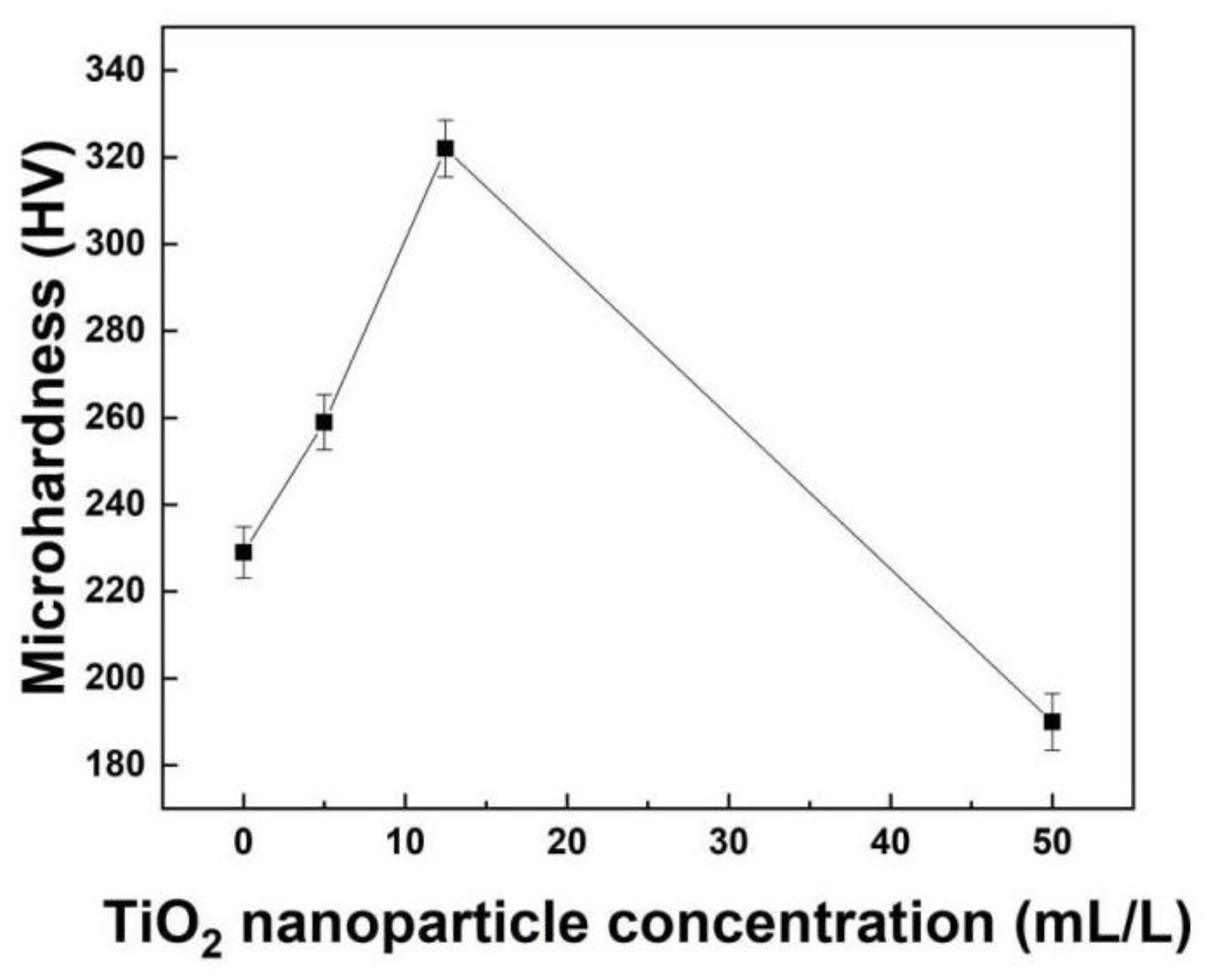
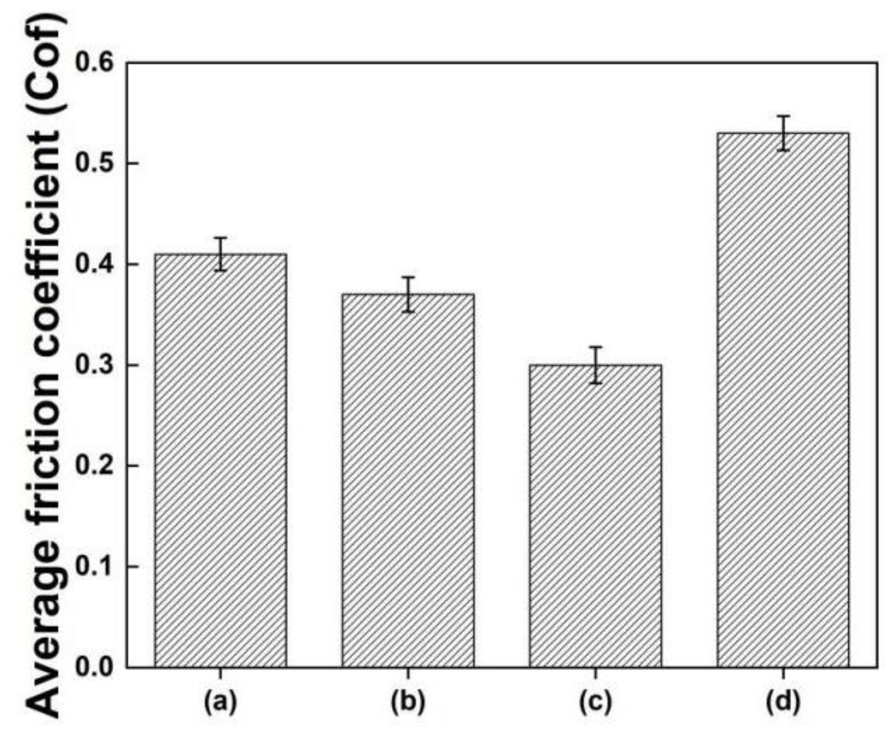
3.3. Mechanical Properties of Co−Ni−TiO2 Films

The microhardness of Co−Ni−TiO2 films at diverse TiO2 sol content has been shown in Figure 7. The microhardness of the Co−Ni film is ~228 HV. Co−Ni-12.5 mL/L TiO2 film possesses the highest microhardness of 323 HV, which is 41% higher than the Co−Ni film’s hardness. However, adding 50 mL/L TiO2 sol reduces the microhardness of the film to approximately 190 HV. Under an appropriate content of TiO2 incorporation, the highly dispersed TiO2 nanoparticles are spread in the grain boundary region, which hinders grain development and enhances fine-grain strengthening. Additionally, the highly dispersed TiO2 nanoparticles perform the second-phase reinforcing effect that further promotes film hardness. In contrast, under an extremely high TiO2 sol concentration, the excessive TiO2 nanoparticle clusters cause agglomerations and reduce the films’ mechanical properties.
Figure 8 illustrates the average friction coefficient of the Co−Ni−TiO2 films. A Co−Ni film with no TiO2 sol has a coefficient of 0.68, whereas a Co−Ni−12.5 mL/L TiO2 film shows a friction coefficient of 0.42. In contrast, when the content of TiO2 sol is 50 mL/L, the coefficient of friction increases to 0.78. The film’s friction resistance will be increased using the proper TiO2 sol (12.5 mL/L) content of the electrodeposition solution.
Figure 9 illustrates the after-wear sample morphologies of Co−Ni films, Co−Ni−5 mL/L TiO2, Co−Ni−12.5 mL/L TiO2, and Co−Ni−50 mL/L TiO2 films, respectively. Under the rising concentration of TiO2 sol, the film’s wear track width reduces first and then rises. A suitable content of TiO2 sol added to the electrolytes decreases the wear track width of Co−Ni−12.5 mL/L TiO2 to 281 mL, while the wear track width of Co−Ni−50 mL/L TiO2 increases to about 417 μm under a higher sol concentration. The same trend is proposed: the wear resistance improves at a proper TiO2 concentration, whereas it reduces by adding excessive TiO2 sol.

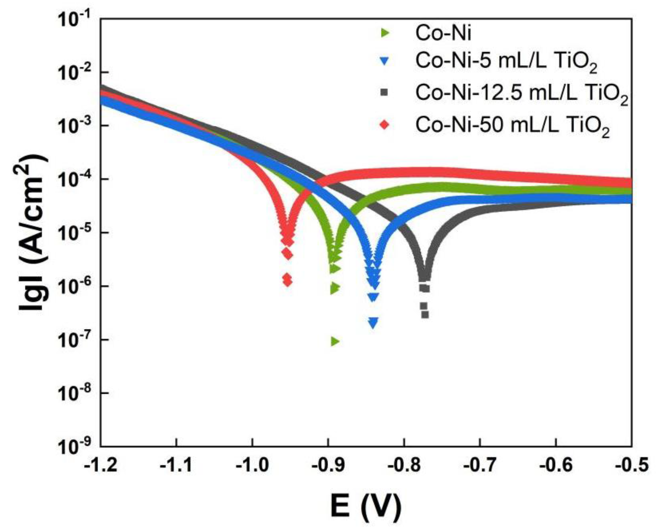
3.4. Corrosion Resistance of Co−Ni−TiO2 Films

The corrosion performance of the produced Co−Ni−TiO2 films was investigated by measuring potentiodynamic polarization, as depicted in Figure 10. The greater the corrosion potential (Ecorr) value of the material and the smaller the corrosion current density, the better the corrosion resistance of the film material. When the concentration of the TiO2 sol rises, the corrosion potential of the film gradually increases. Under a 12.5 mL/L TiO2 sol addition, the corrosion potential is the largest, showing that the film has the most excellent corrosion resistance. It is evident that the film has a worsened corrosion resistance when TiO2 sol content reaches 50 mL/L.
Table 1 below shows the electrochemical parameters by fitting the polarization curves. The corrosion speed of the Co−Ni film is 0.074 mm/a, and the corrosion speed of the Co−Ni−12.5 mL/L TiO2 film is only 0.036 mm/a. According to the findings, the corrosion resistance of the Co−Ni film is improved when the film has 12.5 mL/L TiO2 sol. The corrosion rate for the Co−Ni−50 mL/L TiO2 film is 0.171 mm/a, indicating that the film’s corrosion resistance significantly decreases. Adding appropriate content of TiO2 sol can form a denser film surface with refined crystals, as suggested above, which explains the enhanced corrosion performance. Nevertheless, an excessive concentration of TiO2 sol will cause TiO2 nanoparticles to agglomerate in the coating, impairing the coating’s uniformity and decreasing the corrosion resistance.

4. Conclusions

This study utilized chemical synthesis to create TiO2 sols with high dispersion and chemical stability. Mechanical and corrosion-resistant Co−Ni−TiO2 films were prepared by adding a suitable amount of TiO2 sol (12.5 mL/L) to the electrolytes during electrodeposition. The addition of TiO2 sol controls the nucleation process and the properties of the composite films by generating TiO2 nanoparticles in situ in the electrodeposition process. By producing TiO2 nanoparticles in situ in electrolytes with sizes below 100 nm, nanoparticle agglomeration is largely avoided. The mechanical property testing results reveal that the Co−Ni−12.5 mL/L TiO2 film has the highest hardness of 323 HV, an increase of 41% over the undoped samples. Electrochemical tests demonstrate a 51% reduction in the corrosion rate for the Co−Ni−12.5 mL/L TiO2 film compared to the undoped Co−Ni film sample. Increased nucleation sites can result from the appropriate amount of TiO2 sol, enhancing coating quality through fine-grain and dispersion-strengthening effects. Nevertheless, coarse grains and pores will result from the agglomeration of excessive TiO2 nanoparticles under high TiO2 sol concentration, which will have a detrimental impact on the film’s performance.

Author Contributions

Conceptualization, Y.W. and Z.H.; methodology, Z.M. and J.C.; validation, Z.H. and Z.M.; formal analysis, Z.H.; investigation, Z.M.; writing—original draft preparation, Y.W.; writing—review and editing, Z.H.; visualization, S.Z.; supervision, J.C. and Z.H.; funding acquisition, Z.H. All authors have read and agreed to the published version of the manuscript.

Funding

This work was supported by the Natural Science Foundation of Jiangsu Province by grant BK20201008.

Institutional Review Board Statement

Not applicable.

Informed Consent Statement

Not applicable.

Data Availability Statement

Not applicable.

Conflicts of Interest

The authors declare no conflict of interest.

References

  1. Bouzit, F.Z.; Nemamcha, A.; Moumeni, H.; Rehspringer, J.L. Morphology and Rietveld analysis of nanostructured Co-Ni electrodeposited thin films obtained at different current densities. Surf. Coat. Technol. 2017, 315, 172–180. [Google Scholar] [CrossRef]
  2. Olvera, S.; Arce Estrada, E.M.; Sanchez-Marcos, J.; Palomares, F.J.; Vazquez, L.; Herrasti, P. Effect of the low magnetic field on the electrodeposition of CoxNi100−x alloys. Mater. Charact. 2015, 105, 136–143. [Google Scholar] [CrossRef]
  3. Li, D.; Levesque, A.; Franczak, A.; Wang, Q.; He, J.; Chopart, J.P. Evolution of morphology in electrodeposited nanocrystalline Co-Ni films by in-situ high magnetic field application. Talanta 2013, 110, 66–70. [Google Scholar] [CrossRef] [PubMed]
  4. Karpuz, A.; Kockar, H.; Alper, M. Electrodeposited Co–Ni Films: Electrolyte pH—Property Relationships. J. Supercond. Nov. Magn. 2012, 26, 651–655. [Google Scholar] [CrossRef]
  5. Yu, M.M.; Li, H.Y.; Wang, Y. Study on Present Situation and New Trends of the Electrodeposition of Nickel-Cobalt Alloy. Adv. Mater. Res. 2012, 535–537, 973–976. [Google Scholar] [CrossRef]
  6. Sun, L.F.; Mao, J.M.; Pan, Z.W.; Chang, B.H.; Zhou, W.Y.; Wang, G.; Qian, L.X.; Xie, S.S. Growth of straight nanotubes with a cobalt–nickel catalyst by chemical vapor deposition. Appl. Phys. Lett. 1999, 74, 644–646. [Google Scholar] [CrossRef]
  7. Li, Y.; Xie, Y.; Huang, L.; Liu, X.; Zheng, X. Effect of physical vapor deposited Al2O3 film on TGO growth in YSZ/CoNiCrAlY coatings. Ceram. Int. 2012, 38, 5113–5121. [Google Scholar] [CrossRef]
  8. Cuglietta, M.; Kuhn, J.; Kesler, O. A Novel Hybrid Axial-Radial Atmospheric Plasma Spraying Technique for the Fabrication of Solid Oxide Fuel Cell Anodes Containing Cu, Co, Ni, and Samaria-Doped Ceria. J. Therm. Spray Technol. 2013, 22, 609–621. [Google Scholar] [CrossRef]
  9. Liu, C.; Su, F.; Liang, J. Nanocrystalline Co-Ni alloy coating produced with supercritical carbon dioxide assisted electrodeposition with excellent wear and corrosion resistance. Surf. Coat. Technol. 2016, 292, 37–43. [Google Scholar] [CrossRef]
  10. Hu, H.; Tan, M.; Liu, L. Anomalous codeposition mechanism of Co-Ni alloy nanowires. J. Alloys Compd. 2017, 715, 384–389. [Google Scholar] [CrossRef]
  11. Barzegar, M.; Allahkaram, S.R.; Naderi, R.; Ghavidel, N. Effect of phosphorous content and heat treatment on the structure, hardness and wear behavior of Co-P coatings. Wear 2019, 422–423, 35–43. [Google Scholar] [CrossRef]
  12. Tebbakh, S.; Messaoudi, Y.; Azizi, A.; Fenineche, N.; Schmerber, G.; Dinia, A. The influence of saccharin on the electrodeposition and properties of Co–Ni alloy thin films. Trans. IMF 2015, 93, 196–204. [Google Scholar] [CrossRef]
  13. Altamirano-Garcia, L.; Vazquez-Arenas, J.; Pritzker, M.; Luna-Sánchez, R.; Cabrera-Sierra, R. Effects of saccharin and anions (SO4 2−, Cl) on the electrodeposition of Co–Ni alloys. J. Solid State Electrochem. 2014, 19, 423–433. [Google Scholar] [CrossRef]
  14. Vazquez-Arenas, J.; Treeratanaphitak, T.; Pritzker, M. Formation of Co–Ni alloy coatings under direct current, pulse current and pulse-reverse plating conditions. Electrochim. Acta 2012, 62, 63–72. [Google Scholar] [CrossRef]
  15. Wang, S. Influence of 2-butyne-1, 4-diol on the structure and performance of the Co-Ni alloy plated by electrodeposition. Acta Metall. Sin. (Engl. Lett.) 2008, 21, 50–56. [Google Scholar] [CrossRef]
  16. Bu, A.; Zhang, Y.; Zhang, Y.; Chen, W.; Cheng, H.; Wang, L.; Wang, Y. A Novel Electrolytic Plasma Spraying Preparation SiO2/SiC Coating on Carbon Fiber Fabric. Coatings 2018, 8, 344. [Google Scholar] [CrossRef]
  17. Xiong, C.; Xiao, J.; Zhao, Y.; Wang, Y.; Tay, S.L.; Xu, W. Properties of Ni–ZrO2 nanocomposite coatings by electroplating. Int. J. Mod. Phys. B 2019, 33, 1940024. [Google Scholar] [CrossRef]
  18. Zhang, W.; Du, S.; Li, B.; Mei, T.; Miao, Y.; Chu, H.; Wang, J. Synthesis and characterization of TiN nanoparticle reinforced binary Ni-Co alloy coatings. J. Alloys Compd. 2021, 865, 158722. [Google Scholar] [CrossRef]
  19. Elkhoshkhany, N.; Hafnway, A.; Khaled, A. Electrodeposition and corrosion behavior of nano-structured Ni-WC and Ni-Co-WC composite coating. J. Alloys Compd. 2017, 695, 1505–1514. [Google Scholar] [CrossRef]
  20. Wang, Y.; Hu, B.; Tay, S.L.; Hou, F.; Gao, W.; Xiong, C. The microstructure and properties of sol-enhanced Sn–TiO2 nanocomposite coatings. Int. J. Mod. Phys. B 2017, 31, 1744025. [Google Scholar] [CrossRef]
  21. He, Z.; Cao, D.; Wang, Y.; Yin, L.; Hayat, M.D.; Singh, H. Preparation of Co–P–TiO2 nanocomposite coatings via a pulsed electrodeposition process. Surf. Eng. 2019, 36, 975–981. [Google Scholar] [CrossRef]
  22. Zhang, W.; Cao, D.; Qiao, Y.; He, Z.; Wang, Y.; Li, X.; Gao, W. Microstructure and Properties of Duplex Ni-P-TiO2/Ni-P Nanocomposite Coatings. Mater. Res. 2019, 22 (Suppl. 2), e20180478. [Google Scholar] [CrossRef]
  23. Liu, Y.; Li, L.; Lu, G.-L.; Han, Z.-W.; Liu, J.-D.; Yu, S.-R. Fractal characteristics and wettability of Nano-Al2O3/Ni-Co composite coating prepared by electrodeposition. Trans. Nonferrous Met. Soc. China 2011, 21, s380–s383. [Google Scholar] [CrossRef]
  24. Rasooli, A.; Safavi, M.S.; Kasbkar Hokmabad, M. Cr2O3 nanoparticles: A promising candidate to improve the mechanical properties and corrosion resistance of Ni-Co alloy coatings. Ceram. Int. 2018, 44, 6466–6473. [Google Scholar] [CrossRef]
  25. Bakhit, B.; Akbari, A.; Nasirpouri, F.; Hosseini, M.G. Corrosion resistance of Ni–Co alloy and Ni–Co/SiC nanocomposite coatings electrodeposited by sediment codeposition technique. Appl. Surf. Sci. 2014, 307, 351–359. [Google Scholar] [CrossRef]
  26. Gómez, E.; Pané, S.; Alcobe, X.; Vallés, E. Influence of a cationic surfactant in the properties of cobalt–nickel electrodeposits. Electrochim. Acta 2006, 51, 5703–5709. [Google Scholar] [CrossRef]
Figure 1. TEM images were taken on the generated TiO2 nanoparticles made by adding sol into DI water. (a) TEM image, (b) Enlarged image for nanoparticles.
Coatings 13 00783 g001
Figure 2. The microstructure of a single TiO2 nanoparticle. (a) TEM image, (b) HR−TEM micrograph, (c) electron diffraction pattern.
Coatings 13 00783 g002
Figure 3. Size distribution of TiO2 nanoparticles generated by adding sol into DI water.
Coatings 13 00783 g003
Figure 4. Surface morphology of Co−Ni−TiO2 films prepared with different content of TiO2 sol: (a) 0 mL/L; (b) 5 mL/L; (c) 12.5 mL/L; (d) 50 mL/L.
Coatings 13 00783 g004
Figure 5. X-ray diffractograms recorded on the prepared Co−Ni−TiO2 films.
Coatings 13 00783 g005
Figure 6. Cross-section of Co−Ni−TiO2 films prepared with different content of TiO2 sol: (a) 0 mL/L; (b) 12.5 mL/L; (c) 50 mL/L, and (d) the EDS results of the Co−Ni−12.5 mL/L TiO2 sample.
Coatings 13 00783 g006
Figure 7. Microhardness tested on Co−Ni−TiO2 films with diverse TiO2 sol content.
Coatings 13 00783 g007
Figure 8. The friction coefficient of Co−Ni−TiO2 films prepared with diverse content of TiO2 sol: (a) 0 mL/L; (b) 5 mL/L; (c) 12.5 mL/L; (d) 50 mL/L.
Coatings 13 00783 g008
Figure 9. Wear track of Co−Ni−TiO2 films prepared with different concentrations of TiO2 sol: (a) 0 mL/L; (b) 5 mL/L; (c) 12.5 mL/L; (d) 50 mL/L.
Coatings 13 00783 g009
Figure 10. Potentiodynamic polarization curves of different Co−Ni−TiO2 films.
Coatings 13 00783 g010
Table 1. Potential polarization data of Co−Ni−TiO2 films.
SampleEcorr (V)Icorr (μA/cm2)Corrosion Rate (mm/a)
Co−Ni−0.886.280.074
Co−Ni−5 mL/L TiO2−0.843.690.054
Co−Ni−12.5 mL/L TiO2−0.763.090.036
Co−Ni−50 mL/L TiO2−0.9914.60.171
Disclaimer/Publisher’s Note: The statements, opinions and data contained in all publications are solely those of the individual author(s) and contributor(s) and not of MDPI and/or the editor(s). MDPI and/or the editor(s) disclaim responsibility for any injury to people or property resulting from any ideas, methods, instructions or products referred to in the content.

