Article

Influence of Mesoporous Inorganic Al–B–P Amphiprotic Surfactant Material Resistances of Wood against Brown and White-Rot Fungi (Part 1)

College of Materials Engineering, Fujian Agriculture and Forestry University, Fuzhou 350108, China
*
Author to whom correspondence should be addressed.
Coatings 2020, 10(2), 108; https://doi.org/10.3390/coatings10020108
Submission received: 22 November 2019 / Revised: 14 January 2020 / Accepted: 16 January 2020 / Published: 26 January 2020

Abstract

:
This study describes the application of aluminum sulfate Al2(SO4)3, boric acid H3BO3, phosphoric acid H3PO4 (Al–B–P) and amphiprotic surfactant material synthesis by the sol-gel process, which were adopted as novel precursors for wood modification. The efficacy of Al–B–P-treated wood was tested against Poria placenta and Coriolus versicolor. Untreated wood samples had higher mass losses (>40%) compared to the treated sample, which had the lowest wood mass losses (of 4%) against P. placenta and C. versicolor. To analyze the reaction mechanism of Al–B–P wood, the mechanical properties, chemical structure, crystallinity, thermal analysis, binding energy and wettability was examined by modulus of rupture (MOR), modulus of elasticity (MOE), Fourier transform infrared spectroscopy (FTIR), X-ray powder diffraction (XRD), Thermogravimetric analysis (TG) and X-ray photoelectron spectroscopy (XPS), respectively. Scanning electron microscopy- energy-dispersive X-ray spectroscopy (SEM-EDS) confirmed the wood colonization by fungi, and was used to identify the microstructures and morphologies changes that occurred in the cells during degradation by white and brown-rot fungi. At the same time, X-ray photoelectron spectroscopy (XPS) was employed to analyze the physical and chemical properties of the samples. Therefore, the study confirmed that Al–B–P and amphiprotic surfactant could replace the traditional wood preservative products, and have the potential to extend the service life of wood, particularly in soil contact and outdoor usage.

Graphical Abstract

1. Introduction

Wood is renewable by excellence and is an environmentally friendly material; nevertheless, as a biological material, it suffers to the same extent as many other natural materials, namely degradation, due to its composition and anatomical structure.
The foremost problems are the facts that white-rot fungi use non-enzymatic and enzymatic systems to degrade all cell wall components [1]. Coriolus versicolor and Poria placenta fungi degrade wood in the crystalline region by cellulolytic and ligninolytic enzymes [2]. P. placenta fungi colonize wood by the sequential decomposition of lignin and cellulose using their extracellular enzyme system [3,4,5]. Wood decayed by P. placenta has been further chemically characterized by a number of β-xylosidases and endo-xylanases [2]. C. versicolor fungi are known to degrade polysaccharides efficiently [6].
The most widely used wood chemical preservatives include; copper azole alkaline (CAA) and copper quaternary creosote (CQC), etc. They protect wood bio-deteriorating agents after application; however, their uses pose a threat to the environment, due to the presence of heavy metals that are toxic to the mammals and other organisms in the environment.
In the same way, C. versicolor and P. placenta are also tolerant to copper, a commonly used active ingredient in wood preservation products [7]. Environmental and health concerns are related, which in turn closes the loop by encouraging the use of materials that contain recycled content, reducing their leaching into the environment [8,9]. Moreover, more recently, studies conducted by Saka have been testing inorganic sol-gel, such as TiO2 and SiO2, which, combined with other reagents as Na2OSiO2 gel, can be useful for wood preservation, and the study results showed that integrating inorganic compounds into the wood cavity wall can enhance the characteristics of the wood, such as against fungi (white and brown-rot fungi) and termites [10,11].
Some novel inorganic chemical combinations, based on boron compounds (B), aluminum sulfate compounds (Al), phosphoric acid (P) and amphiprotic surfactant as a reinforcing agent, through a sol-gel process, have been shown to be an excellent anti-fungal agent process, and was a promising and environmentally friendly method to enhance the wood properties against fungi and molds [11]. For this reason, there are significant efforts to investigate the replacement of copper as an active ingredient in protecting wood against copper-tolerant fungi.
Thus, the Al–B–P and the amphiprotic surfactant is perfect for wood preservative, and would not only inhibit the growth of wood-destroying organisms, but would also have low water solubility, which would reduce their leaching into the environment. The decrease hygroscopicity of the wood can also improve dimensional stability because of the presence of aluminum sulfate, which combines with boric acid [12]. The treatment decreases the hygroscopicity of the wood sample, and can also improve the dimensional stability due to the incorporation of the combination of aluminum sulfate and boric acid agents. The application of inorganic sol-gel for wood modification treatment to both enhance and improve resistance to fungi has attracted the attention of many researchers [13,14].
The development of new wood preservatives is aimed to reduce environmental pollution, is cost-effective, and has low viscosity and excellent fungal resistance properties. In recent years several investigations attract the focus of researcher concern on wood surface morphology changed by inorganic agents. Especially Hübert (2018) describes the behavior of nanoparticles compounds against brown-rot fungi [15]. Based on maintaining the characteristics of wood porous structure, it can improve the wood performance on many aspects, including mechanical properties, dimensional stability, being antibacterial, waterproof, and improving the anti-loss of preservatives such as copper and boron, etc. [16,17,18]. Anti-fungi treatment is vital for the efficient utilization of wood raw material [8]. Sol-gel based on aluminum and copper sulfate can lead to an improved resistance against decay degradation and reduced fungal colonization [19]. The deterioration of wood by biological organisms is a definite disadvantage when the wood is used in service. However, the cross-linking mechanism of the chemical bonds formed after impregnation could improve the wood service life [20]. Many studies have reported that Poria placenta mycelium penetrates inside wood cells and causes enormous impact and thus has a higher fungal activity morphology of the mycelium. Novel inorganic aluminum borate and nano metal compounds could improve the antifungal performance of wood, which is in agreement with previous studies [21,22].
Therefore, aluminum sulfate (Al2(SO4)3), boric acid (H3BO3) and the amphiprotic surfactant are considered non-toxic, and also has relatively very low-risk to animals and is non-toxic for humans to significantly improve wood against white-rot fungi and termites, including being low-cost and renewable [23]. Aluminum sulfate and boric acid products are effective against both fungi and termites if used below the limit stated in biocidal products, and have emerged as alternatives to the preservatives based upon heavy metals [24].
The main objective of the present research was mainly focused on preparing inorganic Al–B–P amphiprotic surfactant solutions. Then we used it to improve wood properties, and to study the effect of their interaction on white-rot fungi. Therefore, after the characterization of the impregnation on wood, accelerated biological tests against the white and brown-rot fungi (Coriolus versicolor and Poria placenta), two of the most aggressive fungi against the wood, were run to determine the efficacy of the solution.
To clarify these issues, the processing parameters of wood, an experimental design of the response surface methodology (RSM) model, was used to analyze the problems and get exact results by various independent variables.
Additionally, treated and untreated wood sample surfaces, along with the microstructures, chemical structures, mechanical performance and thermal stability of Al–B–P amphiprotic surfactant is also discussed in this study.

2. Materials and Methods

2.1. Wood Materials

Concerning Pinus (Pinus massoniana) and Sitka spruce (Picea sitchensis), both species have been chosen according to their availability in the region of Fujian, for each species having a relatively straight stem with the age of 15-yrs-old. The Pinus and Spruce density was 0.48 and 0.45 g·cm−3, respectively. The samples prepared from sapwood portions with the dimensions of 50 × 20 × 5 mm3 (longitudinal; tangential; radial) for a wood sample were placed in Petri dishes. We made other dimensions for a sand test (19 × 19 × 19 mm3) and soil contact test (457 × 19 × 19 mm3). Before treatment, we dried wood samples at 103 °C for 24 h. All wood blocks were: having no defects, as knots, and being without visible evidence of fungus infection. All specimens’ size, respectively, was obtained from Fujian Minhou.

2.2. Chemical Products and Fungi

99% aluminum sulfate (Al2(SO4)3), 99% boric acid (H3BO3), 85% phosphoric acid (H3PO4), 99% cupric salt (CuSO4) and amphiprotic surfactant (C5H11NO2HCl) were purchased from Tianjin Fuchen Chemical Co. Ltd. (Tianjin, China), and Sino Pharm Chemical Reagent Co. Ltd. (Shanghai, China). 99.5% sodium tetraborate (Na2B4O7·10H2O) was bought Sigma-Aldrich in Shanghai (China). The fungi used in this study were Poria placenta (brown-rot fungi) and Coriolus versicolor (white-rot fungi) obtained from the collections of the laboratory Beijing Zhongke Quality Inspection Biotechnology Co., Ltd (Beijing, China) and kept at 26–28 °C, and placed in the incubator at 85% of relative humidity.

2.3. Preparation of Al–B–P Amphiprotic Surfactant Solution

In a beaker of 1000 mL, containing 25 mL of Al2(SO4)3, 25 mL of H3BO3, 1.2 g of CuSO4 and 15 g of 85% H3PO4, were dissolved in distilled water at 70 °C and quickly added to a solution of 4.8 mL of amphiprotic surfactant (C5H11NO2HCl), and the entire solution was stirred for 15 min until the solution became blue. The pH of the solution measured was around 2.5 and 3. The optimum parameters for antifungal agents were: the mass ratio of Na2B4O7·10H2O, CuSO4 and Al2(SO4)3 are 1.40%, 2% and 3%, respectively.

2.4. Field Testing Methods

For this study, soil contact analyzed the data collected from Fujian Minhou (26°4′45″ N119°15′30″E). Fujian climate is hot and humid, with a subtropical monsoon climate, with moderate winters, which is suitable for the growth of decaying bacteria and termites, causing severe damage to wood. The field tests aim to visualize the fungal decomposition of wood to test the durability of the treated wood, and then compare it to the untreated wood. Similarly, the method was described by Råberg (2013) [25].
The data average is 15 for each specimen of the mechanical properties of a wood sample with a moisture content at 12%. The size of all samples for MOE and MOR was 457 ×1 9 × 19 mm3 (L × W × H), and they were tested according to the British Standard BS EN 252:2014 [26]. Reported results are the average of six replications. Wood treatment with Al–B–P and amphiprotic surfactant sol-gel influences the static strength (MOR) and elastic modulus (MOE) of the treated wood.

2.5. Preparation Method of Culture of Fungi

75 g of sand, 75 g of pine sawdust, 7.5 g (25 mesh) of cornflour and 4.3 g of brown sugar were added in a flask of 250 mL and mixed. 50 mL of maltose was slowly added to the mixture and sterilized for 1 h at 120 °C. After cooling, before the Potato Dextrose Agar was inoculated with fungi, 90 Petri dishes of (120 by 20 mm) were sterilized by autoclaving at 120 °C for 15 min. After that, we inoculated each petri dish in the center with P. placenta or C. versicolor. We placed the inoculated culture in a growth chamber at 28 °C with 85% relative humidity for 20 days. For Potato Dextrose Agar (PDA), we boiled 200 g of sliced potatoes in 1 liter of water for 30 min into the flask, added 20 g of glucose and 15–20 g of agar. It was then sterilized for 20 min. The medium was prepared according to the methods described by the American Society for Testing and Materials (ASTM) D4445,2015 [27]. Treated and untreated wood was placed in a petri dish (120 mL) containing 25% PDA and introduced into the soil block, then exposed for 16 weeks [28].

2.6. Treatment Procedure of Wood Sample

Wood samples were weighed, and the moisture content of the wood was collected. All wood was first soaked in Al–B–P and amphiprotic surfactant solution by dipping according to the normal pressure for 6 h. Three replicates were used for this experiment. Following impregnation, we dried wood in an oven for 6 h at 80 °C. Following impregnation, we cleaned each wood with cotton and absolute ethanol. The impregnated wood was then kept for the evaluation of the durability. Response surface methodology (RSM) was used to optimize the parameters of the Na2B4O7·10H2O, CuSO4, and Al2(SO4)3, solution concentration, for the effective treatment process. Design-Expert software trial version 8.0.5 was used to draw the analysis diagram based on the regression equation. The Box–Behnken design method was adopted to carry out the test at the level of three factors m(Na2B4O7·10H2O), m(CuSO4) and mAl2(SO4)3, with respective values of 1.4%, 2% and 3%. The test designs were coded as X1, X2 and X3. Table 1, and Table S1 show the results of the response surface (ML (mass loss) of two types of fungi, Poria placenta and Coriolus versicolor).

2.7. Retention Rate of Chemical Effectiveness

The retention of wood preservatives depends upon treatment time, temperature and pH of the solution. All these factors influence the fixation of chemicals on wood [29,30].
Wood was weighed before and after the impregnation of the solution. The chemical retention of the anti-fungi agents was determined using the following Equation (1) [31].
WPG = W 1 W 2 W 2 × 100
where: WPG—Weight Percent Gains
W1 = the oven dried weight before treatment
W2 = the oven dried weight after treatment respectively.

2.8. Evaluation of Fungicide and Testing Procedure

After the characterization of the treated wood sample, their resistance against decay by C. versicolor and P. placenta was assessed following the American standard ASTM D2017-05 [28]. The mass loss of the wood after characterization was evaluated after 16 weeks of exposure to fungi. The mycelium on the surface of the test specimens was a softness removed with care, and the wood blocks were oven-dried for 24 h at 103 °C, weighed, and we saved the final mass loss. The average mass loss rate (AML%) of treated and untreated wood samples was determined by following Equation (2):
AML % = W initial W final   W initial   × 100
where Winitial is initial dried mass of wood before treatment and Wfinal is the dry mass after fungi attack. Table 2 below gave the resistance grade of wood after fungi attacks.

2.9. Materials Characterization

The morphological aspects of treated and untreated wood samples were characterized using a Scanning Electron Microscopy (SEM, Hitachi UHR FE-SEM SU8010, Tokyo, Japan) The cross-sectional of the wood samples had been cut with a microtome and sprayed with carbon to 15 nm before image acquisition and operating at 50 µm 15 kV and variable pressure (5–10 Pa). EDS (Supra 55 Zeiss, Oberkochen, Germany) was carried out using an acceleration voltage of 15 kV and variable pressure (5–10 Pa). We performed the mapping of the wood sample with a distance of 10 mm, and the time of capture was 600 s. We performed the pore size distribution analysis of wood realized by nitrogen absorption desorption on (JW-BK132F, Beijing, China). The sample size of 50 × 20 × 5 mm3 weighing as a 0.5–1 g powder specimen was determined using a blast dryer, under the conditions of 103 ± 3 °C. The pore size of the wood was calculated according to the Barrett– Joyner–Halenda (B–J–H) formula. The surface porosity of the wood was calculated according to the Brunauer, Emmett and Teller (BET) equation. FTIR spectra were obtained with the MIR-FIR spectrum (VERTEX 70, Bruker Instrument, Billerica, MA, USA). The KBr pellet method was employed in each sample by grinding 2 mg of dried wood powder (200 mesh) and potassium bromide powder (1:100) to produce thin sheet disks. The diffraction analysis was performed using an X-ray diffractometer (XRD) (X’Pert PRO Malvern Panalytical Ltd., Almelo, The Netherlands). The wood powder (200 mesh) was pressed into 10 × 10 × 1 mm3 size on a slide. The test of cellulose crystallinity and chemical bond combinations between wood and chemical compound with Cu Kα radiation at a wavelength λ = 1.790 nm) were examined. Nuclear magnetic resonance (NMR) analysis. The 31P and 27A1 MAS NMR spectra of Al–B–P compounds specimens were recorded on a Bruker ADVANCE III 500 spectrometer (Billerica, MA, USA), using a 7 mm zirconia oxide rotor with a spinning speed of 4.5 kHz. We recorded all spectra at room temperature, using dry air as driving gas. The 27A1 MAS NMR spectra were recorded at a frequency of 105 MHz with a pulse length of 1 μs.
The wettability of the treated wood and untreated wood was measured using the contact angle by placing a droplet of water in contact with the wood surface using a micrometer syringe, and the Contact Angle was measured by scanning the droplet profile 0, 30 and 60 s after placing water drops on the wood surface, using an analyzer (Harke-spcaxd contact angle measuring instrument, Beijing, China). The curves of the thermal gravimetric analyser (TGA) were obtained using a thermogravimetric analyser (NETZSCHSTA449F3, Selb, Germany). Under high-purity nitrogen atmosphere, approximately 10 ± 0.5 mg of the sample was heated in air at room temperature to 800 °C. The experiments started with a different heating rate of 10 and 20 °C/min. The reported results are the average of eight specimens. The measurement of the MOR and MOE values of the wood samples after exposure to the ground was performed on the MTS Exceed Series 40 Electromechanical Test system (Eden Prairie, MN, USA) at a constant speed of 100 mm/min. The reported results are the average of 120 samples. The sample preparation was according to TS 2474. Loading was done at the tangential directions of samples. For this purpose, the size of samples was 457 × 19 × 19 mm3 (L × W × H), and the dimension between the centers of the two supports was 240 mm. X-ray photoelectron spectroscopy (XPS) was performed on the ESCALAB 250Xi (Thermo Fisher Scientific Company, Waltham, MA, USA). Experiments were carried out at an ambient temperature in an ultrahigh-vacuum system with Al Kα (λ = 1486.6 eV) a power of 300 W radiation with high sensitivity spectroscopy 500 µm.

3. Results and Discussion

3.1. Inhibition Efficiency

For the anti-fungal effectiveness of Al–B–P and the amphiprotic surfactant, the evaluation was determined by 16 weeks exposure of specimens with different treatments concentration to the different white and brown-rot fungi C. versicolor and P. placenta, as well as under sterile control conditions, according to our previous report [28]. Our results show that Al–B–P and the amphiprotic surfactant were significantly higher than those reported by Lesar 2018 [32]. Of boron–ethanolamine-treated specimens exposed to C. versicolor, the incubation time was thirteen weeks.
There were minor differences in mass loss between CuSO4- and Al2(SO4)3-treated specimens in Figure 1. When exposed to fungi, we observed lower mass loss in treating wood samples compared to the control sample. Also, minor differences in mass loss between the Agar block test and the soil block test treated samples in Figure 1b. However, the test has shown that were found to be more effective against brown-rot fungi (P. placenta) than white-rot fungus (C. versicolor). Untreated Pinus shows a higher weight loss after exposing (C. versicolor) Figure 1a, which is also confirmed through the morphological difference of microstructures between natural wood and Al–B–P and amphiprotic surfactant-treated wood, as shown in Figure 1c. We obtained the average percentage difference for each combination of chemical compounds, where the mass loss of treated specimens exposed to C. versicolor was more than 5%, while in P. placenta it was below 4%. Figure 1b. The higher mass losses observed for C. versicolor compared to the P. placenta mass loss of the control blank sample wood specimens exposed to C. versicolor was above 45%, and it was 40% for P. placenta.
The change in the appearance of the wood samples after exposure to C. versicolor and P. placenta is shown in Figure 1c. Evidently, the control blank sample is severely damaged, while the inorganic Al–B–P and amphiprotic surfactant-treated (optimized) sample Figure 1c had minimal damage for specimens exposed to the C. versicolor and P. placenta. Thus, the inorganic Al–B–P and amphiprotic surfactant protected the wood samples from extensive white-rot fungi deterioration. While though the treated sample exposed to C. versicolor and P. placenta does not show cracks on their surfaces, that is, no visible damage, compared to the control blank samples, which have shrunk considerably because of lignin degradation. These results have shown that the Al–B–P gel provides efficiency, and the protection of wood against fungi. The results obtained from the soil block test were like those reported in the literature [33,34]. When raw wood and soil block test samples were exposed to fungus mass loss, it was observed that the mass loss of untreated wood specimens exposed to C. versicolor was above 35% and it was 31% for Poria placenta after 16 weeks of incubation.

3.2. Optimize Preparation Parameter of Wood

The parameters chosen for the study code and levels are shown in Table 1. The statistical analyses were performed by using the response surface methodology (RSM) to the analysis of preservatives in wood samples. The ordering of three independent and dependent variables of mass loss (ML%) is illustrated in (Table 3 and Table 4, Tables S2 and S3). The final coded factors are given by Equation (3).
ML ( % ) = 6.76 + 1.83 × X 1 3.09 × X 2 + 1.00 × X 3 + 4.56 × X 1 X 2   4.08 × X 1 X 3 + 2.86 × X 2 X 3   + 4.26 × X 1 2 1 + 6.60 × X 2 2 2 + 10.40 × X 3 3 2
where: ML: mass loss, X1: (Na2B4O7·10H2O), X2: Al2 (SO4)3, and X3: (CuSO4) respectively.
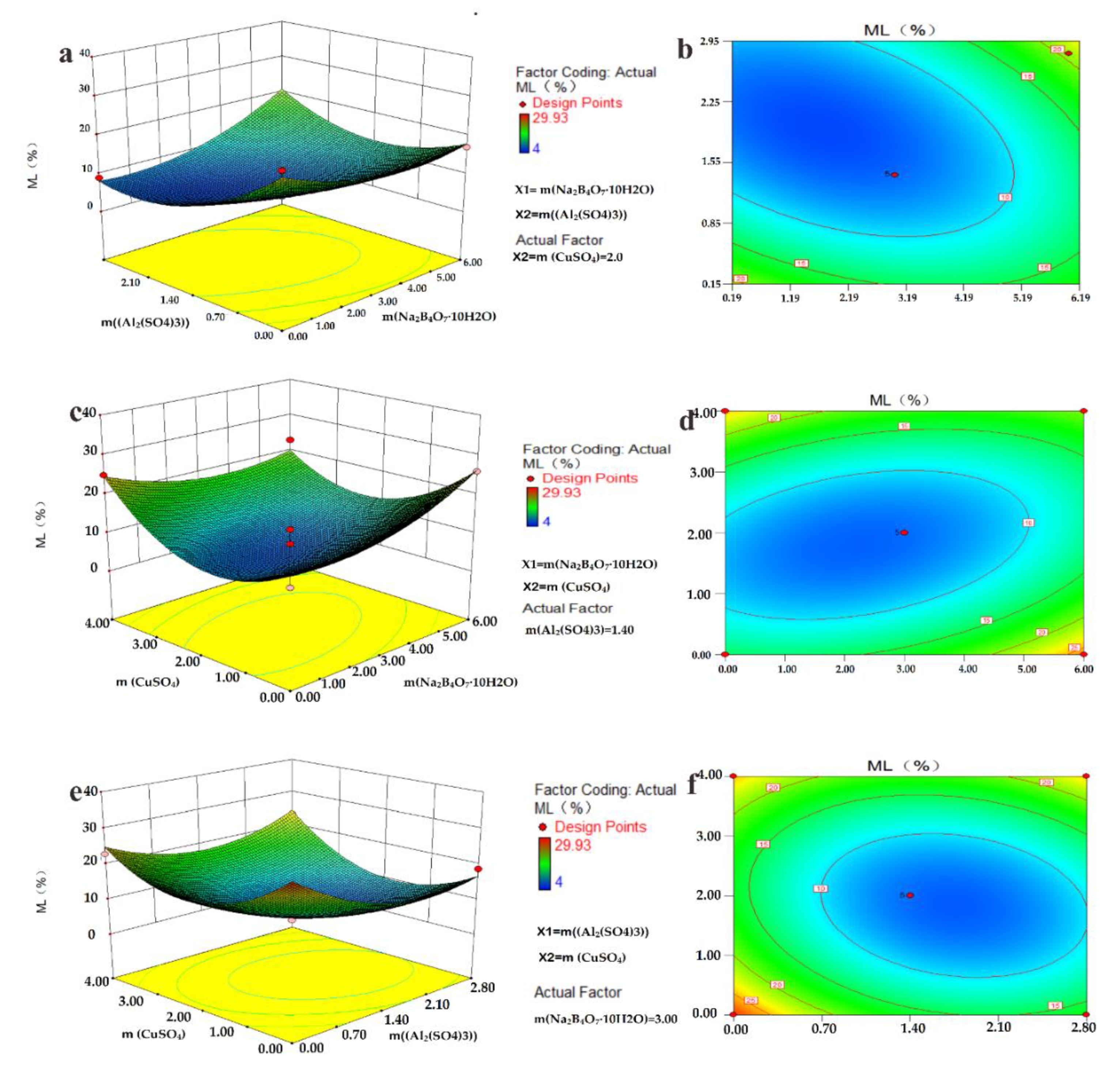
To test the validity of mass loss, the regression equation and the analysis of Variance (ANOVA) was used, and the model coefficients are presented in Table 3. The Na2B4O7 ratio (X1) has the largest influence on the p-value. The model was 0.0013, which shows that the model was significant. In the quadratic’s prediction term, the Na2B4O7·10H2O ratio (X1), reaction Al2(SO4)3 (X2) and reaction CuSO4 (X3) have an extremely interactive effect between them, and the model of the quadratic polynomial model has significant influences because of the F-value 13.19 and the Prob > F is 0.0013. This indicates that the model was significant. As shown in Table S4, the model summary statistics of both R2 = 0.9443 and 0.4740 of each response demonstrates appropriates correlation [8]. This shows an excellent correlation between the experiment and the predicted value for each of the responses between the preparation variables and the polynomial model. The response surface graph is a three-dimensional (3D) response surface plot and 2D contour plots showing the optimal levels composed of specific response value ML% and corresponding factors X1, X2, and X3, which can intuitively reflect the influence of various factors on response value [35].
The response surface analysis results of the various factors on the interaction between Al2(SO4)3, CuSO4 and Na2B4O7·10H2O are shown in Figure 2a–f. As shown in Figure 2e, the Al2(SO4)3 ratio ranged from 0 to 1.4%. Response values present a tendency to increase. When the Al2(SO4)3 ratio exceeds 1.4%, the ML tends to decrease. The effect of CuSO4 on the response value of ML is like the Al2(SO4)3 ratio, indicating that the interaction effect between the Al2(SO4)3 ratio and CuSO4 provide significantly better resistance to fungi activities. Although there are no significant differences, these results are the same as those observed with the ANOVA in Figure 2c, when the reaction of CuSO4 is fixed with the temperature at 70 °C, the reaction of CuSO4 ranges from 0 to 2%, and the response value ML decreases. In summary, when examining the concentration effects of X1, X2 and X3, the interaction of X1X2 is significantly greater than that of X1X3 and X2X3. This is the significance of opting for 3% of concentration, which has an excellent anti-fungal effect.

3.3. Microstructure Analysis of the Inoculated Wood

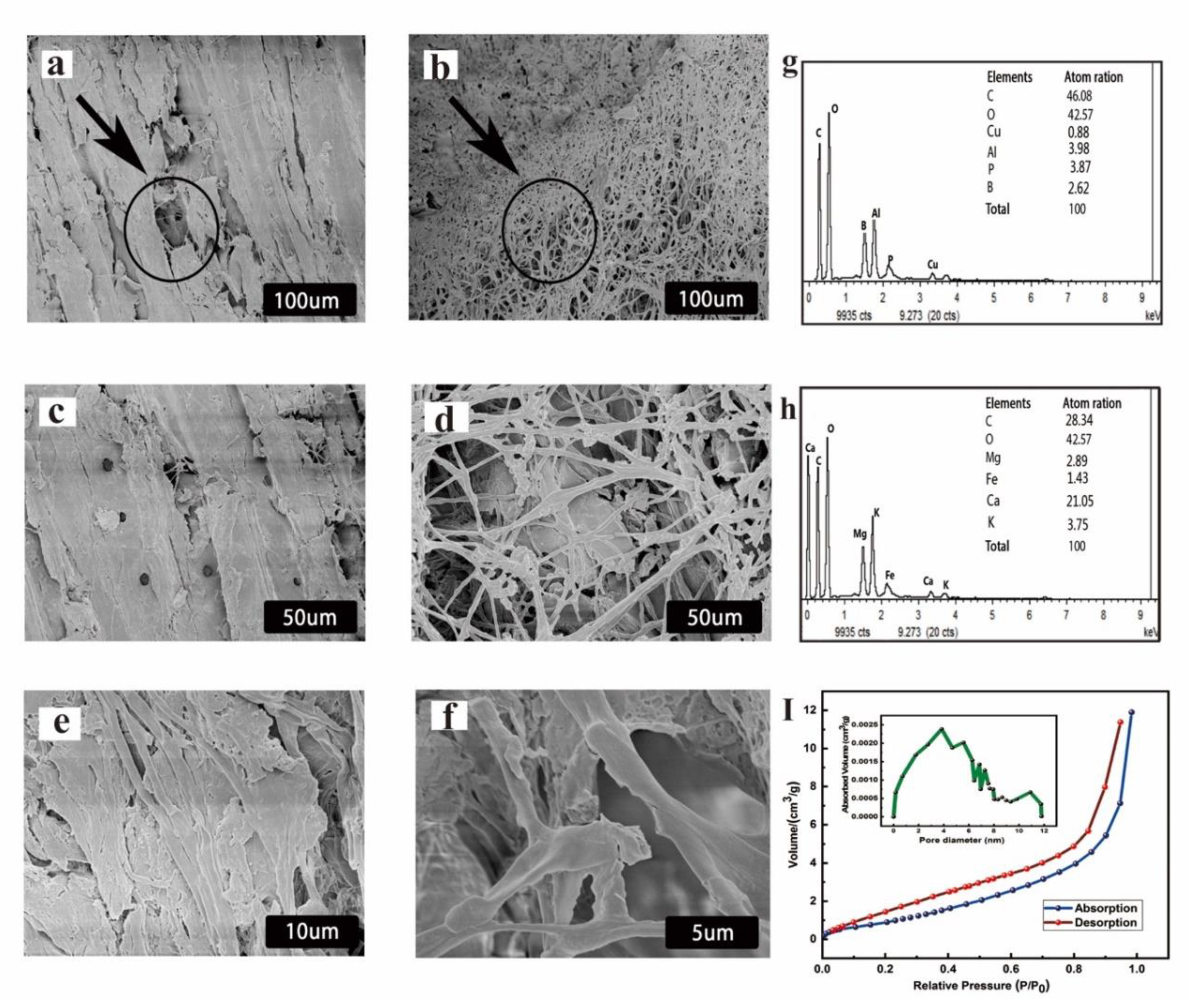
The morphological features of inorganic Al–B–P and the amphiprotic surfactant examined by SEM, as well as the microstructures and morphologies of untreated and treated wood with the solution Al–B–P, can be viewed from the cross-section, and this is displayed in Figure 3. It was observed that the impregnation of wood with Al–B–P revealed the differences between the fungi decay, which can be visible to the naked eye (Figure 3b). Under the higher magnification of 50 μm, it can be seen that the unmodified surface samples showed signs of colonization after 16 weeks of incubation with C. versicolor and P. placenta. Presented in Figure 3d,f, the penetration of the hyphae inside the wood has caused the weakness in the S2 layer because the C. versicolor can the degrade lignin ultimately [36]. It can be noted that the appearance of wood changed following exposure to fungi; the untreated wood was seriously broken-down, compared to the treated wood (Figure 3b,d,f). The Al–B–P-treated samples have minimum damage for the samples exposed to P. placenta and C. versicolor. In Figure 3e, the inorganic film around the wood cell was because of the presence of the amphiprotic surfactant in the aluminosilicate network; the toxic threshold values of the wood treated with a solution of Al–B–P in the cell walls was more effective in enhancing the properties of the wood, and this wood would become effective in enhancing the functionality against H2O2 and Fe [37]. Consequently, we could reasonably conclude that Al–B–P and the coupling agent amphiprotic surfactant both play a vital role to enhance the interfacial interaction between the fungi colonization and wood.
Using Energy-dispersive X-ray (EDX) maps confirms the distribution of chemical composition of inoculated wood. In Figure 3g, it can be seen that the treated sample contains large amounts of Al, P, B, Cu and O elements inside the wood cells, respectively. The inoculated woods with white-rot fungi results showed that in the presence of carbon and oxygen, the obtained elements were Fe, Mg, K, Ca, C and O. White-rot fungi-produced Fe elements have a function to mechanism to decompose wood cellulose or render it more susceptible to attack by conventional cellulases [38]. The absorption and desorption isotherm results of the treated sample with inorganic Al–B–P and amphiprotic surfactant, shown in Figure 3i and Figure S1. The absorption surface capacity progressively increases by the addition of Al–P–B with an augment of relative pressure N = 2 of the EO unit, especially when the relative pressure of N2 is more than 0.9. The hysteresis curve indicates the presence of a porous structure in the Al–B–P compound [39]. The pore diameter and the total pore volume of Al–B–P and the amphiprotic surfactant material are 0−12 nm, while the specific surface area of Al–B–P and the amphiprotic surfactant material was 1.78 × 102 cm3/g, and the pore diameter is 5.02 nm, respectively. The average surface area was 9.11 m2/g. The total BET surface areas and micropore surface areas are 282.7 and 156.2 m2/g, for Pinus and Spruce, respectively. Results show that the integrated mesoporous Al–B–P and amphiprotic surfactant material with hierarchical porosity exhibit unique structure mechanical properties, which provide the improved mechanical properties of a treated sample over that of the untreated sample [39].

3.4. Reaction Mechanism Analysis

To further investigate the probable chemical bonds and interaction between the presences of the functional groups among inorganic Al–B–P amphiprotic surfactant components and wood samples, we present the corresponding FTIR spectra in Figure 4b. The intensity range used for this test was ranged from 410–3740 cm−1. In Figure 4b, we observe differences between Pinus and Spruce wood before and after treatment. The spectra can be separated into two regions; the first region exhibits a strong O–H stretching peak vibration range between 2800–3740 cm−1 [40]. The absorption intensities of treated and untreated samples have shown strong O–H stretching, and the second is the vibration bands between 2900–3540 cm−1. Similarly, the same spectra were observed in treated and untreated spruce at the same frequencies. The intensities of the absorption peaks at 1300–1594 cm−1 can be assigned to the aromatic ring [41,42]. Moreover, the peak at 800 cm−1 that can be attributed to C–H deformation in cellulose and hemicellulose was shifted to 1000 cm−1 for modified spruce. The bands at 1010–1079 cm−1 in the untreated wood were more intense than in the treated wood [43]. The absorption peaks at 1508–1735 cm−1 can be attributed to C=C of hemicelluloses. The impregnation of Al–B–P and amphiprotic surfactant into raw wood could be proven through THE group C–Br band position, stretch at 507–647 cm−1 and C–N stretch of C–H in wood at 1600–2900 cm−1.
The X-ray diffraction method which was employed to test the crystallinity of samples is one of the essential methods to have the characteristics of physical, chemical and other properties of samples [44,45]. To further analyze the formation of Al–B–P and amphiprotic surfactant, the XRD patterns showed the crystalline and non-crystalline regions of the wood sample. It can be seen from Figure 4a that the three major diffraction peaks at the 2θ value of the untreated and treated wood appears around 16°, 20°, 22, and 33°, respectively, corresponding to the (110), (200) and (300) diffraction plane of crystalline cellulose Iβ [46]. The diffraction peaks show a higher increase due to the deposited particles (Al and B), which have not destroyed the crystalline structure of the cellulose on treating wood [47]. compared to untreated samples, curve 1 and curve 3 exposes after exposure to (Poria placenta) and (Coriolus versicolor), they flatten between 30°–35°, samples showing a decrease in the intensity of diffraction plane (300) plane, caused by exposure to white-rot fungi substantially degrading the lignin and decrease the thickness of crystalline cells [48]. In addition, NMR MAS provides the structural knowledge about 31P MAS NMR and 27Al MAS NMR spectra of Al–B–P presented in Figure 4c,d, according to the experimental protocol standard of 31P (100) = 1/2 and 27Al (100) = 5/2 MAS NMR spectra. Resonances are shifted by about 150 ppm. Inorganic Al–B–P structure remains unchanged at the processing temperature below 100 °C with a pH of around 9 [49]. 31P MAS NMR and 27Al MAS NMR spectra indicate a significant quadrupole shift and exhibit one small peak around 115.59 ppm with another sharp peak at −18.20 ppm. These suggest that the degree of aluminum polymerization could have increased. We can assign this Q 0 0   (0Al), Q 1 0   (1Al), Q 2 0   ( 2 Al ) Q 3 0   ( 3 Al ) and Q 4 0   (4Al) of the structural Octahedral network, Figure 4e. Furthermore, this observation was confirmed by the 31P NMR, which shows that the increasing interaction of phosphorus connectivity at these spectra could provide the formation of the Al–P linkages. The 31P characteristic chemical signal presence of peaks near −13 ppm, which was due to the double resonance [50]. Therefore, the schematic reaction between the Al–P compounds and the wood could be described as Figure 4e.

3.5. Mechanical and Physical Properties Analysis

The contact angle was measured on rectangles 20 × 20 × 5 mm3 (T × R × L), distilled water droplets of a 5 μL microsyringe of deionized water were placed onto the tangential, radial and longitudinal surfaces. As shown in Figure 5d, a water droplet was resting on the Al–B–P amphiprotic surfactant wood surface. Contrary to untreated or unmodified wood, a typical wetting behavior is observed immediate absorption and spreading of water inside the untreated wood sample after 4s, and images were taken at a specific frequency. As shown in Figure 5d.
The drop angle was measured 0, 30 and 60 s after the contact of the drop with the wood samples, and the average angles were calculated to determine the wettability. There was a significant difference between the contact angle of the untreated wood and the Al–B–P treated samples. The hydrophobicity of Al–B–P and the surface roughness of the wood is about 98°. Enhancement of the hydrophobicity of the sample CuSO4 and the C5H11NO2HCl was used as the indicator to characterize the wetting property of the treated, exhibiting the property of excellent super-hydrophobicity [51].
Figure 5a,b presents the results of the TG and differential thermogravimetric (DTG) curves of Al–B–P amphiprotic surfactant wood specimens subjected to heat in the range 50−800 °C are shown in Figure 5a,b. There is a total weight loss of 57.21% for the Al–B–P amphiprotic surfactant. Three distinct weight loss stages have accounted for 52.55%, 58.83% and 60.26%, respectively, which are assigned to the loss of free and moisture, cross-linked, and the process of thermal dehydrogenation [39].
The first region starts around 100 °C and corresponds to the dehydration of material leading to 4.16% of mass loss. The second region starts around 240 °C and corresponds to the degradation of cellulose and hemicellulose. Hemicellulose starts its decomposition around 200 °C and ends around 300 °C, while cellulose decomposed between 300 and 350 °C. The third region corresponds to the degradation of lignin and is between 350–800 °C. The entire bonds are broken, a large part of the material is pyrolyzed, and only the ashes remained [52,53]. Nevertheless, we found the many factors that influence the DTG area of the composition of inorganic constituent distribution of the chains within a constituent, the number of inorganic compounds (boric phosphoric cupric content) in the DTG curves Figure 5b. The mass wood reduction was higher in the untreated specimens as compared to the Al–B–P treated specimens with thermal decomposition peak temperatures of 331 and 336 °C. The exposure time of wood exposed to field test makes it susceptible to the attack of decay fungi, resulting in the degradation of cell wall constituents, which consequently causes a loss of mechanical strength [52]. The MOR and MOE of wood changing trends found on the Fujian site are inconsistent, which may be related to factors such as heat treatment temperature and climate [54], after impregnated wood specimens with inorganic Al–B–P mechanism performance can affect, the origin mechanical properties change. The H3PO4 phosphoric acid remaining in the Al–B–P amphiprotic surfactant may react with other wood constituent or deteriorate the wood constituent, which decreases the bonding strength of the wood specimens. In Figure 5c the results show that treated specimens MOR gradually increase by 110, 115 and 120 MPA. Figure 5c compared to untreated specimens gradually decrease by 90 and 80 MPA. Besides, the laboratory test provided a better durability value of the wood treated with Al–B–P amphiprotic surfactant than the soil contact test.

3.6. XPS Surface Chemical Analysis

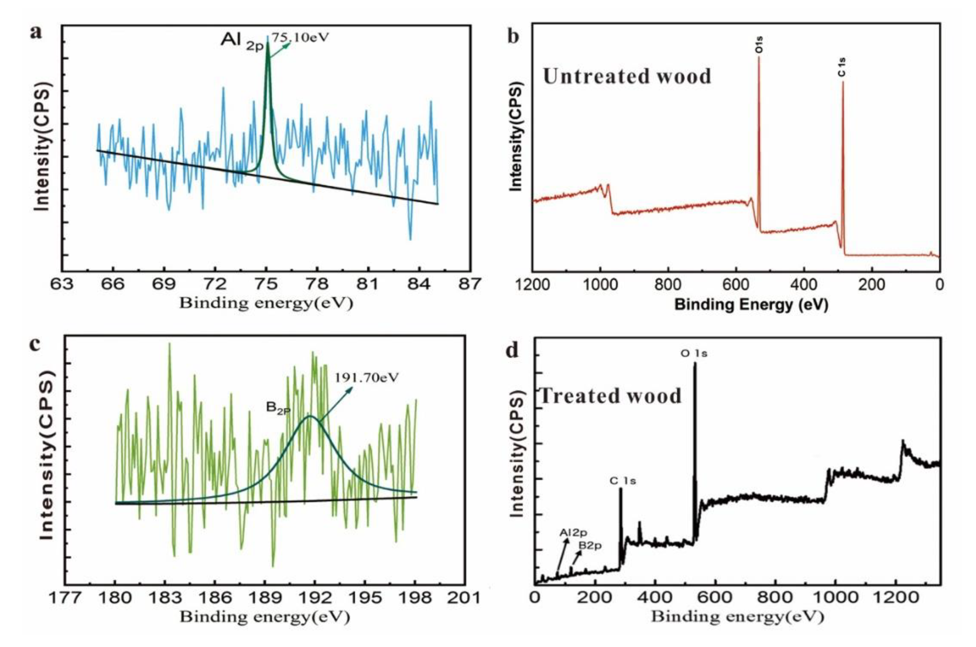
To clarify the specific chemical bond between Al–B–P amphiprotic surfactant and wood specimens, the surface chemistry of Al–B–P and wood was performed by XPS. The results of the experiment have shown that carbon and oxygen are the main elements of a scan of the survey spectrum, as presented in Figure 6b. The survey spectrum provides the elemental surface composition of wood [55]. The carbon C 1s and oxygen O 1s binding energy spectra ranges of 286.4–565 eV and 286.6–573 eV for the treating specimen, indicating the four components were designated C1, C2, C3 and C4, based on their binding energy the C 1s peak corresponds to C–C and C=C aromatic stretch attributed to lignin. The characteristic Al 2p of the Al–B–P amphiprotic surfactant is present at 75.10 ev and B 2p peaks with the binding energy of 191.70 ev (Figure 6a,c). This value is because of the electronegativity of O 1s, C 1s and the presence of the amphiprotic surfactant in the Al–B–P network. Table S5 shows the division peak percentage content for wood modified and unmodified.

4. Conclusions

In this study, new preservative materials based on inorganic chemical combinations based on boron compounds (B), aluminum sulfate compounds (Al), phosphoric acid (P) and amphiprotic surfactant materials have been obtained and impregnated on wood.
The parameters result obtained from the RSM had demonstrated the excellent anti-corrosion properties of Al–B–P amphiprotic surfactant preservatives.
The investigation of mass loss demonstrated the fact that the weight loss of the treated samples with Al–B–P compounds was 4% greater than without Al–B–P compounds wood sample more than 45%.
The decay fungi resistance was tested by exposure to Poria placenta; Coriolus versicolor and monitored using the XRD, FTIR and SEM-EDX methods.
The fixation has a significant effect on the antifungal with more remarkable durability properties in an outdoor application or usage. The test showed that Al–B–P and amphiprotic surfactants have excellent hydrophobicity.
Further research is under progress and very promising for wood conservation based on inorganic compounds for all kinds of fungus organisms’ infections in the global wood trade.

Supplementary Materials

The following are available online at https://www.mdpi.com/2079-6412/10/2/108/s1, Table S1: Mass loss of control and impregnated sample exposed to wood Poria. Placenta and Coriolus versicolor fungi for 3 months according to ASTM D4445-10(2015) [1] procedure. (Al2(SO4)3—aluminum sulfate, Na2B4O7·10H2O—sodium tetraborate; CuSO4–cupric sulfate), Table S2: Lacks of fit tests, Table S3: Sequential model sum of squares, Table S4: Model summary statistics of R2, Figure S1: Nitrogen adsorption-desorption isotherm and mesopore distribution of inorganic Al–B–P and amphiprotic surfactant material, Table S5: Peak, intensity, and atom percent of different element.

Author Contributions

Conceptualization, and data curation K.G.Y.B. and Y.X.; methodology, J.T.A.; software, B.Z.; validation, J.T.A., formal analysis, and investigation, X.H.; resources, D.L. Writing original draft review and editing, K.G.Y.B. and J.T.A. supervision, and project administration, Y.X. All authors have read and agreed to the published version of the manuscript.

Funding

This research is supported by the Scientific Research Foundation of Graduate School of Fujian Agriculture and Forestry University (1122YB033). The authors are grateful for the financial support of the National Science and Technology Support Program (2008BADA9B01) and the National Natural Science Foundation of China (30781982).

Conflicts of Interest

The authors declare no conflict of interest.

Abbreviations

RSMresponse surface methodology
Al2(SO4)3aluminum sulphate
CuSO4copper sulphate
Na2B4O7sodium tetraborate
H3PO4phosphoric acid
H3BO3boric acid
CAAcopper azole alkaline
CQCcopper quaternary creosote
ACQalkaline copper quaternary
C5H11NO2HClamphiprotic surfactant
FTIRFourier Transform infrared spectroscopy
XRDX-ray diffraction
MOEmodulus of elasticity
MORmodulus of rupture
SEMscanning electron microscopy
EDSenergy-dispersive spectroscopy
TGAThermogravimetric analysis
DTGdifferential thermogravimetric
XPSX-ray photoelectron spectroscopy
NMRnuclear magnetic resonance
BETBrunauer−Emmett−Teller equation
ANOVAanalysis of variance
MLRLoss mass rate

References

  1. Bari, E.; Nazarnezhad, N.; Kazemi, S.M.; Ghanbary, M.A.T.; Mohebby, B.; Schmidt, O.; Clausen, C.A. Comparison between degradation capabilities of the white rot fungi Pleurotus ostreatus and Trametes versicolor in beech wood. Int. Biodeterior. Biodegrad. 2015, 104, 231–237. [Google Scholar] [CrossRef]
  2. Goodell, B.; Qian, Y.; Jellison, J. Fungal decay of wood: Soft rot—brown rot—white rot. In Development of Commercial Wood Preservatives; American Chemical Society: Washington, DC, USA, 2008; Volume 982, pp. 9–31. [Google Scholar]
  3. Blanchette, R.A. An unusual decay pattern in brown-rotted wood. Mycologia 1983, 75, 552–556. [Google Scholar] [CrossRef]
  4. Asif, M.B.; Hai, F.I.; Hou, J.; Price, W.E.; Nghiem, L.D. Impact of wastewater derived dissolved interfering compounds on growth, enzymatic activity and trace organic contaminant removal of white rot fungi—A critical review. J. Environ. Manag. 2017, 201, 89–109. [Google Scholar] [CrossRef] [PubMed] [Green Version]
  5. Ohkoshi, M.; Kato, A.; Suzuki, K.; Hayashi, N.; Ishihara, M. Characterization of acetylated wood decayed by brown-rot and white-rot fungi. J. Wood Sci. 1999, 45, 69–75. [Google Scholar] [CrossRef]
  6. Zhang, Z.; Yang, T.; Mi, N.; Wang, Y.; Li, G.; Wang, L.; Xie, Y. Antifungal activity of monoterpenes against wood white-rot fungi. Int. Biodeterior. Biodegrad. 2016, 106, 157–160. [Google Scholar] [CrossRef]
  7. Goodell, B.; Daniel, G.; Jellison, J.; Qian, Y. Iron-reducing capacity of low-molecular-weight compounds produced in wood by fungi. Holzforschung 2006, 60, 630–636. [Google Scholar] [CrossRef]
  8. Wu, Z.; Huang, D.; Wei, W.; Wang, W.; Wang, X.A.; Wei, Q.; Niu, M.; Lin, M.; Rao, J.; Xie, Y. Mesoporous aluminosilicate improves mildew resistance of bamboo scrimber with CuBP anti-mildew agents. J. Clean. Prod. 2019, 209, 273–282. [Google Scholar] [CrossRef]
  9. Mercer, T.; Frostick, L. Evaluating the potential for environmental pollution from chromated copper arsenate (CCA)-treated wood waste: A new mass balance approach. J. Hazard. Mater. 2014, 276, 10–18. [Google Scholar] [CrossRef]
  10. Miyafuji, H.; Saka, S. Na2O-SiO2 wood-inorganic composites prepared by the sol-gel process and their fire-resistant properties. J. Wood Sci. 2001, 47, 483–489. [Google Scholar] [CrossRef]
  11. Lu, Y.; Feng, M.; Zhan, H. Preparation of SiO2–wood composites by an ultrasonic-assisted sol–gel technique. Cellulose 2014, 21, 4393–4403. [Google Scholar] [CrossRef]
  12. Yamaguchi, H. Properties of silicic-acid compounds as chemical-agents for impregnation and fixation of wood. Mokuzai Gakkaishi 1994, 40, 830–837. [Google Scholar]
  13. Campos, K.S.; Lenz e Silva, G.F.L.; Nunes, E.H.; Silva, A.M.; Bestard, G.A.; Vasconcelos, W.L. Surface modification of coked alumina refractories by the deposition of sol-gel derived silica coatings. Ceram. Int. 2019, 45, 8626–8633. [Google Scholar] [CrossRef]
  14. Hübert, T.; Shabir Mahr, M. Sol-gel wood preservation. Handb. Sol-Gel Sci. Technol. 2016, 1, 1–52. [Google Scholar]
  15. Hübert, T.; Shabir Mahr, M. Sol-gel wood preservation. In Handbook of Sol-Gel Science and Technology: Processing, Characterization and Applications; Klein, L., Aparicio, M., Jitianu, A., Eds.; Springer International Publishing: Cham, Switzerland, 2018; pp. 2795–2842. [Google Scholar]
  16. Tsvetkova, I.; Krasil’nikova, L.; Khoroshavina, Y.; Galushko, A.; Yu, V.F.; Kychkin, A.; Shilova, O. Sol-gel preparation of protective and decorative coatings on wood. J. Sol-Gel Sci. Technol. 2019, 92, 474–483. [Google Scholar] [CrossRef]
  17. Tshabalala, M.A.; Gangstad, J.E. Accelerated weathering of wood surfaces coated with multifunctional alkoxysilanes by sol-gel deposition. J. Coat. Technol. 2003, 75, 37–43. [Google Scholar] [CrossRef]
  18. Denes, A.R.; Tshabalala, M.A.; Rowell, R.; Denes, F.; Young, R.A. Hexamethyldisiloxane-plasma coating of wood surfaces for creating water repellent characteristics. Holzforschung 1999, 53, 318–326. [Google Scholar] [CrossRef]
  19. Guo, H.; Bachtiar, E.; Ribera, J.; Heeb, M.; Schwarze, F.; Burgert, I. Non-biocidal preservation of wood against brown-rot fungi with TiO2/Ce Xerogel. Green Chem. 2018, 20, 1375–1382. [Google Scholar] [CrossRef]
  20. Hill, C.A. Wood modification: An update. BioResources 2011, 6, 918–919. [Google Scholar]
  21. Humar, M.; Žlindra, D.; Pohleven, F. Improvement of fungicidal properties and copper fixation of copper-ethanolamine wood preservatives usingoctanoic acid and boron compounds. Holz als Roh- und Werkst. 2007, 65, 17–21. [Google Scholar] [CrossRef]
  22. European Commission. Directive 98/8/EC of the European Parliament and of the Council of 16 February 1998 concerning the placing of biocidal products on the market. Off. J. Eur. Union 1998, 41, 123. [Google Scholar]
  23. Mourant, D.; Yang, D.-Q.; Lu, X.; Riedl, B.; Roy, C. Copper and boron fixation in wood by pyrolytic resins. Bioresour. Technol. 2009, 100, 1442–1449. [Google Scholar] [CrossRef] [PubMed]
  24. Råberg, U.; Terziev, N.; Daniel, G. Degradation of Scots pine and beech wood exposed in four test fields used for testing of wood preservatives. Int. Biodeterior. Biodegrad. 2013, 79, 20–27. [Google Scholar] [CrossRef]
  25. En 252. Field Test Method for Determining the Relative Protective Effectiveness of a Wood Preservative in Ground Contact; British Standards institute: London, UK, 2014. [Google Scholar]
  26. ASTM D4445. Standard Test Method for Fungicides for Controlling Sapstain and Mold on Unseasoned Lumber (Laboratory Method); ASTM International: West Conshohocken, PA, USA, 2015. [Google Scholar]
  27. ASTM D2017-05. Standard Test Method of Accelerated Laboratory Test of Natural Decay Resistance of Woods; ASTM International: West Conshohocken, PA, USA, 2017. [Google Scholar]
  28. Tascioglu, C.; Cooper, P.; Ung, T. Adsorption of ACQ and CuMEA wood preservatives in red pine. Int. Res. Group on Wood Pres. Document No: IRG/WP/05-30374, 2005. [Google Scholar]
  29. Jiang, X. Fixation Chemistry of Amine-Copper Preservatives. Ph.D. Thesis, University of British Columbia, Vancouver, BC, Canada, November 2000. [Google Scholar]
  30. En 84. Wood Preservatives–Accelerated Ageing of Treated Wood Prior to Biological Testing–Leaching Procedure; CEN (European Committee for Standardization): Brussels, Belgium, 1997. [Google Scholar]
  31. Lesar, B.; Podlesnik, B.; Pohleven, F.; Humar, M.; Kralj, P.; Veber, M. Performance of boron-ethanolamine-quaternary ammonium based wood preservatives against leaching, wood decay and blue stain fungi. Wood Res. 2008, 53, 17–26. [Google Scholar]
  32. Mahr, S.; Hübert, T.; Stephan, I.; Militz, H. Decay protection of wood against brown-rot fungi by titanium alkoxide impregnations. Int. Biodeterior. Biodegrad. 2013, 77, 56–62. [Google Scholar] [CrossRef]
  33. Usmani, S.M.; Stephan, I.; Hübert, T.; Kemnitz, E. Nano metal fluorides for wood protection against fungi. ACS Appl. Nano Mater. 2018, 1, 1444–1449. [Google Scholar] [CrossRef]
  34. Dong, Z.; Zhou, Y.; Ren, H. Optimization on ultrasonic-assisted extraction technology of chlorophyll from radish leaf. Trans. Chin. Soc. Agric. Eng. 2011, 27, 288–292. [Google Scholar]
  35. Hyde, S.M.; Wood, P.M. A mechanism for production of hydroxyl radicals by the brown-rot fungus Coniophora puteana: Fe (III) reduction by cellobiose dehydrogenase and Fe (II) oxidation at a distance from the hyphae. Microbiology 1997, 143, 259–266. [Google Scholar] [CrossRef] [Green Version]
  36. Takeshi, F.; Kwnta, S.; Tohru, U. Combination of wood and silicate II: Wood-mineral composites using water glass and reactants of barium chloride, boric acid, and borax, and their properties. Mokuzai Gakkaishi 1992, 38, 448–457. [Google Scholar]
  37. Blanchette, R.A.; Otjen, L.; Effland, M.J.; Eslyn, W.E. Changes in structural and chemical components of wood delignified by fungi. Wood Sci. Technol. 1985, 19, 35–46. [Google Scholar] [CrossRef]
  38. Chen, T.; Xie, Y.; Cai, L.; Zhuang, B.; Wang, X.A.; Wu, Z.; Niu, M.; Lin, M. Mesoporous aluminosilicate material with hierarchical porosity for ultralow density wood fiber composite (ULD_WFC). ACS Sustain. Chem. Eng. 2016, 4, 3888–3896. [Google Scholar] [CrossRef]
  39. Traoré, M.; Kaal, J.; Cortizas, A.M. Application of FTIR spectroscopy to the characterization of archeological wood. Spectrochim. Acta Part A Mol. Biomol. Spectrosc. 2016, 153, 63–70. [Google Scholar] [CrossRef] [PubMed]
  40. Rahman, M.R.; Hamdan, S. Preparation and characterizations of various clay-and monomers-dispersed wood nanocomposites. In Wood Polymer Nanocomposites; Springer: Cham, Switzerland, 2018; pp. 37–68. [Google Scholar]
  41. Cesar, T.; Danevčič, T.; Kavkler, K.; Stopar, D. Melamine polymerization in organic solutions and waterlogged archaeological wood studied by FTIR spectroscopy. J. Cult. Herit. 2017, 23, 106–110. [Google Scholar] [CrossRef]
  42. Özgenç, Ö.; Durmaz, S.; Boyaci, I.H.; Eksi-Kocak, H. Determination of chemical changes in heat-treated wood using ATR-FTIR and FT Raman spectrometry. Spectrochim. Acta Part A Mol. Biomol. Spectrosc. 2017, 171, 395–400. [Google Scholar] [CrossRef] [PubMed]
  43. Andersson, S.; Serimaa, R.; Paakkari, T.; Saranpää, P.; Pesonen, E. Crystallinity of wood and the size of cellulose crystallites in Norway spruce (Picea abies). J. Wood Sci. 2003, 49, 531–537. [Google Scholar] [CrossRef] [Green Version]
  44. Weimer, P.; Hackney, J.; French, A. Effects of chemical treatments and heating on the crystallinity of celluloses and their implications for evaluating the effect of crystallinity on cellulose biodegradation. Biotechnol. Bioeng. 1995, 48, 169–178. [Google Scholar] [CrossRef] [PubMed]
  45. French, A.D. Idealized powder diffraction patterns for cellulose polymorphs. Cellulose 2014, 21, 885–896. [Google Scholar] [CrossRef]
  46. Broda, M.; Mazela, B.; Dutkiewicz, A. Organosilicon compounds with various active groups as consolidants for the preservation of waterlogged archaeological wood. J. Cult. Herit. 2019, 35, 123–128. [Google Scholar] [CrossRef]
  47. Rosu, L.; Varganici, C.-D.; Mustata, F.; Rusu, T.; Rosu, D.; Rosca, I.; Tudorachi, N.; Teacă, C.-A. Enhancing the thermal and fungal resistance of wood treated with natural and synthetic derived epoxy resins. ACS Sustain. Chem. Eng. 2018, 6, 5470–5478. [Google Scholar] [CrossRef]
  48. Eckert, H. Synthesis of non-siliceous glasses and their structural characterization by solid-state NMR. Handb. Sol-Gel Sci. Technol. Process. Charact. Appl. 2018, 1, 1323–1373. [Google Scholar]
  49. Dirken, P.J.; Smith, M.E.; Whitfield, H.J. 17O and 29Si solid state NMR study of atomic scale structure in sol-gel-prepared TiO2-SiO2 materials. J. Phys. Chem. 1995, 99, 395–401. [Google Scholar] [CrossRef]
  50. Wang, X.; Liu, S.; Chang, H.; Liu, J. Sol-gel deposition of TiO2 nanocoatings on wood surfaces with enhanced hydrophobicity and photostability. Wood Fiber Sci 2014, 46, 109–117. [Google Scholar]
  51. Mattos, B.D.; de Cademartori, P.H.; Lourençon, T.V.; Gatto, D.A.; Magalhães, W.L. Biodeterioration of wood from two fast-growing eucalypts exposed to field test. Int. Biodeterior. Biodegrad. 2014, 93, 210–215. [Google Scholar] [CrossRef]
  52. Popescu, C.-M.; Lisa, G.; Manoliu, A.; Gradinariu, P.; Vasile, C. Thermogravimetric analysis of fungus-degraded lime wood. Carbohydr. Polym. 2010, 80, 78–83. [Google Scholar] [CrossRef]
  53. Gašparovič, L.; Koreňová, Z.; Jelemenský, Ľ. Kinetic study of wood chips decomposition by TGA. Chem. Pap. 2010, 64, 174–181. [Google Scholar] [CrossRef]
  54. Wang, X.H.; Liu, J.L.; Fei, B.H. Effect of vacuum heat treatment temperature on physical and mechanical properties of Eucalyptus pellita wood. Wood Fiber Sci. 2014, 46, 368–375. [Google Scholar]
  55. Rowell, R. Surface characterization. In Handbook of wood chemistry and wood composites, 2nd ed.; Rowell, R. CRC Press, Taylor and Francis group: Boca Raton, FL, USA, 2012; pp. 217–252. [Google Scholar]
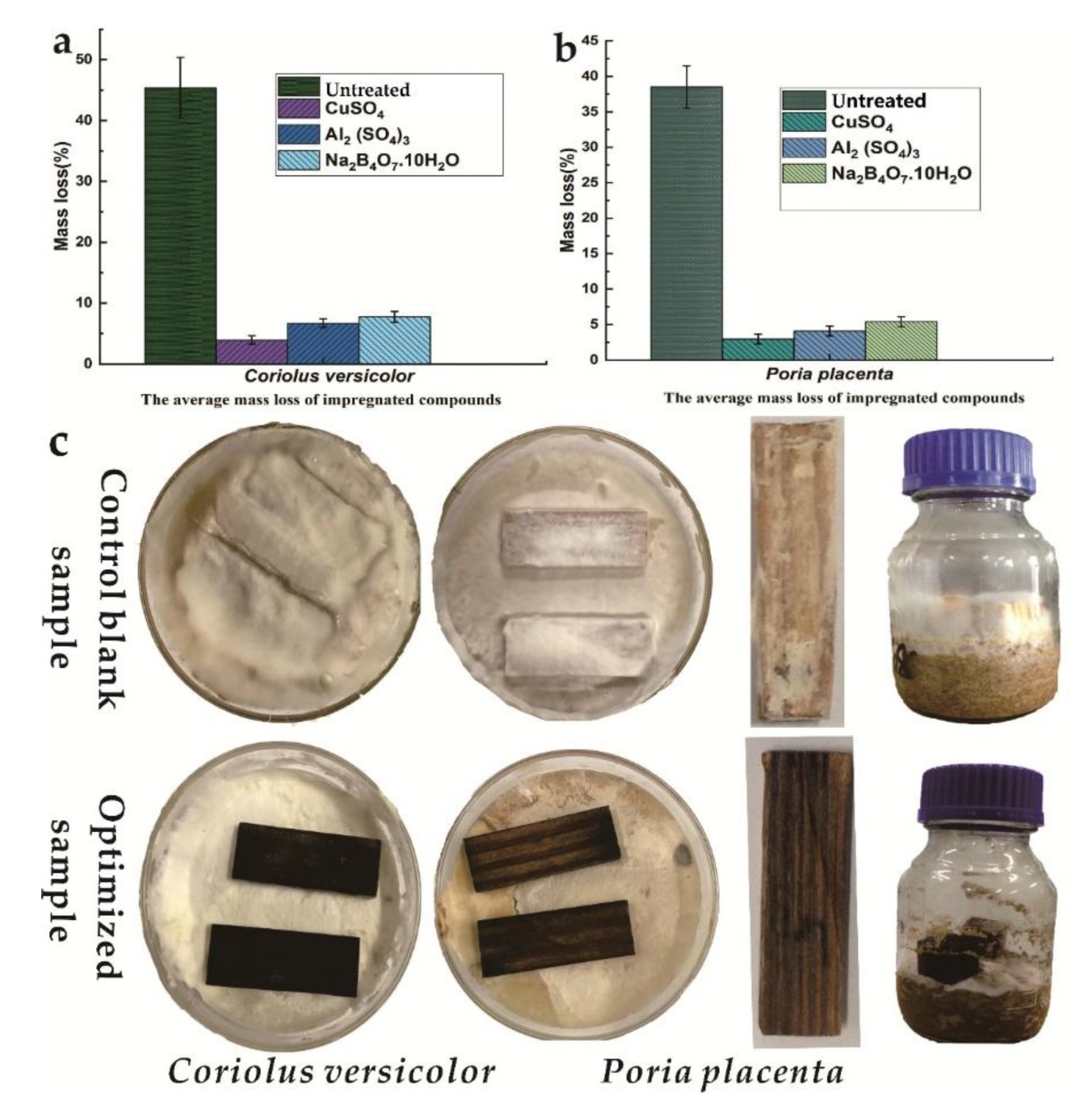
Figure 1. Average mass losses of the wood specimens of different concentrations of Al–B–P and amphiprotic surfactant solution after exposure to Poria placenta and Coriolus versicolor (a,b); Wood specimens after exposure white rot fungi Agar block test and soil-block test in control and treated (optimized) with inorganic Al–B–P and amphiprotic surfactant after exposure to C. versicolor and P. placenta (c).
Coatings 10 00108 g001
Figure 2. Three dimensional (3D) Response surface plots and contour plots for the maximum ML% and two dimensional (2D) contour lines: (a,b) effects of mass rate of Na2B4O7·10H2O and Al2(SO4)3; (c,d) effects of mass rate of Na2B4O7·10H2O and CuSO4; (e,f) effects of mass rate of CuSO4 and Al2(SO4)3.
Coatings 10 00108 g002
Figure 3. Scanning electron microscopy (SEM) morphology of natural, untreated wood after exposure to Poria placenta for 16 weeks (b,d,f); SEM morphology of inorganic Al–B–P and amphiprotic surfactant treated wood after exposure to the white rot fungus (a,c,e); 100, 50, 10, 5 μm respectively. Energy-dispersive X-ray spectroscopy (EDX) profile map energy dispersive solutions compounds inside sample-treated wood specimens (g) natural wood after exposure to P. placenta EDX profile (h); nitrogen adsorption-desorption isotherm and mesopore distribution of inorganic Al–B–P and amphiprotic surfactant material (I).
Coatings 10 00108 g003
Figure 4. XRD analysis of wood treated and untreated by Al–B–P (a) (FT-IR) of treated wood with Al–B–P compounds and without compounds (b) 31P MAS NMR and 27Al MAS NMR spectra of Al–B–P amphiprotic surfactant and wood (c,d) Schematic of the reaction between wood and amphiprotic surfactant compounds (e).
Coatings 10 00108 g004
Figure 5. TG (a) DTG curves (b) of Al–B–P compounds and without Al–B–P compounds. MOE and MOR of untreated and treated wood specimens after 2 years exposure to the field (c) Contact angle images of Al–B–P amphiprotic surfactant a water droplet on a unmodified wood sample 0 s, and modified 0,30 and 60 s respectively (d).
Coatings 10 00108 g005
Figure 6. Typical XPS profiles of C 1s, O 1s untreated wood (b), Typical XPS of C 1s, O 1s, B 2p, and Al 2p of wood after anti-fungal treatment (a,c,d).
Coatings 10 00108 g006
Table 1. Code and level of effective antifungal agents.
FactorsCode and Levels *
–101
X100.71.4
X2012
X301.53
* Combinations at two levels (−1, 0) low (0, 1) high levels.
Table 2. Specific standards evaluation of wood-decay fungi.
(AMLR%) * of Wood Sample Level Class Resistance to Test FungusAverage Residual Weight (%)
0%–10%Strong rot Resistant90–100
11%–24%rot resistant76–89
25%–44%slightly rot resistant56–75
<45%not resistant to rot<55
* Average mass loss rate%.
Table 3. Box–Behnken design and response values.
TestX1%X2%X3%* Mass Loss%
13.002.800.0018.8
23.001.402.0011
36.001.400.0026
43.000.004.0023
53.001.402.005
63.002.804.0023.3
73.001.402.004
83.000.000.0029.93
90.000.002.0025.1
106.002.802.0019.26
110.001.404.0025
126.000.002.0017.08
133.001.402.007.2
140.002.802.009.02
153.001.402.006.58
166.001.404.0023.04
170.001.400.0011.63
* ML: Mass loss of white and brown-rot fungi (Poria placenta; and Coriolus versicolor).
Table 4. Analysis of variance (ANOVA).
SourceSquaresdfMean SquareF-Valuep-Value
Model1079.809119.9813.190.0013
X1 (Na2B4O7)26.75126.752.940.1301
X2 Al2(SO4)376.45176.458.400.0230
X3 (CuSO4)7.9617.960.870.3807
X1X283.36183.369.160.0192
X1X366.67166.677.330.0303
X2X332.66132.663.590.1000
X1276.39176.398.400.0231
X22183.381183.3820.160.0028
X32455.591455.5950.080.0002
Residual63.6879.10
Lack of Fit34.77311.591.600.3219
Pure Error28.9247.23
Cor Total1143.4816
Model1079.809119.9813.190.0013