Article

Biocompatibility of Niobium Coatings

1
Department of Biomedical Engineering, Georgia Institute of Technology, Atlanta, GA 30332, USA
2
Unidad de Materiales, Departamento de Ingeniería Mecánica y Mecátronica, Avenida 30 No. 45-03, Ciudad Unversitaria, Bototá D.C., Colombia
3
Instituto de Investigaciones en Materiales, Universidad Nacional Autónoma de México, Circuito exterior SN, Ciudad Universitaria, 04510 México DF, México
*
Author to whom correspondence should be addressed.
Coatings 2011, 1(1), 72-87; https://doi.org/10.3390/coatings1010072
Submission received: 2 August 2011 / Revised: 1 September 2011 / Accepted: 15 September 2011 / Published: 22 September 2011
(This article belongs to the Special Issue Advances in Medical Device Coatings)

Abstract

: Niobium coatings deposited by magnetron sputtering were evaluated as a possible surface modification for stainless steel (SS) substrates in biomedical implants. The Nb coatings were deposited on 15 mm diameter stainless steel substrates having an average surface roughness of 2 μm. To evaluate the biocompatibility of the coatings three different in vitro tests, using human alveolar bone derived cells, were performed: cellular adhesion, proliferation and viability. Stainless steel substrates and tissue culture plastic were also studied, in order to give comparative information. No toxic response was observed for any of the surfaces, indicating that the Nb coatings act as a biocompatible, bioinert material. Cell morphology was also studied by immune-fluorescence and the results confirmed the healthy state of the cells on the Nb surface. X-ray diffraction analysis of the coating shows that the film is polycrystalline with a body centered cubic structure. The surface composition and corrosion resistance of both the substrate and the Nb coating were also studied by X-ray photoelectron spectroscopy and potentiodynamic tests. Water contact angle measurements showed that the Nb surface is more hydrophobic than the SS substrate.

1. Introduction

Biomaterials are an important aspect in the development of biomedical devices and implants. The surface of the biomaterial is the first to contact the living tissue when the material is placed in the body. Therefore, the initial response of the living tissue to the biomaterial depends on the surface properties. Currently, it is rare that a biomaterial with good bulk properties also possesses the surface characteristics suitable for some clinical applications. For that reason, over the last years, the development of surface modification techniques for biomaterials has been expanding rapidly. In this way, it is possible to make ideal biomaterials with surfaces attributes that are decoupled from the bulk properties. For instance, by altering the surface functionality through the deposition of a thin film, the optimal surface, chemical and physical properties could be attained. One of the surface properties of great importance for orthopedic or dental implants, which is capable of being modified, is the corrosion resistance. The corrosion resistance of the metallic materials determines the device service life, but is also important because of the harmfulness of corrosion products, which can be released and then interact with the living organisms [1]. Refractory metals are known to have excellent corrosion resistance, and this explains the extensive use of Co-Cr and Ti alloys for orthopedic implants. However, apart from these two alloys, there is little information about the biocompatibility of the other refractory metal alloys. Particularly, Ta, Nb and Zr have been studied and used as implant materials, and it has been reported that these metals possess good biocompatibility, viable for their use as biomaterials [2,3]. More recently [4], an in vivo animal study including Titanium, Hafnium, Niobium, Tantalum and Rhenium showed that all these metals have sufficiently good biocompatibility. The authors in [4] showed that healing around the position where the metallic wires were inserted in the animal (near to soft and hard tissues) proceed rapidly. Moreover, the experiments showed the formation of new bone well attached to the surface of the metal implant, i.e., the metals have good biocompatibility and osteoinductivity [4]. Similarly, Ta has recently gained interest for a variety of orthopedic implant applications due to its osteoinductive properties [5] and the possibility of using it as a porous trabecular-like structure for hard tissue generation [6]. Some other reports about the biological response of the cells in contact with Nb came from the evaluation of TiNbx alloys, in which Nb had been introduced to substitute Al or V [7-9]. In all cases it was demonstrated that the short and long-term biocompatibility of Nb is excellent; it is a good alloying substitute providing the mechanical strength to the Ti alloys, as well as a biocompatible element. Eisenbarth et al. [10] tested samples of pure crystalline Nb, (cp-Nb), showing that the cell proliferation, mitochondrial activity and cell volume were similar and superior to that of Ti and stainless steel samples, respectively.
Taking advantage of the fact that the surface characteristics are relevant for the interaction between the implant and the living tissue [11-13], we propose to make use of the thin film technology to modify any base-material with a biocompatible-coating. The coating material could be metallic such as Nb or non-metallic, a metal oxide or nitride—materials that can be easy and inexpensively produced by thin film deposition techniques [14]. Magnetron sputtering constitutes one of the physical vapor deposition techniques that have been widely used in applications involving the metal-mechanic industry with great success [15,16]. For the biomedical field, however, this is an innovative subject with great possibilities which is now under extensive research [17-19]. The coatings should preferably have a high hardness value, resistance to corrosion and mechanical wear, and good adhesion to the substrate on which they are deposited, as well as biocompatibility.
The purpose of this study was to evaluate the biocompatibility of Nb thin films deposited by magnetron sputtering. The biocompatibility was assessed by an in vitro model [20-22]; quantifying the adhesion, proliferation and viability of human osteoblasts cells in contact with the film surface in comparison with samples of medical grade stainless steel and tissue culture plastics, which represents the experimental optimum standard for investigations of osteoblast biology in vitro. Most probably, Nb thin films will not be the best material for real applications since Nb is considered a soft metal, but thin films based on Nb, such as Nb oxides or nitrides have a high potentiality, since they are hard, and wear and corrosion resistant [23,24]. Nevertheless, for biomedical applications, it is very important to determine the toxicity of the isolated elements as well as their compounds, since these may be released from the implant surface due to device corrosion or wear for example.

2. Results and Discussion

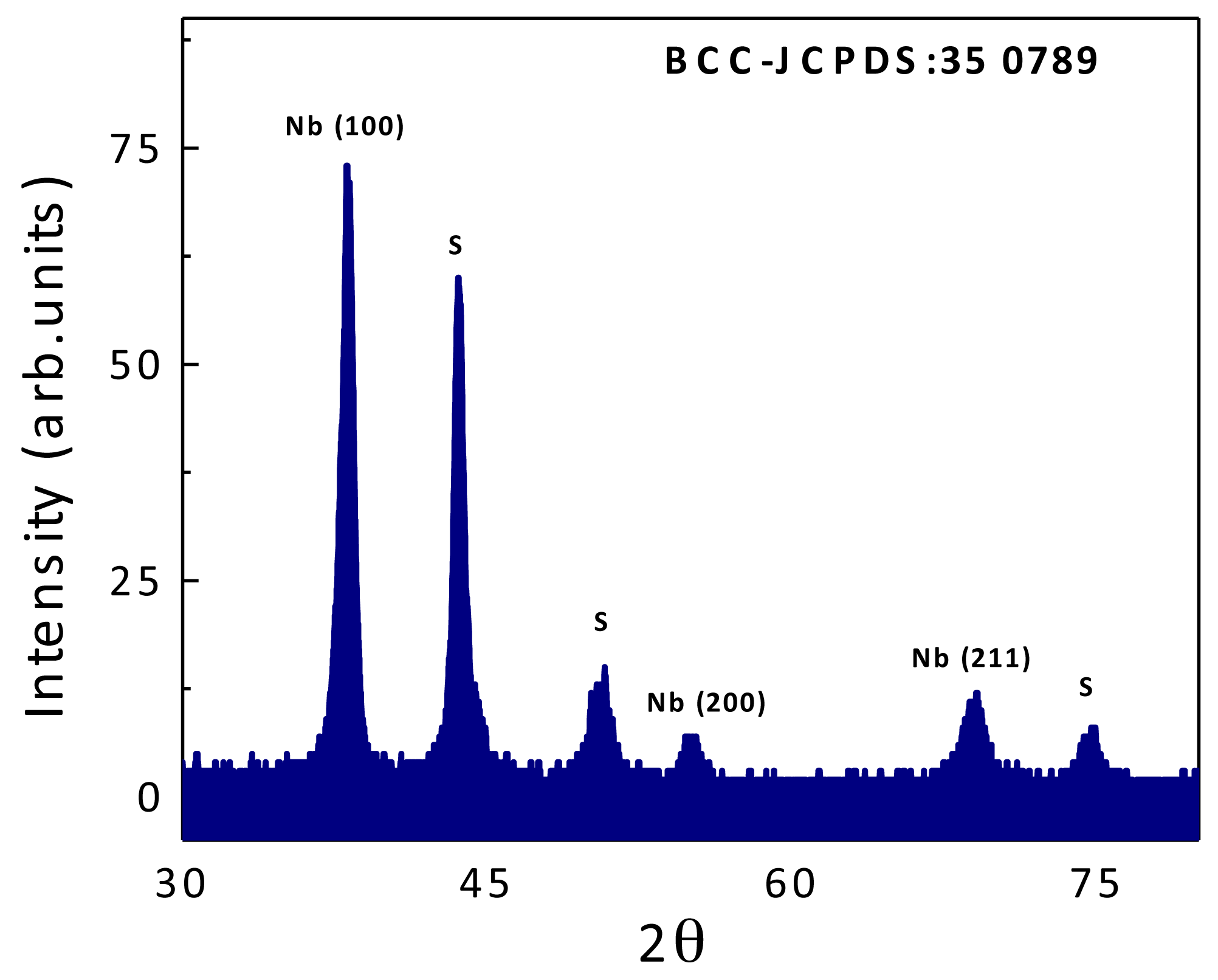
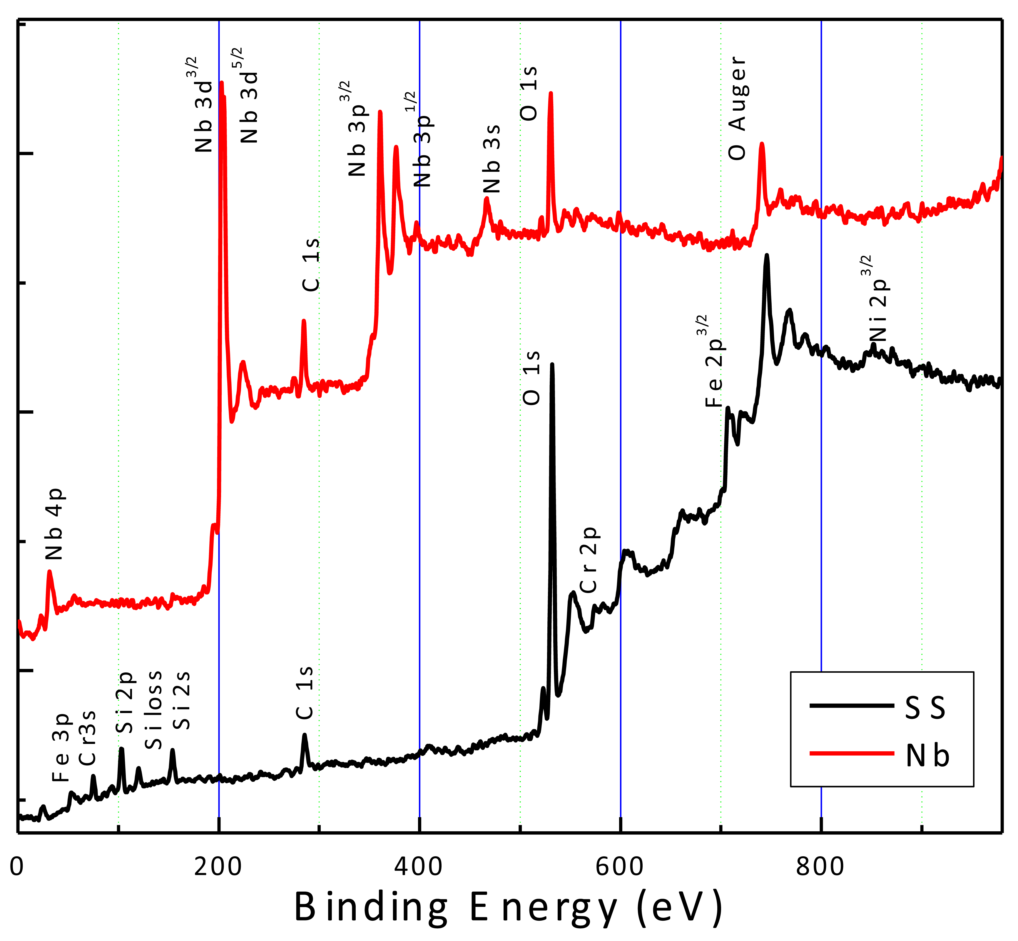
2.1. Coatings Physical Properties

The X-ray diffraction (XRD) spectrum of the Nb film deposited on stainless steel is shown in Figure 1. The diffractogram shows mainly peaks of crystalline-Nb indicating that the film has a bcc (body centre cubic) structure as the bulk Niobium; the remaining peaks belong to the stainless steel substrate.
The surface composition was determined by X-ray photoelectron spectroscopy (XPS) and the spectra of both the substrate and the film are shown in Figure 2. The spectrum of the SS showed the major elements of the AISI 316L; Fe, Cr, Ni and the O from the surface CrOx layer, while for the films no traces of the substrate elements were detected, suggesting a good coating uniformity. Carbon and oxygen signals seen in the Nb spectra, appeared as surface contaminants, which were easily removed by an argon cleaning process, indicating that they were not chemically bonded to the constituent elements. Moreover, it was observed that the binding energy of the Nb peaks corresponded to metallic Nb and not to any NbOx phases.
The surface topography of both Nb and SS, observed by scanning electron microscopy (SEM), consisted of a random distribution of dips and scratches (Figure 3). The surfaces were completely isotropic, without any preferential orientation of the features and this was also reflected by the surface roughness Ra, which was the same when measured in the two opposite directions and the values did not vary significantly after the film deposition (see Table 1).
The water contact angle, on the other hand, was strongly modified by the film deposition changing from 46° on the SS to 67° on the Nb film; these data can be observed in Table 1.
Figure 4 shows the results of the potentiodynamic tests for the Nb film and the stainless steel. It might be seen that the corrosion potential, corrosion and critical-passivation currents were similar for the films and the stainless steel. This is an indication that Nb film also forms a metastable oxide-passive coating (most probably Nb2O5) in the H2SO4 solution. A recent publication regarding the corrosion of bulk niobium in sulfuric and hydrochloric solutions at elevated temperatures shows similar behavior as the Nb film, with a clear surface passivation [25]. Even though the corrosion parameters cannot be directly compared due to the differences in the solution concentration and temperature, the order of magnitude are similar. For organic or ceramic coatings deposited on stainless steel substrates, electrochemistry analysis can be used to determine the exposed metal surface area due to the existence of pores or pinholes in the film. However, the metallic behavior of the Nb film hinders this kind of analysis. Nevertheless, we believed that it is still possible to improve the corrosion resistance, when Nb oxides or nitride films are used, which is the next step of the present research.
The surfaces were also observed using the SEM after the corrosion test in order to determine if some film degradation occurred. The images (not included here) exhibited very similar morphology and features as obtained before the corrosion test with no evidence of blistering or film de-bonding.

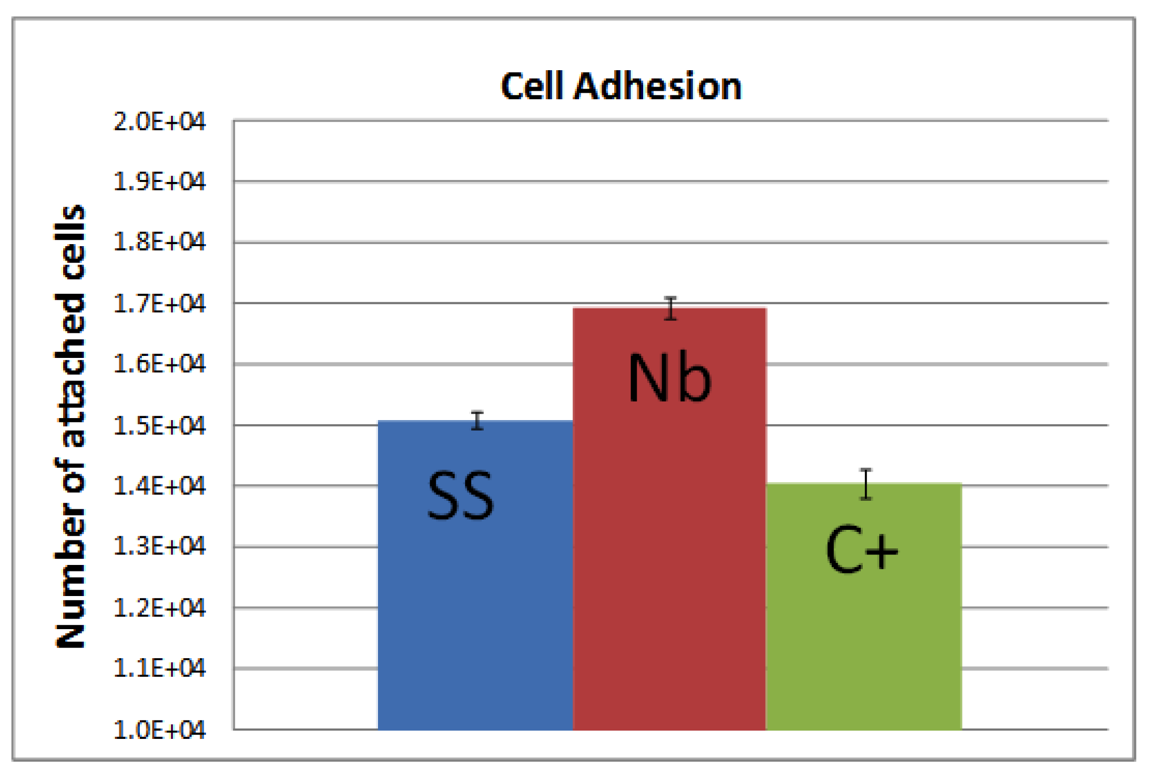
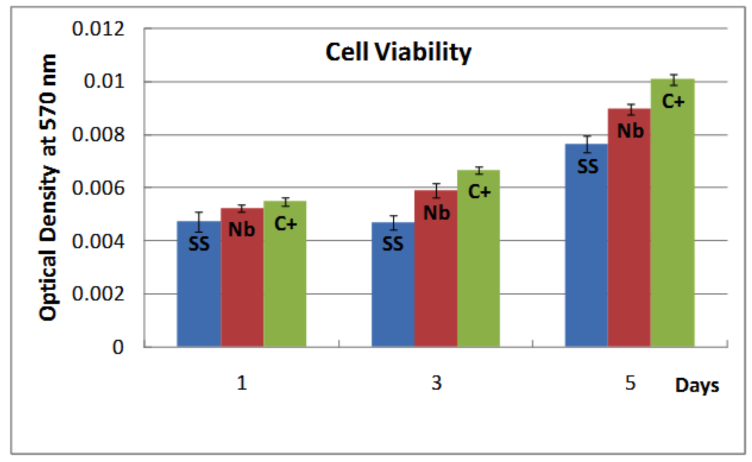
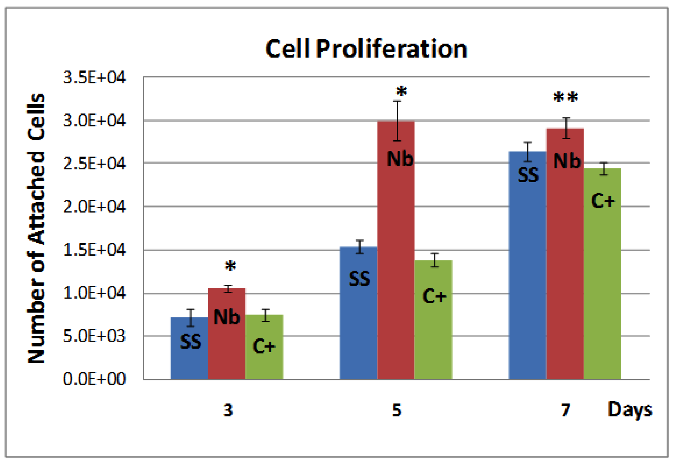
2.2. Biocompatibility
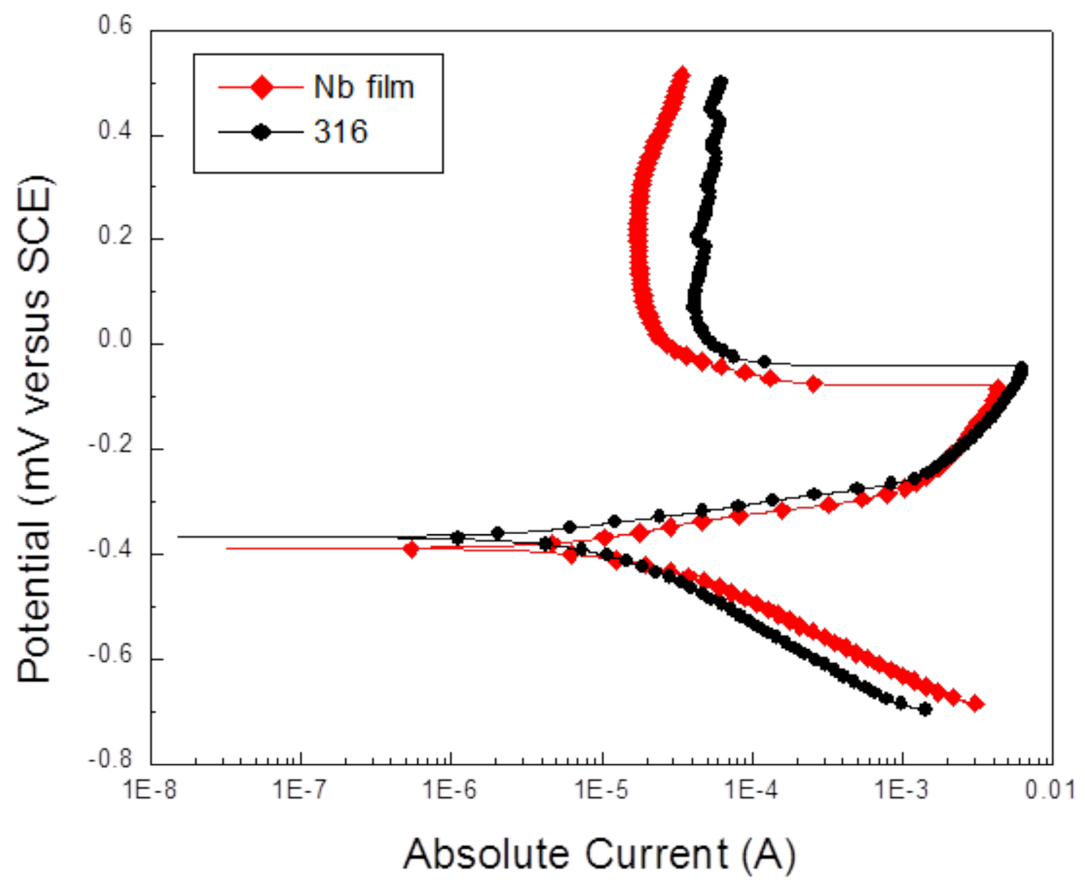
The attachment of human osteoblasts cells on Nb thin films was compared to the medical grade stainless steel (SS) and the tissue culture plastic (C+). Figure 5 shows the absolute number of attached cells after a culture period of 24 hours. It was concluded from this study that the number of attached cells to the Nb film was superior to that of SS and the control. Indeed there was a 20% more attachment on the Nb and only 7% on the SS when compared to the control (considered as 100%). The results therefore indicated that, under the conditions used for this experiment, the osteoblasts cells exhibit a statistically higher preference for the Nb surface, compared to that of stainless steel or the tissue culture plastic surfaces.
Figure 6 shows the results from the proliferation experiments, it shows the number of attached cells to each of the substrates after the incubation period. It can be seen that the number increased approximately 3-fold over a 5 day culture for the Nb film, while the SS and C+ the proliferation rate was lower, attaining the 3-fold factor over a 7 days period. These results are in good agreement with the major attachment of cells on the Nb surface, observed in Figure 4, and indicated that the human osteoblast cells can attach and grow more effectively on the Nb surfaces compared to the substrate and the tissue culture plastic.
The results obtained by the proliferation test were further confirmed by the cell viability assay (MTT) combination, which is very important since proliferation/viability tests provide information about the cell growth and the metabolic activity of the new cells. The results of the MTT assay (Figure 7) are presented as the optical density at 570 nm (OD570, high absorbance, high metabolic activity and less toxicity) as a function of the culture time for each surface. It might be seen that the levels of MTT conversion are similar for the three surfaces. At days 3 and 5, a more significant statistical difference was detected that indicated a better response of the C+ than the Nb film and the SS. Nevertheless, for all the surfaces the metabolic activity increases with time, as expected since there were an increasing number of cells. The results therefore indicated that none of the surfaces exerted toxic effects to the human osteoblast cells.
The images obtained from the immune-fluorescence microscopy are shown in Figure 8; the actin is a protein abundant in many cells that significantly contributes to the cell structure and motility. The actin cytoskeleton is organized into bundles and networks of filaments that supports the plasma membrane and, therefore determines the cell’s shape. The pictures in Figure 8 represented the morphology of the human osteoblast cells cultured on the Nb and SS surfaces. It can be seen that in the Nb surface, the cells rapidly assumed the elongated fibroblastic appearance typical of osteoblast-like cells plated onto tissue culture plates. However, in the SS surfaces, at day 3 the cells have a rounded appearance and even at day 7, the cytoskeleton is not as well organized as in the Nb surface at day 5.

2.3. Discussion

The general requirements of biomaterials are non-toxicity, stability, tissue compatibility, mechanical properties according to the application as well as simple and reproducible fabrication methods. No one material can meet all these requirements, and surface modification is frequently used to improve the properties. The alteration of the surface can vary from a simple modification of the surface topography or activation of adsorbed species to the deposition of a coating having different chemical and physical properties. The modified surface must fulfill another set of requirements defined by the specific application and the biocompatibility is one of them. In this work, we evaluated the biocompatibility of Nb coatings deposited on stainless steel surfaces by an in vitro model, which includes cellular attachment, proliferation, viability and observation of cell morphology. The purpose of the study was to evaluate the feasibility of using Nb-based thin films for biomedical applications.
Human osteoblasts cells were able to attach and grow when they were seeded on Nb-coated stainless steel surfaces. Indeed, the number of viable cells increased with time on the three surfaces investigated (Nb, SS and C+), indicating that none of these surfaces were toxic to the osteoblast cells. Some differences in cell behavior were however noted which were a consequence of the different surface chemistry and properties. Figure 5 shows that the initial attachment of osteoblasts cells was higher on Nb as compared to the SS and the control. This is a significant result since it has been reported that the initial recruitment of osteoblastic cells to a biomaterial is of great importance: the cell number rather than relative expression of phenotype is more important for osteoblast mineralization [26]. Thus, the recruitment of statistically more cells to the Nb samples compared to the SS samples has implications in the long term success of it as an implant material, in which the integration of the surface to the new bone is important. In addition to the results of the cellular attachment, we observed a faster growth rate of cells on Nb, and an excellent metabolic function. In general, we observed a marked difference in cell behavior due to the surface characteristics for all tests, including the cell morphology, which was different. This is a relevant issue since other reports [5,27-29] have indicated that the osteoblasts response is more sensitive to the culture conditions than to the chemical or topographical differences of the surfaces. For example, Findlay et al. [5] found no difference in osteoblastic cell behavior among Ta-bulk, Ta-film, crystalline pure-Ti and Co-Cr alloy having polished and micro-textured surfaces. Similarly, comparison between different types of cells showed that fibroblasts [27] or epithelial cells [28] are more sensitive to the differences in the surface-chemistry or texture than the osteoblast-like cells. In this work, the surface topography of the SS and Nb samples was similar, but the surface composition and water-contact-angle, i.e., the wettability was different and so was the cell response, suggesting that the physicochemical properties of the Nb surface exerted a strong influence on the response of the human osteoblast cells. Nevertheless, more studies are necessary to understand and explain the reasons for this difference, but it is a good indication of the importance of studying the biological response of the cells to niobium samples. The cell morphology showed in Figure 8, through the observation of the organization of the actin cytoskeleton, supported the conclusions about the non-toxicity and the best response of the Nb surfaces, since the organization of the cytoskeleton occurs faster and more efficiently than compared to the SS surface. Niobium surface promoted elongation of the cell projections and rearrangement of the actin cytoskeleton, which could explain also the difference in cell proliferation rate between Nb and SS surfaces. It is well known that well spread cells are more likely to proliferate over cells that are more constrained. Moreover, by observing the well-defined osteoblast morphology of the cells grown on the Nb surface one could speculate that the cells switch from a proliferative state to a more differentiated state in shorter time than on the SS surface. However, this hypothesis must be confirmed by investigation of the proteins associated to the osteoblast differentiation, which are currently under research. It is however important to remark that although our results showed that the cellular response is different between SS and Nb, both materials can be considered as biocompatible. Since surface morphology and cell type used in the experiment were the same for both surfaces, the differences observed in the cellular adhesion, proliferation and morphology must be a consequence of the chemistry or wettability or a synergistic effect of both.

3. Experimental Section

3.1. Disks Preparation

Discs specimens with a thickness of approximately 3 mm were cut from stainless steel (AISI 316L) bars of 15 mm in diameter. Each specimen was polished down to 300 grit specification and then sand blasted using SiO2 particles in order to obtain a uniform roughness for all samples. The SS disk samples were used as the substrates for the film deposition and as a control surface for comparison of the biological response.

3.2. Thin Film Deposition

Prior to deposition, the substrates were ultrasonically cleaned in acetone for 30 min, followed by ultrasonic rinsing in isopropanol (30 min) and washed several times in deionized water and air-dried. After introduction into the vacuum chamber, a base pressure of 6 × 10⁻⁴ Pa was attained and the samples were exposed to an argon-plasma for 10 min to complete the cleaning process.
The Nb Thin films were deposited using an unbalanced [30] magnetron sputtering cathode from Teer Coatings Limited. Films were deposited using a 4-in. diameter 99.95 purity Nb target, in an Ar atmosphere (high purity). The substrate was positioned 50 mm from the target and the working pressure was 3.7 mTorr using a gas flow rate of 4 sccm. The dc magnetron discharge power was 200 W and the deposition time was adjusted to give a coating thickness of approximately 150 nm. During the deposition the substrate temperature increased around 100 °C.

3.3. Physical Properties

The crystallographic phase of the coating was obtained by XRD analysis from the spectra taken using a Siemens D500 system in the Bragg-Brentano mode and CuKα radiation.
The surface chemical composition of the samples was obtained by XPS using a Thermo-Scientific Multilab ESCA 2000 using X-Rays of the Mg Kα radiation (1253.6 eV) operating at 20 mA, with a 500 μm spatial resolution. These conditions provide a full-width half maximum of 1 eV for the Ag3d5/2 peak. Binding energy positions were calibrated using the same silver peak at 367.7 eV and the C1s peak at 284eV, respectively.
The corrosion resistance was evaluated by electrochemical measurements: potentiodynamic anodic polarization was performed using a PCI4/300 Gamry potentiostat. The test was performed on the bare and Nb-coated AISI 316 stainless steel substrate. The test set-up followed the American Society for Testing and Materials standard G5 [31]. The counter electrode was of graphite and the reference was a Saturated Calomel Electrode (SCE). The electrolyte was 0.5 M H2SO4 containing 0.05 M KSCN, it was chosen in order to observe the current passivation current of the stainless steel substrate [32]. Before the tests, the electrolyte solution was kept at room temperature for one hour. The sample was sealed to a wall of the electrochemical apparatus with a Viton O’ring leaving an area of 0.15 cm2 exposed to the solution. The scans were conducted from –300 to 1000 mV at a scan rate of 20 mV/min.
For the numerical description of the surface topography, the average height deviation from the mean plane, known as the average roughness Ra, was used. The height profile was measured using a profilometer (DEKTAK II): two specimens of each sample (substrate and film) were randomly selected for recordings and, up to six line scans of 1 mm length were made in opposite directions.
Water contact angles were obtained using the sessile drop method with a Ramé-Hart Inc. system, model 100/07/00. Average values of the advancing angle were obtained after 10 measurements made at 19 °C and using 10 ml of purified water.

3.4. Cell Preparation

Human alveolar bone derived cells (HABDC) were isolated by an explant technique. Briefly, human alveolar bone pieces provided by the Maxillofacial Surgery Department of the University (UNAM), were washed three times in Dulbecco’s Modified Eagle’s Medium (DMEM) and then were cut into smaller pieces. The bone chips were plated in Petri dishes and cultured in DMEM supplemented with 10% FBS (Fetal bovine serum) and antibiotic solution (Streptomycin 100 μg/mL and penicillin 100 U/mL, Sigma Chem. Co.) until confluence, and the media was changed every two days. The cells were subcultured in 75 cm2 cell-culture flasks in “culture medium” composed of: DMEM, supplemented with 10% FBS and antibiotic solution. The cells were incubated in a 100% humidified environment at 37 °C in an atmosphere of 95% air and 5% CO2.

3.5. Biocompatibility Tests

The Biocompatibility of the Nb thin films was evaluated by quantifying the cellular attachment, proliferation and the mithochondrial activity and, by observation of the cell morphology. The samples were sterilized by autoclave prior to the biological tests. Niobium films (Nb) and bare stainless steel (SS) samples were placed in 24-well culture plates and control cells were cultured directly on tissue culture polystyrene (C+). Cells were plated at density of 2 × 10⁴/well and left to adhere for three hours. After this time, 500 μL of culture medium were added.
All experiments were made by triplicate and measurements repeated at least three times. Statistical analysis was performed using one-way ANOVA and differences of significance p < 0.001, 0.01 and 0.05 are reported.

3.6. Cellular Attachment

For the attachment assay, the cells were incubated for 24 hours in the culture medium at standard conditions, as described above. After this time, unattached cells were washed off three times with phosphate buffered saline (PBS), and the remaining cells were fixed with 3.5% paraformaldehyde for 1 hour and then stained using 0.1% toluidine blue for 3 hours. The dye was extracted with 500 μL of 0.1% sodium dodecyl sulfate (SDS). The number of attached cells was quantified by measuring the optical density (or absorbance) at 605 nm, which correlates closely with cell number, as we have previously described [33].

3.7. Cellular Proliferation

Cell proliferation was measured by culturing the HABDC on direct contact to the Nb, SS and C+ surfaces, as described above, for 3, 5 and 7 days. During the experiment, the cultures were treated every three days with fresh media. After each incubation time, the number of attached cells was obtained following the same procedure than for the attachment test.

3.8. Citotoxicity, MTT Test

The MTT test, also known as a cytotoxicity test, was used to assess cell viability. The assay is based on the cleavage of the tetrazolium salt (3-[4,5-dimethylthiazolyl-2-y]-2,5-diphenyltetrazolium bromide) to formazan by cellular mitochondrial dehydrogenases. The formazan dye produced by viable cells was quantified by measuring the absorbance of the dye solution at 570 nm, which gives a reading directly proportional to the number of viable cells. For this, after the incubation period (1, 3 and 5 days) 50 μL of MTT were added to the medium and incubated for 3 hours. Then, supernatant was removed and 500 μL of dimethyl sulfoxide (DMSO) were added to each well. After 60 minutes of slow shaking the absorbance (OD570) of the dye solution was read.

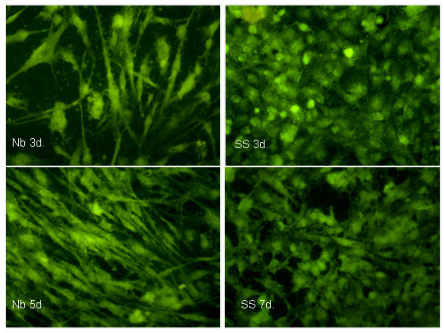
3.9. Morphological Characterization of the Cells Using Fluorescence Microscopy

The variations of the morphological appearance of the cells, due to the composition of the surface on which they were grown, was done after 1, 3, 5 and 7 days of culturing time. The cells were plated and cultured as described in section 2.5 and after the incubation period were fixed using 3.7% formaldehyde and screened for the expression of the β-actin protein by immunofluorescence. Fluorescence antibody labeling methods combined with fluorescence microscopy are useful techniques for visualizing cellular interactions with materials not generally conducive to transmitted light, as was the case for Nb and SS. The cells were treated with (0.1% Triton × 100 in PBS) for permeabilization, then, the cells were incubated with anti-human β-actin diluted 1:200 (Santa Cruz Biotechnology) in PBS containing 1 mg/mL of bovine serum albumin (BSA) for 2 hours at room temperature. The samples were washed twice with ice-cold washing buffer (0.1% Tween-20 in PBS) for 10 minutes and incubated for 1 h at room temperature with the secondary antibody goat-anti-rabbit immunoglobulin conjugated with FITC (Invitrogen) diluted 1:50 in PBS. The samples were washed again twice with washing buffer, and cover-slipped in glycerol-PBS (1:9 v/v) containing 20 mg/mL of 1,2,diazabycyclo (2.2.2) octane (DABCO; triethylenediamine). The cell morphology was visualized by observing the inmunofluorescence produced by the actin cytoskeleton using an epifluorescent microscope (Axioscope 2, Carl Zeiss).

4. Conclusions

Thin films of niobium deposited by magnetron sputtering were tested with structural and biological tools. Comparing with stainless steel 316L and tissue culture plastic, the adhesion and proliferation of human osteoblast cells showed a remarkable difference of the cell behavior. Quantitative analysis of the cell adhesion showed a major initial attachment of osteoblast cells on the Nb surfaces as compared to the SS substrate and the control. The proliferation rate was also higher on the Nb surfaces during the first 5 days of culture. Similarly, a small difference in the cell metabolic activity indicated a better response of the human osteoblast cells to the Nb surface than to the steel. Summarizing, the results of the tests—cellular adhesion, proliferation, viability and morphology—showed that all the surfaces have good biocompatibility, but there is a significant better response on the Nb surface. The relation between the surface properties and the biomedical response is not an easy issue; it was observed that the film microstructure consisted mainly of the metallic crystalline phase known for bulk niobium (bcc), it showed a medium wettability to distilled water and corrosion properties similar to those of the stainless steel. The chemical composition and larger water-contact angle of the films influence positively the biological response, although we cannot provide a simple explanation for these results.
Nevertheless, the result of these experiments confirmed the feasibility of using Nb-based films as coatings for implant materials. Niobium oxide or nitride films have also to be evaluated for biocompatibility, corrosion resistance, mechanical properties, etc., but the in vitro results obtained in this work are encouraging since the response of human osteoblast cells to Nb surfaces was remarkably good.
Figure 1. X-ray diffraction pattern of the Nb thin films. The intensity of the substrate peaks (s) is high due to the small thickness of the films.
Coatings 01 00072f1 1024
Figure 2. XPS spectra of the bare substrate and the Nb-coated steel. Binding energy of the Nb peaks appeared at: Nb 3d3/2 (202 eV), Nb 3d5/2 (205 eV), Nb 3p3/2 (360.6 eV) and Nb 3p1/2 (376.5 eV).
Coatings 01 00072f2 1024
Figure 3. Scanning electron microscopy (SEM) image of the stainless steel (SS) sandblasted substrate. The same morphology is observed after film deposition.
Coatings 01 00072f3 1024
Figure 4. Potential-current curve of the Nb films as compared to the 316L steel substrate.
Coatings 01 00072f4 1024
Figure 5. Absolute numbers of attached cells after 24 hour of incubation for the control (C+), the substrate and the Nb film. Statistics using the one-way ANOVA showed that Nb results are significantly different to SS and C+ (p < 0.001).
Coatings 01 00072f5 1024
Figure 6. Number of cells as a function of the culture time. The initial number of plated cells was 2 × 10⁴ cells/sample, since this number was the same for all surfaces, it can be concluded that Nb promoted the greatest proliferation over the three surfaces at days 3 and 5 (* p < 0.01 and ** p < 0.05).
Coatings 01 00072f6 1024
Figure 7. Cell Viability as a function of the incubation time. The optical density at 570 nm is directly proportional to the number of viable cells. (* p < 0.01 and ** p < 0.05).
Coatings 01 00072f7 1024
Figure 8. Morphology of the human osteoblast cells cultured on the Nb film during 3 (A) and 5 days (B) and stainless steel surfaces at 3 days (C) and 7 days (D). The cytoskeleton of well differentiated osteoblasts normally assumed an elongated appearance in in vitro experiments.
Coatings 01 00072f8 1024
Table 1. Surface average roughness and water contact angles including the standard deviation error after 12 and 10 measurements, respectively.
Roughness and Water Contact angle
SampleRoughness (μm)Water contact angle
SS1.79 ± 0.346 ± 0.67
Nb1.87 ± 0.3567 ± 1.25

Acknowledgments

The authors like to thank to H. Arzate and S. Muhl for cooperation with the realization of this work. Special thanks to L. Huerta for XPS data. The authors acknowledged financial support from CONACYT projects P45833R and 152995.

References and Notes

  1. Jacobs, J.J.; Gilbert, J.L.; Urban, R.M. Corrosion of metal orthopaedic implants. J. Bone Joint Surg. 1998, 80A, 268–282. [Google Scholar]
  2. Kawahara, H.; Ochi, S.; Tanetani, K.; Kato, K.; Isogai, M.; Mizuno, Y.; Yamamoto, H.; Yamagami, A. Biological test of dental biomaterials, effect of pure metals upon the mouse subcutaneous fibroblast, strain L cell in tissue cultura. Japan Soc. Dent. Appar. Mater. 1963, 4, 65–85. [Google Scholar]
  3. Steinemann, S.G. Corrosion of Surgical Implants—In vivo and in vitro Tests, Evaluation of Biomaterials; Wiley: New York, NY, USA, 1980; pp. 1–34. [Google Scholar]
  4. Matsuno, H.; Yokoyama, A.; Watari, F.; Uo, M.; Kawasaki, T. Biocompatibility and osteogenesis of refractoy metal implants, titanium, hafnium, niobium, tantalum and rhenium. Biomaterials 2001, 22, 1253–1262. [Google Scholar] [CrossRef] [PubMed]
  5. Findlay, D.M.; Welldon, K.; Atkins, G.J.; Howie, D.W.; Zannettino, A.C.W.; Bobyn, D. The proliferation and phenotypic expression of human osteoblasts on tantalum metal. Biomaterials 2004, 25, 2215–2227. [Google Scholar] [CrossRef] [PubMed]
  6. Bobyn, J.D.; Toth, K.K.; Hacking, S.A.; Tanzer, M.; Krygier, J.J. Tissue response to porous tantalum acetabular cups: A canine model. J. Arthoplasty 1999, 14, 347–354. [Google Scholar] [CrossRef]
  7. Semlitsch, M.F.; Weber, H.; Sttreicher, R.M.; Schön, R. Joint replacement components made of hot-forged and surface treated Ti-6Al-7Nb alloy. Biomaterials 1992, 13, 781–788. [Google Scholar] [CrossRef] [PubMed]
  8. Godmann, S.B.; Davidson, J.A.; Fornasier, V.L.; Mishra, A.K. Histological response to cylinders of a low modulus Titanium alloy (Ti-13Nb-13Zr) and a wear resistant Zirconium alloy (Zr-2.5Nb) implanted in the rabbit tibia. J. Appl. Biomater. 1993, 4, 331–339. [Google Scholar] [CrossRef]
  9. Metikoš-Huković, M.; Kwokal, A.; Piljac, J. The influence of niobium and vanadium on passivity of titanium-based implants in physiological solution. Biomaterials 2003, 24, 3765–3775. [Google Scholar] [CrossRef] [PubMed]
  10. Eisenbarth, E.; Velten, D.; Müller, M.; Thull, R.; Brene, J. Biocompatibility of β-stabilizing elements of titanium alloys. Biomaterials 2004, 25, 5705–5713. [Google Scholar] [CrossRef] [PubMed]
  11. Castner, D.G.; Ratner, B.D. Biomedical surface science: Foundations to frontiers. Surf. Sci. 2002, 500, 28–60. [Google Scholar] [CrossRef]
  12. Kasemo, B. Biological surface science. Surf. Sci. 2002, 500, 656–677. [Google Scholar] [CrossRef]
  13. Ratner, B.D. Surface modifications of polymers: Chemical, biological and surface analytical challenges. Biosens. Bioelectron. 1995, 10, 797–804. [Google Scholar] [CrossRef] [PubMed]
  14. Smith, D.L. Thin Film Deposition: Principles and Practice; Mc Graw-Hill: New York, NY, USA, 1995. [Google Scholar]
  15. Stals, L.M.M.; Nesladek, M.; Quayhaegens, C. Current industrial practice critical issue in hard PVD and PA-CVD coatings. Surf. Coat. Technol. 1997, 91, 230–239. [Google Scholar] [CrossRef]
  16. Schneider, J.M.; Rohde, S.; Sproul, W.D.; Matthews, A. Recent developments in plasma assisted physical vapour deposition. J. PhysD: Appl. Phys. 2000, 33, R173–R186. [Google Scholar] [CrossRef]
  17. Bolz, A. Application of thin film technology in biomedical engineering. In Encyclopedic Handbook of Biomaterials and Bioengineering, Part B: Applications; Wise, D.L., Trantolo, D.J., Altobelli, D.E., Yaszemski, M.J., Schwartz, E.R., Eds.; Marcel Dekker: New York, NY, USA, 1995. [Google Scholar]
  18. Mandl, S.; Rauschenbach, B. Improving the biocompatibility of medical implants with plasma inmersion ion implantation. Surf. Coat. Technol. 2002, 156, 276–283. [Google Scholar] [CrossRef]
  19. Chu, P.K.; Chen, J.Y.; Wang, L.P.; Huang, N. Plasma-surface modification of biomaterials. Mat. Sci. Eng. R. 2002, 36, 143–206. [Google Scholar] [CrossRef]
  20. Pizzoferrato, A.; Ciapetti, G.; Stea, S.; Cenni, E.; Arciola, C.A.; Granchi, D.; Savarino, L. Cell culture methods for testing biocompatibility. Clin. Mater. 1994, 15, 173–190. [Google Scholar] [CrossRef] [PubMed]
  21. Leitãão, E.; Barbosa, M.A.; De Groot, K. In vitro testing of surface-modified biomaterials. J. Mater. Sci.: Mater. in Med. 1998, 9, 543–548. [Google Scholar] [CrossRef]
  22. Manso, M.; Ogueta, S.; Pérez-Rigueiro, J.; García, J.P.; Martínez-Duart, J.M. Testing biomaterials by in-situ evaluation of cell response. Biomol. Eng. 2002, 19, 239–242. [Google Scholar] [CrossRef] [PubMed]
  23. Olaya, J.J.; Rodil, S.E.; Muhl, S. Comparative study of niobium nitride coatings deposited by unbalanced and balanced magnetron sputtering. Thin Solid Films 2008, 516, 8319–8326. [Google Scholar] [CrossRef]
  24. Ramirez, G.; Rodil, S.E.; Arzate, H.; Muhl, S.; Olaya, J.J. Niobium based coatings for dental implants. Appl. Surf. Sci. 2011, 257, 2555–2559. [Google Scholar] [CrossRef]
  25. Asselin, E.; Ahmed, T.M.; Alfantazi, A. Corrosion of niobium in sulphuric and hydrochloric solutions at 75 and 95 °C. Corr. Sci. 2007, 49, 694–710. [Google Scholar] [CrossRef]
  26. Marie, P.J. Human endosteal osteoblastic cells: Relationship with bone formation. Calcif. Tissue Int. 1995, 56, S13–S16. [Google Scholar] [PubMed]
  27. Schmidt, C.; Kaspar, D.; Sarkar, M.R.; Claes, L.E.; Ignatius, A.A. A scanning electron microscopy study of human osteoblast morphology on five orthopedic metals. J. Biomed. Mater. Res. 2002, 63, 252–261. [Google Scholar] [CrossRef] [PubMed]
  28. Lauer, G.; Wiedmann-Al-Ahmad, M.; Otten, J.E.; Hubner, U.; Schmelzeisen, S.W. The titanium surface texture effects adherence and growth of human gingival keratinocytes and human maxillar osteoblast-like cells in vitro. Biomaterials 2001, 22, 2799–2809. [Google Scholar] [CrossRef] [PubMed]
  29. Anselme, K.; Bigerelle, M.; Noel, B.; Iost, A.; Hardouin, P. Effect of grooved titanium substratum on human osteoblastic cell growth. J. Biomed. Mater. Res. 2002, 60, 529–540. [Google Scholar] [CrossRef] [PubMed]
  30. Rodil, S.E.; Olaya, J.J. Unbalanced magnetic field configuration: Plasma and film properties. J. Phys: Condens. Matter 2006, 18, S1703–S1719. [Google Scholar] [CrossRef]
  31. ASTM G5-4 “Standard reference test method for making potentiostatic and potentyodinamic anodic polarization measurements”. In Annual Book of ASTM standards; 2000; 3, 02 Wear and Erosion; metal Corrosion; p. 57.
  32. Chou, W.J.; Yu, G.P.; Huang, J.H. Corrosion resistance of ZrN films on AISI 304 stainless steel substrate. Surf. Coat. Technol. 2003, 167, 59–67. [Google Scholar] [CrossRef]
  33. Rodil, S.E.; Olivares, R.; Arzate, H.; Muhl, S. Properties of carbon films and their biocompatibility using in vitro tests. Diamond Relat. Mater. 2003, 12, 931–937. [Google Scholar] [CrossRef]

